Article

Variability in Adaptive Resistance of Salmonella Typhimurium to Sublethal Levels of Antibiotics

1
Department of Biomedical Science, Kangwon National University, Chuncheon 24341, Gangwon, Republic of Korea
2
Institute of Bioscience and Biotechnology, Kangwon National University, Chuncheon 24341, Gangwon, Republic of Korea
*
Author to whom correspondence should be addressed.
Antibiotics 2022, 11(12), 1725; https://doi.org/10.3390/antibiotics11121725
Submission received: 10 November 2022 / Revised: 24 November 2022 / Accepted: 28 November 2022 / Published: 1 December 2022

Abstract

:
This study was designed to evaluate the adaptive resistance of Salmonella Typhimurium under continuous sublethal selective pressure. Salmonella Typhimurium ATCC 19585 (STATCC) and S. Typhimurium CCARM 8009 (STCCARM) were sequentially cultured for 3 days at 37 °C in trypticase soy broth containing 1/2 × MICs of cefotaxime (CEF1/2), chloramphenicol (CHL1/2), gentamicin (GEN1/2), and polymyxin B (POL1/2). The STATCC and STCCARM exposed to CEF1/2, CHL1/2, GEN1/2, and POL1/2 were evaluated using antibiotic susceptibility, cross-resistance, and relative fitness. The susceptibilities of STATCC exposed to GEN1/2 and POL1/2 were increased by a 2-fold (gentamicin) and 8-fold (polymyxin B) increase in minimum inhibitory concentration (MIC) values, respectively. The MIC values of STCCARM exposed to CEF1/2, CHL1/2, GEN1/2, and POL1/2 were increased by 4-fold (cefotaxime), 2-fold (chloramphenicol), 2-fold (gentamicin), and 8-fold (polymyxin B). The highest heterogeneous fractions were observed for the STATCC exposed to CEF1/2 (38%) and POL1/2 (82%). The STCCARM exposed to GEN1/2 was cross-resistant to cefotaxime (p < 0.05), chloramphenicol (p < 0.01), and polymyxin B (p < 0.05). The highest relative fitness levels were 0.92 and 0.96, respectively, in STATCC exposed to CEF1/2 and STCCARM exposed to POL1/2. This study provides new insight into the fate of persistent cells and also guidance for antibiotic use.

1. Introduction

Over the last few decades, the emergence and spread of antibiotic resistance have become some of the top threats to global health [1,2]. Bacteria can acquire resistance mechanisms under antibiotic selective pressures [3]. In addition, bacterial populations consist of persisters as tolerant subpopulations [3]. Indeed, persistence, tolerance, and resistance are characterized by the bacterial growth rates during the antibiotic exposure. Bacterial persistence is a metabolically inactive state including dormant cells under an antibiotic stress condition. Tolerance is a slow-growing state, and resistance is an actively growing state in the presence of antibiotic stress [4]. Antibiotic treatment failure is mainly due to antibiotic-resistance-conferring mutations, antibiotic resistance acquisition via horizontal gene transfer (HGT), and the formation of persistent cells [5]. Antibiotic resistance in bacteria is genetically acquired, but persistence is characterized as phenotypic heterogeneity [6]. The mechanisms of antibiotic resistance in bacteria include enzymatic inactivation of antibiotics, alteration in binding affinity of antibiotics, modification of membrane permeability, and use of alternative metabolic pathways [7]. Efflux pumps, DNA damage, stress responses, molecular degradation, and toxin–antitoxin (TA) molecules can affect persister cell formation [8]. However, the formation of persister cells is not clearly understood. Bacteria persistence is a reversible and transient phenomenon that can survive under stressful conditions [9].
Antibiotic tolerance in persister cells is due to dormancy, leading to fewer active target sites for antibiotics [10]. Persister cells are divided into two types; one is type I persistence in stationary phase (triggered persistence), and the other is type II persistence in exponential phase (spontaneous persistence) [10]. Spontaneous persistence depends on a stochastic switch to persister cells which rarely occurs, while triggered persistence relies on metabolic and environmental stresses [11,12]. Persister cells can revitalize in favorable conditions [10]. Hence, not only antibiotic resistance but also tolerance and persistence can cause serious public health problems [1]. However, persister cells are relatively less often investigated and are underestimated with regard to the risk of infection. Furthermore, it is still unknown whether tolerance and persistence as bacterial survival strategies can affect the development of resistance [3]. Therefore, this study aimed to characterize in terms of antibiotic susceptibility, cross-resistance, and relative fitness the Salmonella Typhimurium wild-type strain (STATCC), which serves as the original parent strain, and the antibiotic-resistant strain (STCCARM), which encodes pre-resistome genes that can respond to several stress conditions [13], by exposing them to sublethal levels of antibiotics.

2. Results and Discussion

In the early stage of antibiotic discovery, antibiotics became a successful treatment for bacterial infections. However, the overuse and misuse of antibiotics have become major causes of the development of antibiotic resistance in bacteria [14]. Bacteria evolve survival strategies under unfavorable antibiotic treatments [9]. For instance, persistence is an intrinsic defense system which protects bacteria from stress-induced damage [15]. Bacterial persisters are subpopulations that reversibly tolerate antibiotics and cause recalcitrant chronic infections [11,12,16,17,18,19,20]. However, bacterial persistence is overlooked due to a lack of information [2]. Therefore, differentiation between persister cells and antibiotic-resistant cells is needed for the design of effective antibiotic therapy [12].

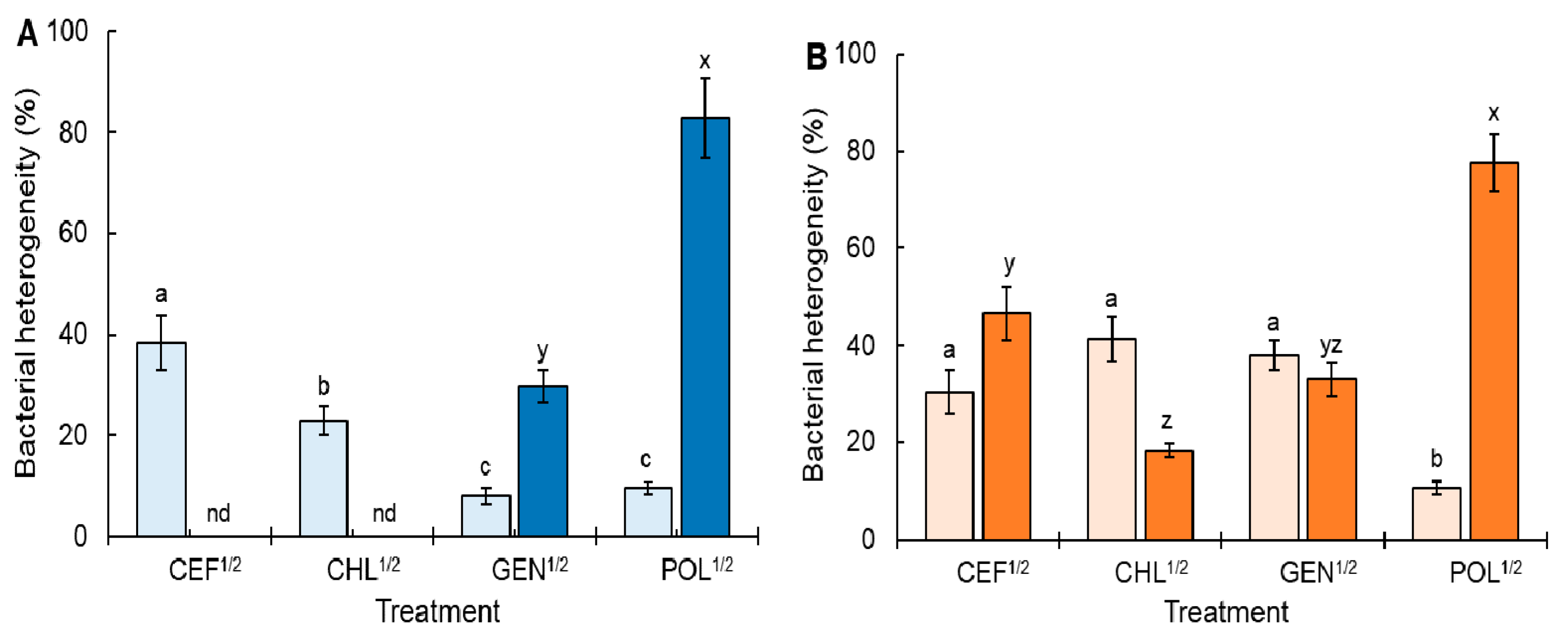
2.1. Antibiotic Susceptibility and Heterogeneous Subpopulation under Continuous Sublethal Selective Pressure

Antibiotics including cefotaxime, chloramphenicol, gentamicin, and polymyxin B were used to evaluate the antibiotic susceptibilities of STATCC and STCCARM exposed to a half MICs of antibiotics for 3 days at 37 °C (CEF1/2, CHL1/2, GEN1/2, and POL1/2, respectively) (Figure 1). For STATCC, no changes in the MICs of cefotaxime and chloramphenicol were observed after exposure to CEF1/2 and CHL1/2 (Figure 1A,B), corresponding to the absence of antibiotic resistance (Figure 2A). STATCC is antibiotic-sensitive bacteria with the absence of pre-resistance genes [21]. Thus, STATCC may not transform from the antibiotic-sensitive to the antibiotic-resistant phenotype. In addition, the most common mechanisms of resistance to β-lactam and chloramphenicol in bacteria are the production of antibiotic-degrading enzymes such as β-lactamases and the presence of efflux pumps [21]. On the other hand, other studies also mentioned that β-lactams may act as persistence effectors through the induction of SOS responses [22]. Thus, this observation suggests that cefotaxime and chloramphenicol are more likely to be associated with the persistence formation in STATCC rather than resistance development. However, the susceptibilities of STATCC to gentamicin and polymyxin B were decreased after exposure to GEN1/2 and POL1/2, showing a 2-fold and 8-fold increase in MIC values, respectively (Figure 1C,D). For STCCARM, all treatments, CEF1/2, CHL1/2, GEN1/2, and POL1/2, showed increased MIC values of cefotaxime (4-fold; Figure 1E), chloramphenicol (2-fold; Figure 1F), gentamicin (2-fold; Figure 1G), and polymyxin B (8-fold; Figure 1H). Bacteria are easily adapted to subinhibitory concentrations of antibiotics, resulting in enhanced antibiotic resistance [6]. In general, enhanced persistence level is attributed to serial antibiotic exposure, which can also lead to the development of bacterial tolerance to different classes of antibiotics [23].
The bacterial heterogeneity of STATCC and STCCARM exposed to CEF1/2, CHL1/2, GEN1/2, and POL1/2 was evaluated with the formation of persister and resistant cells (Figure 2). Compared to the control, the highest persistence fraction was observed for STATCC exposed to CEF1/2 (38%), followed by CHL1/2 (23%). This is in good agreement with a previous report which stated that the continuous exposure of bacteria to antibiotics can promote the formation of persister cells [24]. The formation of persisters is induced by the inhibition of replication, transcription, and translation [8,16,25] and is also influenced by experimental conditions and antibiotic classes; thereby, it is known as a complex phenomenon [26,27]. For instance, antibiotics such as tetracycline, aminoglycosides, β-lactams, and fluoroquinolones act as persistence effectors that inhibit DNA replication and protein synthesis [28,29]. In addition, β-lactams are involved in the induction of an SOS response through lexA/recA regulators [22]. These regulators play an important role in the expression of the class I toxin–antitoxin (TA) system, resulting in bacterial growth inhibition and persister cell formation [30]. Furthermore, the SOS response is also associated with the formation of small colony variants (SCVs) which typically enhance intracellular residence in host cells, promote persistence, and increase the risk of antibiotic tolerance and resistance [23]. Persister cells are characterized by morphological variation (small colony and cell wall-deficient state), metabolic alterations (reduced ATP production and cell membrane modification), gene expression changes (fitness- and survival-associated regulators and TA mechanism), and stress responses (pH and oxygen) [29,31]. STATCC exposed to POL1/2 showed the highest resistance percentage (83%), followed by GEN1/2 (30%) (Figure 2A). Resistant cells were not detected in STATCC exposed to CEF1/2 and CHL1/2. The STCCARM cells exposed to CEF1/2, CHL1/2, and GEN1/2 were more than 30% persister cells (Figure 2B). Similar to STATCC, the highest resistance fraction was observed in STCCARM exposed to POL1/2 (78%).
Salmonella Typhimurium has several resistance mechanisms to antimicrobial peptides, such as polymyxin B, which regulate the activation of the two-component regulatory PmrAB system and the increased secretion of outer membrane vesicles containing antimicrobial agents from the bacterial envelope [32]. This implies that the STATCC and STCCARM strains used in this study are able to activate the resistance mechanisms that can lead to easy adaptation to polymyxin B and a large proportion of polymyxin-B-resistant cells. This observation suggests that the PmrAB operon and outer membrane vesicles are more likely to be associated with resistance development than persistence formation [32]. Persistence can be an intermediator for the evolution of antibiotic resistance [1]. Therefore, persister cells can involve the emergence of antibiotic-resistant development [6]. The survival curves of persister cells exposed to antibiotics showed characteristic biphasic behaviors, representing a rapid decline in a majority of the vulnerable populations at the early phase, followed by a slow decrease in a minority of the tolerant populations at the late phase [17,33].

2.2. Cross-Resistance under Continuous Sublethal Selective Pressure

The development of cross-resistance by STATCC and STCCARM exposed to CEF1/2, CHL1/2, GEN1/2, and POL1/2 was evaluated using different classes of antibiotic disks (Figure 3). All treatments, including CEF1/2, CHL1/2, GEN1/2, and POL1/2, showed significant resistance to the same antibiotics, cefotaxime, chloramphenicol, gentamicin, and polymyxin B, respectively, except for STATCC exposed to GEN1/2 against gentamicin (Figure 3C). STATCC cells exposed to POL1/2 (p < 0.05) were cross-resistant to cefotaxime (Figure 3A), CEF1/2 (p < 0.05) to chloramphenicol (Figure 3B), and CEF1/2 (p < 0.05), GEN1/2 (p < 0.001), and POL1/2 (p < 0.001) to polymyxin B (Figure 3D). The cross-resistance of STCCARM exposed to GEN1/2 was observed with cefotaxime (Figure 3E), chloramphenicol (Figure 3F), gentamicin (Figure 3G), and polymyxin B (Figure 3H). The enhanced cross-resistance of STATCC exposed to CEF1/2, CHL1/2, and POL1/2 and STCCARM exposed to CEF1/2, CHL1/2, GEN1/2, and POL1/2 with cefotaxime, chloramphenicol, gentamicin, and polymyxin B resulted from the continuous sublethal selective pressure [34,35]. In general, persisters are responsible for de novo mutation when exposed to antibiotics [36]. Moreover, cell-wall-deficient spheroplasts formed under β-lactam antibiotic treatment are responsible for enhanced antibiotic tolerance and persistence [17,37]. The antibiotic tolerance of persister cells is also attributed to enhanced efflux pump activity [38]. It demonstrates that metabolically active cells can induce persister formation through the efflux pump [18]. This implies that persisters highly express multidrug efflux pumps which decrease intracellular antibiotic concentrations, resulting in enhanced antibiotic resistance [38,39]. Although enhanced susceptibility was not noticeable in this study, STATCC exposed to GEN1/2 (Figure 3A) and STCCARM exposed to CHL1/2 and POL1/2 (Figure 3G) showed increased susceptibility to cefotaxime and gentamicin, respectively. This result is known as negative cross-resistance, where the acquisition of resistance to one antibiotic results in increased susceptibility to another antibiotic [40]. Persister cells become susceptible to antibiotics due to the awakening of persisters triggered by environmental signals [8,41,42]. Therefore, selection inversion approaches can enhance the antibiotic susceptibility of antibiotic-resistant bacteria [43].

2.3. Relative Fitness under Continuous Sublethal Selective Pressure

The relative fitness was determined to evaluate the cost of resistance of STATCC and STCCARM exposed to CEF1/2, CHL1/2, GEN1/2, and POL1/2 (Figure 4). The STATCC cells exposed to CEF1/2 showed the highest relative fitness level (0.92), while those exposed to CHL1/2 showed the lowest relative fitness level (0.47), indicating the highest fitness cost. The highest fitness level achieved was that of STCCARM exposed to POL1/2 (0.96), followed by that achieved by GEN1/2 (0.82) and CHL1/2 (0.70) (Figure 4). Antibiotic-susceptible bacterial cells outcompete antibiotic-resistant cells that have low fitness when cultured in the absence of antibiotics [21]. Low fitness costs are likely to acquire antibiotic resistance in bacteria [44]. Therefore, a low fitness cost might be associated with the increase in the frequency of antibiotic resistance and cross-resistance of STATCC and STCCARM exposed to CEF1/2, CHL1/2, GEN1/2, and POL1/2 when compared to other classes of antibiotics (Figure 3). As a result, this allows bacteria to survive under antibiotic exposure, implying that bacteria can quickly adapt to new environments by optimizing the fitness [42]. Fitness is an important factor for maintaining antibiotic resistance [21]. The growth of antibiotic-resistant bacteria depends on the fitness cost in the absence of antibiotics [3]. The fitness of persister cells is reduced in favorable growth conditions, but their phenotypic heterogeneity allows them to adapt to stressful conditions [45]. The resistance acquired at sublethal concentrations of antibiotics shows higher fitness than that acquired under lethal concentrations [46]. Compensatory mutations can help antibiotic-resistant bacteria to survive in the absence of antibiotics [44]. In contrast, antibiotic-tolerant persisters revert to the antibiotic-susceptible wild type when exposed to antibiotic-free media [33]. The antibiotic susceptibility of persister cells can be increased when treated with metabolic enhancers such as daptomycin with glucose [47], fluoroquinolones with L-serine [48], and gentamicin with L-arginine [49], resulting in elevated uptake of proton motive force-dependent antibiotics [50].

3. Materials and Methods

3.1. Bacterial Strains and Culture Conditions

Strains of Salmonella enterica subsp. enterica serovar Typhimurium ATCC 19585 (STATCC) and S. Typhimurium CCARM 8009 (STCCARM) were obtained from the American Type Culture Collection (ATCC, Manassas, VA, USA) and the Culture Collection of Antibiotic Resistant Microbes (CCARM, Seoul, Korea), respectively. STATCC and STCCARM were used as representative of wild-type, antibiotic-sensitive and antibiotic-resistant strains, respectively [13]. All strains were cultured in trypticase soy broth (TSB; BD; Becton, Dickinson and Co., Sparks, MD) at 37 °C for 20 h and washed three times with phosphate-buffered saline (PBS; pH 7.2) by a centrifugation at 6000× g for 15 min at 4 °C. The collected cells were diluted with PBS to 108 CFU/mL for further assays.

3.2. Antibiotic Susceptibility Assay

The antibiotic susceptibilities of STATCC and STCCARM were determined according to the microbroth dilution assay [51]. The antibiotics used in this study are listed in Table 1. Antibiotic stock solutions were prepared by dissolving in ethanol for chloramphenicol and distilled water for cefotaxime, gentamicin, and polymyxin B to obtain a final concentration of 10.24 mg/mL. Each antibiotic stock solution was serially (1:2) diluted with fresh TSB in 96-well microtiter plates (BD Falcon, San Jose, CA, USA). Approximately 105 CFU/mL of STATCC or STCCARM was inoculated in the microtiter plates. After inoculation, the plates were incubated at 37 °C for 18 h to measure optical density (OD) at 600 nm using a microplate reader (BioTek Instruments, Inc., Winooski, VT, USA) and to determine minimum inhibitory concentration (MIC) where no visible growth was observed.

3.3. Analysis of Heterogeneous Cell Populations

STATCC and STCCARM were successively cultured in TSB containing 1/2 × MICs of cefotaxime (CEF1/2; 0.004 and 0.002 μg/mL), chloramphenicol (CHL1/2; 2 and 2 μg/mL), gentamicin (GEN1/2; 2 and 4 μg/mL), and polymyxin B (POL1/2; 0.5 and 0.5 μg/mL) for 3 days at 37 °C. The antibiotic-treated STATCC and STCCARM cells (CEF1/2, CHL1/2, GEN1/2, and POL1/2; 10 μL each) were daily resuspended in 1 mL of TSB containing a half MIC of each antibiotic. After exposure to antibiotics, the growths of untreated control and STATCC and STCCARM exposed to CEF1/2, CHL1/2, GEN1/2, and POL1/2 as a function of antibiotic concentration were used to estimate the fractions of heterogeneous cells. The area under curve was calculated using sub-MIC and resistance-selective windows [52]. The selective windows of STATCC and STCCARM exposed to CEF1/2, CHL1/2, GEN1/2, and POL1/2 were compared with those of the untreated control to measure heterogeneity.

3.4. Disk Diffusion Susceptibility Test

The cross-resistance of STATCC and STCCARM exposed to CEF1/2, CHL1/2, GEN1/2, and POL1/2 was evaluated using antibiotic disk diffusion assay. Antibiotic disks (Oxoid Ltd., Hampshire, United Kingdom) used in this study were cefotaxime (30 μg), chloramphenicol (30 μg), gentamicin (10 μg), and polymyxin B (300 μg). The antibiotic-exposed STATCC and STCCARM cells were spread on Mueller–Hinton agar plates. After the plates were dried for 10 min, antibiotic disks were placed on the surface of agar plates, and the plates were incubated at 37 °C for 24 h. The diameter of the inhibition zone was measured using a digital vernier caliper (The L.S. Starrett Co., Athol, MA, USA).

3.5. Relative Fitness

The relative fitness of STATCC and STCCARM exposed to CEF1/2, CHL1/2, GEN1/2, and POL1/2 was determined by culturing in antibiotic-free TSB at 37 °C for 24 h. The relative fitness was calculated to evaluate the cost of resistance of STATCC and STCCARM exposed to CEF1/2, CHL1/2, GEN1/2, and POL1/2, which were expressed as the ratio of the growth of antibiotic-treated cells to antibiotic-untreated control cells cultured in antibiotic-free TSB.

3.6. Statistical Analysis

Data were analyzed using the Statistical Analysis System (SAS) software. All analyses were performed with three biological replicates. The general linear model (GLM) and Fisher’s least significant difference (LSD) procedures were used to determine mean differences at 5%, 1%, and 0.1% levels of significance.

4. Conclusions

In conclusion, the most significant finding was that bacterial persistence can cause a significant impact on the development of antibiotic resistance. Bacteria exposed to antibiotic selection pressure promote the emergence and spread of antibiotic resistance. Antibiotic resistance and persistence are characterized by transferable genetic variation and transient phenotypic variations, respectively, in bacteria. Although bacterial persistence can contribute to the evolution of antibiotic resistance, antibiotic-tolerant bacteria show growth defects in the absence of antibiotics. The population-based study of persistence is not sufficient to specifically identify and characterize persister cells in mixed populations. Thus, single-cell-based approaches need to accurately identify specific markers and distinguish persister cells. The evolution of antibiotic persistence cannot go unnoticed in clinical practice. It is essential to understand the mechanisms of persister formation when designing effective antibiotic strategies.

Author Contributions

J.D. conducted all experiments and also wrote the manuscript. J.A. designed the experiment and contributed to the data analysis. All authors have read and agreed to the published version of the manuscript.

Funding

This research was supported by Basic Science Research Program through the National Research Foundation of Korea (NRF) funded by the Ministry of Education (NRF-2016R1D1A3B0100830416).

Informed Consent Statement

Not applicable.

Conflicts of Interest

The authors declare no conflict of interest.

References

  1. Hossain, T.; Deter, H.S.; Peters, E.J.; Butzin, N.C. Antibiotic tolerance, persistence, and resistance of the evolved minimal cell, Mycoplasma mycoides JCVI-Syn3B. iScience 2021, 24, 102391. [Google Scholar] [CrossRef] [PubMed]
  2. Windels, E.M.; Michiels, J.E.; Van den Bergh, B.; Fauvart, M.; Michiels, J. Antibiotics: Combatting tolerance to stop resistance. mBio 2019, 10, e02095-19. [Google Scholar] [CrossRef] [Green Version]
  3. Windels, E.M.; Bergh, B.V.D.; Michiels, J. Bacteria under antibiotic attack: Different strategies for evolutionary adaptation. PLoS Pathog. 2020, 16, e1008431. [Google Scholar] [CrossRef] [PubMed]
  4. Fernández-García, L.; Fernandez-Cuenca, F.; Blasco, L.; López-Rojas, R.; Ambroa, A.; Lopez, M.; Pascual, Á.; Bou, G.; Tomás, M. Relationship between tolerance and persistence mechanisms in Acinetobacter baumannii strains with AbkAB toxin-antitoxin system. Antimicrob. Agents Chemother. 2018, 62, e00250-18. [Google Scholar] [CrossRef] [Green Version]
  5. Harms, A.; Maisonneuve, E.; Gerdes, K. Mechanisms of bacterial persistence during stress and antibiotic exposure. Science 2016, 354, aaf4268. [Google Scholar] [CrossRef] [PubMed]
  6. Levin-Reisman, I.; Ronin, I.; Gefen, O.; Braniss, I.; Shoresh, N.; Balaban, N.Q. Antibiotic tolerance facilitates the evolution of resistance. Science 2017, 355, 826–830. [Google Scholar] [CrossRef] [PubMed]
  7. Cox, G.; Wright, G.D. Intrinsic antibiotic resistance: Mechanisms, origins, challenges and solutions. Int. J. Med. Microbiol. 2013, 303, 287–292. [Google Scholar] [CrossRef]
  8. Wilmaerts, D.; Windels, E.; Verstraeten, N.; Michiels, J. General Mechanisms Leading to Persister Formation and Awakening. Trends Genet. 2019, 35, 401–411. [Google Scholar] [CrossRef]
  9. Rocha-Granados, M.C.; Zenick, B.; Englander, H.E.; Mok, W.W. The social network: Impact of host and microbial interactions on bacterial antibiotic tolerance and persistence. Cell. Signal. 2020, 75, 109750. [Google Scholar] [CrossRef]
  10. Goormaghtigh, F.; Melderen, L.V. Single-Cell imaging and characterization of Escherichia coli persister cells to ofloxacin in exponential cultures. Sci Adv. 2019, 5, eaav9462. [Google Scholar] [CrossRef]
  11. Balaban, N.Q.; Helaine, S.; Lewis, K.; Ackermann, M.; Aldridge, B.; Andersson, D.I.; Brynildsen, M.P.; Bumann, D.; Camilli, A.; Collins, J.J.; et al. Definitions and guidelines for research on antibiotic persistence. Nat. Rev. Microbiol. 2019, 17, 441–448. [Google Scholar] [CrossRef] [PubMed] [Green Version]
  12. Brauner, A.; Fridman, O.; Gefen, O.; Balaban, N.Q. Distinguishing between resistance, tolerance and persistence to antibiotic treatment. Nat. Rev. Microbiol. 2016, 14, 320–330. [Google Scholar] [CrossRef] [PubMed]
  13. Kim, J.; Jo, A.; Ding, T.; Lee, H.-Y.; Ahn, J. Assessment of altered binding specificity of bacteriophage for ciprofloxacin-induced antibiotic-resistant Salmonella Typhimurium. Arch. Microbiol. 2016, 198, 521–529. [Google Scholar] [CrossRef] [PubMed]
  14. Ghosh, D.; Veeraraghavan, B.; Elangovan, R.; Perumal, V. Antibiotic resistance and epigenetics: More to it than meets the eye. Antimicrob. Agents Chemother. 2020, 64, e02225-19. [Google Scholar] [CrossRef] [PubMed]
  15. Völzing, K.G.; Brynildsen, M.P. Stationary-Phase Persisters to Ofloxacin Sustain DNA Damage and Require Repair Systems Only during Recovery. mBio 2015, 6, e00731-15. [Google Scholar] [CrossRef] [Green Version]
  16. Long, Y.; Fu, W.; Li, S.; Ren, H.; Li, M.; Liu, C.; Zhang, B.; Xia, Y.; Fan, Z.; Xu, C.; et al. Identification of novel genes that promote persister formation by repressing transcription and cell division in Pseudomonas aeruginosa. J. Antimicrob. Chemother. 2019, 74, 2575–2587. [Google Scholar] [CrossRef]
  17. Balaban, N.Q.; Merrin, J.; Chait, R.; Kowalik, L.; Leibler, S. Bacterial Persistence as a Phenotypic Switch. Science 2004, 305, 1622–1625. [Google Scholar] [CrossRef] [Green Version]
  18. Zou, J.; Peng, B.; Qu, J.; Zheng, J. Are bacterial persisters dormant cells only? Front. Microbiol. 2021, 12, 708580. [Google Scholar] [CrossRef]
  19. Liu, S.; Wu, N.; Zhang, S.; Yuan, Y.; Zhang, W.; Zhang, Y. Variable persister gene interactions with (p)ppGpp for persister formation in Escherichia coli. Front. Microbiol. 2017, 8, 1795. [Google Scholar] [CrossRef]
  20. Claudi, B.; Spröte, P.; Chirkova, A.; Personnic, N.; Zankl, J.; Schürmann, N.; Schmidt, A.; Bumann, D. Phenotypic Variation of Salmonella in Host Tissues Delays Eradication by Antimicrobial Chemotherapy. Cell 2014, 158, 722–733. [Google Scholar] [CrossRef]
  21. Andersson, D.I.; Hughes, D. Antibiotic resistance and its cost: Is it possible to reverse resistance? Nat. Rev. Microbiol. 2010, 8, 260–271. [Google Scholar] [CrossRef] [PubMed]
  22. Debbia, E.; Roveta, S.; Schito, A.; Gualco, L.; Marchese, A. Antibiotic Persistence: The Role of Spontaneous DNA Repair Response. Microb. Drug Resist. 2001, 7, 335–342. [Google Scholar] [CrossRef] [PubMed]
  23. Trastoy, R.; Manso, T.; Fernández-García, L.; Blasco, L.; Ambroa, A.; del Molino, M.L.P.; Bou, G.; García-Contreras, R.; Wood, T.K.; Tomás, M. Mechanisms of bacterial tolerance and persistence in the gastrointestinal and respiratory environments. Clin. Microbiol. Rev. 2018, 31, e00023-18. [Google Scholar] [CrossRef] [Green Version]
  24. Goneau, L.W.; Yeoh, N.S.; MacDonald, K.W.; Cadieux, P.A.; Burton, J.P.; Razvi, H.; Reid, G. Selective Target Inactivation Rather than Global Metabolic Dormancy Causes Antibiotic Tolerance in Uropathogens. Antimicrob. Agents Chemother. 2014, 58, 2089–2097. [Google Scholar] [CrossRef] [PubMed] [Green Version]
  25. Kim, Y.; Wood, T.K. Toxins Hha and CspD and small RNA regulator Hfq are involved in persister cell formation through MqsR in Escherichia coli. Biochem. Biophys. Res. Commun. 2010, 391, 209–213. [Google Scholar] [CrossRef] [Green Version]
  26. Helaine, S.; Kugelberg, E. Bacterial persisters: Formation, eradication, and experimental systems. Trends Microbiol. 2014, 22, 417–424. [Google Scholar] [CrossRef] [PubMed]
  27. Kaldalu, N.; Hauryliuk, V.; Tenson, T. Persisters—As elusive as ever. Appl. Microbiol. Biotechnol. 2016, 100, 6545–6553. [Google Scholar] [CrossRef] [Green Version]
  28. Dewachter, L.; Verstraeten, N.; Fauvart, M.; Michiels, J. An integrative view of cell cycle control in Escherichia coli. FEMS Microbiol. Rev. 2018, 42, 116–136. [Google Scholar] [CrossRef] [Green Version]
  29. Page, R.; Peti, W. Toxin-Antitoxin systems in bacterial growth arrest and persistence. Nat. Chem. Biol. 2016, 12, 208–214. [Google Scholar] [CrossRef]
  30. Podlesek, Z.; Bertok, D.Ž. The DNA Damage Inducible SOS Response Is a Key Player in the Generation of Bacterial Persister Cells and Population Wide Tolerance. Front. Microbiol. 2020, 11, 1785. [Google Scholar] [CrossRef]
  31. Chang, J.; Lee, R.-E.; Lee, W. A pursuit of Staphylococcus aureus continues: A role of persister cells. Arch. Pharmacal Res. 2020, 43, 630–638. [Google Scholar] [CrossRef] [PubMed]
  32. Tamayo, R.; Ryan, S.S.; McCoy, A.J.; Gunn, J.S. Identification and genetic characterization of PmrA-regulated genes and genes involved in polymyxin B resistance in Salmonella enterica serovar typhimurium. Infect. Immun. 2002, 70, 6770–6778. [Google Scholar] [CrossRef] [PubMed] [Green Version]
  33. Michiels, J.E.; Van den Bergh, B.; Verstraeten, N.; Michiels, J. Molecular mechanisms and clinical implications of bacterial persistence. Drug Resist. Updat. 2016, 29, 76–89. [Google Scholar] [CrossRef]
  34. Chellat, M.F.; Raguž, L.; Riedl, R. Targeting Antibiotic Resistance. Angew. Chem. Int. Ed. 2016, 55, 6600–6626. [Google Scholar] [CrossRef] [PubMed]
  35. Wistrand-Yuen, E.; Knopp, M.; Hjort, K.; Koskiniemi, S.; Berg, O.G.; Andersson, D.I. Evolution of high-level resistance during low-level antibiotic exposure. Nat. Commun. 2018, 9, 1599. [Google Scholar] [CrossRef] [PubMed] [Green Version]
  36. Sebastian, J.; Swaminath, S.; Nair, R.R.; Jakkala, K.; Pradhan, A.; Ajitkumar, P. De Novo Emergence of Genetically Resistant Mutants of Mycobacterium tuberculosis from the Persistence Phase Cells Formed against Antituberculosis Drugs In Vitro. Antimicrob. Agents Chemother. 2017, 61, e01343-16. [Google Scholar] [CrossRef] [PubMed] [Green Version]
  37. Dörr, T.; Davis, B.M.; Waldor, M.K. Endopeptidase-Mediated Beta Lactam Tolerance. PLoS Pathog. 2015, 11, e1004850. [Google Scholar] [CrossRef] [Green Version]
  38. Pu, Y.; Zhao, Z.; Li, Y.; Zou, J.; Ma, Q.; Zhao, Y.; Ke, Y.; Zhu, Y.; Chen, H.; Baker, M.A.; et al. Enhanced Efflux Activity Facilitates Drug Tolerance in Dormant Bacterial Cells. Mol. Cell 2016, 62, 284–294. [Google Scholar] [CrossRef] [Green Version]
  39. Bergmiller, T.; Andersson, A.M.; Tomasek, K.; Balleza, E.; Kiviet, D.J.; Hauschild, R.; Tkačik, G.; Guet, C.C. Biased partitioning of the multidrug efflux pump AcrAB-TolC underlies long-lived phenotypic heterogeneity. Science 2017, 356, 311–315. [Google Scholar] [CrossRef]
  40. Liakopoulos, A.; Aulin, L.B.S.; Buffoni, M.; Fragkiskou, E.; van Hasselt, J.G.C.; Rozen, D.E. Allele-specific collateral and fitness effects determine the dynamics of fluoroquinolone resistance evolution. Proc. Natl. Acad. Sci. USA 2022, 119, e2121768119. [Google Scholar] [CrossRef]
  41. Edelmann, D.; Berghoff, B.A. A Shift in Perspective: A Role for the Type I Toxin TisB as Persistence-Stabilizing Factor. Front. Microbiol. 2022, 13, 871699. [Google Scholar] [CrossRef] [PubMed]
  42. Barbosa, C.; Trebosc, V.; Kemmer, C.; Rosenstiel, P.; Beardmore, R.; Schulenburg, H.; Jansen, G. Alternative Evolutionary Paths to Bacterial Antibiotic Resistance Cause Distinct Collateral Effects. Mol. Biol. Evol. 2017, 34, 2229–2244. [Google Scholar] [CrossRef] [PubMed] [Green Version]
  43. Baym, M.; Stone, L.K.; Kishony, R. Multidrug evolutionary strategies to reverse antibiotic resistance. Science 2016, 351, aad3292. [Google Scholar] [CrossRef] [PubMed] [Green Version]
  44. Durão, P.; Balbontín, R.; Gordo, I. Evolutionary mechanisms shaping the maintenance of antibiotic resistance. Trends Microbiol. 2018, 26, 677–691. [Google Scholar] [CrossRef] [PubMed] [Green Version]
  45. Kussell, E.; Leibler, S. Phenotypic Diversity, Population Growth, and Information in Fluctuating Environments. Science 2005, 309, 2075–2078. [Google Scholar] [CrossRef] [PubMed] [Green Version]
  46. Westhoff, S.; van Leeuwe, T.M.; Qachach, O.; Zhang, Z.; van Wezel, G.P.; Rozen, D.E. The evolution of no-cost resistance at sub-MIC concentrations of streptomycin in Streptomyces coelicolor. ISME J. 2017, 11, 1168–1178. [Google Scholar] [CrossRef] [PubMed] [Green Version]
  47. Prax, M.; Mechler, L.; Weidenmaier, C.; Bertram, R. Glucose Augments Killing Efficiency of Daptomycin Challenged Staphylococcus aureus Persisters. PLoS ONE 2016, 11, e0150907. [Google Scholar] [CrossRef] [PubMed] [Green Version]
  48. Duan, X.; Huang, X.; Wang, X.; Yan, S.; Guo, S.; Abdalla, A.E.; Huang, C.; Xie, J. l-Serine potentiates fluoroquinolone activity against Escherichia coli by enhancing endogenous reactive oxygen species production. J. Antimicrob. Chemother. 2016, 71, 2192–2199. [Google Scholar] [CrossRef] [Green Version]
  49. Lebeaux, D.; Chauhan, A.; Létoffé, S.; Fischer, F.; de Reuse, H.; Beloin, C.; Ghigo, J.M. pH-Mediated potentiation of aminoglycosides kills bacterial persisters and eradicates In vivo biofilms. J. Infect. Dis. 2014, 210, 1357–1366. [Google Scholar] [CrossRef] [Green Version]
  50. Allison, K.R.; Brynildsen, M.P.; Collins, J.J. Metabolite-enabled eradication of bacterial persisters by aminoglycosides. Nature 2011, 473, 216–220. [Google Scholar] [CrossRef]
  51. CLSI. Performance Standards for Antimicrobial Susceptibility Testing, 29th ed.; CLSI Supplement; Clinical and Laboratory Standards Institute: Wayne, PA, USA, 2019. [Google Scholar]
  52. Khan, S.; Beattie, T.K.; Knapp, C.W. The use of minimum selectable concentrations (MSCs) for determining the selection of antimicrobial resistant bacteria. Ecotoxicology 2017, 26, 283–292. [Google Scholar] [CrossRef] [PubMed]
Figure 1. Antibiotic susceptibilities of Salmonella Typhimurium ATCC 19585 (STATCC); (AD) and S. Typhimurium CCARM 8009 (STCCARM: EH) exposed to a half minimum inhibitory concentration (MIC) of cefotaxime (CEF1/2; A,E), chloramphenicol (CHL1/2; B,F), gentamicin (GEN1/2; C,G), and polymyxin B (POL1/2; E,H) for 0 (control; ○, ○) and 3 (, ) days. The optical density (OD) of STATCC and the STCCARM untreated control and after exposure to CEF1/2, CHL1/2, GEN1/2, and POL1/2 for 3 days was measured after incubating at 37 °C for 18 h.
Antibiotics 11 01725 g001
Figure 2. Frequencies of persistence (, ) and resistance (, ) in Salmonella Typhimurium ATCC 19585 (STATCC; A) and S. Typhimurium CCARM 8009 (STCCARM: B) exposed to a half minimum inhibitory concentration (MIC) of cefotaxime (CEF1/2), chloramphenicol (CHL1/2), gentamicin (GEN1/2), and polymyxin B (POL1/2). Different letters (a–c) on the bars show significant differences among treatments within persistence at p < 0.05, and different letters (x–z) on the bars show significant difference among treatments within resistance at p < 0.05. nd represents not determined.
Antibiotics 11 01725 g002
Figure 3. Cross-resistance of Salmonella Typhimurium ATCC 19585 (STATCC; AD) and S. Typhimurium CCARM 8009 (STCCARM: EH) exposed to a half minimum inhibitory concentration (MIC) of cefotaxime (CEF1/2), chloramphenicol (CHL1/2), gentamicin (GEN1/2), and polymyxin B (POL1/2) to cefotaxime (A,E), chloramphenicol (B,F), gentamicin (C,G), and polymyxin B (D,H). The dotted lines represent untreated STATCC and STCCARM. ns indicates no significant difference and *, **, and *** denote significance differences at p < 0.05, p < 0.01, and p < 0.001, respectively.
Antibiotics 11 01725 g003
Figure 4. Relative fitness of Salmonella Typhimurium ATCC 19585 (STATCC; ) and S. Typhimurium CCARM 8009 (STCCARM; ) exposed to a half minimum inhibitory concentration (MIC) of cefotaxime (CEF1/2), chloramphenicol (CHL1/2), gentamicin (GEN1/2), and polymyxin B (POL1/2). Different letters (a–c) on the bars show significant differences among treatments within persistence at p < 0.05, and different letters (x–z) on the bars show significant differences among treatments within resistance at p < 0.05.
Antibiotics 11 01725 g004
Table 1. Antimicrobial property of antibiotics used in this study.
ClassAntibioticSpectrumActivityAffinityInhibitory Mechanisms
CephemsCefotaximeBroadCidalHydrophilicCell wall synthesis
AminoglycosidesGentamicinNarrowCidalHydrophilicProtein synthesis (30S)
GlycopeptidesPolymyxin BNarrowCidalHydrophilicMembrane permeability
PhenicolsChloramphenicolBroadStaticHydrophobicProtein synthesis (50S)
Publisher’s Note: MDPI stays neutral with regard to jurisdictional claims in published maps and institutional affiliations.

