Article

Design of a Bacteriophage Cocktail Active against Shigella Species and Testing of Its Therapeutic Potential in Galleria mellonella

1
Wound Infections Department, Bacterial Diseases Branch, Walter Reed Army Institute of Research, Silver Spring, MD 20910, USA
2
Multidrug-Resistant Organism Repository and Surveillance Network, Bacterial Diseases Branch, Walter Reed Army Institute of Research, Silver Spring, MD 20910, USA
3
Department of Enteric Diseases, Armed Forces Research Institute of Medical Sciences, Bangkok 10400, Thailand
4
J. Craig Venter Institute, Rockville, MD 20850, USA
5
Bacterial Diseases Branch, Walter Reed Army Institute of Research, Silver Spring, MD 20910, USA
*
Author to whom correspondence should be addressed.
The author is since deceased.
Antibiotics 2022, 11(11), 1659; https://doi.org/10.3390/antibiotics11111659
Submission received: 27 October 2022 / Revised: 15 November 2022 / Accepted: 16 November 2022 / Published: 19 November 2022

Abstract

:
Shigellosis is a leading global cause of diarrheal disease and travelers’ diarrhea now being complicated by the dissemination of antibiotic resistance, necessitating the development of alternative antibacterials such as therapeutic bacteriophages (phages). Phages with lytic activity against Shigella strains were isolated from sewage. The genomes of 32 phages were sequenced, and based on genomic comparisons belong to seven taxonomic genera: Teetrevirus, Teseptimavirus, Kayfunavirus, Tequatrovirus, Mooglevirus, Mosigvirus and Hanrivervirus. Phage host ranges were determined with a diverse panel of 95 clinical isolates of Shigella from Southeast Asia and other geographic regions, representing different species and serotypes. Three-phage mixtures were designed, with one possessing lytic activity against 89% of the strain panel. This cocktail exhibited lytic activity against 100% of S. sonnei isolates, 97.2% of S. flexneri (multiple serotypes) and 100% of S. dysenteriae serotypes 1 and 2. Another 3-phage cocktail composed of two myophages and one podophage showed both a broad host range and the ability to completely sterilize liquid culture of a model virulent strain S. flexneri 2457T. In a Galleria mellonella model of lethal infection with S. flexneri 2457T, this 3-phage cocktail provided a significant increase in survival.

1. Introduction

Shigellosis, or bacillary dysentery, is a disease caused by invasion of the colonic, rectal and distal ileal epithelium by Shigella spp. Shigellosis is a leading cause of diarrheal disease worldwide, particularly in developing countries [1,2], and is a continuing problem for civilian and military travelers visiting endemic regions [3,4]. Vaccine development [5,6] and other prophylactic measures [1] remain a high priority given the disease burden [7], increasing antibiotic resistance [8,9], and gaining appreciation of the post-infectious sequelae associated with shigellosis [10]. Shigella encompasses four species subdivided into serotypes and subserotypes, Shigella dysenteriae (15 serotypes and 2 provisional serotypes), Shigella flexneri (7 serotypes and 15 subserotypes), Shigella sonnei (one serotype), and Shigella boydii (20 serotypes) [8,11]. S. sonnei is the most common species found in high-income countries [1]. S. flexneri accounts for 30–60% of shigellosis cases in developing regions, necessitating coverage of prevalent S. flexneri serotypes in a multivalent Shigella vaccine or therapeutic [12]. Data from studies where culture-independent diagnosis was assessed, such as quantitative polymerase chain reaction (qPCR) for Shigella, indicate that traditional culture-based methods significantly underestimate the global burden of Shigella-associated illness [13,14]. Estimates from the Global Enteric Multicenter Study (GEMS) found that analysis using qPCR resulted in a 2 to 2.5 fold increase in the attributable fraction of Shigella-associated moderate-severe diarrheal disease [13,15].
Historically, diarrhea has been the most common illness reported by U.S. military service members during numerous military exercises and mobilizations to regions/theaters where sanitation conditions were poor [16,17]. Shigella and enterotoxigenic Escherichia coli (ETEC) are major bacterial etiological agents of travelers’ diarrhea [3,4]. Additionally, antimicrobial resistance to common antibiotics used for the treatment of travelers’ diarrhea, including ciprofloxacin, is significantly increasing in ETEC and Shigella isolates, especially from countries in Africa and South and Southeast Asia [8,18,19]. Thus, prophylaxis of shigellosis and ETEC infection among military and civilian contingents is a priority, since no licensed vaccine is available [5,6].
Bacteriophages (phages) have shown therapeutic efficacy against various multidrug-resistant infections in laboratory animals and humans, including individual compassionate use and several promising clinical trials [20,21]. A single lytic phage prevented S. flexneri adherence and invasion in vitro, using a cultured human colorectal adenocarcinoma cell model [22]. A cocktail of five ATCC phages was able to lyse 62/65 (95%) of Shigella strains that belong to all four species and oral gavage of mice with this cocktail shortly before and/or after S. sonnei infection significantly reduced bacterial burden in fecal and cecum samples and did not distort gut microbiota [23]. Similar phage effects were observed in mice treated with phages against oral Listeria monocytogenes [24] and E. coli O157:H7 infections [25]. In addition, oral administration of a cocktail of three phages prevented Vibrio cholerae infection in infant mice and rabbits [26].
The oral administration of phages was used in different countries for treatment against dysentery in humans since 1919 and appeared to result in more frequent positive outcomes than in patients who did not receive phage (for reviews, see [27,28]). Two double-blind placebo-controlled clinical trials were conducted in the USSR, designed according to contemporary WHO standards and enrolling thousands of subjects. In these trials, a 2.5-fold and 3.9-fold reduction in the prevalence of dysentery was observed in groups of children receiving a lyophilized and pectinized mixture of Shigella-specific lytic phages once every seven [29] or every three days [30], compared with control groups receiving a placebo.
The deliberate rational design of phage cocktails for prophylactic and/or therapeutic use is needed to ensure these antimicrobials will work well in concert with standard of care antibiotic treatments, and also to overcome the bacterial resistance that emerges naturally [31]. Phages are isolated from the environment and selected as candidates for use in cocktails based on attributes including burst size, host range within the diversity of the target pathogen, anti-biofilm activity, synergies with treatment antibiotics, and selection of host receptor diversity [20]. The main aim of this work was to initiate the rational design of phage cocktails against target Shigella species and serotypes for the development of antimicrobials with activity against emerging multidrug-resistant variants. Toward this aim, we isolated new lytic phages, designed initial 3-phage prototype cocktails with broad host range against key Shigella pathogens, and tested the efficacy of a lead cocktail in protecting against S. flexneri infection in a Galleria mellonella model.

2. Results

2.1. Phage Isolation

Using the eight Shigella strains selected for phage enrichment (Table 1), three fractions of Washington DC wastewater collected before any chemical treatment (Materials and Methods) yielded a high prevalence of phages with lytic activity against Shigella. Thirty-two phages were isolated from the sewage samples that were distinct and diverse based on the EcoRV restriction digestion patterns of their genomic DNA, each with its own unique restriction profile (data not shown).

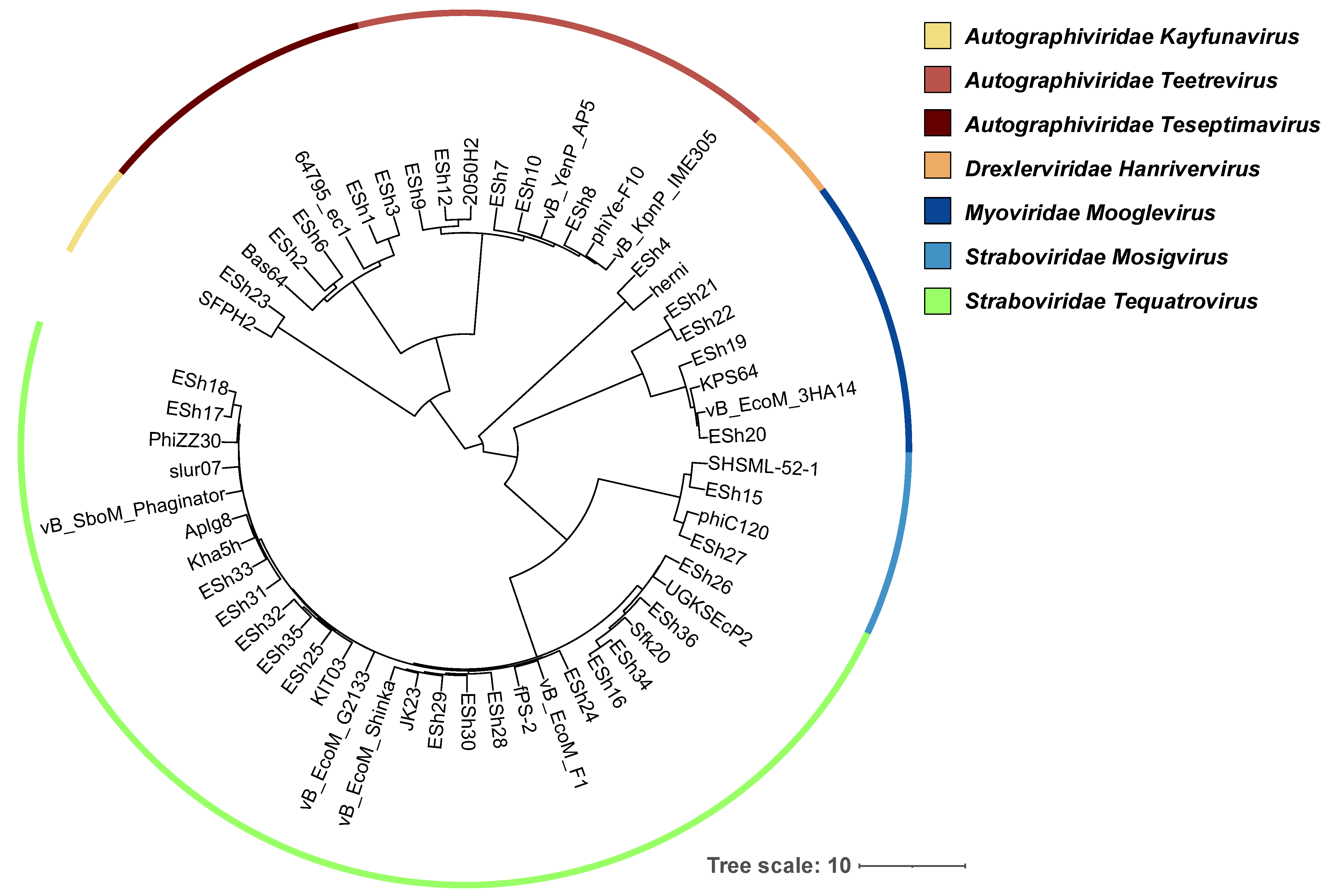
2.2. Phage Genome Analysis

All 32 phages were sequenced, with genome sizes ranging from 38,701 to 170,646 bp (Table 2). Based on nucleotide BLAST analysis, these phages were classified into seven genera, Tequatrovirus (ESh16-18, 24-26, 28-36), Mosigvirus (ESh15, ESh27), Teetrevirus (ESh7-12), Teseptimavirus (ESh1-3, 6), Kayfunavirus (ESh23), Mooglevirus (ESh19-22), and Hanrivervirus (ESh4) within four families (Straboviridae, Autographiviridae, Myoviridae and Drexlerviridae) (See Table 2). An average nucleotide distance phylogenetic tree of these Shigella phages, based on whole genome sequences, is presented in Figure 1. Bioinformatic analysis indicated that these phage genomes do not contain genes that are potentially deleterious for phage therapeutic application, such as putative determinants of transduction, genes encoding antimicrobial resistance, toxins and other virulence factors. While all of the phages appear to be strictly lytic, four of them (ESh19-22) belong to subfamily Ounavirinae within the former family Myoviridae. Some representatives of this subfamily called “superspreaders” have been found to efficiently release intact plasmid DNA upon lysis and thus to stimulate horizontal gene transfer by transformation [32]. For example, the genome of phage ESh19 showed 84% identity at the nucleotide level with superspreaders SUSP1 and SUSP2 [32] using BLAST analysis. Therefore, before using them as therapeutics, phages ESh19-22 should be tested to exclude the ability to enhance gene transfer.
Table 1. Shigella strains used in this work.
#StrainSerotypeOrigin#StrainSerotypeOrigin
1S. flexneri 271Vietnam50S. flexneri 833aKenya
2S. flexneri 461Thailand51S. flexneri 843aKenya
3S. flexneri 131aBhutan52S. flexneri J17B *3aJapan
4S. flexneri 281aVietnam53S. flexneri 383bVietnam
5S. flexneri 611aNepal54S. flexneri 94Cambodia
6S. flexneri 21bCambodia55S. flexneri 224Bhutan
7S. flexneri 31bCambodia56S. flexneri 394Vietnam
8S. flexneri 141bBhutan57S. flexneri 544Thailand
9S. flexneri 151bBhutan58S. flexneri 704Nepal
10S. flexneri 191bBhutan59S. flexneri 404aVietnam
11S. flexneri 291bVietnam60S. flexneri 414aVietnam
12S. flexneri 301bVietnam61S. flexneri 555Thailand
13S. flexneri 471bThailand62S. flexneri M90T5aUSA
14S. flexneri 621bNepal63S. flexneri M90T55 b*5aLaboratory
15S. flexneri 821bKenya64S. flexneri 106Cambodia
16S. flexneri 631cNepal65S. flexneri 116Cambodia
17S. flexneri 162Bhutan66S. flexneri 236Bhutan
18S. flexneri 312Vietnam67S. flexneri 246Bhutan
19S. flexneri 482Thailand68S. flexneri 426Vietnam
20S. flexneri 42aCambodia69S. flexneri 436Vietnam
21S. flexneri 52aCambodia70S. flexneri 566Thailand
22S. flexneri 172aBhutan71S. flexneri 576Thailand
23S. flexneri 182aBhutan72S. flexneri 716Nepal
24S. flexneri 322aVietnam73S. flexneri 726Nepal
25S. flexneri 332aVietnam74S. flexneri 856Kenya
26S. flexneri 492aThailand75S. flexneri SSU2415 *6USA
27S. flexneri 502aThailand76S. flexneri CCH060 *6Unknown
28S. flexneri 642aNepal77S. flexneri 58var. XThailand
29S. flexneri 652aNepal78S. flexneri 44var. YVietnam
30S. flexneri 812aKenya79S. sonnei 1NACambodia
31S. flexneri 2457T *2aJapan80S. sonnei 12NABhutan
32S. flexneri BS103 a*2aLaboratory81S. sonnei 26NAVietnam
33S. flexneri 62bCambodia82S. sonnei 45 NAThailand
34S. flexneri 342bVietnam83S. sonnei 60NANepal
35S. flexneri 662bNepal84S. sonnei Moseley *NAUSA
36S. flexneri ATCC 120222bUnknown85S. sonnei ATCC 25931NAPanama
37S. flexneri 352abVietnam86S. dysenteriae 591Thailand
38S. flexneri 513Thailand87S. dysenteriae 731Nepal
39S. flexneri 73aCambodia88S. dysenteriae 1617 *1Guatemala
40S. flexneri 83aCambodia89S. dysenteriae 742Nepal
41S. flexneri 203aBhutan90S. dysenteriae 759Nepal
42S. flexneri 213aBhutan91S. dysenteriae 7612Nepal
43S. flexneri 363aVietnam92S. dysenteriae 8712Kenya
44S. flexneri 373aVietnam93S. boydii 771Nepal
45S. flexneri 523aThailand94S. boydii 252Bhutan
46S. flexneri 533aThailand95S. boydii 782Nepal
47S. flexneri 673aNepal96S. boydii 862Kenya
48S. flexneri 683aNepal97S. boydii 7910Nepal
49S. flexneri 693aNepal98S. boydii 8012Nepal
a Non-invasive plasmid-cured strain of 2457T [33]; b non-invasive plasmid-cured strain of M90T [34]; * strain selected for phage enrichments. NA, not applicable.
Table 2. The characteristics of 32 Shigella phages isolated in this study.
Phage IDGenome Size, bpAccession No.Phage taxonomy aClosest Relative in NCBI Database b
FamilySubfamilyGenusDefinitionAccession No.
ESh139,034ON528715AutographiviridaeStudiervirinaeTeseptimavirus64795_ec1KU927499
ESh239,818ON528716AutographiviridaeStudiervirinaeTeseptimavirusJeanTinguely Bas64MZ501081
ESh339,180ON528717AutographiviridaeStudiervirinaeTeseptimavirus64795_ec1KU927499
ESh451,077ON528718DrexlerviridaeTempevirinaeHanrivervirusherniNC_049823
ESh639,381ON528719AutographiviridaeStudiervirinaeTeseptimavirusJeanTinguely Bas64MZ501081
ESh739,724ON528720AutographiviridaeStudiervirinaeTeetrevirusvB_KpnP_IME305OK149215
ESh838,701ON528721AutographiviridaeStudiervirinaeTeetrevirusphiYe-F10NC_047755
ESh939,308ON528722AutographiviridaeStudiervirinaeTeetrevirus2050H2NC_047844
ESh1038,729ON528723AutographiviridaeStudiervirinaeTeetrevirusvB_YenP_AP5KM253764
ESh1239,704ON528724AutographiviridaeStudiervirinaeTeetrevirus2050H2NC_047844
ESh15168,076ON528725StraboviridaeTevenvirinaeMosigvirusSHSML-52-1KX130865
ESh16165,784ON528726StraboviridaeTevenvirinaeTequatrovirusSfk20MW341595
ESh17166,355ON528727StraboviridaeTevenvirinaeTequatrovirusslur07LN881732
ESh18165,470ON528728StraboviridaeTevenvirinaeTequatrovirusKha5hNC_054905
ESh1987,867ON528729Myoviridae  cOunavirinaeMooglevirusvB_EcoM_3HA14MN342151
ESh2089,515ON528730Myoviridae  cOunavirinaeMooglevirusvB_EcoM_3HA14MN342151
ESh2186,414ON528731Myoviridae  cOunavirinaeMooglevirusKPS64MK562502
ESh2288,154ON528732Myoviridae  cOunavirinaeMooglevirusvB_EcoM_3HA14MN342151
ESh2340,156ON528733AutographiviridaeStudiervirinaeKayfunavirusSFPH2NC_048025
ESh24167,086ON528734StraboviridaeTevenvirinaeTequatrovirusvB_EcoM_F1NC_054912
ESh25166,499ON528735StraboviridaeTevenvirinaeTequatrovirusAplg8NC_054902
ESh26167,539ON528736StraboviridaeTevenvirinaeTequatrovirusUGKSEcP2OV876900
ESh27168,955ON528737StraboviridaeTevenvirinaeMosigvirusphiC120NC_055718
ESh28164,289ON528738StraboviridaeTevenvirinaeTequatrovirusJK23MK962752
ESh29166,160ON528739StraboviridaeTevenvirinaeTequatrovirusvB_EcoM_ShinkaMZ502379
ESh30170,189ON528740StraboviridaeTevenvirinaeTequatrovirusfPS-2NC_054943
ESh31167,224ON528741StraboviridaeTevenvirinaeTequatrovirusPhiZZ30NC_054938
ESh32169,173ON528742StraboviridaeTevenvirinaeTequatrovirusvB_EcoM_G2133MK327928
ESh33166,484ON528743StraboviridaeTevenvirinaeTequatrovirusvB_SboM_PhaginatorOL615012
ESh34167,055ON528744StraboviridaeTevenvirinaeTequatrovirusSfk20MW341595
ESh35166,919ON528745StraboviridaeTevenvirinaeTequatrovirusKIT03NC_054923
ESh36170,646ON528746StraboviridaeTevenvirinaeTequatrovirusT4_ev151LR597660
a General taxonomy for all phages: Viruses, Duplodnaviria (realm), Heunggongvirae (kingdom), Uroviricota (phylum), Caudoviricetes (class), Caudovirales (order), then families, subfamilies and genera as indicated in Table 2. b National Center for Biotechnology Information. c The family assignment of subfamily Ounavirinae viruses is unclear in the current taxonomy, so the previous family assignment is retained herein.

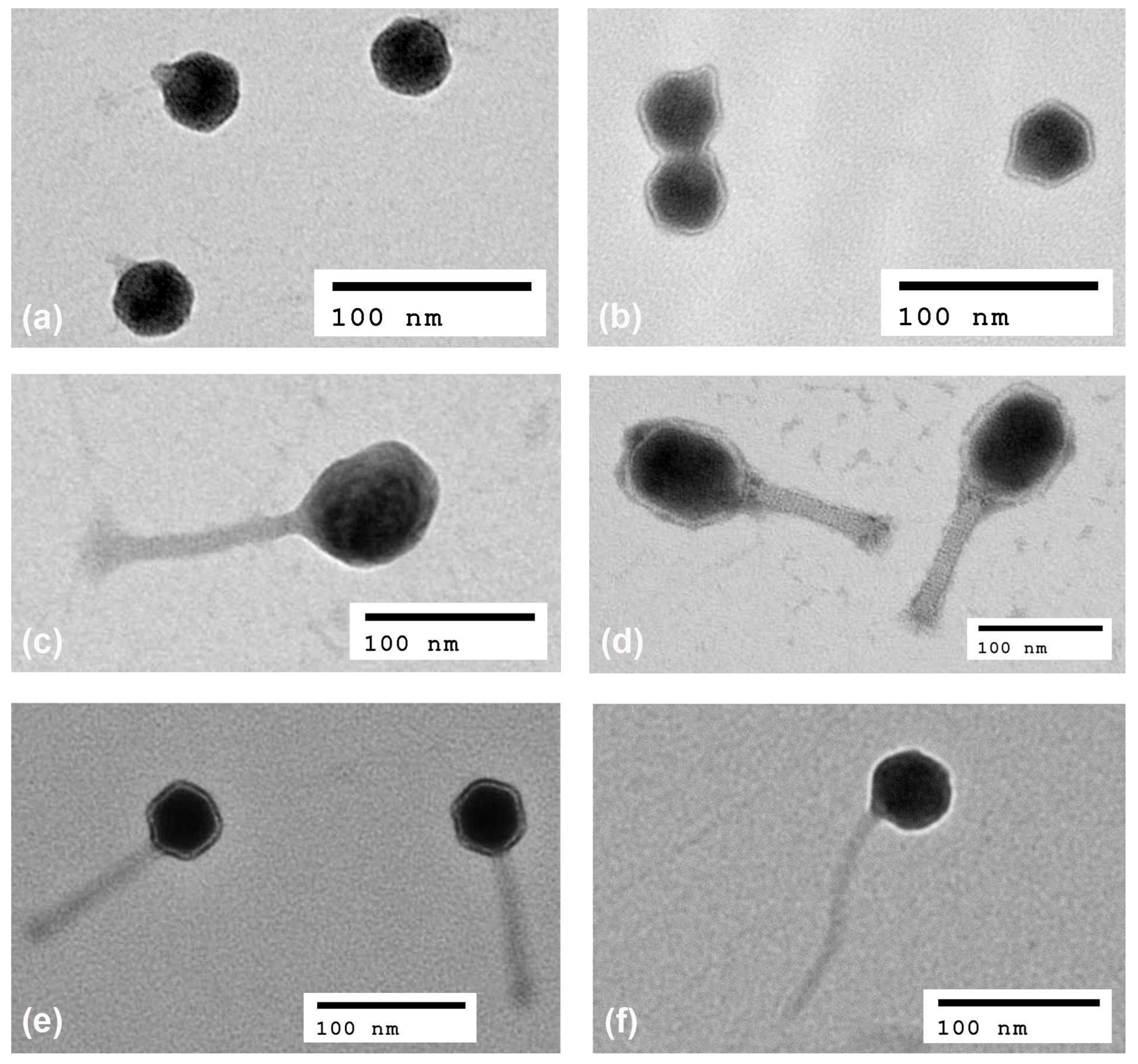
2.3. Phage Morphology

The morphology of phage virions was studied using transmission electron microscopy (Figure 2). Phage particles exhibited typical morphology associated with their family classification: myovirus phages with long contractile tails in genera Tequatrovirus, Mosigvirus (both now reclassified from Myoviridae to family Straboviridae) and Mooglevirus (family Myoviridae); podophages in genera Teetrevirus, Teseptimavirus and Kayfunavirus with short non-contractile tails (family Autographiviridae); and a siphophage with long non-contractile tail in the genus Hanrivervirus (family Drexlerviridae). Virion morphologies were consistent with what was expected based on the morphologies of phages with similar genome sequences.

2.4. Prototype Phage Cocktails

First, a panel of 12 phages was selected from the larger collection for further testing based on breadth of lytic host range across bacterial strains that represented the chief Shigella serotypes being targeted (shown in Table 3). Four mixtures or cocktails consisting of three phages each were then developed based on the initial lytic properties of individual candidate phages. Three out of the four mixtures (##1, 2 and 4) demonstrated the ability to completely clarify and sterilize liquid cultures of S. flexneri 2a 2457T (Table 4).

2.5. Host Range Testing

Activities of the 12 selected phages and the four 3-phage cocktails were tested against a panel of 95 Shigella strains assembled by the Armed Forces Research Institute of Medical Sciences composed mainly of clinical isolates from Southeast Asia, but also from East Asia, Africa, South America and the USA (Table 5 and Table S1). Overall, the 12 phages were able to lyse 86/95 (90.5%) of Shigella strains (Table S1). All of the 3-phage cocktails showed broad host ranges and killed 100% of S. sonnei isolates, 82–97% of S. flexneri (including serotypes 1, 1a, 1b, 1c, 2, 2a, 2b, 3a, 3b, 4, 4a, 5, and 6, as well as X and Y variants) and 100% of S. dysenteriae serotypes 1 and 2 (Table 5). Since 89/95 (93.7%) of the strains were pigmented on Congo Red agar (data not shown) and thus carried the virulence plasmid, it appears that these phage mixtures successfully kill virulent strains of Shigella. The representatives of some Shigella groups were not susceptible to this initial collection of candidate therapeutic phages: S. dysenteriae serotypes 9 and 12 and most S. boydii isolates.

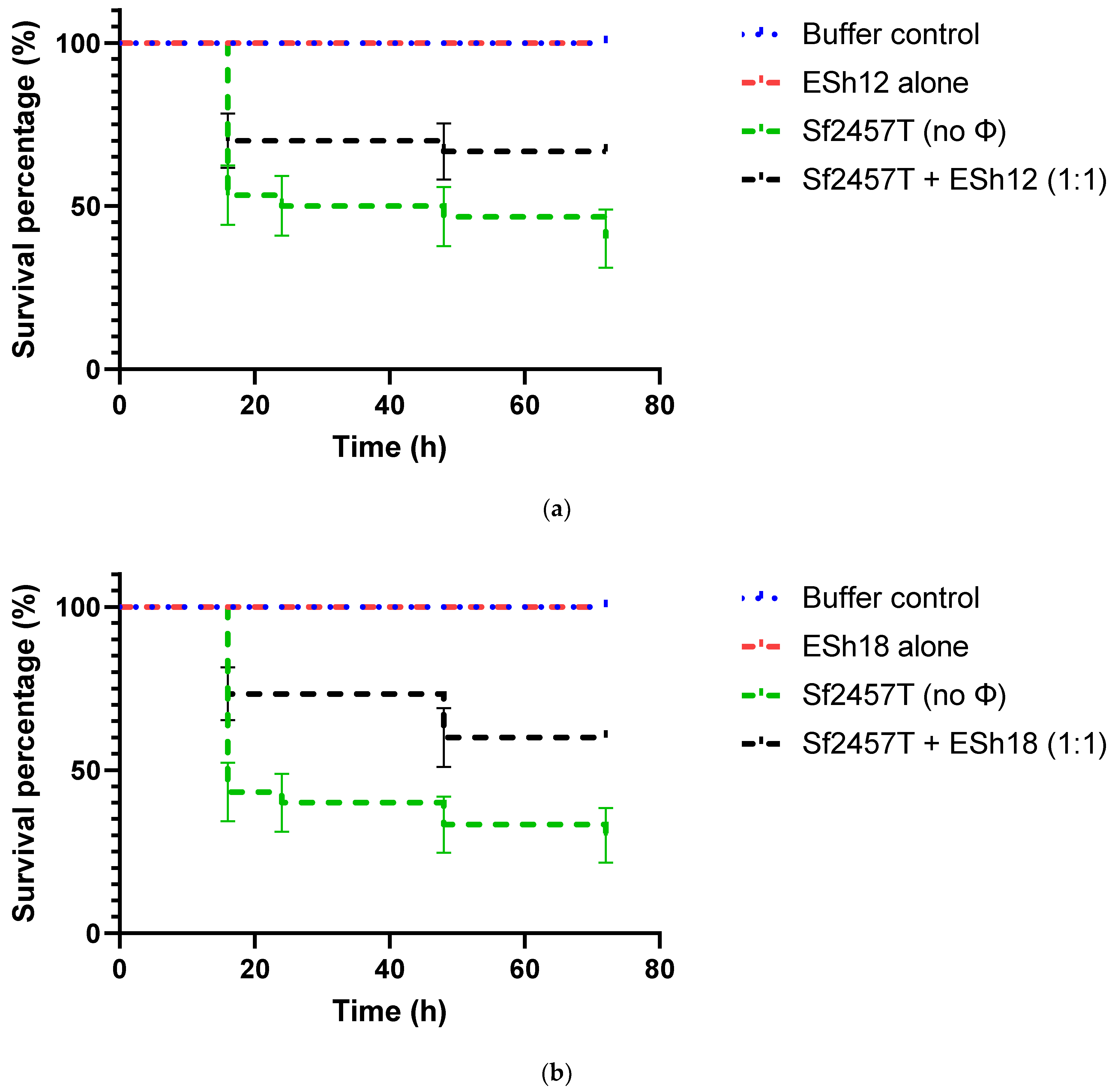
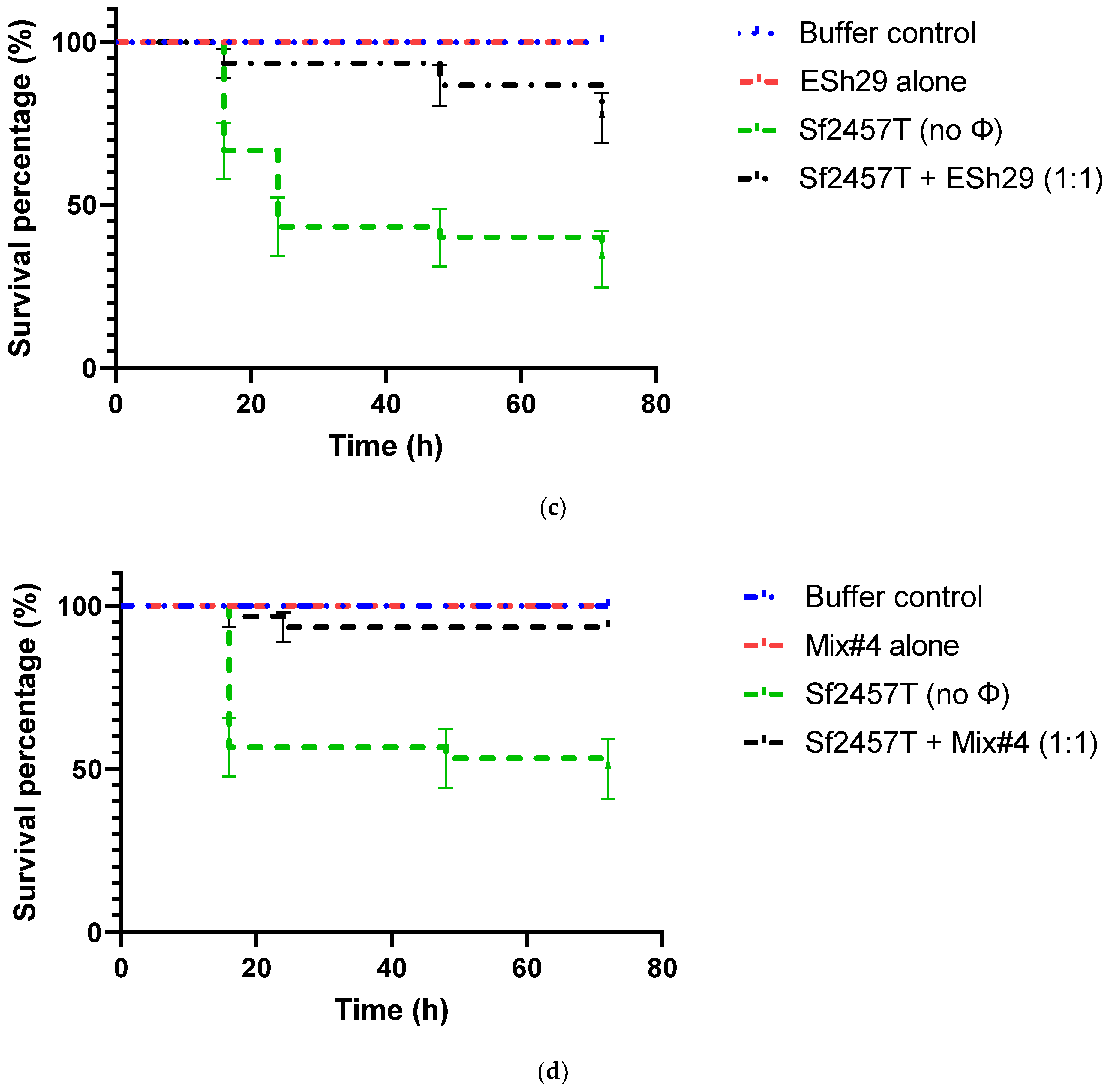
2.6. Phage Treatment of Shigella Infection of G. mellonella Larvae

Based on the plating efficiencies of phages ESh12, ESh18 and ESh29, their individual lytic activities, and also their combined killing effect in liquid culture and host range, prototype cocktail #4 was selected to test therapeutic efficacy in the wax moth (G. mellonella) larvae infection model as an initial, more rapid and economical in vivo assessment. The therapeutic effect using the individual component phages and the 3-phage mixture was tested in the treatment of S. flexneri strain 2457T infection of G. mellonella larvae. Administration of the phages and the cocktail at the doses used did not cause adverse effects on the larvae, a concern because of endotoxin carryover in phage purification (not shown). Survival was extended in larvae infected with a lethal dose of strain 2457T and treated 30 min later with either the individual phages or the cocktail (Figure 3). The rate of survival of the larvae after 72 h increased from 40–50% without phage treatment to 55–85% with treatment using the individual phages or the cocktail. Phage ESh29 alone (Figure 3c) or the 3-phage cocktail (Figure 3d) each provided an increase in survival to about 85%; this indicates that ESh29 provides the predominant therapeutic effect of the cocktail against strain 2457T, though all three component phages provided an increase in survival in this model (Figure 3). Higher doses of the three individual phages or the mixture did not correlate with higher survival after treatment, however (not shown).

3. Discussion

The high prevalence and severity of shigellosis, particularly in developing countries among both residents and travelers [1,4], and increasing drug resistance in Shigella spp. isolates [8] indicate that new antibacterials are needed to augment antibiotics. Phages are a promising option for the prophylaxis and therapy of shigellosis. Previous prophylactic oral treatment of children with a mixture of lytic phages resulted in a significant reduction of shigellosis in comparison with control groups receiving a placebo [29,30]. A 5-phage cocktail administered orally to mice before and/or after S. sonnei infection significantly reduced the numbers of bacteria in fecal and cecum specimens [23]. Optimization of a phage cocktail and more frequent phage application (perhaps every day or every other day) could potentially result in an even higher efficacy of shigellosis prophylaxis than what was observed in these studies. More research is required to isolate lytic phages of Shigella and characterize them in vitro and in vivo toward developing robust fixed phage cocktails to prevent and treat drug-resistant shigellosis, with potential benefits for civilian travelers and deployed military personnel.
The purpose of this work was to isolate a panel of lytic phages with broad activity against diverse Shigella strains, to develop prototype phage cocktails and evaluate the efficacy of phage treatment in a waxworm model. The eight strains of Shigella used for phage enrichment included S. flexneri (serotypes 2a, 3a, 5a, and 6), S. sonnei, and S. dysenteriae (serotype 1) (Table 1). Thirty-two lytic phages active against Shigella species (Table 2) were isolated from three fractions of Washington DC sewage (grit chamber water, secondary effluent and blend sludge) collected on the same day. Whole-genome sequencing and analysis enabled classification of the phages into seven viral genera (Table 2, Figure 1).
The virus family represented by the largest number of phages in the panel (17) was Straboviridae (formerly Myoviridae), including subfamily Tevenvirinae, genera Tequatrovirus (T4-like, 15 phages) and Mosigvirus (2 phages). T4-like phages are strictly lytic, do not show significant DNA sequence identity with bacterial genomes [37,38] and have broad host ranges among Shigella [23,39], pathogenic strains of E. coli [38,40,41,42], both E. coli and Salmonella [43,44], E. coli, Salmonella and Shigella [45], Yersinia pestis and Yersinia pseudotuberculosis [46], even Acinetobacter baumannii [47] and Stenotrophomonas maltophilia [48]. However, S. maltophilia T4-like phage DLP6 encodes a transposon that might stimulate gene transfer and thus is not a favorable candidate for phage therapy [48]. This suggests that genomes of even strictly lytic phages should be analyzed in depth to exclude potentially detrimental gene content before using them as therapeutics. Mosigvirus phages are similar to those belonging to the genus Tequatrovirus and were also considered T4-like phages until recently, when they were reclassified into a separate genus. They are also obligately lytic and demonstrate broad activity against Shigella [23,49] and pathogenic E. coli [41]. Four of the 32 phages belonged to subfamily Ounavirinae within family Myoviridae, genus Mooglevirus.
These 21 myoviral phages within Straboviridae and Ounavirinae isolated in this study did not show any significant DNA sequence similarity to genes encoding integrases, recombinases, transposases, excisionases, and repressors of the lytic cycle, nor to any bacterial genes, including drug resistance and pathogenicity determinants. The seventeen phages that belong to subfamily Tevenvirinae within Straboviridae appear to be safe for therapeutic application based on gene content. Subfamily Ounavirinae was named after diagnostic lytic Salmonella phage O1 (or Felix O1), proposed for therapy and control of Salmonella in food [50]. Although four Ounavirinae phages discovered by our team (ESh19-22) appear to be virulent, they share high genome identity with phages SUSP1 and SUSP2 (subfamily Ounavirinae, genus Suspvirus). SUSP1 and SUSP2, called “superspreaders,” have the demonstrated ability to efficiently release intact plasmid DNA upon lysis, followed by enhanced horizontal gene transfer via transformation [32]. Unless this ability can be experimentally excluded for ESh19-22, these four phages cannot be recommended for therapeutic use because of the risk of potentially spreading drug resistance or virulence determinants.
Ten of the phages were classified as members of three genera within family Autographiviridae (formerly Podovoridae) and subfamily Studiervirinae, including Teetrevirus (T3-like phages, ESh7-10 and ESh12), Teseptimavirus (T7-like phages, ESh1-3 and ESh6) and Kayfunavirus (ESh23). Shigella phages that belong to subfamily Studiervirinae appear to be relatively rare. For example, among 69 Shigella phages deposited in GenBank and listed in a recent review article by Subramanian et al. [51], there are 27 myophages of subfamily Tevenvirinae (18 of which are members of genus Tequatrovirus), while there is only one representative of podovirus subfamily Studiervirinae, Kayfunavirus phage SFPH2, and no T3- or T7-like phages. SFPH2 has been shown to lyse strains of S. flexneri 2, 2a and Y [52]. However, it was observed long ago that E. coli phages T3 and T7 are able to lyse some S. sonnei strains [53,54]. T3- and T7-like phages are virulent, have robust lytic activity [55,56,57], do not encode toxic proteins [58] and thus appear to be promising as candidate therapeutics. Teetrevirus phage KPP-5 exhibited a broad host range for Klebsiella pneumoniae strains [59] and Teseptimavirus phage EG1 was specific for uropathogenic isolates of E. coli [58]. Another T7-like phage, φA1122, is capable of lysing the vast majority of diverse Y. pestis strains [60]. Genomic analysis of all 10 podophages isolated in this study revealed no potentially detrimental genetic information, so these can be considered as candidate therapeutics. Finally, one phage, ESh4, belonged to family Drexlerviridae (formerly Siphoviridae), subfamily Tempevirinae, genus Hanrivervirus. The first representative of this genus, virulent phage pSf-1 isolated in Korea, showed lytic activity against S. flexneri, S. boydii and S. sonnei [61]. ESh4 also seems to be a candidate therapeutic phage because no potentially deleterious genes were found in its genome. Transmission electron microscopy confirmed that the 32 phages isolated in this work belong to myo-, podo- and siphoviruses (Figure 2).
Initial use of host range testing against a small Shigella strain panel allowed for the selection of 12 phages with broader activity for further characterization (Table 3). This selection included representatives of genera Tequatrovirus (ESh16-18, ESh29, ESh31, ESh33, and ESh35), Mosigvirus (ESh27), Mooglevirus (ESh22), Teetrevirus (ESh9 and ESh12), and Teseptimavirus (ESh1). Four 3-phage cocktails were developed from these phages and tested for the ability to lyse and kill S. flexneri 2a 2457T, a fully virulent challenge strain that has been used globally in animal trials as a virulent Shigella challenge [34]. Three of these cocktails (mixtures ##1, 2 and 4) were able to completely lyse and sterilize broth cultures of strain 2457T (Table 4), suggesting that these phage combinations successfully kill the entire bacterial population, including any mutants resistant to the individual phages. Bacterial resistance is developed less frequently to phage cocktails than to single phages because well designed cocktails usually contain phages that use different cell surface receptors, and mutants resistant to one phage can be lysed by other cocktail components [62]. These results also indicated that strain 2457T does not rapidly develop resistance to these three 3-phage cocktails.
The host ranges of the 12 selected phages and four 3-phage cocktails were evaluated using a diversity panel of 95 Shigella clinical strains isolated in Southeast Asia and East Asia, Africa, South America, and the USA (Table 5 and Table S1). The lytic spectra of individual phages ranged from 59% to 87%, and altogether the 12 phages were able to lyse 86/95 (90.5%) of the Shigella strains listed in Table S1. All four 3-phage cocktails had broad host ranges, with activity against 100% of S. sonnei isolates, 82–97% of S. flexneri (serotypes 1, 1a, 1b, 1c, 2, 2a, 2b, 3a, 3b, 4, 4a, 5, and 6, as well as X and Y variants) and 100% of S. dysenteriae serotypes 1 and 2 (Table 5). Therefore, they had killing activity against virtually all of the Shigella subtypes that cause travelers’ diarrhea in Southeast Asia. The lytic activity observed for mixture #15 against the entire Shigella diversity panel was 88.8%. Importantly, 94% of the strains (89/95) were pigmented on Congo Red agar and thus possessed the virulence plasmid, indicating the selected phages and phage cocktails can efficiently kill virulent Shigella strains. S. dysenteriae serotypes 9 and 12 and most S. boydii isolates were resistant to all of the phages tested. Our data on the broad host ranges of individual Shigella phages and their mixtures agree with the results of others, but the bacterial strain panel used in this study may be better characterized and more diverse. For example, commercially available INTESTI, PYO and Septaphage phage cocktail products manufactured in Georgia, respectively possessed lytic activity against 19/20 (95%), 19/20 and 11/20 (55%) strains of Shigella spp. isolated in Switzerland (species/serotype breakdown not provided) [63]. A cocktail of two T1-like phages was able to lyse 85% of MDR isolates of S. sonnei (44) and S. flexneri (26, without serotype breakdown) from different provinces of Iran [9]. A cocktail of five ATCC phages that belong to genera Tequatrovirus (3), Mosigvirus (1) and Tequintavirus (1; family Demerecviridae, subfamily Markadamsvirinae) provided a lytic effect against all strains of S. sonnei (18), S. dysenteriae (5), S. boydii (4), and 35/38 S. flexneri isolates (without serotype breakdown for the latter three species) [23].
Use of waxworm (G. mellonella larvae) infection models to study bacterial virulence and test new antimicrobials, including phages, offers low cost, technical simplicity and lack of ethical restrictions, in contrast to vertebrate models [64]. G. mellonella larvae have been used in Shigella virulence studies: injection of S. flexneri 2a 2457T was lethal for waxworms, while oral force feeding did not cause any death or clinical manifestations [65]. In this effort, we used a waxworm injection model to evaluate phage therapeutic efficacy against S. flexneri 2a 2457T infection. Prototype cocktail #4 was selected for this model based on the plating efficiencies of phages ESh12, ESh18 and ESh29 on strain 2457T, their individual lytic activities, the complete sterilization of liquid culture by the cocktail, and also host range of the mixture. Administration of each individual phage and the cocktail at the doses used did not cause adverse effects on the larvae, a concern because of endotoxin carryover in phage purification (not shown). Survival was significantly extended in larvae infected with a lethal dose of strain 2457T and treated 30 min later with the individual phages or the cocktail (Figure 3). The rate of survival of the larvae after 72 h increased from 40–50% without phage treatment to 55–85% with treatment using the individual phages or the cocktail (Figure 3). Both phage ESh29 alone and the 3-phage mixture provided an increase in survival to about 85%; this indicated that ESh29 may provide the majority of the therapeutic effect of the cocktail against strain 2457T, though each of the three component phages alone provided an increase in survival in this model (Figure 3). However, providing higher MOIs of the three individual phages or the mixture did not correlate with higher survival after treatment (not shown), perhaps because treatment efficacy was already saturating at the 1:1 MOI. The phage therapeutic effects observed in this study are comparable with those observed by others for lytic phages used in waxworms infected with Burkholderia cepacia [66], Clostridium difficile [67], K. pneumoniae [68], vancomycin-resistant Enterococcus faecium [69], and methicillin-resistant Staphylococcus aureus [70], though in the latter case, the observed phage effect was dose-dependent. One group first showed phage efficacy against Pseudomonas aeruginosa and A. baumannii infections in Galleria and then confirmed it in a mouse acute pneumonia model to indicate the relevance of the Galleria model [71,72].
The initial stages of rational phage cocktail design were employed in this study, with a focus on component phage host range and cocktail lytic activity. This effort built candidate phage mixtures for preclinical testing and potential development for prophylaxis and treatment of the common pathogens that cause shigellosis, a significant medical problem for large populations in developing countries, deployed military service members, and travelers. Initially we had anticipated the need to design different cocktails against representatives of the predominant Shigella species and serotypes circulating regionally in different parts of the world, but this initial effort indicated that it may well be possible to address the key Shigella pathogens on a global level using a single phage cocktail formulation, if well designed. A treatment effect against S. flexneri 2a was demonstrated here in the Galleria model, but further work must be done to determine whether such phage cocktails are also effective in treating Shigella infections in relevant mammalian models, and eventually in humans. Prophylaxis may be the main role for phages if they have a limited effect on human disease once underway because of the Shigella invasive and intracellular lifestyle [1]. Additionally, it will be necessary to demonstrate that the in vitro lytic spectrum of the phage cocktail translates to breadth of activity in vivo. The Galleria model can be used to test the efficacy of treatment against the main serotypes of S. flexneri and S. sonnei that cause the majority of disease and also allow for testing against a greater variety of pathogen strains than is feasible using mammalian models.

4. Materials and Methods

4.1. Bacterial Strains and Culture Media

The Shigella strains used in this work are presented in Table 1 and Table S1. Bacteria were grown in Heart Infusion Broth (HIB, Becton, Dickinson and Co., Franklin Lakes, NJ, USA) or on 1.5% HIB agar plates at 37 °C. Semisolid 0.7% HIB agar was used as overlay for phage plating [73].

4.2. Isolation of Phages

Wastewater samples from three different sites and reactors (grit chamber water, secondary effluent and blend sludge) within the Blue Plains Wastewater Treatment Plant (Washington DC) were used as source materials for phage isolation. Eight strains were used for phage enrichment: S. flexneri 2a 2457T (virulent model strain broadly used in in vitro and in vivo studies [74]), BS103 (avirulent derivative of 2457T [75]), S. flexneri 3a J17B (wild type S. flexneri 3a strain from the Walter Reed Army Institute of Research/WRAIR collection [74]), S. flexneri 5a M90T55 (avirulent derivative of M90T [76]), S. flexneri 6 SSU2515 (wild type S. flexneri 6 strain from WRAIR collection; the source was a sheep outbreak in Florida in 1973 [Malabi Venkatesan, personal communication]), S. flexneri 6 CCH060 (wild type S. flexneri 6 strain from WRAIR collection [74]), S. sonnei Moseley (type strain from WRAIR collection [77]), and S. dysenteriae 1 1617 (wild type S. dysenteriae 1 from WRAIR collection [78]) (Table 1). The wastewater samples were centrifuged for 60 min at 5500× g. Centrifugation was repeated for blend sludge. Grit chamber water and secondary effluent supernatants were filtered using sterile 0.22-μm filters, while the blend sludge supernatant was consecutively filtered through 0.8-μm, 0.45-μm and 0.22-μm filters (MilliporeSigma, Bollington, MA, USA). Then, the samples were processed as previously described [79]. Briefly, 5× HIB was mixed with each filter-sterilized sample at a 1:5 ratio, and overnight broth culture of each enrichment Shigella strain was added to create 24 enrichment mixtures (three wastewater fractions by eight enrichment strains). The enrichment mixtures were incubated overnight at 37 °C with shaking at 200 rpm, and the supernatant was sterilized using a 0.22-μm filter. The resulting lysates were evaluated for plaque formation on double-layer HIB agar plates [73]. Phage purification was performed by three rounds of single plaque isolation.

4.3. Phage Propagation

Bacteriophages were propagated on S. flexneri strain BS103. The host bacteria were grown in HIB supplemented with 5 mM calcium chloride and incubated at 37 °C with shaking at 200 rpm. Phage lysate was added to 250 mL of an early exponential phase bacterial culture grown in HIB (OD600 of 0.1–0.2) at a multiplicity of infection (MOI) of 0.01–0.1 and incubated in a 500-mL plastic Erlenmeyer flask at 37 °C and 200 rpm until visible lysis occurred. Phage lysate was treated with chloroform (5%, vol./vol.). Bacterial debris was removed by centrifugation for 15 min at 5500× g. Phage particles from the supernatant were concentrated by centrifugation for 3 h at 13,250× g. Phage pellets were resuspended in 1/40 vol. of SM buffer (Teknova, Hollister, CA, USA). Bacterial debris was removed by centrifugation for 15 min at 5500× g, supernatant was collected and filtered through a sterile 0.22-μm PVDF membrane (MilliporeSigma, Sigma-Aldrich Inc., Saint Louis, MO, USA). Endotoxin levels in phage suspensions were tested with the Endosafe nexgen-PTS device (Charles River Laboratories, Wilmington, MA, USA), and if needed, further purified using EndoTrap bulk resin (Hyglos GmbH, Bernried am Starnberger See, Germany) according to the manufacturer’s protocol, to ensure that the endotoxin level was below 500 EU per 109 PFU (plaque-forming units), approximating the U.S. Food & Drug Administration guidance for human use.

4.4. Phage DNA Isolation, Restriction Analysis and Genome Sequencing

Phage genomic DNA was extracted as described previously [80]. To identify unique phages, DNA samples were treated with restriction endonuclease EcoRV (New England BioLabs Inc., Ipswich, MA, USA) according to the manufacturer’s protocol and DNA fragments were separated using agarose gel electrophoresis. Phage genomic DNA of 32 phages with unique restriction profiles was sequenced on a MiSeq benchtop sequencer (Illumina, Inc., San Diego, CA, USA). Libraries were constructed using the Kapa HyperPlus library preparation kit (Roche Diagnostics, Indianapolis, IN, USA). Libraries were quantified using the Kapa library quantification kit Illumina/Bio-Rad iCycler (Roche Diagnostics) on a CFX96 real-time cycler (Bio-Rad, Hercules, CA, USA). For the MiSeq, libraries were normalized to 2 nM, pooled, denatured, and diluted to 20 pM. The pooled samples were further diluted to a final concentration of 14 pM. Samples were sequenced using MiSeq reagent kit v3 (Illumina; 600 cycles; 2 × 300 bp). Short-read sequencing data were trimmed for adapter sequence content and quality using Btrim64. Overlapping sequence reads were merged using FLASH. De novo assembly was performed using Newbler (v2.7). Minimum thresholds for contig size and coverage were set at 200 bp and 49.5×, respectively. The annotation of open reading frames and sequence similarity searches were performed as described earlier [79].

4.5. Phage Phylogenetic Tree

A phage whole genome phylogeny was generated from an ANI (Average Nucleotide Identity)-based distance matrix calculated with the Mash program [35] as described previously [81]. Briefly, a sketch file was created from the 32 described Shigella phage genomes isolated and sequenced in this study, plus 25 obtained from GenBank (11 Escherichia, 6 Shigella, 3 Yersinia, 2 Serratia, 1 Klebsiella and 2 Enterobacteria phages) with BLASTN [Basic Local Alignment Search Tool searching the NCBI nucleotide database] matches to the Shigella phages), with 5000 13mers generated per genome (i.e., mash sketch -k 13 -s 5000). The sketch file was then compared to all the phage genome sequences to generate the ANI matrix using the Mash distance command using default settings. The Gaussian Genome Representative Selector with Prioritization (GGRaSP) [36] R-package was used to calculate the UPGMA (Unweighted Pair Group Method with Arithmetic Mean) phylogeny from the ANI distance matrix, using GGRaSP options -e 5 -d 100 -m average. The resulting dendrogram was translated into Newick format within GGRaSP using the APE R package [82], loaded into the iTOL tree viewer [83], and annotated with taxonomic information.

4.6. Transmission Electron Microscopy

Phages were prepared for transmission electron microscopy as described previously [84], with minor modifications. Briefly, phage suspensions were washed twice with 0.1% ammonium acetate using centrifugation for 3 h at 13,250× g and phage titers were adjusted to 109 PFU/mL. Phage particles were deposited on 300 mesh carbon-coated copper grids (Electron Microscopy Sciences, Hatfield, PA, USA), stained with 2% uranyl acetate for 1 min and examined in a JEOL JEM-1400 electron microscope at 80 kV. Images were analyzed with Image J software v. 1.53 (National Institutes of Health, Bethesda, MD, USA).

4.7. Phage Host Range Testing

Phage host ranges were determined using a micro-spot plating assay [80]. Briefly, 10-fold serial phage dilutions were prepared in a sterile flat-bottomed 96-well plate. A 2-μL aliquot of each phage dilution, ranging from 10−1 to 10−8, was spotted with a multichannel pipette on 0.7% HIB agar overlay infused with Shigella culture and incubated overnight at 37 °C. The morphology of individual plaques was evaluated and the results were scored from 4+ to 0 as follows: 4+, totally clear spots, isolated large clear plaques in the highest phage dilutions (highly positive result); 3+, clear spots, clear plaques of medium or small size, or large turbid plaques (positive result); 2+, clear or turbid spots, tiny clear or turbid plaques, sometimes barely countable (slightly positive result); 1+, lysis from without indicated by very faint, turbid spots or clear spots in first dilutions, no plaques (negative result); 0, no lysis spots or plaques (negative result).

4.8. Assessment of Phage Protection against Infection of G. mellonella Larvae with Shigella Strains

To determine if phages would provide an in vivo therapeutic effect against Shigella, a G. mellonella larva (wax worm) model of infection was utilized [72]. S. flexneri 2457T was grown to exponential phase, washed and resuspended in PBS to approximately 1 × 10⁷ CFU per mL. Waxworms (Vanderhorst, Inc., St. Marys, OH, USA) in the final-instar larval stage and weighing 200–300 mg were saved and housed in clean plastic Petri dishes, 10 worms per group. Worms were inoculated with 10 μL of the bacterial suspension prepared above into the last left proleg using a 300-μL BD Insulin syringe (Becton Dickinson, 1 Becton Drive, Franklin Lakes, NJ, USA). After 30 min, 10 μL of phage suspension (in dilutions to deliver MOIs of 1:1, 10:1 or 100:1 [pfu/CFU]) or its vehicle buffer was injected in the opposite proleg. After these infection and treatment injections, worms were incubated in plastic Petri dishes at 37 °C and monitored for death over 4 days. Worms were considered dead when they displayed no movement in response to tactile stimuli. Two control groups were included in the experiment, an “untouched” control group that did not receive any injections, to ensure the health of the worms after shipping, and a PBS control group that was injected with PBS instead of bacteria, to control for any detrimental effects from injection.

4.9. Statistical Analysis

Kaplan-Meier survival curves were compared using the Log-rank (Mantel-Cox) test with Bonferroni’s correction for multiple comparisons. Significance was established at p < 0.05. Statistical analysis was done using GraphPad software (http://www.graphpad.com/quickcalcs/), accessed on May 2022.

4.10. Accession Numbers

GenBank accession numbers for all phages are listed in Table 2.

Supplementary Materials

The following supporting information can be downloaded at: https://www.mdpi.com/article/10.3390/antibiotics11111659/s1, Table S1: Host range testing of 12 phages against 95 Shigella isolates.

Author Contributions

Conceptualization, M.P.N., A.A.F. and K.V.S.; methodology, A.A.F., K.V.S., W.S., A.S., D.E.F. and Y.H.; validation, A.A.F., K.V.S., W.S., W.L., P.T.M. and D.W.E.; formal analysis, A.A.F., W.S., K.V.S., E.C.S., R.T.K., D.E.F. and M.P.N.; resources, B.E.S., W.L., D.W.E., P.T.M. and M.P.N.; data curation, A.A.F., K.V.S., R.T.K. and W.S.; writing—original draft preparation, M.P.N.; writing—review and editing, A.A.F., M.P.N., D.W.E., W.S., R.T.K., D.E.F. and B.E.S.; supervision, M.P.N., D.W.E. and B.E.S.; project administration, M.P.N.; funding acquisition, M.P.N. All authors have read and agreed to the published version of the manuscript.

Funding

This research was funded by the Defense Health Program through Military Infectious Disease Research Program project D0559_17_WR and the WRAIR command, and supported by Congressionally Directed Medical Research Program grant PR182667 (Peer Reviewed Medical Research Program).

Institutional Review Board Statement

Not applicable.

Informed Consent Statement

Not applicable.

Data Availability Statement

Not applicable.

Acknowledgments

Material has been reviewed by the Walter Reed Army Institute of Research. There is no objection to its presentation and/or publication. The opinions or assertions contained herein are the private views of the author, and are not to be construed as official, or as reflecting true views of the Department of the Army or the Department of Defense. We thank Akhil Reddy and Austin Puchany (WRAIR WID) and Kamonporn Poramathikul (AFRIMS) for their technical assistance, Edwin Oaks for advice and providing type strains from the WRAIR Bacterial Diseases Branch collection, Emil Lesho and the staff at the WRAIR Multidrug-resistant organism Repository and Surveillance Network (MRSN) for whole-genome sequencing and bioinformatic analyses, Nathan Brown (University of Leicester, Leicester, England) for his early-stage genomic analysis, and Edward Asafo-Adjei (WRAIR Pathology) for the electron micrographs.

Conflicts of Interest

The authors declare no conflict of interest. The funders had no role in the design of the study; in the collection, analyses, or interpretation of data; in the writing of the manuscript, or in the decision to publish the results.

References

  1. Kotloff, K.L.; Riddle, M.S.; Platts-Mills, J.A.; Pavlinac, P.; Zaidi, A.K.M. Shigellosis. Lancet 2018, 391, 801–812. [Google Scholar] [CrossRef]
  2. Baker, S.; The, H.C. Recent insights into Shigella. Curr. Opin. Infect. Dis. 2018, 31, 449–454. [Google Scholar] [CrossRef]
  3. Steffen, R.; Hill, D.R.; DuPont, H.L. Traveler’s diarrhea: A clinical review. JAMA 2015, 313, 71–80. [Google Scholar] [CrossRef] [PubMed]
  4. Olson, S.; Hall, A.; Riddle, M.S.; Porter, C.K. Travelers’ diarrhea: Update on the incidence, etiology and risk in military and similar populations—1990–2005 versus 2005–2015, does a decade make a difference? Trop. Dis. Travel Med. Vaccines 2019, 5, 1. [Google Scholar] [CrossRef] [PubMed]
  5. Barry, E.; Cassels, F.; Riddle, M.; Walker, R.; Wierzba, T. Vaccines against Shigella and enterotoxigenic Escherichia coli: A summary of the 2018 VASE Conference. Vaccine 2019, 37, 4768–4774. [Google Scholar] [CrossRef] [PubMed]
  6. Chapartegui-González, I.; Bowser, S.; Torres, A.G.; Khakhum, N. Recent progress in Shigella and Burkholderia pseudomallei vaccines. Pathogens 2021, 10, 1353. [Google Scholar] [CrossRef] [PubMed]
  7. Khalil, I.A.; Troeger, C.; Blacker, B.F.; Rao, P.C.; Brown, A.; Atherly, D.E.; Brewer, T.G.; Engmann, C.M.; Houpt, E.R.; Kang, G.; et al. Morbidity and mortality due to shigella and enterotoxigenic Escherichia coli diarrhoea: The Global Burden of Disease Study 1990–2016. Lancet Infect. Dis. 2018, 18, 1229–1240. [Google Scholar] [CrossRef] [Green Version]
  8. Puzari, M.; Sharma, M.; Chetia, P. Emergence of antibiotic resistant Shigella species: A matter of concern. J. Infect. Public. Health 2018, 11, 451–454. [Google Scholar] [CrossRef] [PubMed]
  9. Shahin, K.; Bouzari, M.; Komijani, M.; Wang, R. A new phage cocktail against multidrug, ESBL-producer isolates of Shigella sonnei and Shigella flexneri with highly efficient bacteriolytic activity. Microb. Drug Resist. 2020, 26, 831–841. [Google Scholar] [CrossRef] [PubMed] [Green Version]
  10. Connor, B.A.; Riddle, M.S. Post-infectious sequelae of travelers’ diarrhea. J. Travel Med. 2013, 20, 303–312. [Google Scholar] [CrossRef]
  11. Sethuvel, D.P.M.; Ragupathi, N.K.D.; Anandan, S.; Veeraraghavan, B. Update on: Shigella new serogroups/serotypes and their antimicrobial resistance. Lett. Appl. Microbiol. 2017, 64, 8–18. [Google Scholar] [CrossRef] [PubMed]
  12. Livio, S.; Strockbine, N.A.; Panchalingam, S.; Tennant, S.M.; Barry, E.M.; Marohn, M.E.; Antonio, M.; Hossain, A.; Mandomando, I.; Ochieng, J.B.; et al. Shigella isolates from the global enteric multicenter study inform vaccine development. Clin. Infect. Dis. 2014, 59, 933–941. [Google Scholar] [CrossRef] [PubMed]
  13. Lindsay, B.; Ochieng, J.B.; Ikumapayi, U.N.; Toure, A.; Ahmed, D.; Li, S.; Panchalingam, S.; Levine, M.M.; Kotloff, K.; Rasko, D.A.; et al. Quantitative PCR for detection of Shigella improves ascertainment of Shigella burden in children with moderate-to-severe diarrhea in low-income countries. J. Clin. Microbiol. 2013, 51, 1740–1746. [Google Scholar] [CrossRef] [Green Version]
  14. Pavlinac, P.B.; Platts-Mills, J.A.; Tickell, K.D.; Liu, J.; Juma, J.; Kabir, F.; Nkeze, J.; Okoi, C.; Operario, D.J.; Uddin, J.; et al. The clinical presentation of culture-positive and culture-negative, quantitative polymerase chain reaction (qPCR)-attributable shigellosis in the Global Enteric Multicenter Study and derivation of a Shigella severity score: Implications for pediatric Shigella vaccine trials. Clin. Infect. Dis. 2021, 73, e569–e579. [Google Scholar] [CrossRef] [PubMed]
  15. Liu, J.; Platts-Mills, J.A.; Juma, J.; Kabir, F.; Nkeze, J.; Okoi, C.; Operario, D.J.; Uddin, J.; Ahmed, S.; Alonso, P.L.; et al. Use of quantitative molecular diagnostic methods to identify causes of diarrhoea in children: A reanalysis of the GEMS case-control study. Lancet 2016, 388, 1291–1301. [Google Scholar] [CrossRef] [Green Version]
  16. Sanders, J.W.; Putnam, S.D.; Gould, P.; Kolisnyk, J.; Merced, N.; Barthel, V.; Rozmajzl, P.J.; Shaheen, H.; Fouad, S.; Frenck, R.W. Diarrheal illness among deployed U.S. military personnel during Operation Bright Star 2001--Egypt. Diagn. Microbiol. Infect. Dis. 2005, 52, 85–90. [Google Scholar] [CrossRef]
  17. Porter, C.K.; Olson, S.; Hall, A.; Riddle, M.S. Travelers’ diarrhea: An update on the incidence, etiology, and risk in military deployments and similar travel populations. Mil. Med. 2017, 182, 4–10. [Google Scholar] [CrossRef] [PubMed] [Green Version]
  18. Bodhidatta, L.; McDaniel, P.; Sornsakrin, S.; Srijan, A.; Serichantalergs, O.; Mason, C.J. Case-control study of diarrheal disease etiology in a remote rural area in Western Thailand. Am. J. Trop. Med. Hyg. 2010, 83, 1106–1109. [Google Scholar] [CrossRef] [Green Version]
  19. Swierczewski, B.E.; Odundo, E.A.; Koech, M.C.; Ndonye, J.N.; Kirera, R.K.; Odhiambo, C.P.; Cheruiyot, E.K.; Wu, M.T.; Lee, J.E.; Zhang, C.; et al. Surveillance for enteric pathogens in a case-control study of acute diarrhea in Western Kenya. Trans. R. Soc. Trop. Med. Hyg. 2013, 107, 83–90. [Google Scholar] [CrossRef] [PubMed] [Green Version]
  20. Nikolich, M.P.; Filippov, A.A. Bacteriophage therapy: Developments and directions. Antibiotics 2020, 9, 135. [Google Scholar] [CrossRef] [PubMed]
  21. Uyttebroek, S.; Chen, B.; Onsea, J.; Ruythooren, F.; Debaveye, Y.; Devolder, D.; Spriet, I.; Depypere, M.; Wagemans, J.; Lavigne, R.; et al. Safety and efficacy of phage therapy in difficult-to-treat infections: A systematic review. Lancet Infect. Dis. 2022, 22, E208–E220. [Google Scholar] [CrossRef]
  22. Llanos-Chea, A.; Citorik, R.J.; Nickerson, K.P.; Ingano, L.; Serena, G.; Senger, S.; Lu, T.K.; Fasano, A.; Faherty, C.S. Bacteriophage therapy testing against Shigella flexneri in a novel human intestinal organoid-derived infection model. J. Pediatr. Gastroenterol. Nutr. 2019, 68, 509–516. [Google Scholar] [CrossRef] [PubMed]
  23. Mai, V.; Ukhanova, M.; Reinhard, M.K.; Li, M.; Sulakvelidze, A. Bacteriophage administration significantly reduces Shigella colonization and shedding by Shigella-challenged mice without deleterious side effects and distortions in the gut microbiota. Bacteriophage 2015, 5, e1088124. [Google Scholar] [CrossRef] [PubMed] [Green Version]
  24. Mai, V.; Ukhanova, M.; Visone, L.; Abuladze, T.; Sulakvelidze, A. Bacteriophage administration reduces the concentration of Listeria monocytogenes in the gastrointestinal tract and its translocation to spleen and liver in experimentally infected mice. Int. J. Microbiol. 2010, 2010, 624234. [Google Scholar] [CrossRef] [Green Version]
  25. Dissanayake, U.; Ukhanova, M.; Moye, Z.D.; Sulakvelidze, A.; Mai, V. Bacteriophages reduce pathogenic Escherichia coli counts in mice without distorting gut microbiota. Front. Microbiol. 2019, 10, 1984. [Google Scholar] [CrossRef] [PubMed]
  26. Yen, M.; Cairns, L.S.; Camilli, A. A cocktail of three virulent bacteriophages prevents Vibrio cholerae infection in animal models. Nat. Commun. 2017, 8, 14187. [Google Scholar] [CrossRef] [PubMed] [Green Version]
  27. Goodridge, L.D. Bacteriophages for managing Shigella in various clinical and non-clinical settings. Bacteriophage 2013, 3, e25098. [Google Scholar] [CrossRef] [PubMed] [Green Version]
  28. Tang, S.S.; Biswas, S.K.; Tan, W.S.; Saha, A.K.; Leo, B.F. Efficacy and potential of phage therapy against multidrug resistant Shigella spp. PeerJ 2019, 7, e6225. [Google Scholar] [CrossRef] [Green Version]
  29. Solodovnikov, I.P.; Pavlova, L.I.; Emel’ianov, P.I.; Garnova, N.A.; Nogteva, I.B.; Sotemskiĭ, I.S.; Bogdashich, O.M.; Arshinova, V.V. The prophylactic use of dry polyvalent dysentery bacteriophage with pectin in preschool children’s institutions. I. Results of a strictly controlled epidemiologic trial (Yaroslavl, 1968). Zh. Mikrobiol. Epidemiol. Immunobiol. 1970, 47, 131–137. [Google Scholar] [PubMed]
  30. Solodovnikov, I.P.; Pavlova, L.I.; Garnova, N.A.; Nogteva, I.B.; Sotemskiĭ, I.S. Preventive use of dry polyvalent dysentery bacteriophage in preschool institutions. II. Principles of present-day tactics and application schedule of bacteriophage. Zh. Mikrobiol. Epidemiol. Immunobiol. 1971, 48, 123–127. [Google Scholar] [PubMed]
  31. Labrie, S.J.; Samson, J.E.; Moineau, S. Bacteriophage resistance mechanisms. Nat. Rev. Microbiol. 2010, 8, 317–327. [Google Scholar] [CrossRef] [PubMed]
  32. Keen, E.C.; Bliskovsky, V.V.; Malagon, F.; Baker, J.D.; Prince, J.S.; Klaus, J.S.; Adhya, S.L. Novel “superspreader” bacteriophages promote horizontal gene transfer by transformation. mBio 2017, 8, e02115-16. [Google Scholar] [CrossRef] [Green Version]
  33. Maurelli, A.T.; Blackmon, B.; Curtiss, R., III. Loss of pigmentation in Shigella flexneri 2a is correlated with loss of virulence and virulence-associated plasmid. Infect. Immun. 1984, 43, 397–401. [Google Scholar] [CrossRef] [PubMed] [Green Version]
  34. Formal, S.B.; Oaks, E.V.; Olsen, R.E.; Wingfield-Eggleston, M.; Snoy, P.J.; Cogan, J.P. Effect of prior infection with virulent Shigella flexneri 2a on the resistance of monkeys to subsequent infection with Shigella sonnei. J. Infect. Dis. 1991, 164, 533–537. [Google Scholar] [CrossRef] [PubMed]
  35. Ondov, B.D.; Treangen, T.J.; Melsted, P.; Mallonee, A.B.; Bergman, N.H.; Koren, S.; Phillippy, A.M. Mash: Fast genome and metagenome distance estimation using MinHash. Genome Biol. 2016, 17, 132. [Google Scholar] [CrossRef] [PubMed] [Green Version]
  36. Clarke, T.H.; Brinkac, L.M.; Sutton, G.; Fouts, D.E. GGRaSP: A R-package for selecting representative genomes using Gaussian mixture models. Bioinformatics 2018, 34, 3032–3034. [Google Scholar] [CrossRef]
  37. Denou, E.; Bruttin, A.; Barretto, C.; Ngom-Bru, C.; Brüssow, H.; Zuber, S. T4 phages against Escherichia coli diarrhea: Potential and problems. Virology 2009, 388, 21–30. [Google Scholar] [CrossRef] [Green Version]
  38. de Almeida Kumlien, A.C.M.; Pérez-Vega, C.; González-Villalobos, E.; Borrego, C.M.; Balcázar, J.L. Genome analysis of a new Escherichia phage vB_EcoM_C2-3 with lytic activity against multidrug-resistant Escherichia coli. Virus Res. 2022, 307, 198623. [Google Scholar] [CrossRef] [PubMed]
  39. Doore, S.M.; Schrad, J.R.; Dean, W.F.; Dover, J.A.; Parent, K.N. Shigella phages isolated during a dysentery outbreak reveal uncommon structures and broad species diversity. J. Virol. 2018, 92, e02117-17. [Google Scholar] [CrossRef] [Green Version]
  40. Zhang, C.; Li, W.; Liu, W.; Zou, L.; Yan, C.; Lu, K.; Ren, H. T4-like phage Bp7, a potential antimicrobial agent for controlling drug-resistant Escherichia coli in chickens. Appl. Environ. Microbiol. 2013, 79, 5559–5565. [Google Scholar] [CrossRef]
  41. Kaczorowska, J.; Casey, E.; Neve, H.; Franz, C.M.A.P.; Noben, J.-P.; Lugli, G.A.; Ventura, M.; van Sinderen, D.; Mahony, J. A quest of great importance-developing a broad spectrum Escherichia coli phage collection. Viruses 2019, 11, 899. [Google Scholar] [CrossRef] [Green Version]
  42. Niu, Y.D.; Liu, H.; Du, H.; Meng, R.; Mahmoud, E.S.; Wang, G.; McAllister, T.A.; Stanford, K. Efficacy of individual bacteriophages does not predict efficacy of bacteriophage cocktails for control of Escherichia coli O157. Front. Microbiol. 2021, 12, 616712. [Google Scholar] [CrossRef]
  43. Pham-Khan, N.H.; Sunahara, H.; Yamadeya, H.; Sakai, M.; Nakayama, T.; Yamamoto, H.; Bich, V.T.T.; Miyanaga, K.; Kamei, K. Isolation, characterisation and complete genome sequence of a Tequatrovirus phage, Escherichia phage KIT03, which simultaneously infects Escherichia coli O157:H7 and Salmonella enterica. Curr. Microbiol. 2019, 76, 1130–1137. [Google Scholar] [CrossRef]
  44. Zhou, Y.; Li, L.; Han, K.; Wang, L.; Cao, Y.; Ma, D.; Wang, X. A polyvalent broad-spectrum Escherichia phage Tequatrovirus EP01 capable of controlling Salmonella and Escherichia coli contamination in foods. Viruses 2022, 14, 286. [Google Scholar] [CrossRef]
  45. Hamdi, S.; Rousseau, G.M.; Labrie, S.J.; Tremblay, D.M.; Kourda, R.S.; Slama, K.B.; Moineau, S. Characterization of two polyvalent phages infecting Enterobacteriaceae. Sci. Rep. 2017, 7, 40349. [Google Scholar] [CrossRef] [Green Version]
  46. Skurnik, M.; Jaakkola, S.; Mattinen, L.; von Ossowski, L.; Nawaz, A.; Pajunen, M.I.; Happonen, L.J. Bacteriophages fEV-1 and fD1 infect Yersinia pestis. Viruses 2021, 13, 1384. [Google Scholar] [CrossRef]
  47. Jansen, M.; Wahida, A.; Latz, S.; Krüttgen, A.; Häfner, H.; Buhl, E.M.; Ritter, K.; Horz, H.-P. Enhanced antibacterial effect of the novel T4-like bacteriophage KARL-1 in combination with antibiotics against multi-drug resistant Acinetobacter baumannii. Sci. Rep. 2018, 8, 14140. [Google Scholar] [CrossRef] [Green Version]
  48. Peters, D.L.; Stothard, P.; Dennis, J.J. The isolation and characterization of Stenotrophomonas maltophilia T4-like bacteriophage DLP6. PLoS ONE 2017, 12, e0173341. [Google Scholar] [CrossRef] [Green Version]
  49. Schofield, D.A.; Wray, D.J.; Molineux, I.J. Isolation and development of bioluminescent reporter phages for bacterial dysentery. Eur. J. Clin. Microbiol. Infect. Dis. 2015, 34, 395–403. [Google Scholar] [CrossRef]
  50. Whichard, J.M.; Sriranganathan, N.; Pierson, F.W. Suppression of Salmonella growth by wild-type and large-plaque variants of bacteriophage Felix O1 in liquid culture and on chicken frankfurters. J. Food Prot. 2003, 66, 220–225. [Google Scholar] [CrossRef] [PubMed]
  51. Subramanian, S.; Parent, K.N.; Doore, S.M. Ecology, Structure, and Evolution of Shigella Phages. Annu. Rev. Virol. 2020, 7, 121–141. [Google Scholar] [CrossRef]
  52. Yang, C.; Wang, H.; Ma, H.; Bao, R.; Liu, H.; Yang, L.; Liang, B.; Jia, L.; Xie, J.; Xiang, Y.; et al. Characterization and genomic analysis of SFPH2, a novel T7virus infecting Shigella. Front. Microbiol. 2018, 9, 3027. [Google Scholar] [CrossRef]
  53. Goebel, W.F.; Jesaitis, M.A. The somatic antigen of a phage-resistant variant of phase II Shigella sonnei. J. Exp. Med. 1952, 96, 425–438. [Google Scholar] [CrossRef] [PubMed] [Green Version]
  54. Hausmann, R. The genetics of T-odd phages. Annu. Rev. Microbiol. 1973, 27, 51–68. [Google Scholar] [CrossRef] [PubMed]
  55. Krüger, D.H.; Schroeder, C. Bacteriophage T3 and bacteriophage T7 virus-host cell interactions. Microbiol. Rev. 1981, 45, 9–51. [Google Scholar] [CrossRef]
  56. Dunn, J.J.; Studier, F.W. Complete nucleotide sequence of bacteriophage T7 DNA and the locations of T7 genetic elements. J. Mol. Biol. 1983, 166, 477–535. [Google Scholar] [CrossRef]
  57. Pajunen, M.I.; Elizondo, M.R.; Skurnik, M.; Kieleczawa, J.; Molineux, I.J. Complete nucleotide sequence and likely recombinatorial origin of bacteriophage T3. J. Mol. Biol. 2002, 319, 1115–1132. [Google Scholar] [CrossRef]
  58. Gu, Y.; Xu, Y.; Xu, J.; Yu, X.; Huang, X.; Liu, G.; Liu, X. Identification of novel bacteriophage vB_EcoP-EG1 with lytic activity against planktonic and biofilm forms of uropathogenic Escherichia coli. Appl. Microbiol. Biotechnol. 2019, 103, 315–326. [Google Scholar] [CrossRef]
  59. Sofy, A.R.; El-Dougdoug, N.K.; Refaey, E.E.; Dawoud, R.A.; Hmed, A.A. Characterization and full genome sequence of novel KPP-5 lytic phage against Klebsiella pneumoniae responsible for recalcitrant Infection. Biomedicines 2021, 9, 342. [Google Scholar] [CrossRef]
  60. Garcia, E.; Elliott, J.M.; Ramanculov, E.; Chain, P.S.; Chu, M.C.; Molineux, I.J. The genome sequence of Yersinia pestis bacteriophage φA1122 reveals an intimate history with the coliphage T3 and T7 genomes. J. Bacteriol. 2003, 185, 5248–5262. [Google Scholar] [CrossRef]
  61. Jun, J.W.; Kim, J.H.; Shin, S.P.; Han, J.E.; Chai, J.Y.; Park, S.C. Characterization and complete genome sequence of the Shigella bacteriophage pSf-1. Res. Microbiol. 2013, 164, 979–986. [Google Scholar] [CrossRef] [PubMed]
  62. Rohde, C.; Resch, G.; Pirnay, J.-P.; Blasdel, B.G.; Debarbieux, L.; Gelman, D.; Górski, A.; Hazan, R.; Huys, I.; Kakabadze, E.; et al. Expert opinion on three phage therapy related topics: Bacterial phage resistance, phage training and prophages in bacterial production strains. Viruses 2018, 10, 178. [Google Scholar] [CrossRef] [PubMed] [Green Version]
  63. Bernasconi, O.J.; Donà, V.; Tinguely, R.; Endimiani, A. In vitro activity of 3 commercial bacteriophage cocktails against Salmonella and Shigella spp. isolates of human origin. Pathog. Immun. 2018, 3, 72–81. [Google Scholar] [CrossRef] [PubMed]
  64. Ménard, G.; Rouillon, A.; Cattoir, V.; Donnio, P.-Y. Galleria mellonella as a suitable model of bacterial infection: Past, present and future. Front. Cell. Infect. Microbiol. 2021, 11, 782733. [Google Scholar] [CrossRef]
  65. Barnoy, S.; Gancz, H.; Zhu, Y.; Honnold, C.L.; Zurawski, D.V.; Venkatesan, M.M. The Galleria mellonella larvae as an in vivo model for evaluation of Shigella virulence. Gut Microbes 2017, 8, 335–350. [Google Scholar] [CrossRef] [Green Version]
  66. Seed, K.D.; Dennis, J.J. Experimental bacteriophage therapy increases survival of Galleria mellonella larvae infected with clinically relevant strains of the Burkholderia cepacia complex. Antimicrob. Agents Chemother. 2009, 53, 2205–2208. [Google Scholar] [CrossRef] [Green Version]
  67. Nale, J.Y.; Chutia, M.; Carr, P.; Hickenbotham, P.T.; Clokie, M.R.J. ‘Get in early’; biofilm and wax moth (Galleria mellonella) models reveal new insights into the therapeutic potential of Clostridium difficile bacteriophages. Front. Microbiol. 2016, 7, 1383. [Google Scholar] [CrossRef] [Green Version]
  68. Thiry, D.; Passet, V.; Danis-Wlodarczyk, K.; Lood, C.; Wagemans, J.; De Sordi, L.; van Noort, V.; Dufour, N.; Debarbieux, L.; Mainil, J.G.; et al. New bacteriophages against emerging lineages ST23 and ST258 of Klebsiella pneumoniae and efficacy assessment in Galleria mellonella larvae. Viruses 2019, 11, 411. [Google Scholar] [CrossRef] [Green Version]
  69. El Haddad, L.; Harb, C.P.; Stibich, M.; Chemaly, R.F.; Chemaly, R.F. 735. Bacteriophage therapy improves survival of Galleria mellonella larvae injected with vancomycin-resistant Enterococcus faecium. Open Forum Infect. Dis. 2019, 6, S329. [Google Scholar] [CrossRef]
  70. Tkhilaishvili, T.; Wang, L.; Tavanti, A.; Trampuz, A.; Di Luca, M. Antibacterial efficacy of two commercially available bacteriophage formulations, Staphylococcal bacteriophage and PYO bacteriophage, against methicillin-resistant Staphylococcus aureus: Prevention and eradication of biofilm formation and control of a systemic infection of Galleria mellonella larvae. Front. Microbiol. 2020, 11, 110. [Google Scholar] [CrossRef]
  71. Jeon, J.; Yong, D. Two novel bacteriophages improve survival in Galleria mellonella infection and mouse acute pneumonia models infected with extensively drug-resistant Pseudomonas aeruginosa. Appl. Environ. Microbiol. 2019, 85, e02900-18. [Google Scholar] [CrossRef] [Green Version]
  72. Jeon, J.; Park, J.H.; Yong, D. Efficacy of bacteriophage treatment against carbapenem-resistant Acinetobacter baumannii in Galleria mellonella larvae and a mouse model of acute pneumonia. BMC Microbiol. 2019, 19, 70. [Google Scholar] [CrossRef] [PubMed]
  73. Sambrook, J.; Fritsch, E.F.; Maniatis, T. Molecular Cloning: A Laboratory Manual, 2nd ed.; Cold Spring Harbor Laboratory Press: New York, NY, USA, 1989. [Google Scholar]
  74. Formal, S.B.; Kent, T.H.; May, H.C.; Palmer, A.; Falkow, S.; LaBrec, E.H. Protection of monkeys against experimental shigellosis with a living attenuated oral polyvalent dysentery vaccine. J. Bacteriol. 1966, 92, 17–22. [Google Scholar] [CrossRef] [PubMed]
  75. Andrews, G.P.; Hromockyj, A.E.; Coker, C.; Maurelli, A.T. Two novel virulence loci, mxiA and mxiB, in Shigella flexneri 2a facilitate excretion of invasion plasmid antigens. Infect. Immun. 1991, 59, 1997–2005. [Google Scholar] [CrossRef] [PubMed] [Green Version]
  76. Tigyi, Z.; Kishore, A.R.; Maeland, J.A.; Forsgren, A.; Naidu, A.S. Lactoferrin-binding proteins in Shigella flexneri. Infect. Immun. 1992, 60, 2619–2626. [Google Scholar] [CrossRef] [PubMed] [Green Version]
  77. Hartman, A.B.; Venkatesan, M.M. Construction of a stable attenuated Shigella sonnei ΔvirG vaccine strain, WRSS1, and protective efficacy and immunogenicity in the guinea pig keratoconjunctivitis model. Infect. Immun. 1998, 66, 4572–4576. [Google Scholar] [CrossRef] [PubMed]
  78. Hale, T.L.; Guerry, P.; Seid, R.C., Jr.; Kapfer, C.; Wingfield, M.E.; Reaves, C.B.; Baron, L.S.; Formal, S.B. Expression of lipopolysaccharide O antigen in Escherichia coli K-12 hybrids containing plasmid and chromosomal genes from Shigella dysenteriae 1. Infect. Immun. 1984, 46, 470–475. [Google Scholar] [CrossRef] [PubMed]
  79. Mencke, J.L.; He, Y.; Filippov, A.A.; Nikolich, M.P.; Belew, A.T.; Fouts, D.E.; McGann, P.T.; Swierczewski, B.E.; Getnet, D.; Ellison, D.W.; et al. Identification and characterization of vB_PreP_EPr2, a lytic bacteriophage of pan-drug resistant Providencia rettgeri. Viruses 2022, 14, 708. [Google Scholar] [CrossRef]
  80. Sergueev, K.V.; Filippov, A.A.; Farlow, J.; Su, W.; Kvachadze, L.; Balarjishvili, N.; Kutateladze, M.; Nikolich, M.P. Correlation of host range expansion of therapeutic bacteriophage Sb-1 with allele state at a hypervariable repeat locus. Appl. Environ. Microbiol. 2019, 85, e0109-19. [Google Scholar] [CrossRef] [Green Version]
  81. Duan, Y.; Llorente, C.; Lang, S.; Brandl, K.; Chu, H.; Jiang, L.; White, R.C.; Clarke, T.H.; Nguyen, K.; Torralba, M.; et al. Bacteriophage targeting of gut bacterium attenuates alcoholic liver disease. Nature 2019, 575, 505–511. [Google Scholar] [CrossRef] [PubMed]
  82. Paradis, E.; Schliep, K. ape 5.0: An environment for modern phylogenetics and evolutionary analyses in R. Bioinformatics 2019, 35, 526–528. [Google Scholar] [CrossRef] [PubMed]
  83. Letunic, I.; Bork, P. Interactive tree of life (iTOL) v3: An online tool for the display and annotation of phylogenetic and other trees. Nucleic Acids Res. 2016, 44, W242–W245. [Google Scholar] [CrossRef] [PubMed]
  84. Ackermann, H.W. Basic phage electron microscopy. Methods Mol. Biol. 2009, 501, 113–126. [Google Scholar] [CrossRef]
Figure 1. A whole-genome average nucleotide distance phylogenetic tree of the phages in this study. This tree was constructed for 57 total phage genomes from an ANI-based distance matrix calculated with MASH [35] using a sketch size of s = 5000, a k-mer size of k = 13 and GGRaSP [36] (see Section 4). Color strips denote genus-level taxonomic assignments (see key). The scale bar represents percent average nucleotide divergence. Genomes of the following phages were used as reference sequences: UGKSEcP2, Shigella phage Sfk20, Escherichia phage vB_EcoM_F1, Yersinia phage fPS-2, Shigella phage JK23, Escherichia phage vB_EcoM_Shinka, Escherichia phage vB_EcoM_G2133, Escherichia phage KIT03, Enterobacteria phage Kha5H, Enterobacteria phage Aplg8, Shigella phage vB_SboM_Phaginator, Escherichia phage slur07, Serratia phage PhiZZ30, Shigella phage SFPH2, Escherichia phage JeanTinguely strain Bas64, Escherichia phage 64795_ec1, Serratia phage 2050H2, Yersinia phage vB_YenP_AP5, Yersinia phage phiYe-F10, Klebsiella phage vB_KpnP_IME305, Escherichia phage herni, Shigella phage KPS64, Escherichia phage vB_EcoM_3HA14, Shigella phage SHSML-52-1, and Escherichia phage phiC120.
Antibiotics 11 01659 g001
Figure 2. The morphology of Shigella phage particles via transmission electron microscopy. Podoviral morphology: (a) ESh9 (genus Teetrevirus); (b) ESh3 (genus Teseptimavirus). Phages with long contractile tails: (c) ESh15 (genus Mosigvirus); (d) ESh18 (genus Tequatrovirus); (e) ESh19 (genus Mooglevirus). Phage with long non-contractile tail: (f) ESh4 (genus Hanrivervirus).
Antibiotics 11 01659 g002
Figure 3. The survival of G. mellonella infected with Shigella flexneri strain 2457T: (a) treatment with phage ESh12 alone; (b) treatment with phage ESh18 alone; (c) treatment with phage ESh29 alone; (d) treatment with the 3-phage mixture. In each experiment the phage-treated group that received a single phage dose with multiplicity of infection (MOI) of 1:1 is shown. Controls used in each experiment: vehicle buffer alone, bacterial dose without phage treatment, phage treatment alone. Triplicates of experiments were conducted using ten worms per group. Pairwise comparisons of survival curves of treated versus untreated infected groups using the Mantel-Cox test: ESh12-treated (1:1) vs. untreated, p = 0.0439; ESh18-treated (1:1) vs. untreated, p = 0.0144; ESh29-treated (1:1) vs. untreated, p = 0.0003; Mix#4 (cocktail)-treated (1:1) vs. untreated, p = 0.0002.
Antibiotics 11 01659 g003aAntibiotics 11 01659 g003b
Table 3. The lytic activity of 12 Shigella phages selected for use in prototype therapeutic mixtures.
PhageBacterial HostPlaque Phenotype
S. flexneri 2aS. flexneri 3aS. flexneri 5S. flexneri 6S. sonneiS. dysenteriae 1
ESh1++++Large
ESh9++++Large
ESh12++++Very large
ESh16+++++Large turbid
ESh17+++++Small turbid
ESh18+++++Small turbid
ESh22++++Large, halo
ESh27+++++Small turbid
ESh29+++++Small turbid
ESh31+++++Small clear
ESh33+++++Small clear
ESh35++++Small clear
Table 4. A test of sterility conferred via lytic activity of prototype phage mixtures upon broth cultures of S. flexneri 2457T after 24 h of incubation.
MixturePhage ComponentsSterility Test Result after 24 h Incubation
#1ESh1ESh18ESh27Sterile
#2ESh12ESh18ESh27Sterile
#4ESh12ESh18ESh29Sterile
#15ESh1ESh31ESh33Low secondary growth
Table 5. The host range of prototype phage cocktails against Shigella clinical isolates in the 95-strain diversity panel by species and serotype.
Bacterial Isolates Lytic Activity of Phage Mixtures (%)
n#1#2#4#15
S. sonnei7100100100100
S. flexneri7585.981.783.197.2
S. dysenteriae 1, 24100100100100
S. dysenteriae 9, 1230000
S. boydii616.716.716.716.7
Overall Shigella collection9576.476.477.588.8
Publisher’s Note: MDPI stays neutral with regard to jurisdictional claims in published maps and institutional affiliations.

