Article

Metabolomic Profiling, In Vitro Antimalarial Investigation and In Silico Modeling of the Marine Actinobacterium Strain Rhodococcus sp. UR111 Associated with the Soft Coral Nephthea sp.

by
Noha M. Gamaleldin
1,
Hebatallah S. Bahr
2,
Yaser A. Mostafa
3,
Bryant F. McAllister
4,
Amr El Zawily
4,5,*,
Che J. Ngwa
6,
Gabriele Pradel
6,
Hossam M. Hassan
7,
Usama Ramadan Abdelmohsen
8,9,*,
Dalal Hussien M. Alkhalifah
10 and
Wael N. Hozzein
11
1
Department of Microbiology, Faculty of Pharmacy, The British University in Egypt (BUE), Cairo 11837, Egypt
2
Department of Pharmacognosy, Faculty of Pharmacy, Nahda University, Beni-Suef 62764, Egypt
3
Pharmaceutical Organic Chemistry Department, Faculty of Pharmacy, Assiut University, Assiut 71526, Egypt
4
Department of Biology, University of Iowa, Iowa City, IA 52242-1324, USA
5
Department of Plant and Microbiology, Faculty of Science, Damanhour University, Damanhour 22511, Egypt
6
Division of Cellular and Applied Infection Biology, Institute of Zoology, RWTH Aachen University, 52074 Aachen, Germany
7
Department of Pharmacognosy, Faculty of Pharmacy, Beni-Suef University, Beni-Suef 62511, Egypt
8
Department of Pharmacognosy, Faculty of Pharmacy, Minia University, Minia 61519, Egypt
9
Department of Pharmacognosy, Faculty of Pharmacy, Deraya University, Minia 61111, Egypt
10
Department of Biology, College of Science, Princess Nourah Bint Abdulrahman University, Riyadh 11671, Saudi Arabia
11
Botany and Microbiology Department, Faculty of Science, Beni-Suef University, Beni-Suef 62511, Egypt
*
Authors to whom correspondence should be addressed.
Antibiotics 2022, 11(11), 1631; https://doi.org/10.3390/antibiotics11111631
Submission received: 1 October 2022 / Revised: 28 October 2022 / Accepted: 3 November 2022 / Published: 15 November 2022

Abstract

:
Malaria is a persistent illness with a great public health concern. To combat this fatal disease, developing effective antimalarial medications has become a necessity. In the present study, we described the actinomycetes associated with the Red Sea soft coral Nephthea sp. and isolated a strain that was sub-cultured in three different media (M1, ISP2, and OLIGO). Actinomycete isolate’s phylogenetic analysis of the 16S rRNA gene revealed that it belongs to the genus Rhodococcus. In vitro screening of the antimalarial activity for three extracts against Plasmodium falciparum was carried out. Non-targeted metabolomics for the chemical characterization of the isolated actinomycete species UA111 derived extracts were employed using high-resolution liquid chromatography–mass spectrometry (LC-HR-MS) for dereplication purposes. Additionally, statistical analysis of the vast LC-MS data was performed using MetaboAnalyst 5.0. Finally, an in silico analysis was conducted to investigate the potential chemical compounds that could be the source of the antimalarial potential. The results revealed that ISP2 media extract is the most effective against Plasmodium falciparum, according to antimalarial screening (IC50 8.5 µg/mL), in contrast, OLIGO media extract was inactive. LC-HRMS-based metabolomics identified a range of metabolites, mainly alkaloids, from the genus Rhodococcus. On the other hand, multivariate analysis showed chemical diversity between the analyzed samples, with ISP2 extract being optimal. The docking analysis was able to anticipate the various patterns of interaction of the annotated compounds with three malarial protein targets (P. falciparum kinase, P. falciparum cytochrome bc1 complex, and P. falciparum lysyl-tRNA synthetase). Among all of the test compounds, perlolyrine (11) and 3097-B2 (12) displayed the best docking profiles. In conclusion, this work demonstrated the value of the established method for the metabolic profiling of marine actinomycetes using the data from liquid chromatography–mass spectrometry (LC-MS), which helps to streamline the difficult isolation stages required for their chemical characterization. In addition, the antimalarial efficacy of this strain has intriguing implications for future pharmaceutical development.

1. Introduction

Natural products have always been regarded as a rich source of prospective drug candidates for the creation of novel naturally based treatments [1]. Macro- and microorganisms that can resist pressure, salt, and temperature extremes make up the taxonomically and biologically varied marine ecosystems. Marine inhabitants have the ability to create distinctive chemicals with innovative medicinal uses that are not present in their terrestrial equivalents [2]. Marine actinomycetes are a diverse collection of aerobic Gram-positive microbes that have been identified as a source of numerous commercially marketed medications as well as novel therapeutic prospects. These bacterial species are prolific makers of new substances with a variety of biological and pharmacological effects, including, among others, anticancer, antioxidant, antibacterial, and enzyme inhibition [2,3]. Rhodococcus is a member of the taxonomic group actinobacteria and is a member of the Nocardiaceae family and the order actinomycetales; they are classified as aerobic, Gram-positive, non-motile, non-sporulating, mycolate-containing, and non-cardioform actinomycetes [4,5], which have remarkable metabolic capacity to manufacture common potent antibacterial substances such rhodopeptins, aurachins, lariatins, rhodostreptomycins, and rhodozepinone. Additionally, it has been demonstrated that they can biosynthesize a few indole derivatives with effective inhibitory properties against bacterial biofilms, as well as peptides, antioxidant carotenoids, siderophores, and certain enzymes [1,3]. Malaria is a public health issue that has persisted for a long time and presumably originated in Africa. Globally, it affects 30 million people and claims roughly 2.5 million lives annually, the majority of whom are young children. All endemic regions of the world now have widespread chloroquine-resistant P. falciparum strains, and there is evidence of the evolution of resistance against artemisinin-based combination therapies, which are recently used as first-line treatments for P. falciparum [6]. Consequently, there is an increasing need to find new medications [7,8]. As a potent and versatile approach that has the advantage of simultaneously analyzing and characterizing multiple active components in complex mixtures without requiring extensive chromatographic work, metabolomics is attracting increasing attention from experts in a variety of fields related to natural products [9]. In light of this, metabolomic profiling based on liquid chromatography–mass spectrometry (LC-MS) techniques has become a common and successful method for examining the chemical complexity of natural sources with minimal experimental effort, enabling fast drug discovery [1]. As part of our ongoing study on actinomycetes generated from marine microorganisms, in this study, the actinomycete associated with the Red Sea soft coral Nephthea sp. was isolated, identified, and cultivated in different culture media using the OSMAC technique. To evaluate the efficacy of fermented extracts from Rhodococcus species against Plasmodium falciparum, antimalarial screening was carried out. To limit the amount of data obtained and find correlation and differentiation among the tested materials, a chemical investigation supported by LC-HRESIMS was conducted, followed by multivariate data statistical analysis (MVDA). This work investigates the biological potential and chemical metabolites of the marine soft-coral-associated bacterium Rhodococcus sp. UA111 as a promising source of new therapeutic agents.

2. Material and Methods

2.1. Soft Coral Collection

The marine soft coral Nephthea sp. (1.5 kg) was obtained in March 2021 at a depth of 7 m off the coast of Hurghada on the Red Sea (2,715,048″ N, 334,903″ E). The Invertebrates Department, National Institute of Oceanography and Fisheries, Red Sea Branch, Hurghada, Egypt, was provided with a voucher sample (NIOF730/2021). The soft coral was identified by El-Sayd Abed El-Aziz (Department of Invertebrates Lab., National Institute of Oceanography and Fisheries, Red Sea Branch, 84511 Hurghada, Egypt). The soft coral biomass was transferred to a plastic bag containing seawater and transported to the laboratory. The soft coral specimens were rinsed in sterile seawater, cut into pieces of ca. 1 cm3, and then thoroughly homogenized in a sterile mortar with 10 volumes of sterile seawater. The supernatant was diluted in a ten-fold series (10−1, 10−2, and 10−3) and subsequently plated on agar plates.

2.2. Strain Isolation

Three different media [M1, ISP medium 2, and Oligotrophic medium (OLIGO) were used for the isolation of the actinobacteria. All of the media were supplemented with 0.2 µm pore size filtered cycloheximide (100 µg/mL), nystatin (25 µg/mL), and nalidixic acid (25 µg/mL). Cycloheximide and nystatin inhibit fungal growth, while nalidixic acid inhibits many fast-growing Gram-negative bacteria [10]. All of the media contained Difco Bacto agar (18 g/L) and were prepared in 1 L of artificial seawater (NaCl 234.7 g, MgCl2·6 H2O 106.4 g, Na2SO4 39.2 g, CaCl2 11.0 g, NaHCO3 1.92 g, KCl 6.64 g, KBr 0.96 g, H3BO3 0.26 g, SrCl2 0,24 g, NaF 0.03 g and ddH2O to 10.0 L). The inoculated plates were incubated at 30 °C for 6–8 weeks. Distinct colony morphotypes were picked and re-streaked until visually free of contaminants. Thirty isolates were picked upon morphological differences. The isolates were maintained on plates for short-term storage, and long-term strain collections were set up in a medium supplemented with 30% glycerol at −80 °C.

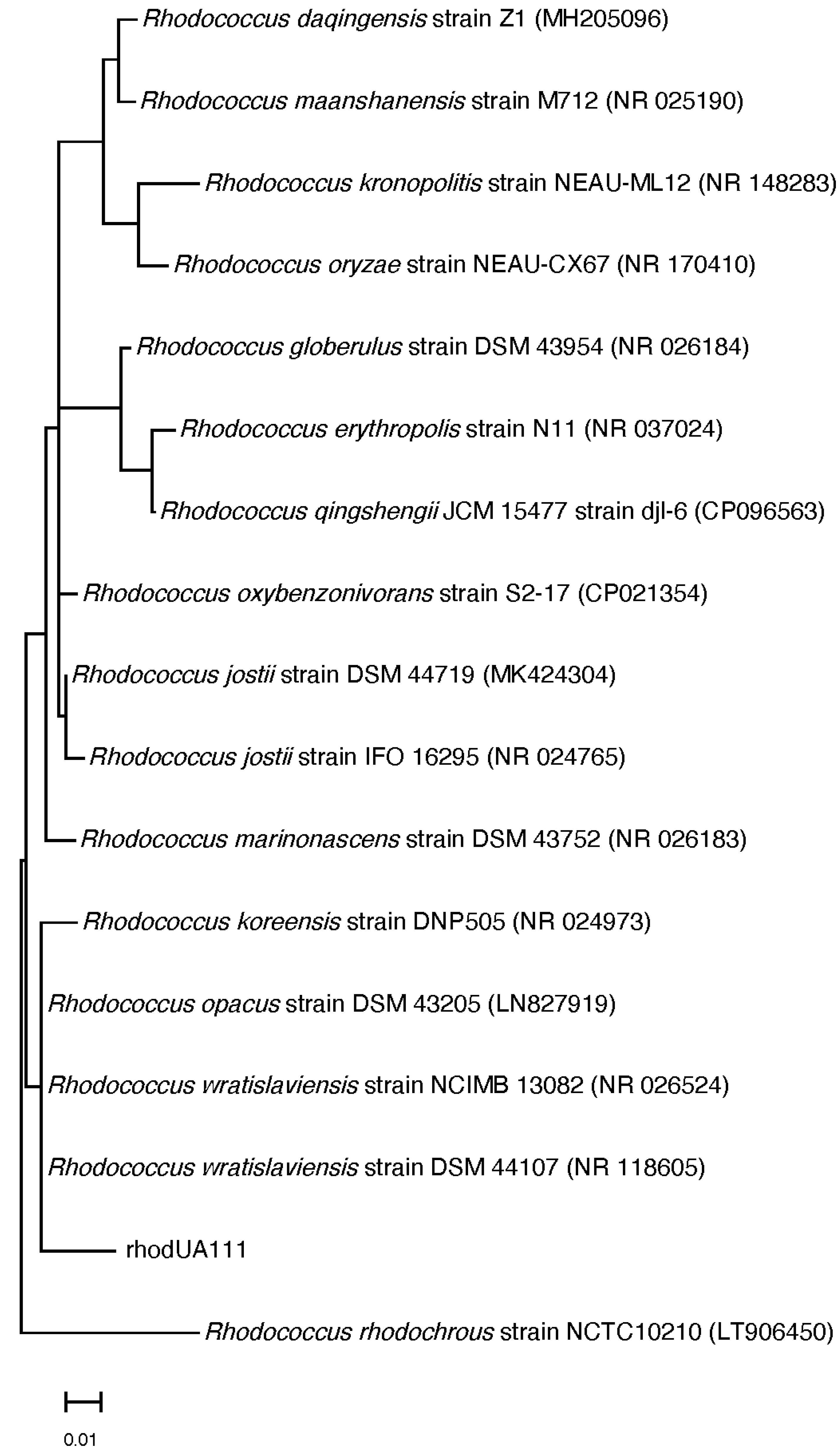
2.3. Molecular Identification and Phylogenetic Analysis

The 16S rRNA gene amplification, cloning, and sequencing were performed according to Hentschel et al., 2015, using the universal primers 27F and 1492R. Chimeric sequences were identified by using the Pintail program. The genus-level affiliation of the sequence was validated using the Ribosomal Database Project Classifier. The genus-level identification of all the sequences was conducted with RDP Classifier (-g 16srrna, -f allrank) and validated with the SILVA Incremental Aligner (SINA) (search and classify option). An alignment was calculated again using the SINA web aligner (variability profile: bacteria). The gap-only positions were removed with trimAL (-noallgaps). For phylogenetic tree construction, the best fitting model was estimated initially with Model Generator. RAxML (-f a m GTRGAMMA –x 12345 –p 12345 -# 1000) and the estimated model was used with 1000 bootstrap resamples to generate the maximum-likelihood tree. The sequences of 16S rRNA from the type strains of Rhodococcus species were identified using the NCBI BLAST server implementing the megablast search program with the RhodUA111 16S rRNA sequence as a query. Preliminary phylogenetic analyses were used to identify the Rhodococcus species most closely to RhodUA111. The sequences were obtained from the NCBI database. The sequence alignment and phylogenetic analysis were performed with MEGA11. A multispecies sequence alignment was obtained with Muscle, and the phylogenetic relationships were revealed using Maximum Likelihood with the TN93+G+I substitution model.

2.4. Extract Preparation

The strain (low sequence similarity) was cultivated using 3 different production media (M1, ISP2, and OLIGO) liquid approaches to produce 3 organic extracts. The liquid cultures were grown for 10 days at 30 °C while shaking at 150 rpm. The culture was then filtered, and the supernatant was extracted with ethyl acetate while the cells and mycelia were extracted by shaking with methanol for 4 h. The ethyl acetate extracts were stored at 4 °C.

2.5. Antimalarial Assay

To determine the antiplasmodial effect of the fungal extracts on P. falciparum erythrocytic replication in vitro, the Malstat assay was used as described. To synchronize the NF54 culture, parasites with many ring stages were centrifuged, and the pellet was resuspended in five times pellet volume of 5% w/v sorbitol /ddH20 and incubated for 10 min at room temperature. The cells were washed once with RPMI to remove sorbitol and further cultivated. Synchronized ring-stage parasites with 1% parasitaemia of P. falciparum NF54 strains were plated in triplicate in 96-well plates (200 µL/well) in the presence of a serial dilution of the extracts dissolved in 0.5% v/v dimethyl sulfoxide (DMSO). The parasites were incubated with the extracts for 72 h at 37 °C in the presence of nitrogen-containing 5% O2 and 5% CO2. The incubation of parasites with DMSO at a concentration of 0.5% alone was used as negative control, and 20% was used as positive control. Afterwards, 20 µL was removed and added to 100 µL of the Malstat reagent (1% Triton X-100, 10 mg of l-lactate, 3.3 mg Tris and 0.33 mg of APAD (3-Acetylpyridine adenine dinucleotide) dissolved in 1 mL of distilled water, pH 9.0) in a new 96-well microtiter plate. The plasmodial lactate dehydrogenase activity was then assessed by adding a 20 μL mixture of NBT (Nitro Blue Tetrazolium)/Diaphorase (1:1; 1 mg/mL stock each) to the Malstat reaction. The optical densities were measured at 630 nm, and the IC50 values were calculated from variable-slope sigmoidal dose–response curves using the GraphPad Prism program version 5.

2.6. LC-MS Profiling

Three ethyl acetate extracts from the samples were prepared at 1 mg/mL for mass spectrometry analysis. The recovered ethyl acetate extract was subjected to metabolic analysis using LC-HR-ESI-MS, according to Abdelmohsen et al., 2014. An Acquity Ultra Performance Liquid Chromatography system connected to a Synapt G2 HDMS quadrupole time-of-flight hybrid mass spectrometer (Waters, Milford, CT, USA) was used. The positive and negative ESI ionization modes were utilized to carry out the high-resolution mass spectrometry coupled with a spray voltage at 4.5 kV; the capillary temperature was 320 °C, and the mass ranged from m/z 150 to 1500. The MS dataset was processed, and the data were extracted using MZmine 2.20 based on the established parameters. Mass ion peaks were detected and accompanied by a chromatogram builder and a chromatogram deconvolution. The local minimum search algorithm was addressed, and isotopes were also distinguished via the isotopic peaks of the grouper. The missing peaks were displayed using the gap-filling peak finder. An adduct search, along with a complex search, was carried out. The processed data set was next subjected to molecular formula prediction and peak identification. The positive and negative ionization mode data sets from the respective extract were dereplicated against the DNP (Dictionary of Natural Products) databases. ChemBioDraw Ultra 14.0 software was used to create the chemical structure drawings.

2.7. In silico Molecular Docking Simulations

Molecular Operating Environment (MOE® 2014.0901) software was used to evaluate and examine the possible interactions of the dereplicated molecules (3, 4, 5, 6, 11, and 12) within the crystal structure of 3 target proteins: P. falciparum kinase (PfPK5; PDB ID: 1V0P), P. falciparum cytochrome bc1 complex (Cyt bc1; PDB ID: 4PD4), and finally, P. falciparum lysyl-tRNA synthetase (PfKRS1; PDB ID: 6AGT). The docking results in the form of a docking score (S; kcal/mol), docking accuracy (root-mean-square deviation; RMSD in Å), and binding interactions with various amino acid residues lining the active site were listed (as shown in Table 1 and Table 2). The preparation of the structural formulas of the test molecules and the structure of the target proteins were performed as reported by [11]. The validation of the prepared protein structures was conducted via the re-docking of the co-crystallized ligands with their protein crystal structure obtained from the RSCB protein data bank (https://www.rcsb.org/ accessed on 8 August 2022), and their docking score and RMSD values were within an acceptable range for running docking simulations within the target proteins (as listed in Table 1). The docking simulations were performed according to a docking protocol reported elsewhere [11], and the results are reported in Table 1 and Table 2. The visual inspections of the produced docking poses (10 poses/molecule) for the binding interactions with various amino acid residues lining the active site of 3 target proteins crystal structures are listed in Table 2 and are represented as 2D and 3D diagrams.

2.8. ADME Parameters and Drug-Likeness Computational Analysis

SwissADME free web tool for prediction of physicochemical parameters, pharmacokinetics, and drug-likeness of small molecules (http://www.swissadme.ch/ accessed on 7 August 2022), was used to measure such parameters of compounds VIa-Vik, and the results are listed in Table 3. Additionally, the medicinal chemistry filters (Lipinski, Ghose, Veber, Egan, and Muegge) were applied to all of the test compounds, and none violated such filters. PAINS (Pan-Assay of Interference Compounds) were used to examine the specificity of such a class of compounds as possible anticancer agents, and all compounds yielded zero scores, as shown in Table 3.

3. Results and Discussion

3.1. Identification of Red Sea Soft Coral Associated Actinobacteria

The actinobacterium strains linked with Red Sea soft coral were isolated and taxonomically identified as Rhodococcus sp. UR111 based on morphology, 16S rRNA genome sequencing, and phylogenetic analysis (Figure 1).

3.2. Antimalarial Assay

As shown in Table 4, the activity of the culture extracts was tested in vitro against Plasmodium falciparum. The results revealed that the extract ISP2 exhibited the highest inhibitory activity, with IC50 values of 8.5 µg/mL, while Oligo was inactive when compared to the positive control drug chloroquine’s IC50 value (0.022 µg/mL).

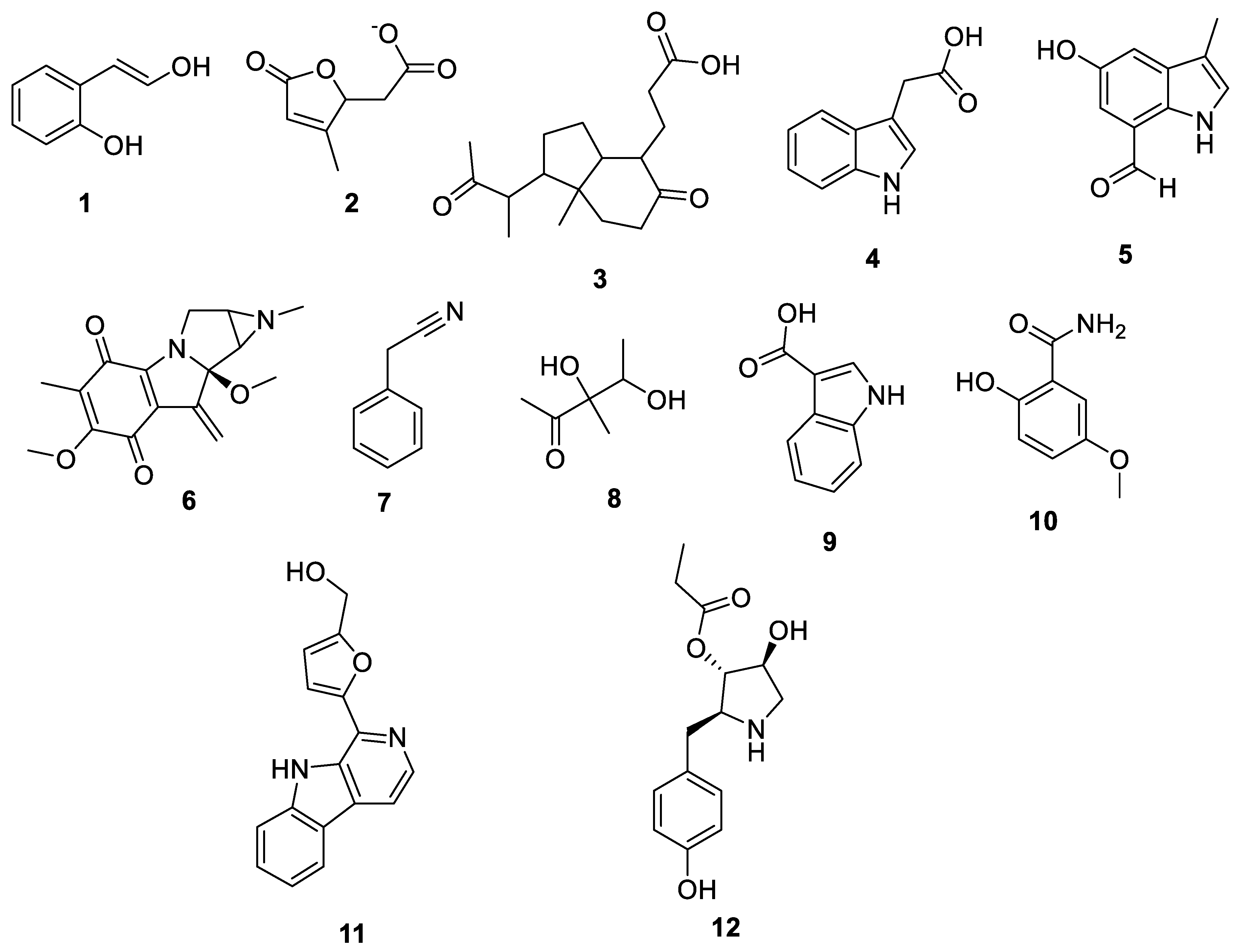
3.3. Metabolomic Analysis

Since marine actinomycetes are producing an increasing number of natural products, particularly species of Streptomyces, Nocardia, Salinispora, Micromonospora, and Rhodococcus, these microbes have also helped researchers find a number of clinically significant drugs. In addition, the obtained results of the antimalarial assay encouraged us to investigate the chemical profile of each of the tested extracts in order to identify the active antimalarial chemical components. Given this, metabolomic profiling of the actinomycete Rhodococcus sp. UA111 obtained from soft marine coral using LC-HR-MS for dereplication purposes has led to the identification of a number of metabolites, of which alkaloids were identified to predominate. LC-HRMS analysis was performed using both positive and negative ionization modes to detect the greatest number of metabolites possible. From the DNP database (Table 5 and Figure 2), the mass ion peak at m/z 135.0442 [M_H]_ (RT, 3.0118min) for the suggested molecular formulas C8H8O2 was identified as 2-(2′-Hydroxyphenyl) ethen-1-ol, which was previously obtained from Rhodococcus benzothiophene [12]. The mass ion peak at m/z 293.1755 [M_H]_ (RT, 4.668 min), in accordance with the molecular formula C17H26O4 was recognized as octahydro-7α-methyl-1-(1-methyl-2-oxo-propyl)-5-oxo-1H-indene-4-propanoic acid that was previously isolated from Rhodococcus corallines [13]. Whereas that at m/z 174.1022 [M_H]_ (RT, 2.6807min), corresponding to the molecular formula C10H9NO2, was suggested to be indole-3-acetic acid, which was previously isolated from Rhodococcus sp. BFI 332 [14]. The mass ion peak at m/z 155.0237 [M_H]_ (RT, 2.58059 min), in accordance with the molecular formula C7H8O4, was recognized as 2,5-Dihydro-3-methyl-5-oxo-2-furanacetic acid (S-form), which was previously isolated from Rhodococcus rhodochrous [10]. Additionally, another metabolite with the molecular formula C10H9NO2, for the mass ion peak at m/z 176.0712 [M_H]+ (RT, 4.1051 min), was characterized as rhodinodohyde, which was previously described from Rhodococcus sp. UA13 [3]. The metabolite, namely Mitomycin-K, with the molecular formula C16H18N2O4, was also dereplicated from the mass ion peak at m/z 303.134 [M_H]+ (RT, 2.684719 min); this compound was previously reported from Rhodococcus sp. UR59 [15]. Furthermore, the mass ion peak at m/z 116.0497 [M_H]_ (RT, 3.077 min) for the predicted molecular formula C8H7N was distinguished as 2-Phenylacetonitrile, which was isolated before from marine Streptomyces sp. GWS-BW-H5 [16]. The mass ion peak at m/z 131.0707 [M_H]_ (RT, 4.6038 min), for the suggested molecular formulas C6H12O3 was identified as 3,4-Dihydroxy-3-methylpentan-2-one that was previously obtained from Streptomyces griseorubens sp. ASMR4 [17]. Moreover, the mass ion peak at m/z 160.0395 [M_H]_ (RT, 3.08194 min), corresponding to the proposed molecular formula C9H7NO2, was identified as Indole-3-carboxylic acid, which was earlier obtained from Streptomyces sp. TK-VL_333 [18]. The mass ion peak at m/z 166.0503 [M_H]- (RT, 2.3045 min), with the molecular formula C8H9NO3, was recognized as 2-Hydroxy-5-methoxybenzamide that was previously isolated from marine Streptomyces sp. [19]. The mass ion peak at m/z 265.0973 [M_H]+ (RT, 3.67572 min), in accordance with the molecular formula C16H12N2O2, was recognized as Perlolyrin, which was previously isolated from a terrestrial Streptomyces sp. GW11/1695 [20] and a marine Streptomyces sp. [21] The metabolite, 3097-B2, with the molecular formula C14H19NO4, was also dereplicated from the mass ion peak at m/z 264.1238 [M_H]_ (RT, 2.9919 min); this compound was previously reported from marine Streptomyces sp. [22].

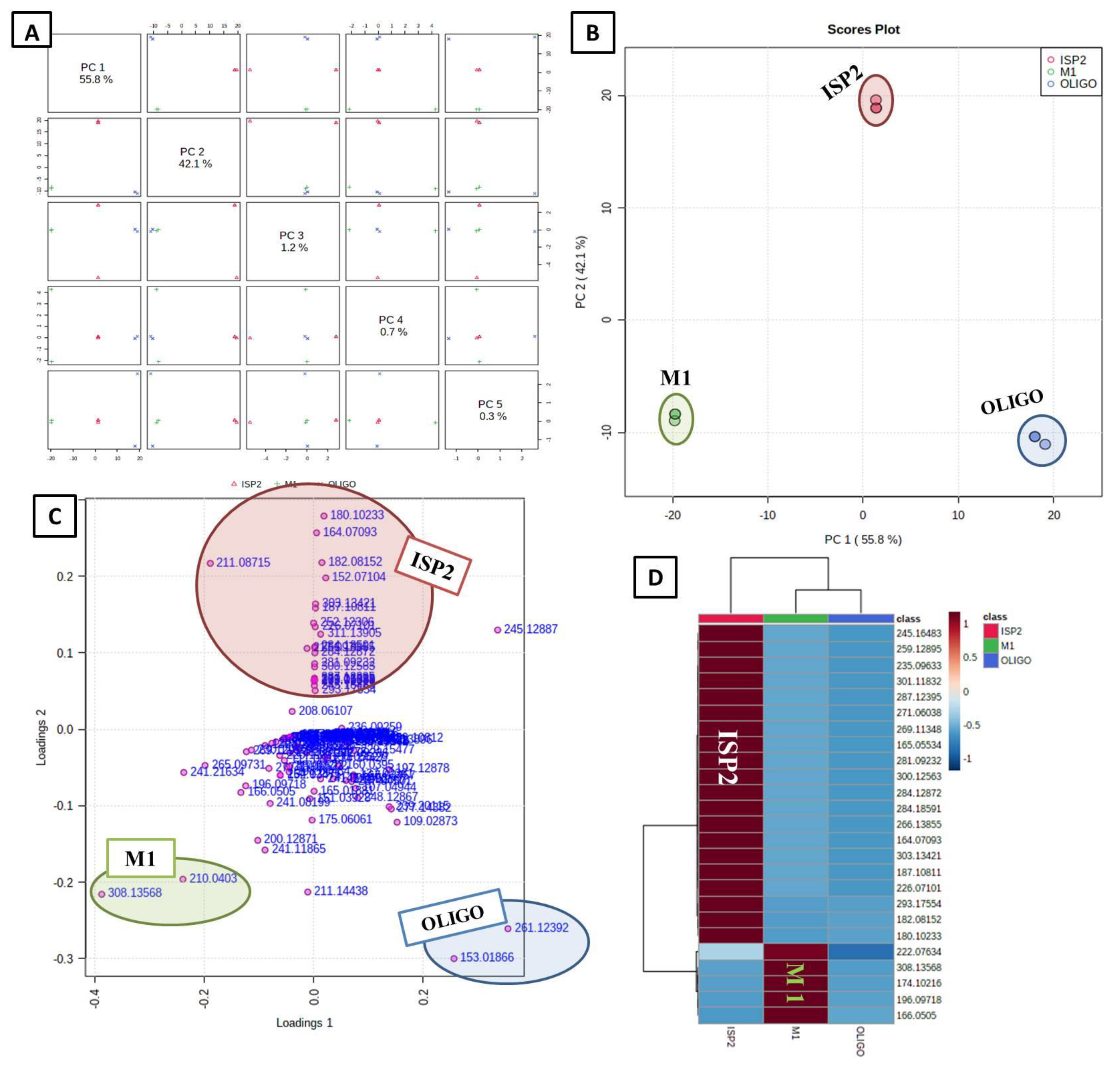
3.4. Multivariate Data Analysis

After being processed by the mzmine software, an MVA on the gathered LC-HRMS data was carried out in order to condense the enormous amount of data obtained and identify correlation and distinction among the tested samples. The LC-MS analysis dataset was statistically treated using MetaboAnalyst 5.0 [23,24].

3.4.1. Unsupervised Analysis

As an initial “blind” step, the unsupervised principal component analysis (PCA) method was implemented first. As shown in the PCA pairwise score plots (Figure 3A), there are five PCA components (PCs) that explained 100.1% of the total variation, in which the first and second PCs separately contributed to 97.9% of the total variation (PC1 and PC2 represent 55.8% and 42.1%, respectively) (Figure 3A). The samples were primarily allocated to three distinct areas between PC1 and PC2, which revealed statistically significant differences between the three extracts, indicating their chemical variation in the PCA 2D scores plot (Figure 3B). On the other hand, the heat map plot (Figure 3D) showed distinct patterns for the culture extracts ISP2 and M1. The metabolites (m/z) that contributed to the fluctuation of the anomalous samples were shown by the PCA loadings plot (Figure 3C); the unique clusters those metabolites (m/z) formed in the loading plot correspond to the locations of the anomalous samples in the scores plot. Such metabolites were dereplicated using the Dictionary of Natural Products (DNP); the annotated discriminatory compounds for ISP2 corresponding to m/z (retention time in min) 180.10233 [M_H]+ (2.4039) was identified as p-tolyl-3-aminopropanoate (C10H13NO2) [25]. The mass ion peak at m/z 164.07093 [M_H] (RT, 1.95985 min), corresponding to the proposed molecular formula C9H11NO2, was identified as Anthranilic acid ethyl ester [26]. The mass ion peak at m/z 182.08152 [M_H]+ (RT, 2.997325 min), in accordance with the molecular formula C9H11NO3, was recognized as tyrosol carbamate [27]. In contrast, that at m/z 152.07104 [M_H] (RT, 2.9552 min) corresponding to the molecular formula C8H9NO2, was suggested to be streptokordin [28]. The mass ion peak at m/z 211.08715 [M_H]+ (RT, 3.872317 min), in accordance with the molecular formula C13H10N2O, was identified as penicinoline E [29]. On the other hand, the annotated discriminatory compounds for M1, corresponding to m/z (retention time in min) 308.13568 [M_H]+ (2.36095) and 210.0403 [M+H] (2.304102) were putatively identified as antibiotic A 53868A (C11H22N3O5P) and antibiotic T0007 B2 (C9H9NO5) [30], respectively.

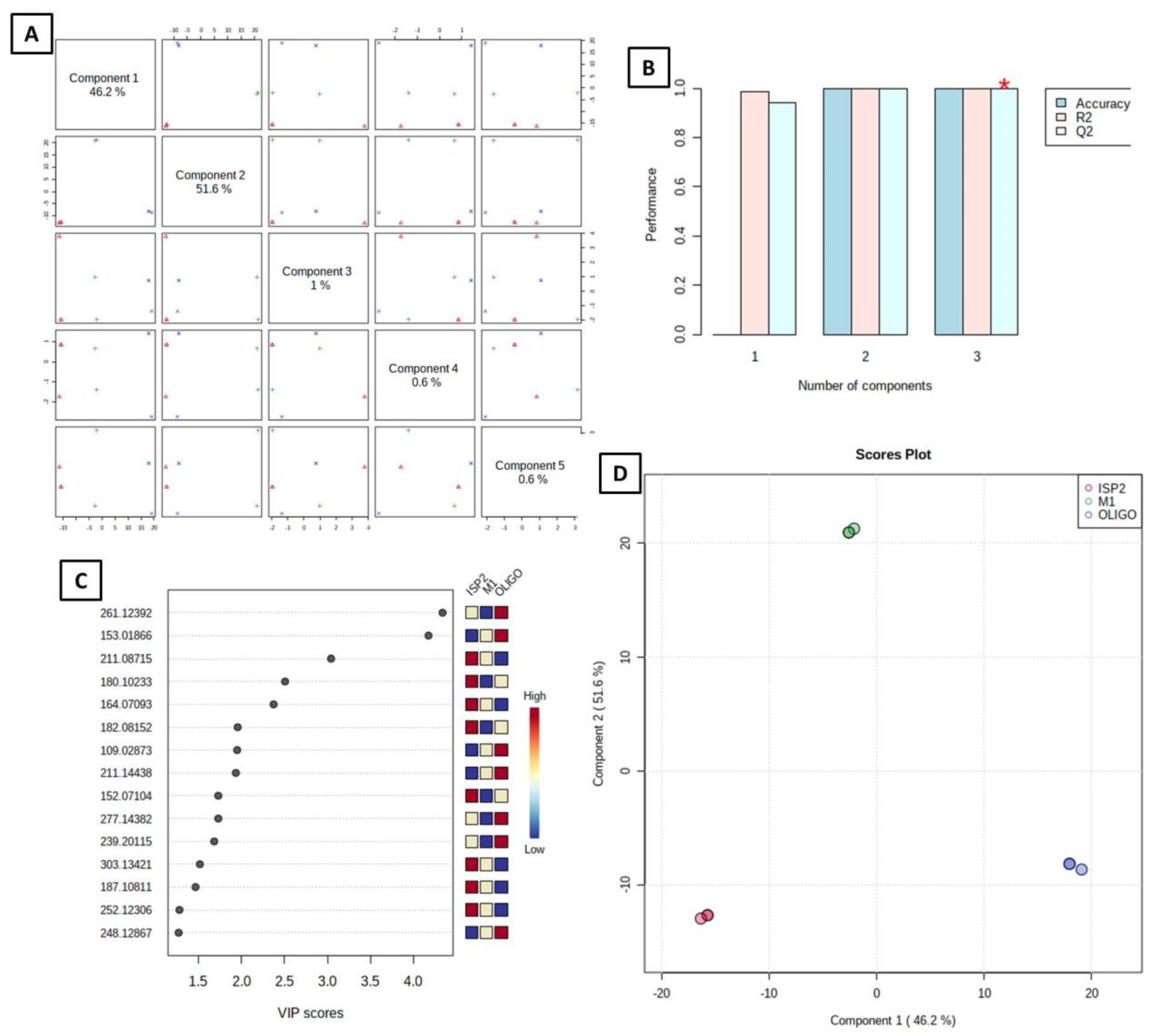
3.4.2. Supervised Analysis

The supervised approach frequently employed in metabolomics studies for classification and supervised biomarker discovery, partial least squares discriminant analysis (PLS-DA), makes use of multivariate linear regression techniques [31]. Both the prediction and the performance of the developed model were good (predictive power of models, Q2 = 1.0; model goodness, R2 = 1.0); a high Q2 value indicates good predictions (Q2 values were those that were very near to 1.0) (Figure 4B). The differences between the samples were confirmed after PLS-DA supervised analysis; in the PLS-DA 2D paired score plot (Figure 4A), five components accounted for 100 percent of the total variation. Two PLS components (PCs) explained 97.8% of the overall variances in the PLS-DA 2D scores plot, with the first and second PCs contributing 46.2 and 51.6 percent, respectively (Figure 4D). One of the crucial measurements in PLS-DA is the variable Importance in Projection (VIP) (Figure 4C), which is shown in Figure 4C. The top 15 most significant traits with the highest value, according to PLS-DA, were demonstrated by VIP. The top 15 significant metabolites (m/z) were listed on the vertical axis in descending order according to the scores they received on the horizontal axis, and the relative concentrations of the important metabolites in the tested samples are shown in the attached colored box on the right (Figure 4B) [32].
The observed variations emphasize how crucial it is to use a diverse range of media to improve the isolation effectiveness of metabolites from actinomycetes associated with the marine environment. Based on prior experience and published reports, the actinomycete cultivation media M1, ISP2, and OLIGO were selected [2]. The results showed that ISP2 media was preferred for growing the genus Rhodococcus and the production of bioactive antimalarial compounds.

3.5. Molecular Docking Simulations

From our interest to explore the possible cellular targets upon which these dereplicated molecules could affect within P. falciparum species, we ran molecular docking simulations within the crystal structures of various proteins found in P. falciparum species, such as the Plasmodium falciparum cell cycle regulator and non-human-derived cyclin-dependent kinase, called P. falciparum kinase (PfPK5; PDB ID: 1V0P). Additionally, the P. falciparum cytochrome bc1 complex (Cyt bc1; PDB ID: 4PD4), and finally, P. falciparum lysyl-tRNA synthetase (PfKRS1; PDB ID: 6AGT). These simulations revealed a moderate to strong docking score (S = −4.45 to −7.11 kcal/mol) with all test molecules within the three active sites used in this study (as listed in Table 1). Molecules 11 (Perlolyrine) and 12 (3097-B2) showed the best docking profile among all test molecules as found with their docking score and binding interactions with various amino acid residues lining the proteins’ active sites (as illustrated in Figure 5, Figure 6 and Figure 7 and Figures S1–S3 in the Supporting Information File).
Briefly, in silico molecular docking simulations within the active site of P. falciparum’s kinase (PDB ID: 1V0P) revealed that both molecules 11 and 12 succeed in having binding interactions (either H-bonding or pi-H interactions, as listed in Table 2) with at least one of the key amino acid residues (VAL18, THR14, LEU82, ILE10, and/or LYS88) lining this active site, as shown in Figure 5.
Moreover, both molecules 11 and 12 showed an interesting binding mode within the P. falciparum mitochondrial cytochrome bc1 complex (PDB ID: 4PD4) active site mediated through H-bond donor and acceptor interactions with key amino acid residues lining the active site (GLU272, MET295, and/or MET139), as listed in Table 2. Such types of interactions provided better stabilization of the molecules within the target protein and could be used as a scaffold for the further development of antimalarial agents (as shown in Figure 6).
Finally, the docking scores and the docking accuracy of the test molecules within the crystal structure of P. falciparum lysyl-tRNA synthetase (PfKRS1; PDB ID: 6AGT) showed the highest and best possibility of being a target by such compounds and could be used to illustrate their antimalarial biological activities. In brief, the docking simulations within lysyl-tRNA synthetase resulted in the best docking score and number of binding interactions within the active site of lysyl-tRNA synthetase crystal structures, especially with molecules 11 and 12, which have a high and comparable docking score within all other dereplicated molecules and to the co-crystallized ligand (9X0), as shown in Table 2 and Figure 7.

3.5.1. ADME Parameters and Drug-likeness Computational Analysis

Investigating the oral bioavailability of such dereplicated molecules was achieved by measuring the physicochemical descriptors, pharmacokinetics, drug-like nature, and medicinal chemistry friendliness of six of these dereplicated molecules (3, 4, 5, 6, 11, and 12) using the SwissADME free web tool (http://www.swissadme.ch/ accessed on 7 August 2022). Starting with the role of five (Lipinski’s RO5) [33,34], all of the test molecules have values for physicochemical within the required ranges of candidate drugs, as shown in Table 3.
Briefly, having H-bonding centers (either acceptor or donor) helps in enhancing water solubility and H-bond formation with various amino acid residues lining the target’s active sites, in the same way, having an acceptable number of rotating bonds helps in the adaptation and flexibility alignment of such molecules within the target active sites. Additionally, having a partition coefficient (i.e., lipophilicity parameter) within −0.5 to ≤5, and both TPSA and MR values below 140 and 130, respectively, generally indicates a good probability of penetration through cell membranes, gastrointestinal penetration, and hence bioavailability. Fortunately, all of the test molecules (except molecule 11) were found to not be a substantial substrate for permeability glycoprotein (also known as multidrug resistance protein, which is responsible for the efflux of drugs out of cells and hence reduces their pharmacological efficacy) and having an Abbott oral bioavailability score above zero, indicating their high probability of medicinal impact and biological activities in clinical trials [35].
Finally, all the test molecules pass the medicinal chemistry filters of Lipinski. In the same way, all the test molecules also pass (except molecule 6) the Pan-assay of interference compounds (PAINS), indicating their high probability of being a drug candidate for drug discovery and development, as shown in Table 3.

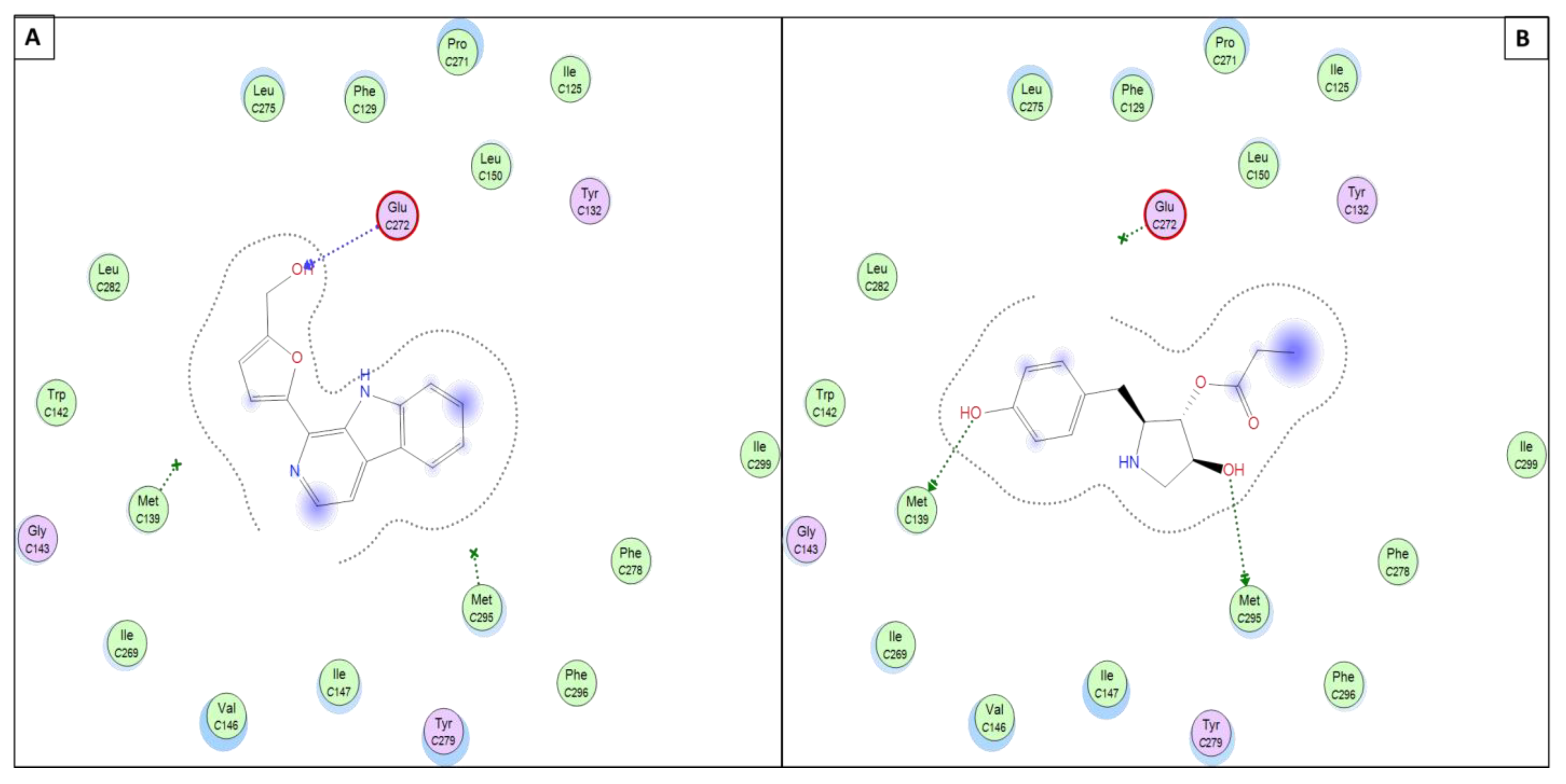
3.5.2. Perlolyrine (11) and 3097-B2 (12)

Perlolyrine (11) is a β-carboline alkaloid that was previously isolated from the roots of Codonopsis pilosula. It was also reported in rye grass (Lolium perenne L.) [36], Codonopsis lanceolata, Panax gingseng [37], and recently isolated from a terrestrial Streptomyces species [20]. On the other hand, 3097-B2 (12) is a pyrrolidine derivative structurally relative to the antifungal drug anisomycin; it was previously isolated from the culture broth of Streptomyces strain SA3097 and showed cytotoxic activity in vitro against the cancer cell lines, human lung carcinoma (LU99) and human breast cancer (MCF-7) [22]. Moreover, Bhattarai et al. identified 3097-B2 (12) in the fermented culture of Streptomyces species isolated from the soils of Nepal using an untargeted metabolomics study [38].

4. Conclusions

Rhodococcus sp., an infrequently studied marine bacterium, was investigated utilizing the established process using LC-HR-ESI-MS-based analytical techniques. Herein, the OSMAC technique was used to isolate and subculture the actinomycete, Rhodococcus associated with the Red Sea soft coral Nephthea sp. in three different media (M1, ISP2, and OLIGO). The extracts were then examined for their antimalarial activity and chemical diversity. The ISP2 extract showed substantial antimalarial efficacy. On the other hand, several metabolites of various structural kinds have been identified as a result of our study of this species, belonging to different classes, mainly alkaloids. In silico studies revealed that the metabolites perlolyrine (11) and 3097-B2 (12) have a high and comparable docking score within all other dereplicated molecules. This study emphasized the unique function of actinomycetes associated with marine organisms as an unexplored source of active metabolites for the creation of effective natural antimalarial drugs. By combining the analytical, statistical, and dereplication approaches for examining their bioactive principles, it is possible to effectively shorten or replace the time-consuming bioassay-guided isolation schemes and complex chromatographic procedures. Biochemometrics approaches have been widely used in natural product research as an initial evaluation step in the process of finding new drugs from actinomycetes. Targeting the bioactive antimalarial metabolites in complicated crude extracts can therefore be more successful with greater integration with in silico technologies such as neural network-based virtual screening. Using elicitation approaches, cryptic gene clusters in microorganisms could be a new approach to finding new antimalarial drug leads.

Supplementary Materials

The following are available online at https://www.mdpi.com/article/10.3390/antibiotics11111631/s1, Figure S1. Presumptive mode of interaction of molecules 3, 4, 5, 6, and PVB within the active site of P. falciparum kinase (PDB ID: 1V0P). Figure S2. Presumptive mode of interaction of molecules 3, 4, 5, 6, and AOQ within the active site of P. falciparum mitochondrial cytochrome bc1 complex (PDB ID: 4PD4). Figure S3. Presumptive mode of interaction of molecules 3, 4, 5, 6, and 9X0 within the active site of P. falciparum lysyl-tRNA synthetase (PfKRS1; PDB ID: 6AGT).

Author Contributions

Conceptualization, U.R.A., H.S.B., C.J.N. and G.P.; methodology, U.R.A., C.J.N., A.E.Z., B.F.M. and G.P.; Software, Y.A.M., A.E.Z. and B.F.M.; supervision, U.R.A. and N.M.G.; visualization, H.S.B., Y.A.M., B.F.M., A.E.Z. and U.R.A.; data curation, U.R.A. and H.S.B.; writing—original draft preparation, H.S.B.; writing—review and editing, U.R.A., H.S.B., Y.A.M., A.E.Z., B.F.M., H.M.H., D.H.M.A., N.M.G. and W.N.H. All authors have read and agreed to the published version of the manuscript.

Funding

This research received no external funding.

Institutional Review Board Statement

Not applicable.

Informed Consent Statement

Not applicable.

Data Availability Statement

The data are contained within the article or Supplementary Material.

Acknowledgments

The authors acknowledge the support from Princess Nourah bint Abdulrahman University Researchers Supporting Project number (PNURSP2022R15), Princess Nourah bint Abdulrahman University, Riyadh, Saudi Arabia.

Conflicts of Interest

The authors declare no conflict of interest.

References

  1. Elsayed, Y.; Refaat, J.; Abdelmohsen, U.R.; Othman, E.M.; Stopper, H.; Fouad, M.A. Metabolomic Profiling and Biological Investigation of the Marine Sponge-Derived Bacterium Rhodococcus sp. UA13. Phytochem. Anal. 2018, 29, 543–548. [Google Scholar] [CrossRef] [PubMed]
  2. Abdelmohsen, U.R.; Yang, C.; Horn, H.; Hajjar, D.; Ravasi, T.; Hentschel, U. Actinomycetes from Red Sea Sponges: Sources for Chemical and Phylogenetic Diversity. Mar. Drugs 2014, 12, 2771–2789. [Google Scholar] [CrossRef] [PubMed] [Green Version]
  3. Elsayed, Y.; Fahim, J.; Abdelmohsen, U.; Fouad, M. Chemical and Biological Investigation of the Marine Bacterium Rhodococcus Sp. UA13. J. Adv. Biomed. Pharm. Sci. 2021, 4, 1–6. [Google Scholar] [CrossRef]
  4. Bell, K.S.; Philp, J.C.; Aw, D.W.J.; Christofi, N. A Review: The Genus Rhodococcus. J. Appl. Microbiol. 1998, 85, 195–210. [Google Scholar] [CrossRef]
  5. Elsayed, Y.; Refaat, J.; Yasmin Elsayed, C.; Abdelmohsen, U.R.; Fouad, M.A. The Genus Rhodococcus as a Source of Novel Bioactive Substances: A Review. J. Pharmacogn. Phytochem. 2017, 6, 83–92. [Google Scholar]
  6. Uwimana, A.; Legrand, E.; Stokes, B.H.; Ndikumana, J.L.M.; Warsame, M.; Umulisa, N.; Ngamije, D.; Munyaneza, T.; Mazarati, J.B.; Munguti, K.; et al. Emergence and Clonal Expansion of in Vitro Artemisinin-Resistant Plasmodium Falciparum Kelch13 R561H Mutant Parasites in Rwanda. Nat. Med. 2020, 26, 1602–1608. [Google Scholar] [CrossRef]
  7. Fotie, J. Quinones and Malaria. Antiinfect. Agents Med. Chem. 2006, 5, 357–366. [Google Scholar] [CrossRef]
  8. Hussain, H.; Specht, S.; Sarite, S.R.; Saeftel, M.; Hoerauf, A.; Schulz, B.; Krohn, K. A New Class of Phenazines with Activity against a Chloroquine Resistant Plasmodium falciparum Strain and Antimicrobial Activity. J. Med. Chem. 2011, 54, 4913–4917. [Google Scholar] [CrossRef]
  9. El-Hawary, S.S.; Mohammed, R.; Bahr, H.S.; Attia, E.Z.; El-Katatny, M.H.; Abelyan, N.; Al-Sanea, M.M.; Moawad, A.S.; Abdelmohsen, U.R. Soybean-Associated Endophytic Fungi as Potential Source for Anti-COVID-19 Metabolites Supported by Docking Analysis. J. Appl. Microbiol. 2021, 131, 1193–1211. [Google Scholar] [CrossRef]
  10. Cha, C.J. Biological Production of Optically Active Muconolactones by Rhodococcus Rhodochrous. Appl. Microbiol. Biotechnol. 2001, 56, 453–457. [Google Scholar] [CrossRef]
  11. Abdel-Wahab, N.M.; Gomaa, A.A.-R.; Mostafa, Y.A.; Hajjar, D.; Makki, A.A.; Alaaeldin, E.; Refaat, H.; Bringmann, G.; Zayed, A.; Abdelmohsen, U.R. Diterpenoids Profile of the Marine Sponge Chelonaplysilla erecta and Candidacy as Potential Antitumor Drugs Investigated by Molecular Docking and Pharmacokinetic Studies. Nat. Prod. Res. 2022, 1–5. [Google Scholar] [CrossRef] [PubMed]
  12. Onaka, T.; Matsui, T.; Maruhashi, K. Metabolite Detection in Rhodococcus benzothiophene Desulfurization Using Reversed-Phase Solid-Phase Extraction and a Derivatization Technique. Biotechnol. Lett. 2002, 24, 1961–1966. [Google Scholar] [CrossRef]
  13. Pereliaeva, E.V.; Dmitrieva, M.E.; Morgunova, M.M.; Belyshenko, A.Y.; Imidoeva, N.A.; Ostyak, A.S.; Axenov-Gribanov, D.V. The Use of Baikal Psychrophilic Actinobacteria for Synthesis of Biologically Active Natural Products from Sawdust Waste. Fermentation 2022, 8, 213. [Google Scholar] [CrossRef]
  14. Lee, J.H.; Kim, Y.G.; Kim, C.J.; Lee, J.C.; Cho, M.H.; Lee, J. Indole-3-Acetaldehyde from Rhodococcus sp. BFI 332 Inhibits Escherichia Coli O157:H7 Biofilm Formation. Appl. Microbiol. Biotechnol. 2012, 96, 1071–1078. [Google Scholar] [CrossRef] [PubMed]
  15. Alhadrami, H.A.; Thissera, B.; Hassan, M.H.A.; Behery, F.A.; Ngwa, C.J.; Hassan, H.M.; Pradel, G.; Abdelmohsen, U.R.; Rateb, M.E. Bio-Guided Isolation of Antimalarial Metabolites from the Coculture of Two Red Sea Sponge-Derived Actinokineospora and Rhodococcus spp. Mar. Drugs 2021, 19, 9. [Google Scholar] [CrossRef] [PubMed]
  16. Miki, Y.; Asano, Y. Biosynthetic Pathway for the Cyanide-Free Production of Phenylacetonitrile in Escherichia Coli by Utilizing Plant Cytochrome P450 79A2 and Bacterial Aldoxime Dehydratase. Appl. Environ. Microbiol. 2014, 80, 6828–6836. [Google Scholar] [CrossRef] [Green Version]
  17. Hamed, A.; Abdel-Razek, A.S.; Frese, M.; Wibberg, D.; El-Haddad, A.F.; Ibrahim, T.M.A.; Kalinowski, J.; Sewald, N.; Shaaban, M. New Oxaphenalene Derivative from Marine-Derived Streptomyces Griseorubens sp. ASMR4. Zeitschrift fur Naturforsch.—Sect. B J. Chem. Sci. 2017, 72, 53–62. [Google Scholar] [CrossRef]
  18. Kavitha, A.; Prabhakar, P.; Vijayalakshmi, M.; Venkateswarlu, Y. Purification and Biological Evaluation of the Metabolites Produced by Streptomyces sp. TK-VL_333. Res. Microbiol. 2010, 161, 335–345. [Google Scholar] [CrossRef]
  19. Mohamed, M.A.A. Ent-Homoabyssomicins A and B, Two New Spirotetronates, Khatmiamycin, a Zoosporicidal Naphthoquinone, and Further New Biologically Active Secondary Metabolites; Cuvillier Verlag: Göttingen, Germany, 2010; ISBN 3736935013. [Google Scholar]
  20. Fotso, S.; Maskey, R.P.; Schröder, D.; Ferrer, A.S.; Grün-Wollny, I.; Laatsch, H. Furan Oligomers and β-Carbolines from Terrestrial Streptomycetes. J. Nat. Prod. 2008, 71, 1630–1633. [Google Scholar] [CrossRef]
  21. El-Bondkly, A.; Abd-Alla, H.; Shaaban, M.; Shaaban, K. The Electrospray Ionization—Mass Spectra of Erythromycin A Obtained from a Marine Streptomyces sp. Mutant. Indian J. Pharm. Sci. 2008, 70, 310–319. [Google Scholar] [CrossRef] [Green Version]
  22. Hosoya, Y.; Kameyama, T.; Naganawa, H.; Okami, Y.; Takeuchi, T. Anisomycin and new congeners active against human tumor cell lines. J. Antibiot. (Tokyo) 1993, 46, 1300–1302. [Google Scholar] [CrossRef] [PubMed]
  23. Raheem, D.J.; Tawfike, A.F.; Abdelmohsen, U.R.; Edrada-Ebel, R.A.; Fitzsimmons-Thoss, V. Application of Metabolomics and Molecular Networking in Investigating the Chemical Profile and Antitrypanosomal Activity of British Bluebells (Hyacinthoides Non-Scripta). Sci. Rep. 2019, 9, 2547. [Google Scholar] [CrossRef] [PubMed] [Green Version]
  24. Tawfike, A.F.; Romli, M.; Clements, C.; Abbott, G.; Young, L.; Schumacher, M.; Edrada-Ebel, R.; Diederich, M.; Farag, M.; Edrada-Ebel, R. Isolation of Anticancer and Anti-Trypanosome Secondary Metabolites from the Endophytic Fungus Aspergillus Flocculus via Bioactivity Guided Isolation and MS Based Metabolomics. J. Chromatogr. B 2019, 1106, 71–83. [Google Scholar] [CrossRef] [PubMed] [Green Version]
  25. Xu, D.B.; Ye, W.W.; Han, Y.; Deng, Z.X.; Hong, K. Natural Products from Mangrove Actinomycetes. Mar. Drugs 2014, 12, 2590–2613. [Google Scholar] [CrossRef] [Green Version]
  26. Varnavas, A.; Lassiani, L.; Valenta, V.; Berti, F.; Mennuni, L.; Makovec, F. Anthranilic Acid Derivatives: A New Class of Non-Peptide CCK1 Receptor Antagonists. Bioorg. Med. Chem. 2003, 11, 741–751. [Google Scholar] [CrossRef]
  27. Gautschi, J.T.; Tenney, K.; Compton, J.; Crews, P. Chemical Investigations of a Deep Water Marine-Derived Fungus: Simple Amino Acid Derivatives from an Arthrinium sp. Nat. Prod. Commun. 2007, 2, 1934578X0700200. [Google Scholar] [CrossRef] [Green Version]
  28. Jeong, S.Y.; Shin, H.J.; Kim, T.S.; Lee, H.S.; Park, S.K.; Kim, H.M. Streptokordin, a New Cytotoxic Compound of the Methylpyridine Class from a Marine-Derived Streptomyces Sp. KORDI-3238. J. Antibiot. (Tokyo) 2006, 59, 234–240. [Google Scholar] [CrossRef]
  29. Shao, C.L.; Wang, C.Y.; Gu, Y.C.; Wei, M.Y.; Pan, J.H.; Deng, D.S.; She, Z.G.; Lin, Y.C. Penicinoline, a New Pyrrolyl 4-Quinolinone Alkaloid with an Unprecedented Ring System from an Endophytic Fungus Penicillium sp. Bioorganic Med. Chem. Lett. 2010, 20, 3284–3286. [Google Scholar] [CrossRef]
  30. Zhang, H. Isolation and Identification of Antibiotic T0007B_1 and T0007B_2. Zhongguo Kangshengsu Zazhi 1990, 15, 321–324. [Google Scholar]
  31. Szymańska, E.; Saccenti, E.; Smilde, A.K.; Westerhuis, J.A. Double-Check: Validation of Diagnostic Statistics for PLS-DA Models in Metabolomics Studies. Metabolomics 2012, 8, 3–16. [Google Scholar] [CrossRef] [Green Version]
  32. Solanki, H.; Pierdet, M.; Thomas, O.P.; Zubia, M. Insights into the Metabolome of the Cyanobacterium Leibleinia Gracilis from the Lagoon of Tahiti and First Inspection of Its Variability. Metabolites 2020, 10, 215. [Google Scholar] [CrossRef] [PubMed]
  33. Lipinski, C.A. Drug-like Properties and the Causes of Poor Solubility and Poor Permeability. J. Pharmacol. Toxicol. Methods 2000, 44, 235–249. [Google Scholar] [CrossRef]
  34. Lipinski, C.A.; Lombardo, F.; Dominy, B.W.; Feeney, P.J. Experimental and Computational Approaches to Estimate Solubility and Permeability in Drug Discovery and Development Settings. Adv. Drug Deliv. Rev. 2012, 64, 4–17. [Google Scholar] [CrossRef]
  35. Martin, Y.C. A Bioavailability Score. J. Med. Chem. 2005, 48, 3164–3170. [Google Scholar] [CrossRef]
  36. Jeffreys, J.A.D. The Alkaloids of Perennial Rye-Grass (Lolium perenne L.). Part 1V.l Isolation of a New Base, Perlolyrine; the Crystal Structure of Its Hydro-Bromide Dihydrate, and the Synthesis of the Base. J. Chem. Soc. 1970, 8, 1091–1103. [Google Scholar]
  37. Liu, T.; Liang, W.; Tu, G. Perlolyrine: A β-Carboline Alkaloid from Codonopsis Pilosula. Planta Med. 1988, 54, 472–473. [Google Scholar] [CrossRef]
  38. Bhattarai, B.R.; Khadayat, K.; Aryal, N.; Aryal, B.; Lamichhane, U.; Bhattarai, K.; Rana, N.; Regmi, B.P.; Adhikari, A.; Thapa, S.; et al. Untargeted Metabolomics of Streptomyces Species Isolated from Soils of Nepal. Processes 2022, 10, 1173. [Google Scholar] [CrossRef]
Figure 1. Phylogenetic tree of the 16S rRNA gene-marking Rhodococcus sp. UR111 isolate and its closest relatives.
Antibiotics 11 01631 g001
Figure 2. Metabolites dereplicated from Rhodococcus sp. crude extract.
Antibiotics 11 01631 g002
Figure 3. Metabolomics multivariate analysis. (A) PCA pairwise score plot of the unsupervised method; (B) 2D PCA scores plot of the unsupervised method; (C) 2D PCA loadings plot of the unsupervised method; (D) Heatmap plot.
Antibiotics 11 01631 g003
Figure 4. Metabolomics multivariate analysis. (A) PLS pairwise score plot; (B) 2D PCA scores plot of the unsupervised method; (C) 2D PCA loadings plot of the unsupervised method; (D) Heatmap plot.
Antibiotics 11 01631 g004
Figure 5. Presumptive mode of interaction of molecules 11 and 12 within active site of P. falciparum kinase (PDB ID: 1V0P); (A) Perlolyrine (11); (B) 3097-B2 (12).
Antibiotics 11 01631 g005
Figure 6. Presumptive mode of interaction of molecule 11 (A) and 12 (B) within active site of P. falciparum mitochondrial cytochrome bc1 complex (PDB ID: 4PD4). showing H-bond in blue and/or green arrows and solvent accessible contour as black-dotted line around whole molecule.
Antibiotics 11 01631 g006
Figure 7. Presumptive mode of interaction of molecules 11, 12, and 9X0 within active site of P. falciparum lysyl-tRNA synthetase (PfKRS1; PDB ID: 6AGT). (A) Perlolyrine (11); (B) 3097-B2 (12).
Antibiotics 11 01631 g007
Table 1. Docking simulations of dereplicated molecules and co-crystallized ligands within three of P. falciparum proteins.
#Cpd
ID
Isolated Molecules1V0P a4PD4 b6AGT c
S gRMSD hSRMSDSRMSD
13Antibiotics 11 01631 i001
Indene propanoic acid derivative
−5.741.67−5.551.97−5.880.92
24Antibiotics 11 01631 i002
Indole-3-acetic acid
−4.451.40−5.161.77−5.151.10
35Antibiotics 11 01631 i003
Rhodinodohyde
−5.010.96−4.850.81−5.371.53
46Antibiotics 11 01631 i004
Mitomycin-K
−5.391.68−5.370.73−6.091.16
511Antibiotics 11 01631 i005
Perlolyrine
−5.981.04−5.841.69−6.911.12
612Antibiotics 11 01631 i006
3097-B2
−5.891.99−6.501.56−7.111.97
Co-crystallized Ligand−7.32 d1.21−6.45 e1.64−7.98 f0.63
a 1V0P: P. falciparum kinase; b 4PD4: cytochrome bc1; and c 6AGT: lysyl-tRNA synthetase (PfKRS1) active sites; d Purvalanol B (PVB); e 2-[trans-4-(4-chlorophenyl)cyclohexyl]-3-hydroxynaphthalene-1,4-dione (AOQ); f N-(cyclohexylmethyl)-4-oxo-4H-1-benzopyran-2-carboxamide (9X0); g Docking Score (S; Kcal/mol); h Root-Mean-Square Deviation (Å).
Table 2. Binding interactions of dereplicated molecules 11 and 12 within three P. falciparum proteins.
Molecule ID1V0P a4PD4 b6AGT c
Typea.a.
Residue
Distance eTypea.a.
Residue
DistanceTypea.a.
Residue
Distance
11H-acceptorTHR143.20H-acceptorGLU2723.49H-acceptorGLY5563.46
H-acceptorARG3303.15
Pi-HVAL184.21Pi-PiPHE3423.81
Pi-PiPHE3423.83
12H-donorLEU822.90H-donorMET2953.33H-acceptorARG3303.15
H-acceptorTHR142.86H-donorMET1393.83Pi-PiPHE3423.90
Pi-HILE103.90
Co-crystallized Ligands dH-donorGLU803.17Pi-HILE 1254.15H-acceptorASN3393.05
H-donorLEU822.76
H-acceptorLEU823.01Pi-PiPHE3423.58
IonicLYS883.91
Pi-HVAL184.01
a 1V0P: P. falciparum kinase; b 4PD4: cytochrome bc1 complex; and c 6AGT: lysyl-tRNA synthetase (PfKRS1) active sites; d Co-crystallized ligands: PVB for 1V0P, AOQ for 4PD4, and 9X0 for 6AGT; e Bond distance (Å).
Table 3. ADMET, Pharmacokinetic and PAINS analysis of dereplicated molecules (3, 4, 5, 6, 11, and 12).
IDM.F. aM.W. bNrotb cHBA dHBD eMR fTPSA giLogP hWater SolubilityHIA% iBBB Permeant jPgp Substrate kF lPAINS m
3C17H26O4294.3954181.7871.442.16SolubleHighYesNo0.850
4C10H9NO2175.1822249.8453.091.01SolubleHighYesNo0.850
5C10H9NO2175.1812250.6853.091.08SolubleHighYesNo0.550
6C16H18N2O4302.3325085.3858.852.82Very solubleHighNoNo0.551
11C16H12N2O2264.2823277.4362.052.36SolubleHighYesYes0.550
12C14H19NO4265.355374.1378.792.11SolubleHighNoNo0.550
RO5≤500≤10≤10≤5≤130≤140≤5
a MF, Molecular Formula; b M.W., Molecular Wight; c nrotb, # of rotatable bonds; d HBA, Hydrogen Bond Acceptor; e HBD, Hydrogen bond donor; f MR, Molar Refractivity; g TPSA, Total Polar Surface Area; h iLogP, octanol/water partition coefficient; i HIA%, human gastrointestinal absorption; j BBB permeant, Blood–brain barrier penetration; k Pgp substrate, Permeability glycoprotein; l F, Abbott oral bioavailability score; m PAINS, Pan-Assay Interference Compounds.
Table 4. In Vitro antimalarial activity of Rhodococcus fermented extracts against Plasmodium falciparum.
SampleIC50 (µg/mL)
M124.7
ISP28.5
Oligo>50
Chloroquine0.022
Table 5. Metabolites dereplicated from Rhodococcus crude extract.
#Ionizationm/zRtNameFormulaKey FragmentsSourceReference
1N135.04423.01182-(2′-Hydroxyphenyl) ethen-1-olC8H8O2 Rhodococcus benzothiophene[12]
2N155.02372.5805922,5-Dihydro-3-methyl-5-oxo-2-furanacetic acid (S-form)C7H8O4 Rhodococcus rhodochrous[10]
3N293.17554.668813Octahydro-7a-methyl-1-(1-methyl-2-oxo-propyl)-5-oxo-1H-indene-4-propanoic acidC17H26O4221.13Rhodococcus corallinus[13]
4N174.10222.6807Indole-3-acetic acidC10H9NO2 Rhodococcus sp. BFI 332[14]
5P176.07124.105067RhodinodohydeC10H9NO272.07Rhodococcus sp. UA13[3]
6P303.13422.684719Mitomycin-KC16H18N2O4257.06Rhodococcus sp. UR59[15]
7N116.04973.077592
2.708363
2-PhenylacetonitrileC8H7N marine Streptomyces sp. GWS-BW-H5[16]
8N131.07074.6038223,4-Dihydroxy-3-methylpentan-2-oneC6H12O387.04Streptomyces griseorubens sp. ASMR4[17]
9N160.03953.081942Indole-3-carboxylic acidC9H7NO2 Streptomyces sp. TK-VL_333[18]
10N166.05032.3045832-Hydroxy-5-methoxybenzamideC8H9NO3 marine Streptomyces[19]
11P265.09733.675722PerlolyrinC16H12N2O2264.87, 165.64Streptomyces sp. isolate GW11/1695[20]
Marine streptomyces[21]
12N264.12382.9919533097-BC14H19NO4 marine Streptomyces[22]
Publisher’s Note: MDPI stays neutral with regard to jurisdictional claims in published maps and institutional affiliations.

