Article

Assessment and Antibiotic Resistance Profiling in Vibrio Species Isolated from Wild Birds Captured in Danube Delta Biosphere Reserve, Romania

1
Department of Infectious Diseases, Faculty of Veterinary Medicine, University of Agricultural Sciences and Veterinary Medicine, 400372 Cluj-Napoca, Romania
2
Subdivision of the Federal State Budgetary Institution of Science “Kazan Scientific Center of Russia Academy of Sciences”, Tatar Scientific Research Institute of Agriculture, 420088 Kazan, Russia
3
Institute of Research and Development for Agro Montanology, 557085 Cristian, Sibiu, Romania
4
Instiute of Veterinary Medicine, Warsaw University of Life Sciences WULS, 02-786 Warsaw, Poland
*
Authors to whom correspondence should be addressed.
Antibiotics 2021, 10(3), 333; https://doi.org/10.3390/antibiotics10030333
Submission received: 15 February 2021 / Revised: 16 March 2021 / Accepted: 17 March 2021 / Published: 22 March 2021
(This article belongs to the Special Issue Antimicrobial Resistance and the Environment: One Health Approach)

Abstract

:
Antimicrobial and multidrug-resistant bacteria are a major problem worldwide and, consequently, the surveillance of antibiotic-resistant bacteria and assessment of the dissemination routes are essential. We hypothesized that migratory birds, coming from various environments, would carry more numerous Vibrio strains than sedentary species, with increased risk to be passed to their contacts or environment in habitats they transit or nest in. Similarly, we presumed that strains from migratory birds will show multidrug resistance. A total of 170 oral and rectal swabs were collected from wild birds captured in different locations of the Danube Delta (Malic, Sfantu-Gheorghe, Letea Forest) and processed using standardized selective media. V. cholerae strains were confirmed by serology and molecular methods and, subsequently, their susceptibility was evaluated. The prevalence of Vibrio species by host species, habitat type, and location was interpreted. The isolated Vibrio species were identified as Vibrio cholerae 14.33%, V. fluvialis 13.33%, V. alginolyticus 12%, V. mimicus 17.33%, V. vulnificus 10.88%, with V. parahaemolyticus and V. metschnikovii (16%) also being prevalent. Of the 76 Vibrio spp. isolates, 18.42% were resistant towards at least three antimicrobials, and 81.57% demonstrated a multidrug resistance phenotype, including mainly penicillins, aminoglycosides, and macrolides. The results of the present study indicate higher numbers of Vibrio strains in migratory (74.66%) than in sedentary birds (25.33%), confirming our hypothesis. Furthermore, the increased pathogenicity of Vibrio spp. strains, isolated from wild migratory and sedentary birds, was confirmed by their increased multiple antibiotic resistance (MAR) index (0.09–0.81).

1. Introduction

The Danube is the second largest river in Europe, after the Volga, and also one of the most important rivers in Romania [1,2]. The Danube flows into the Black Sea through three branches: Chilia, Sulina, and Sfîntul Gheorghe, thus forming the Danube Delta. The Danube Delta is located in the northwestern part of the Black Sea basin and it is delineated by Sasik Lake, Midia Cape, and Ceatal Ismail (south) and by Chilia in the east [3]. In the Delta region, the Danube River splits into three main arms: Chilia (Kilia), Sulina, and Sf. Gheorghe (St. George) [4].
Letea Forest is the oldest natural reservation and protected area in the Danube Delta Biosphere Reserve. It is located between Sulina and Chilia branches of the Danube. It covers an area of approximately 2825 ha (6980 acres). On the other hand, the Sf. Gheorghe area represents a highly touristic region, with thousands of visitors coming in every year to enjoy the wonders of this peculiar location [5].
The Danube Delta is considered a paradise for birds, being visited annually by more than 330 species of wild migratory birds [6]. The Sfântu Gheorghe arm mouth [7] is an important starting point for the migration of Palearctic birds. Since the carriage of bacterial pathogens has been demonstrated in the case of several wild bird species, they may contribute to environmental contamination and transfer to humans [8,9], thus playing an important role in the epidemiology of human-associated zoonoses [10,11,12].
Vibrio species are natural residents of aquatic environments [13,14], characterized by high salinity and temperatures varying from 10 to 30 °C [15,16,17]. Numerous species (V. cholerae, V. harveyi, V. parahaemolyticus, V. alginolyticus, V. anguillarum, V. splendidus, V. mimicus, V. vulnificus, V. hollisae, and V. fluvialis, etc.) are responsible for severe infections in humans and animals [17,18,19]. Among these, Vibrio cholerae is the etiological agent for cholera, while V. parahaemolyticus, V. mimicus, V. vulnificus, V. hollisae, V. furnissii, V. metschnikovii, and V. fluvialis can cause acute gastroenteritis in humans following consumption of contaminated seafood [20,21,22,23], as well as V. alginolytius and V. damsel, which may be involved in wound and eye infection after exposure to sea water [18]. Another member of the genus, V. harveyi, may provoke mass mortality in marine invertebrates [24,25].
In addition to the many facets of their virulence, different species are characterized by antibiotic resistance. Drug-resistant strains of V. cholerae occur with increasing frequency [26,27,28]. Resistance genes to several antibiotics are located on large conjugative SXT elements that are integrated into prfC on the V. cholerae chromosome [27,28,29,30,31,32]. V. cholerae becomes drug-resistant by exporting drugs through efflux pumps, by chromosomal mutations, or by developing genetic resistance [33,34]. Streptomycin B resistance gene strB [27,33,35], sulfonamide resistance gene sul2, and trimethoprim resistance genes dfrA1 and dfr18 [27,36,37] also play a role in the acquisition of antibiotic resistance.
Such aspects are described for Vibrio species isolated from aquatic environments (surface water, sediment) and habitats from several regions [27,38,39,40] but no report characterizes the presence of Vibrio spp. strains isolated from wild birds captured in different locations of the Danube Delta, Romania. Therefore, we hypothesized that migratory birds, coming from various environments, would carry more numerous Vibrio strains than sedentary species, with increased risk to be passed to their contacts or environment in habitats they transit or nest in. Similarly, we presumed that strains from migratory birds will show multidrug resistance (MDR). The research carried out aimed at verifying these hypotheses.

2. Materials and Methods

A total of 170 samples from wild birds were collected over a two-week period, during both spring and autumn, in Sfantu Gheorghe and Letea Forest of the Danube Delta (Figure 1).
The samples were collected from 44 migratory birds and 41 sedentary birds, such as the Eurasian sparrowhawk (Accipiter nisus), common chaffinch (Fringilla coelebs), great tit (Parus major), reed bunting (Emberiza schoeniclus), common snipe (Gallinago gallinago), wood sandpiper (Tringa glareola), black-crowned night heron (Nycticorax nycticorax), squacco heron (Ardeola ralloides), Eurasian hobby (Falco subbuteo), hooded crow (Corvus corone cornix), Eurasian teal (Anas crecca), common greenshank (Tringa nebularia), Eurasian tree sparrow (Passer montanus), lesser whitethroat (Sylvia curucca), red-backed shrike (Lanius collurio), icterine warbler (Hippolais icterina), red-footed falcon (Falco vespertinus), garden warbler (Sylvia borin), common whitethroat (Sylvia communis), common kingfisher (Alcedo atthis), Eurasian blackcap (Sylvia atricapilla), barred warbler (Sylvia nisoria), black-and-white magpie (Pica pica), western jackdaw (Corvus monedula), Eurasian blue tit (Parus caeruleus), long tailed tit (Aegithalos caudatus), bearded reedling (Panurus biarmicus), hawfinch (Coccothraustes coccothraustes), and song thrush (Turdus philomelos), were sampled from both indicated areas. Both oral and cloacal swabs were collected individually from apparently healthy birds (n = 85) captured using mist nets according to standard practices. The birds were captured for scientific purposes (banding) by the Romanian Ornithological Centre (Institute of Biology, Romanian Academy of Sciences), were evaluated morphologically and physiologically, and were released upon the completion of the operation.
For initial bacterial isolation, the samples were inoculated in sterile alkaline peptone water (pre-enrichment) (APW, Oxoid) and incubated for 24 h at 30 °C. A total volume of 150 µL of enriched samples was added to thiosulphate–citrate–bile salts–sucrose agar (TCBS, Oxoid) and incubated for another 24 h for isolation of sucrose- and non-sucrose-fermenting Vibrio spp. detected based on the assessment of the cultural characteristics (green, yellow, bluish, and colorless colonies).
The strains presenting typical aspect (large yellow colonies, 2–3 mm diameter, colonies with blue to green centers, 3 mm diameter, large yellow mucoidal colonies, green colonies, 2–3 mm) were selected and cultured at 37 °C on trypticase soy agar (TSA, Oxoid) supplemented with 2% NaCl. All isolates were subjected to an oxidase test (Microbact) and species identification was carried out using the Rapid One NF Plus (ThermoFischer Scientific, Remel) system. Oxidase-positive isolates were further confirmed serologically by a slide agglutination test using Vibrio cholerae polyvalent agglutinating sera (Oxoid).
Vibrio spp. strains were subjected to PCR analysis using bacterial cell lysate as the source of template DNA. PCR primers for outer membrane protein (ompW) and regulatory protein (toxR) were used as described previously [41]. The ompW gene is involved in increasing the adaptability of virulent strains to different environmental conditions. The toxR gene is responsible for virulence through the production of specific proteins [42].
Bacterial DNA amplification was performed in a 25 µL reaction mixture containing 3 µL of template DNA (lysate) and 1 µL of each primer. The reaction mixture was subjected to amplification for 30 cycles, each of which consisted of three steps in the following order: denaturation of template DNA at 94 °C for 30 s, annealing of the template DNA at 64 °C for 30 s, and extension of the primers at 72 °C for 30 s. The amplified DNA fragments were separated on 1.5% agarose gel (Lonza) and visualized by staining with 5 µL RedSafe (Intron). One 100 bp DNA ladder (Bioline) was used as a DNA molecular weight standard.
The sensitivity patterns of the isolated Vibrio spp. strains were evaluated using the Kirby–Bauer disk diffusion technique on Mueller–Hinton agar (Oxoid) towards eleven antimicrobials: penicillin (P), 10 UI (Oxoid), erythromycin (E), 15 µg (Oxoid), ampicillin (AMP), 10 µg (Oxoid), chloramphenicol (C), 30 µg (Oxoid), amikacin (AK), 30 μg (Oxoid), kanamycin (K), 30 µg (Oxoid), oxytetracycline (OT), 30 µg (Oxoid), tetracycline (T), 25 mcg (Oxoid), enrofloxacin (ENF), 5 µg (KRKA), marbofloxacin (MAR), 5 µg (KRKA), and ciprofloxacin (CIP), 5 µg (Oxoid), belonging to six antimicrobial drug classes (tetracyclin, cloramphenicol, macrolides, penicillins, floroquinolones, aminoglycosides). The results were interpreted in accordance with CLSI 2020 guidelines. The multiple antibiotic resistance index was assessed based on the procedure described by Krumperman [43], calculating the MAR index as the number of antibiotics to which the isolate was resistant/total number of antibiotics against which the isolate was tested. Values lower than 0.2 were considered to represent low risk while those higher than 0.2 indicated a high risk [43]. Similarly, the MAR index for each antibiotic was calculated as the number of isolates resistant to the selected antibiotics, divided by the sum of the number of used antibiotics multiplied by the number of isolates [44]. Multidrug resistance (MDR) and extensive drug resistance (XDR) were considered as resistance to at least one agent in three or more antimicrobial categories or a lack of susceptibility to at least one agent in all but two or fewer antimicrobial categories, respectively, as according to Magiorakos and coworkers [45]. The results were analyzed with GraphPad Prism 5.00 software (GraphPad Software Inc., La Jolla, CA, USA) and Microsoft Excel. Average values were used to simplify the interpretation of the grouped data (GraphPad), while the graphical construal was supported by Excel.

3. Results

The present studies were conducted to assess the avian wildlife reservoir of Vibrio spp. in the Danube Delta and also evaluate their resistance to antibiotics.
The results of the microbiological assessment of the oral and cloacal samples are presented in Table 1.
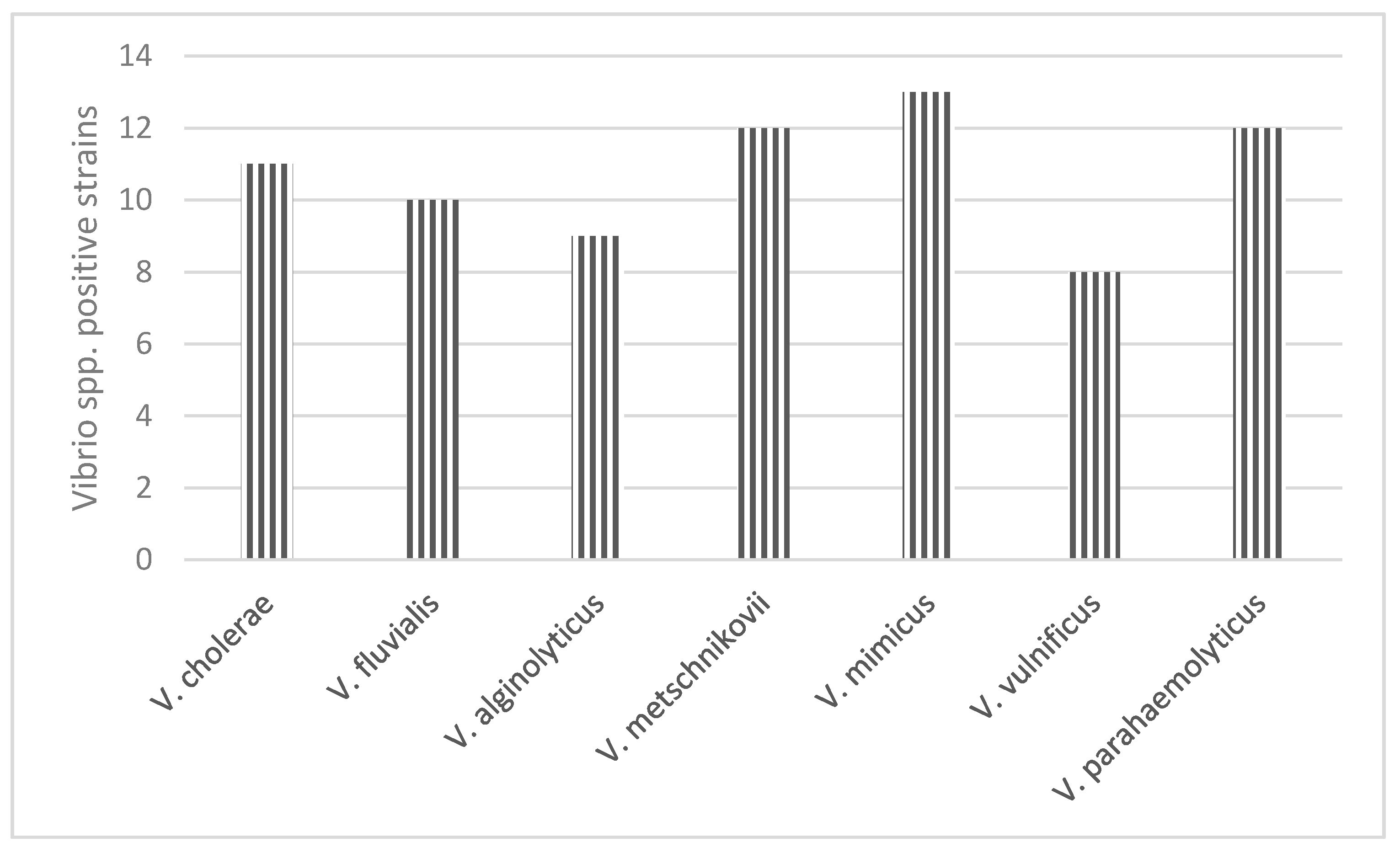
Based on the cultural characteristics and the results of the biochemical properties testing using the Rapid One NF plus system, seven species belonging to the genus Vibrio were detected and confirmed: Vibrio vulnificus 10.66% (n = 8), Vibrio cholerae 14.66% (n = 12), Vibrio fluvialis 13.33% (n = 10), Vibrio parahaemolyticus 16% (n = 12), Vibrio mimicus 17.33% (n = 13), Vibrio metschnikovii 16% (n = 12), and Vibrio alginolyticus 12% (n = 9) (Figure 2).
Most of these strains were isolated from samples collected from Eurasian hobby (Falco subbuteo), lesser whitethroat (Sylvia curucca), red-backed shrike (Lanius collurio), red-footed falcon (Falco vespertinus), common kingfisher (Alcedo atthis), hooded crow (Corvus corone cornix), and Eurasian blue tit (Parus caeruleus).
The majority (59.64%) of Vibrio spp. strains were isolated from migratory birds. Out of 75 samples, eleven (14.67%) isolates were positive in the PCR employing ompW and toxR primers. Nine of the DNA samples yielded a single amplicon of 588 bp in an ompW-based PCR assay and six samples yielded a 336 bp PCR product for toxR. About one third (36.36%) of the samples were positive for both toxR and ompW (Table 1).
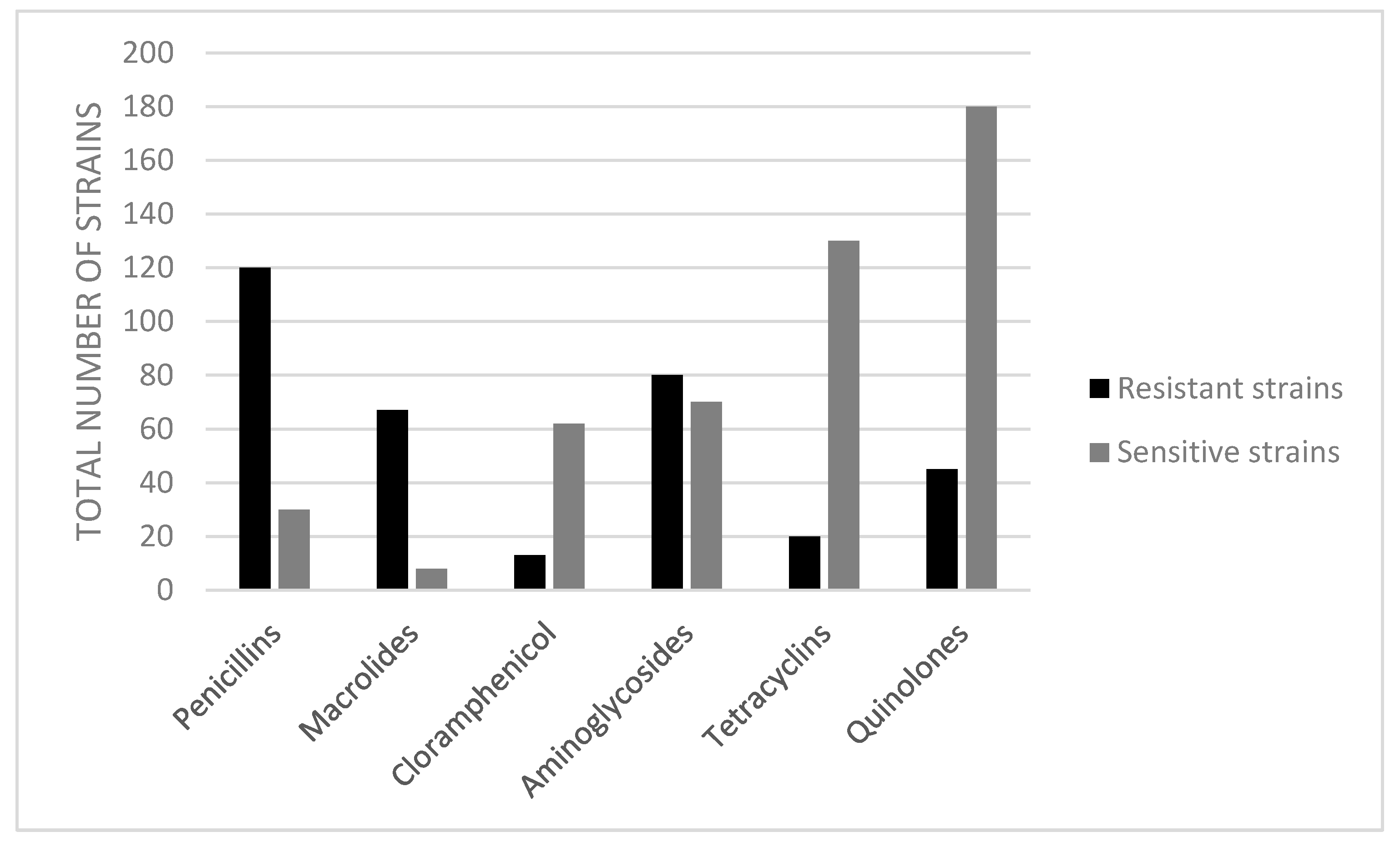
The isolated Vibrio spp. strains were evaluated for their level of antibiotic resistance. Based on the values of the inhibition diameters as compared against the CLSI guidelines 2020 in the Kirby–Bauer method, the bacterial strains were classified into resistant (R), intermediate (I), and susceptible (S) categories. The numbers of resistant strains in migratory and sedentary birds are indicated in Table 2. The percentage of total bacterial isolates demonstrating resistance or sensitivity to the 11 tested antibiotics is presented in Figure 3. In the present study, the resistance to erythromycin was the highest (88.16%), followed by penicillin (86.84%), amikacin (81.57%), and ampicillin (71.05%) and statistically significantly (p < 0.05) lower percentages were found for kanamycin (23.68%), quinolone class (enrofloxacine, marbofloxacine, and ciprofloxacine with 19.74%), and tetracycline class (tetracycline and oxitetracycline, both with 13.15%).
A MAR index value ≥0.2 was observed in 89.33% of the resistant pathogens. The MAR index calculated for isolated strains was 0.8 in 4 strains (all V.cholerae strains isolated from migratory birds Ardeola ralloides, Turdus philomelos, Accipiter nisus), 0.72 in 2 strains (V. cholerae strains isolated from Falco subbuteo and Accipiter nisus), 0.63 in 2 strains (V. cholerae strains isolated from Falco subbuteo and Fringilla coelebs), 0.54 in 4 strains, (V. cholerae isolated from Gallinago gallinago, V. fluvialis isolated from Falco vespertinus, V. mimicus from Parus major, and Vibrio parahaemolyticus isolated from Sylvia borin), 0.45 in 16 strains, 0.36 in 30 strains, 0.27 in 8 strains, 0.18 in 5 stains, and 0.09 in one strain (V. fluvialis associated with Alcedo atthis).
The isolates showed a high frequency of resistance to commonly used antibiotics, such as for each penicillin, erythromycin, and amikacin, between 90.66% and 92.00%, ampicillin (72%), and enrofloxacin (18.66%). Oxytetracycline, tetracycline, and ciprofloxacin were the most effective, with 86.66% of the strains being sensitive to oxytetracycline and tetracycline while 80% were sensitive to ciprofloxacin. The MAR indexes for antibiotics showed values ranging from 0.1 to 0.8 (Table 2) and the encountered resistance types are presented in Table 3. The result indicated the presence of MAR, MDR, and also XDR in the isolated Vibrio spp.

4. Discussion

In our study, we investigated the incidence of clinically important Vibrio spp. in wild birds captured in the Danube Delta Biosphere Reserve, Romania. Vibrio species are described as natural residents of aquatic environments [16,26], with species such as Vibrio parahaemolyticus and Vibrio vulnificus detected in the natural flora of estuarine and coastal marine environments worldwide [46,47], and also from the sea and brackish water and sediments of both tropical and temperate regions, as well as from a variety of seafood [17,46]. Seven clinically important Vibrio spp. were isolated, predominantly, V. mimicus, V. parahaemolyticus, and V. metschnikovii, followed by V. cholerae, V. vulnificus, V. alginolyticus, and V. fluvialis. Our results indicated a high frequency of all these Vibrio species in both wild migratory and sedentary birds.
The presence of these bacteria has medical relevance, as V. parahaemolyticus, V. cholerae, and V. vulnificus are the main species associated with seafood-borne infections, while V. alginolyticus, V. mimicus, and Grimontia hollisae (previously known as V. hollisae) have been sporadically isolated in disease outbreaks [48]. Vibrio vulnificus is one of the emerging food- and waterborne zoonotic bacteria, indigenous to estuarine waters and shellfish worldwide; it is involved in gastroenteritis and primary septicemia after consumption of contaminated oysters [49,50,51,52].
Toxigenic Vibrio cholerae (O1, O139) represent a major public health problem in many areas of the developing world [53,54,55]. The importance of wild birds as potential vectors of disease has received recent renewed empirical interest, especially regarding human health [9,23]. Besides other Vibrio spp., our results indicated the presence of V. cholerae in both sedentary and migratory birds, but with a higher frequency in migratory birds. Wild birds possess an important role in the epidemiology of Vibrio spp.-associated outbreaks [56,57] due to the particularities of their habitat and the interrelations with human activities (boat fishing, fish processing areas, and shellfish beds). Prolonged survival and persistence of Vibrio spp. in wild birds facilitate the possible contamination of the environment, especially in the case of migratory birds regarded as carriers for several bacterial pathogens, including several Vibrio species. Wild aquatic birds are a vehicle of V. vulnificus and V. parahaemolyticus in winter [58], so these bacteria can be potentially transported long distances [59]. Carrier status of V. cholerae in birds has a major implication for public health. The infection in birds occurs during their feeding in areas polluted by human or animal V. cholerae O1 carriers. Migratory birds may transport this pathogenic organism to different areas. Additionally, the transformation of non-O1 V. cholerae to an O1 serovar was demonstrated at the intestinal level of the birds [60].
The results of Nandi et al. (2000) [41] indicate that the toxR gene is involved in the regulation and expression of several genes of V. cholerae. The ToxR regulon (regulatory protein) is required in Vibrio cholerae for transcriptional activation of the toxT gene, which, in turn, activates numerous genes involved in the virulence of the bacterium [61,62,63]. Nevertheless, a low prevalence (7.9%) of this gene was found in the present study, indicating a potentially reduced overall virulence of the isolated strains, but not necessarily their decreased pathogenicity. Several studies indicate different degrees of resistance to antibiotics of isolated Vibrio spp. strains. The results of Li and coworkers [64] indicated a sensitivity of Vibrio species against streptomycin, rifampicin, kanamycin, tetracycline, and polymyxin B. These results could not be correlated with the studies of Okoh and Igbinosa (2010) [24] which reported percentages of resistance of 100, 90, 70, and 80 to trimethoprim, penicillin, cotrimoxazole, and streptomycin, respectively, in Vibrio fluvialis; 92, 82 90, and 100, respectively, to cephalothin in V. vulnificus, V. parahaemolyticus, V fluvialis, and V. metschnikovii, as well as resistance to ampicillin in all isolated Vibrio strains. Our findings were similar to those of Okoh and Igbinosa [24], especially in terms of strains resistant to penicillin and ampicillin. The authors isolated strains which exhibited various degrees of resistance toward tested drugs: tetracycline, marfloxin, and chloramphenicol (50%), penicillin, amikacin, and erythromycin 100%, ciprofloxacin (41.66%), oxytetracycline (58.33%), and ampicillin (83.33%), with six different resistance patterns being observed. You and coworkers (2016) [65] also isolated antibiotic-resistant and multidrug-resistant Vibrio spp. from aquatic environments, with high frequencies of resistance against erythromycin (81.8–95.7%), ampicillin (42–82%), and mecillinam (42–55%).
Similarly, the percentage of resistant strains was dependent on the antibiotic rather than on the class, and high resistance percentages indicated the differences in antimicrobial potency between the antibiotics in current human and veterinary use. Furthermore, the MAR values in the present study, as high as 0.2 to 0.8, supported the assumption that the isolated strains originated from a high-risk source of contamination and presented increased risk to public health. This hypothesis is further sustained by the presence of MDR and XDR in a large proportion (Table 3). Deng et al. (2020) [66] isolated Vibrio species, with a high prevalence of resistance to vancomycin, amoxicillin, midecamycin, and furazolidone, moderate prevalence of resistance to tobramycin, rifampicin, gentamicin, and tetracycline, and low prevalence of resistance to erythromycin, trimethoprim–sulfamethoxazole, doxycycline, and chloramphenicol. We found similar results for penicillins, but not for tetracyclines and erythromycin, were the results showed the opposite, with low and high prevalence of resistance, respectively.
Matyar et al. (2008) [67] showed multidrug resistance in Gram-negative bacteria isolated from aquatic environments and shrimp samples. Okoh and Igbinosa (2010) [24] identified multidrug-resistant non-cholera Vibrio spp. isolates showing resistance to all the antibiotics traditionally used to treat cholera. The study of Shivakumaraswamy et al. (2019) [68] revealed a high occurrence of antibiotic resistance in bacteria from animals and other natural environments.
Miyasaka and coworkers [59] evaluated the spatial and temporal distribution of Vibrio spp. in wild birds from Japan. The most important species of Vibrio were Vibrio parahaemolyticus and Vibrio vulnificus. The isolation of these species from feces demonstrated that the investigated avian species represent a vehicle for V. parahaemolyticus and V. vulnificus and also a habitat to survive during winter [59]. In this study, we isolated 12 strains of V. parahaemolyticus and eight strains of V. vulnificus, these strains mainly coming from migratory birds (Tringa glareola, Nycticorax nycticorax, Sylvia curucca, Sylvia borin, Sylvia nisoria, Ardeola ralloides, Emberiza schoeniclus, Accipiter nisus, Anas crecca, Hippolais icterina).
The emergence and spread of multidrug-resistant bacteria in natural environments represent a serious impact on animal and human health [69,70,71].
Wild birds have been not only postulated, but also demonstrated, as sentinels, reservoirs, and potential spreaders of antibiotic resistance [72,73,74,75,76]. Based on the results recorded by these authors, solid scientific evidence indicates the significant epidemiological role of wild birds in the dissemination of multidrug-resistant bacteria through migration.
Isolation of resistant bacterial strains from wilds birds highlights the potential hazard to both humans and animals given the transmission to humans and animals and vice versa [74,75,77]. The fecal shedding of the resistant strains allows environmental contamination.
However, most of these published studies are focused on Escherichia coli and/or Enterococcus spp. [73,78]. There is little information regarding the antimicrobial resistance level of Vibrio species isolated from wild birds, therefore, we believe that our study brings important additional information on the prevalence of Vibrio spp. in wild birds.
Antimicrobial resistance was determined for the Vibrio cholerae strains isolated from the Danube River in Slovakia [36]. All strains were susceptible only to three antimicrobials: chloramphenicol, rifampicin, and tetracycline, while resistance was displayed towards kanamycin and streptomycin [36]. Complementarily, our study evaluates antibiotic resistance to all isolated strains. Antimicrobial resistance is a growing problem worldwide and represents a major medical and public health problem [24,28,70]; a high prevalence of multidrug resistance indicates a serious need for antibiotic surveillance programs [79].

5. Conclusions

Our study confirmed the presence of MDR Vibrio spp. in migratory and sedentary wild birds captured from two different locations in the Danube Delta. Both migratory and sedentary birds from sampling sites were positive for a broad spectrum of potentially pathogenic Vibrio spp., indicating a great risk to public health. The present results indicate that wild-living birds may be a latent source for further microbial pollution of various habitats, including nesting places, and thus transfer it to offspring in the Danube Delta region. Further studies are intended to assess the antibiotic resistance determinants to underline the role of wild birds as a reservoir of multidrug-resistant Vibrio spp., including V. cholerae. Additional samples should be taken for a better assessment of carriage frequency.

Author Contributions

E.P., R.K.R. and M.S. designed the study; E.P. and G.D. performed the PCR, study design, and data interpretation; C.D.Ș. and G.F.B. defined antibiotic resistance; M.N., C.C., and D.O. completed the cultivation and identification of the strains; S.Z. and G.D. performed the serological identification of the strains; M.R. (Magdalna Rzewuska) was responsible of the methodology of the study; A.V.P. and A.V. analyzed the data. E.P., M.S., M.R. (Mariana Rusu), S.Z. and C.C. wrote the article. All authors have read and agreed to the published version of the manuscript.

Funding

This research received no funding.

Institutional Review Board Statement

Ethical review and approval were waived for this study, due to the fact that there was no direct interaction of the involved institutions/participants with the birds (see also Materials and Methods). The Romanian Ornithological Centre (Institute of Biology, Romanian Academy of Sciences), part of Euring, the organization coordinating European bird ringing schemes, collected the samples while banding the birds under ethical principles applied by Euring and the provisions of Directive 2009/147/EC on the conservation of wild birds (consolidated version, http://data.europa.eu/eli/dir/2009/147/2019-06-26, accessed on 21 February 2021).

Informed Consent Statement

Not applicable.

Data Availability Statement

The data presented in this study are available on request from the corresponding author.

Acknowledgments

The authors express their gratitude to the Romanian Ornithological Centre (Institute of Biology, Romanian Academy of Sciences), namely, to Mircea Gogu-Bogdan and Ioana Cobzaru, for kindly providing the samples while carrying out wild bird banding in the Danube Delta research areas.

Conflicts of Interest

The authors declare that the research was conducted in the absence of any commercial or financial relationships that could be construed as a potential conflict of interest.

References

  1. Coman, A.; Maftei, D.N.; Chereches, R.M.; Zavrotchi, E.; Bria, P.; Dragnea, C.; Mckenzie, P.P.; Valentine, M.A.; Gray, G.C. Avi-an Influenza Surveillance in the Danube Delta Using Sentinel Geese and Ducks. Influ. Res. Treat. 2014, 2014, 1–6. [Google Scholar] [CrossRef] [PubMed]
  2. Páll, E.; Niculae, M.; Kiss, T.; Şandru, C.D.; Spînu, M. Human impact on the microbiological water quality of the rivers. J. Med. Microbiol. 2013, 62, 1635–1640. [Google Scholar] [CrossRef]
  3. Romanescu, G. Alluvial transport processes and the impact of anthropogenic intervention on the Romanian littoral of the Danube Delta. Ocean Coast. Manag. 2013, 73, 31–43. [Google Scholar] [CrossRef]
  4. Ionita, C.; Bogdan, T.A.; Ipate, I.; Ivana, S.; Ionita, L.; Gogu-Bogdan, M. Strategies for conserving birds biodiversity in Danube del-ta. Ocean Coast. Manag. 2010, 15, 641–644. [Google Scholar]
  5. Giosan, L.; Donnelly, J.P.; Vespremeanu, E. River delta morphodynamics: Examples from the Danube Delta. River Deltas-Concepts Models Ex. 2005, 83, 393–411. [Google Scholar]
  6. Danube Delta-UNESCO World Heritage Centre. Available online: https://whc.unesco.org/en/list/588/ (accessed on 12 January 2021).
  7. Păunescu, C. A study of migration in time of the Sacalin Island, Danube Delta, Romania. Proc. Rom. Acad. Ser. B 2012, 2, 156–160. [Google Scholar]
  8. Greig, J.; Rajić, A.; Young, I.; Mascarenhas, M.; Waddell, L.; Lejeune, J. A scoping review of the role of wildlife in the transmis-sion of bacterial pathogens and antimicrobial resistance to the food chain. Zoonoses Public Health 2015, 62, 269–284. [Google Scholar] [CrossRef] [PubMed]
  9. Khan, S.A.; Imtiaz, M.A.; Sayeed, M.A.; Shaikat, A.H.; Hassan, M.M. Antimicrobial resistance pattern in domestic animal-Wildlife-environmental niche via the food chain to humans with a Bangladesh perspective; A systematic review. BMC Vet. Res. 2020, 16. [Google Scholar] [CrossRef]
  10. Abulreesh, H.H.; Goulder, R.; Scott, G.W. Wild birds and human pathogens in the context of ringing and migration. Ringing Migr. 2007, 23, 193–200. [Google Scholar] [CrossRef]
  11. Lord, A.T.K.; Mohandas, K.; Somanath, S.; Ambu, S. Multidrug resistant yeasts in synanthropic wild birds. Ann. Clin. Microbiol. Antimicrob. 2010, 9. [Google Scholar] [CrossRef] [PubMed] [Green Version]
  12. Elmberg, J.; Berg, C.; Lerner, H.; Waldenström, J.; Hessel, R. Health perspective. Infect. Ecol. Epidemiol. 2017, 7, 1300450. [Google Scholar] [CrossRef] [PubMed]
  13. Wang, Y.; Leung, P.C.; Qian, P.Y.; Gu, J.D. Antibiotic resistance and plasmid profile of environmental isolates of Vibrio species from Mai Po Nature Reserve, Hong Kong. Ecotoxicology 2006, 15, 371–378. [Google Scholar] [CrossRef]
  14. Li, B.; Liu, J.; Zhou, S.; Fu, L.; Yao, P.; Chen, L.; Yang, Z.; Wang, X.; Zhang, X.-H. Vertical variation in Vibrio community com-position in Sansha Yongle Blue Hole and its ability to degrade macromolecules. Mar. Life Sci. Technol. 2020, 2, 60–72. [Google Scholar] [CrossRef] [Green Version]
  15. Moghimi, S.M.; Hunter, A.C.; Murray, J.C. Long-Circulating and Target-Specific Nanoparticles: Theory to Practice. Pharmacol. Rev. 2001, 53, 283–318. [Google Scholar]
  16. Sarita, G.B. Multiple Antibiotic Resistances of Vibrio Isolates from Coastal and Brackish Water Areas. Am. J. Biochem. Biotechnol. 2005, 1, 201–206. [Google Scholar]
  17. Chikwendu, C. Multiple Antimicrobial Resistance in Vibrio spp Isolated from River and Aquaculture Water Sources in Imo State, Nigeria. Br. Microbiol. Res. J. 2014, 4, 560–569. [Google Scholar] [CrossRef]
  18. Brooks, G.F.; Carroll, K.C.; Butel, J.S.; Morse, S.A. Vibrio, Campylobacters, Helicobacter and associated bacteria. Jawetz Melnock Adelberg’s Med. Microbiol. 2007, 27e, 270–279. [Google Scholar]
  19. Osunla, C.A.; Okoh, A.I. Vibrio Pathogens: A Public Health Concern in Rural Water Resources in Sub-Saharan Africa. Int. J. Environ. Res. Public Health 2017, 14, 1188. [Google Scholar] [CrossRef] [Green Version]
  20. Adebayo-Tayo, B.C.; Okonko, I.O.; Esen, C.U.; Odu, N.N.; Onoh, C.C.; Igwiloh, N.J.P. Incidence of Potentially Pathogenic Vib-rio Spp. In Fresh Seafood from Itu Creek in Uyo, Akwa Ibom State, Nigeria. World Appl. Sci. J. 2011, 15, 985–991. [Google Scholar]
  21. Adeleye, I.A.; Daniels, F.V.; Enyinnia, V.A. Characterization and pathogenicity of Vibrio spp. contaminating seafoods in La-gos, Nigeria. Internet J. Food Saf. 2010, 12, 1–9. [Google Scholar]
  22. Ballal, M.; Shetty, V.; Bangera, S.R.; Prabhu, M.; Umakanth, S. Vibrio furnissii, an emerging pathogen causing acute gastroen-teritis: A Case Report. JMM Case Rep. 2017, 4. [Google Scholar] [CrossRef] [Green Version]
  23. Willey, J.M.; Sherwood, L.M.; Woolverton, C.J. Presscott, Harley, and Klein’s Microbiology; McGraw-Hill: New York, NY, USA, 2008; ISBN 0073302082. [Google Scholar]
  24. Baker-Austin, C.; Oliver, J.D.; Alam, M.; Ali, A.; Waldor, M.K.; Qadri, F.; Martinez-Urtaza, J. Vibrio spp. infections. Nat. Rev. Dis. Prim. 2018, 4, 8. [Google Scholar] [CrossRef]
  25. Montánchez, I.; Ogayar, E.; Plágaro, A.H.; Esteve-Codina, A.; Gómez-Garrido, J.; Orruño, M.; Arana, I.; Kaberdin, V.R. Analysis of Vibrio harveyi adaptation in sea water microcosms at elevated temperature provides insights into the putative mechanisms of its persistence and spread in the time of global warming. Sci. Rep. 2019, 9, 289. [Google Scholar] [CrossRef] [Green Version]
  26. Mukhopadhyay, A.K.; Garg, S.; Mitra, R.; Basu, A.; Rajendran, K.; Dutta, D.; Bhattacharya, S.K.; Shimada, T.; Takeda, T.; Takeda, Y.; et al. Temporal Shifts in Traits of Vibrio cholerae Strains Isolated from Hospitalized Patients in Calcutta: A 3-Year (1993 to 1995) Analysis. J. Clin. Microbiol. 1996, 34, 2537–2543. [Google Scholar] [CrossRef] [Green Version]
  27. Okoh, A.I.; Igbinosa, E.O. Antibiotic susceptibility profiles of some Vibrio strains isolated from wastewater final effluents in a rural community of the Eastern Cape Province of South Africa. BMC Microbiol. 2010, 10, 143. [Google Scholar] [CrossRef] [Green Version]
  28. Das, B.; Verma, J.; Kumar, P.; Ghosh, A.; Ramamurthy, T. Antibiotic resistance in Vibrio cholerae: Understanding the ecology of resistance genes and mechanisms. Vaccine 2020, 38, A83–A92. [Google Scholar] [CrossRef] [PubMed]
  29. Wang, R.; Yu, D.; Yue, J.; Kan, B. Variations in SXT elements in epidemic Vibrio cholerae O1 El Tor strains in China. Sci. Rep. 2016, 6, 22733. [Google Scholar] [CrossRef] [PubMed] [Green Version]
  30. Hochhut, B.; Lotfi, Y.; Mazel, D.; Faruque, S.M.; Woodgate, R.; Waldor, M.K. Molecular analysis of antibiotic resistance gene clusters in Vibrio cholerae O139 and O1 SXT constins. Antimicrob. Agents Chemother. 2001, 45, 2991–3000. [Google Scholar] [CrossRef] [PubMed] [Green Version]
  31. Sarkar, A.; Morita, D.; Ghosh, A.; Chowdhury, G.; Mukhopadhyay, A.K.; Okamoto, K.; Ramamurthy, T. Altered Integrative and Conjugative Elements (ICEs) in Recent Vibrio cholerae O1 Isolated From Cholera Cases, Kolkata, India. Front. Microbiol. 2019, 10. [Google Scholar] [CrossRef]
  32. Waldor, M.K.; Tscha¨pe, H.; Tscha¨pe, T.; Mekalanos, J.J. A New Type of Conjugative Transposon Encodes Resistance to Sul-famethoxazole, Trimethoprim, and Streptomycin in Vibrio cholerae O139. J. Bacteriol. 1996, 178, 4157–4165. [Google Scholar] [CrossRef] [Green Version]
  33. Kitaoka, M.; Miyata, S.T.; Unterweger, D.; Pukatzki, S. Antibiotic resistance mechanisms of Vibrio cholerae. J. Med. Microbiol. 2011, 60, 397–407. [Google Scholar] [CrossRef] [PubMed]
  34. Dengo-Baloi, L.C.; Semá-Baltazar, C.A.; Manhique, L.V.; Chitio, J.E.; Inguane, D.L.; Langa, J.P. Antibiotics resistance in El Tor Vibrio cholerae 01 isolated during cholera outbreaks in Mozambique from 2012 to 2015. PLoS ONE 2017, 12, e0181496. [Google Scholar] [CrossRef] [Green Version]
  35. Falbo, V.; Carattoli, A.; Tosini, F.; Pezzella, C.; Dionisi, A.M.; Luzzi, I. Antibiotic Resistance Conferred by a Conjugative Plas-mid and a Class I Integron in Vibrio cholerae O1 El Tor Strains Isolated in Albania and Italy. Am. Soc. Microbiol. 1999, 43, 693–696. [Google Scholar]
  36. Amita, M.; Chowdhury, S.R.; Thungapathra, M.; Ramamurthy, T.; Nair, G.B.; Ghosh, A. Class I integrons and SXT elements in El Tor strains isolated before and after 1992 Vibrio cholerae 0139 outbreak, Calcutta, India. Emerg. Infect. Dis. 2003, 9, 500–502. [Google Scholar] [CrossRef]
  37. Mohapatra, H.; Mohapatra, S.S.; Mantri, C.K.; Colwell, R.R.; Singh, D. V Vibrio cholerae non-O1, non-O139 strains isolated before 1992 from Varanasi, India are multiple drug resistant, contain intSXT, dfr18 and aadA5 genes. Environ. Microbiol. 2008, 10, 866–873. [Google Scholar] [CrossRef]
  38. Bhanumathi, R.; Sabeena, F.; Isac, S.R.; Shukla, B.N.; Singh, D. V Molecular Characterization of Vibrio cholerae O139 Bengal Isolated from Water and the Aquatic Plant Eichhornia crassipes in the River Ganga, Varanasi, India. Appl. Environ. Microbiol. 2003, 69, 2389–2394. [Google Scholar] [CrossRef] [Green Version]
  39. Seman, M.; Prokšová, M.; Rosinský, J.; Ferianc, P. Isolation, identification, and characterization of Vibrio cholerae from the Danube River in Slovakia. Folia Microbiol. 2012, 57, 191–197. [Google Scholar] [CrossRef]
  40. Oprea, M.; Njamkepo, E.; Cristea, D.; Zhukova, A.; Clark, C.G.; Kravetz, A.N.; Monakhova, E.; Ciontea, A.S.; Cojocaru, R.; Rauzier, J.; et al. The seventh pandemic of cholera in Europe revisited by microbial genomics. Nat. Commun. 2020, 11, 5347. [Google Scholar] [CrossRef]
  41. Nandi, B.; Nandy, R.K.; Mukhopadhyay, S.; Nair, G.B.; Shimada, T.; Ghose, A.C. Rapid Method for Species-Specific Identifi-cation of Vibrio cholerae Using Primers Targeted to the Gene of Outer Membrane Protein OmpW. J. Clin. Microbiol. 2000, 38, 4145–4151. [Google Scholar] [CrossRef] [Green Version]
  42. Lembke, M.; Höfler, T.; Walter, A.N.; Tutz, S.; Fengler, V.; Schild, S.; Reidl, J. Host stimuli and operator binding sites control-ling protein interactions between virulence master regulator ToxR and ToxS in Vibrio cholerae. Mol. Microbiol. 2020, 114, 262–278. [Google Scholar] [CrossRef] [PubMed] [Green Version]
  43. Krumperman, P.H. Multiple Antibiotic Resistance Indexing of Escherichia coli to Identify High-Risk Sources of Fecal Con-tamination of Foodst. Appl. Environ. Microbiol. 1983, 46, 165–170. [Google Scholar] [CrossRef] [Green Version]
  44. Tambekar, D.H.; Dhanorkar, D.V.; Gulhane, S.R.; Khandelwal, V.K.; Dudhane, M.N. Antibacterial susceptibility of some urinary tract pathogens to commonly used antibiotics. Afr. J. Biotechnol. 2006, 5, 1562–1565. [Google Scholar]
  45. Magiorakos, A.P.; Srinivasan, A.; Carey, R.B.; Carmeli, Y.; Falagas, M.E.; Giske, C.G.; Harbarth, S.; Hindler, J.F.; Kahlmeter, G.; Olsson-Liljequist, B.; et al. Multidrug-resistant, extensively drug-resistant and pandrug-resistant bacteria: An international expert proposal for interim standard definitions for acquired resistance. Clin. Microbiol. Infect. 2012, 18, 268–281. [Google Scholar] [CrossRef] [Green Version]
  46. Kirs, M.; Depaola, A.; Fyfe, R.; Jones, J.L.; Krantz, J.; Van Laanen, A.; Cotton, D.; Castle, M. A survey of oysters (Crassostrea gigas) in New Zealand for Vibrio parahae-molyticus and Vibrio vulnificus. Int. J. Food Microbiol. 2010, 147, 149–153. [Google Scholar] [CrossRef]
  47. Johnson, C.N.; Bowers, J.C.; Griffitt, K.J.; Molina, V.; Clostio, R.W.; Pei, S.; Laws, E.; Paranjpye, R.N.; Strom, M.S.; Chen, A.; et al. Ecology of Vibrio parahaemolyticus and Vibrio vulnificus in the Coastal and Estuarine Waters of Louisiana, Maryland, Mississippi, and Washington (United States). Am. Soc. Microbiol. 2012. [Google Scholar] [CrossRef] [PubMed] [Green Version]
  48. Kokashvili, T.; Whitehouse, C.A.; Tskhvediani, A.; Grim, C.J.; Elbakidze, T.; Mitaishvili, N.; Janelidze, N.; Jaiani, E.; Haley, B.J.; Lashkhi, N.; et al. Occurrence and Diversity of Clinically Important Vibrio Species in the Aquatic Environment of Geor-gia. Front. Public Health 2015, 3. [Google Scholar] [CrossRef] [PubMed]
  49. Lai, C.-H.; Hwang, C.-K.; Chin, C.; Lin, H.-H.; Wong, W.-W.; Liu, C.-Y. Severe watery diarrhoea and bacteraemia caused by Vibrio fluvialis. J. Infect. 2006, 52, 95–98. [Google Scholar] [CrossRef]
  50. Horseman, M.A.; Surani, S. A comprehensive review of Vibrio vulnificus: An important cause of severe sepsis and skin and soft-tissue infection. Int. J. Infect. Dis. 2011, 15, 157–166. [Google Scholar] [CrossRef] [Green Version]
  51. Chase, E.; Harwood, V.J. Comparison of the Effects of Environmental Parameters on Growth Rates of Vibrio vulnificus Bio-types I, II, and III by Culture and Quantitative PCR Analysis. Appl. Environ. Microbiol. 2011, 77, 4200–4207. [Google Scholar] [CrossRef] [Green Version]
  52. Heng, S.P.; Letchumanan, V.; Deng, C.Y.; Mutalib, N.S.A.; Khan, T.M.; Chuah, L.H.; Chan, K.G.; Goh, B.H.; Pusparajah, P.; Lee, L.H. Vibrio vulnificus: An environmental and clinical burden. Front. Microbiol. 2017, 8, 997. [Google Scholar] [CrossRef] [Green Version]
  53. Faruque, S.M.; Albert, M.J.; Mekalanos, J.J. Epidemiology, Genetics, and Ecology of Toxigenic Vibrio cholerae. Microbiol. Mol. Biol. Rev. 1998, 62, 1301–1314. [Google Scholar] [CrossRef] [PubMed] [Green Version]
  54. Faruque, S.M.; Islam, M.J.; Ahmad, Q.S.; Biswas, K.; Faruque AS, G.; Nair, G.B.; Sack, R.B.; Sack, D.A.; Mekalanos, J.J. An Improved Technique for Isolation of Environmental Vibrio cholerae with Epi-demic Potential: Monitoring the Emergence of a Multiple-Antibiotic–Resistant Epidemic. J. Infect. Dis. 2006, 193, 1029–1036. [Google Scholar] [CrossRef] [Green Version]
  55. Rodriguez Ojeda, J.A.; Chadi, I.K. Vibrio Cholerae; StatPearls Publishing: Treasure Island, FL, USA, 2020. Available online: https://www.ncbi.nlm.nih.gov/books/NBK526099/ (accessed on 18 March 2020).
  56. Benskin, C.M.W.H.; Wilson, K.; Jones, K.; Hartley, I.R. Bacterial pathogens in wild birds: A review of the frequency and effects of infection. Biol. Rev. 2009, 84, 349–373. [Google Scholar] [CrossRef] [PubMed]
  57. Laviad-Shitrit, S.; Izhaki, I.; Halpern, M. Accumulating evidence suggests that some waterbird species are potential vectors of Vibrio cholerae. PLoS Pathog. 2019, 15, e1007814. [Google Scholar] [CrossRef] [PubMed]
  58. Lutz, C.; Erken, M.; Noorian, P.; Sun, S.; McDougald, D. Environmental reservoirs and mechanisms of persistence of Vibrio cholerae. Front. Microbiol. 2013, 4. [Google Scholar] [CrossRef] [Green Version]
  59. Miyasaka, J.; Yahiro, S.; Arahira, Y.; Tokunaga, H.; Katsuki, K.; Hara-Kudo, Y. Isolation of Vibrio parahaemolyticus and Vibrio vulnificus from wild aquatic birds in Japan. Epidemiol. Infect. 2006, 134, 780–785. [Google Scholar] [CrossRef]
  60. Ogg, J.E.; Ryder, R.A.; Smith, H.L. Isolation of Vibrio cholerae from Aquatic Birds in Colorado and Utah. Appl. Environ. Microbiol. 1989, 55, 95–99. [Google Scholar] [CrossRef] [Green Version]
  61. Liang, W.; Wang, L.; Liang, P.; Zheng, X.; Zhou, H.; Zhang, J.; Zhang, L.; Kan, B. Sequence polymorphisms of rfbT among the Vibrio cholerae O1 strains in the Ogawa and Inaba serotype shifts. BMC Microbiol. 2013, 13, 173. [Google Scholar] [CrossRef] [Green Version]
  62. Krukonis, E.S.; Yu, R.R.; DiRita, V.J. The Vibrio cholerae ToxR/TcpP/ToxT virulence cascade: Distinct roles for two mem-brane-localized transcriptional activators on a single promoter. Mol. Microbiol. 2000, 38, 67–84. [Google Scholar] [CrossRef] [Green Version]
  63. Kunkle, D.E.; Bina, T.F.; Bina, X.R.; Bina, J.E. Vibrio cholerae OmpR Represses the ToxR Regulon in Response to Membrane Intercalating Agents That Are Prevalent in the Human Gastrointestinal Tract. Am. Soc. Microbiol. 2020. [Google Scholar] [CrossRef]
  64. Li, J.; Yie, J.; Foo, W.T.; Ling Julia, M.L.; Huaishu, X.; Norman, Y.S.W. Antibiotic resistance and plasmid profiles of Vibrio isolates from cultured silver sea bream, Sparus sarba. Mar. Poll. Bull. 1999, 39, 245–249. [Google Scholar] [CrossRef]
  65. You, K.G.; Bong, C.W.; Lee, C.W. Antibiotic resistance and plasmid profiling of Vibrio spp. in tropical waters of Peninsular Malaysia. Environ. Monit. Assess. 2016, 188, 171. [Google Scholar] [CrossRef] [PubMed]
  66. Deng, Y.; Xu, L.; Chen, H.; Liu, S.; Guo, Z.; Cheng, C.; Ma, H.; Feng, J. Prevalence, virulence genes, and antimicrobial resistance of Vibrio species isolated from diseased marine fish in South China. Sci. Rep. 2020, 10, 14329. [Google Scholar] [CrossRef]
  67. Matyar, F.; Kaya, A.; Dinçer, S. Antibacterial agents and heavy metal resistance in Gram-negative bacteria isolated from sea-water, shrimp and sediment in Iskenderun Bay, Turkey. Sci. Total Environ. 2008, 407, 279–285. [Google Scholar] [CrossRef] [PubMed]
  68. Karunasagar, I.; Shivakumaraswamy, S.K.; Deekshit, V.K.; Vittal, R.; Akhila, D.S.; Mundanda, D.M.; Raj, J.R.M.; Chakraborty, A. Phenotypic & genotypic study of antimicrobial profile of bacteria isolates from environmental samples. Indian J. Med. Res. 2019, 149, 232–239. [Google Scholar]
  69. Prestinaci, F.; Pezzotti, P.; Pantosti, A. Antimicrobial resistance: A global multifaceted phenomenon. Pathog. Glob. Health 2015, 109, 309–318. [Google Scholar] [CrossRef] [Green Version]
  70. Roca, I.; Akova, M.; Baquero, F.; Carlet, J.; Cavaleri, M.; Coenen, S.; Cohen, J.; Findlay, D.; Gyssens, I.; Heure, O.E.; et al. The global threat of antimicrobial resistance: Science for intervention. New Microbes New Infect. 2015, 6, 22–29. [Google Scholar] [CrossRef] [PubMed] [Green Version]
  71. Wellington, E.M.; Boxall, A.B.; Cross, P.; Feil, E.J.; Gaze, W.H.; Hawkey, P.M.; Hawkey, P.M.; Johnson-Rollings, A.S.; Jones, D.L.; Lee, N.M.; et al. The role of the natural environment in the emergence of antibiotic resistance in Gram-negative bacteria. Lancet Infect. Dis. 2013, 13, 155–165. [Google Scholar] [CrossRef]
  72. Fallacara, D.M.; Monahan, C.M.; Morishita, T.Y.; Diseases, R.F.W. Fecal shedding and antimicrobial susceptibility of selected bacterial pathogens and a survey of intestinal parasites in free-living waterfowl. Avian Dis. 2001, 45, 128–135. [Google Scholar] [CrossRef]
  73. Dolejska, M.; Cizek, A.; Literak, I. High prevalence of antimicrobial-resistant genes and integrons in Escherichia coli isolates from Black-headed Gulls in the Czech Republic. J. Appl. Microbiol. 2007, 103, 11–19. [Google Scholar] [CrossRef] [PubMed]
  74. Bonnedahl, J.; Järhult, J.D. Antibiotic resistance in wild birds. Upsala J. Med. Sci. 2014, 119, 113–116. [Google Scholar] [CrossRef] [PubMed] [Green Version]
  75. Radhouani, H.; Silva, N.; Poeta, P.; Torres, C.; Correia, S.; Igrejas, G. Potential impact of antimicrobial resistance in wildlife, environment, and human health. Front. Microbiol. 2014, 5, 23. [Google Scholar] [CrossRef] [Green Version]
  76. Suárez-Pérez, A.; Corbera, J.A.; González-Martín, M.; Donázar, J.A.; Rosales, R.S.; Morales, M.; Tejedor-Junco, M.T. Micro-organisms Resistant to Antimicrobials in Wild Canarian Egyptian Vultures (Neophron percnopterus majorensis). Animals 2020, 10, 970. [Google Scholar] [CrossRef] [PubMed]
  77. Ong, K.H.; Khor, W.C.; Quek, J.Y.; Low, Z.X.; Arivalan, S.; Humaidi, M.; Chua, C.; Seow, K.L.G.; Guo, S.; Tay, M.Y.F.; et al. Oc-currence and Antimicrobial Resistance Traits of Escherichia coli from Wild Birds and Rodents in Singapore. Int. J. Environ. Res. Public Health 2020, 17, 5606. [Google Scholar] [CrossRef]
  78. Guenther, S.; Grobbel, M.; Lübke-Becker, A.; Goedecke, A.; Friedrich, N.D.; Wieler, L.H.; Ewers, C. Antimicrobial resistance profiles of Escherichia coli from common European wild bird species. Vet. Microbiol. 2010, 144, 219–225. [Google Scholar] [CrossRef]
  79. Gandra, S.; Alvarez-Uria, G.; Turner, P.; Joshi, J.; Limmathurotsakul, D.; van Doorn, H.R. Antimicrobial Resistance Surveil-lance in Low- and Middle-Income Countries: Progress and Challenges in Eight South Asian and Southeast Asian Countries. Clin. Microbiol. Rev. 2020, 33, e00048-19. [Google Scholar] [CrossRef] [PubMed]
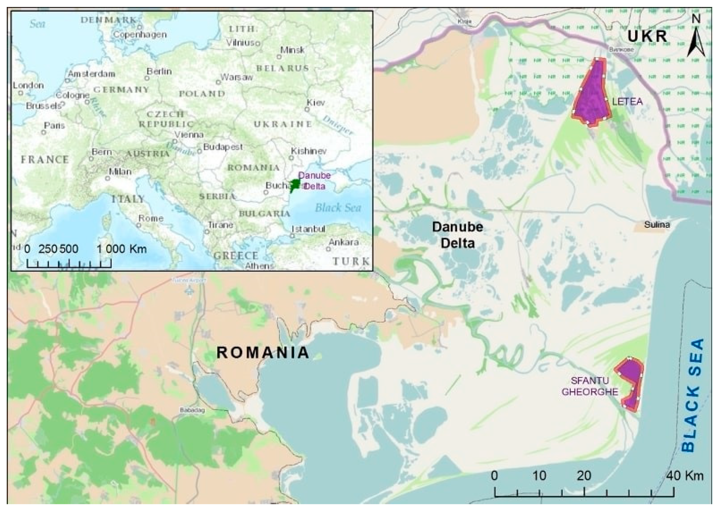
Figure 1. Sampling sites in the Danube Delta (Letea and Sfantu Georghe marked in purple, a total of 199 samples were collected from wild sedentary and migratory birds).
Antibiotics 10 00333 g001
Figure 2. Vibrio spp. strains isolated from wild birds captured in Danube Delta Biosphere, Romania.
Antibiotics 10 00333 g002
Figure 3. Incidence of antimicrobial resistance (represented by classes of antibiotics) in Vibrio spp. isolates from wild birds.
Antibiotics 10 00333 g003
Table 1. Vibrio spp. isolated from cloacal and oral specimens collected (n = 170) from wild birds in Danube Delta Biosphere, Romania (samples harvested from migratory and sedentary birds, isolated and characterized bacterial strains isolated by microbiology and molecular biology techniques).
SpeciesHabitat TypeNo. Captured BirdsPositive Samples for Vibrio spp.Bacterial IsolatePCR
ompWtoxR
Squacco heron
(Ardeola ralloides)
Migratory22V. cholerae++
V. alginolyticus-+
V. parahaemolyticus--
Common greenshank
(Tringa nebularia)
Migratory22V. fluvialis-+
V. alginolyticus--
Reed bunting
(Emberiza schoeniclus)
Migratory21V. metschnikovii--
V. fluvialis--
V. parahaemolyticus--
Song thrush
(Turdus philomelos)
Migratory21V. cholerae+-
V. alginolyticus--
V. mimicus--
Eurasian hobby
(Falco subbuteo)
Migratory22V. cholerae++
V. fluvialis--
V. alginolyticus--
V. mimicus--
Wood sandpiper
(Tringa glareola)
Migratory22V. mimicus--
V. vulnificus--
Black-crowned night heron
(Nycticorax nycticorax)
Migratory22V. vulnificus--
V. parahaemolyticus--
Common snipe
(Gallinago gallinago)
Migratory32V. cholerae++
V. mimicus--
Eurasian sparrowhawk
(Accipiter nisus)
Migratory21V. cholerae+-
V. parahaemolyticus--
Common chaffinch
(Fringilla coelebs)
Migratory22V. mimicus--
Eurasian teal
(Anas crecca)
Migratory21V. mimicus--
V. vulnificus--
--
Eurasian tree sparrow
(Passer montanus)
Sedentary22V. cholerae+-
V. fluvialis--
Lesser whitethroat
(Sylvia curucca)
Migratory11V. metschnikovii--
V. mimicus--
V. vulnificus--
Red-backed shrike
(Lanius collurio)
Migratory11V. fluvialis--
V. alginolyticus--
V. metschnikovii--
V. parahaemolyticus--
Icterine warbler
(Hippolais icterina)
Migratory32V. parahaemolyticus--
Red-footed falcon
(Falco vespertinus)
Migratory22V. cholerae+-
V. fluvialis--
V. alginolyticus--
V. metschnikovii--
Garden warbler
(Sylvia borin)
Migratory43V. vulnificus--
V. parahaemolyticus--
Common whitethroat
(Sylvia communis)
Migratory54V. alginolyticus--
V. metschnikovii--
Common kingfisher
(Alcedo atthis)
Migratory11V. cholerae--
V. fluvialis--
V. metschnikovii--
V. mimicus--
Eurasian blackcap
(Sylvia atricapilla)
Migratory11V. parahaemolyticus--
Barred warbler
(Sylvia nisoria)
Migratory31V. metschnikovii--
V. vulnificus--
Black-and-white magpie
(Pica pica)
Sedentary52V. cholerae++
V. metschnikovii-
V. parahaemolyticus--
Western jackdaw
(Corvus monedula)
Sedentary42V. cholerae--
V. fluvialis--
Hooded crow
(Corvus corone cornix)
Sedentary73V. metschnikovii--
V. mimicus--
V. vulnificus--
V. parahaemolyticus--
Eurasian tree sparrow
(Passer montanus)
Sedentary42V. cholerae+-
V. fluvialis--
Eurasian blue tit
(Parus caeruleus)
Sedentary22V. metschnikovii--
V. mimicus--
V. vulnificus--
V. parahaemolyticus--
Long tailed tit
(Aegithalos caudatus)
Sedentary20---
Bearded reedling
(Panurus biarmicus)
Sedentary21V. alginolyticus--
V. metschnikovii--
V. mimicus--
Common chaffinch
(Fringilla coelebs)
Sedentary32V. cholerae--
V. mimicus--
Hawfinch
(Coccothraustes coccothraustes)
Sedentary85V. alginolyticus--
V. metschnikovii--
V. mimicus--
Great tit
(Parus major)
Sedentary22V. fluvialis--
V. mimicus--
Table 2. Multiple antibiotic resistance (MAR) index of antibiotics against isolated Vibrio spp.
Antimicrobial ClassDrugTotal Number of Resistant Strains Isolated from Migratory Birds, by AntimicrobialTotal Number of Resistant Strains Isolated from Sedentary Birds, by AntimicrobialMAR Index for the Tested Antibiotics
PenicillinsPenicillin48180.8
Ampicillin37170.6
MacrolidesErythromycin47200.8
CloramphenicolChloramphenicol1210.1
AminoglycosidesAmikacin45170.7
Kanamycin1170.2
TetracyclinesOxytetracycline1000.1
Tetracycline820.1
QuinolonesEnrofloxacin1140.1
Marbofloxacin1050.1
Ciprofloxacin960.1
Table 3. Resistance types observed in the isolated Vibrio spp.
Number of Antibiotic Classes to Which Isolated Strains Showed ResistanceNumber/% of Resistant StrainsResistance Type
11 (1.32)-
25 (6.58)MAR
335 (46.05)MDR
421 (27.63)XDR
510 (13.16)XDR
64 (5.26)XDR
MAR-multiple antibiotic resistance, MDR-multidrug resistance, XDR-extensively drug resistance.
Publisher’s Note: MDPI stays neutral with regard to jurisdictional claims in published maps and institutional affiliations.