Article

blaNDM and mcr-1 to mcr-5 Gene Distribution Characteristics in Gut Specimens from Different Regions of China

1
Department of Epidemiology and Health Statistics, The School of Public Health of Qingdao University, Qingdao 266021, China
2
State Key Laboratory of Infectious Disease Prevention and Control, National Institute for Communicable Disease Control and Prevention, Chinese Center for Disease Control and Prevention, Beijing 102206, China
*
Authors to whom correspondence should be addressed.
These authors contributed equally to this article.
Antibiotics 2021, 10(3), 233; https://doi.org/10.3390/antibiotics10030233
Submission received: 23 January 2021 / Revised: 19 February 2021 / Accepted: 23 February 2021 / Published: 25 February 2021
(This article belongs to the Section Antibiotics Use and Antimicrobial Stewardship)

Abstract

:
Antibiotic resistance has become a global public health concern. To determine the distribution characteristics of mcr and blaNDM in China, gene screening was conducted directly from gut specimens sourced from livestock and poultry, poultry environments, human diarrhea patients, and wild animals from 10 regions, between 2010–2020. The positive rate was 5.09% (356/6991) for mcr and 0.41% (29/6991) for blaNDM, as detected in gut specimens from seven regions, throughout 2010 to 2019, but not detected in 2020. The detection rate of mcr showed significant differences among various sources: livestock and poultry (14.81%) > diarrhea patients (1.43%) > wild animals (0.36%). The detection rate of blaNDM was also higher in livestock and poultry (0.88%) than in diarrhea patients (0.17%), and this was undetected in wildlife. This is consistent with the relatively high detection rate of multiple mcr genotypes in livestock and poultry. All instances of coexistence of the mcr-1 and blaNDM genes, as well as coexistence of mcr genotypes within single specimens, and most new mcr subtypes came from livestock, and poultry environments. Our study indicates that the emergence of mcr and blaNDM genes in China is closely related to the selective pressure of carbapenem and polymyxin. The gene-based strategy is proposed to identify more resistance genes of concern, possibly providing guidance for the prevention and control of antimicrobial resistance dissemination.

1. Introduction

Antibiotic resistance has become a major global public health concern in the 21st century. Carbapenems and polymyxins are among the last-resort antibiotics for defending against Gram-negative bacterial infections [1]. Among the various mechanisms, the blaNDM (New Delhi metallo-β-lactamase) gene and the mcr (mobile colistin resistance) gene, conferring resistance to carbapenem and polymyxin respectively, exhibit cross-species and cross-region transmission [2]. The blaNDM-1 gene was first discovered in patients in 2009 [3], and 29 genotypes have since been identified [4,5,6,7,8,9]. Carbapenems are mainly used to treat human respiratory infections, and resistant bacterial strains often exhibit multidrug and broad-spectrum drug resistance [10]. Although carbapenem usage has not been approved for use in the breeding industry in China, new blaNDM subtypes and an epidemic of resistant bacterial strains have appeared in livestock and poultry [11,12]. The mcr-1 gene was first discovered in pigs in 2016 [13] and 10 genotypes and multiple subtypes have since been found [14,15,16,17,18]. Polymyxins were once widely used as feed additives and for disease prevention in livestock and poultry in China, but have been banned as animal growth promoters in China since 2017. The colistin-resistant Escherichia coli (CREC) and mcr-1 positive Escherichia coli (MCRPEC) seem to have declined [19], however, our results from multiple sources did not show a decrease [20]. The mcr-positive or polymyxin-resistant strains are not only found in livestock, humans, and environments [21,22,23], but also in wild animals such as macaques and migratory birds [24,25,26].
Antibiotic overuse [27] and the emergence of drug resistance are linked [28]. China has become the largest consumer of antibiotics due to antibiotic use in the livestock and poultry industries [29]. Our previous work showed that most of the mcr or blaNDM positive strains were from normal flora, identified in isolates from wildlife, patients, livestock and poultry, and environmental specimens [20], with these appearing to be increased by antibiotic selective pressures. However, normal flora with resistance may not always be detected in this way due to the limited sensitivity of isolation techniques. This study was further conducted to determine the broader picture of mcr and blaNDM in China, in the context of these genes being carried by bacteria selected by antibiotic pressures and the normal flora with resistance, providing guidance for drug resistance control measures.

2. Results

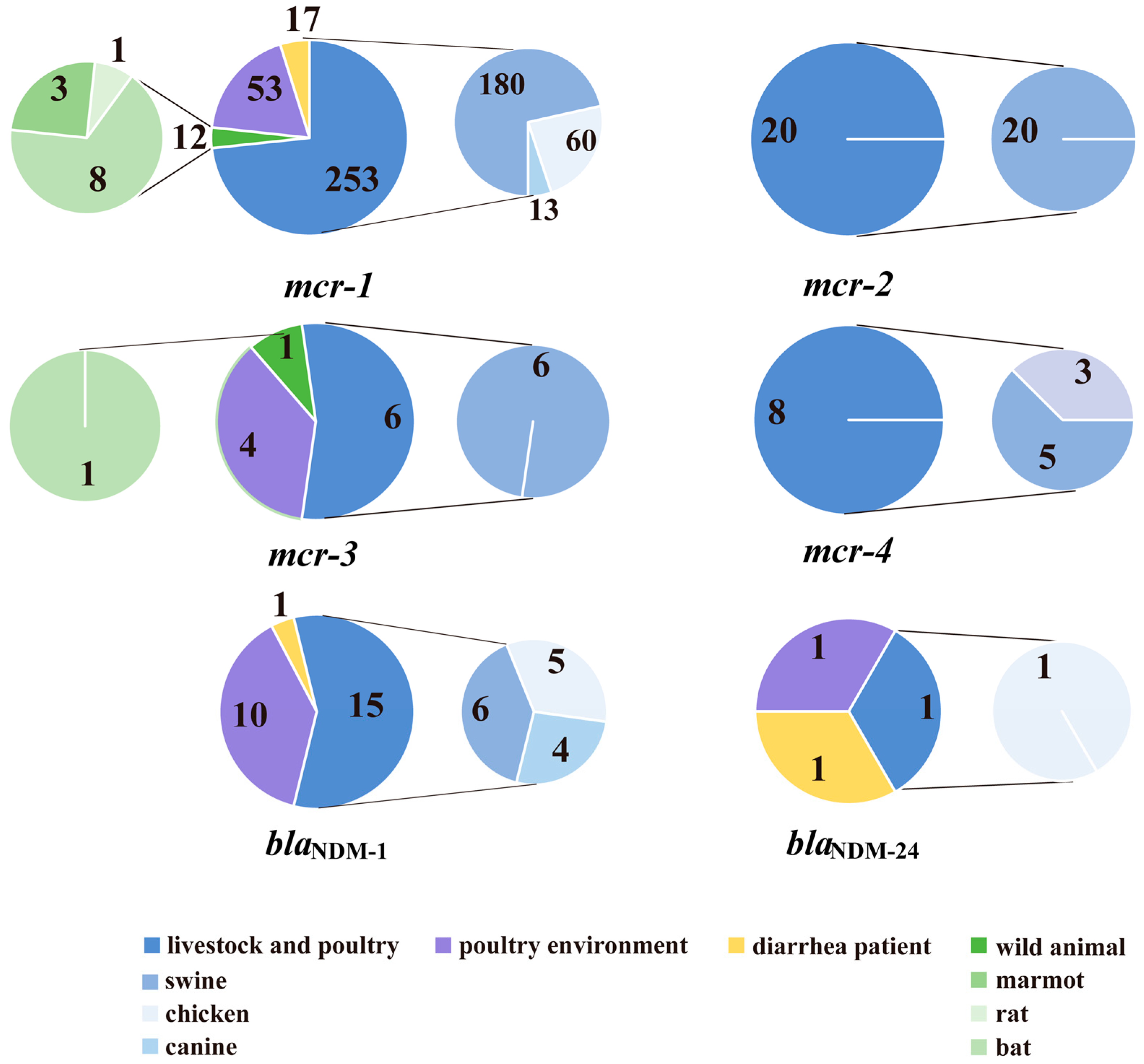
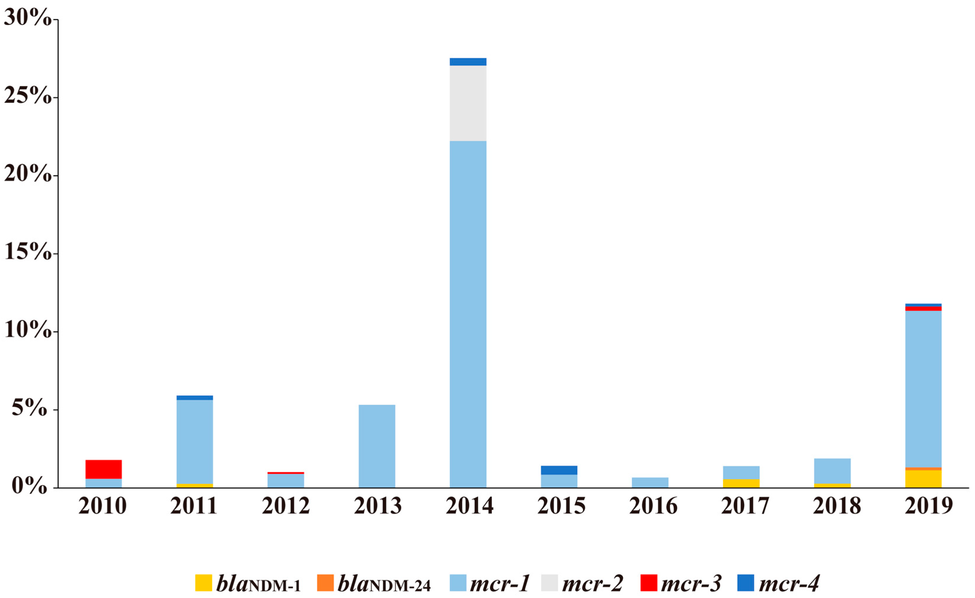
2.1. Distributions of blaNDM and mcr

Among the 6991 gut specimens collected, 0.41% were positive for blaNDM (29/6991) and 5.09% were positive for mcr (356/6991) (Table 1, detailed background in Table S1). For the blaNDM gene, the detection rate of blaNDM-1 (0.37%, 26/6991) was the highest, followed by blaNDM-24 (0.04%, 3/6991). For the mcr gene, the detection rate of mcr-1 (4.79%, 335/6991) was highest, followed by mcr-2 (0.29%, 20/6991), mcr-3 (0.16%, 11/6991), and mcr-4 (0.11%, 8/6991). No gut specimen was positive for mcr-5.
The majority of blaNDM and mcr genes were found in livestock and poultry. The positive rates of the blaNDM gene in livestock and poultry, poultry environments, and diarrhea patients were 0.88 (16/1823), 3.14 (11/350), and 0.17% (2/1186), respectively; it was not detected in wild animals. The blaNDM gene rates between various sources showed significant differences (Fisher exact test χ2 = 22.66, p < 0.05). The positive rates of the mcr gene in livestock and poultry, poultry environments, diarrhea patients, and wild animals were 14.81 (270/1823), 16.00 (56/350), 1.43 (17/1186), and 0.36% (13/3632), respectively. The mcr gene rates between various sources showed significant differences (Pearson χ2 = 643.72, p < 0.05). The detection rate of blaNDM and mcr within single gut specimens was 0.38 (7/1823) and 0.86% (3/350) for livestock and poultry, and poultry environments, respectively. Among the positive gut specimens from livestock and poultry, intensively reared animals (swine, chickens and fish) accounted for a greater proportion than non-intensively reared breeding animals (yak, goats and canines) (Figure 1). The positive rates of these two kinds of reared animals were 1.02 (12/1179) and 0.62% (4/644) for the blaNDM gene, 21.80 (257/1179) and 2.02% (13/644) for the mcr gene. In wild animals, the mcr gene was detected in various species ranging from marmots and rats to bats (Figure 1).
Regarding gut specimen collection year, blaNDM-positive gut specimens were detected in 2011 (0.27%, 1/372), 2017 (0.56%, 2/356), 2018 (0.28%, 5/1797), and 2019 (1.33%, 21/1575), and mcr-positive gut specimens were detected throughout 2010–2019, with detection rates of 1.80 (9/501), 5.38 (20/372), 1.01 (9/889), 5.33 (17/319), 22.95 (95/414), 1.41 (5/354), 0.67 (2/299), 0.84 (3/356), 1.61 (29/1797), and 10.60% (167/1575). More specifically, blaNDM-1 was found in 2011, 2017, 2018, and 2019, blaNDM-24 was found in 2019, mcr-1 was found throughout 2010–2019 (with detection rates peaking in 2014 and 2019), mcr-2 was found in 2014, mcr-3 was found in 2010, 2012, and 2019, and mcr-4 was found in 2011, 2014, 2015, and 2019. New mcr subtypes were found in 2010 (mcr-3.32), 2012 (mcr-3.31), 2014 (mcr-1.30, mcr-2.4, mcr-2.5, mcr-2.6, mcr-2.7), and 2018 (mcr-1.29) (Figure 2).

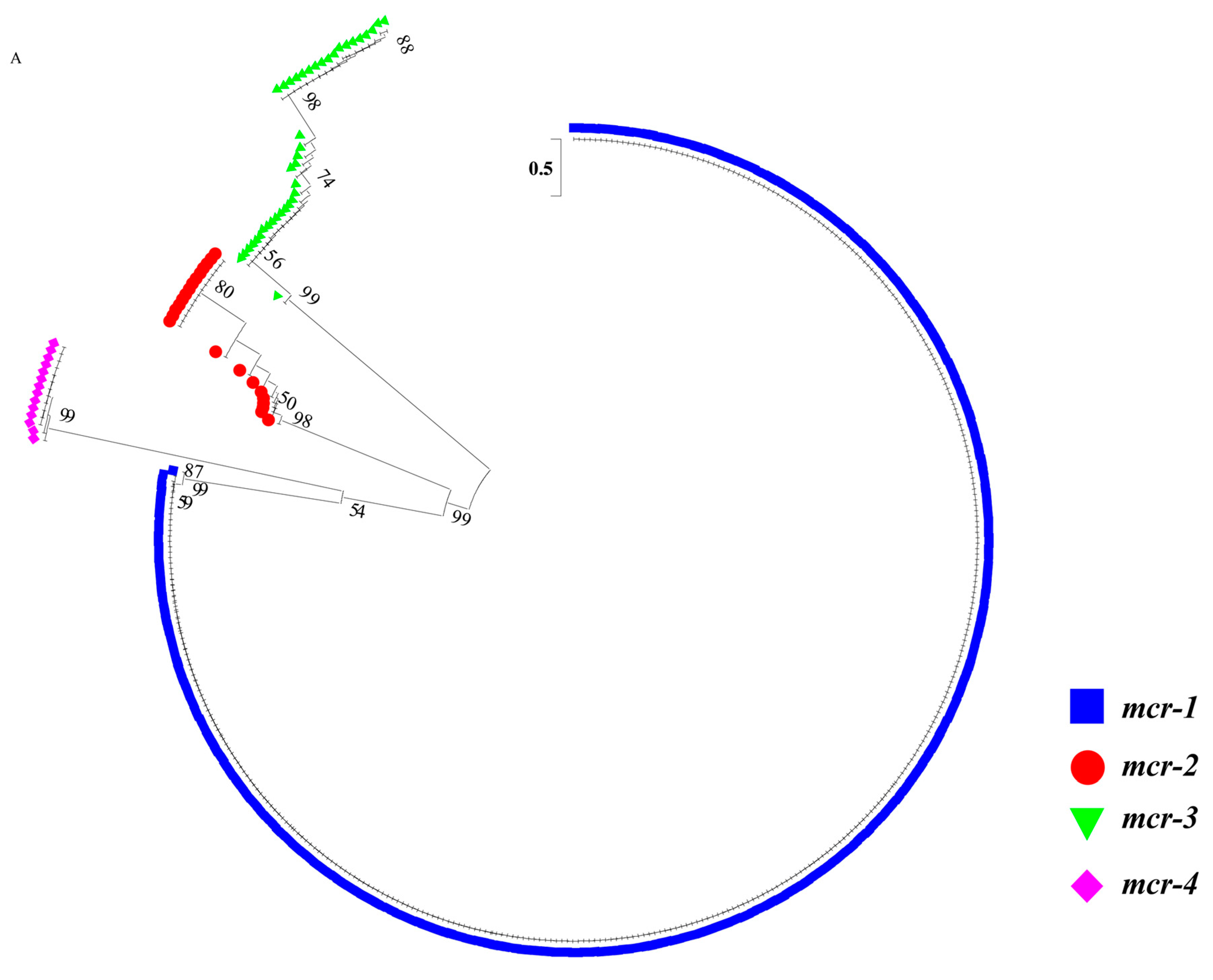
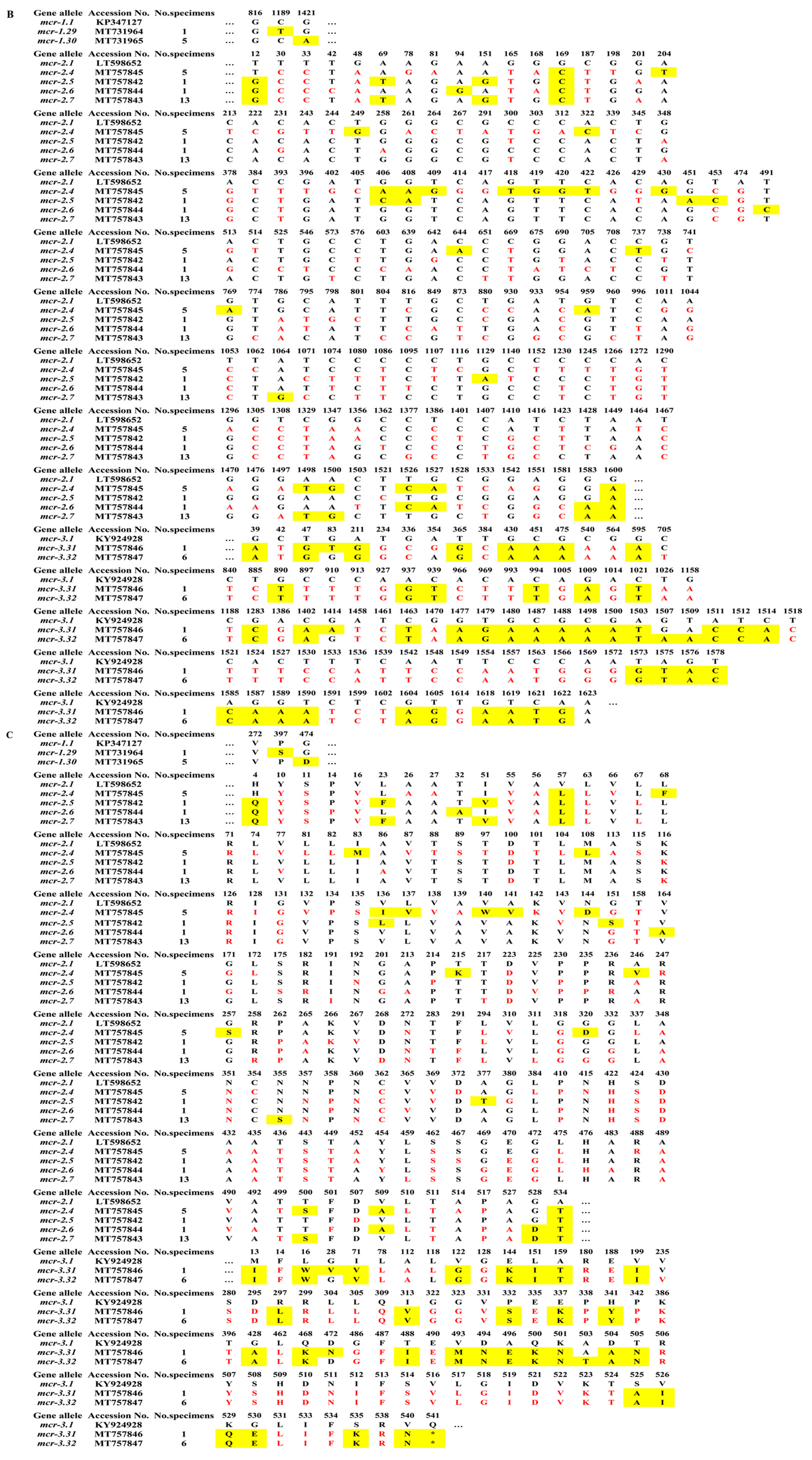
2.2. Sequence Analysis of blaNDM and mcr

Among the blaNDM-positive gut specimens, no new mutations were found. Among these gut specimens, 89.66% (26/29) were identical to NDM-1 (Accession No.: WP_004201164.1) and 10.34% (3/29) were identical to NDM-24 (Accession No.: WP_111672913.1). The amino acid (aa) identity between NDM-1 and NDM-24 was 99.8%. Among the mcr-positive gut specimens, 8.15% (29/356) involved new subtypes of mcr-1 to mcr-3. No new mutations were found in mcr-4, which all belonged to MCR-4.3 (Accession No.: WP_011638903.1). A cluster analysis of the mcr genotypes is shown in Figure 3.
Among the mcr-1-positive gut specimens, 98.21% (329/335) were identical to MCR-1.1 (Accession No.: WP_049589868.1). The remaining six gut specimens formed two new subtypes, all with a sense mutation (Figure 3). The aa identities of each of the new subtypes compared with MCR-1.1 were: 99.8 (MCR-1.29) and 99.8% (MCR-1.30). The amino acid mutation of MCR-1.29 is P397S, and MCR-1.30 is G474D.
All 20 mcr-2 positive gut specimens in this study were new subtypes. Compared to MCR-2.1 (Accession No.: WP_065419574.1), there were numerous sense and nonsense mutations (Figure 3). Compared with MCR-2.1, the aa identity of MCR-2.4 was 97.0%, and of the 3% mutations, 81.81% were nonsense and 18.18% were sense; the aa identity of MCR-2.5 was 98.5%, and of the 1.5% mutations, 84.91% were nonsense and 15.09% were sense; the aa identity of MCR-2.6 was 98.7%, and of the 1.3% mutations, 83.39% were nonsense and 10.61% were sense; the aa identity of MCR-2.7 was 98.5%, and of the 1.5% mutations, 85.96% were nonsense and 14.04% were sense.
Among the 11 mcr-3 positive gut specimens, two were consistent with MCR-3.18 (Accession No.: WP_111273847.1) and two were identical to MCR-3.3 (Accession No.: WP_099982814.1). The remaining seven gut specimens formed two new subtypes, both involving the premature stop codon that was one codon before the expected stop codon (Figure 3). The aa identities of each of the new subtype compared with MCR-3.1 (Accession No.: WP_039026394.1) were: 94.4% (MCR-3.31, sense mutation: 41.03%, nonsense mutation: 58.97%) and 94.6% (MCR-3.32, sense mutation: 40.26%, nonsense mutation: 59.74%). Both MCR-3.31 and MCR-3.32 had a premature stop codon at aa 541 out of 542.

2.3. Distribution of Coexisting Genes/Genotypes and New Subtypes

The gut specimens with coexisting genes/genotypes or new subtypes were mostly from livestock and poultry and poultry environment, with only the new subtype mcr-3.31 being derived from wild animals (Table 2). Ten gut specimens harbored both blaNDM and mcr-1, and 18 gut specimens harbored two mcr genotypes. The coexistence of blaNDM and mcr-1 within a single gut specimen was only observed in Anhui. The coexistence of mcr genotypes a within single gut specimen was mostly found in Guangxi. The new mcr subtypes were from Guangxi, Anhui and Yunnan.

3. Discussion

Antibiotic resistance may be a survival strategy for bacteria, with antibiotics triggering specific bacterial responses [30,31]. This study shows that antibiotic selective pressure might be reflected by resistance gene pools of various sources. In combination with the findings of our previous study, this shows that the emergence of polymyxin and carbapenem resistance strains in China is closely related to the selective pressure of antibiotics. The mcr or blaNDM strains originating from livestock and poultry, patients, and wildlife, are mainly non-pathogenic organisms [20], which is consistent with findings from studied conducted in 47 countries across six continents with mcr-positive strains [32], which showed a tendency to be increased under antibiotic selection pressures. Due to the limited sensitivity of isolation, some normal flora with resistance may not be detected. To further determine the distribution of mcr and blaNDM from different sources—carried by the bacteria selected by antibiotic selective pressures and normal flora with resistance—this study was conducted based on gut specimen detection strategy and a One Health approach. Overall, the positive rate of the mcr gene was much higher than that of the blaNDM gene for each specimen source. This is in accordance with positive-strain isolation [20]. The positive rates of the mcr gene showed significant differences among sources: livestock and poultry (14.81%) > diarrhea patients (1.43%) > wild animals (0.36%) (Table 1), consistent with the relative isolation rates of polymyxin-resistant strains among these sources [20]. Though polymyxin-resistant strains had not been isolated in wildlife, the mcr gene was detected. Livestock and poultry (0.88%) were found to contain the blaNDM gene more frequently than diarrhea patients (0.17%), but this gene was not detected in wildlife (Table 1). Carbapenem-resistant strains were also not isolated from wildlife in a previous study [20]. Compared with other sources, no polymyxin- or carbapenem-resistant strains [20], lower rates of mcr and blaNDM genes (Table 1) and less mcr genotypes (Table 2) were found in wildlife samples, which supports the hypothesis that wild animals are a net sink rather than a source of clinically relevant drug resistance [33]. The phenotypic diversity of drug resistant strains in wildlife is also low [33]. Since wild animals have less chance of being exposed to antibiotics, the emergence of resistance genes possibly reflects the resistance genes carried by normal flora. Similarly, Salmonella enterica—isolated from diarrhea patients and asymptomatic individuals—showed equal carriage of mcr carriers, suggesting the mcr gene is carried by normal flora [34]. In this study, the detection rates of mcr and blaNDM in diarrhea patients were far lower than in livestock and poultry, and higher than in wild animals (Table 2). This is in accordance with the relatively low use of polymyxin and carbapenem in this population.
The gene pools of mcr or blaNDM reflect resistance genes carried by normal flora when antibiotic pressure is low, and genes carried by the bacteria selected by antibiotic pressure. When the pressure is relatively high, such as in livestock, poultry and humans, the relative levels of the mcr and blaNDM genes—to a certain extent—possibly reflects the antibiotic selective pressure. In particular, in livestock and poultry, there higher rates of the blaNDM and mcr gene (Table 1) and more mcr genotypes were found (Table 2 and Figure 3). Polymyxins are often used as therapeutic drugs and feed additives for animals, and they are used more frequently for farmed animals in China [29], where the highest number of mcr-positive strains was reported [32]. During the intensive feeding period, antibiotics are required for animal treatment and disease prevention, which involves large doses and long-term use [35]. We found that the positive rate of the mcr gene was much higher in intensively reared animals (21.80%, 257/1179, swine, chickens, etc.) than in non-intensively reared breeding animals (2.02%, 13/644, yak, goats, canine, etc.) (Figure 1). The ban of polymyxin use as an animal growth promoter in 2017 seems to have reduced CREC and MCRPEC [20,36]. However, the observation that the mcr detection rates peaked in 2019 in this study (10.60%, 167/1575) (Figure 2), is consistent with the notion that mcr-1 isolates successively recovered from 2017 to 2019, which indicates the possibility that polymyxin resistance still exists in livestock and poultry. Carbapenem drug-resistant strains have appeared and are prevalent in poultry and livestock. New genotypes of the blaNDM gene have been found in livestock and poultry-derived strains around the world [11,28]. Firstly, carbapenems might be applied when treating animal diseases. Secondly, their use in humans pollutes the environment and results in indirect exposure of animals to the drug. Last but not least, bacteria with the blaNDM gene may exist in normal gut flora [37]. In summary, the emergence of drug resistance genes is due to the selective pressure caused by the overuse of antibiotics. The strategy of gene detection can be used for resistance gene profiles and supervision.
In this study, livestock and poultry were not only the main source of the mcr and blaNDM gene pool (Table 1), but they were also sources of mcr-1 and blaNDM co-harbored genes. Additionally, livestock and poultry were the source of multiple mcr genotypes within single gut specimens (Table 2). Similar findings were not shown in diarrhea patients or wild animals. In general, the coexistence of the mcr-1 and blaNDM genes was only found in Anhui, and the coexistence of mcr genotypes mostly came from Guangxi, indicating that livestock and poultry in some regions may be exposed to higher or more complex antibiotic selective pressures. Considering that no strain carrying both the mcr and blaNDM genes had been isolated in the previous study [20], a past and present coexistence of the mcr-1 and blaNDM genes within one gut specimen is more likely to come from different clones (e.g., one clone harboring mcr-1, other clone harboring blaNDM). It is also possible that a single clone carried both genes. In either case, the drug resistance conferred by mcr and blaNDM genes may be transmitted from livestock and poultry to humans, possibly even resulting in the emergence of polymyxin and carbapenem resistant strains. Recently, mcr-1 and blaNDM coexistence was also reported in the United States, Venezuela, and Japan [38,39,40], which reduces treatment options for multidrug-resistant bacterial infections and increases the incidence and mortality of the infections, leading to stricter antibiotic controls. It is necessary to strengthen antimicrobial resistance surveillance in livestock and poultry.
This study revealed the gene distribution of mcr and blaNDM in livestock and poultry, diarrhea patients and wild animals, demonstrating that relative level of the resistance genes may reflect the selective pressure of antibiotic exposure of various hosts, which is expected to become a strategy of antibiotic usage oversight. Potential antimicrobial usage of colistin, and others, plays a role in the enrichment of antimicrobial resistance genes in gut specimens, which are needed to further support culture-based data. Compared with the culture-based strategy, the gene-based strategy is more sensitive. The positive rates of gene detection among various gut specimens were about two to three times those of isolation rates [20]. On the other hand, bacterial culture and genetic background information is not available through the gene-based strategy. The fact that more positive specimens found by gene detection than culture detection, may come from the normal flora with resistance that cannot always be isolated, and gene positive results do not always equate to phenotype positive results [25]. Additionally, searching for new variants is limited by the current PCR method. Although this method is improving over time [34,41], it is based on known genotype data which often cannot be used to discover an unknown variant. The gene detection method could be developed into a strategy based on metagenomic sequencing [42], identifying more concerned drug resistance genes and genetic information coming from various sources, and providing guidance for the prevention and control of drug-resistant bacteria and for supervision of antibiotic usage.

4. Materials and Methods

4.1. Gut Specimen Sources

Nucleic acid samples were obtained from 6991 gut specimens from livestock and poultry (26.08%, 1823/6991) including swine, chickens, canine, yak, goats, etc., poultry environments (5.01%, 350/6991), including breeding or slaughter environment, human diarrhea patients (16.96%, 1186/6991), and wild animals (51.95%, 3632/6991), including bats, marmots, rats, etc. (Table 1). The gut specimens were obtained in 2010–2020 from 10 regions of China (Beijing, Anhui, Gansu, Yunnan, Guangxi, Guizhou, Ningxia, Inner Mongolia, Qinghai, and Zhejiang) (Figure S1), and they were retrospectively screened for the target genes. Gut specimen types of this study included human feces, animal anal swab, feces, intestinal content/swab or oral-pharyngeal swab, and poultry environment specimens related to gut environment, including drinking water, cage swab, depilator swab, cleaning sewage, chopping board swab, and soil. Unified protocols for specimen collection, transportation, and process were applied by professionals from local CDC (Center for Disease Control and Prevention) facilities, Institutes for Endemic Disease Prevention and Control, and hospitals. Specimens were collected and transported in Cary–Blair Transport Medium, processed and nucleic acids extracted using a genomic extraction kit (TIANamp Bacteria DNA Kit, Beijing, China). The nucleic acid samples were frozen for storage.

4.2. blaNDM and mcr-1 to mcr-5 Screening of Gut Specimens and Sequence Analysis

The target genes blaNDM and mcr were screened for, sequenced, and aligned with reference sequences from the National Center for Biotechnology Information (NCBI) database. The screening primers (Table S2) for blaNDM and mcr (mcr-1 to mcr-5) were previously described [20,41]. The original amplification of mcr-1 to mcr-5 involved multiplex PCR, but single PCR was conducted in this study. The CDS (coding sequences) of gut specimens with mutations in the screening sequences were further amplified, cloned (Transgene, Beijing, China), and sequenced. The number of PCR cycles for gene screening is 25 to 30, for CDS amplification it is 30. The PCR was performed using a 20 μL volume containing 10 μL Premix Taq version 2.0 (Takara, Beijing, China), 8 μL ultrapure distilled water, 0.5 μL (10 μM) of each forward and reverse primer and 1 μL of DNA template. The amplified products were detected using gel electrophoresis and sequenced in both directions using an Applied Biosystems 3730xl DNA Analyzer (Tsingke Biological Technology, Beijing, China). Phylogenic tree was constructed based on CDS sequences of mcr gene including sequences of this study and reference sequences (mcr-1 to mcr-4) and sequence analysis of mcr and MCR were conducted (Figure 3).

4.3. Statistical Analysis

Pearson’s chi-square test (theoretical frequency T ≥ 5) was used to compare positive rates among different sources. As one theoretical frequency is 1 < T < 5, the Fisher exact test was also applied when comparing rates among different sources. Bonferroni correction was used to compare the positive rates between two sources. p < 0.05 was considered statistically significant. The statistical analysis was conducted by SPSS Version 19.0.

4.4. Nucleotide Sequence Accession Numbers

The CDSs of the following new subtypes (mcr-1 to mcr-3) were deposited in the GenBank database: mcr-1.29 (GenBank: MT731964), mcr-1.30 (GenBank: MT731965), mcr-2.4 (GenBank: MT757845), mcr-2.5 (GenBank: MT757842), mcr-2.6 (GenBank: MT757844), mcr-2.7 (GenBank: MT757843), mcr-3.31 (GenBank: MT757846), and mcr-3.32 (GenBank: MT757847).

4.5. Ethics Statement

The study was approved by the ethics committee of the National Institute for Communicable Disease Control and Prevention of the Chinese Center for Disease Control and Prevention (IACUC Issue No. 2020-008). Verbal consent was obtained from the included diarrhea patients.

5. Conclusions

This study is first to determine the distribution characteristics of blaNDM and mcr genes from various sources of China. The positive rate of the mcr gene was much higher than that of the blaNDM gene for all sources, from highest to lowest was: livestock and poultry, diarrhea patients, and wild animals. The mcr or blaNDM gene pool of certain source reflect the resistance gene carried by normal flora when antibiotic pressure is low, and genes carried by the bacteria selected by antibiotic pressure. Livestock and poultry were not only the main source of the mcr and blaNDM gene pool, but also the source of co-harbored mcr-1 and blaNDM genes. The antimicrobial resistance surveillance in livestock and poultry needs to be strengthened. In conclusion, the study demonstrated that the selective pressure of antibiotic exposure of various hosts maybe reflected by relative level of the resistance genes, which is expected to become a strategy of antibiotic usage oversight.

Supplementary Materials

The following are available online at https://www.mdpi.com/2079-6382/10/3/233/s1, Table S1. Background and genotypes of mcr or blaNDM positive gut specimens. Table S2. Primers for blaNDM and mcr screening and CDS amplification. Figure S1. Geographic distribution and frequency of the specimens collected from 2010 to 2020. The color shades represent different years and the pie area reflects the number of samples collected in each region. Abbreviations: BJ = Beijing, AH = Anhui, GS = Gansu, YN = Yunnan, GX = Guangxi, GZ = Guizhou, NX = Ningxia, IM = Inner Mongolia, QH = Qinghai, ZJ = Zhejiang.

Author Contributions

Conceptualization, H.J. and X.W.; data curation, D.L., R.D., R.F., H.M., J.L., M.X., Z.H., S.Q. and J.Y.; formal analysis, D.L., R.D., R.F. and Z.W.; funding acquisition, H.J. and X.W.; investigation, D.L., R.D., R.F., H.M., M.X., Z.H., H.J., Z.W. and X.W.; methodology, H.J. and X.W.; project administration, H.J. and X.W.; resources, H.J. and X.W.; supervision, H.J., Z.W. and X.W.; validation, H.J. and X.W.; visualization, D.L. and R.D.; Writing—original draft, D.L., R.D., R.F., H.M., J.L., S.Q., H.J., Z.W. and X.W.; writing—review and editing, D.L., R.D., R.F., H.M., J.L., M.X., Z.H., S.Q., J.Y., H.J., Z.W. and X.W. Conceptualization, H.J. and X.W.; data curation, D.L., R.D., R.F., H.M., J.L., M.X., Z.H., S.Q. and J.Y.; formal analysis, D.L., R.D., R.F. and Z.W.; funding acquisition, H.J. and X.W.; investigation, D.L., R.D., R.F., H.M., M.X., Z.H., H.J., Z.W. and X.W.; methodology, H.J. and X.W.; project administration, H.J. and X.W.; resources, H.J. and X.W.; supervision, H.J., Z.W. and X.W.; validation, H.J. and X.W.; visualization, D.L. and R.D.; writing—original draft, D.L., R.D., R.F., H.M., J.L., S.Q., H.J., Z.W. and X.W.; writing—review and editing, D.L., R.D., R.F., H.M., J.L., M.X., Z.H., S.Q., J.Y., H.J., Z.W. and X.W. All authors have read and agreed to the published version of the manuscript.

Funding

This work was supported by National Science and Technology Major Project (2018ZX10713-003-002 and 2018ZX10713-001-002).

Institutional Review Board Statement

The study was conducted according to the guidelines of the Declaration of Helsinki, and approved by the National Institute for Communicable Disease Control and Prevention of the Chinese Center for Disease Control and Prevention (IACUC Issue No. 2020-008).

Informed Consent Statement

Not applicable.

Acknowledgments

We thank the Charlesworth Group’s author services for their critical editing and helpful comments regarding our manuscript (Order#: 74493).

Conflicts of Interest

The authors declare that the research was conducted in the absence of any commercial or financial relationships that could be construed as a potential conflict of interest.

References

  1. Nordmann, P.; Dortet, L.; Poirel, L. Carbapenem resistance in Enterobacteriaceae: Here is the storm! Trends Mol. Med. 2012, 18, 263–272. [Google Scholar] [CrossRef] [PubMed]
  2. El-Sayed Ahmed, M.A.E.; Zhong, L.L.; Shen, C.; Yang, Y.; Doi, Y.; Tian, G.B. Colistin and its role in the Era of antibiotic resistance: An extended review (2000–2019). Emerg. Microbes Infect. 2020, 9, 868–885. [Google Scholar] [CrossRef] [PubMed] [Green Version]
  3. Kumarasamy, K.K.; Toleman, M.A.; Walsh, T.R.; Bagaria, J.; Butt, F.; Balakrishnan, R.; Chaudhary, U.; Doumith, M.; Giske, C.G.; Irfan, S.; et al. Emergence of a new antibiotic resistance mechanism in India, Pakistan, and the UK: A molecular, biological, and epidemiological study. Lancet Infect. Dis. 2010, 10, 597–602. [Google Scholar] [CrossRef]
  4. Kaase, M.; Nordmann, P.; Wichelhaus, T.A.; Gatermann, S.G.; Bonnin, R.A.; Poirel, L. NDM-2 carbapenemase in Acinetobacter baumannii from Egypt. J. Antimicrob. Chemother. 2011, 66, 1260–1262. [Google Scholar] [CrossRef] [Green Version]
  5. Cuzon, G.; Bonnin, R.A.; Nordmann, P. First identification of novel NDM carbapenemase, NDM-7, in Escherichia coli in France. PLoS ONE 2013, 8, e61322. [Google Scholar] [CrossRef] [PubMed]
  6. Tada, T.; Miyoshi-Akiyama, T.; Dahal, R.K.; Sah, M.K.; Ohara, H.; Kirikae, T.; Pokhrel, B.M. NDM-8 metallo-β-lactamase in a multidrug-resistant Escherichia coli strain isolated in Nepal. Antimicrob. Agents Chemother. 2013, 57, 2394–2396. [Google Scholar] [CrossRef] [Green Version]
  7. Zou, D.; Huang, Y.; Zhao, X.; Liu, W.; Dong, D.; Li, H.; Wang, X.; Huang, S.; Wei, X.; Yan, X.; et al. A novel New Delhi metallo-beta-lactamase variant, NDM-14, isolated in a Chinese Hospital possesses increased enzymatic activity against carbapenems. Antimicrob. Agents Chemother. 2015, 59, 2450–2453. [Google Scholar] [CrossRef] [Green Version]
  8. Shrestha, B.; Tada, T.; Miyoshi-Akiyama, T.; Shimada, K.; Ohara, H.; Kirikae, T.; Pokhrel, B.M. Identification of a novel NDM variant, NDM-13, from a multidrug-resistant Escherichia coli clinical isolate in Nepal. Antimicrob. Agents Chemother. 2015, 59, 5847–5850. [Google Scholar] [CrossRef] [PubMed] [Green Version]
  9. Liu, L.; Feng, Y.; McNally, A.; Zong, Z. blaNDM-21, a new variant of blaNDM in an Escherichia coli clinical isolate carrying blaCTX-M-55 and rmtB. J. Antimicrob. Chemother. 2018, 73, 2336–2339. [Google Scholar] [CrossRef]
  10. Kaye, K.S.; Pogue, J.M. Infections Caused by Resistant Gram-Negative Bacteria: Epidemiology and Management. Pharmacotherapy 2015, 35, 949–962. [Google Scholar] [CrossRef] [PubMed]
  11. Liu, Z.; Wang, Y.; Walsh, T.R.; Liu, D.; Shen, Z.; Zhang, R.; Yin, W.; Yao, H.; Li, J.; Shen, J. Plasmid-Mediated Novel bla(NDM-17) Gene Encoding a Carbapenemase with Enhanced Activity in a Sequence Type 48 Escherichia coli Strain. Antimicrob. Agents Chemother. 2017, 61. [Google Scholar] [CrossRef] [Green Version]
  12. Liu, Z.; Li, J.; Wang, X.; Liu, D.; Ke, Y.; Wang, Y.; Shen, J. Novel Variant of New Delhi Metallo-β-lactamase, NDM-20, in Escherichia coli. Front. Microbiol. 2018, 9, 248. [Google Scholar] [CrossRef] [PubMed] [Green Version]
  13. Liu, Y.Y.; Wang, Y.; Walsh, T.R.; Yi, L.X.; Zhang, R.; Spencer, J.; Doi, Y.; Tian, G.; Dong, B.; Huang, X.; et al. Emergence of plasmid-mediated colistin resistance mechanism MCR-1 in animals and human beings in China: A microbiological and molecular biological study. Lancet Infect. Dis. 2016, 16, 161–168. [Google Scholar] [CrossRef]
  14. Xavier, B.B.; Lammens, C.; Ruhal, R.; Kumar-Singh, S.; Butaye, P.; Goossens, H.; Malhotra-Kumar, S. Identification of a novel plasmid-mediated colistin-resistance gene, mcr-2, in Escherichia coli, Belgium, June 2016. Euro Surveill. Bull. Eur. Sur Les Mal. Transm. Eur. Commun. Dis. Bull. 2016, 21. [Google Scholar] [CrossRef]
  15. Yin, W.; Li, H.; Shen, Y.; Liu, Z.; Wang, S.; Shen, Z.; Zhang, R.; Walsh, T.R.; Shen, J.; Wang, Y. Novel Plasmid-Mediated Colistin Resistance Gene mcr-3 in Escherichia coli. mBio 2017, 8. [Google Scholar] [CrossRef] [Green Version]
  16. Carattoli, A.; Villa, L.; Feudi, C.; Curcio, L.; Orsini, S.; Luppi, A.; Pezzotti, G.; Magistrali, C.F. Novel plasmid-mediated colistin resistance mcr-4 gene in Salmonella and Escherichia coli, Italy 2013, Spain and Belgium, 2015 to 2016. Euro Surveill. Bull. Eur. Sur Les Mal. Transm. Eur. Commun. Dis. Bull. 2017, 22. [Google Scholar] [CrossRef] [PubMed] [Green Version]
  17. Borowiak, M.; Fischer, J.; Hammerl, J.A.; Hendriksen, R.S.; Szabo, I.; Malorny, B. Identification of a novel transposon-associated phosphoethanolamine transferase gene, mcr-5, conferring colistin resistance in d-tartrate fermenting Salmonella enterica subsp. enterica serovar Paratyphi B. J. Antimicrob. Chemother. 2017, 72, 3317–3324. [Google Scholar] [CrossRef] [Green Version]
  18. Yang, Y.Q.; Li, Y.X.; Lei, C.W.; Zhang, A.Y.; Wang, H.N. Novel plasmid-mediated colistin resistance gene mcr-7.1 in Klebsiella pneumoniae. J. Antimicrob. Chemother. 2018, 73, 1791–1795. [Google Scholar] [CrossRef] [PubMed] [Green Version]
  19. Liu, Y.Y.; Zhou, Q.; He, W.; Lin, Q.; Yang, J.; Liu, J.H. mcr-1 and plasmid prevalence in Escherichia coli from livestock. Lancet Infect. Dis. 2020, 20, 1126. [Google Scholar] [CrossRef]
  20. Fan, R.; Li, C.; Duan, R.; Qin, S.; Liang, J.; Xiao, M.; Lv, D.; Jing, H.; Wang, X. Retrospective Screening and Analysis of mcr-1 and bla (NDM) in Gram-Negative Bacteria in China, 2010–2019. Front. Microbiol. 2020, 11, 121. [Google Scholar] [CrossRef] [Green Version]
  21. Cui, L.; Lei, L.; Lv, Y.; Zhang, R.; Liu, X.; Li, M.; Zhang, F.; Wang, Y. blaNDM-1-producing multidrug-resistant Escherichia coli isolated from a companion dog in China. J. Glob. Antimicrob. Resist. 2018, 13, 24–27. [Google Scholar] [CrossRef]
  22. Yang, Y.Q.; Li, Y.X.; Song, T.; Yang, Y.X.; Jiang, W.; Zhang, A.Y.; Guo, X.Y.; Liu, B.H.; Wang, Y.X.; Lei, C.W.; et al. Colistin Resistance Gene mcr-1 and Its Variant in Escherichia coli Isolates from Chickens in China. Antimicrob. Agents Chemother. 2017, 61. [Google Scholar] [CrossRef] [Green Version]
  23. Perry, J.D.; Naqvi, S.H.; Mirza, I.A.; Alizai, S.A.; Hussain, A.; Ghirardi, S.; Orenga, S.; Wilkinson, K.; Woodford, N.; Zhang, J.; et al. Prevalence of faecal carriage of Enterobacteriaceae with NDM-1 carbapenemase at military hospitals in Pakistan, and evaluation of two chromogenic media. J. Antimicrob. Chemother. 2011, 66, 2288–2294. [Google Scholar] [CrossRef] [PubMed] [Green Version]
  24. Bachiri, T.; Lalaoui, R.; Bakour, S.; Allouache, M.; Belkebla, N.; Rolain, J.M.; Touati, A. First Report of the Plasmid-Mediated Colistin Resistance Gene mcr-1 in Escherichia coli ST405 Isolated from Wildlife in Bejaia, Algeria. Microb. Drug Resist. (Larchmt. N. Y.) 2018, 24, 890–895. [Google Scholar] [CrossRef] [PubMed]
  25. Swift, B.M.C.; Bennett, M.; Waller, K.; Dodd, C.; Murray, A.; Gomes, R.L.; Humphreys, B.; Hobman, J.L.; Jones, M.A.; Whitlock, S.E.; et al. Anthropogenic environmental drivers of antimicrobial resistance in wildlife. Sci. Total Environ. 2019, 649, 12–20. [Google Scholar] [CrossRef]
  26. Ahmed, Z.S.; Elshafiee, E.A.; Khalefa, H.S.; Kadry, M.; Hamza, D.A. Evidence of colistin resistance genes (mcr-1 and mcr-2) in wild birds and its public health implication in Egypt. Antimicrob. Resist. Infect. Control 2019, 8, 197. [Google Scholar] [CrossRef]
  27. Goossens, H.; Ferech, M.; Vander Stichele, R.; Elseviers, M. Outpatient antibiotic use in Europe and association with resistance: A cross-national database study. Lancet (Lond. Engl.) 2005, 365, 579–587. [Google Scholar] [CrossRef]
  28. Mather, A.E.; Matthews, L.; Mellor, D.J.; Reeve, R.; Denwood, M.J.; Boerlin, P.; Reid-Smith, R.J.; Brown, D.J.; Coia, J.E.; Browning, L.M.; et al. An ecological approach to assessing the epidemiology of antimicrobial resistance in animal and human populations. Proc. Biol. Sci. 2012, 279, 1630–1639. [Google Scholar] [CrossRef] [PubMed] [Green Version]
  29. Shen, Z.; Wang, Y.; Shen, Y.; Shen, J.; Wu, C. Early emergence of mcr-1 in Escherichia coli from food-producing animals. Lancet Infect. Dis. 2016, 16, 293. [Google Scholar] [CrossRef] [Green Version]
  30. Fajardo, A.; Martínez, J.L. Antibiotics as signals that trigger specific bacterial responses. Curr. Opin. Microbiol. 2008, 11, 161–167. [Google Scholar] [CrossRef]
  31. Bunce, J.T.; Hellyer, P. Antibiotic resistance and antibiotic prescribing by dentists in England 2007-2016. Br. Dent. J. 2018, 225, 81–84. [Google Scholar] [CrossRef] [PubMed]
  32. Elbediwi, M.; Li, Y.; Paudyal, N.; Pan, H.; Li, X.; Xie, S.; Rajkovic, A.; Feng, Y.; Fang, W.; Rankin, S.C.; et al. Global Burden of Colistin-Resistant Bacteria: Mobilized Colistin Resistance Genes Study (1980–2018). Microorganisms 2019, 7, 461. [Google Scholar] [CrossRef] [PubMed] [Green Version]
  33. Hassell, J.M.; Ward, M.J.; Muloi, D.; Bettridge, J.M.; Robinson, T.P.; Kariuki, S.; Ogendo, A.; Kiiru, J.; Imboma, T.; Kang’ethe, E.K.; et al. Clinically relevant antimicrobial resistance at the wildlife-livestock-human interface in Nairobi: An epidemiological study. Lancet Planet. Health 2019, 3, e259–e269. [Google Scholar] [CrossRef] [Green Version]
  34. Paudyal, N.; Pan, H.; Wu, B.; Zhou, X.; Zhou, X.; Chai, W.; Wu, Q.; Li, S.; Li, F.; Gu, G.; et al. Persistent Asymptomatic Human Infections by Salmonella enterica Serovar Newport in China. mSphere 2020, 5. [Google Scholar] [CrossRef] [PubMed]
  35. Wang, Y.; Hu, Y.; Cao, J.; Bi, Y.; Lv, N.; Liu, F.; Liang, S.; Shi, Y.; Jiao, X.; Gao, G.F.; et al. Antibiotic resistance gene reservoir in live poultry markets. J. Infect. 2019, 78, 445–453. [Google Scholar] [CrossRef]
  36. Ministry of Agriculture, People’s Republic of China. No. 2428 Announcement. 2016. Available online: http://www.moa.gov.cn/nybgb/2016/dibaqi/201712/t20171219_6102822.htm (accessed on 9 October 2020).
  37. Walsh, T.R.; Toleman, M.A. The emergence of pan-resistant Gram-negative pathogens merits a rapid global political response. J. Antimicrob. Chemother. 2012, 67, 1–3. [Google Scholar] [CrossRef] [PubMed] [Green Version]
  38. Mediavilla, J.R.; Patrawalla, A.; Chen, L.; Chavda, K.D.; Mathema, B.; Vinnard, C.; Dever, L.L.; Kreiswirth, B.N. Colistin- and Carbapenem-Resistant Escherichia coli Harboring mcr-1 and blaNDM-5, Causing a Complicated Urinary Tract Infection in a Patient from the United States. mBio 2016, 7. [Google Scholar] [CrossRef] [Green Version]
  39. Delgado-Blas, J.F.; Ovejero, C.M.; Abadia-Patiño, L.; Gonzalez-Zorn, B. Coexistence of mcr-1 and blaNDM-1 in Escherichia coli from Venezuela. Antimicrob. Agents Chemother. 2016, 60, 6356–6358. [Google Scholar] [CrossRef] [Green Version]
  40. Uchida, H.; Tada, T.; Sugahara, Y.; Kato, A.; Miyairi, I.; Kirikae, T. A clinical isolate of Escherichia coli co-harbouring mcr-1 and blaNDM-5 in Japan. J. Med Microbiol. 2018, 67, 1047–1049. [Google Scholar] [CrossRef]
  41. Jousset, A.B.; Bernabeu, S.; Bonnin, R.A.; Creton, E.; Cotellon, G.; Sauvadet, A.; Naas, T.; Dortet, L. Development and validation of a multiplex polymerase chain reaction assay for detection of the five families of plasmid-encoded colistin resistance. Int. J. Antimicrob. Agents 2019, 53, 302–309. [Google Scholar] [CrossRef]
  42. Wang, Y.; Hu, Y.; Liu, F.; Cao, J.; Lv, N.; Zhu, B.; Zhang, G.; Gao, G.F. Integrated metagenomic and metatranscriptomic profiling reveals differentially expressed resistomes in human, chicken, and pig gut microbiomes. Environ. Int. 2020, 138, 105649. [Google Scholar] [CrossRef]
Figure 1. Gut specimen sources of each blaNDM and mcr subtype.
Antibiotics 10 00233 g001
Figure 2. Positive detection rates of mcr or blaNDM gene from 2010 to 2019. Neither was detected in 2020.
Antibiotics 10 00233 g002
Figure 3. Cluster analysis of mcr genotypes (A) and nucleotide (nt) and amino acid (aa) alignments of new mcr subtypes (B,C). (A) Phylogenic tree of mcr-1 to mcr-4 based on nt sequences. (continued). (B) nt mutations of new mcr subtypes. Sense mutation is shown in yellow, nonsense mutation is shown in red. (C) aa mutations of new mcr subtypes. Sense mutation is shown in yellow, nonsense mutation is shown in red. * termination codon.
Antibiotics 10 00233 g003aAntibiotics 10 00233 g003b
Table 1. Positive rate of blaNDM and mcr in gut specimens from various sources.
SourceNo. Specimensmcr (%) *blaNDM (%) *mcr and blaNDM (%)
Livestock and poultry182314.81 a0.88 a0.38
Poultry environments35016.00 a3.14 b0.86
Diarrhea patients11861.43 b0.17 c-
Wild animals36320.36 c--
Total69915.090.410.14
* The positive rate shows significant differences between different sources (p < 0.05). a, b, c: each subscript letter denotes a subset of source categories whose column proportions do not differ significantly from each other.
Table 2. Distribution of genotypes and coexisting genes/genotypes.
GeneGenotypeLivestock and PoultryPoultry EnvironmentsDiarrhea PatientWild AnimalsTotal
blaNDM or mcrblaNDM-1981 18
blaNDM-24 1 1
mcr-1.1228481712305
mcr-1.29 * 1 1
mcr-1.30 *1 1
mcr-2.4 *3 3
mcr-2.6 *1 1
mcr-3.18 2 2
mcr-3.3 1 1
mcr-3.31 * 11
mcr-3.32 *6 6
mcr-4.37 7
mcr-1.1, mcr-2.4 *2 2
mcr-1.1, mcr-2.5 *1 1
mcr-1.1, mcr-2.7 *9 9
mcr-1.1, mcr-3.3 1 1
mcr-1.1, mcr-4.31 1
mcr-1.30 *, mcr-2.7 *4 4
blaNDM and mcrblaNDM-1, mcr-1.162 8
blaNDM-24, mcr-1.111 2
Total279641913375
* new subtypes found in this study.
Publisher’s Note: MDPI stays neutral with regard to jurisdictional claims in published maps and institutional affiliations.

