Article

Activity of Two Antimicrobial Peptides against Enterococcus faecalis in a Model of Biofilm-Mediated Endodontic Infection

1
Dentistry Center, Department of Medicine and Surgery, University of Parma, 43126 Parma, Italy
2
Laboratory of Microbiology and Virology, Department of Medicine and Surgery, University of Parma, 43126 Parma, Italy
*
Author to whom correspondence should be addressed.
Antibiotics 2021, 10(10), 1220; https://doi.org/10.3390/antibiotics10101220
Submission received: 11 August 2021 / Revised: 28 September 2021 / Accepted: 1 October 2021 / Published: 7 October 2021

Abstract

:
Enterococcus faecalis is a common cause of biofilm-associated opportunistic infections, which are often difficult to treat. The formation of E. faecalis biofilms on the dentinal walls of the root canal is frequently the cause of endodontic treatment failure and secondary apical periodontitis. In a preliminary work, two recognized antifungal peptides, KP and L18R, showed antibacterial activity against planktonic E. faecalis cells at micromolar concentrations. Moreover, L18R proved to reduce the biomass in the early stage of E. faecalis biofilm development on polystyrene plates, while a qualitative biofilm inhibition was demonstrated on hydroxyapatite disks by confocal laser scanning microscopy (CLSM). The aim of this study was to better characterize the effect of both peptides on E. faecalis biofilm. A reduction in metabolic activity after peptide treatment was detected by Alamar Blue assay, while a remarkable impairment in the architecture of E. faecalis biofilms on hydroxyapatite disks, along with a significant reduction in viable bacteria, was caused mostly by L18R, as assessed by CLSM and scanning electron microscopy. The lack of cytotoxicity of the investigated peptides against L929 murine fibroblasts was also determined. Obtained results suggest L18R as a promising candidate for the development of new strategies for endodontic infection control.

Graphical Abstract

1. Introduction

Apical periodontitis (AP) is a dental pathology that involves an inflammatory lesion of the periradicular tissues caused by microbial infection of the dental pulp and biofilm formation on the dentinal walls of the root canal system [1]. Current AP treatment implies the chemo-mechanical disinfection of the root canals followed by three-dimensional obturation to prevent reinfection [2]. The most common antiseptics employed during canal irrigation are sodium hypochlorite and chlorhexidine [3]. Under clinical conditions, the efficacy of these substances is limited by the complexity of the root canal anatomy, which may limit the volume of irrigants that can reach the microorganisms [4]. Another major problem is the higher resistance of bacteria within biofilm communities to antimicrobial agents [5,6]. In particular, antimicrobial failure has been associated with reduced penetration through the biofilm matrix, biofilm-specific expression of efflux pumps, and protection against oxidative stress [5,7].
Root canal disinfection could be enhanced by placing an inter-appointment intracanal medication in order to extend the time of exposure [8,9,10,11]. The most used endodontic dressing is calcium hydroxide, Ca(OH)2, which inhibits the growth of many pathogens by the release of hydroxyl ions and induction of strongly alkaline conditions [12,13]. However, in the radicular environment, the activity of Ca(OH)2 is limited by the inherent buffer effect of dentine, and some endodontic pathogens may survive, leading to persistent infections and secondary AP [14,15].
Primary root canal infections are polymicrobial, dominated by anaerobic Gram-negative bacteria and composed of 10–30 species per canal [16]. In secondary, post-treatment infections, 1–5 species are detected, mostly Gram-positive facultative anaerobes [16,17,18]. In particular, Enterococcus faecalis is the species most frequently associated with cases of endodontic failure and persistent AP [17]. This microorganism normally inhabits the human gastrointestinal tract. However, it is a frequent cause of biofilm-associated opportunistic infections of the urinary tract and wounds, and can cause endocarditis, which are often healthcare-associated and difficult to treat due to the intrinsic resistance of E. faecalis to β-lactam antibiotics and the marked ability to acquire antimicrobial-resistance genes [19].
In the root canal environment, the inherent ability of E. faecalis to invade dentinal tubules, survive under unfavorable environmental conditions, such as starvation and alkaline pH, and form biofilms may contribute to its persistence after currently used treatments [20,21]. Due to these features, E. faecalis biofilms can be used as in vitro models to preliminarily assess the effect of novel antibiofilm agents for the development of alternative and more effective root canal disinfection strategies [22].
In recent years, there has been a growing interest in the possible use of antimicrobial peptides (AMPs) in endodontic decontamination [23]. AMPs are short, low-molecular-weight peptides of various origins, with a wide spectrum of antimicrobial activities [24,25,26,27]. The main reported mechanisms of action of AMPs involve microbial cell membrane permeabilization, but more complex interactions with diverse intracellular targets have been proposed [28].
In a preliminary work, we reported the activity of two recognized antifungal peptides, KP and L18R, against planktonic E. faecalis cells. Moreover, both peptides proved to reduce the biomass of E. faecalis biofilms on polystyrene plates, while a qualitative biofilm inhibition was demonstrated on hydroxyapatite (HA) disks by confocal laser scanning microscopy (CLSM) [29]. KP is a decapeptide derived from the sequence of the variable region of a single-chain recombinant anti-idiotypic antibody that represents the functional internal image of a wide-spectrum yeast killer toxin [30]. KP showed a remarkable activity against taxonomically unrelated pathogens, including protozoa, fungi, bacteria, and viruses [31]. L18R was synthesized on the basis of the sequence of immunoglobulin gene J (locus heavy, IGHJ2). L18R proved to display a strong fungicidal activity in vitro and to be therapeutic against Candida albicans experimental infection in Galleria mellonella [32].
The aim of this study was to better characterize the effect of KP and L18R on E. faecalis early-stage biofilms on polystyrene plates and mature biofilms on HA disks. Their efficacy on mature biofilms was compared with Ca(OH)2, a commonly used endodontic dressing. Both peptides proved to be effective against E. faecalis biofilms, while lacking cytotoxic activity against in vitro-cultured mammalian cells. Based on the obtained results, L18R is envisaged as a promising candidate for the development of new strategies for endodontic infection control.

2. Results

2.1. Cytotoxic Effect of The Investigated Peptides

KP and L18R were tested for their cytotoxic activity against eukaryotic cells by the MTT assay. At all the tested concentrations (up to 400 µg/mL), mean absorbance values were generally higher than those of control cells in the absence of peptides, although no statistically significant difference was observed. In Table 1, results of the cytotoxicity assay are expressed as % cell viability (control in the absence of peptides, 100% viability).

2.2. Activity of KP and L18R against E. faecalis Biofilm on Polystyrene Plates

The capability of KP and L18R to interfere with E. faecalis biofilm formation on polystyrene plates was investigated by Alamar Blue assay. Both peptides proved to reduce biofilm metabolic activity in a dose-related manner (Figure 1). The half maximal effective concentration (EC50) value for KP was 123.3 µg/mL (confidence interval 79.41–191.6), corresponding to 1.235 × 10⁻⁴ M (confidence interval 0.795–1.920). L18R proved to be more active, showing an EC50 value of 32.77 µg/mL (confidence interval 27.72–38.73), corresponding to 1.585 × 10⁻⁵ M (confidence interval 1.340–1.873).

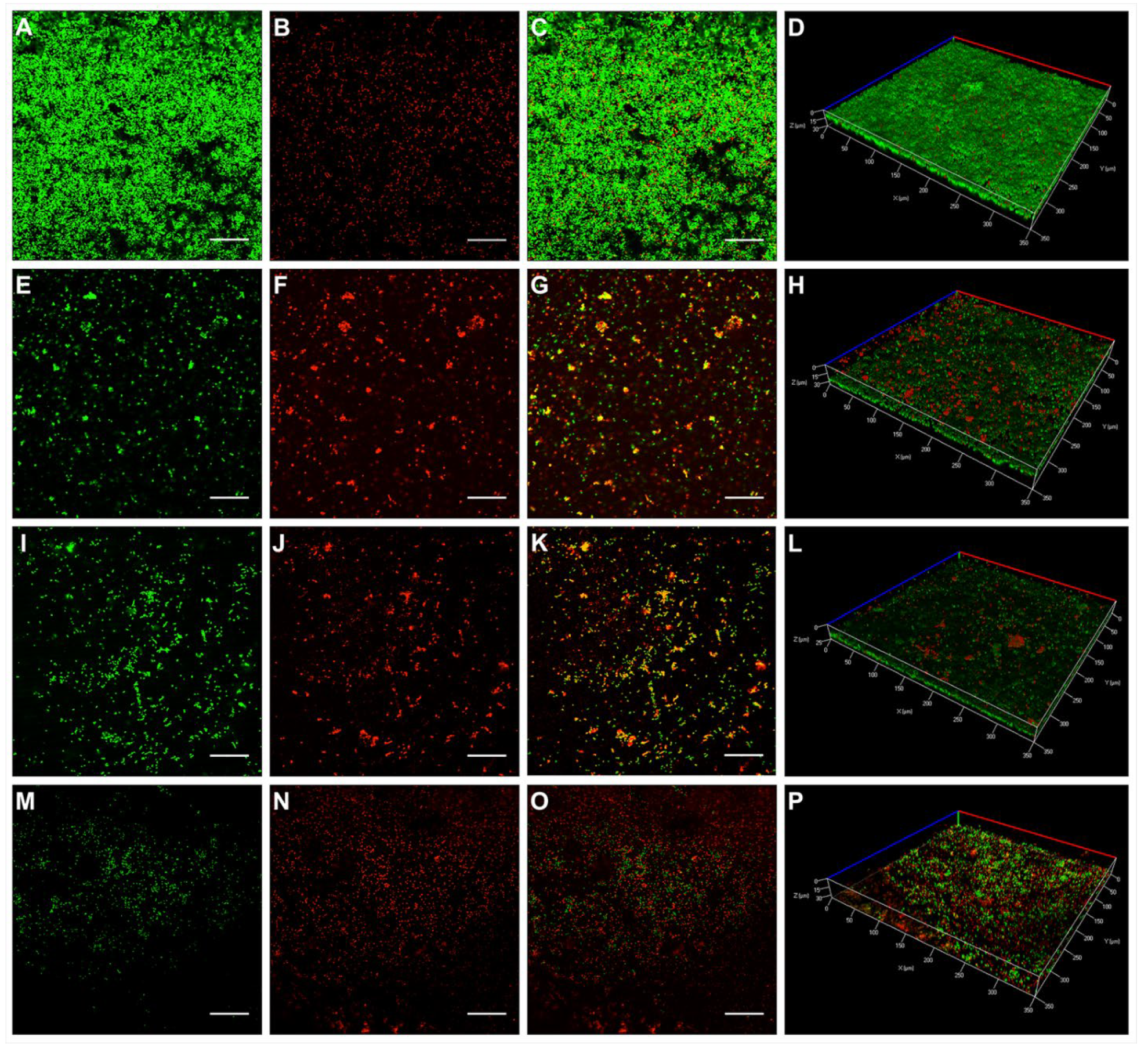
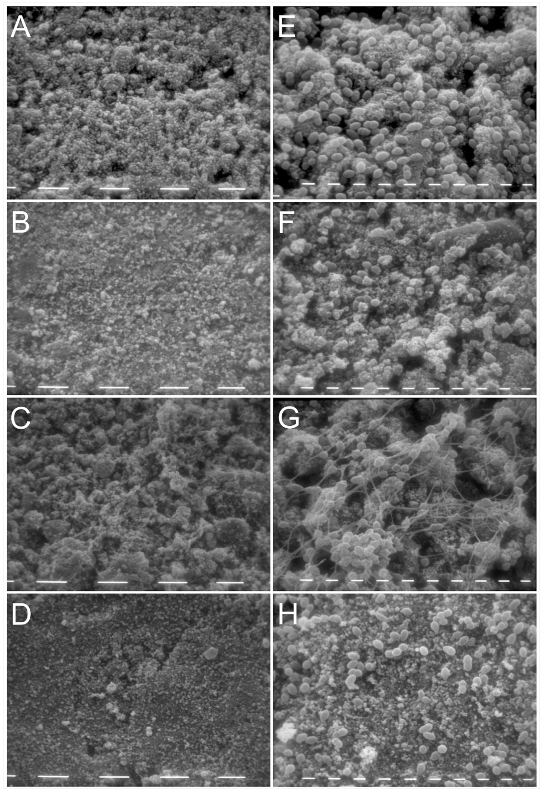
2.3. Activity of KP and L18R against E. faecalis Biofilm on Hydroxyapatite Disks

The activity of KP and L18R against 48 h-old E. faecalis biofilm grown on HA disks was assessed by CLSM and scanning electron microscopy (SEM) and compared with samples treated with a saturated Ca(OH)2 endodontic dressing solution. CLSM images (Figure 2A–C) and the 3D reconstruction (Figure 2D) of untreated E. faecalis biofilms on HA disks showed mainly viable cells organized in a homogeneous and robust biofilm layer. As compared with the untreated controls, biofilms grown on HA disks and exposed to a Ca(OH)2-saturated solution (Figure 2E–G), 100 μg/mL of KP (Figure 2I–K), and 50 μg/mL of L18R (Figure 2M–O) showed a consistent number of dead cells, along with fewer adhering bacteria for samples treated with Ca(OH)2 and L18R, as evidenced by the 3D reconstruction images (Figure 2H,L,P for Ca(OH)2, KP, and L18R, respectively). The reduction in biofilm thickness was particularly evident after treatment with L18R.
A quantitative analysis of the fluorescence intensities (FI), performed on the 3D CLSM reconstruction of four random fields from each disk, revealed a detaching effect of Ca(OH)2 and L18R treatments. In fact, the sum of total FI (viable and dead cells) was clearly reduced from 42.93 ± 4.95 for controls, to 24.18 ± 10.70 and 10.87 ± 4.33 for Ca(OH)2 and L18R, respectively. A different effect was observed after KP treatment, as the total FI resulted to be 76.88 ± 12.
The proportion of dead bacteria in the treated biofilm is shown in Figure 3. A significant increase in dead cells followed treatment with L18R and Ca(OH)2. Notably, in the biofilm treated with L18R, the percentage of dead cells was significantly higher than that obtained after treatment with the conventional endodontic dressing. KP treatment caused an increase, although not significant, in dead cells.
These results were confirmed by SEM images, which showed a lower number of cells in the biofilm grown on HA disks treated with Ca(OH)2 and L18R as compared to the untreated control sample (Figure 4). L18R treatment caused the highest detachment. In KP-treated samples, a network of fibril-like structures on adhering cells was observed.

3. Discussion

In the root canal environment, the ability of E. faecalis to resist harsh environmental conditions and form biofilms on the inner surface of the tooth canals makes its elimination extremely difficult [21]. These features can explain the high prevalence of this bacterium in secondary and persistent endodontic infections [17].
In a previous published preliminary study, in order to look for new alternative and nontoxic antibacterial substances to improve root canal disinfection, the effects of the synthetic peptides KP and L18R against planktonic cells and E. faecalis biofilms on polystyrene plates in early-stage development were assessed [29]. The results demonstrated a good antibacterial activity of both peptides against E. faecalis planktonic cells at micromolar concentrations. L18R proved to be the most effective with an EC50 value of 3.624 × 10⁻⁷ M, while KP EC50 resulted to be 4.520 × 10⁻⁶ M. KP and L18R were also shown to interfere with E. faecalis biofilm formation by reducing biofilm mass. As for the activity against planktonic bacteria, L18R resulted to be more effective than KP in biofilm inhibition. On the basis of these promising results, new investigations were performed to better characterize the effect of both peptides on E. faecalis biofilms.
In the present work, the KP and L18R activities against E. faecalis on polystyrene plates were confirmed by the assessment of their ability to reduce biofilm metabolic activity. Notably, the comparison of the results obtained in this assay with those of the previously described experiment that evaluated the reduction in biofilm mass [29] showed a similar behavior for both peptides. In fact, taking into consideration, as an example, the concentration of 50 µg/mL, KP caused a reduction in biofilm mass of approximately 35% and in biofilm viability of approximately 43%. Similarly, L18R, at the same concentration, caused reductions in biofilm mass and viability of approximately 73% and 70%, respectively.
KP and L18R were effective against E. faecalis biofilms at concentrations higher than those against planktonic cells, as commonly found with conventional antibiotics. This behavior suggests a mechanism of action, during the early phases of E. faecalis biofilm formation, not distinct from the killing activity against planktonic bacterial cells. On the contrary, some peptides, such as the human cathelicidin LL-37, are able to inhibit and disperse preformed bacterial biofilms at concentrations lower than or equal to concentrations effective against free-floating cells, implying an action on biofilm-specific targets rather than ubiquitous microbial structures [33].
From previous studies on yeasts, it has been hypothesized that the first step of KP killing activity is an interaction with cell-wall glucan-like structures [31]. For L18R, a direct penetration via an energy-independent pathway involving stable or transient destabilization and peptide folding on the lipid portion of the membrane was shown [32], indicating that the activity of this peptide may involve different mechanisms of action. Possible multi-modal mechanisms of action would render the peptides potentially advantageous in targeting different biofilm sub-populations. The mechanism of action of the investigated peptides against bacterial cells is not known, and further studies will be necessary to elucidate it.
Notably, both peptides proved to be nontoxic to murine fibroblasts at active concentrations, in agreement with previous studies that demonstrated the lack of detectable toxicity in vitro to other cell lines, erythrocytes, and peripheral blood mononuclear cells [31,32,34]. As for other AMPs, the selective antimicrobial action of KP and L18R may be explained by the cationic nature of these molecules, which promotes the interaction with the negatively charged membranes of bacteria and not with the zwitterionic membranes of mammalian cells [35]. Conversely, for other endodontic dressings of clinical use, such as Ca(OH)2 and cresol, a certain degree of toxicity against host cells was demonstrated [36,37].
In the preliminary study, the antibacterial effects of KP and L18R were also tested in a model of endodontic infection consisting of preformed E. faecalis biofilms grown on dentin-mimicking hydroxyapatite disks, in comparison with a saturated Ca(OH)2 endodontic dressing solution. This model, which represents a simplification of the clinical reality of the infected root canal, was used for a qualitative CLSM analysis [29].
In the present study, the same in vitro model was used for quantitative CLSM and qualitative SEM analyses. Deep alterations in the architecture and spatial distribution of the treated biofilms, with the highest detaching effect induced by L18R, were shown. In KP-treated samples, SEM images revealed a network of fibril-like structures on adhering cells, confirming the self-assembling properties previously shown by the peptide when challenged against yeast cells [31,38]. L18R treatment did not involve the formation of fibril-like structures on biofilms, according to previous observations on C. albicans cells [32]. It has been previously described that KP molecules easily dimerize in solution, due to the formation of disulfide bridges, and, with time, KP dimers self-assemble, giving rise to fibril-like aggregates that can be visualized by transmission electron microscopy. Moreover, KP aggregates are readily formed in the presence of soluble 1,3-β-glucans and after incubation with C. albicans cells exposing 1,3-β-glucans on their surface [38]. Notably, this peculiar property of KP, able to confer protection against proteases, has been associated to KP therapeutic activity in vivo against experimental fungal infections. Further studies are needed to establish which surface components on E. faecalis cells are able to induce KP assembly in a fibril-like network. The peculiar aggregation of KP in the presence of the targeted microorganisms could explain the data obtained by the quantitative analysis of FI in 3D reconstructions of CLSM images, i.e., the sum of total FI (viable and dead cells), which resulted to be higher for the KP-treated sample (76.88 ± 12) in comparison to the control (42.93 ± 4.95). It is conceivable that the KP fibril-like network may hinder the detachment of bacterial cells (viable and dead) during biofilm washing procedures. Likewise, the above-mentioned difference observed for KP activity against E. faecalis biofilms on polystyrene plates between the reduction in biofilm mass (35%) and in biofilm viability (43%), although not very high, could have the same explanation.
The determination of the viability profile of E. faecalis cells allowed the detection of a significantly higher ratio of dead-to-total cells in L18R-treated samples compared to the control and to the samples treated with Ca(OH)2 (Figure 3). These findings suggest the potential of L18R for E. faecalis biofilm treatment, showing its possible benefit over the established inter-appointment medicament Ca(OH)2, and confirm the already reported partial resistance to the commercial endodontic dressing [39,40].
Further aspects, such as the spectrum of anti-biofilm activity of L18R and its activity against multispecies consortia, which may represent a greater challenge toward biofilm eradication, should be investigated. The synergistic interaction with conventional antimicrobial drugs may also be tested. Nonetheless, the obtained results indicate L18R as a promising candidate for further development as an anti-biofilm agent to be used, alone or in combination with classic endodontic dressings, as an innovative intracanal medicament to reduce endodontic failures.

4. Materials and Methods

4.1. Peptides and Bacterial Strain

KP (AKVTMTCSAS, molecular mass 998.17) was synthesized in its active dimeric form by NeoMPS (PolyPeptide Group, Strasbourg, France), while L18R (LLVLRSLGPWHPGHCLLR, molecular mass 2068.1) was synthesized at the CRIBI Biotechnology Center (University of Padua, Padua, Italy), as previously described [32,34]. Peptides were solubilized in DMSO (starting solution, 20 mg/mL) and diluted prior to use. In all experiments, controls (without peptides) contained DMSO at the proper concentration.
The reference E. faecalis ATCC 29212 strain was maintained in Brain Heart Infusion Agar (BHA; Sigma-Aldrich, St. Louis, MO, USA) plates. Subcultures were made two times a week.

4.2. Peptide Cytotoxicity Assay

The cytotoxicity of the peptides against L929 murine fibroblasts was assessed by a 3-(4,5-dimethyl-2-thiazolyl)-2,5-diphenyl-2H-tetrazolium bromide (MTT) assay. Cells cultured in Dulbecco’s modified Eagle’s medium (DMEM; Sigma-Aldrich, St. Louis, MO, USA) with 10% fetal bovine serum, 100 U/mL of penicillin, and 100 μg/mL of streptomycin were seeded in a 96-well microplate (100 μL/well, 4 × 10⁵ cells/mL) and incubated for 24 h at 37 °C in a 5% CO2 atmosphere. Cells were then treated for 24 h with the peptides at 50, 100, 200, and 400 μg/mL in DMEM with 2% FBS. Cells in medium without peptides were used as a control. The medium was discarded and 100 µL of MTT at the concentration of 0.5 mg/mL in serum-free DMEM was added in each well. After 4 h of incubation at 37 °C, 100 μL of the proper reagent (10% Triton-X 100 in acidic isopropanol 0.1 N HCl) was added to solubilize formazan crystals formed following the reduction in MTT by viable cells, and the absorbance was measured at 540 nm. Each assay was run in triplicate. Results, from two independent experiments, were expressed as the percentage of viable cells in comparison to the control.

4.3. Treatment of E. faecalis Biofilm Formed on Polystyrene Surfaces

The effect of KP and L18R was investigated in early stages of the biofilm developed on polystyrene plates, as previously described [29]. Briefly, E. faecalis cells (7.5 × 10⁶ cells/mL, 200 μL/well) were incubated for 90 min at 37 °C; then, planktonic bacteria were removed and adherent cells were exposed to serial concentrations of peptides for 5 h at 37 °C. Cells incubated in water served as the control. After washing, the plates were further incubated at 37 °C for 48 h, and then the evaluation of biofilm metabolic activity was performed by Alamar Blue (CellTiter-Blue, Invitrogen, Carlsbad, CA, USA) assay. The plates were washed with PBS before the addition of 200 µL/well of the cell viability reagent. After 1 h incubation at 37 °C, the fluorescence was measured using a microplate reader (EnSpire® Multimode Plate Reader, PerkinElmer, Waltham, MA, USA), setting excitation at 570 nm and emission at 585 nm. Each assay was run in triplicate. Four independent experiments were performed. The results were expressed as the percentage of biofilm viability reduction with reference to the untreated control (100% viability). EC50 values were calculated using Graph Pad Prism 4.01 software.

4.4. Treatment of E. faecalis Biofilm Formed on Hydroxyapatite Disks

In order to evaluate the activity of KP and L18R on mature biofilms, an in vitro model of root canal infection on hydroxyapatite (HA) disks was employed, as previously described [29]. Sterilized HA disks were placed in wells of flat-bottom 24-well plates and inoculated with 500 μL of a 7.5 × 10⁶ cells/mL bacterial suspension prepared as previously described. After 48 h of incubation at 37 °C, the medium was gently washed off and the disks were treated with 500 μL/well of a saturated solution of a Ca(OH)2 endodontic dressing, KP (100 μg/mL), L18R (50 μg/mL), or sterile water (control) for 24 h at 37 °C. The effect of the treatments on the biofilm preformed on HA was assessed by confocal laser scanning and scanning electron microscopy. Two independent experiments were performed.

4.4.1. Confocal Laser Scanning Microscopy

For each treatment, half of the disks were examined by CLSM to determine the biofilm architecture and the viability of bacteria. After washing with PBS, bacteria on HA disks were stained using 500 μL of a live/dead kit (LIVE/DEAD FilmTracer™ Biofilm Viability Kit, Invitrogen, Paisley, UK) solution, containing two component dyes (0.3% SYTO-9, 0.3% propidium iodide), according to the manufacturer’s instructions. After 20 min, the disks were washed again and fluorescence emission was detected using a LSM 510 Meta scan head integrated with the Axiovert 200 M inverted microscope (Carl Zeiss, Jena, Germany). The excitation/emission wavelengths were 480/500 nm for the SYTO-9 live cell stain and 490/635 nm for the propidium iodide dead cell stain. The samples were observed using a 40 × NA1.3 oil immersion lens, and four random fields were scanned in each sample. A stack of 80–100 slices in 0.5 μm step sizes was captured along the Z-axis from the top to bottom of the biofilm. CLSM images were acquired and three-dimensional (3D) reconstructions were produced using the microscope manufacturer’s software (Axiovision module inside 4D release 4.5, Carl Zeiss, Jena, Germany). The ratio of red fluorescence intensity (FI) to green-and-red FI, calculated with the Imaris 9.5.0 software (Bitplane AG, Zurich, Switzerland), indicated the proportion of dead cells for treatment groups.

4.4.2. Scanning Electron Microscopy

The remaining half of HA disks were processed for SEM. Briefly, the disks were washed with PBS and dried at room temperature for 15 min. The samples were then fixed with a solution of glutaraldehyde 2.5% in 0.1 M of sodium cacodylate for 1 h at room temperature, dehydrated in graded series of ethanol (25%, 50%, 75%, 90%, 100%; 30 min between each passage), immersed in absolute acetone, and subjected to critical-point drying. The disks were mounted on aluminum stubs and covered with a 60 nm gold film using a metal sputtering device. The samples were observed using a Philips 501 microscope equipped with a Nikon Coolpix digital camera for acquisition of the images.

4.5. Statistical Analysis

Statistical analysis was performed using Prism 4.01 (GraphPad software, San Diego, CA, USA). An ANOVA test followed by Tukey’s post-hoc was used for multiple comparisons. Values of p < 0.05 were considered significant.

Author Contributions

Conceptualization, G.M. and L.G.; Formal Analysis, G.M. and L.G.; Investigation, G.M., P.B., T.C. and L.G.; Methodology, G.M. and L.G.; Project Administration, M.M. and S.C.; Resources, G.M. and L.G.; Supervision, M.M.; Validation, G.M., M.M. and L.G.; Visualization, G.M. and L.G.; Writing—Original Draft, G.M. and L.G.; Writing—Review and Editing, S.C. All authors have read and agreed to the published version of the manuscript.

Funding

This research received no external funding.

Data Availability Statement

The data supporting the findings of this study are available within the article.

Acknowledgments

The authors thank Silvana Belletti and Davide Dallatana for technical assistance in confocal microscopy and SEM studies. The confocal images were obtained in the Laboratory of Confocal Microscopy of the Department of Medicine and Surgery of the University of Parma. Francesca Poggia, Federica Placa, and Chiara Mirabile are also acknowledged for their support during the laboratory work.

Conflicts of Interest

The authors declare no conflict of interest.

References

  1. Berman, L.; Hargreaves, K. Cohen’s Pathways of the Pulp, 11th ed.; Elsevier: St. Louis, MO, USA, 2016. [Google Scholar]
  2. European Society of Endodontology. Quality guidelines for endodontic treatment: Consensus report of the European Society of Endodontology. Int. Endod. J. 2006, 39, 921–930. [Google Scholar] [CrossRef]
  3. Haapasalo, M.; Shen, Y.; Wang, Z.; Gao, Y. Irrigation in endodontics. Br. Dent. J. 2014, 216, 299–303. [Google Scholar] [CrossRef] [PubMed]
  4. Nair, P.N.R.; Henry, S.; Cano, V.; Vera, J. Microbial status of apical root canal system of human mandibular first molars with primary apical periodontitis after “one-visit” endodontic treatment. Oral Surg. Oral Med. Oral Pathol. Oral Radiol. Endod. 2005, 99, 231–252. [Google Scholar] [CrossRef]
  5. Flemming, H.-C.; Wingender, J.; Szewzyk, U.; Steinberg, P.; Rice, S.A.; Kjelleberg, S. Biofilms: An emergent form of bacterial life. Nat. Rev. Microbiol. 2016, 14, 563–575. [Google Scholar] [CrossRef] [PubMed]
  6. Stewart, P.S.; Costerton, J.W. Antibiotic resistance of bacteria in biofilms. Lancet 2001, 358, 135–138. [Google Scholar] [CrossRef]
  7. Van Acker, H.; Van Dijck, P.; Coenye, T. Molecular mechanisms of antimicrobial tolerance and resistance in bacterial and fungal biofilms. Trends Microbiol. 2014, 22, 326–333. [Google Scholar] [CrossRef] [PubMed]
  8. Sjogren, U.; Figdor, D.; Spangberg, L.; Sundqvist, G. The antimicrobial effect of calcium hydroxide as a short-term intracanal dressing. Int. Endod. J. 1991, 24, 119–125. [Google Scholar] [CrossRef]
  9. Shuping, G.B.; Orstavik, D.; Sigurdsson, A.; Trope, M. Reduction of intracanal bacteria using nickel-titanium rotary instrumentation and various medications. J. Endod. 2000, 26, 751–755. [Google Scholar] [CrossRef]
  10. Siqueira, J.F., Jr.; Magalhaes, K.M.; Rocas, I.N. Bacterial reduction in infected root canals treated with 2.5% NaOCl as an irrigant and calcium hydroxide/camphorated paramonochlorophenol paste as an intracanal dressing. J. Endod. 2007, 33, 667–672. [Google Scholar] [CrossRef]
  11. Vera, J.; Siqueira, J.F., Jr.; Ricucci, D.; Loghin, S.; Fernandez, N.; Flores, B.; Cruz, A.G. One- versus two-visit endodontic treatment of teeth with apical periodontitis: A histobacteriologic study. J. Endod. 2012, 38, 1040–1052. [Google Scholar] [CrossRef] [PubMed]
  12. Estrela, C.; Pimenta, F.C.; Ito, I.Y.; Bammann, L.L. In vitro determination of direct antimicrobial effect of calcium hydroxide. J. Endod. 1998, 24, 15–17. [Google Scholar] [CrossRef]
  13. Gomes, B.P.; Ferraz, C.C.; Garrido, F.D.; Rosalen, P.L.; Zaia, A.A.; Teixeira, F.B.; de Souza-Filho, F.J. Microbial susceptibility to calcium hydroxide pastes and their vehicles. J. Endod. 2002, 28, 758–761. [Google Scholar] [CrossRef] [PubMed]
  14. Wang, J.D.; Hume, W.R. Diffusion of hydrogen ion and hydroxyl ion from various sources through dentine. Int. Endod. J. 1988, 21, 17–26. [Google Scholar] [CrossRef] [PubMed]
  15. Evans, M.; Davies, J.K.; Sundqvist, G.; Figdor, D. Mechanisms involved in the resistance of Enterococcus faecalis to calcium hydroxide. Int. Endod. J. 2002, 35, 221–228. [Google Scholar] [CrossRef] [PubMed]
  16. Siqueira, J.F., Jr.; Rocas, I.N. Diversity of endodontic microbiota revisited. J. Dent. Res. 2009, 88, 969–981. [Google Scholar] [CrossRef] [PubMed]
  17. Siqueira, J.F., Jr.; Rôças, I.N. Distinctive features of the microbiota associated with different forms of apical periodontitis. J. Oral Microbiol. 2009, 1, 2009. [Google Scholar] [CrossRef]
  18. Rôças, I.N.; Siqueira, J.F., Jr. Identification of bacteria enduring endodontic treatment procedures by a combined reverse transcriptase-polymerase chain reaction and reverse-capture checkerboard approach. J. Endod. 2010, 36, 45–52. [Google Scholar] [CrossRef] [PubMed]
  19. Ch’ng, J.-H.; Chong, K.K.L.; Lam, L.N.; Wong, J.J.; Kline, K.A. Biofilm-associated infection by enterococci. Nat. Rev. Microbiol. 2019, 17, 82–94. [Google Scholar] [CrossRef]
  20. Nakajo, K.; Komori, R.; Ishikawa, S.; Ueno, T.; Suzuki, Y.; Iwami, Y.; Takahashi, N. Resistance to acidic and alkaline environments in the endodontic pathogen Enterococcus faecalis. Oral Microbiol. Immunol. 2006, 21, 283–288. [Google Scholar] [CrossRef]
  21. Stuart, C.H.; Schwartz, S.A.; Beeson, T.J.; Owatz, C.B. Enterococcus faecalis: Its role in root canal treatment failure and current concepts in retreatment. J. Endod. 2006, 32, 93–98. [Google Scholar] [CrossRef]
  22. Swimberghe, R.C.D.; Coenye, T.; De Moor, R.J.G.; Meire, M.A. Biofilm model systems for root canal disinfection: A literature review. Int. Endod. J. 2019, 52, 604–628. [Google Scholar] [CrossRef] [PubMed] [Green Version]
  23. Lima, S.M.F.; de Padua, G.M.; Sousa, M.; Freire, M.S.; Franco, O.L.; Rezende, T.M.B. Antimicrobial peptide-based treatment for endodontic infections--biotechnological innovation in endodontics. Biotechnol. Adv. 2015, 33, 203–213. [Google Scholar] [CrossRef]
  24. Marr, A.K.; Gooderham, W.J.; Hancock, R.E. Antibacterial peptides for therapeutic use: Obstacles and realistic outlook. Curr. Opin. Pharmacol. 2006, 6, 468–472. [Google Scholar] [CrossRef] [PubMed]
  25. Boto, A.; Perez de la Lastra, J.M.; Gonzalez, C.C. The road from host-defense peptides to a new generation of antimicrobial drugs. Molecules 2018, 23, 311. [Google Scholar] [CrossRef] [PubMed] [Green Version]
  26. Gomes, B.; Augusto, M.T.; Felicio, M.R.; Hollmann, A.; Franco, O.L.; Goncalves, S.; Santos, N.C. Designing improved active peptides for therapeutic approaches against infectious diseases. Biotechnol. Adv. 2018, 36, 415–429. [Google Scholar] [CrossRef]
  27. Boparai, J.K.; Sharma, P.K. Mini review on antimicrobial peptides, sources, mechanism and recent applications. Protein Pept. Lett. 2020, 27, 4–16. [Google Scholar] [CrossRef]
  28. Hale, J.D.F.; Hancock, R.E.W. Alternative mechanisms of action of cationic antimicrobial peptides on bacteria. Expert Rev. Anti-Infect. Ther. 2007, 5, 951–959. [Google Scholar] [CrossRef]
  29. Mergoni, G.; Manfredi, M.; Bertani, P.; Ciociola, T.; Conti, S.; Giovati, L. Antibacterial effects of two synthetic peptides against Enterococcus faecalis biofilms: A preliminary in vitro study. G. Ital. Endod. 2020, 34, 47–54. [Google Scholar] [CrossRef]
  30. Polonelli, L.; Magliani, W.; Ciociola, T.; Giovati, L.; Conti, S. From Pichia anomala killer toxin through killer antibodies to killer peptides for a comprehensive anti-infective strategy. Antonie Leeuwenhoek 2011, 99, 35–41. [Google Scholar] [CrossRef] [PubMed]
  31. Magliani, W.; Conti, S.; Ciociola, T.; Giovati, L.; Zanello, P.P.; Pertinhez, T.; Spisni, A.; Polonelli, L. Killer peptide: A novel paradigm of antimicrobial, antiviral and immunomodulatory auto-delivering drugs. Future Med. Chem. 2011, 3, 1209–1231. [Google Scholar] [CrossRef] [PubMed]
  32. Polonelli, L.; Ciociola, T.; Sperinde, M.; Giovati, L.; D’Adda, T.; Galati, S.; Travassos, L.R.; Magliani, W.; Conti, S. Fungicidal activity of peptides encoded by immunoglobulin genes. Sci. Rep. 2017, 7, 10896. [Google Scholar] [CrossRef] [PubMed] [Green Version]
  33. Dostert, M.; Belanger, C.R.; Hancock, R.E.W. Design and assessment of anti-biofilm peptides: Steps toward clinical application. J. Innate Immun. 2019, 11, 193–204. [Google Scholar] [CrossRef] [PubMed]
  34. Giovati, L.; Santinoli, C.; Mangia, C.; Vismarra, A.; Belletti, S.; D’Adda, T.; Fumarola, C.; Ciociola, T.; Bacci, C.; Magliani, W.; et al. Novel activity of a synthetic decapeptide against Toxoplasma gondii tachyzoites. Front. Microbiol. 2018, 9, 753. [Google Scholar] [CrossRef] [PubMed]
  35. Matsuzaki, K. Control of cell selectivity of antimicrobial peptides. Biochim. Biophys. Acta 2009, 1788, 1687–1692. [Google Scholar] [CrossRef] [PubMed] [Green Version]
  36. Ehlinger, C.; Dartevelle, P.; Zaet, A.; Kurashige, Y.; Haïkel, Y.; Metz-Boutigue, M.-H.; Marban, C. A new combination with D-cateslytin to eradicate root canal pathogens. Int. J. Pept. Res. Ther. 2019, 25, 1679–1687. [Google Scholar] [CrossRef] [Green Version]
  37. Kobayashi, M.; Tsutsui, T.W.; Kobayashi, T.; Ohno, M.; Higo, Y.; Inaba, T.; Tsutsui, T. Sensitivity of human dental pulp cells to eighteen chemical agents used for endodontic treatments in dentistry. Odontology 2013, 101, 43–51. [Google Scholar] [CrossRef]
  38. Pertinhez, T.A.; Conti, S.; Ferrari, E.; Magliani, W.; Spisni, A.; Polonelli, L. Reversible Self-Assembly: A Key Feature for a New Class of Autodelivering Therapeutic Peptides. Mol. Pharm. 2009, 6, 1036–1039. [Google Scholar] [CrossRef]
  39. Bystrom, A.; Claesson, R.; Sundqvist, G. The antibacterial effect of camphorated paramonochlorophenol, camphorated phenol and calcium hydroxide in the treatment of infected root canals. Endod. Dent. Traumatol. 1985, 1, 170–175. [Google Scholar] [CrossRef]
  40. Orstavik, D.; Haapasalo, M. Disinfection by endodontic irrigants and dressings of experimentally infected dentinal tubules. Endod. Dent. Traumatol. 1990, 6, 142–149. [Google Scholar] [CrossRef]
Figure 1. Effect of the investigated peptides against E. faecalis ATCC 29212 biofilm. Biofilm metabolic activity was determined by Alamar Blue assay after treatment with different concentrations of (A) KP and (B) L18R. Results are reported as percent reduction in biofilm viability as compared to untreated samples. Data are presented as mean ± SD of at least 3 independent experiments.
Antibiotics 10 01220 g001
Figure 2. Representative CLSM images of E. faecalis biofilms on HA disks. Images of 48 h-old E. faecalis ATCC 29212 biofilm were acquired by CLSM after exposure to H2O (control, AC), Ca(OH)2-saturated solution (EG), KP (100 μg/mL, IK), and L18R (50 μg/mL, MO) for 24 h. Shown in each line is the same field (single focal plane). From left to right: SYTO-9 (green, viable cells), propidium iodide (red, dead cells), merged image of viable and dead cells. 3D reconstructions of the selected fields (full thickness, merged images) are shown in panels (D) (control), (H) (exposed to Ca(OH)2-saturated solution), (L) (exposed to KP), and (P) (exposed to L18R). Notably, in the L18R-treated sample, the 3D reconstruction shows a reduced thickness of the biofilm in comparison to the untreated control. Bar = 50 µm.
Antibiotics 10 01220 g002
Figure 3. Dead cells (%) after treatment of E. faecalis biofilm pre-grown on HA disks. Values were obtained by 3D CLSM analysis of 48 h-old E. faecalis ATCC 29212 biofilms after exposure to H2O (control), Ca(OH)2-saturated solution, KP (100 μg/mL), and L18R (50 μg/mL) for 24 h. Red and green fluorescence intensity measures were carried out with Imaris 7.2 on four random fields of each disk. The results are expressed as means ± SD (**, p < 0.01 vs. control; ***, p < 0.001 vs. control; ###, p < 0.001 vs. Ca(OH)2).
Antibiotics 10 01220 g003
Figure 4. Representative scanning electron microscopy (SEM) images of E. faecalis biofilms on HA disks. Images of 48 h-old E. faecalis ATCC 29212 biofilms were acquired by SEM after exposure to H2O (control; A,E), Ca(OH)2-saturated solution (B,F), KP (100 μg/mL; C,G), and L18R (50 μg/mL; D,H) for 24 h. Magnification 1250× (AD, bar = 10 µm) and 5000× (EH, bar = 1 µm).
Antibiotics 10 01220 g004
Table 1. In vitro cytotoxic activity of the investigated peptides against L929 cells.
Concentration (µg/mL)Cell Viability (%) after Treatment with
KPL18R
400104 ± 17127 ± 24
200121 ± 1599 ± 20
100145 ± 34102 ± 0
50120 ± 39120 ± 16
Publisher’s Note: MDPI stays neutral with regard to jurisdictional claims in published maps and institutional affiliations.

