Review

3D Printing Assisted Wearable and Implantable Biosensors

1
Department of Radiology, University of Michigan, Ann Arbor, MI 48109, USA
2
Department of Smart Health Science and Technology, Kangwon National University (KNU), Chuncheon-si 24341, Republic of Korea
3
Department of Bioengineering and Biotechnology, School of Basic and Applied Sciences, Galgotias University, Greater Noida 203201, India
4
Department of Mechanical and Biomedical, Mechatronics Engineering, Kangwon National University (KNU), Chuncheon-si 24341, Republic of Korea
*
Author to whom correspondence should be addressed.
These authors contributed equally to this work.
Biosensors 2025, 15(9), 619; https://doi.org/10.3390/bios15090619
Submission received: 14 July 2025 / Revised: 9 September 2025 / Accepted: 12 September 2025 / Published: 17 September 2025
(This article belongs to the Special Issue Biological Sensors Based on 3D Printing Technologies)

Abstract

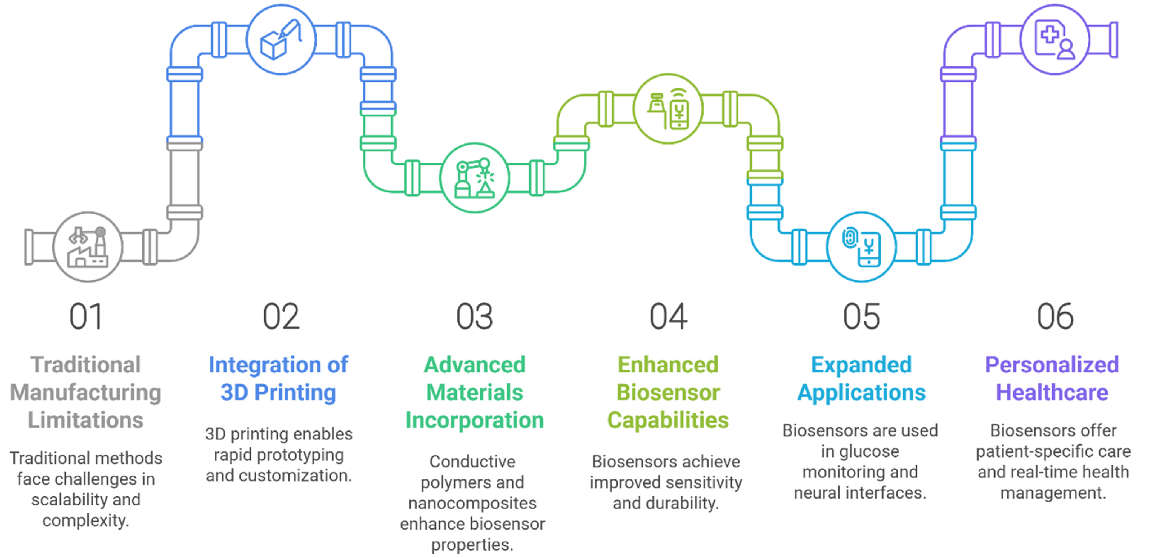
Biosensors have undergone transformative advancements, evolving into sophisticated wearable and implantable devices capable of real-time health monitoring. Traditional manufacturing methods, however, face limitations in scalability, cost, and design complexity, particularly for miniaturized, multifunctional biosensors. The integration of 3D printing technology addresses these challenges by enabling rapid prototyping, customization, and the production of intricate geometries with high precision. This review explores how additive manufacturing techniques facilitate the fabrication of flexible, stretchable, and biocompatible biosensors. By incorporating advanced materials like conductive polymers, nanocomposites, and hydrogels, 3D-printed biosensors achieve enhanced sensitivity, durability, and seamless integration with biological systems. Innovations such as biodegradable substrates and multi-material printing further expand applications in continuous glucose monitoring, neural interfaces, and point-of-care diagnostics. Despite challenges in material optimization and regulatory standardization, the convergence of 3D printing with nanotechnology and smart diagnostics heralds a new era of personalized, proactive healthcare, offering scalable solutions for both clinical and remote settings. This synthesis underscores the pivotal role of additive manufacturing in advancing wearable and implantable biosensor technology, paving the way for next-generation devices that prioritize patient-specific care and real-time health management.

1. Introduction to Biosensors

Biosensors are sophisticated analytical devices that combine biological recognition elements with electronic systems (transducers) to detect and measure specific biological and chemical substances [1]. These devices operate by converting biological responses into electrical signals, enabling the quantitative analysis of various analytes. Their significance spans multiple sectors, including healthcare, environmental monitoring, food safety, and biotechnology, due to their capacity for real-time, sensitive, and accurate detection [2]. Although biosensors contain many components (such as an immobilization matrix, signal processor, and display unit), the main core components that it operates on are a biological recognition element (bioreceptor) and a transducer (Figure 1). Upon introduction of the target molecule (analyte), an interaction occurs with the bioreceptor, leading to biochemical changes that are crucial for signal transduction. These changes are detected by a transducer, which converts the biochemical response into an electrical signal, thus quantifying the concentration of the analyte in question [3]. With abilities to detect low concentrations of analyte, portability, rapid results, and low costs have made them valuable tools in various fields, including healthcare, environmental monitoring, and food safety. Biosensors can be divided into several categories based on their components, detection methods, and target applications (Table 1). Table 1 presents the common target analytes and their detection strategies in modern biosensors. Biosensors can be divided into several categories based on their components, detection methods, and target applications, which are presented in the Supplementary Materials (Table S1).
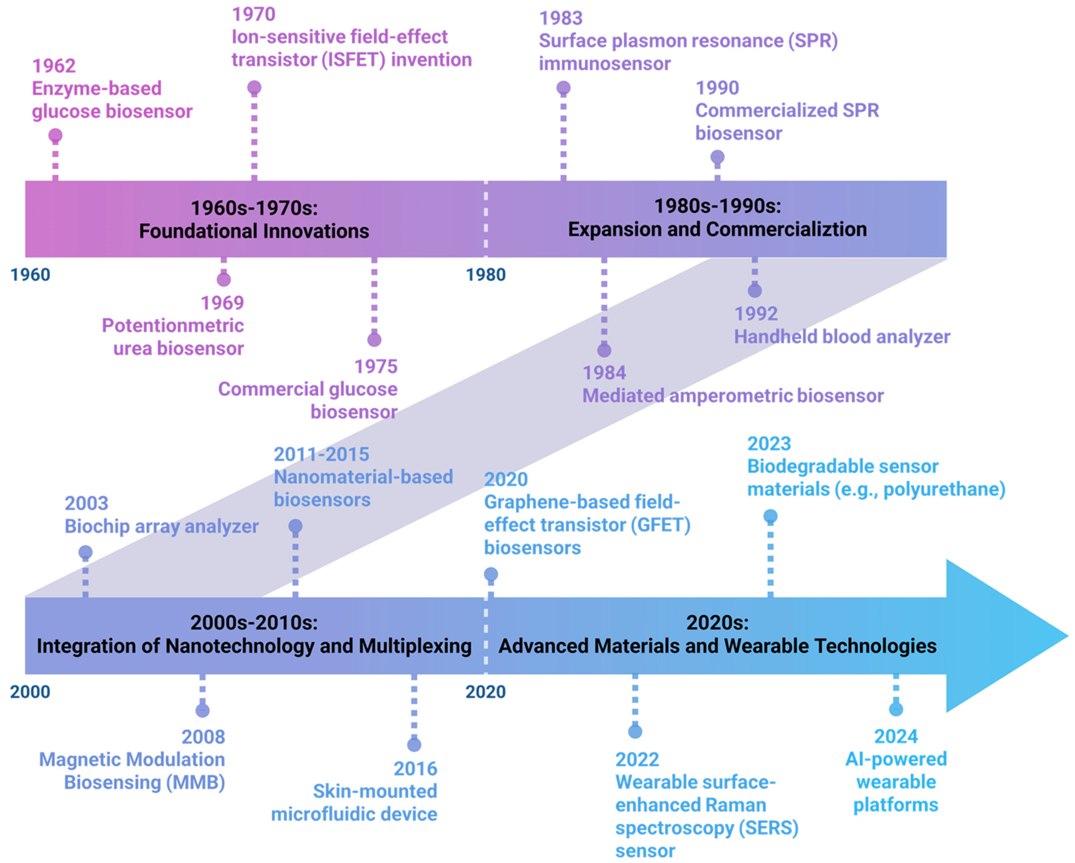
The story of biosensors begins in 1956 with Leland C. Clark, often called the “Father of Biosensors” who laid the groundwork for modern biosensors (Figure 2). Clark’s breakthrough came in 1962 when he modified his oxygen electrode to detect glucose. He did this by trapping glucose oxidase (an enzyme) against the electrode using a dialysis membrane. This created the first true biosensor, which worked by measuring how much oxygen was consumed when the enzyme reacted with glucose [25,26]. This fundamental design—combining a biological component with an electrode—established the basic principle that biosensors still use today. Later, it was observed that the first-generation biosensors faced critical oxygen dependency issues, where glucose oxidase relied on dissolved oxygen as the natural electron acceptor, making measurements highly susceptible to fluctuating oxygen concentrations in biological environments. This limitation rendered these sensors unreliable for in vivo applications, particularly in oxygen-deficient tissues or during physiological changes affecting oxygen availability. In the next couple of decades (1970s–1980s; often termed as the second-generation biosensors), scientists made biosensors more efficient by improving how signals were detected. Instead of measuring oxygen consumption, they developed ways to directly detect the electrons produced in biological reactions. For example, derivatives of ferrocene (an introduction of artificial electron mediators) replaced oxygen as electron acceptors, thereby improving electron transfer efficiency, reducing dependence on environmental oxygen, and enabling more reliable amperometric detection [27,28]. Some other developments of new immobilization techniques [29,30], such as the integration of enzymes with microelectrodes, facilitated the miniaturization and creation of the first commercial biosensors (developed by Yellow Spring Instruments, YSI) primarily for glucose monitoring in diabetes management. While this innovation significantly improved reproducibility and enabled the development of commercial glucose test strips, second-generation systems introduced new limitations, including mediator leaching, potential toxicity, and restricted applicability for implantable devices. The next generation brought about “reagent less” biosensors, where the biological component was directly connected to the electrode [31,32]. Some other novel introductions, like conducting polymers [33,34], new biological recognition elements [35], and integration with microelectronics and miniaturization techniques occurred [36]. The post-2000s era, often referred to as the “modern era” of biosensing, marks the convergence of multiple enabling technologies. A central driver has been the incorporation of nanomaterials, such as quantum dots, nanoparticles, graphene, and carbon nanotubes. The introduction of nanomaterials dramatically increased the surface area-to-volume ratio, exponentially enhancing the available active sites for biomolecule immobilization and catalytic reactions [37]. This significantly enhanced sensor sensitivity and enabled the detection of extremely small quantities of target molecules [38,39]. Nanomaterials facilitate a direct electron transfer (DET) between enzymes and electrodes, eliminating the need for mediators entirely in previous-generation biosensors. This was achieved through the unique electronic properties of nanomaterials, particularly their enhanced electrical conductivity and favorable electron transfer kinetics [40]. The quantum confinement effects in nanomaterials such as quantum dots introduced tunable optical and electronic properties that were impossible to achieve with bulk materials [41]. These advances solved the core miniaturization challenge that had previously confined diagnostic testing to centralized laboratories and allowed healthcare providers to conduct real-time diagnostics at the patient’s bedside, in remote areas, or in resource-limited settings [42,43]. Consequently, this shift has catalyzed the development of smart biosensors that are capable of continuous, multiplexed monitoring, wireless data transmission, and seamless integration into healthcare systems [44,45].
As demand grew for continuous and non-invasive monitoring, biocompatible flexible substrates and adhesives such as silicone, hydrogel, and natural biomaterials (e.g., silk fibroin, cellulose) were engineered to conform to the skin, mitigate irritation, and maintain sensor performance even with movement [46]. These advances were essential for transitioning biosensors from intermittent use to flexible patches, textiles, and accessories integrated into everyday life [47]. Innovations in bioinspired and biomineralized coatings, as well as biodegradable polymers, allowed the development of implantable sensors to remain functional inside the body for months while safely interfacing with tissues [48]. Engineered bioinks and 3D-printed biocompatible matrices further improved integration and the miniaturization needed for chronic implants. This material progress transformed biosensors from sporadic standalone tools to seamless health platforms embedded in daily life and clinical practice [49,50,51].
Figure 2. Timeline and key developments in biosensor technology. The field began with Leland C. Clark Jr.’s enzyme-based glucose biosensor (1962) [26], followed by the first potentiometric urea biosensor by Guilbault and Montalvo (1969) [29], and Piet Bergveld’s invention of the ISFET for ion detection (1970) [52]. The first commercial glucose biosensor was launched by Yellow Springs Instruments (1975) [53]. The era of expansion saw the development of the first surface plasmon resonance (SPR) immunosensor (1983) [54] and mediated amperometric biosensors (1984) [27], culminating in the commercialization of SPR-based biosensors (1990) [55] and handheld blood analyzers (1992) [56]. The 21st century marked integration with nanotechnology, multiplexing, and wearable formats, such as the biochip array analyzer (2003) [57], magnetic modulation biosensing for enhanced sensitivity (2008) [58], and nanomaterial-based sensors (2011–2015) [59]. The introduction of skin-mounted microfluidic devices (2016) [60], ultra-sensitive graphene-based FET biosensors (2020) [61], and wearable SERS sensors (2022) [62] facilitated non-invasive diagnostics. Recent innovations include biodegradable sensor materials (2023) [63] and AI-powered wearable platforms for biomedical applications (2024) [64].
Biosensors 15 00619 g002

2. Wearable and Implantable Biosensors

Wearable biosensors are portable devices that are designed to be worn on, inside, or near the human body to continuously monitor physiological or biochemical parameters that indicate a patient’s health status. These sensors provide real-time measurements of physiological parameters, generating digital outputs that are easy to interpret and act upon. By allowing patients quick access to clinical information, these devices promote proactive health management in a more convenient and cost-effective manner, improving user compliance [65,66]. In recent years, various wearable technologies have been introduced in scientific research, particularly for personalized medicine, point-of-care diagnostics, and fitness or home health monitoring. These platforms collect physiological data through wearable components such as patches [67], lenses [68], headbands [69], wristbands [70], eyeglasses [71], and skin-conformal tattoos [72]. They are capable of detecting a range of health indicators, including glucose levels, blood pressure, heart rate, oxygen saturation, respiratory and tactile parameters, body motion, skin temperature, and brain activity [73].
Although wearable biosensing devices have become popular with their real-time and quick measurements that are easy to interpret and act upon, there are certain challenges with wearable biosensing devices, such as they are unfeasible for providing data by penetrating deep into the tissue, while contaminants from the environment might affect the results, and only global information is provided. Therefore, in addition to these flexible wearable biosensing devices, there is also an urgent need to have easy implantable devices that can bring forth the efficacious pathway of not only better diagnostic but also therapeutic options. For example, cardiac monitors have revolutionized cardiology by providing continuous heart rhythm tracking, allowing for early detection and intervention in life-threatening arrhythmias [74]. Implantable pressure sensors have facilitated the management of conditions such as hydrocephalus and traumatic brain injury by allowing for continuous intracranial pressure monitoring [75]. These advancements highlight the adaptability and transformative impact of implantable sensors in tackling various health conditions, paving the way for a new era of personalized and proactive healthcare.
The growing acceptance of wearable and implantable biosensors is driven by advancements in fabrication techniques that enable features like flexibility, stretchability, ultra-thinness, and lightweight designs, ensuring a seamless integration with the body [76,77]. Considerable research has focused on enhancing the interaction between these multifunctional devices and biological systems. Inspired by the properties of soft materials, ultra-thin biosensors are designed to conform to biological surfaces for improved performance [78,79,80]. Currently, a variety of fabrication processes, including lithography-based techniques, microchannel molding, and deposition techniques such as vapor or electrochemical deposition, laser ablation, roll-to-roll printing, and micromachining, are used to build the sensing interfaces [81,82,83]. However, there are many challenges that this technique faces in fabricating sensitive and flexible biological sensors, such as limited manufacturing scalability, high production costs, reduced durability, and restricted adaptability, which continue to hinder the widespread adoption of these cutting-edge wearable biosensors [84,85]. The shortcomings of conventional manufacturing methods have been addressed by additive manufacturing (AM), sometimes referred to as 3D printing, which has grown substantially in recent years.

3. The Requirement of 3D Printing Technology

Three-dimensional printing, a form of additive manufacturing, has revolutionized every manufacturing sector. Its ability to produce completely three-dimensional structures with intricate features in a single step makes it particularly appealing [86]. Charles Hull was the first to create this technique in 1986 [87]. Three-dimensional printing uses computer-controlled procedures based on three-dimensional (3D) digital representations of the item to be printed to fabricate a variety of structures by printing successive layers of materials that are built on top of one another. The academic and corporate communities have recently shown a great deal of interest in additive manufacturing, and it has been referred to as a third industrial revolution [86,88]. According to market research, the worldwide 3D printing market was valued at USD 16.54 billion in 2021 and is projected to increase at a CAGR of 21.0% between 2021 and 2028. By 2028, the worldwide market is expected to grow to over USD 63 billion [89]. Biomedical applications [90,91], electronics and sensors [92], lightweight engineering materials [93,94,95], and multifunctional composites [96,97,98] are just a few of the many fields in which it has attracted industry and scientific attention.
In the production of biosensors or biosensor components, 3D printing technologies present a promising advance. As mentioned above, wearable biosensors have multiple miniaturized complex structures, which are difficult to fabricate in a single unit using conventional manufacturing techniques like coating and injection molding. Three-dimensional printing offers numerous benefits over traditional manufacturing, such as increased versatility, reduced waste, greater design freedom, low fabrication costs, high automation, and a short fabrication cycle time [87,99]. In this regard, several unique aspects of 3D printing have found their way into the fabrication of biomedical sensors. For instance, 3D printing streamlines the production process by using material deposition and curing/sintering to create the individual components. Secondly, the miniaturization and compactness of the biosensors were easily achievable with the use of 3D printing, as it enables the integration of different materials during the fabrication process. For example, by integrating many sensing modalities into a single sensor via extrusion printing, researchers have achieved downsizing by lowering the device’s total footprint in comparison to using separate sensors [100]. Also, through the progressive addition of material digitally controlled by computer-aided design (CAD) systems, 3D printing enables the very precise fabrication of personalized parts [101]. Another example showcasing the 3D printing capability of tackling the problem of complicated geometries is the development of Clark platinum electrodes. Previously, the Clark platinum electrodes with special shapes were developed for ongoing oxygen concentration measurement in cardiovascular surgery. However, because each sensory component was made individually and manually integrated, these sensors were challenging to mass-produce. Later, 3D printing was used to tackle the problem of developing the multi-component complicated shapes of the electrodes as a single unit. Product customization poses a difficulty for conventional manufacturers, mostly due to the substantial expenses associated with mold fabrication, particularly for small-scale manufacturing of bespoke items. Conversely, 3D printing can produce limited numbers of tailored plastic objects at far lower prices than conventional mold-based manufacturing. This is particularly beneficial in biomedical disciplines, where individualized patient-specific goods are also necessary [87]. By combining complex geometries into specific microstructures, 3D printing enables the on-demand production of customizable sensing devices. Along with miniaturization and customization, the widespread acceptance of 3D printing relies on the fabrication of materials with critical characteristics such as stretchability, flexibility, ultra-thinness, and lightweight that fuel the development of more efficient wearable and implantable platforms (Figure 3) [73,102]. In recent years, 3D printing has become more popular in academic research because of its ease in rapid manufacturing when dealing with emerging multifunctional materials [103,104]. Interestingly, these sophisticated multifunctional and multipurpose materials are being investigated for use in 3D-printed bio-integrated devices to give doctors, patients, healthcare professionals, and healthy individuals ways to track their health. Through the use of diverse soft functional materials, 3D printing technology may enable the meticulous creation of patient-specific geometry in the context of wearable biomedical devices, directly on the preferred surfaces [85,97,103,105].
This review highlights the latest developments in 3D-printed bio-integrated sensor technologies, with a focus on wearable and implantable biosensors. We begin by exploring how innovative 3D printing techniques are enabling the fabrication of flexible 3D structures using advanced printable soft materials. Subsequently, we emphasize various 3D printing approaches used in the development of wearable and implantable biosensors. Later, this article concentrates on 3D-printed wearable (bio)sensors designed for various applications, such as detecting electrophysiological signals, biochemical signals, and signals from the dynamics of vascular flow patterns. Additionally, readers are directed to several recently published studies on 3D-printed biosensors and (bio)analytical sensors for further exploration.

4. 3D Printing—Materials and Methods

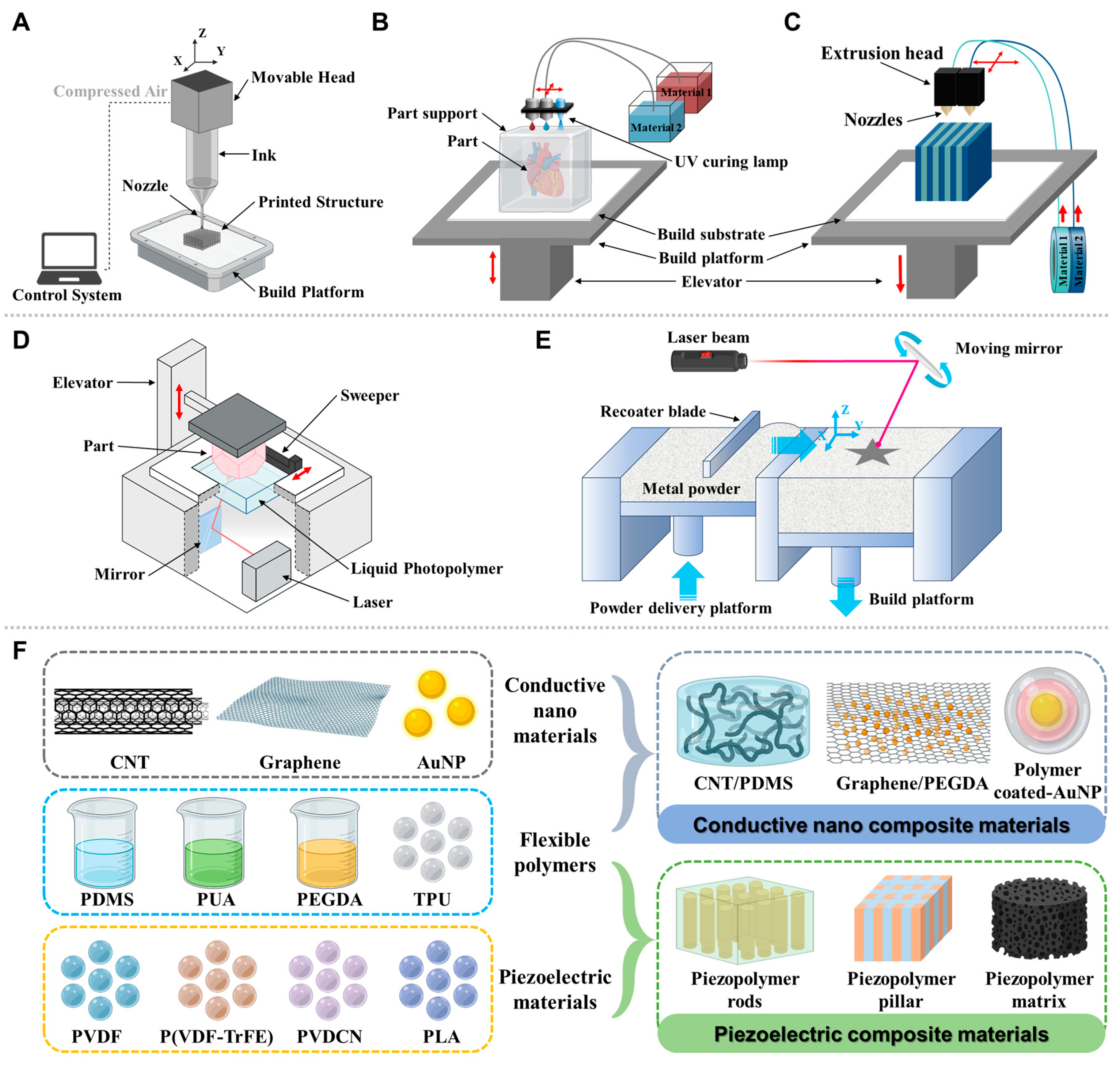
Regarding key 3D printing technologies for wearable biosensor fabrication, several 3D printing technologies have emerged as particularly suitable for wearable biosensor fabrication, each offering distinct advantages for specific sensing applications (Figure 4). Direct ink writing (DIW) has emerged as a versatile 3D printing technique for wearable biosensor fabrication, offering exceptional control over material deposition and structural features. This approach employs computer-controlled extrusion of functional inks through fine nozzles to create precise patterns of sensing elements and supporting structures [106]. This technology enables the incorporation of nanomaterials, including carbon nanotubes, graphene, and metallic nanoparticles, into inks with carefully tailored rheological properties, resulting in high-performance electrodes with enhanced conductivity and sensing capabilities [106,107]. One significant advantage of DIW is its compatibility with a wide range of substrate materials, including flexible polymers, hydrogels that conform to body contours, facilitating improved sensor-skin interfaces and enhanced signal acquisition [108]. DIW also enables the incorporation of nanomaterials, including carbon nanotubes, graphene, and metallic nanoparticles, into inks with carefully tailored rheological properties, resulting in high-performance electrodes with enhanced conductivity and sensing capabilities [106,107]. Unlike high-temperature FDM or laser-based SLS, DIW can work with low-temperature printing that allows compatibility with biomolecules and living cells. Another wide approach that inkjet printing technology offers is the unique advantages for wearable biosensor fabrication, particularly for creating high-resolution conductive traces and precise deposition of functional materials. Inkjet printing eliminates the need for masks, molds, and complex lithography steps, reducing production costs and enabling mass fabrication [109]. It is ideal for rapid prototyping and large-scale manufacturing of biosensors. With droplet volumes in picoliters and feature sizes down to 50–100 µm, inkjet printing achieves fine patterns essential for miniaturized sensors and high-density arrays [106,110]. The non-contact drop-on-demand approach reduces material waste and prevents substrate contamination, crucial for sensitive biological components. It supports sequential printing of diverse materials, including conductors (metal and carbon-based nanoparticles) and biologics (enzymes and small molecules) on flexible substrates (e.g., PDMS, plastics, paper, and textiles) [111,112]. Using high-resolution 3D-scanned body-shape data, researchers have demonstrated the fabrication of on-demand personalized wearable sensors that accurately conform to individual anatomical features [110]. Inkjet-printed electrodes on flexible substrates (e.g., textiles) enable the real-time monitoring of biomarkers in sweat (e.g., lactate and glucose) or electrophysiological signals (ECG and EEG) [113,114]. Extrusion-based approaches, including fused filament fabrication (FFF), provide cost-effective solutions for creating structural components and housings for wearable sensing platforms [115,116]. These methods allow for the integration of different functional materials within a single printing process, enabling the creation of multifunctional sensing systems with improved mechanical and electrical properties. FFF supports a broad range of thermoplastics and composites, including conductive (e.g., Proto-Pasta® CB-PLA, and Black Magic® graphene-PLA), biodegradable (PLA), and biocompatible materials (polycaprolactone, PCL) [117,118]. For example, in one of the studies, FFF was used to print bespoke filaments with optimized conductive filler ratios (PLA (60 wt%) graphite (40 wt%)) to improve electron transfer, enabling detection of biomarkers like the SARS-CoV-2 spike protein at pM levels [119]. A quantitative comparison of various 3D-printed methods used for the fabrication of wearable and implantable biosensors is presented in Table 2.
Materials advances that enable 3D-printed biosensors are a critical component of many 3D-printed biosensors is the electrode, which requires electrically conductive materials. The most common base polymers for these composites include polylactic acid (PLA) and acrylonitrile butadiene styrene (ABS), favored for their compatibility with fused deposition modeling (FDM), a widely accessible 3D printing method [120]. To achieve conductivity, these polymers are embedded with carbon-based materials such as graphene, carbon nanotubes (CNTs), and carbon black [107]. Graphene-layered substrates have been successfully synthesized and integrated into flexible wearable biosensor platforms, providing exceptional electrical conductivity while maintaining the mechanical compliance necessary for skin-interfaced applications [121]. The primary advantage of these composites lies in their ability to create bespoke electrode geometries, enhancing the sensitivity and performance of the resulting biosensor. However, challenges remain in achieving uniform dispersion of conductive fillers within the polymer matrix, which can affect the consistency and reliability of the sensor’s performance. Piezoelectric composites have received widespread attention for their ability to convert mechanical forces into charge signals, making them particularly valuable for motion-sensing applications and energy harvesting in wearable devices [122]. These materials enable self-powered sensing capabilities that reduce or eliminate the need for external power sources.
For biosensors intended for medical applications, particularly those that come into contact with biological fluids or tissues, biocompatibility is paramount. Materials like polycaprolactone (PCL) and various photocurable resins used in stereolithography (SLA) and digital light processing (DLP) are often chosen for their non-toxic and biocompatible properties [123]. In this domain, hydrogels are emerging as a particularly promising class of materials. These water-rich polymer networks are inherently biocompatible and can be 3D-printed with high precision [124]. An example of a hydrogel used for 3D printing biosensors is the conductive GelMA (Gelatin Methacryloyl) hydrogel that is designed for electrochemical sensing. This material combines the excellent biocompatibility of a natural polymer with the electrical properties (by dispersing graphene nanoplatelets) needed for a functional sensor [125]. The field is continuously evolving with the introduction of novel functional polymers. For example, advancements in multi-material 3D printing are enabling the integration of different polymers with distinct properties into a single biosensor, such as combining rigid housing with flexible, conductive sensing elements. One such example is PEDOT:PSS, (poly (3,4-ethylenedioxythiophene):poly (styrene sulfonate)), which stands out as a particularly promising material for wearable health monitors due to its unique combination of excellent conductivity, biocompatibility, and flexibility, which are well-suited for the fabrication of complex, customized bioelectronic devices. PEDOT:PSS’s unique combination of aqueous processability enables its formulation into inks compatible with emerging fabrication technologies like inkjet printing, direct ink writing (DIW), and electrohydrodynamic printing, facilitating cost-effective manufacturing of high-resolution devices. Most significantly, its electrical conductivity is highly tunable through post-treatment with solvents, enabling performance optimization for specific sensing applications, ranging from electrophysiological monitoring to electrochemical biomarker detection [126,127,128]. Recent advancements have focused on enhancing its mechanical durability and stretchability through the formation of composites with elastomers or hydrogels, ensuring robust operation under physical deformation [129,130,131]. Flexible substrate materials such as polydimethylsiloxane (PDMS) serve as ideal platforms for accommodating the complex geometries and integrated functionalities of modern wearable biosensors [132]. These substrates provide the necessary mechanical compliance for maintaining stable contact with the skin during movement while supporting the integration of various sensing modalities and electronic components [100].
Although 3D-printed biosensors have advanced quickly, there are still challenges to overcome. One major issue is the limited availability of high-performance materials made specifically for 3D printing in biosensing. Another important challenge is ensuring that these devices remain stable and reliable over time, especially in complex biological environments. As new polymers and composites with enhanced conductivity, biocompatibility, and functionality are developed, the creation of low-cost, customizable, and highly sensitive wearable and implantable biosensors will become increasingly accessible, paving the way for next-generation healthcare and personalized medicine.
Table 2. Quantitative comparison of 3D printing methods over conventional methods in biosensor fabrication.
Fabrication MethodTypical Feature Size/ResolutionSensor Transduction TypeSensitivity/Limit of DetectionFabrication Speed (per Device or Batch)Approx. Cost per Device (Prototype vs. Scaled)Notable Advantages/LimitationsReferences
SLA/DLP 3DPrintingSub-50 μm features achievable in some resins; practical ~50–200 μm walls and microchannelsElectrochemical, optical, or impinging microfluidic integrationLimit of quantity in pM–nM range for some electrochemical sensors; depends on electrode surface area and functionalizationMinutes to hours per device for single parts; rapid prototyping; multi-part assemblies possiblePrototype cost is low to moderate; scalable with batch printingHigh-resolution smooth surfaces; post-processing (washing, curing, and sealing) can influence performance[133,134]
FDM (thermoplastic)Typical feature ~100–300 μm; printers ~50–100 μm with high-end nozzles; Electrochemical, colorimetric, or integrated microfluidicsLimit of quantity often higher than SLA/DLP, but acceptable for glucose, urea, with surface modificationsSlow per device due to layer-by-layer deposition; batch printing feasible for simple housingsLow material cost; high-volume tooling not required; unit cost higher at small runsBest for rugged housings and disposable cartridges; limited microchannel resolution[118]
Powder-BasedSintering/SLS~100–200 μm features; complex geometries possibleElectrochemical, adhered membranes, microfluidic networksVariable; often in μM–nM for optimized electrode surfaces; not all SLS surfaces are chemically activeModerate; build time scales with part volume; post-processing (debinding, sintering) adds timeModerate tooling; no molds, but material costs are higher; post-processing adds stepsGood for robust, solvent-resistant parts; surface chemistry can be challenging[135]
Inkjet 3DPrinting (droplet-based)High resolution for membranes and films; ~tens of micrometers in thicknessOptical, colorimetric, enzyme filmsOften high sensitivity with surface coatings; limit of detection in μM–nM depending on biofunctionalizationModerate; drop-on-demand patterns; faster for small arraysModerate for consumables; no tooling, scalable for arraysFlexible sensor patterning and rapid multiplexing[136]
Photolithography/MicrofabricationSub-micron to micron-scale features (e.g., microfluidic channels)Electrochemical, optical, and enzymaticLimit of detection depending on electrode design; e.g., pM–nM range in optimized electrodesHigh-volume throughput; batch processing possibleHigh upfront tooling (photomasks, molds) but very low per-unit cost at scaleExcellent control, repeatability, and scalability; long-established ecosystems[137]
ScreenPrinting50–200 μm typical channel and electrode featuresElectrochemicalCompetitive Limit of detection for well-established assays (e.g., glucose) with functionalized inksHigh-throughput; rapid batch productionVery low per-unit cost at scale; expensive for molds/tools upfrontSimple, cost-effective for disposable sensors; limited complex 3D geometry[138]
InjectionMoldingMicrofluidic channels down to ~100 μm in optimized moldsElectrochemical, opticalHigh signal-to-noise with well-defined net surfacesVery high when production volumes are largeHigh tooling cost; low per-unit cost at scaleBest for mass production of disposable biosensors; long lead time to set up[139]

5. Applications for Wearable Biosensors by 3D Printing Technology

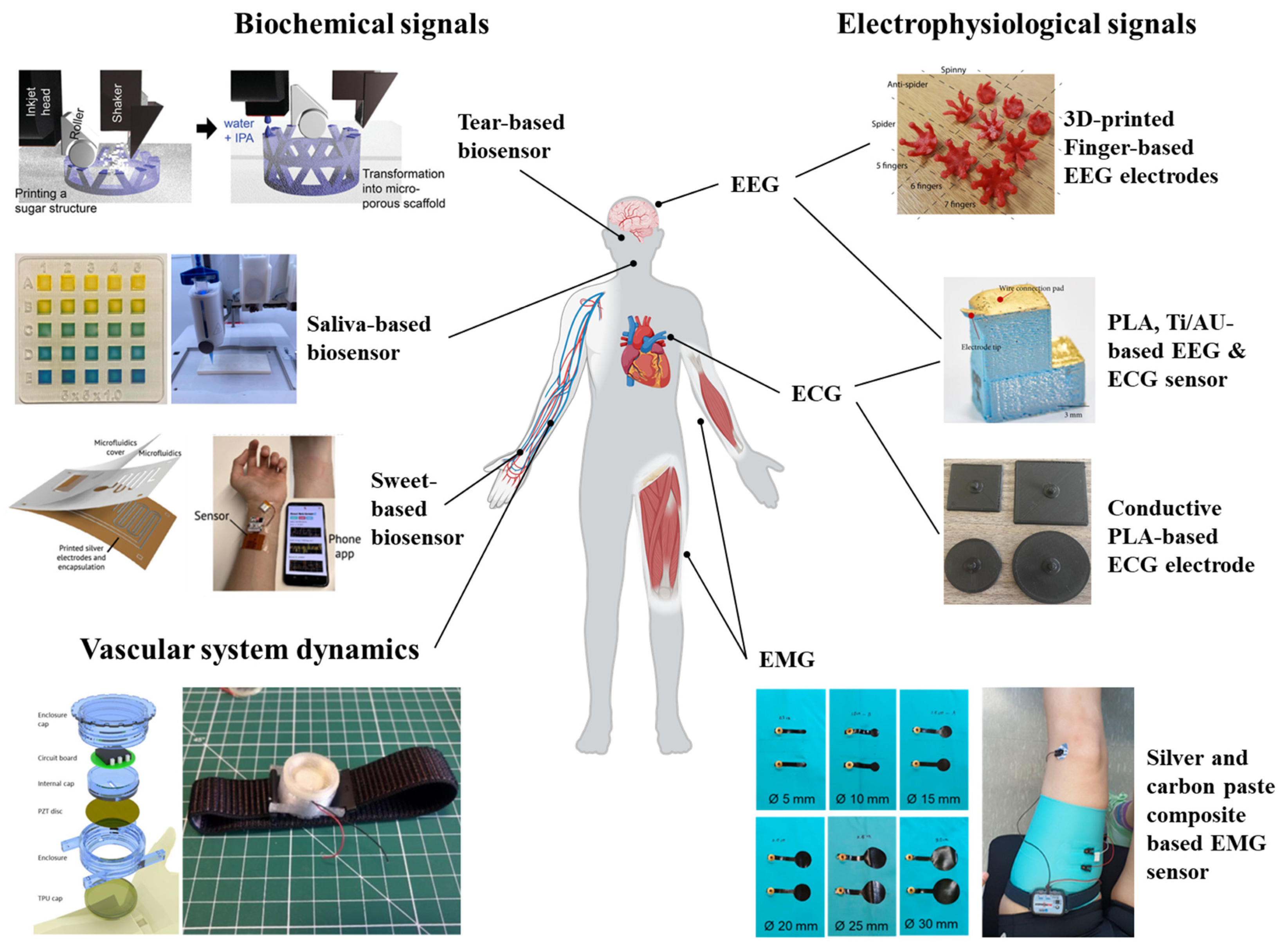
Three-dimensional-printed biosensors have demonstrated remarkable potential for continuous health monitoring and disease management across various clinical contexts. This continuous real-time monitoring is mostly suitable when used for skin-wearable sensor applications. In this section, we have highlighted some significant contributions of 3D printing technologies towards developing wearable biosensors based on biophysical and biochemical signals. In this review, physiological signals are divided into three subtypes: (i) electrophysiological signals, (ii) biochemical signals, and (iii) vascular systems. A schematic with the application of 3D-printed biosensors on the types of physiological signals is presented in Figure 5.

5.1. Electrophysiological Signals

Electrophysiological signals are electrical manifestations of biological processes, particularly from the brain, heart, muscles, and nerves. These signals are crucial for diagnosing and monitoring medical conditions. Depending on the sensing position, it is possible to monitor electrophysiological signals from the skin. Electrocardiography (ECG), electromyography (EMG), and electroencephalography (EEG) are examples of electrophysiological biosignals that are frequently monitored.
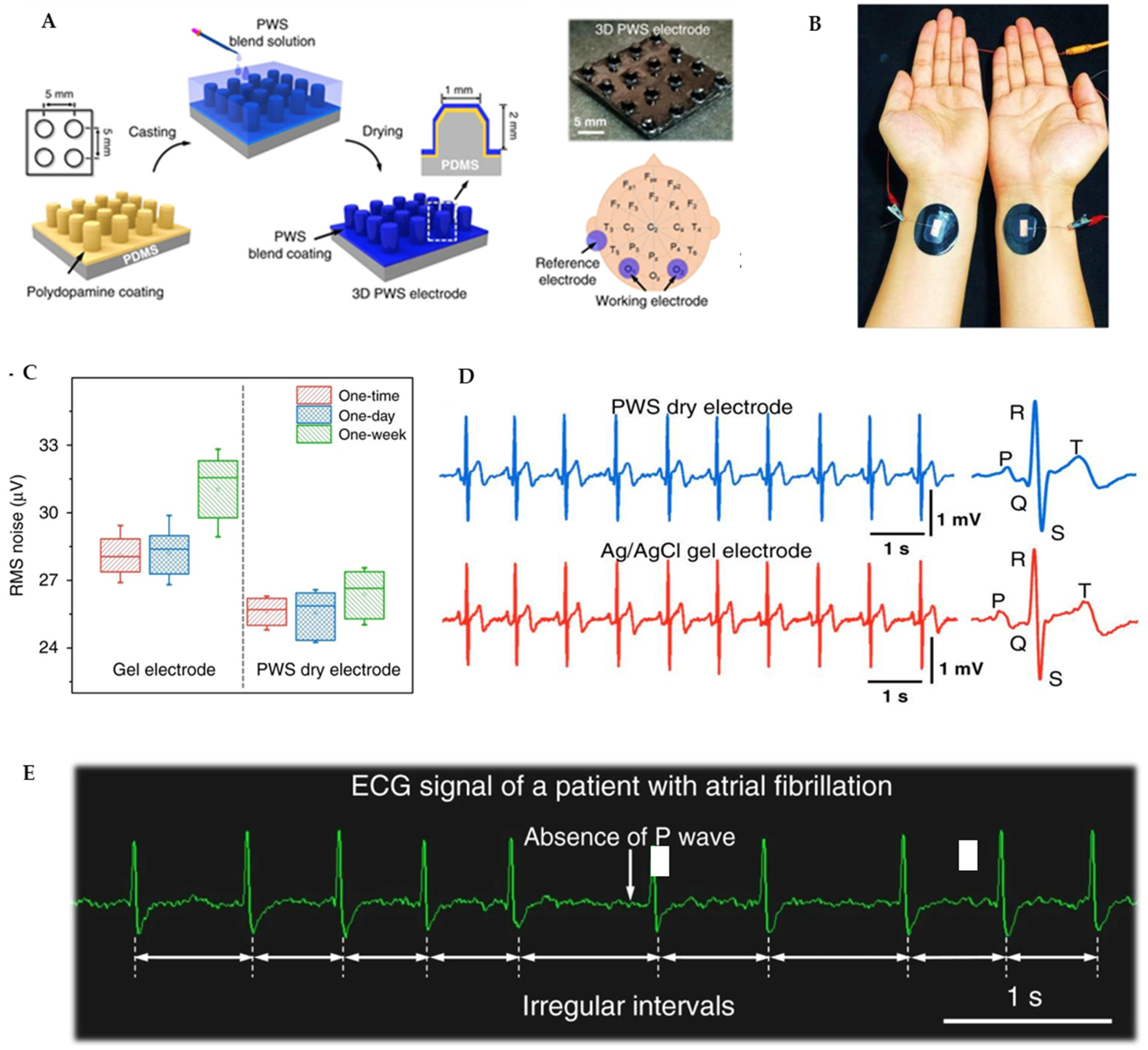
Electrocardiography (ECG) measures the heart’s electrical activity and rhythm by detecting the signals produced with each heartbeat. This diagnostic tool helps identify and track various cardiac conditions. Traditional ECG monitoring typically relies on wet electrodes that use conductive gels to improve electrical conductivity between the skin and the electrode. However, these conventional approaches present limitations for extended monitoring periods, including skin irritation, signal degradation over time, and user discomfort. In response to these challenges, 3D printing technology helps to develop dry electrodes that offer better comfort while maintaining signal quality for long-term cardiac monitoring. Microneedle array electrodes represent a key advancement in dry electrode technology, as their design allows penetration into the epidermis, reducing the insulating effect of the stratum corneum and significantly lowering skin–electrode impedance. In a notable approach, 3D-printed PLA molds with cylindrical holes (≈1.5 mm diameter and 2 mm depth) were used to form micro-pillars by casting a PEDOT:PSS–WPU–D-sorbitol blend, curing at ~60 °C, and peeling off the patterned film [147] (Figure 6A–E). This fabrication yields electrodes with well-defined microstructures that achieve impedance levels comparable to or lower than traditional wet electrodes. The experimental results further confirm their superior ability to provide high-quality recordings of essential ECG components compared to conventional surface electrodes. In another study, Aloqalaa et al. evaluated the performance of 3D-printed ECG dry electrodes fabricated using four commercially available polylactic acid (PLA) conductive filaments [142]. The researchers designed, built, and tested electrodes specifically for acquiring ECG signals, comparing their performance based on signal-to-noise ratio (SNR) and their ability to accurately measure heart rate using the Pan–Tompkins algorithm. All the printed electrodes demonstrated acceptable efficiency, achieving SNR values equal to or exceeding 18.89 dB—a threshold that indicates sufficient signal quality for reliable heart rate measurement. In another study, the scientists developed a 3D-printed sensor capable of measuring both electroencephalogram (EEG) and electrocardiogram signals from zebrafish [141]. This approach demonstrated the potential for creating highly miniaturized biosensors suitable for small animal studies, which had previously been challenging due to size constraints. The work is particularly noteworthy considering the extremely small cranial area of zebrafish (approximately 2 mm × 2 mm), which requires exceptional precision in electrode placement and signal detection. Another recent study demonstrated the fabrication of wet electrodes using screen printing with silver nanowire ink, electrode gel, and gentle adhesive on flexible substrates [148]. The system integrated (1) multilayer screen-printed flexible electrodes with gel and biocompatible adhesive, (2) a compact 3D-printed wireless AoP readout connected via pogo pins, and (3) mobile/cloud-based analytics for continuous monitoring. Tested on 20 volunteers, the printed electrodes were rated significantly higher in comfort and ease of removal compared to commercial electrodes, highlighting the potential of 3D-printed, conformable electrodes for applications beyond EEG, including ECG. However, limitations remain, such as (1) performance degradation from gel drying (wet electrodes) or sweat/dirt accumulation (dry electrodes), (2) higher resistance in fully 3D-printed dry electrodes with conductive filaments, and (3) noise artifacts from EMG interference and baseline drift during motion [142]. Looking ahead, advances in hybrid nanomaterial inks (e.g., graphene and advanced polymer blends), flexible electronics for ultra-low impedance, and miniaturized, energy-efficient modules could enable true patch-like wearables. Moreover, direct printing onto garments or biocompatible hydrogels opens pathways toward next-generation “second-skin” or implantable monitoring systems [149].
Electromyography (EMG) is a diagnostic procedure that evaluates the health of muscles and the nerve cells (motor neurons) that control them. This technique involves recording and analyzing the electrical activity generated by skeletal muscles during contraction and relaxation. When muscles activate, they produce electrical signals that can be detected and measured, providing valuable information about muscle function and neuromuscular communication pathways. Traditional EMG measurements occur in clinical settings using either needle electrodes inserted directly into the muscle (invasive EMG) or surface electrodes placed on the skin overlying the muscle (non-invasive surface EMG or sEMG). Surface EMG has become particularly significant in the development of wearable biosensors as it allows for non-invasive, continuous monitoring of muscle activity. This approach forms the foundation for the expanding field of wearable EMG technology that enables monitoring outside clinical environments during everyday activities. Despite their promise, wearable EMG systems face several technical challenges. Maintaining stable skin–electrode contact during movement is particularly difficult, as poor contact can introduce motion artifacts and degrade signal quality [150]. To address this problem, Wan et al. developed a high-performance bioelectronic interface created using 3D printing of a novel poly (vinyl alcohol-formaldehyde) (PVAF)-PEDOT:PSS composite ink. This ink supports the precise printing of complex structures and yields a hydrogel interface with excellent conductivity, strong adhesion, and stable electrochemical properties (over 100 S/m). High adhesion (~31 kPa) and near-skin mechanical modulus prevent delamination and minimize motion-induced artifacts. The electrode comfortably conforms to dynamic skin movements, a distinct improvement over rigid or less-adhesive alternatives. When tested in EMG recording, the hydrogel electrodes outperform commercial types (such as Ag/AgCl) in signal stability, maintaining a high signal-to-noise ratio (>10 dB) under variable stresses and repetitive motion [151]. The integration of electrodes into textiles represents another significant advancement in wearable EMG technology. These “textrodes” can be incorporated directly into clothing, improving comfort and enabling unobtrusive monitoring during daily activities. Studies have shown that factors such as the electrode shape, fabric density, and applied pressure significantly affect signal quality [150]. For instance, wave-type embroidered electrodes have demonstrated greater morphological stability than circular designs, maintaining their shape even under strains up to 30% [152]. Additionally, research indicates that a minimum pressure of approximately 10 mmHg is necessary for textile electrode performance that is comparable to conventional Ag/AgCl electrodes [143]. Textrodes excel in wear comfort and enable continuous, unobtrusive monitoring in real-life settings. In a study, Li et al. developed a photocurable 3D printing approach for manufacturing graphene-enhanced polymer electrodes with programmable geometries. When incorporated into textile substrates, these printed nanocomposites demonstrated clinical-grade biosensing capabilities for both cardiac (ECG) and muscular (EMG) activity monitoring while maintaining comfort and durability for everyday use [153]. Another significant advancement in wearable EMG technology has been the development of ultra-low power systems that extend a battery’s life while maintaining high sampling rates. One notable approach uses two different frequency microcontroller clock sources and a ping-pong buffer memory architecture to achieve significant power savings [154]. These optimizations have resulted in power consumption reductions of up to 92.72% compared to commercial devices, with corresponding increases in battery life by more than nine times. Such advancements make continuous, long-term EMG monitoring increasingly practical for everyday use. Wearable EMG technologies are rapidly evolving with innovations in hydrogels, textiles, and nanocomposites that improve comfort, adhesion, and signal quality. However, challenges remain, including material instability from environmental changes, limited durability under mechanical stress, pressure-dependent signal quality in textiles, and unresolved concerns about biocompatibility, manufacturing scalability, and cost. Despite these hurdles, research is advancing toward hybrid and self-healing materials, multifunctional smart textiles, and fully integrated garments that combine sensing, electronics, and wireless connectivity.
Electroencephalography (EEG) measures the brain’s electrical activity through electrodes placed on the scalp, capturing postsynaptic potentials from cortical neurons. Traditional rigid EEG electrodes often cause discomfort, especially in hairy regions. Three-dimensional printing now enables flexible electrodes using materials like thermoplastic polyurethane (TPU), which adapt to scalp contours while maintaining conductivity. These non-conductive electrodes are coated with silver/silver-chloride (Ag/AgCl) to reduce contact impedance and noise, outperforming earlier rigid designs [155]. Few studies have also shown that 3D-printed fingered electrodes may be customized and that various electrode configurations can be employed for various individuals or for various head regions [140]. The flexible bases can part hair without snapping, enhancing long-term usability. Conductive filaments like Multi3D Electrifi allow fully 3D-printed dry electrodes, eliminating post-printing processing. These electrodes achieve contact impedances below 550 Ω (20 Hz–10 kHz) and successfully detect alpha wave fluctuations during eye-open/closed states [156]. Their customizable shapes cater to individual anatomies, addressing fit issues common in standardized designs. In another interesting study, researchers have transformed earbuds into EEG devices using 3D-printed stretchable sensors [157]. These conform to the ear’s dynamic structure, measuring brain activity and sweat lactate simultaneously. This dual sensing aids in epilepsy monitoring and performance tracking during exercise. The ear’s proximity to the brain and sweat glands makes it ideal for unobtrusive, high-fidelity data collection. These innovations open pathways for unobtrusive systems in healthcare, performance tracking, and even space or mobility-constrained settings. However, challenges remain in durability, signal stability under motion, scalability, and standardization, as customized geometries complicate universal benchmarking. Looking forward, hybrid sensing platforms, smart self-healing materials, and AI-guided optimization could enhance reliability and personalization, ultimately driving EEG technology toward robust, long-term, and user-friendly applications in both clinical and real-world environments. A compilation of studies demonstrating biosensing applications for electrophysiological signals with 3D-printed wearable biosensors is provided in Table 3.

5.2. Biochemical Signals

Traditional methods for measuring biochemical signals from the body have often relied on invasive techniques, primarily involving the extraction and analysis of blood samples. However, the limitations and inconvenience associated with these procedures spurred the development of non-invasive approaches utilizing alternative biofluids like saliva, tear fluid, and sweat. The ability to access biochemical information through these less intrusive means has paved the way for continuous and non-invasive monitoring, a capability that has significantly fueled the burgeoning field of wearable technology, enabling real-time health and wellness tracking.
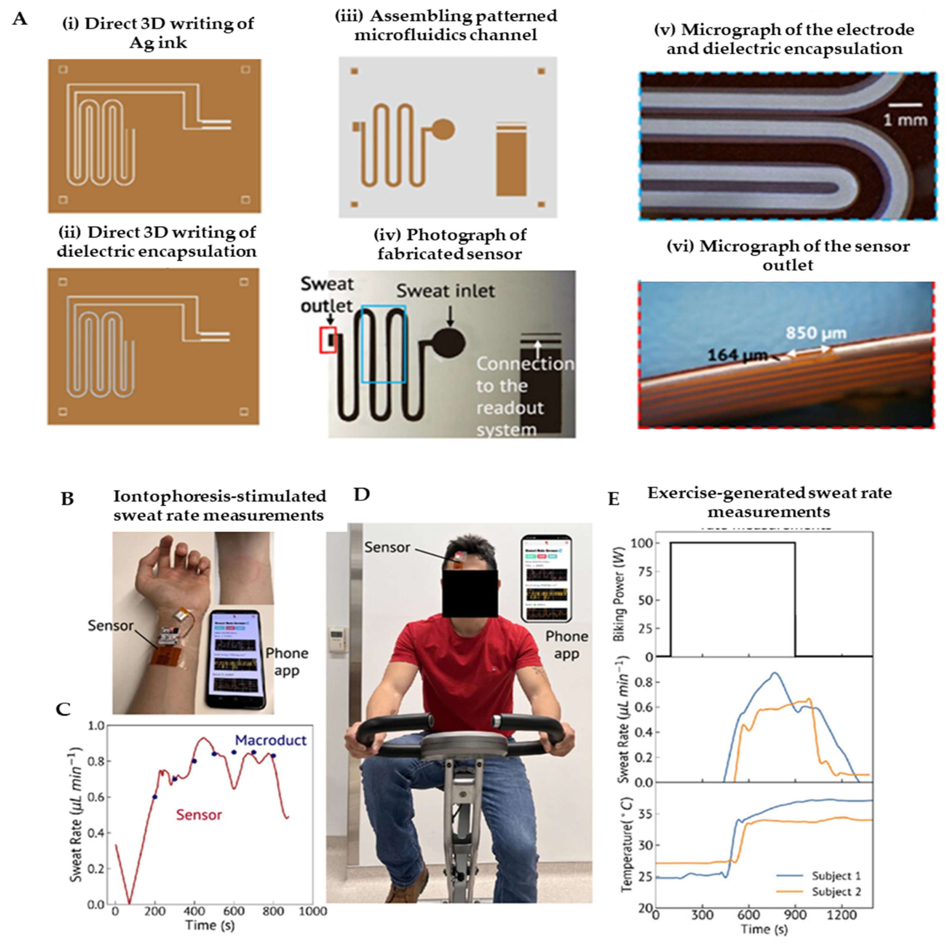
Sweat-based biosensors have emerged as a pivotal tool for non-invasive, real-time health monitoring, leveraging sweat’s rich content of biomarkers like electrolytes, metabolites, and hormones. These biosensors use electrochemical platforms, such as potentiometric and amperometric sensors, to detect key indicators like lactate, glucose, and cortisol, offering insights into physiological states, including stress and fatigue [174]. Additionally, electrolytes such as chloride and copper, as well as pH levels in sweat, have been successfully measured using 3D-printed microfluidic systems with integrated microcuvettes [175]. Islam et al. created a flexible sweat rate sensor utilizing 3D-printed microfluidic channels combined with capacitive electrodes on a flexible substrate (Figure 7A–E). The device achieved a high sensitivity of 0.01 μL/min, enabling real-time hydration tracking that was seamlessly integrated with a mobile application for data visualization. Its major advantages include the capacity for continuous monitoring and its high sensitivity, making it an ideal tool for athletes and workers in labor-intensive environments. A key limitation is that the sensor primarily measures the sweat volume and flow rate without providing data on biochemical composition; furthermore, its performance under fluctuating ambient humidity conditions requires further validation [145]. Complementing this, Liu et al. designed an eco-friendly chromic device, fabricating a 3D-printed flexible patch that incorporated materials that visually change color in response to UV exposure, temperature, and sweat pH. The device produced stable visible readouts and was lauded for its fully recyclable and environmentally conscious design. Its primary advantages stem from the complete absence of electronic circuitry, which drastically reduces both cost and electronic waste, while also enabling intuitive visual interpretation. However, a significant limitation is its inherent lack of quantitative precision, as interpreting color shifts can be subjective, and the platform is not multiplexed for the detection of numerous analytes simultaneously [176]. Koukouviti et al. developed an enzyme-free glucose sensor featuring a 3D-printed PLA electrode modified with an Fe(III) cluster for direct voltammetric detection [177]. The sensor demonstrated selective glucose quantification within the acidic environment of sweat (pH~4–6) and maintained excellent stability by eliminating reliance on fragile biological enzymes. Its key advantages include overcoming a major hurdle in wearable biosensing—enzyme instability—while utilizing a low-cost PLA substrate. A primary limitation is that the study exclusively focused on glucose, leaving its performance and potential cross-reactivity in the complex, multi-analyte milieu of real sweat unclear. The study has a significant potential in expanding this robust enzyme-free platform to detect other biomarkers like lactate, cortisol, or uric acid, thereby enabling the development of comprehensive multi-analyte sensing devices. In another study, Mi et al. combined MOF-derived porous carbon nanorods with a 3D-printed microfluidic chip to enable simultaneous monitoring of uric acid and potassium ions in sweat, showcasing the platform’s multiplexing potential and high analytical performance [178]. Across recent studies, 3D printing is demonstrably democratizing biosensor fabrication by offering highly customizable, scalable, and low-cost production methods. However, key limitations persist, including a lack of standardization in biomarker thresholds, susceptibility to environmental interference like variable sweat rates and pH, and a general inability to effectively multiplex beyond one or two analytes. Many devices also remain in early validation stages, tested under controlled conditions rather than during prolonged real-life use, and some still depend on external readers. Future directions are consequently focused on developing fully integrated multiplex systems that combine the sweat rate, metabolites, and electrolytes, leveraging AI for data interpretation and personalized calibration to individual physiology.
Saliva-based biosensors are gaining significant traction as non-invasive, point-of-care diagnostic tools capable of detecting a wide array of biomarkers, from metabolic and infectious diseases to stress and hormonal imbalances. These sensors utilize the rich biochemical milieu of saliva—including electrolytes, enzymes, nucleic acids, and metabolites—to enable real-time health monitoring with minimal patient discomfort. Electrochemical biosensors, particularly organic electrochemical transistors (OECTs) and aptamer-based platforms, offer high sensitivity and have been successfully deployed to detect analytes like glucose, uric acid, and SARS-CoV-2, demonstrating potential as alternatives to conventional blood-based diagnostics [179,180]. Several 3D printing methods have demonstrated utility in creating saliva-based sensors. Fused deposition modeling (FDM) has emerged as a versatile approach, enabling the production of highly customizable and complex geometries (e.g., microfluidic channels and multi-analyte systems) that are essential for advanced clinical diagnostics. A significant trend is the development of bespoke conductive filaments (e.g., PLA composites with graphene, carbon nanotubes, or metal nanoparticles), which are tailored to enhance electrochemical performance by optimizing conductivity and biocompatibility, surpassing the limitations of commercial filaments like Black Magic® or Proto-Pasta® that often contain impurities affecting sensor reliability [118]. Wrobel Von Zuben et al. developed a 3D-printed amperometric sensor using a polylactic acid (PLA) thermoplastic composite infused with graphene flakes for the enzyme-free detection of ethanol in saliva samples. The fused deposition modeling (FDM) technique enabled the fabrication of electrodes that demonstrated strong sensitivity and reproducible signals, leveraging the conductive properties of graphene within the PLA matrix to facilitate efficient electron transfer during ethanol oxidation. A key advantage of this non-enzymatic design is its avoidance of the stability issues commonly associated with biologically modified electrodes, enhancing durability and reducing operational complexities. However, the study noted that the dispersion quality of graphene flakes within the PLA filament significantly influenced electrical conductivity and sensor-to-sensor reproducibility, highlighting a critical manufacturing challenge that requires further optimization [181]. These composite materials combine the printability of thermoplastics with the electrical conductivity and sensing capabilities of nanomaterials. Sunil et al. developed a sophisticated 3D-printed microfluidic SERS biosensor platform incorporating Cu@Ag core–shell nanoparticle-decorated carbon nanofibers (Cu@Ag/CNFs) for the non-invasive, label-free detection of salivary biomarkers associated with oral cancer [182]. The 3D-printed system demonstrated strong Raman enhancement, enabling high-sensitivity identification of biomarkers in both simulated and clinical saliva samples with a high signal-to-noise ratio. The advantages of the system include the platform’s capacity for high-throughput screening and its integration with AI-assisted spectral analysis, which enhances diagnostic accuracy by automating the interpretation of complex Raman spectra. However, challenges remain in the fabrication complexity and batch-to-batch reproducibility of nanoparticle deposition, while the cost of the device currently exceeds that of conventional electrochemical biosensors. Another recent development is a 3D bioprinted hydrogel sensor for salivary pH detection, employing a sodium alginate–polyvinylpyrrolidone matrix and bromothymol blue to provide a robust colorimetric response in the pH range of 3.5 to 9.0 [146]. This design supports easy visualization and digital analysis via RGB component extraction, making it suitable for wearable intraoral applications. These innovations underscore the versatility of 3D printing for tailoring sensor architecture, improving sensitivity, and enhancing user compliance. However, key limitations remain, such as biofouling from salivary mucins and enzymes, variability between individuals requiring personalized calibration, and a general lack of multi-analyte capability in current devices. Looking forward, the integration of hybrid detection modalities, machine learning for biomarker pattern recognition—as seen in AI-aided SERS platforms—and the development of self-powered intraoral wearables present promising pathways toward standardized, energy-autonomous, and clinically validated salivary diagnostic systems for continuous health monitoring.
Another emerging transformative tool in non-invasive diagnostics is tear-based biosensors. These biosensors harness ocular platforms, particularly smart contact lenses and eye patches, to continuously monitor biomarkers such as glucose, lactate, electrolytes, pH, and proteins. Although it is still an emerging field, 3D-printed wearable tear-based biosensors are showing immense promise in non-invasive, real-time health monitoring, particularly via smart ocular platforms. While specific studies directly focusing on 3D-printed tear biosensors are limited, several reviews and technology demonstrations indicate a strong potential for adapting 3D-printed methods for tear analysis. Kalkal et al. highlight advancements in the 3D printing of biosensors for wearable healthcare, noting the feasibility of miniaturized devices for ocular use through vat photopolymerization and material extrusion—techniques that are compatible with soft bio-functional materials that are suitable for tear interfaces [183]. Rachim and Park expand on this by discussing in situ 3D printing directly onto curved, non-planar surfaces, like the human eye, enabling personalized, bio-integrated sensors for tear fluid detection [184]. Ho et al. demonstrated 3D-printed sugar-based scaffolds for wearable sensors, which offer high flexibility and biocompatibility—essential traits for ocular applications—though these are not yet specifically applied to tear fluid [110]. The integration of piezoelectric elements to power 3D-printed sensors, as shown by Sobianin et al., also hints at future self-powered tear biosensing platforms [144]. While 3D-printed tear biosensors are still largely conceptual, these foundational studies support their eventual realization, potentially via soft contact lenses or eye patches tailored through additive manufacturing. Table 4 summarizes studies that explore biosensing applications of 3D-printed wearable biosensors for biochemical signal monitoring.

5.3. Vascular System Dynamics

Vascular dynamics refer to the complex patterns of blood flow through the body’s circulatory system. These dynamics are governed by fluid–structure interactions between blood and vessel walls, creating measurable waveforms that contain valuable diagnostic information. Continuous monitoring of these dynamics enables early detection and prevention of cardiovascular diseases, which remain a leading cause of mortality worldwide.
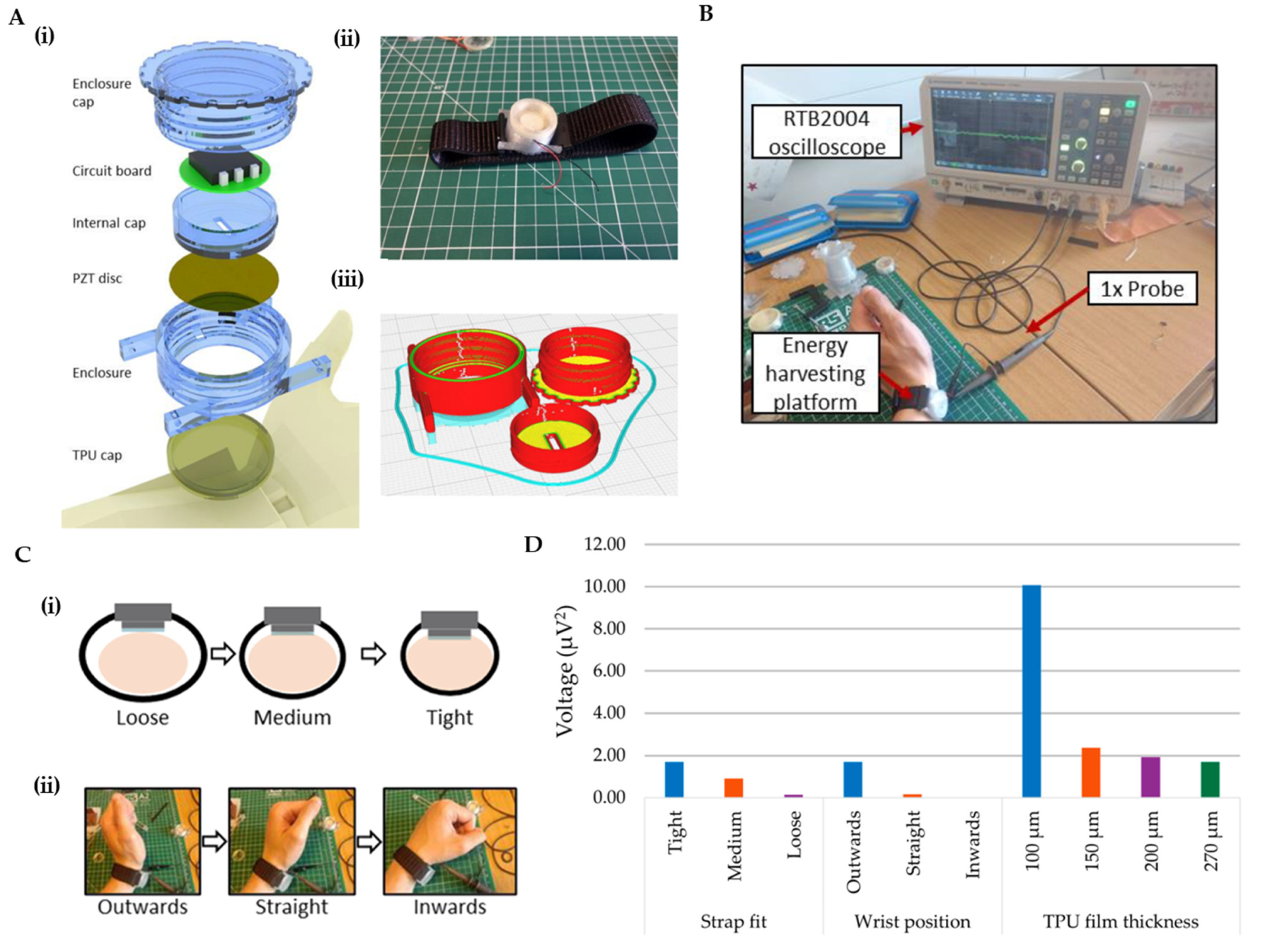
Multiple sensing mechanisms have been employed in 3D-printed wearable devices for detecting vascular-related biosignals. Pulse-wave monitoring represents one of the most common applications for 3D-printed wearable vascular sensors. Self-powered, high-sensitivity printed e-tattoo sensors have been developed for unobtrusive arterial pulse-wave monitoring [206]. Continuous blood pressure monitoring is crucial for real-time assessment and early prevention of cardiovascular diseases. Wearable continuous blood pressure monitoring devices based on pulse-wave transit time have received significant attention due to their excellent dynamic response characteristics and high accuracy [207]. These systems typically combine photoplethysmogram (PPG) and ECG measurements to calculate the transit time between arterial sites, which correlates with blood pressure. Numerous clinical and consumer devices measure the PPG signal, which has become a useful tool for determining the age and function of the arteries. Variations in blood pressure, atherosclerosis, changes in arterial stiffness, and natural vascular aging all affect the PPG pulse’s waveform and timing [207,208]. Piezoelectric sensing technologies have been incorporated into 3D-printed wearable devices to detect mechanical vibrations originating from arterial pulsations (Figure 8A–D). These components can not only sense vascular activity but also harvest energy from arterial pulsations, potentially enabling self-powered operation. This dual functionality makes piezoelectric-based sensors particularly attractive for long-term monitoring applications [144]. Three-dimensional-printed wearable ring sensors incorporating MEMS piezo-resistive pressure sensors have demonstrated accurate monitoring of real-time human blood pressure pulse waveform as an indicator for cardiovascular conditions [209]. Their ring achieved an accurate heart rate (HR) and HRV readings that are comparable to a clinical ECG strap and could track the entire blood pressure pulse’s waveform over long-term wear. Because it captures the waveform shape, it has potential for early hypertension screening: the authors noted the ring could detect subtle changes or abnormalities in the waveform, enabling early diagnosis. Phonoangiography (PAG) represents another valuable sensing approach that captures acoustic signals generated by blood flow through vessels. Custom-designed 3D-printed wearable devices combining PAG and PPG techniques have shown promise for the early and accurate detection of arteriovenous access (AVA) stenosis in hemodialysis patients. Research has demonstrated that after percutaneous transluminal angioplasty (PTA), the amplitudes of both PAG and PPG signals increased in patients, corresponding with improved blood flow [210].
The materials used in 3D-printed vascular monitoring devices significantly influence their performance, comfort, and durability. Conductive hydrogels have emerged as promising materials for 3D-printed wearable sensors due to their excellent biocompatibility, flexibility, and electrical conductivity [211]. These materials can be 3D-printed while maintaining their conductive properties, making them ideal for direct skin contact applications like pulse sensing. Elastic thermoplastic polyurethane (TPU) films are frequently incorporated to provide better skin adherence, protect sensitive electronic components, and electrically isolate the device from the human body. In one notable application, a TPU film forms an air chamber between the skin and the piezoelectric disc electrode, improving adsorption to the skin while preventing damage to the piezoelectric component [144]. Triboelectric materials have also been utilized in wearable sensors for pulse wave monitoring. Wearable triboelectric nanogenerator (TENG)-based sensors offer compelling advantages, including self-powered operation, lightweight construction, and superior sensitivity [212]. Table 5 presents a list of studies on biosensing applications for signals from vascular dynamics using 3D-printed wearable biosensors. A compilation of studies demonstrating biosensing applications due to body mechanical deformation (strain sensor), touch sense (tactile sensor), and other miscellaneous physiological signals with 3D-printed wearable biosensors is provided in Table 6.

6. Implantable Devices by 3D Printing Technology

Implantable biosensing devices represent a frontier in healthcare monitoring, offering direct access to physiological parameters and biomolecular markers within the body. These devices enable continuous monitoring of internal biological processes that cannot be accessed through non-invasive means, providing critical data for disease diagnosis and management. Recent advances in 3D printing have significantly expanded the possibilities for implantable sensor design, allowing for the creation of biocompatible structures that can interface effectively with surrounding tissues while minimizing foreign body responses. Three-dimensional printing methods, such as fused deposition modeling (FDM), stereolithography (SLA), selective laser melting (SLM), and direct ink writing (DIW), can fabricate structures from metals, polymers, hydrogels, or composites with customized shape and porosity. For example, FDM and SLA are widely used for electrode scaffolds and microfluidic chips, while DIW can extrude viscous inks (e.g., graphene or conductive polymers) for soft or tissue-like probes [248]. A notable innovation in this field is the development of 3D-printed liquid-core hydrogel platforms for encapsulating single-walled carbon nanotube (SWNT) sensors, which can detect important signaling molecules such as nitric oxide (NO) and hydrogen peroxide (H2O2) [249]. This approach addresses previous challenges related to sensor stability and compatibility with biological environments, providing a promising solution for long-term implantable molecular sensing.
Implantable biosensors typically rely on electrochemical or optical transduction. Electrochemical modes include amperometric (enzymatic or redox sensors), potentiometric (ion- or pH-sensitive), and impedance-based detectors [248]. For instance, implanted amperometric electrodes can monitor local neurotransmitter or metabolite levels in real time [250]. Optical approaches (fluorescence or luminescence) have also been integrated into 3D-printed scaffolds for reporter-based sensing. Other modalities (capacitive, piezoelectric, and thermal) are more common in mechanical/strain-sensing implants [248].

6.1. Implantable Biosensors for Neurological Applications

Neurological disorders represent a critical application area for implantable biosensors, as continuous monitoring of neural activity and neurotransmitters can provide invaluable insights for diagnosis and treatment. Momin et al. created a 3D-printed flexible neural probe with a porous, tissue-like silicone–carbon composite structure [251]. Their DIW device matched the brain’s compliance and achieved low impedance for high-fidelity recordings at the single-neuron level. Other groups have DIW-printed electrodes from graphene or PEDOT:PSS [252]. Shao et al. reported 3D-printed carbon nanoneedles (via photopolymerization and pyrolysis) that were sharp enough to measure dopamine release in a Drosophila nerve cord. Graphene-based 3D-printed sensors are especially promising for Parkinson’s monitoring. Dopamine depletion is a hallmark of Parkinson’s disease, so that implantable dopamine sensors could aid early diagnosis or therapy. Shin et al. created a minimally invasive system using a graphene electrode array in very encouraging research. This is the first integrated system to concurrently demonstrate biocompatibility, wearability, removability, target specificity, and wireless control [253]. Animal models of Parkinson’s disease have shown that cortical motor surface stimulation normalizes brain waves and restores motor function, which corresponds to potentiated synaptic responses. Moreover, the overexpression of the D5 dopamine receptor (D5R, Drd5) and metabotropic glutamate receptor 5 (mGluR5, Grm5) genes in the glutamatergic synapse is linked to these alterations. The wireless capabilities of this neural implant enable both real-time diagnostics and targeted therapeutics, suggesting significant potential for clinical applications in Parkinson’s disease treatment. A recent review notes extensive development of graphene-modified electrodes for dopamine detection [250]. Graphene can be formulated into printable inks or composites: for instance, a graphene/PLA filament was 3D-printed into a microneedle-like electrode array for ex vivo dopamine sensing. In general, such electrochemical implants would use chronoamperometry or voltammetry to detect neural transmitters or metabolites with high sensitivity and spatial resolution. These systems must also integrate with telemetry or onboard electronics to transmit data out of the body.

6.2. Bone Regeneration and Orthopedic Sensors

Three-dimensional printing has been widely applied to bone repair scaffolds and implants. Smart bone scaffolds can combine biocompatible matrices (e.g., titanium, ceramics, or polymers) with sensing functionality. For instance, Huang et al. demonstrated a 3D-printed porous scaffold of carboxymethyl chitosan reinforced with 0.5% (w/v) carboxylated carbon nanotubes (CNT) for bone defects [254]. This CNT/CMC scaffold was electrically conductive and exhibited “electrochemical responsiveness”: cyclic voltammetry and impedance spectra changed sensitively as osteogenic cells differentiated, thus reporting on new bone formation. Notably, the CNTs also enhanced scaffold strength and stimulated stem cell osteogenesis. This is an example of a biosensing scaffold—it does not have discrete electronics, but its material itself acts as a sensor. Other approaches embed miniature sensors into orthopedic implants. For example, Lavdas et al. designed a titanium orthopedic spacer with a cavity housing a temperature sensor and wireless telemetry for monitoring post-operative infection in knee arthroplasty [255]. The sensor electronics were encapsulated in a 3D-printed Ti case within the bone cement. Similarly, Feynman-track strain gauges and piezoelectric elements have been integrated into 3D-printed knee or spine implants for load sensing.
A clinical case report describes the use of a 3D-printed titanium mesh implant with a plate construct for managing critical-size bone defects in distal tibial open wounds [256]. The porotic nature of the mesh facilitated bone ingrowth, with CT scans at 1.5 years post-surgery confirming good bone integration and restored ambulation. This case demonstrates how 3D-printed implants can effectively integrate with biological tissues, suggesting potential for incorporating biosensing elements that could monitor bone healing processes in real-time. However, as noted by recent reviews, electrochemical sensors (chemical or biochemical) are still rare in orthopedics [248]. When used, implantable sensors may target pH (to detect inflammation/infection), phosphate or cytokine levels, or pressure (strain) at the bone–implant interface. In all cases, biocompatibility and long-term stability are major concerns—implants must function reliably for months or years without leaching or significant drift.

6.3. Tumor and Cancer Biomarker Sensors

Implantable sensors for oncology are an emerging frontier. The goal is to measure tumor-specific biomarkers (proteins, DNA, metabolites) in situ, enabling personalized therapy monitoring. One concept is an implantable electrochemical aptasensor: for example, a printed graphene-based electrode functionalized with a DNA aptamer for cancer antigen (like HER2 or MUC1) could be inserted near a tumor. Similarly, 3D-printed microdialysis probes or hydrogel reservoirs can continuously sample interstitial tumor fluid, feeding analytes to an on-chip sensor [248]. In general, electrochemical biosensors are well-suited to detect proteins, nucleic acids, or small molecules.

6.4. Biocompatibility, Stability, and Regulatory Considerations

Although additive manufacturing supports patient-specific geometries (e.g., matching bone defect topology) and rapid prototyping of multi-material systems; however, printing implants mandates rigorous material control and post-processing (such as cleaning, sterilization, and curing) to meet the biocompatibility and mechanical requirements [248]. Any implantable device must use biocompatible materials (ISO 10993 compliant) and be thoroughly sterilizable [257]. Metals (Ti, CoCr) and ceramics (hydroxyapatite, beta-TCP) used in bone implants have well-known biocompatibility. Polymers like medical-grade PLA/PCL, PEEK, parylene, polyimide, or silicone are also common. Three-dimensional printable hydrogels based on conducting polymers have emerged as promising materials for creating implantable bioelectronics with tissue-like mechanical compliance and robust electrochemical properties [258]. These materials offer advantages such as Young’s modulus values around 650 kPa (like soft tissues), strong bioadhesion properties (interfacial toughness of 200 J m−2 and shear strength of 120 kPa), and tunable electrical properties.
Longevity is a key challenge: implant sensors must operate for months or years without significant drift. Chronic implantation leads to protein adsorption and encapsulation by fibrotic tissue, which can degrade sensor signals. Strategies to mitigate fouling include anti-biofouling coatings (PEG or zwitterionic materials) or self-cleaning surfaces. Recent research has demonstrated promising advances in sensor longevity, with some 3D-printed hydrogel-encapsulated sensors maintaining stable performance for extended periods. For instance, (AT)15-wrapped-SWNT NO sensors encapsulated in 3D-printed self-healing hydrogels have shown a negligible decrease in fluorescence intensity after 90 days at 37 °C, with statistical analyses indicating that the change in intensity was not significant [249].
Reliability testing in vitro and in vivo (accelerated aging, bioreactor flow) is essential. For example, carbon electrodes may suffer surface oxidation; polymers may hydrolyze, and printed layers may delaminate. Thus, multi-month animal studies and benchtop soak tests are typically required before clinical use. In recent work, an electrophysiological study in rat heart models has demonstrated the capability of 3D-printed hydrogel bioelectronics to establish conformal interfaces with dynamic organs, enabling long-term and high-precision spatiotemporal epicardial monitoring [258]. These developments highlight the potential of 3D-printed implantable biosensors for chronic disease monitoring and management applications that require stable, long-term performance.
Three-dimensional-printed implantable sensors fall under medical device regulations (e.g., FDA Class III or EU MDR, depending on the risk). Regulatory bodies require demonstration of safety, efficacy, and manufacturing control. This includes adherence to ISO standards (biocompatibility ISO 10993, sterilization ISO 11135/11737, electrical safety ISO 60601) and good manufacturing practices [257]. For additive-manufactured devices, the FDA has issued guidance (2017) emphasizing the validation of printing processes, material traceability, and post-process quality control [259]. Custom or patient-specific implants must follow stringent design controls. In practice, this means every novel printed implantable sensor needs bench testing (electrical/analytical performance), biocompatibility studies, and often animal studies (to show no toxicity or adverse tissue response) before human trials.
Despite the hurdles, several 3D-printed implants (mostly orthopedic scaffolds and dental implants) have already reached clinical use, indicating feasibility [260]. The field of 3D-printed biosensors is younger, but momentum is growing. Combining additive manufacturing with advances in flexible electronics and nanomaterials holds promise for next-generation “smart implants” that monitor health from within the body. In conclusion, 3D printing provides unique advantages (such as customization, integration, and speed) for implantable biosensors, and recent studies have demonstrated prototypes in bone, neural, and microfluidic domains [250,254,261].

7. Challenges and Future Perspectives

Recent advancements in 3D printing technologies have revolutionized the development of biosensing devices, offering unprecedented opportunities for personalized health monitoring through wearable and implantable sensors. These innovations enable continuous, real-time monitoring of physiological and biochemical parameters, potentially transforming disease diagnosis and management. Despite these advances, numerous challenges persist regarding material selection, printing resolution, biocompatibility, and long-term reliability.

7.1. Material Challenges in 3D Printing of Biosensors

Material selection represents a fundamental challenge in 3D printing of biosensing devices, as the materials must simultaneously satisfy requirements for printability, sensor functionality, and biocompatibility. Traditional 3D printing materials often lack the necessary electrical, optical, or chemical properties required for effective biosensing applications. For instance, stereolithographic (SLA) photopolymers typically yield parts with low mechanical compliance, which are unsuitable for applications requiring tissue-like flexibility. Researchers have addressed this challenge by developing tunable resins with polydimethylsiloxane (PDMS)-like elastic modulus for stereolithographic 3D printing, enabling the creation of more compliant structures for biosensing applications [262]. Additionally, material compatibility with different 3D printing methods presents a significant bottleneck in the development of functional biosensing devices [123]. Each printing technique (extrusion-based, stereolithographic, inkjet, etc.) imposes specific requirements on material viscosity, curing mechanisms, and thermal properties, limiting the range of viable materials for specific biosensor designs.
The incorporation of nanomaterials into printable formulations offers enhanced functionality but introduces additional challenges related to dispersion stability and printing reliability. Nanocomposites incorporated into 3D printing for biosensors include carbon nanotubes, metal nanoparticles, and conductive polymers, which can significantly enhance sensor sensitivity and selectivity [107]. However, achieving uniform dispersion of these nanomaterials within printing resins or inks without agglomeration remains challenging, particularly for high-aspect-ratio nanomaterials such as carbon nanotubes. The tendency of nanomaterials to settle or aggregate during the printing process can lead to inconsistencies in sensor performance across different parts of the printed structure. Furthermore, the incorporation of nanomaterials can alter the rheological properties of printing inks, potentially compromising printability and resolution [263]. These challenges necessitate careful formulation development and process optimization to ensure reliable production of functional nanocomposite-enhanced biosensors.
The biocompatibility and biofunctionality of printed materials present another layer of complexity, particularly for implantable biosensing applications. Materials must not only be non-toxic and non-immunogenic but also maintain their functional properties in the biological environment over extended periods. The development of biomaterials capable of withstanding a harsh physiological environment while maintaining sensing capabilities requires careful consideration of surface chemistry, degradation behavior, and protein adsorption characteristics [123]. Additionally, the potential release of unreacted monomers, photoinitiators, or degradation products from printed materials can cause adverse biological responses, necessitating thorough biocompatibility evaluations. Recent advances in this area include the development of 3D printable hydrogels with self-healing properties and robust bioadhesion, which can establish stable interfaces with dynamic biological tissues [258]. Despite these advances, the limited range of biocompatible materials suitable for different 3D printing technologies continues to constrain the design space for wearable and implantable biosensing devices.

7.2. Technical Challenges in 3D Printing of Biosensors

Printing resolution remains a significant technical challenge in the fabrication of biosensing devices, particularly for applications requiring microscale or nanoscale features. While commercial 3D printers have made remarkable progress in resolution capabilities, there still exists a substantial gap between the resolution achievable with current 3D printing technologies and the feature sizes required for optimal biosensor performance [123]. This limitation is particularly pronounced for sensors targeting the detection of small biomolecules or requiring high-density electrode arrays. High-resolution techniques such as two-photon polymerization can achieve sub-micron features but suffer from limited throughput and material compatibility issues.
Multi-material printing capability is essential for creating integrated biosensing systems, but it introduces numerous technical complexities. Biosensors typically require the integration of materials with disparate properties-such as conductive elements for signal transduction, flexible substrates for comfort and conformability, and bioactive components for molecular recognition [107]. Researchers have developed approaches such as “Pause-Print” protocols (3P-printing) to fabricate high-resolution multi-material parts with desktop SLA printers without requiring post-assembly [261]. However, challenges persist related to material compatibility, adhesion between different materials, and preventing cross-contamination during the printing process. The integration of electronic components and circuitry with 3D-printed structures presents another layer of technical complexity. Most biosensors require not only the sensing element itself but also associated electronics for signal conditioning, processing, and transmission [264]. Recent innovations have explored approaches for printing electronic circuits directly within 3D structures using conductive inks or embedding pre-fabricated electronic components during the printing process [258]. However, these approaches face challenges related to ensuring proper electrical connectivity, preventing thermal damage to electronic components during printing, and maintaining mechanical integrity at the interface between rigid electronics and potentially flexible printed substrates.

7.3. Operational Challenges

Sensor performance and reliability under real-world conditions represent significant operational challenges for 3D-printed biosensing devices. Many biosensors demonstrate excellent performance in controlled laboratory environments but fail to maintain consistent functionality when exposed to the complex, dynamic conditions of actual use [264]. For wearable sensors, factors such as motion artifacts, skin perspiration, temperature fluctuations, and mechanical deformation during body movement can significantly impact sensor readings and reliability [107]. Implantable sensors face even more challenging conditions, including protein biofouling, immune responses, tissue encapsulation, and potential degradation in the physiological environment [249]. These challenges are compounded for 3D-printed sensors, as the layer-by-layer fabrication process can introduce structural heterogeneities that compromise mechanical integrity and sensing performance under stress.
Power management presents a critical operational challenge, particularly for wireless and implantable biosensing devices. The continuous operation of sensors for real-time health monitoring demands efficient power utilization strategies to extend device lifespan between charging cycles or battery replacements [264]. For implantable sensors, battery replacement typically requires invasive procedures, making long-term power sustainability especially crucial. Traditional approaches to power miniaturized sensors include small batteries, wireless power transfer, and energy harvesting from the environment or body [265]. However, the integration of these power solutions with 3D-printed structures introduces additional design and fabrication complexities. Recent research has explored the potential of 3D printing to create energy storage systems directly within sensor structures, as well as the development of self-powered devices that can harvest energy from physiological processes or environmental sources [123]. Despite these advances, achieving a balance between power consumption, sensor performance, and device size remains challenging, particularly for continuous monitoring applications that require frequent data acquisition and transmission.

7.4. Future Perspectives

The integration of artificial intelligence and machine learning with 3D-printed biosensing devices represents a promising frontier for enhancing sensor capabilities and clinical utility. As biosensors continue to generate increasingly complex and voluminous data, AI algorithms can help identify subtle patterns and correlations that might indicate early disease onset or treatment efficacy [264]. Machine learning approaches can also compensate for sensor limitations by filtering out noise, correcting drift, and improving overall accuracy through adaptive calibration techniques. Furthermore, AI-enabled personalization of sensor systems could allow for adapting detection parameters based on individual physiological baselines and health histories, leading to more meaningful and actionable health insights [266]. The combination of 3D printing’s customization capabilities with AI’s analytical power could enable the development of highly personalized biosensing systems tailored to individual patient needs, anatomical considerations, and specific health monitoring requirements. This convergence of technologies is likely to significantly enhance the clinical value of biosensing devices while expanding their applications across diverse healthcare settings.
Emerging materials and printing technologies are poised to revolutionize the capabilities of wearable and implantable biosensors. Advanced bioinks incorporating stimuli-responsive polymers, self-healing materials, and biomimetic structures are being developed to improve sensor biocompatibility, longevity, and functionality in complex biological environments [258]. For instance, 3D-printable hydrogels with self-healing properties and strong bioadhesion have demonstrated the ability to maintain stable interfaces with dynamic organs, enabling long-term electrophysiological monitoring. Novel 4D printing approaches, where printed structures can change shape or properties in response to environmental stimuli, offer exciting possibilities for creating adaptive biosensors that can respond to physiological changes or optimize their positioning within tissues [123]. Additionally, advances in high-resolution printing technologies, such as two-photon polymerization and microsterolithography, are enabling the fabrication of biosensors with nanoscale features, potentially leading to significant improvements in sensitivity, specificity, and miniaturization [263]. These materials and technological innovations are expanding the design space for biosensing devices while addressing many of the current limitations related to biocompatibility, durability, and performance.
The clinical translation and commercialization of 3D-printed biosensing technologies represent critical steps toward realizing their full potential in healthcare. While numerous innovative 3D-printed biosensors have been demonstrated in research settings, relatively few have progressed to clinical validation and commercial availability [107]. Bridging this translational gap requires addressing several key challenges, including scaling up manufacturing processes while maintaining quality and consistency, establishing regulatory pathways for novel device approval, and demonstrating clear clinical benefits and cost-effectiveness compared to existing monitoring approaches [264]. Additionally, developing sustainable business models that balance device affordability with manufacturing costs will be essential for widespread adoption, particularly in resource-limited healthcare settings [266]. Despite these challenges, the unique advantages offered by 3D-printed biosensing devices—including customization, rapid prototyping, and potential for point-of-care manufacturing—position them favorably for future clinical integration, potentially transforming approaches to disease monitoring, management, and personalized healthcare delivery.

8. Conclusions

The evolution of biosensor technology has traversed a remarkable journey from Clark’s pioneering glucose electrode in 1962 to today’s sophisticated wearable and implantable devices. This review has highlighted how 3D printing technology is revolutionizing biosensor fabrication, addressing critical limitations of conventional manufacturing methods. The unique capabilities of additive manufacturing—including design freedom, cost-effectiveness, rapid prototyping, and material versatility—have enabled the production of complex, miniaturized biosensing platforms that were previously unattainable. Particularly significant is 3D printing’s ability to create customized, patient-specific devices with intricate geometries while integrating multiple materials and sensing modalities into unified structures. These advantages have accelerated the development of flexible, stretchable, and lightweight biosensors that conform seamlessly to biological surfaces, enhancing both comfort and performance. As we look toward the future, the convergence of 3D printing with advanced nanomaterials, multifunctional bioinks, and AI-powered analytics promises to further transform biosensor technology. While challenges remain in material optimization, durability, and regulatory standardization, the trajectory is clear: 3D-printed biosensors are poised to dramatically expand point-of-care diagnostics, enable continuous health monitoring, and ultimately drive a new paradigm of personalized, proactive healthcare with profound implications for both clinical practice and patient outcomes.

Supplementary Materials

The following supporting information can be downloaded at: https://www.mdpi.com/article/10.3390/bios15090619/s1, Table S1. Classification of biosensors: components and detection method.

Author Contributions

Conceptualization, S.M. and M.K. and H.L.; methodology, S.M. and M.K.; writing—original draft preparation, S.M., M.K. and R.K.; writing—review and editing, S.M., M.K., R.K. and H.L.; supervision S.M. and H.L.; funding acquisition, H.L. All authors have read and agreed to the published version of the manuscript.

Funding

This work was supported by a National Research Foundation of Korea (NRF) grant funded by the Korea government (MSIT) (No. RS-2024-00423107). This research was also supported by a grant from the Korea Health Technology R&D Project through the Korea Health Industry Development Institute (KHIDI), funded by the Ministry of Health and Welfare, Republic of Korea (grant number: RS-2025-24535069).

Institutional Review Board Statement

Not applicable.

Informed Consent Statement

Not applicable.

Data Availability Statement

The data presented in this study are available on request from the corresponding author.

Conflicts of Interest

The authors declare no conflicts of interest.

References

  1. Thevenot, D.R.; Tóth, K.; Durst, R.A.; Wilson, G.S. Electrochemical Biosensors: Recommended Definitions and Classification. Pure. Appl. Chem. 1999, 71, 2333–2348. [Google Scholar] [CrossRef]
  2. Mehrotra, P. Monitoring and Control Blood and Tissue Oxygen. J. Oral. Biol. Craniofacial. Res. 2016, 6, 153–159. [Google Scholar] [CrossRef] [PubMed]
  3. Long, F.; Zhu, A.; Shi, H. Recent Advances in Optical Biosensors for Environmental Monitoring and Early Warning. Sensors 2013, 13, 13928–13948. [Google Scholar] [CrossRef] [PubMed]
  4. Yang, M.; Fu, D.; Gao, C.; Liu, Y. 2D Material-Based Electrochemical Sensors for Early Diabetes Detection: A Review of Progress and Prospects. Int. J. Electrochem. Sci. 2025, 20, 101123. [Google Scholar] [CrossRef]
  5. Sharma, S.K.; Leblanc, R.M. Biosensors Based on β-Galactosidase Enzyme: Recent Advances and Perspectives. Anal. Biochem. 2017, 535, 1–11. [Google Scholar] [CrossRef]
  6. Lakard, S.; Pavel, I.-A.; Lakard, B. Electrochemical Biosensing of Dopamine Neurotransmitter: A Review. Biosensors 2021, 11, 179. [Google Scholar] [CrossRef]
  7. Botewad, S.N.; Gaikwad, D.K.; Girhe, N.B.; Thorat, H.N.; Pawar, P.P. Urea Biosensors: A Comprehensive Review. Biotech. App Biochem. 2023, 70, 485–501. [Google Scholar] [CrossRef]
  8. Narwal, V.; Deswal, R.; Batra, B.; Kalra, V.; Hooda, R.; Sharma, M.; Rana, J.S. Cholesterol Biosensors: A Review. Steroids 2019, 143, 6–17. [Google Scholar] [CrossRef]
  9. Ding, Y.; Yang, L.; Wen, J.; Ma, Y.; Dai, G.; Mo, F.; Wang, J. A Comprehensive Review of Advanced Lactate Biosensor Materials, Methods, and Applications in Modern Healthcare. Sensors 2025, 25, 1045. [Google Scholar] [CrossRef]
  10. Sivagnanam, S.; Mahato, P.; Das, P. An Overview on the Development of Different Optical Sensing Platforms for Adenosine Triphosphate (ATP) Recognition. Org. Biomol. Chem. 2023, 21, 3942–3983. [Google Scholar] [CrossRef]
  11. Wankhade, U.A.; Thakare, Y.N.; Hardas, B.M.; Pande, R.S. Cortisol Detection Methods for Stress Monitoring: Current Insight and Future Prospect: A Review. IEEE Sens. J. 2024, 24, 23389–23400. [Google Scholar] [CrossRef]
  12. Bahadır, E.B.; Sezgintürk, M.K. Electrochemical Biosensors for Hormone Analyses. Biosens. Bioelectron. 2015, 68, 62–71. [Google Scholar] [CrossRef]
  13. Psoma, S.D.; Kanthou, C. Wearable Insulin Biosensors for Diabetes Management: Advances and Challenges. Biosensors 2023, 13, 719. [Google Scholar] [CrossRef] [PubMed]
  14. Negahdary, M.; Barros Azeredo, N.F.; Santos, B.G.; De Oliveira, T.G.; De Oliveira Lins, R.S.; Dos Santos Lima, I.; Angnes, L. Electrochemical Nanomaterial-Based Sensors/Biosensors for Drug Monitoring. CTMC 2023, 23, 295–315. [Google Scholar] [CrossRef] [PubMed]
  15. Garzón, V.; Pinacho, D.; Bustos, R.-H.; Garzón, G.; Bustamante, S. Optical Biosensors for Therapeutic Drug Monitoring. Biosensors 2019, 9, 132. [Google Scholar] [CrossRef]
  16. Velusamy, K.; Periyasamy, S.; Kumar, P.S.; Rangasamy, G.; Nisha Pauline, J.M.; Ramaraju, P.; Mohanasundaram, S.; Nguyen Vo, D.-V. Biosensor for Heavy Metals Detection in Wastewater: A Review. Food Chem. Toxicol. 2022, 168, 113307. [Google Scholar] [CrossRef]
  17. Sassolas, A.; Prieto-Simón, B.; Marty, J.-L. Biosensors for Pesticide Detection: New Trends. AJAC 2012, 03, 210–232. [Google Scholar] [CrossRef]
  18. Ali, A.A.; Altemimi, A.B.; Alhelfi, N.; Ibrahim, S.A. Application of Biosensors for Detection of Pathogenic Food Bacteria: A Review. Biosensors 2020, 10, 58. [Google Scholar] [CrossRef]
  19. Guliy, O.I.; Zaitsev, B.D.; Larionova, O.S.; Borodina, I.A. Virus Detection Methods and Biosensor Technologies. Biophysics 2019, 64, 890–897. [Google Scholar] [CrossRef]
  20. V, K. DNA Biosensors-A Review. J. Bioeng. Biomed. Sci. 2017, 07. [Google Scholar] [CrossRef]
  21. Du, Y.; Dong, S. Nucleic Acid Biosensors: Recent Advances and Perspectives. Anal. Chem. 2017, 89, 189–215. [Google Scholar] [CrossRef]
  22. Khanmiri, H.H.; Yazdanfar, F.; Mobed, A.; Rezamohammadi, F.; Rahmani, M.; Haghgouei, T. Biosensors; Noninvasive Method in Detection of C-Reactive Protein (CRP). Biomed. Microdevices 2023, 25, 27. [Google Scholar] [CrossRef]
  23. Gerdan, Z.; Saylan, Y.; Denizli, A. Biosensing Platforms for Cardiac Biomarker Detection. ACS Omega. 2024, 9, 9946–9960. [Google Scholar] [CrossRef] [PubMed]
  24. Mobed, A.; Shakouri, S.K.; Dolati, S. Biosensors: A Novel Approach to and Recent Discovery in Detection of Cytokines. Cytokine 2020, 136, 155272. [Google Scholar] [CrossRef] [PubMed]
  25. Clark, L.C., Jr. Monitor and Control of Blood and Tissue Oxygen Tensions. Trans.- Am. Soc. Artif. Intern. Organs. 1956, 2, 41–48. [Google Scholar]
  26. Clark, L.C.; Lyons, C. Electrode Systems for Continuous Monitoring in Cardiovascular Surgery. Ann. New York Acad. Sci. 1962, 102, 29–45. [Google Scholar] [CrossRef]
  27. Cass, A.E.G.; Davis, G.; Francis, G.D.; Hill, H.A.O.; Aston, W.J.; Higgins, I.J.; Plotkin, E.V.; Scott, L.D.L.; Turner, A.P.F. Ferrocene-Mediated Enzyme Electrode for Amperometric Determination of Glucose. Anal. Chem. 1984, 56, 667–671. [Google Scholar] [CrossRef]
  28. Heller, A. Electrical Wiring of Redox Enzymes. Acc. Chem. Res. 1990, 23, 128–134. [Google Scholar] [CrossRef]
  29. Guilbault, G.G.; Montalvo, J.G. Urea-Specific Enzyme Electrode. J. Am. Chem. Soc. 1969, 91, 2164–2165. [Google Scholar] [CrossRef]
  30. Barcelona, M.J.; Liljestrand, H.M.; Morgan, J.J. Determination of Low Molecular Weight Volatile Fatty Acids in Aqueous Samples. Anal. Chem. 1980, 52, 321–325. [Google Scholar] [CrossRef]
  31. Wollenberger, U. Chapter 2 Third Generation Biosensors—Integrating Recognition and Transduction in Electrochemical Sensors. In Comprehensive Analytical Chemistry; Gorton, L., Ed.; Elsevier Science: Amsterdam, The Netherlands, 2005; pp. 65–130. ISBN 978-0-444-50715-0. [Google Scholar]
  32. Sekretaryova, A.N.; Vokhmyanina, D.V.; Chulanova, T.O.; Karyakina, E.E.; Karyakin, A.A. Reagentless Biosensor Based on Glucose Oxidase Wired by the Mediator Freely Diffusing in Enzyme Containing Membrane. Anal. Chem. 2012, 84, 1220–1223. [Google Scholar] [CrossRef]
  33. Kranz, C.; Wohlschläger, H.; Schmidt, H.; Schuhmann, W. Controlled Electrochemical Preparation of Amperometric Biosensors Based on Conducting Polymer Multilayers. Electroanalysis 1998, 10, 546–552. [Google Scholar] [CrossRef]
  34. Gerard, M. Application of Conducting Polymers to Biosensors. Biosens. Bioelectron. 2002, 17, 345–359. [Google Scholar] [CrossRef]
  35. Tombelli, S.; Minunni, M.; Mascini, M. Analytical Applications of Aptamers. Biosens. Bioelectron. 2005, 20, 2424–2434. [Google Scholar] [CrossRef] [PubMed]
  36. Manz, A.; Graber, N.; Widmer, H.M. Miniaturized Total Chemical Analysis Systems: A Novel Concept for Chemical Sensing. Sens. Actuators B Chem. 1990, 1, 244–248. [Google Scholar] [CrossRef]
  37. Ramesh, M.; Janani, R.; Deepa, C.; Rajeshkumar, L. Nanotechnology-Enabled Biosensors: A Review of Fundamentals, Design Principles, Materials, and Applications. Biosensors 2022, 13, 40. [Google Scholar] [CrossRef] [PubMed]
  38. Wang, J. Nanomaterial-Based Electrochemical Biosensors. Analyst 2005, 130, 421. [Google Scholar] [CrossRef] [PubMed]
  39. Justino, C.I.L.; Gomes, A.R.; Freitas, A.C.; Duarte, A.C.; Rocha-Santos, T.A.P. Graphene Based Sensors and Biosensors. TrAC Trends Anal. Chem. 2017, 91, 53–66. [Google Scholar] [CrossRef]
  40. Juska, V.B.; Pemble, M.E. A Critical Review of Electrochemical Glucose Sensing: Evolution of Biosensor Platforms Based on Advanced Nanosystems. Sensors 2020, 20, 6013. [Google Scholar] [CrossRef]
  41. Hai, X.; Feng, J.; Chen, X.; Wang, J. Tuning the Optical Properties of Graphene Quantum Dots for Biosensing and Bioimaging. J. Mater. Chem. B 2018, 6, 3219–3234. [Google Scholar] [CrossRef]
  42. Yager, P.; Edwards, T.; Fu, E.; Helton, K.; Nelson, K.; Tam, M.R.; Weigl, B.H. Microfluidic Diagnostic Technologies for Global Public Health. Nature 2006, 442, 412–418. [Google Scholar] [CrossRef]
  43. Yadav, R.; Vaishnav, Y.; Verma, S.; Kaur, A.; Manjunath, K.; Pandey, A. Point of Care Diagnostics—Using Nanomaterials as Detection Probes. RJPT 2023, 3483–3488. [Google Scholar] [CrossRef]
  44. Gao, W.; Emaminejad, S.; Nyein, H.Y.Y.; Challa, S.; Chen, K.; Peck, A.; Fahad, H.M.; Ota, H.; Shiraki, H.; Kiriya, D.; et al. Fully Integrated Wearable Sensor Arrays for Multiplexed in Situ Perspiration Analysis. Nature 2016, 529, 509–514. [Google Scholar] [CrossRef] [PubMed]
  45. Yang, Y.; Gao, W. Wearable and Flexible Electronics for Continuous Molecular Monitoring. Chem. Soc. Rev. 2019, 48, 1465–1491. [Google Scholar] [CrossRef]
  46. Bhui, A.K.; Singh, P.; Baig, Y.R.; Shukla, S.; Sen, S.; Dey, A.; Patel, R. Materials Advancement, Biomaterials, and Biosensors. In Mechanical Engineering in Biomedical Applications; Srivastava, J.P., Kozak, D., Ranjan, V., Kumar, P., Kumar, R., Tayal, S., Eds.; Wiley: Weinheim, Germany, 2024; pp. 291–325. ISBN 978-1-394-17452-2. [Google Scholar]
  47. Pandya, A.; Mahato, K. (Eds.) Advances in Materials for Wearable Biosensors. In Progress in Molecular Biology and Translational Science; Elsevier: Amsterdam, The Netherlands, 2025; Volume 215, pp. 155–179. ISBN 978-0-443-34447-3. [Google Scholar]
  48. Abdelhamid, M.A.A.; Ki, M.-R.; Pack, S.P. Biominerals and Bioinspired Materials in Biosensing: Recent Advancements and Applications. IJMS 2024, 25, 4678. [Google Scholar] [CrossRef]
  49. Swan, H.J.C.; Ganz, W.; Forrester, J.; Marcus, H.; Diamond, G.; Chonette, D. Catheterization of the Heart in Man with Use of a Flow-Directed Balloon-Tipped Catheter. N. Engl. J. Med. 1970, 283, 447–451. [Google Scholar] [CrossRef]
  50. Sanders, G.D.; Hlatky, M.A.; Owens, D.K. Cost-Effectiveness of Implantable Cardioverter–Defibrillators. N. Engl. J. Med. 2005, 353, 1471–1480. [Google Scholar] [CrossRef]
  51. Burny, F.; Donkerwolcke, M.; Moulart, F.; Bourgois, R.; Puers, R.; Van Schuylenbergh, K.; Barbosa, M.; Paiva, O.; Rodes, F.; Bégueret, J.B.; et al. Concept, Design and Fabrication of Smart Orthopedic Implants. Med. Eng. Phys. 2000, 22, 469–479. [Google Scholar] [CrossRef]
  52. Bergveld, P. Development of an Ion-Sensitive Solid-State Device for Neurophysiological Measurements. IEEE Trans. Biomed. Eng. 1970, 70–71. [Google Scholar] [CrossRef]
  53. Updike, S.J.; Hicks, G.P. The Enzyme Electrode. Nature 1967, 214, 986–988. [Google Scholar] [CrossRef]
  54. Liedberg, B.; Nylander, C.; Lunström, I. Surface Plasmon Resonance for Gas Detection and Biosensing. Sens. Actuators 1983, 4, 299–304. [Google Scholar] [CrossRef]
  55. Jönsson, U.; Fägerstam, L.; Ivarsson, B.; Johnsson, B.; Karlsson, R.; Lundh, K.; Löfås, S.; Persson, B.; Roos, H.; Rönnberg, I. Real-Time Biospecific Interaction Analysis Using Surface Plasmon Resonance and a Sensor Chip Technology. Biotechniques 1991, 11, 620–627. [Google Scholar] [PubMed]
  56. Erickson, K.A.; Wilding, P. Evaluation of a Novel Point-of-Care System, the i-STAT Portable Clinical Analyzer. Clin. Chem. 1993, 39, 283–287. [Google Scholar] [CrossRef] [PubMed]
  57. Molloy, R.M.; Mc Connell, R.I.; Lamont, J.V.; FitzGerald, S.P. Automation of Biochip Array Technology for Quality Results. Clin. Chem. Lab. Med. (CCLM) 2005, 43. [Google Scholar] [CrossRef]
  58. Danielli, A.; Porat, N.; Ehrlich, M.; Arie, A. Magnetic Modulation Biosensing for Rapid and Homogeneous Detection of Biological Targets at Low Concentrations. CPB 2010, 11, 128–137. [Google Scholar] [CrossRef]
  59. Zhang, G.-J.; Ning, Y. Silicon Nanowire Biosensor and Its Applications in Disease Diagnostics: A Review. Anal. Chim. Acta 2012, 749, 1–15. [Google Scholar] [CrossRef]
  60. Koh, A.; Kang, D.; Xue, Y.; Lee, S.; Pielak, R.M.; Kim, J.; Hwang, T.; Min, S.; Banks, A.; Bastien, P.; et al. A Soft, Wearable Microfluidic Device for the Capture, Storage, and Colorimetric Sensing of Sweat. Sci. Transl. Med. 2016, 8. [Google Scholar] [CrossRef]
  61. Seo, G.; Lee, G.; Kim, M.J.; Baek, S.-H.; Choi, M.; Ku, K.B.; Lee, C.-S.; Jun, S.; Park, D.; Kim, H.G.; et al. Rapid Detection of COVID-19 Causative Virus (SARS-CoV-2) in Human Nasopharyngeal Swab Specimens Using Field-Effect Transistor-Based Biosensor. ACS Nano. 2020, 14, 5135–5142. [Google Scholar] [CrossRef]
  62. Liu, L.; Martinez Pancorbo, P.; Xiao, T.; Noguchi, S.; Marumi, M.; Segawa, H.; Karhadkar, S.; Gala De Pablo, J.; Hiramatsu, K.; Kitahama, Y.; et al. Highly Scalable, Wearable Surface-Enhanced Raman Spectroscopy. Adv. Opt. Mater. 2022, 10, 2200054. [Google Scholar] [CrossRef]
  63. Liu, Z.; Li, C.; Zhang, X.; Zhou, B.; Wen, S.; Zhou, Y.; Chen, S.; Jiang, L.; Jerrams, S.; Zhou, F. Biodegradable Polyurethane Fiber-Based Strain Sensor with a Broad Sensing Range and High Sensitivity for Human Motion Monitoring. ACS Sustain. Chem. Eng. 2022, 10, 8788–8798. [Google Scholar] [CrossRef]
  64. Zhang, Z.; Wang, L.; Jiang, F.; Yu, S.; Ji, F.; Sun, T.; Zhang, H.; Zhu, Y.; Chang, H.; Li, T.; et al. Fully Integrated Wearable Control System for Micro/Nanorobot Navigation. Int. J. Extrem. Manuf. 2025, 7, 035505. [Google Scholar] [CrossRef]
  65. Johnston, L.; Wang, G.; Hu, K.; Qian, C.; Liu, G. Advances in Biosensors for Continuous Glucose Monitoring Towards Wearables. Front. Bioeng. Biotechnol. 2021, 9, 733810. [Google Scholar] [CrossRef] [PubMed]
  66. Kim, J.; Campbell, A.S.; De Ávila, B.E.-F.; Wang, J. Wearable Biosensors for Healthcare Monitoring. Nat. Biotechnol. 2019, 37, 389–406. [Google Scholar] [CrossRef] [PubMed]
  67. Punjiya, M.; Rezaei, H.; Zeeshan, M.A.; Sonkusale, S. A Flexible pH Sensing Smart Bandage with Wireless CMOS Readout for Chronic Wound Monitoring. In Proceedings of the 2017 19th International Conference on Solid-State Sensors, Actuators and Microsystems (TRANSDUCERS), Kaohsiung, Taiwan, 18–22 June 2017; pp. 1700–1702. [Google Scholar]
  68. Liao, Y.-T.; Yao, H.; Lingley, A.; Parviz, B.; Otis, B.P. A 3-µW CMOS Glucose Sensor for Wireless Contact-Lens Tear Glucose Monitoring. IEEE J. Solid-State Circuits 2012, 47, 335–344. [Google Scholar] [CrossRef]
  69. Dehghani, M.; Dangelico, R.M. Smart Wearable Technologies: Current Status and Market Orientation through a Patent Analysis. In Proceedings of the 2017 IEEE International Conference on Industrial Technology (ICIT), Toronto, ON, Canada, 2–25 March 2017; pp. 1570–1575. [Google Scholar]
  70. Sun, B.; Zhang, Z. Photoplethysmography-Based Heart Rate Monitoring Using Asymmetric Least Squares Spectrum Subtraction and Bayesian Decision Theory. IEEE Sens. J. 2015, 15, 7161–7168. [Google Scholar] [CrossRef]
  71. Holz, C.; Wang, E.J. Glabella: Continuously Sensing Blood Pressure Behavior Using an Unobtrusive Wearable Device. Proc. ACM Interact. Mob. Wearable Ubiquitous Technol. 2017, 1, 1–23. [Google Scholar] [CrossRef]
  72. Kedambaimoole, V.; Kumar, N.; Shirhatti, V.; Nuthalapati, S.; Sen, P.; Nayak, M.M.; Rajanna, K.; Kumar, S. Laser-Induced Direct Patterning of Free-Standing Ti3C2 MXene Films for Skin Conformal Tattoo Sensors. ACS Sens. 2020, 5, 2086–2095. [Google Scholar] [CrossRef]
  73. Heikenfeld, J.; Jajack, A.; Rogers, J.; Gutruf, P.; Tian, L.; Pan, T.; Li, R.; Khine, M.; Kim, J.; Wang, J.; et al. Wearable Sensors: Modalities, Challenges, and Prospects. Lab Chip 2018, 18, 217–248. [Google Scholar] [CrossRef]
  74. Tarakji, K.G.; Wazni, O.M.; Callahan, T.; Kanj, M.; Hakim, A.H.; Wolski, K.; Wilkoff, B.L.; Saliba, W.; Lindsay, B.D. Using a Novel Wireless System for Monitoring Patients after the Atrial Fibrillation Ablation Procedure: The iTransmit Study. Heart Rhythm. 2015, 12, 554–559. [Google Scholar] [CrossRef]
  75. Po-Jui, C.; Rodger, D.C.; Saati, S.; Humayun, M.S.; Yu-Chong, T. Microfabricated Implantable Parylene-Based Wireless Passive Intraocular Pressure Sensors. J. Microelectromech. Syst. 2008, 17, 1342–1351. [Google Scholar] [CrossRef]
  76. Bariya, M.; Nyein, H.Y.Y.; Javey, A. Wearable Sweat Sensors. Nat. Electron. 2018, 1, 160–171. [Google Scholar] [CrossRef]
  77. Dervisevic, M.; Alba, M.; Prieto-Simon, B.; Voelcker, N.H. Skin in the Diagnostics Game: Wearable Biosensor Nano- and Microsystems for Medical Diagnostics. Nano Today 2020, 30, 100828. [Google Scholar] [CrossRef]
  78. Muth, J.T.; Vogt, D.M.; Truby, R.L.; Mengüç, Y.; Kolesky, D.B.; Wood, R.J.; Lewis, J.A. Embedded 3D Printing of Strain Sensors within Highly Stretchable Elastomers. Adv. Mater. 2014, 26, 6307–6312. [Google Scholar] [CrossRef]
  79. Zhou, L.; Gao, Q.; Fu, J.; Chen, Q.; Zhu, J.; Sun, Y.; He, Y. Multimaterial 3D Printing of Highly Stretchable Silicone Elastomers. ACS Appl. Mater. Interfaces 2019, 11, 23573–23583. [Google Scholar] [CrossRef] [PubMed]
  80. Zhu, Z.; Guo, S.; Hirdler, T.; Eide, C.; Fan, X.; Tolar, J.; McAlpine, M.C. 3D Printed Functional and Biological Materials on Moving Freeform Surfaces. Adv. Mater. 2018, 30, 1707495. [Google Scholar] [CrossRef] [PubMed]
  81. Pradhan, R.; Raisa, S.A.; Kumar, P.; Kalkal, A.; Kumar, N.; Packirisamy, G.; Manhas, S. Optimization, Fabrication, and Characterization of Four Electrode-Based Sensors for Blood Impedance Measurement. Biomed. Microdevices 2021, 23, 9. [Google Scholar] [CrossRef]
  82. Gonzalez-Macia, L.; Morrin, A.; Smyth, M.R.; Killard, A.J. Advanced Printing and Deposition Methodologies for the Fabrication of Biosensors and Biodevices. Analyst 2010, 135, 845. [Google Scholar] [CrossRef]
  83. Ahmad, R.; Wolfbeis, O.S.; Hahn, Y.-B.; Alshareef, H.N.; Torsi, L.; Salama, K.N. Deposition of Nanomaterials: A Crucial Step in Biosensor Fabrication. Mater. Today Commun. 2018, 17, 289–321. [Google Scholar] [CrossRef]
  84. Pradhan, R.; Kalkal, A.; Jindal, S.; Packirisamy, G.; Manhas, S. Four Electrode-Based Impedimetric Biosensors for Evaluating Cytotoxicity of Tamoxifen on Cervical Cancer Cells. RSC Adv. 2021, 11, 798–806. [Google Scholar] [CrossRef]
  85. Abdollahi, S.; Markvicka, E.J.; Majidi, C.; Feinberg, A.W. 3D Printing Silicone Elastomer for Patient—Specific Wearable Pulse Oximeter. Adv Healthc. Mater. 2020, 9, 1901735. [Google Scholar] [CrossRef]
  86. Waheed, S.; Cabot, J.M.; Macdonald, N.P.; Lewis, T.; Guijt, R.M.; Paull, B.; Breadmore, M.C. 3D Printed Microfluidic Devices: Enablers and Barriers. Lab Chip 2016, 16, 1993–2013. [Google Scholar] [CrossRef] [PubMed]
  87. Ngo, T.D.; Kashani, A.; Imbalzano, G.; Nguyen, K.T.Q.; Hui, D. Additive Manufacturing (3D Printing): A Review of Materials, Methods, Applications and Challenges. Compos. Part B Eng. 2018, 143, 172–196. [Google Scholar] [CrossRef]
  88. Lu, B.; Li, D.; Tian, X. Development Trends in Additive Manufacturing and 3D Printing. Engineering 2015, 1, 085–089. [Google Scholar] [CrossRef]
  89. 3D Printing Market Size and Share | Industry Report, 2030. Available online: https://www.grandviewresearch.com/industry-analysis/3d-printing-industry-analysis (accessed on 20 February 2025).
  90. Sydney Gladman, A.; Matsumoto, E.A.; Nuzzo, R.G.; Mahadevan, L.; Lewis, J.A. Biomimetic 4D Printing. Nat. Mater 2016, 15, 413–418. [Google Scholar] [CrossRef]
  91. Lin, K.; Sheikh, R.; Romanazzo, S.; Roohani, I. 3D Printing of Bioceramic Scaffolds—Barriers to the Clinical Translation: From Promise to Reality, and Future Perspectives. Materials 2019, 12, 2660. [Google Scholar] [CrossRef]
  92. Frutiger, A.; Muth, J.T.; Vogt, D.M.; Mengüç, Y.; Campo, A.; Valentine, A.D.; Walsh, C.J.; Lewis, J.A. Capacitive Soft Strain Sensors via Multicore–Shell Fiber Printing. Adv. Mater. 2015, 27, 2440–2446. [Google Scholar] [CrossRef]
  93. Compton, B.G.; Lewis, J.A. 3D-Printing of Lightweight Cellular Composites. Adv. Mater. 2014, 26, 5930–5935. [Google Scholar] [CrossRef]
  94. Muth, J.T.; Dixon, P.G.; Woish, L.; Gibson, L.J.; Lewis, J.A. Architected Cellular Ceramics with Tailored Stiffness via Direct Foam Writing. Proc. Natl. Acad. Sci. USA 2017, 114, 1832–1837. [Google Scholar] [CrossRef]
  95. Mueller, J.; Raney, J.R.; Shea, K.; Lewis, J.A. Architected Lattices with High Stiffness and Toughness via Multicore-Shell 3D Printing. Adv. Mater. 2018, 30, 1705001. [Google Scholar] [CrossRef]
  96. Raney, J.R.; Compton, B.G.; Mueller, J.; Ober, T.J.; Shea, K.; Lewis, J.A. Rotational 3D Printing of Damage-Tolerant Composites with Programmable Mechanics. Proc. Natl. Acad. Sci. U.S.A. 2018, 115, 1198–1203. [Google Scholar] [CrossRef]
  97. Abshirini, M.; Charara, M.; Marashizadeh, P.; Saha, M.C.; Altan, M.C.; Liu, Y. Functional Nanocomposites for 3D Printing of Stretchable and Wearable Sensors. Appl. Nanosci. 2019, 9, 2071–2083. [Google Scholar] [CrossRef]
  98. Guo, S.; Yang, X.; Heuzey, M.-C.; Therriault, D. 3D Printing of a Multifunctional Nanocomposite Helical Liquid Sensor. Nanoscale 2015, 7, 6451–6456. [Google Scholar] [CrossRef] [PubMed]
  99. Marro, A.; Bandukwala, T.; Mak, W. Three-Dimensional Printing and Medical Imaging: A Review of the Methods and Applications. Curr. Probl. Diagn. Radiol. 2016, 45, 2–9. [Google Scholar] [CrossRef] [PubMed]
  100. Kim, T.; Yi, Q.; Hoang, E.; Esfandyarpour, R. A 3D Printed Wearable Bioelectronic Patch for Multi-Sensing and In Situ Sweat Electrolyte Monitoring. Adv. Mater. Technol. 2021, 6, 2001021. [Google Scholar] [CrossRef]
  101. Paterson, A.M.; Donnison, E.; Bibb, R.J.; Ian Campbell, R. Computer-Aided Design to Support Fabrication of Wrist Splints Using 3D Printing: A Feasibility Study. Hand Ther. 2014, 19, 102–113. [Google Scholar] [CrossRef]
  102. Choi, J.; Bandodkar, A.J.; Reeder, J.T.; Ray, T.R.; Turnquist, A.; Kim, S.B.; Nyberg, N.; Hourlier-Fargette, A.; Model, J.B.; Aranyosi, A.J.; et al. Soft, Skin-Integrated Multifunctional Microfluidic Systems for Accurate Colorimetric Analysis of Sweat Biomarkers and Temperature. ACS Sens. 2019, 4, 379–388. [Google Scholar] [CrossRef]
  103. Wei, P.; Leng, H.; Chen, Q.; Advincula, R.C.; Pentzer, E.B. Reprocessable 3D-Printed Conductive Elastomeric Composite Foams for Strain and Gas Sensing. ACS Appl. Polym. Mater. 2019, 1, 885–892. [Google Scholar] [CrossRef]
  104. Truby, R.L.; Lewis, J.A. Printing Soft Matter in Three Dimensions. Nature 2016, 540, 371–378. [Google Scholar] [CrossRef]
  105. Zhang, Y.; Shi, G.; Qin, J.; Lowe, S.E.; Zhang, S.; Zhao, H.; Zhong, Y.L. Recent Progress of Direct Ink Writing of Electronic Components for Advanced Wearable Devices. ACS Appl. Electron. Mater. 2019, 1, 1718–1734. [Google Scholar] [CrossRef]
  106. Gao, W. Skin-Interfaced Wearable Sweat Biosensors. Meet. Abstr. 2021, MA2021-01, 1389. [Google Scholar] [CrossRef]
  107. Parupelli, S.K.; Desai, S. The 3D Printing of Nanocomposites for Wearable Biosensors: Recent Advances, Challenges, and Prospects. Bioengineering 2023, 11, 32. [Google Scholar] [CrossRef]
  108. Chen, C.; Fu, Y.; Sparks, S.S.; Lyu, Z.; Pradhan, A.; Ding, S.; Boddeti, N.; Liu, Y.; Lin, Y.; Du, D.; et al. 3D-Printed Flexible Microfluidic Health Monitor for In Situ Sweat Analysis and Biomarker Detection. ACS Sens. 2024, 9, 3212–3223. [Google Scholar] [CrossRef]
  109. Hussain, A.; Abbas, N.; Ali, A. Inkjet Printing: A Viable Technology for Biosensor Fabrication. Chemosensors 2022, 10, 103. [Google Scholar] [CrossRef]
  110. Ho, D.H.; Hong, P.; Han, J.T.; Kim, S.; Kwon, S.J.; Cho, J.H. 3D-Printed Sugar Scaffold for High-Precision and Highly Sensitive Active and Passive Wearable Sensors. Adv. Sci. 2020, 7, 1902521. [Google Scholar] [CrossRef] [PubMed]
  111. Mass, M.; Veiga, L.S.; Garate, O.; Longinotti, G.; Moya, A.; Ramón, E.; Villa, R.; Ybarra, G.; Gabriel, G. Fully Inkjet-Printed Biosensors Fabricated with a Highly Stable Ink Based on Carbon Nanotubes and Enzyme-Functionalized Nanoparticles. Nanomaterials 2021, 11, 1645. [Google Scholar] [CrossRef] [PubMed]
  112. Komuro, N.; Takaki, S.; Suzuki, K.; Citterio, D. Inkjet Printed (Bio)Chemical Sensing Devices. Anal. Bioanal. Chem. 2013, 405, 5785–5805. [Google Scholar] [CrossRef]
  113. Baldini, G.; Albini, A.; Maiolino, P.; Cannata, G. An Atlas for the Inkjet Printing of Large-Area Tactile Sensors. Sensors 2022, 22, 2332. [Google Scholar] [CrossRef]
  114. Bai, Y.; Guo, Q.; Xiao, J.; Zheng, M.; Zhang, D.; Yang, J. An Inkjet-Printed Smartphone-Supported Electrochemical Biosensor System for Reagentless Point-of-Care Analyte Detection. Sens. Actuators B Chem. 2021, 346, 130447. [Google Scholar] [CrossRef]
  115. Gupta, A.; Kumar, N.; Sachdeva, A. Flexible Wearable Devices Using Extrusion-Based 3D Printing Approach: A Review. Mater. Today Proc. 2024, 113, 79–86. [Google Scholar] [CrossRef]
  116. Bakhtiari, H.; Aamir, M.; Tolouei-Rad, M. Effect of 3D Printing Parameters on the Fatigue Properties of Parts Manufactured by Fused Filament Fabrication: A Review. Appl. Sci. 2023, 13, 904. [Google Scholar] [CrossRef]
  117. Crapnell, R.D.; Banks, C.E. Electroanalysis Overview: Additive Manufactured Biosensors Using Fused Filament Fabrication. Anal. Methods 2024, 16, 2625–2634. [Google Scholar] [CrossRef]
  118. Silva, L.R.G.; Lopes, C.E.C.; Tanaka, A.A.; Dantas, L.M.F.; Silva, I.S.; Stefano, J.S. Electrochemical Biosensors 3D Printed by Fused Deposition Modeling: Actualities, Trends, and Challenges. Biosensors 2025, 15, 57. [Google Scholar] [CrossRef] [PubMed]
  119. Stefano, J.S.; Guterres, E.; Silva, L.R.; Rocha, R.G.; Brazaca, L.C.; Richter, E.M.; Abarza Muñoz, R.A.; Janegitz, B.C. New Conductive Filament Ready-to-Use for 3D-Printing Electrochemical (Bio)Sensors: Towards the Detection of SARS-CoV-2. Anal. Chim. Acta 2022, 1191, 339372. [Google Scholar] [CrossRef] [PubMed]
  120. Omar, M.H.; Razak, K.A.; Ab Wahab, M.N.; Hamzah, H.H. Recent Progress of Conductive 3D-Printed Electrodes Based upon Polymers/Carbon Nanomaterials Using a Fused Deposition Modelling (FDM) Method as Emerging Electrochemical Sensing Devices. RSC Adv. 2021, 11, 16557–16571. [Google Scholar] [CrossRef] [PubMed]
  121. Matham, M.V.; Pae, J.Y.; Medwal, R.; Rawat, R.S. Synthesis and the development of graphene-layered substrates for flexible wearable biosensors. In Proceedings of the 3rd International Conference on Theoretical and Applied Nanoscience and Nanotechnology, London, UK, 13–15 July 2025. [Google Scholar]
  122. Li, S.; Shan, Y.; Chen, J.; Chen, X.; Shi, Z.; Zhao, L.; He, R.; Li, Y. 3D Printing and Biomedical Applications of Piezoelectric Composites: A Critical Review. Adv Mater. Technol. 2025, 10, 2401160. [Google Scholar] [CrossRef]
  123. Zhu, Y.; Guo, S.; Ravichandran, D.; Ramanathan, A.; Sobczak, M.T.; Sacco, A.F.; Patil, D.; Thummalapalli, S.V.; Pulido, T.V.; Lancaster, J.N.; et al. 3D-Printed Polymeric Biomaterials for Health Applications. Adv Healthc. Mater. 2025, 14, 2402571. [Google Scholar] [CrossRef]
  124. Distler, T.; Boccaccini, A.R. 3D Printing of Electrically Conductive Hydrogels for Tissue Engineering and Biosensors—A Review. Acta Biomater. 2020, 101, 1–13. [Google Scholar] [CrossRef]
  125. Wei, K.; Sun, J.; Gao, Q.; Yang, X.; Ye, Y.; Ji, J.; Sun, X. 3D “Honeycomb” Cell/Carbon Nanofiber/Gelatin Methacryloyl (GelMA) Modified Screen-Printed Electrode for Electrochemical Assessment of the Combined Toxicity of Deoxynivalenol Family Mycotoxins. Bioelectrochemistry 2021, 139, 107743. [Google Scholar] [CrossRef]
  126. Tseghai, G.B.; Mengistie, D.A.; Malengier, B.; Fante, K.A.; Van Langenhove, L. PEDOT:PSS-Based Conductive Textiles and Their Applications. Sensors 2020, 20, 1881. [Google Scholar] [CrossRef]
  127. Seiti, M.; Giuri, A.; Corcione, C.E.; Ferraris, E. Advancements in Tailoring PEDOT: PSS Properties for Bioelectronic Applications: A Comprehensive Review. Biomater. Adv. 2023, 154, 213655. [Google Scholar] [CrossRef]
  128. Yu, J.; Wan, R.; Tian, F.; Cao, J.; Wang, W.; Liu, Q.; Yang, H.; Liu, J.; Liu, X.; Lin, T.; et al. 3D Printing of Robust High-Performance Conducting Polymer Hydrogel-Based Electrical Bioadhesive Interface for Soft Bioelectronics. Small 2024, 20, 2308778. [Google Scholar] [CrossRef]
  129. Yao, Y.; Wang, D.; Ma, Y.; Zhang, S.; Zhou, Y.; Chen, W.; Liu, T.; Cai, Y.; Fang, L.; Zhang, J.; et al. One-Step Electrochemical Modification of PEDOT:PSS/PBNPs Hybrid Hydrogel on the Screen-Printed Electrode Surface for Highly Sensitive Detection of Creatinine. ACS Appl. Mater. Interfaces 2024, 16, 70352–70361. [Google Scholar] [CrossRef]
  130. Yuan, Q.; Qin, C.; Xu, D.; Qiu, Y.; Hu, J.; Wan, H.; Hu, N.; Wang, P. PEDOT: PSS-Modified Organic Flexible and Implantable Microelectrode for Internal Bi-Directional Electrophysiology of Three-Dimensional Cardiomyocyte Spheroid. ACS Sens. 2025, 10, 460–469. [Google Scholar] [CrossRef] [PubMed]
  131. Han, Y.; Sun, M.; Lu, X.; Xu, K.; Yu, M.; Yang, H.; Yin, J. A 3D Printable Gelatin Methacryloyl/Chitosan Hydrogel Assembled with Conductive PEDOT for Neural Tissue Engineering. Compos. Part B Eng. 2024, 273, 111241. [Google Scholar] [CrossRef]
  132. Kim, T.; Uddin, M.A.; Yi, Q.; Esfandyarpour, R. Molecularly Imprinted Polymer Based Cortisol Sensor with Organic Electrochemical Transistor for Wearable Applications. In Proceedings of the 2024 IEEE Biosensors Conference (BioSensors), Cambridge, UK, 28 July 2024. [Google Scholar]
  133. Liang, M.; Liu, X.; Chong, Y.; Ye, Z.; Zhao, L.; Yu, Q.; Tang, K.; Geng, A.; Hu, B.; Ge, G.; et al. Engineering Biosensors and Biomedical Detection Devices from 3D-Printed Technology. ECS Sens. Plus 2023, 2, 030604. [Google Scholar] [CrossRef]
  134. Zhang, J.; Ye, S.; Liu, H.; Chen, X.; Chen, X.; Li, B.; Tang, W.; Meng, Q.; Ding, P.; Tian, H.; et al. 3D Printed Piezoelectric BNNTs Nanocomposites with Tunable Interface and Microarchitectures for Self-Powered Conformal Sensors. Nano Energy 2020, 77, 105300. [Google Scholar] [CrossRef]
  135. Raghavendra Rao, R.; Sharath, B.; Pradeep, S.; Hareesha, M.; Navaneet, D. Critical Review on Additive Manufacturing Based Biomedical and Biosensors Application. Proc. Inst. Mech. Eng. H. 2025, 239, 721–735. [Google Scholar] [CrossRef]
  136. Paul, A.A.; Aladese, A.D.; Marks, R.S. Additive Manufacturing Applications in Biosensors Technologies. Biosensors 2024, 14, 60. [Google Scholar] [CrossRef]
  137. Fruncillo, S.; Su, X.; Liu, H.; Wong, L.S. Lithographic Processes for the Scalable Fabrication of Micro- and Nanostructures for Biochips and Biosensors. ACS Sens. 2021, 6, 2002–2024. [Google Scholar] [CrossRef]
  138. Paimard, G.; Ghasali, E.; Baeza, M. Screen-Printed Electrodes: Fabrication, Modification, and Biosensing Applications. Chemosensors 2023, 11, 113. [Google Scholar] [CrossRef]
  139. Ageyeva, T.; Horváth, S.; Kovács, J.G. In-Mold Sensors for Injection Molding: On the Way to Industry 4.0. Sensors 2019, 19, 3551. [Google Scholar] [CrossRef]
  140. Velcescu, A.; Lindley, A.; Cursio, C.; Krachunov, S.; Beach, C.; Brown, C.A.; Jones, A.K.P.; Casson, A.J. Flexible 3D-Printed EEG Electrodes. Sensors 2019, 19, 1650. [Google Scholar] [CrossRef]
  141. Cho, S.-J.; Byun, D.; Nam, T.-S.; Choi, S.-Y.; Lee, B.-G.; Kim, M.-K.; Kim, S. A 3D-Printed Sensor for Monitoring Biosignals in Small Animals. J. Healthc. Eng. 2017, 2017, 1–6. [Google Scholar] [CrossRef] [PubMed]
  142. Aloqalaa, Z.M. 3D-Printed Electrocardiogram Dry Electrodes Using Four Commercially Available Polylactic Acid Conductive Filaments. J. Sens. 2023, 2023, 8468466. [Google Scholar] [CrossRef]
  143. Kim, S.; Lee, S.; Jeong, W. EMG Measurement with Textile-Based Electrodes in Different Electrode Sizes and Clothing Pressures for Smart Clothing Design Optimization. Polymers 2020, 12, 2406. [Google Scholar] [CrossRef] [PubMed]
  144. Sobianin, I.; Psoma, S.D.; Tourlidakis, A. A 3D-Printed Piezoelectric Microdevice for Human Energy Harvesting for Wearable Biosensors. Micromachines 2024, 15, 118. [Google Scholar] [CrossRef]
  145. Islam, M.S.; Cha, S.; Hassan, M.F.; Cai, W.; Saniat, T.S.; Leach, C.R.; Khan, Y. Printed Wearable Sweat Rate Sensor for Continuous In Situ Perspiration Measurement. Adv. Intell. Syst. 2025, 7, 2400927. [Google Scholar] [CrossRef]
  146. Łabowska, M.B.; Krakos, A.; Kubicki, W. 3D Printed Hydrogel Sensor for Rapid Colorimetric Detection of Salivary pH. Sensors 2024, 24, 3740. [Google Scholar] [CrossRef]
  147. Zhang, L.; Kumar, K.S.; He, H.; Cai, C.J.; He, X.; Gao, H.; Yue, S.; Li, C.; Seet, R.C.-S.; Ren, H.; et al. Fully Organic Compliant Dry Electrodes Self-Adhesive to Skin for Long-Term Motion-Robust Epidermal Biopotential Monitoring. Nat. Commun. 2020, 11, 4683. [Google Scholar] [CrossRef]
  148. Rauf, S.; Bilal, R.M.; Li, J.; Vaseem, M.; Ahmad, A.N.; Shamim, A. Fully Screen-Printed and Gentle-to-Skin Wet ECG Electrodes with Compact Wireless Readout for Cardiac Diagnosis and Remote Monitoring. ACS Nano. 2024, 18, 10074–10087. [Google Scholar] [CrossRef]
  149. Gharleghi, R.; Dessalles, C.A.; Lal, R.; McCraith, S.; Sarathy, K.; Jepson, N.; Otton, J.; Barakat, A.I.; Beier, S. 3D Printing for Cardiovascular Applications: From End-to-End Processes to Emerging Developments. Ann. Biomed. Eng. 2021, 49, 1598–1618. [Google Scholar] [CrossRef]
  150. Etana, B.B.; Malengier, B.; Timothy, K.; Wojciech, S.; Krishnamoorthy, J.; Van Langenhove, L. A Review on the Recent Developments in Design and Integration of Electromyography Textile Electrodes for Biosignal Monitoring. J. Ind. Text. 2023, 53, 1–34. [Google Scholar] [CrossRef]
  151. Wan, R.; Liu, S.; Li, Z.; Li, G.; Li, H.; Li, J.; Xu, J.; Liu, X. 3D Printing of Highly Conductive and Strongly Adhesive PEDOT:PSS Hydrogel-Based Bioelectronic Interface for Accurate Electromyography Monitoring. J. Colloid Interface Sci. 2025, 677, 198–207. [Google Scholar] [CrossRef] [PubMed]
  152. Kim, H.; Kim, S.; Lim, D.; Jeong, W. Development and Characterization of Embroidery-Based Textile Electrodes for Surface EMG Detection. Sensors 2022, 22, 4746. [Google Scholar] [CrossRef] [PubMed]
  153. Li, J.-W.; Lee, J.C.-M.; Chuang, K.-C.; Chiu, C.-W. Photocured, Highly Flexible, and Stretchable 3D-Printed Graphene/Polymer Nanocomposites for Electrocardiography and Electromyography Smart Clothing. Prog. Org. Coat. 2023, 176, 107378. [Google Scholar] [CrossRef]
  154. Wu, Y.-D.; Ruan, S.-J.; Lee, Y.-H. An Ultra-Low Power Surface EMG Sensor for Wearable Biometric and Medical Applications. Biosensors 2021, 11, 411. [Google Scholar] [CrossRef]
  155. Lee, M.S.; Paul, A.; Xu, Y.; Hairston, W.D.; Cauwenberghs, G. Characterization of Ag/AgCl Dry Electrodes for Wearable Electrophysiological Sensing. Front. Electron. 2022, 2, 700363. [Google Scholar] [CrossRef]
  156. Tong, A.; Perera, P.; Sarsenbayeva, Z.; McEwan, A.; De Silva, A.C.; Withana, A. Fully 3D-Printed Dry EEG Electrodes. Sensors 2023, 23, 5175. [Google Scholar] [CrossRef]
  157. De Fazio, R.; Cascella, I.; Visconti, P.; De Vittorio, M.; Al-Naami, B. EEG Signal Acquisition from the Forehead and Ears through Textile-Based 3D-Printed Electrodes to Be Integrated into a Sensorized Face-Mask for Astronauts’ Sleep Monitoring. In Proceedings of the 2024 Second Jordanian International Biomedical Engineering Conference (JIBEC), Amman, Jordan, 27 November 2024. [Google Scholar]
  158. Choi, Y.Y.; Ho, D.H.; Cho, J.H. Self-Healable Hydrogel–Liquid Metal Composite Platform Enabled by a 3D Printed Stamp for a Multimodular Sensor System. ACS Appl. Mater. Interfaces 2020, 12, 9824–9832. [Google Scholar] [CrossRef]
  159. Salvo, P.; Raedt, R.; Carrette, E.; Schaubroeck, D.; Vanfleteren, J.; Cardon, L. A 3D Printed Dry Electrode for ECG/EEG Recording. Sens. Actuators A Phys. 2012, 174, 96–102. [Google Scholar] [CrossRef]
  160. Lee, J.H.; Kim, H.; Hwang, J.-Y.; Chung, J.; Jang, T.-M.; Seo, D.G.; Gao, Y.; Lee, J.; Park, H.; Lee, S.; et al. 3D Printed, Customizable, and Multifunctional Smart Electronic Eyeglasses for Wearable Healthcare Systems and Human—Machine Interfaces. ACS Appl. Mater. Interfaces 2020, 12, 21424–21432. [Google Scholar] [CrossRef]
  161. Sung-Joon, C.; Tai-Seung, N.; Seok-Yong, C.; Myeong-Kyu, K.; Sohee, K. 3D Printed Multi-Channel EEG Sensors for Zebrafish. In Proceedings of the 2015 IEEE Sensors, Busan, Republic of Korea, 11 June 2015. [Google Scholar]
  162. Zhu, Z.; Park, H.S.; McAlpine, M.C. 3D Printed Deformable Sensors. Sci. Adv. 2020, 6, eaba5575. [Google Scholar] [CrossRef] [PubMed]
  163. Liu, J.; Zhou, Y.; Lu, J.; Cai, R.; Zhao, T.; Chen, Y.; Zhang, M.; Lu, X.; Chen, Y. Injectable, Tough and Adhesive Zwitterionic Hydrogels for 3D-Printed Wearable Strain Sensors. Chem. Eng. J. 2023, 475, 146340. [Google Scholar] [CrossRef]
  164. DemiRciOğlu, P.; Böğrekci, İ.; Uymaz, Ş.C. 3D Printed Holter Electrocardiogram (ECG). Int. J. 3D Print. Technol. Digit. Ind. 2022, 6, 429–437. [Google Scholar] [CrossRef]
  165. Abdou, A.; Mistry, N.; Krishnan, S. 3D Printed Dry Electrodes for Single-Lead Newborn ECG Monitoring. In Proceedings of the 2023 Computing in Cardiology (CinC), Atlanta, GA, USA, 1 October 2023. [Google Scholar]
  166. Alsharif, A.; Cucuri, N.; Dakhaikh, L.; Al-Modaf, F.; El-Atab, N. Structured 3D Printed Dry ECG Electrodes Using Copper Based Filament. ECS Trans. 2022, 109, 3–8. [Google Scholar] [CrossRef]
  167. Foster, M.; Erb, P.; Plank, B.; West, H.; Russenberger, J.; Gruen, M.; Daniele, M.; Roberts, D.L.; Bozkurt, A. 3D-Printed Electrocardiogram Electrodes for Heart Rate Detection in Canines. In Proceedings of the 2018 IEEE Biomedical Circuits and Systems Conference (BioCAS), Cleveland, OH, USA, 17–19 October 2021. [Google Scholar]
  168. Ahmmed, P.; Reynolds, J.; Hamada, S.; Regmi, P.; Bozkurt, A. Novel 3D-Printed Electrodes for Implantable Biopotential Monitoring. In Proceedings of the 2021 43rd Annual International Conference of the IEEE Engineering in Medicine & Biology Society (EMBC), Guadalajara, Mexico, 1 November 2021. [Google Scholar]
  169. Lopez-Larrea, N.; Criado-Gonzalez, M.; Dominguez-Alfaro, A.; Alegret, N.; Agua, I.D.; Marchiori, B.; Mecerreyes, D. Digital Light 3D Printing of PEDOT-Based Photopolymerizable Inks for Biosensing. ACS Appl. Polym. Mater. 2022, 4, 6749–6759. [Google Scholar] [CrossRef]
  170. Xing, L.; Casson, A.J. 3D-Printed, Directly Conductive and Flexible Electrodes for Personalized Electroencephalography. Sens. Actuators A Phys. 2023, 349, 114062. [Google Scholar] [CrossRef]
  171. Ramasamy, M.; Varadan, V.K. 3D Printing of Wearable Fractal-Based Sensor Systems for Neurocardiology and Healthcare. In Nanosensors, Biosensors, Info-Tech Sensors and 3D Systems; Varadan, V.K., Ed.; SPIE: Portland, OR, USA, 2017; Volume 10167, pp. 24–29. [Google Scholar]
  172. Schuhknecht, A.; Fadanelli, E.; Patel, M.; Hanson, A.J.; Maddipatla, D.; Atashbar, M.Z. Development of a Flexible and Conformable EEG Sensors Using 3D Printing Process. In Proceedings of the 2021 IEEE Sensors, Sydney, NJ, Australia, 31 October 2021. [Google Scholar]
  173. Krachunov, S.; Casson, A. 3D Printed Dry EEG Electrodes. Sensors 2016, 16, 1635. [Google Scholar] [CrossRef]
  174. Ma, J.; Li, H.; Anwer, S.; Umer, W.; Antwi-Afari, M.F.; Xiao, E.B. Evaluation of Sweat-Based Biomarkers Using Wearable Biosensors for Monitoring Stress and Fatigue: A Systematic Review. Int. J. Occup. Saf. Ergon. 2024, 30, 677–703. [Google Scholar] [CrossRef]
  175. Yang, D.S.; Wu, Y.; Kanatzidis, E.E.; Avila, R.; Zhou, M.; Bai, Y.; Chen, S.; Sekine, Y.; Kim, J.; Deng, Y.; et al. 3D-Printed Epidermal Sweat Microfluidic Systems with Integrated Microcuvettes for Precise Spectroscopic and Fluorometric Biochemical Assays. Mater. Horiz. 2023, 10, 4992–5003. [Google Scholar] [CrossRef]
  176. Liu, Y.; Li, H.; Feng, Q.; Su, H.; Li, D.; Shang, Y.; Chen, H.; Li, B.; Dong, H. A Three-Dimensional-Printed Recyclable, Flexible, and Wearable Device for Visualized UV, Temperature, and Sweat pH Sensing. ACS Omega 2022, 7, 9834–9845. [Google Scholar] [CrossRef]
  177. Koukouviti, E.; Plessas, A.K.; Economou, A.; Thomaidis, N.; Papaefstathiou, G.S.; Kokkinos, C. 3D Printed Voltammetric Sensor Modified with an Fe(III)-Cluster for the Enzyme-Free Determination of Glucose in Sweat. Biosensors 2022, 12, 1156. [Google Scholar] [CrossRef] [PubMed]
  178. Mi, Z.; Xia, Y.; Dong, H.; Shen, Y.; Feng, Z.; Hong, Y.; Zhu, H.; Yin, B.; Ji, Z.; Xu, Q.; et al. Microfluidic Wearable Electrochemical Sensor Based on MOF-Derived Hexagonal Rod-Shaped Porous Carbon for Sweat Metabolite and Electrolyte Analysis. Anal. Chem. 2024, 96, 16676–16685. [Google Scholar] [CrossRef] [PubMed]
  179. Liu, J.; Tang, Y.; Cheng, Y.; Huang, W.; Xiang, L. Electrochemical Biosensors Based on Saliva Electrolytes for Rapid Detection and Diagnosis. J. Mater. Chem. B 2023, 11, 33–54. [Google Scholar] [CrossRef]
  180. Nguyen, M.-D.; Nguyen, K.-N.; Malo, S.; Banerjee, I.; Wu, D.; Du-Thumm, L.; Dauphin-Ducharme, P. Electrochemical Aptamer-Based Biosensors for Measurements in Undiluted Human Saliva. ACS Sens. 2023, 8, 4625–4635. [Google Scholar] [CrossRef]
  181. Wrobel Von Zuben, T.; Kalinke, C.; Campos Janegitz, B.; Gonçalves Salles, A.; Alves Bonacin, J. 3D-printed Amperometric Sensor for the Detection of Ethanol in Saliva. Electroanalysis 2023, 35, e202300044. [Google Scholar] [CrossRef]
  182. Sunil, N.; Unnathpadi, R.; Seenivasagam, R.K.; Abhijith, T.; Latha, R.; Sheen, S.; Pullithadathil, B. Development of an AI-Derived, Non-Invasive, Label-Free 3D-Printed Microfluidic SERS Biosensor Platform Utilizing Cu@Ag/Carbon Nanofibers for the Detection of Salivary Biomarkers in Mass Screening of Oral Cancer. J. Mater. Chem. B 2025, 13, 3405–3419. [Google Scholar] [CrossRef]
  183. Kalkal, A.; Kumar, S.; Kumar, P.; Pradhan, R.; Willander, M.; Packirisamy, G.; Kumar, S.; Malhotra, B.D. Recent Advances in 3D Printing Technologies for Wearable (Bio)Sensors. Addit. Manuf. 2021, 46, 102088. [Google Scholar] [CrossRef]
  184. Rachim, V.P.; Park, S.-M. Review of 3D-Printing Technologies for Wearable and Implantable Bio-Integrated Sensors. Essays Biochem. 2021, 65, 491–502. [Google Scholar] [CrossRef]
  185. Nesaei, S.; Song, Y.; Wang, Y.; Ruan, X.; Du, D.; Gozen, A.; Lin, Y. Micro Additive Manufacturing of Glucose Biosensors: A Feasibility Study. Anal. Chim. Acta 2018, 1043, 142–149. [Google Scholar] [CrossRef]
  186. Gowers, S.A.N.; Curto, V.F.; Seneci, C.A.; Wang, C.; Anastasova, S.; Vadgama, P.; Yang, G.-Z.; Boutelle, M.G. 3D Printed Microfluidic Device with Integrated Biosensors for Online Analysis of Subcutaneous Human Microdialysate. Anal. Chem. 2015, 87, 7763–7770. [Google Scholar] [CrossRef]
  187. Guan, H.; Zhong, T.; He, H.; Zhao, T.; Xing, L.; Zhang, Y.; Xue, X. A Self-Powered Wearable Sweat-Evaporation-Biosensing Analyzer for Building Sports Big Data. Nano Energy 2019, 59, 754–761. [Google Scholar] [CrossRef]
  188. Nolan, J.K.; Nguyen, T.N.H.; Le, K.V.H.; DeLong, L.E.; Lee, H. Simple Fabrication of Flexible Biosensor Arrays Using Direct Writing for Multianalyte Measurement from Human Astrocytes. SLAS Technol. 2020, 25, 33–46. [Google Scholar] [CrossRef] [PubMed]
  189. Nguyen, T.N.H.; Nolan, J.K.; Park, H.; Lam, S.; Fattah, M.; Page, J.C.; Joe, H.-E.; Jun, M.B.G.; Lee, H.; Kim, S.J.; et al. Facile Fabrication of Flexible Glutamate Biosensor Using Direct Writing of Platinum Nanoparticle-Based Nanocomposite Ink. Biosens. Bioelectron. 2019, 131, 257–266. [Google Scholar] [CrossRef] [PubMed]
  190. Samper, I.C.; Gowers, S.A.N.; Rogers, M.L.; Murray, D.-S.R.K.; Jewell, S.L.; Pahl, C.; Strong, A.J.; Boutelle, M.G. 3D Printed Microfluidic Device for Online Detection of Neurochemical Changes with High Temporal Resolution in Human Brain Microdialysate. Lab Chip 2019, 19, 2038–2048. [Google Scholar] [CrossRef] [PubMed]
  191. Calabria, D.; Lazzarini, E.; Pace, A.; Trozzi, I.; Zangheri, M.; Cinti, S.; Difonzo, M.; Valenti, G.; Guardigli, M.; Paolucci, F.; et al. Smartphone-Based 3D-Printed Electrochemiluminescence Enzyme Biosensor for Reagentless Glucose Quantification in Real Matrices. Biosens. Bioelectron. 2023, 227, 115146. [Google Scholar] [CrossRef]
  192. Wei, H.; Han, L.; Yin, R.; Yang, T.; Liu, Y.; Mou, C.; Pang, F.; Wang, T. Micro-3D Printed Concanavalin A Hydrogel Based Photonic Devices for High-Sensitivity Glucose Sensing. Sens. Actuators B Chem. 2023, 386, 133707. [Google Scholar] [CrossRef]
  193. Lee, J.; Maji, S.; Lee, H. Fabrication and Integration of a Low-cost 3D Printing-based Glucose Biosensor for Bioprinted Liver-on-a-chip. Biotechnol. J. 2023, 18, 2300154. [Google Scholar] [CrossRef]
  194. Adams, A.; Malkoc, A.; La Belle, J.T. The Development of a Glucose Dehydrogenase 3D-Printed Glucose Sensor: A Proof-of-Concept Study. J. Diabetes Sci. Technol. 2018, 12, 176–182. [Google Scholar] [CrossRef]
  195. Silva, M.N.T.; Rocha, R.G.; Richter, E.M.; Munoz, R.A.A.; Nossol, E. Nickel Oxy-Hydroxy/Multi-Wall Carbon Nanotubes Film Coupled with a 3D-Printed Device as a Nonenzymatic Glucose Sensor. Biosensors 2023, 13, 646. [Google Scholar] [CrossRef]
  196. Shar, A.; Glass, P.; Park, S.H.; Joung, D. 3D Printable One-Part Carbon Nanotube-Elastomer Ink for Health Monitoring Applications. Adv. Funct. Mater. 2023, 33, 2211079. [Google Scholar] [CrossRef]
  197. Tan, P.; Xi, Y.; Chao, S.; Jiang, D.; Liu, Z.; Fan, Y.; Li, Z. An Artificial Intelligence-Enhanced Blood Pressure Monitor Wristband Based on Piezoelectric Nanogenerator. Biosensors 2022, 12, 234. [Google Scholar] [CrossRef] [PubMed]
  198. Rivera, K.R.; Pozdin, V.A.; Young, A.T.; Erb, P.D.; Wisniewski, N.A.; Magness, S.T.; Daniele, M. Integrated Phosphorescence-Based Photonic Biosensor (iPOB) for Monitoring Oxygen Levels in 3D Cell Culture Systems. Biosens. Bioelectron. 2019, 123, 131–140. [Google Scholar] [CrossRef] [PubMed]
  199. Contardi, U.A.; Morikawa, M.; Brunelli, B.; Thomaz, D.V. MAX30102 Photometric Biosensor Coupled to ESP32-Webserver Capabilities for Continuous Point of Care Oxygen Saturation and Heartrate Monitoring. In Proceedings of the 2nd International Electronic Conference on Biosensors, Rome, Italy, 14 October 2021. [Google Scholar]
  200. Nah, J.S.; Barman, S.C.; Zahed, M.A.; Sharifuzzaman, M.; Yoon, H.; Park, C.; Yoon, S.; Zhang, S.; Park, J.Y. A Wearable Microfluidics-Integrated Impedimetric Immunosensor Based on Ti3C2T MXene Incorporated Laser-Burned Graphene for Noninvasive Sweat Cortisol Detection. Sens. Actuators B Chem. 2021, 329, 129206. [Google Scholar] [CrossRef]
  201. Parate, K.; Rangnekar, S.V.; Jing, D.; Mendivelso-Perez, D.L.; Ding, S.; Secor, E.B.; Smith, E.A.; Hostetter, J.M.; Hersam, M.C.; Claussen, J.C. Aerosol-Jet-Printed Graphene Immunosensor for Label-Free Cytokine Monitoring in Serum. ACS Appl. Mater. Interfaces 2020, 12, 8592–8603. [Google Scholar] [CrossRef]
  202. Padash, M.; Carrara, S. A 3D Printed Wearable Device for Sweat Analysis. In Proceedings of the 2020 IEEE International Symposium on Medical Measurements and Applications (MeMeA), Bari, Italy, 7 July 2020. [Google Scholar]
  203. Wu, C.-H.; Ma, H.J.H.; Baessler, P.; Balanay, R.K.; Ray, T.R. Skin-Interfaced Microfluidic Systems with Spatially Engineered 3D Fluidics for Sweat Capture and Analysis. Sci. Adv. 2023, 9, eadg4272. [Google Scholar] [CrossRef]
  204. Song, Y.; Tay, R.Y.; Li, J.; Xu, C.; Min, J.; Shirzaei Sani, E.; Kim, G.; Heng, W.; Kim, I.; Gao, W. 3D-Printed Epifluidic Electronic Skin for Machine Learning–Powered Multimodal Health Surveillance. Sci. Adv. 2023, 9, eadi6492. [Google Scholar] [CrossRef]
  205. Weng, X.; Fu, Z.; Zhang, C.; Jiang, W.; Jiang, H. A Portable 3D Microfluidic Origami Biosensor for Cortisol Detection in Human Sweat. Anal. Chem. 2022, 94, 3526–3534. [Google Scholar] [CrossRef]
  206. Laurila, M.-M.; Peltokangas, M.; Montero, K.L.; Verho, J.; Haapala, M.; Oksala, N.; Vehkaoja, A.; Mäntysalo, M. Self-Powered, High Sensitivity Printed e-Tattoo Sensor for Unobtrusive Arterial Pulse Wave Monitoring. Nano Energy 2022, 102, 107625. [Google Scholar] [CrossRef]
  207. Zhou, Z.-B.; Cui, T.-R.; Li, D.; Jian, J.-M.; Li, Z.; Ji, S.-R.; Li, X.; Xu, J.-D.; Liu, H.-F.; Yang, Y.; et al. Wearable Continuous Blood Pressure Monitoring Devices Based on Pulse Wave Transit Time and Pulse Arrival Time: A Review. Materials 2023, 16, 2133. [Google Scholar] [CrossRef]
  208. Charlton, P.H.; Paliakaitė, B.; Pilt, K.; Bachler, M.; Zanelli, S.; Kulin, D.; Allen, J.; Hallab, M.; Bianchini, E.; Mayer, C.C.; et al. Assessing Hemodynamics from the Photoplethysmogram to Gain Insights into Vascular Age: A Review from VascAgeNet. Am. J. Physiol. Heart Circ. Physiol. 2022, 322, H493–H522. [Google Scholar] [CrossRef] [PubMed]
  209. Young, B.; Luo, W.; Young, D.J. A 3D-Printed Wearable Ring Sensor for Long-Term Accurate Monitoring of Human Cardiovascular Condition. In Proceedings of the 2022 IEEE Sensors, Dallas, TX, USA, 30 October 2022. [Google Scholar]
  210. Chen, W.-L.; Lin, C.-H.; Yang, T.-L.; Lin, C.-W.; Kan, C.-D. Custom-Designed Sensors Embedded 3D-Printed Wearable Device for Improving the Hemodialysis-Related Vascular Dysfunction Detection. THC 2023, 31, 1969–1979. [Google Scholar] [CrossRef] [PubMed]
  211. Lin, Y.; Yang, R.; Wu, X. Recent Progress in the Development of Conductive Hydrogels and the Application in 3D Printed Wearable Sensors. RSC Appl. Polym. 2023, 1, 132–157. [Google Scholar] [CrossRef]
  212. Wang, Y.; Wang, X.; Nie, S.; Meng, K.; Lin, Z. Recent Progress of Wearable Triboelectric Nanogenerator-Based Sensor for Pulse Wave Monitoring. Sensors 2023, 24, 36. [Google Scholar] [CrossRef]
  213. Li, J.; Long, Y.; Yang, F.; Wei, H.; Zhang, Z.; Wang, Y.; Wang, J.; Li, C.; Carlos, C.; Dong, Y.; et al. Multifunctional Artificial Artery from Direct 3D Printing with Built-In Ferroelectricity and Tissue-Matching Modulus for Real-Time Sensing and Occlusion Monitoring. Adv. Funct. Mater. 2020, 30, 2002868. [Google Scholar] [CrossRef]
  214. Park, J.; Kim, J.-K.; Kim, D.-S.; Shanmugasundaram, A.; Park, S.A.; Kang, S.; Kim, S.-H.; Jeong, M.H.; Lee, D.-W. Wireless Pressure Sensor Integrated with a 3D Printed Polymer Stent for Smart Health Monitoring. Sens. Actuators B Chem. 2019, 280, 201–209. [Google Scholar] [CrossRef]
  215. Zhao, C.; Xia, Z.; Wang, X.; Nie, J.; Huang, P.; Zhao, S. 3D-Printed Highly Stable Flexible Strain Sensor Based on Silver-Coated-Glass Fiber-Filled Conductive Silicon Rubber. Mater. Des. 2020, 193, 108788. [Google Scholar] [CrossRef]
  216. Le, T.; Song, B.; Liu, Q.; Bahr, R.A.; Moscato, S.; Wong, C.-P.; Tentzeris, M.M. A Novel Strain Sensor Based on 3D Printing Technology and 3D Antenna Design. In Proceedings of the 2015 IEEE 65th Electronic Components and Technology Conference (ECTC), San Diego, CA, USA, 26 May 2015. [Google Scholar]
  217. Votzke, C.; Daalkhaijav, U.; Mengue, Y.; Johnston, M.L. Highly-Stretchable Biomechanical Strain Sensor Using Printed Liquid Metal Paste. In Proceedings of the 2018 IEEE Biomedical Circuits and Systems Conference (BioCAS), Cleveland, OH, USA, 17–19 October 2018. [Google Scholar]
  218. Agarwala, S.; Goh, G.L.; Yeong, W.Y. Aerosol Jet Printed Strain Sensor: Simulation Studies Analyzing the Effect of Dimension and Design on Performance. IEEE Access 2018, 6, 63080–63086. [Google Scholar] [CrossRef]
  219. Agarwala, S.; Goh, G.L.; Dinh Le, T.-S.; An, J.; Peh, Z.K.; Yeong, W.Y.; Kim, Y.-J. Wearable Bandage-Based Strain Sensor for Home Healthcare: Combining 3D Aerosol Jet Printing and Laser Sintering. ACS Sens. 2019, 4, 218–226. [Google Scholar] [CrossRef]
  220. Tetsu, Y.; Yamagishi, K.; Kato, A.; Matsumoto, Y.; Tsukune, M.; Kobayashi, Y.; Fujie, M.G.; Takeoka, S.; Fujie, T. Ultrathin Epidermal Strain Sensor Based on an Elastomer Nanosheet with an Inkjet-Printed Conductive Polymer. Appl. Phys. Express 2017, 10, 087201. [Google Scholar] [CrossRef]
  221. Siddique, S.; Park, J.G.; Andrei, P.; Liang, R. M3D Aerosol Jet Printed Buckypaper Multifunctional Sensors for Composite Structural Health Monitoring. Results Phys. 2019, 13, 102094. [Google Scholar] [CrossRef]
  222. Xiao, T.; Qian, C.; Yin, R.; Wang, K.; Gao, Y.; Xuan, F. 3D Printing of Flexible Strain Sensor Array Based on UV-Curable Multiwalled Carbon Nanotube/Elastomer Composite. Adv. Mater. Technol. 2021, 6, 2000745. [Google Scholar] [CrossRef]
  223. Davoodi, E.; Montazerian, H.; Haghniaz, R.; Rashidi, A.; Ahadian, S.; Sheikhi, A.; Chen, J.; Khademhosseini, A.; Milani, A.S.; Hoorfar, M.; et al. 3D-Printed Ultra-Robust Surface-Doped Porous Silicone Sensors for Wearable Biomonitoring. ACS Nano 2020, 14, 1520–1532. [Google Scholar] [CrossRef] [PubMed]
  224. Davoodi, E.; Fayazfar, H.; Liravi, F.; Jabari, E.; Toyserkani, E. Drop-on-Demand High-Speed 3D Printing of Flexible Milled Carbon Fiber/Silicone Composite Sensors for Wearable Biomonitoring Devices. Addit. Manuf. 2020, 32, 101016. [Google Scholar] [CrossRef]
  225. Huang, K.; Dong, S.; Yang, J.; Yan, J.; Xue, Y.; You, X.; Hu, J.; Gao, L.; Zhang, X.; Ding, Y. Three-Dimensional Printing of a Tunable Graphene-Based Elastomer for Strain Sensors with Ultrahigh Sensitivity. Carbon 2019, 143, 63–72. [Google Scholar] [CrossRef]
  226. Su, X.; Borayek, R.; Li, X.; Herng, T.S.; Tian, D.; Lim, G.J.H.; Wang, Y.; Wu, J.; Ding, J. Integrated Wearable Sensors with Bending/Stretching Selectivity and Extremely Enhanced Sensitivity Derived from Agarose-Based Ionic Conductor and Its 3D-Shaping. Chem. Eng. J. 2020, 389, 124503. [Google Scholar] [CrossRef]
  227. Yin, X.-Y.; Zhang, Y.; Cai, X.; Guo, Q.; Yang, J.; Wang, Z.L. 3D Printing of Ionic Conductors for High-Sensitivity Wearable Sensors. Mater. Horiz. 2019, 6, 767–780. [Google Scholar] [CrossRef]
  228. Vella, S.; Smithson, C.; Halfyard, K.; Shen, E.; Chrétien, M. Integrated Capacitive Sensor Devices Aerosol Jet Printed on 3D Objects. Flex. Print. Electron. 2019, 4, 045005. [Google Scholar] [CrossRef]
  229. Guo, S.; Qiu, K.; Meng, F.; Park, S.H.; McAlpine, M.C. 3D Printed Stretchable Tactile Sensors. Adv. Mater. 2017, 29, 1701218. [Google Scholar] [CrossRef]
  230. Lo, L.; Shi, H.; Wan, H.; Xu, Z.; Tan, X.; Wang, C. Inkjet-Printed Soft Resistive Pressure Sensor Patch for Wearable Electronics Applications. Adv Mater. Technol. 2020, 5, 1900717. [Google Scholar] [CrossRef]
  231. Song, J.H.; Kim, Y.; Cho, S.; Song, W.; Moon, S.; Park, C.; Park, S.; Myoung, J.M.; Jeong, U. Surface-Embedded Stretchable Electrodes by Direct Printing and Their Uses to Fabricate Ultrathin Vibration Sensors and Circuits for 3D Structures. Adv. Mater. 2017, 29, 1702625. [Google Scholar] [CrossRef]
  232. Jing, Q.; Choi, Y.S.; Smith, M.; Ou, C.; Busolo, T.; Kar-Narayan, S. Freestanding Functional Structures by Aerosol-Jet Printing for Stretchable Electronics and Sensing Applications. Adv. Mater. Technol. 2019, 4, 1900048. [Google Scholar] [CrossRef]
  233. Shi, G.; Lowe, S.E.; Teo, A.J.T.; Dinh, T.K.; Tan, S.H.; Qin, J.; Zhang, Y.; Zhong, Y.L.; Zhao, H. A Versatile PDMS Submicrobead/Graphene Oxide Nanocomposite Ink for the Direct Ink Writing of Wearable Micron-Scale Tactile Sensors. Appl. Mater. Today 2019, 16, 482–492. [Google Scholar] [CrossRef]
  234. Schouten, M.; Prakken, B.; Sanders, R.; Krijnen, G. Linearisation of a 3D Printed Flexible Tactile Sensor Based on Piezoresistive Sensing. In Proceedings of the 2019 IEEE Sensors, Montreal, QC, Canada, 27 October 2019. [Google Scholar]
  235. Zou, Q.; Ma, Z.; Li, S.; Lei, Z.; Su, Q. Tunable Ionic Pressure Sensor Based on 3D Printed Ordered Hierarchical Mesh Structure. Sens. Actuators A Phys. 2020, 308, 112012. [Google Scholar] [CrossRef]
  236. Gao, Y.; Xu, M.; Yu, G.; Tan, J.; Xuan, F. Extrusion Printing of Carbon Nanotube-Coated Elastomer Fiber with Microstructures for Flexible Pressure Sensors. Sens. Actuators A Phys. 2019, 299, 111625. [Google Scholar] [CrossRef]
  237. Gao, Y.; Yu, G.; Shu, T.; Chen, Y.; Yang, W.; Liu, Y.; Long, J.; Xiong, W.; Xuan, F. 3D-Printed Coaxial Fibers for Integrated Wearable Sensor Skin. Adv. Mater. Technol. 2019, 4, 1900504. [Google Scholar] [CrossRef]
  238. Li, H.; Fang, X.; Li, R.; Liu, B.; Tang, H.; Ding, X.; Xie, Y.; Zhou, R.; Zhou, G.; Tang, Y. All-Printed Soft Triboelectric Nanogenerator for Energy Harvesting and Tactile Sensing. Nano Energy 2020, 78, 105288. [Google Scholar] [CrossRef]
  239. Nag, A.; Feng, S.; Mukhopadhyay, S.C.; Kosel, J.; Inglis, D. 3D Printed Mould-Based Graphite/PDMS Sensor for Low-Force Applications. Sens. Actuators A Phys. 2018, 280, 525–534. [Google Scholar] [CrossRef]
  240. Haque, R.I.; Chandran, O.; Lani, S.; Briand, D. Self-Powered Triboelectric Touch Sensor Made of 3D Printed Materials. Nano Energy 2018, 52, 54–62. [Google Scholar] [CrossRef]
  241. Qu, J.; Wu, Q.; Clancy, T.; Fan, Q.; Wang, X.; Liu, X. 3D-Printed Strain-Gauge Micro Force Sensors. IEEE Sens. J. 2020, 20, 6971–6978. [Google Scholar] [CrossRef]
  242. Yi, Q.; Najafikhoshnoo, S.; Das, P.; Noh, S.; Hoang, E.; Kim, T.; Esfandyarpour, R. All-3D-Printed, Flexible, and Hybrid Wearable Bioelectronic Tactile Sensors Using Biocompatible Nanocomposites for Health Monitoring. Adv Mater. Technol. 2022, 7, 2101034. [Google Scholar] [CrossRef]
  243. Lo Presti, D.; Dimo, A.; Zoboli, L.; Bianchi, D.; Massaroni, C.; Altomare, V.; Grasso, A.; Oddo, C.M.; Gizzi, A.; Schena, E. A 3-D-Printed Tactile Probe Based on Fiber Bragg Grating Sensors for Noninvasive Breast Cancer Identification. IEEE Sens. J. 2023, 23, 24489–24499. [Google Scholar] [CrossRef]
  244. Bito, J.; Bahr, R.; Hester, J.; Kimionis, J.; Nauroze, A.; Su, W.; Tehrani, B.; Tentzeris, M.M. Inkjet-/3D-/4D-Printed Autonomous Wearable RF Modules for Biomonitoring, Positioning and Sensing Applications; George, T., Dutta, A.K., Islam, M.S., Eds.; SPIE: Anaheim, CA, USA, 2017; p. 101940Z. [Google Scholar]
  245. Su, W.; Wu, Z.; Fang, Y.; Bahr, R.; Raj, P.M.; Tummala, R.; Tentzeris, M.M. 3D Printed Wearable Flexible SIW and Microfluidics Sensors for Internet of Things and Smart Health Applications. In Proceedings of the 2017 IEEE MTT-S International Microwave Symposium (IMS), Honololu, HI, USA, 9 June 2017. [Google Scholar]
  246. Kostecki, R.; Arman, A.; Zhang, B.; Yang, K.; Narayan, R.J.; Hutchinson, M.R.; Ebendorff-Heidepriem, H. Dynamic in Vivo Protein Carbonyl Biosensor for Measuring Oxidative Stress. Med. Devices Sens. 2020, 3, e10135. [Google Scholar] [CrossRef]
  247. Williams, N.X.; Noyce, S.; Cardenas, J.A.; Catenacci, M.; Wiley, B.J.; Franklin, A.D. Silver Nanowire Inks for Direct-Write Electronic Tattoo Applications. Nanoscale 2019, 11, 14294–14302. [Google Scholar] [CrossRef] [PubMed]
  248. Wang, J.; Chu, J.; Song, J.; Li, Z. The Application of Impantable Sensors in the Musculoskeletal System: A Review. Front. Bioeng. Biotechnol. 2024, 12, 1270237. [Google Scholar] [CrossRef] [PubMed]
  249. Sadak, O.; Kuss, M.; Shi, W.; Duan, B.; Iverson, N.M. Development of a 3D Printed Liquid-Core Hydrogel Platform for Real-Time Carbon Nanotube Sensors: A Breakthrough in Minimally Invasive Health Monitoring. Meet. Abstr. 2024, MA2024-01, 855. [Google Scholar] [CrossRef]
  250. Kujawska, M.; Bhardwaj, S.K.; Mishra, Y.K.; Kaushik, A. Using Graphene-Based Biosensors to Detect Dopamine for Efficient Parkinson’s Disease Diagnostics. Biosensors 2021, 11, 433. [Google Scholar] [CrossRef]
  251. Momin, M.; Feng, L.; Ahmed, S.; Ren, J.; Hossain, A.; Zhang, S.; Zhou, T. 3D-Printed Flexible Neural Probes for Recordings at Single-Neuron Level. Device 2024, 2, 100519. [Google Scholar] [CrossRef]
  252. Shao, Z.; Zhao, H.; Dunham, K.E.; Cao, Q.; Lavrik, N.V.; Venton, B.J. 3D-Printed Carbon Nanoneedle Electrodes for Dopamine Detection in Drosophila. Angew. Chem. Int. Ed. Engl. 2024, 63, e202405634. [Google Scholar] [CrossRef]
  253. Shin, H.; Kim, K.; Lee, J.; Nam, J.; Baeg, E.; You, C.; Choi, H.; Kim, M.; Chung, C.K.; Kim, J.G.; et al. A Wireless Cortical Surface Implant for Diagnosing and Alleviating Parkinson’s Disease Symptoms in Freely Moving Animals. Adv. Healthc. Mater. 2025, 2405179. [Google Scholar] [CrossRef]
  254. Huang, Y.; Zhang, L.; Ji, Y.; Deng, H.; Long, M.; Ge, S.; Su, Y.; Chan, S.Y.; Loh, X.J.; Zhuang, A.; et al. A Non-Invasive Smart Scaffold for Bone Repair and Monitoring. Bioact. Mater. 2023, 19, 499–510. [Google Scholar] [CrossRef]
  255. Lavdas, M.K.; Willing, R.; Lanting, B.A.; Teeter, M.G. Embedded Sensing Package for Temporary Bone Cement Spacers in Infected Total Knee Arthroplasty. J. Mech. Behav. Biomed. Mater. 2021, 115, 104301. [Google Scholar] [CrossRef] [PubMed]
  256. Kachare, A.; Goregaonkar, A.B.; Purohit, S.; Munde, K.; Renthlei, L. Surgical Planning and 3D-Printed Mesh Implant for Effective Bone Gap Management: A Case Report. J. Orthop. Case Rep. 2024, 14, 203–207. [Google Scholar] [CrossRef] [PubMed]
  257. Morrison, R.J.; Kashlan, K.N.; Flanagan, C.L.; Wright, J.K.; Green, G.E.; Hollister, S.J.; Weatherwax, K.J. Regulatory Considerations in the Design and Manufacturing of Implantable 3D-Printed Medical Devices. Clin. Transl. Sci. 2015, 8, 594–600. [Google Scholar] [CrossRef] [PubMed]
  258. Wang, F.; Xue, Y.; Chen, X.; Zhang, P.; Shan, L.; Duan, Q.; Xing, J.; Lan, Y.; Lu, B.; Liu, J. 3D Printed Implantable Hydrogel Bioelectronics for Electrophysiological Monitoring and Electrical Modulation. Adv. Funct. Mater. 2024, 34, 2314471. [Google Scholar] [CrossRef]
  259. Di Prima, M.; Coburn, J.; Hwang, D.; Kelly, J.; Khairuzzaman, A.; Ricles, L. Additively Manufactured Medical Products—The FDA Perspective. 3D Print. Med. 2016, 2, 1. [Google Scholar] [CrossRef]
  260. Cândido, T.C.D.O.; Silva, D.N.D.; Borges, M.M.C.; Barbosa, T.G.; Trindade, S.O.D.D.; Pereira, A.C. 3D-Printed Electrochemical Sensors: A Comprehensive Review of Clinical Analysis Applications. Analytica 2024, 5, 552–575. [Google Scholar] [CrossRef]
  261. Podunavac, I.; Djocos, M.; Vejin, M.; Birgermajer, S.; Pavlovic, Z.; Kojic, S.; Petrovic, B.; Radonic, V. 3D-Printed Microfluidic Chip for Real-Time Glucose Monitoring in Liquid Analytes. Micromachines 2023, 14, 503. [Google Scholar] [CrossRef]
  262. Ahmadianyazdi, A.; Miller, I.J.; Folch, A. Tunable Resins with PDMS-like Elastic Modulus for Stereolithographic 3D-Printing of Multimaterial Microfluidic Actuators. Lab Chip 2023, 23, 4019–4032. [Google Scholar] [CrossRef]
  263. Remaggi, G.; Zaccarelli, A.; Elviri, L. 3D Printing Technologies in Biosensors Production: Recent Developments. Chemosensors 2022, 10, 65. [Google Scholar] [CrossRef]
  264. Vo, D.-K.; Trinh, K.T.L. Advances in Wearable Biosensors for Healthcare: Current Trends, Applications, and Future Perspectives. Biosensors 2024, 14, 560. [Google Scholar] [CrossRef]
  265. Koucheryavy, Y.; Yastrebova, A.; Martins, D.P.; Balasubramaniam, S. A Review on Bio-Cyber Interfaces for Intrabody Molecular Communications Systems. arXiv 2021, arXiv:10.48550/arXiv.2104.14944. [Google Scholar]
  266. Rajendran, J.; Esfandyarpour, R. Revolutionizing Personalized Health: The Frontier of Wearable Biomolecule Sensors Through 3D Printing Innovation. Biomed. Mater. Devices 2024, 3, 818–834. [Google Scholar] [CrossRef]
Figure 1. Schematic representation of biosensor operating principles: detection of the target analyte by a specific receptor molecule, followed by signal transduction and output generation.
Biosensors 15 00619 g001
Figure 3. Evolution of biosensors with the integration of 3D printing technologies. The schematic illustrates the progression from traditional manufacturing limitations to the era of personalized healthcare. Key milestones include the adoption of 3D printing for rapid prototyping, incorporation of advanced functional materials, enhanced sensor performance, and expansion into diverse biomedical applications such as glucose monitoring and neural interfaces—ultimately enabling patient-specific, real-time health management. This trajectory highlights the transformative impact of 3D printing in developing next-generation wearable and implantable biosensors.
Biosensors 15 00619 g003
Figure 4. Overview of various 3D printing techniques and printable materials for the fabrication of wearable biosensors: (A) Direct ink writing (DIW); (B) inkjet printing; (C) fused filament fabrication (FFF); (D) stereolithography (SLA); (E) selective laser melting (SLM). Printable functional materials include (F) conductive nanocomposite materials and piezoelectric composite materials, which enable the development of flexible and high-performance wearable bioelectronics. Abbreviations: Carbon nanotube—CNT; gold nanoparticle—AuNP; polydimethylsiloxane—PDMS; polyurethane acrylate—PUA; polyethylene glycol diacrylate—PEGDA; Thermoplastic polyurethane—TPU; polyvinylidene fluoride—PVDF; poly (vinylidene fluoride-trifluoroethylene)—P (VDF-TrFE); polyvinylidene chloride—PVDCN; poly lactic acid—PLA.
Biosensors 15 00619 g004
Figure 5. Applications for wearable biosensors by 3D printing technology. Clockwise from top: flexible finger-based EEG electrodes fabricated by FDM printing using TPU (reproduced with permission from [140], copyright 2019, MDPI). EEG and ECG sensors based on PLA and Ti/Au, fabricated using the FDM method (reproduced with permission from [141], copyright 2017, Wiley). Conductive PLA-based ECG electrode fabricated using the FDM method (reproduced with permission from [142], copyright 2023, Wiley). EMG sensors fabricated using a silver and carbon paste composite (reproduced with permission from [143], copyright 2020, MDPI). Wearable biosensor fabricated by FFF printing using piezoelectric materials for continuous monitoring of mechanical vibrations from the artery (reproduced with permission from [144], copyright 2024, MDPI). Wearable sweat sensor with integrated microfluidic channels fabricated by DIW 3D printing (reproduced with permission from [145], copyright 2025, Wiley.). A hydrogel-based colorimetric biosensor fabricated by 3D printing for saliva analysis (reproduced with permission from [146], copyright 2024, MDPI). A conductive sugar scaffold fabrication by inkjet printing for wearable biosensor applications (reproduced with permission from [110], copyright 2020, Wiley).
Biosensors 15 00619 g005
Figure 6. Studies with biosensing applications on electrophysiological signals: (A) Fully Organic Compliant Dry Electrodes Self-Adhesive to Skin for Long-Term Motion-Robust Epidermal Biopotential Monitoring (reproduced with permission from [147], copyright 2020, Nature)—Fabrication process of 3D PWS electrodes. (B) Photographs of PWS dry electrodes adhered to the wrist. The electrodes remained attached for 16 h and could be peeled off without causing skin irritation or redness. (C) RMS noise comparison between Ag/AgCl gel electrodes and PWS dry electrodes during ECG recording at one time point, after 1 day, and after 1 week. (D) ECG signal comparison using PWS dry electrodes versus commercial Ag/AgCl gel electrodes. (E) Clinical evaluation of PWS electrodes. ECG signals reveal variability in R-R intervals and absence of P-waves, indicative of atrial fibrillation.
Biosensors 15 00619 g006
Figure 7. Studies with biosensing applications on biochemical signals: (A) Printed Wearable Sweat Rate Sensor for Continuous In Situ Perspiration Measurement (reproduced with permission from [145], copyright 2025, Nature)— The fabrication process involves: (i) direct 3D writing of two parallel Ag electrodes; (ii) direct 3D writing of dielectric encapsulation; (iii) integration of a double-sided microfluidic tape, patterned with a desktop cutter machine, onto the encapsulated Ag electrodes; (iv) photographs of a completed SR sensor patch highlighting the sweat inlet, outlet, and connectors for readout electronics; (v) micrographs of the dielectric encapsulation around the metal electrodes (scale bar: 1 mm); and (vi) micrographs of the sensor outlet, which has a thickness of 164 μm and a width of 850 μm. (B) On-body SR sensing. A photograph shows the sensor worn on the left forearm along with a snapshot of the mobile application. The inset highlights the iontophoresis area. The SR values obtained from the rate sensor match those measured using the Macroduct sweat collection system. (C) A photograph shows the sensor worn on the forehead while the subject exercises on a stationary bike, accompanied by a snapshot of the mobile application. (D) Power output of the stationary bike during exercise and on-body SR measurements from two subjects. Sweating begins after a certain time, and SR decreases once the bike power is reduced to zero. (E) On-body temperature measurements. Both subjects show an increase in temperature at the onset of sweating.
Biosensors 15 00619 g007
Figure 8. Studies with biosensing applications on vascular dynamics. (A) A 3D-Printed Piezoelectric Microdevice for Human Energy Harvesting for Wearable Biosensors (reproduced with permission from [144], copyright 2024, MDPI)—(i) schematic of the energy-harvesting platform assembly; (ii) photograph of the manufactured prototype; (iii) sliced PET components.; (B) experimental setup; (C) (i) Strap fit configurations; (ii) wrist positions evaluated; (D) comparative results obtained from the parametric study.
Biosensors 15 00619 g008
Table 1. Biosensor: Common targets, detection strategies, and applications.
AnalyteBioreceptorDetection Method (Transducer)ApplicationsReference
GlucoseGlucose oxidase, Glucose dehydrogenaseElectrochemical (Amperometric, Potentiometric), OpticalDiabetes monitoring, metabolic studies[4]
Lactoseβ-Galactosidase, Lactose oxidaseElectrochemical (Amperometric), OpticalFood quality control, lactose intolerance testing[5]
DopamineTyrosinase, Aptamers, MIPsElectrochemical (Voltammetric, Amperometric), OpticalNeurological disorder diagnosis (Parkinson’s, schizophrenia)[6]
Uric acidUricase, AptamersElectrochemical (Amperometric, Voltammetric)Gout and kidney disorder diagnosis[7]
CholesterolCholesterol oxidase, Cholesterol esteraseElectrochemical (Amperometric, Potentiometric), OpticalCardiovascular risk assessment[8]
Lactic acidLactate oxidaseElectrochemical (Amperometric), OpticalSports medicine, sepsis monitoring[9]
ATPAptamers, EnzymesLuminescent, ElectrochemicalCellular metabolism, cancer detection[10]
Cortisol (Hormone)Antibodies, Aptamers, MIPsElectrochemical (Impedimetric), Optical (SPR, fluorescence)Stress monitoring, endocrine disorder detection[11]
EstrogenAntibodies, Aptamers, MIPsElectrochemical, OpticalReproductive health, cancer diagnostics[12]
InsulinAntibodies, AptamersElectrochemical (Impedimetric), OpticalDiabetes management[13]
Drugs (e.g., Antibiotics, Narcotics)Aptamers, Antibodies, EnzymesElectrochemical (Voltammetric), Optical (SPR, Fluorescence)Drug abuse detection, therapeutic drug monitoring[14,15]
Heavy metals (Pb2+, Hg2+, Cd2+)DNAzymes, AptamersElectrochemical, OpticalEnvironmental monitoring, food/water safety[16]
Pesticides (e.g., organophosphates)Acetylcholinesterase (AChE)Electrochemical (Amperometric), OpticalAgricultural and environmental safety[17]
Pathogenic bacteria (E. coli, Salmonella)Antibodies, Aptamers, BacteriophagesElectrochemical (Impedimetric), Piezoelectric, OpticalFood safety, clinical diagnostics[18]
Viruses (SARS-CoV-2, Influenza, HIV)Antibodies, Aptamers, DNA probesElectrochemical, Optical (SPR, plasmonic), Piezoelectric (QCM)Infectious disease detection[19]
DNA (genetic targets)DNA probes, CRISPR-Cas systemsElectrochemical, Optical (FRET, SPR)Genetic testing, personalized medicine[20]
RNA (viral genomes, miRNA)RNA aptamers, CRISPR-CasElectrochemical (Voltammetric), Optical (Fluorescence)Viral diagnostics, cancer biomarker detection[21]
CRP (C-reactive protein)Antibodies, AptamersElectrochemical (Impedimetric), Optical (SPR)Inflammation monitoring, cardiovascular risk[22]
Troponin (Cardiac biomarker)Antibodies, AptamersElectrochemical (Impedimetric), OpticalHeart attack (myocardial infarction) diagnosis[23]
Cytokines (IL-6, TNF-α)Antibodies, AptamersElectrochemical, OpticalImmune response monitoring, inflammatory disease[24]
Table 3. List of studies with biosensing applications on electrophysiological signals by 3D-printed biosensors.
S. No.Biosensing Application3D Printing MethodStatistical Data/PerformanceAdvantageSocial/Environmental ImpactReference
ELECTROCARDIOGRAM (ECG)
1Self-healable hydrogel–liquid metal ECG sensorCustom 3D-printed moldsEffective ECG signal acquisitionSelf-healing, flexible electrodesLonger device lifespan, reduced waste[158]
2ECG and EEG dry electrode recording3D-printed electrode arraysHigh resolution, repeatableAffordable, scalable vs. gel electrodesAccessible monitoring, low-cost healthcare[159]
3Multifunctional wearable biosensing (EEG, EOG, motion, and UV)3D-printed eyeglass frameDemonstrated integrated biosensingCustomizable, multifunctionalEnhances personal healthcare and HMI[160]
4Underwater EEG sensing (zebrafish)3D-printed multichannel arraysFeasible in aquatic conditionsEnables biosensing in non-human speciesAdvances marine neuroscience[161]
5On-body biosensing (aerogels)Freeform closed-loop 3D printingFunctional printing on skinPrints on moving, curved surfacesReal-time, non-invasive monitoring[80]
6On-tissue electrical impedance sensingClosed-loop 3D printing on deformable surfacesReal-time EIT on porcine lungCompensates for motion, deformationImproves surgical and diagnostic tools[162]
7Smart clothing (ECG/EMG)Photocuring-based 3D printing of graphene/polymerFlexible, stretchable electrodesWearable, washable integrationEco-friendly smart textiles[153]
8Wearable strain and heartbeat sensors3D printing of injectable DN hydrogelsBiocompatible, adhesive, toughBetter flexibility and adhesion vs. gelsSafer, reusable wearable healthcare[163]
9Remote ECG monitoring (Holter)3D-printed casing and electronicsMobile app, SMS, GPRS enabledCost-effective, user-friendlyRemote healthcare, reduced hospital visits[164]
10Newborn ECG monitoring3D-printed dry electrodes92.1% accuracy (rapid HR)Non-invasive, safe for neonatesSupports remote infant care[165]
11Customizable ECG electrodesFFF with copper-based filamentFlat designs are optimal for conductivityAdjustable structures for performanceLower cost, reusable[166]
12Veterinary ECG (canines)3D-printed fur-friendly electrodesIn vivo trials: equivalent to sticky electrodesNon-invasive, reusableEnhances animal welfare, reduces waste[167]
13Subcutaneous ECG implants (animals)Custom conductive 3D-printed electrodesComparable to commercial implantsMiniaturized, customizableBiomedical and veterinary research boost[168]
14Flexible ECG/EMG biosensorsDLP printing of PEDOT inksConductivity: 10−1–10−2 S/cmSuperior to Ag/AgCl electrodesBiocompatible, flexible wearables[169]
ELECTROENCEPHALOGRAM (EEG)
1EEG monitoring (SSVEPs)Direct 3D printing of conductive flexible materialsOptimized electrical and mechanical performanceLow-cost, flexible, customizable electrodesAffordable brain–computer interface applications[170]
2EEG and ECG monitoring in small animals3D-printed biosignal sensor fabricationTime and cost-efficient fabricationAlternative to microfabrication, non-invasiveAdvances animal studies with lower cost[141]
3EMG, EDA, EEG, and strain sensingHigh-resolution 3D printing with sugar scaffoldsHigh sensitivity, precisionCustom-fit, flexible, multimodal sensingPromotes personalized wearable health tech[110]
4Neurocardiology wearable biosensing3D fabrication of flexible fractal-based sensorsDemonstrated functional wearable systemLow-cost, fractal design improves flexibilityExpands neurocardiology and remote healthcare[171]
5EEG monitoring3D printing of flexible, conformable sensorsComparable signal quality to commercial electrodesEnhanced comfort, long-term usabilityImproves patient compliance in long studies[172]
6EEG monitoring3D printing with Ag/AgCl-coated electrodesReduced noise, improved impedanceBetter performance than earlier 3D-printed sensorsIncreases reliability for medical use[140]
7EEG monitoring (dry electrodes)Low-cost 3D printing of dry electrodesComparable to wet electrodesReusable, cost-efficient, non-invasiveAccessible BCI applications, reduced waste[173]
Table 4. List of studies with biosensing applications on biochemical signals by 3D-printed biosensors.
S. No.Biosensing Application3D Printing MethodStatistical Data/PerformanceAdvantageSocial/Environmental ImpactReference
GLUCOSE SENSOR
1Electrochemical tattoo glucose sensorDirect ink writing (DIW)Sensitivity: 17.5 nA M−1; Range: 100–1000 µMHigh sensitivity and specificity vs. screen printingNon-invasive, wearable, and enhances continuous health monitoring[185]
2Glucose/lactose ratio in athletes3D-printed microfluidics (unspecified)Real-time tissue metabolite trackingMiniaturization, portability vs. conventional probesPromotes athlete safety and performance monitoring[186]
3Self-powered sweat lactate sensorPorous carbon film (3D-printed base)Stable lactate detection with wireless data transferEnergy autonomy, wearable vs. benchtop assaysSupports sports analytics and big-data-driven health[187]
4Multi-analyte biosensor (glucose, lactate, and neurotransmitters)DIWFlexible array; compatible with organ-on-chipMultiplexing vs. single-analyte sensorsAdvances neuroscience and clinical diagnostics[188]
5In vivo glutamate biosensorDIWHigh signal stability, PtNPs-based electrodeDirect integration, enhanced electrochemical activityEnables real-time brain monitoring[189]
6Neurochemical monitoring (brain)3D-printed microfluidicsHigh temporal resolution microdialysisPortable, integrated vs. bulky lab devicesSupports brain disorder studies and neurology research[190]
7Smartphone-enabled glucose biosensor3D-printed ECL deviceAffordable, reagentless glucose detectionPoint-of-care adaptability, reagent-freeImproves accessibility in low-resource settings[191]
8Photonic glucose sensorDLP micro-3D printingSensitivity: 0.206 nm/mM; linear responseOptical detection vs. enzymatic electrochemistryEnvironmentally friendly (UV-cured hydrogel); reusable[192]
9Liver-on-a-chip glucose biosensorFDM with conductive PLA + MWCNTEnhanced sensitivity via nanocompositesLow-cost fabrication vs. lithographySustainable bioprinting; organ-on-chip integration[193]
10GDH-based glucose biosensor3D printing (unspecified)Meets industrial performance standardsRobustness, manufacturability vs. manual assemblySupports scalable diabetic treatment solutions[194]
11Disposable non-enzymatic glucose sensor3D-printed support + MWCNT/NiOOHStable electrochemical signalsEnzyme-free, cost-effective vs. enzymatic testsDisposable design reduces costs and broadens testing access[195]
OXYGEN SENSOR
1Finger/toe wearable pulse oximeterFreeform embedding (FRE) printing with PDMSPDMS cuff customized to patient anatomy; accurate SpO2 and pulse monitoringPatient-specific fit; better comfort and accuracy than rigid commercial probesReduces clinical device waste via custom fabrication; improves patient compliance[85]
2Flexible wireless smart bandage for wound oxygenation3D printing with TangoPlus (FLX930)Bandage integrates a galvanic oximeter + printed elastomer; continuous wound oxygenation monitoringWearable, non-invasive wound care; replaces bulky equipmentSupports remote therapy for chronic wounds, reduces hospital visits[196]
3Blood pressure and oxygen monitoring wristbandDirect ink writing (DIW)Substrate + electrodes printed via DIW; surface mount electronics assembled; integrated platformCombines biosensing and electronics in one step; lightweight vs. traditional cuffsPromotes home healthcare and reduces clinical dependency[197]
4Photonic biosensor for 3D cell culture (iPOB)3D-printed chamber with integrated biosensor (unspecified)Phosphorescence-based oxygen monitoring; 3D-printed culture chamber allows gas exchangeHigh-resolution, non-invasive cell monitoring; better than manual samplingAdvances biomedical research while minimizing chemical waste[198]
5IoT-enabled photometric biosensor system (MAX30102)3D-printed case with MAX30102 sensorContinuous SpO2 and HR monitoring; integrated with ESP32 + webserver for IoTPortable, low-cost, real-time remote monitoring vs. hospital devicesExpands access to point-of-care diagnostics; low environmental burden[199]
SWEAT SENSOR
1Sweat electrolyte monitoring (multi-ion, real-time)3D printing of flexible bioelectronic patch (AIIW)Real-time multi-ion tracking in human sweatLow-cost, customizable, continuous biochemical monitoringNoninvasive health tracking; democratizes personalized medicine[100]
2Cortisol detection for stress monitoring3D-printed microfluidic mold + laser-burned graphene with MXeneContinuous cortisol quantification in sweatHigh sensitivity, non-invasive stress biosensingReduces reliance on blood tests; stress monitoring for mental health[200]
3Cytokine detection in serumAerosol Jet Printing (AJP) of graphene ink on polyamideHigh sensitivity in real samplesLabel-free, flexible immunosensingEnables inflammation monitoring; minimal sample prep[201]
4Glucose detection in sweat3D-printed voltammetric sensor with Fe(III)-clusterEnzyme-free, stable response under acidic sweatCost-effective, avoids enzyme instabilityPortable, low-cost diabetes screening[177]
5Sweat analyte collection and analysisMulti-Jet Modeling (MJM) with flexible polymersReal-time sweat biofluid acquisitionRapid, direct-on-skin collectionEnhances wearable diagnostics; reusability reduces waste[202]
6Sweat sample segmentation and spatial analysisDigital Light Processing (DLP) for fluidic channelsMulti-compartment sweat capture (“sweatainer”)Enables parallel analysis of different analytesAdvanced diagnostics, scalable to public health[203]
7Multimodal sensing (alcohol inhibition, behavior)Extrusion-based 3D printing of elastic e-skin (e3-skin)Continuous multimodal data; ML for behavioral predictionIntegrates biochemical + behavioral sensingSupports substance abuse monitoring and safety[204]
8Smartphone-linked cortisol monitoringCompact 3D-printed origami microfluidic sensorPortable, low-cost, real-sweat analysisEasy integration with smartphonesExpands access to stress diagnostics globally[205]
Table 5. List of studies with biosensing applications on the vascular system by 3D-printed biosensors.
S. No.Biosensing Application3D Printing MethodStatistical Data/PerformanceAdvantageSocial/Environmental ImpactReference
Blood Pressure Sensor
1Ferroelectric artificial artery for BP and occlusion monitoringElectric field-assisted 3D printingIn situ-poled artery with ferroelectric properties; real-time, battery-free BP sensing; thrombosis detectionTissue-mimicking modulus; self-powered sensing, unlike battery-dependent cuffsReduces device replacement waste; improves patient safety through early clot detection[213]
2Wireless pressure sensor in a smart stent3D-printed biocompatible polymer stent + MEMSPressure sensor integrated into a stent, enabling wireless recording of biological signalsCombines structural implant + sensor; avoids invasive monitoring post-surgeryEnables continuous monitoring for cardiac patients; reduces the need for hospital readmission[214]
3Wearable ring sensor for BP waveform monitoring3D printing of ring housing + embedded MEMSMEMS piezo-resistive sensor in 3D-printed ring; monitors BP waveforms and HRVComfortable, long-term use; Better fidelity than cuff-based devicesPromotes at-home monitoring; lowers healthcare system burden[209]
Table 6. List of studies with biosensing applications on signals due to body mechanical deformation (strain sensor), touch sense (tactile sensor), and other miscellaneous physiological signals by 3D-printed biosensors.
S. No.Biosensing Application3D Printing MethodStatistical Data/PerformanceAdvantageSocial/Environmental ImpactReference
STRAIN SENSOR
1Human joint motion monitoringDIW with AGF/CF in PDMSGF 8–10; FFT for load distinctionHigh stability, accurate joint trackingNon-invasive rehab monitoring[215]
2Antenna-based strain sensingFDM with Ninjaflex + ECADetects strain via antenna signal lossWireless, antenna-integrated sensingLow-cost and scalable with consumer FDM[216]
3Motion and gesture detectionEmbedded 3DP (e-3DP)Reliable under 0–100% strain cyclesLiquid ink encapsulated, robustSupports prosthetics and human–computer interaction[78]
4Human joint motion tracking3D printing of liquid metal in silicone>375 cycles at 200% strain; near-zero hysteresisHighly stretchable and durableSafer for long-term wearable use[217]
5Wearable motion monitoringExtrusion printing of MWCNT/PDMSStrain up to 146%; GF = 12.15High linearity and stretchabilityPromotes next-gen fitness/rehab devices[97]
6General wearable strain sensingDIW with nanosilica-modified siliconeTunable rheology; improved printabilityFaster, accurate elastomer fabricationOptimizes material efficiency[79]
7Structural and wearable monitoringAerosol Jet Printing (AgNP ink)Optimized grid design; high precisionHigh-resolution, miniaturized sensorsReduced waste via an additive approach[218]
8Wearable home healthcareAJP + laser sintering on a bandageStable over 700 bending cyclesLow-cost, disposable, biocompatibleAt-home continuous monitoring[219]
9Skin motion detectionInkjet printing PEDOT:PSS + AuNPGF 0.73 ± 0.1; 0–6% strain; ~1 μm thicknessUltra-thin, epidermal precisionMinimally invasive, reduced material use[220]
10Structural health monitoringAJP on Buckypaper (CNT)High conductivity and flexibilityDirect integration in compositesExtends infrastructure lifetime[221]
11Motion detection (array)DLP with UV-curable MWCNT/elastomerLinear 0.01–45% strain; GF ≈ 8.94Multi-point, flexible, resilientSupports robotics and wearable analytics[222]
12Robust wearable biomonitoringFDM sacrificial molds + graphene coatingGF = 10 at 2–10% strain; >75% strain durabilityResistant to solvents and harsh cyclesSustainable via mold reusability[223]
13Human joint motion detectionMaterial Jetting (silicone + CF)High GF; flexible and foldablePrecise drop-on-demand fabricationEnergy efficient, scalable[224]
14Strain + VOC gas sensingDIW TPU/CB foamLinear up to ~80% strain; selective VOC responseDual sensing capability (strain + gas)Environmental VOC detection + wearable use[103]
15High-precision monitoringDIW graphene/PDMS compositeStable GF after 100 cyclesHigh sensitivity and repeatabilityEnables precision diagnostics[225]
16Selective stretch/bend sensing3D elastomer molds + agarose ionic gelGF = 17; up to 500% strainBiocompatible, high stretch selectivityEco-friendly ionic materials[226]
17Strain and pressure sensingDLP hydrogel (PAAm-PEGDA)High sensitivity; static and dynamic detectionCapacitive, flexible, multi-sensingSustainable hydrogels for wearables[227]
TACTILE SENSOR
1Capacitive touch sensing on curved 3D surfacesAerosol jet printing (AJP) of AgNPs inkFunctional sensors on ABS, PC, PVCIntegrates on complex geometriesExpands IoT and robotics interfaces[228]
2Finger motion and pulse monitoringCustomized 3D printing on freeform surfacesSkin-conforming detection of motion/pulseFlexible, wearable integrationEnhances personalized health tracking[229]
3Soft pressure sensing (acoustic/pulse)Inkjet printing of AgNPs on PDMSSensitivity: 0.48 kPa−1High reproducibility, wearableImproves low-cost health electronics[230]
4Ultrathin vibration sensingDirect ink writing (DIW) + chemical reductionDetects subtle vibrations and weak pulsesStretchable, ultra-thin electrodesWearable for biomedical and robotics[231]
5Strain and humidity sensingAerosol jet printing (Pt/AgNP inks, free-standing films)Highly flexible free-standing structuresEnables multifunctional sensingSupports sustainable wearable systems[232]
6Wearable tactile sensing (high strain tolerance)DIW with PDMS/GO nanocompositeStrain range ~40%, low resistivityImproved mechanical robustnessDurable and reduces sensor replacement[233]
7Piezo-resistive tactile sensingFDM with conductive filamentAchieved SINAD = 18 dBLow-cost, 3D-printed, flexibleScalable for robotics and prosthetics[234]
8Ionic pressure sensing (ultra-low pressure, pulse)3D-printed ordered hierarchical meshSensitivity: 72.86 kPa; Durability: 7300 cyclesTunable and durableReal-time health + communication tools[235]
9Dual-mode resistive/capacitive pressure sensingExtrusion printing of CNT-elastomerCapacitive: 0.02 kPa, 25 ms; Resistive: 5 Pa, 20 msRapid, multimodal detectionUseful for prosthetics and HMI devices[236]
10Integrated pressure and strain sensingCoaxial extrusion AM of fibersDetects shear, twist, bend, and pressMultifunctional e-skinHuman–machine interaction, robotics[237]
11Health monitoring, tissue engineeringSingle-component CNT–silicone inkHigh conductivity and flexibilitySimplifies fabricationReusable, supports medical bioplatforms[196]
12Tactile sensing + energy harvestingInkjet + DIW of triboelectric nanogeneratorAll-printed TENG, tactile + power genEnergy self-sufficientReduces battery waste in wearables[238]
13Multi-parameter sensing (force, temp, gas)Mold-based 3D printing with PDMS/graphiteLow-force sensing patchesLow-cost, multi-signal monitoringAffordable environmental diagnostics[239]
14Self-powered tactile sensing3D printing of soft triboelectric materialsDistinct responses to force/frequencyOperates without batteriesPromotes sustainable e-skin devices[240]
15Microforce sensing (µN resolution)FDM + SLADetects micro-Newton forcesHigh sensitivity, customizableUseful for biomedical microsurgery[241]
16Real-time wearable monitoring (respiration, pulse)All-3D-printed hybrid nanocomposite sensorsMonitors multiple signalsLow-cost, biocompatibleExpands access to wearable healthcare[242]
17Breast cancer identification3D-printed tactile probe + FBG sensorsImproved force sensitivity, non-invasiveOvercomes the limits of manual palpationEarly cancer screening reduces biopsies[243]
MISCELLANEOUS
1RF electronics and sensors for biomonitoringInkjet/3D/4D printing on paper and polymer substratesDemonstrated scalable RF modulesLow-cost, flexible, system-level integrationEnables affordable, wide-access wearable biomonitoring[244]
2Wearable smart health and food quality sensorsSLA 3D printing + metallizationGood sensitivity, IoT-enabledCombines SIW and microfluidics, flexible designSupports IoT in healthcare and food safety[245]
3Oxidative stress monitoring (protein carbonylation)3D printing of optical fiber biosensorsDynamic in vivo protein carbonyl detectionReal-time, non-invasive stress monitoringApplications in chronic disease, sports, and livestock health[246]
4Wearable biomedical devices and electronic tattoosAerosol jet printing (AJP) of silver nanowiresHigh conductivity, strong adhesionUltra-thin, flexible, fast dryingEco-friendly, reusable e-tattoos for health monitoring[247]
Disclaimer/Publisher’s Note: The statements, opinions and data contained in all publications are solely those of the individual author(s) and contributor(s) and not of MDPI and/or the editor(s). MDPI and/or the editor(s) disclaim responsibility for any injury to people or property resulting from any ideas, methods, instructions or products referred to in the content.