Organic Afterglow Materials for Tumor Diagnosis and Therapy


Abstract
1. Introduction

| Examples | Real-Time Excitation Light | λem [nm] | Biocompatibility | Biological Applications | Ref. | |
|---|---|---|---|---|---|---|
| Organic Afterglow Materials | MEHPPV | Not Required | 595 | Good | Lymph nodes, tumors and drug-induced hepatotoxicity imaging | [7,10] |
| PFODBT | 690 | Real-time monitoring of photodynamic therapy, visualization of immune response and tumor imaging | [14] | |||
| mMB | 710 | Early detection of inflammation, peritoneal metastatic tumors imaging and intraoperative navigation | [15] | |||
| Ce4 | 680 | Peritoneal metastatic tumors imaging and intraoperative navigation | [13] | |||
| TAD | 625–650 | Colorectal cancer, glioblastoma and other tumors imaging, Granzyme B imaging and vascular lesion visualization | [16] | |||
| Inorganic Afterglow Materials | SrAl2O4 | Not Required | 520 | Poor | Thermoluminescence and photoconductivity research | [8] |
| ZnSn2O4:Cr,Eu(ZSO) | 800 | Breast cancer imaging and tumor targeting capacity | [9,17] | |||
| NaGd (Y) F4:Ln@NaGd (Y) F4(Ln = Er/Tm/Ho/Nd) | 1525, 1475, 1180, 1064 | Vascular and tumor imaging and real-time ureter tracking | [9,18] | |||
| mSiO2@Zn0.6Ca0.4Ga2O4:Cr3+,Yb3+ | 650–800 | Tumor imaging and dynamic monitoring | [9,19] | |||
| NIR dyes | ICG | Required | 780–830 | Good | Hepatic function assessment, vascular imaging and tumor surgical navigation | [20,21] |
| BODIPY | NIR-I(700–900), NIR-II(1000–1700) | Photodynamic/photothermal therapy, deep tissue imaging and biosensors | ||||
| IR-780 | 780–820 | Tumor photothermal therapy, fluorescence-guided surgery and drug delivery monitoring | ||||
| SOD9-TPP | 600–800 | Antioxidant therapy, mitochondrial protection and oxidative stress imaging | ||||
| Quantum dots | PbS | Required | 800–1800 | Poor | Tumor imaging, biosensors and photodynamic therapy monitoring | [22,23] |
| CdSe | 500–1000 | Fluorescence labeling, biological imaging and nucleic acid testing | ||||
| InAs | 1000–3000 | Vascular and tumor imaging, biocompatible devices | ||||
| Ag2Te | 1000–1300 | Tumor angiogenesis detection, drug delivery tracking, photothermal therapy monitoring and biosensors |
2. Light-Induced Afterglow Imaging
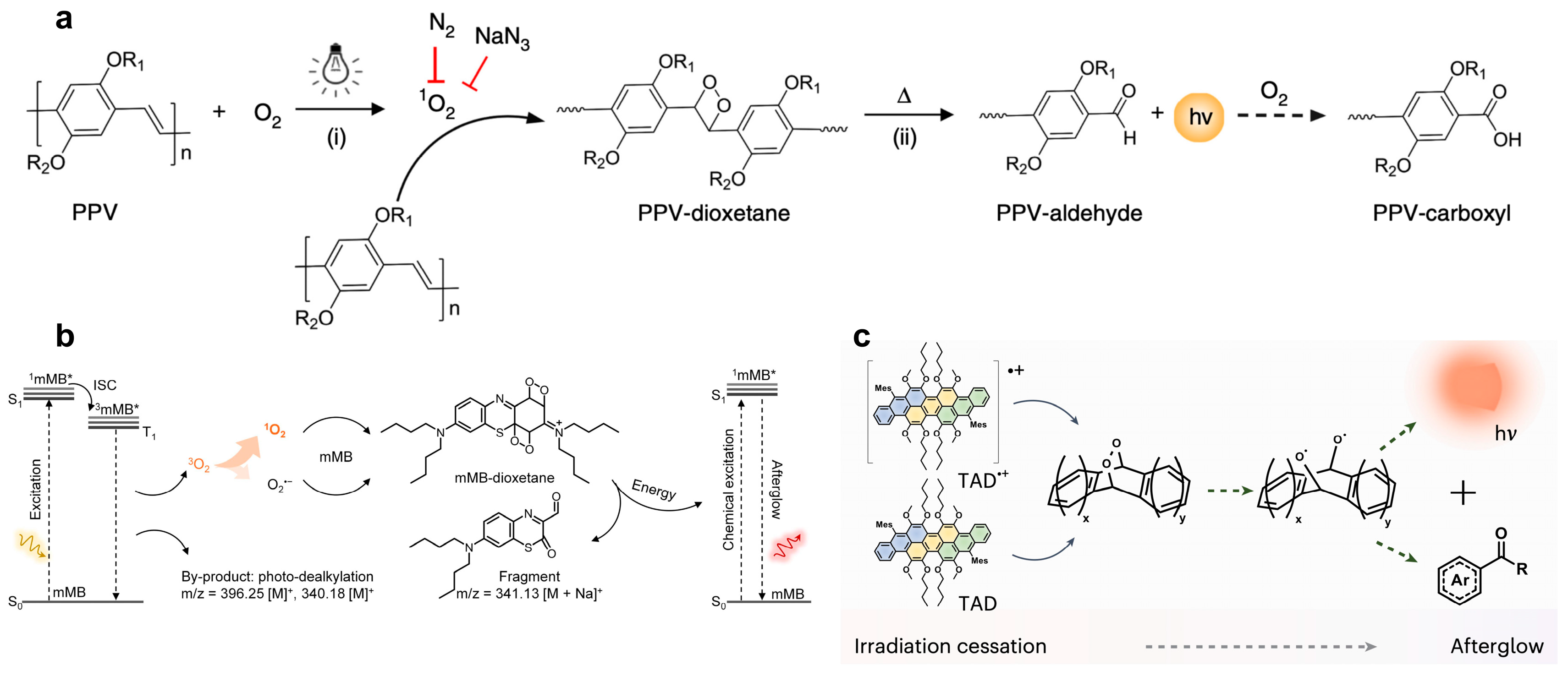
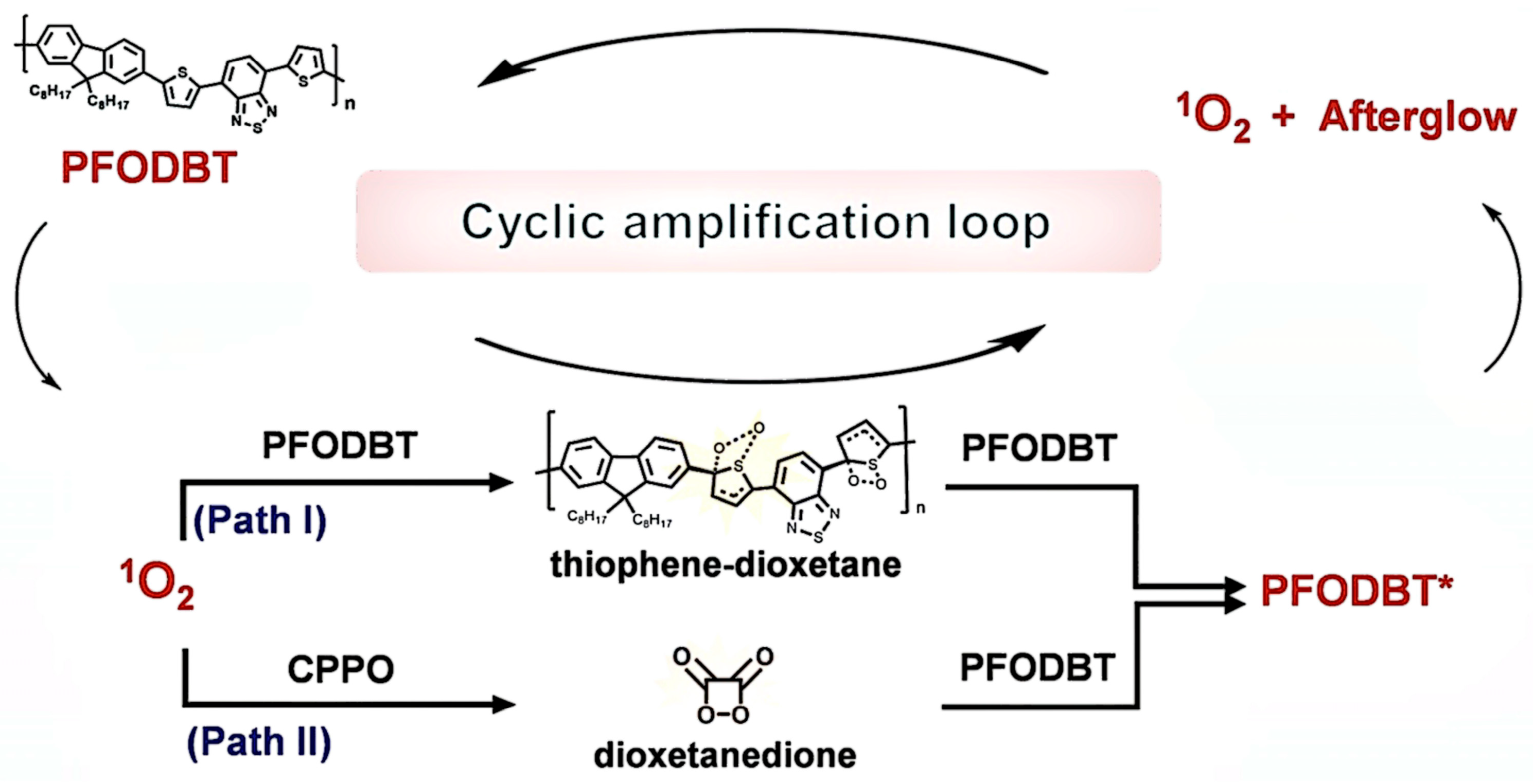
2.1. Semiconducting Polymers (SPs)
2.2. Heterocyclic Compounds
2.3. Trianthracene Derivatives (TADs)
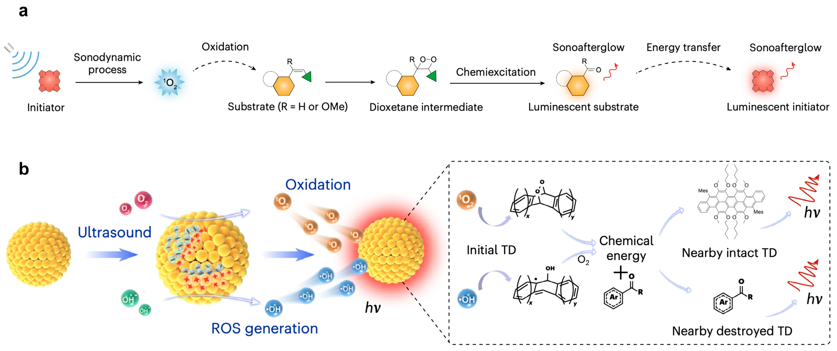
3. Ultrasound-Induced Afterglow Imaging
3.1. Sonoafterglow Nanoparticles (SNAPs)
3.2. Molecules Luminescence Through Two-Step Intraparticle Energy Conversion
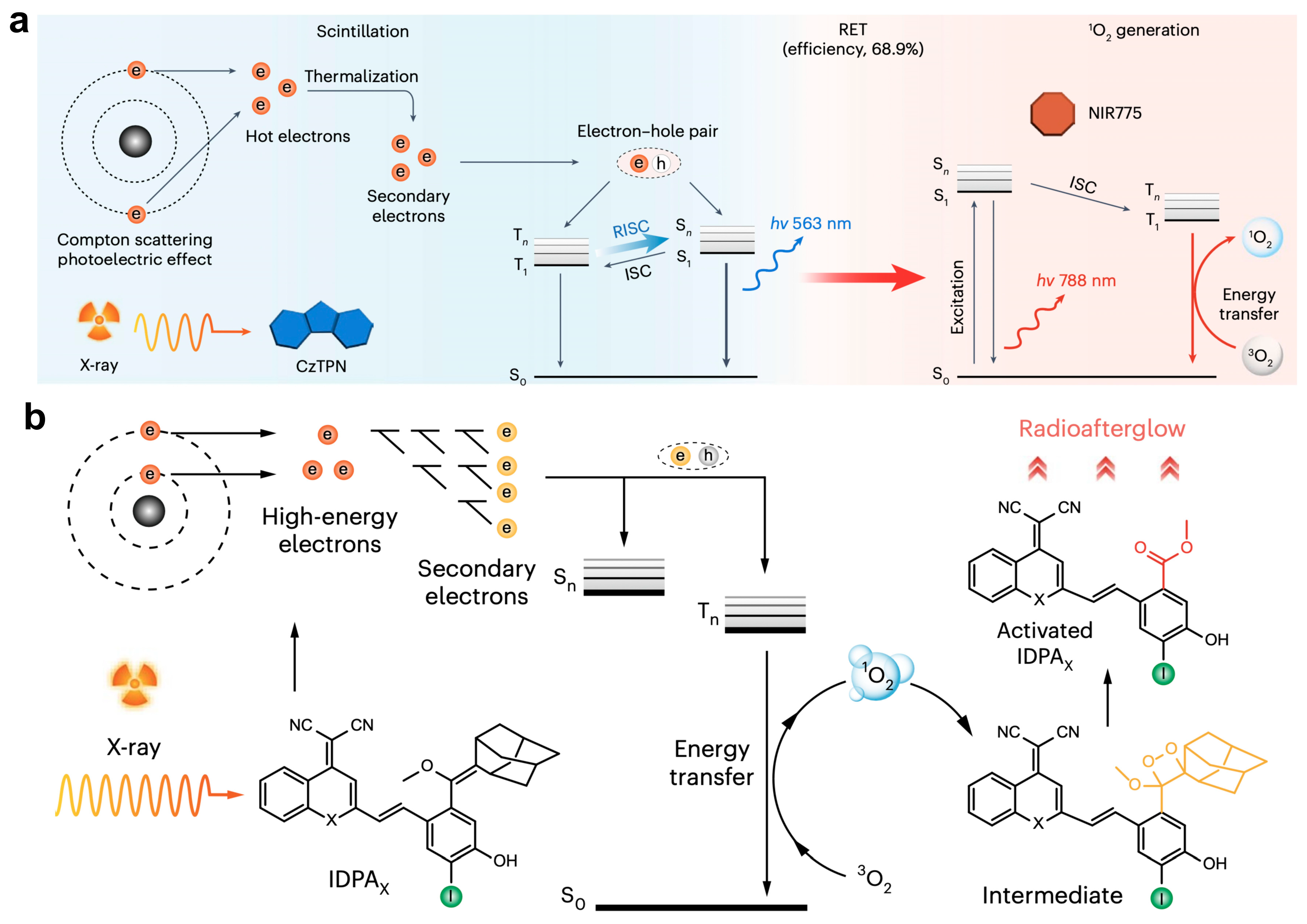
4. X-Ray-Induced Afterglow Imaging
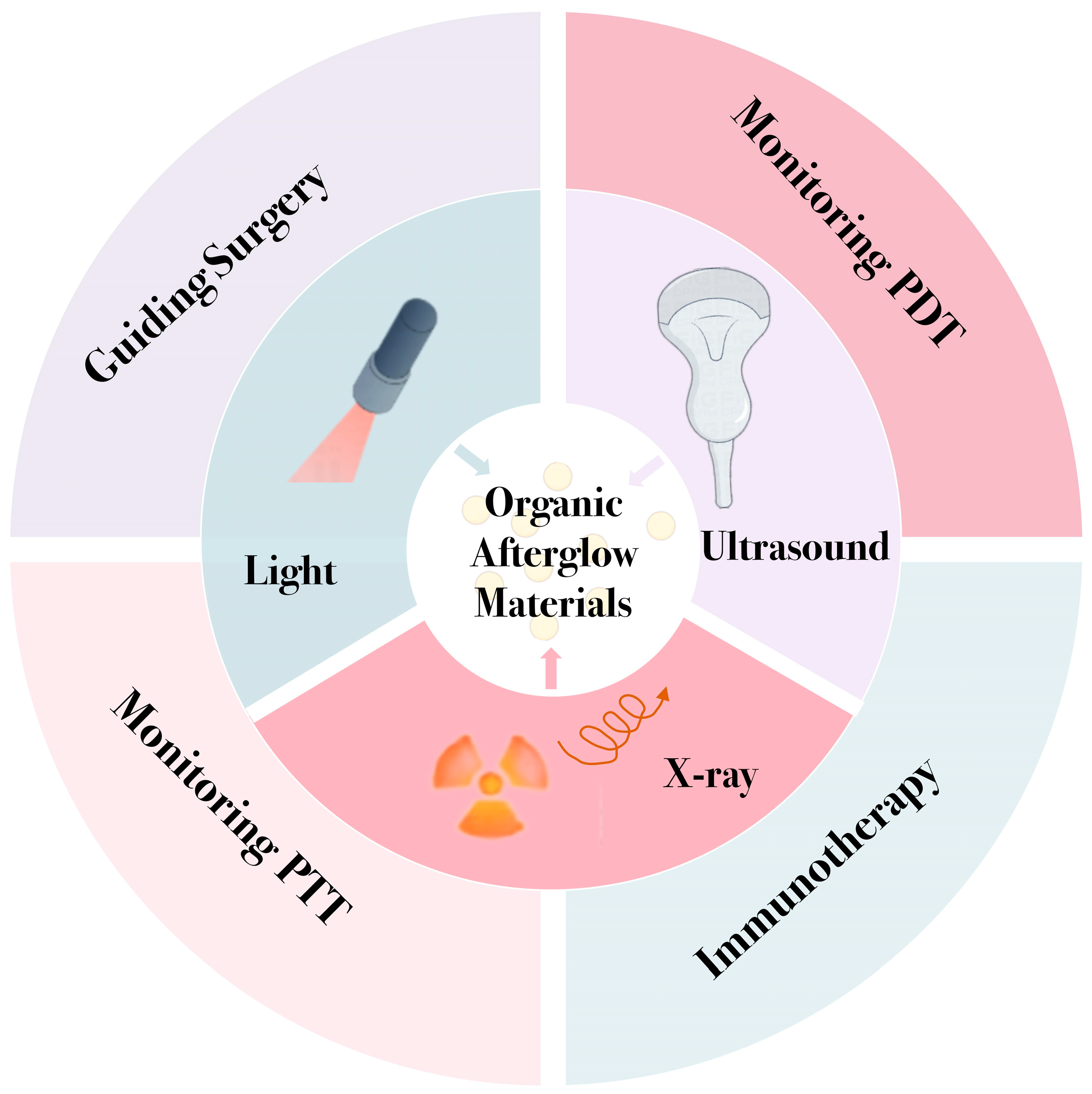
5. Organic Afterglow Nanoparticles for Cancer Therapy
5.1. Afterglow Imaging Guiding Surgery
5.2. Afterglow Imaging Monitoring Photodynamic Therapy
5.3. Afterglow Imaging Monitoring Photothermal Therapy
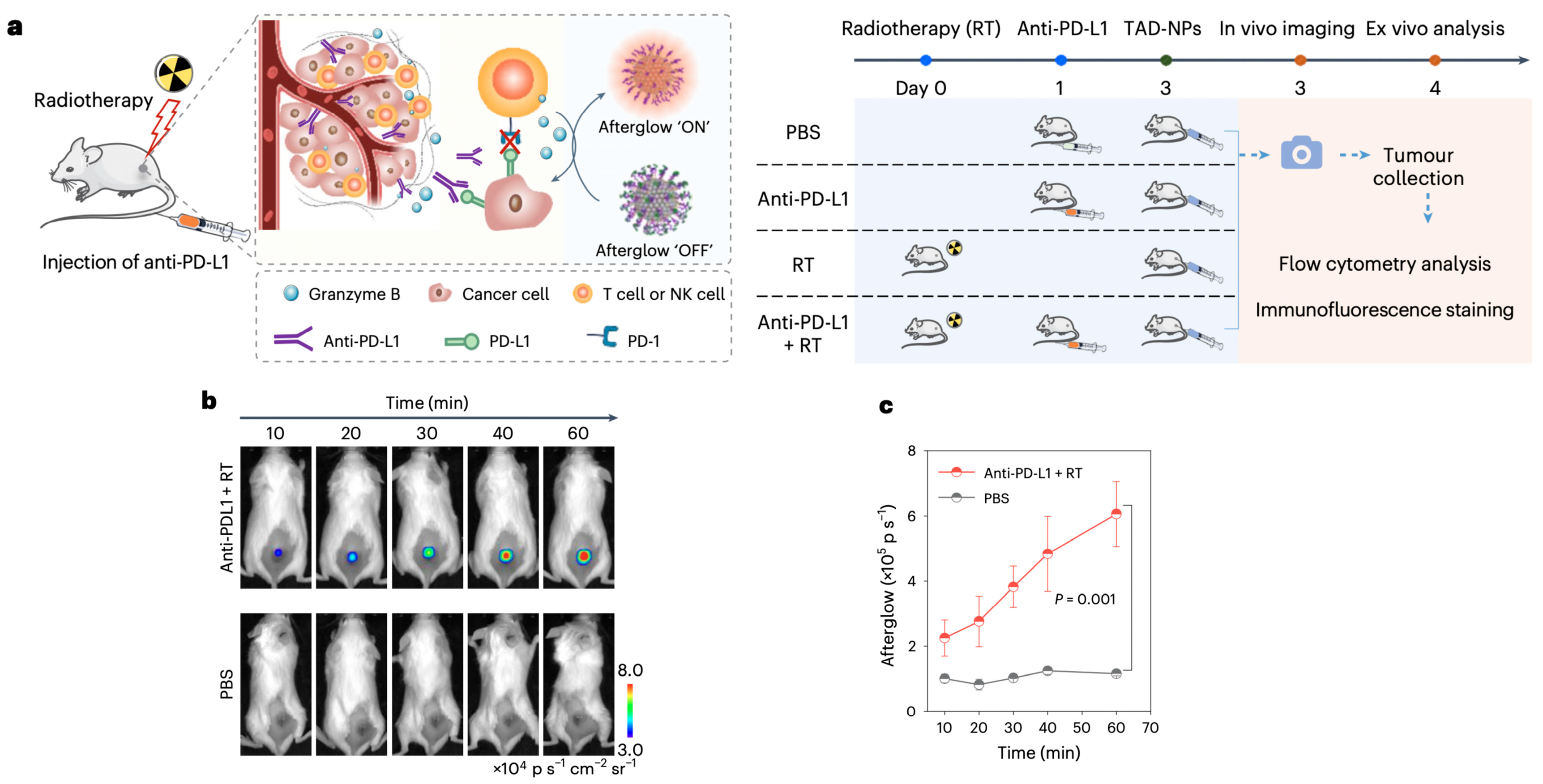
5.4. Afterglow Imaging Immunotherapy
6. Conclusions and Outlook
Author Contributions
Funding
Conflicts of Interest
References
- Maldiney, T.; Bessière, A. The in Vivo Activation of Persistent Nanophosphors for Optical Imaging of Vascularization, Tumours and Grafted Cells. Nat. Mater. 2014, 13, 418–426. [Google Scholar] [CrossRef]
- Li, H.; Kim, Y. Near-Infrared (NIR) Fluorescence-Emitting Small Organic Molecules for Cancer Imaging and Therapy. Chem. Soc. Rev. 2022, 51, 8957–9008. [Google Scholar] [CrossRef] [PubMed]
- Vacher, M.; Fdez Galván, I. Chemi- and Bioluminescence of Cyclic Peroxides. Chem. Rev. 2018, 118, 6927–6974. [Google Scholar] [CrossRef] [PubMed]
- Huang, J.; Su, L. Molecular Radio Afterglow Probes for Cancer Radiodynamic Theranostics. Nat. Mater. 2023, 22, 1421–1429. [Google Scholar] [CrossRef]
- Liu, Y.; Teng, L. “Four-In-One” Design of a Hemicyanine-Based Modular Scaffold for High-Contrast Activatable Molecular Afterglow Imaging. J. Am. Chem. Soc. 2023, 145, 5134–5144. [Google Scholar] [CrossRef]
- Yang, X.; Waterhouse, G.I.N. Recent Advances in the Design of Afterglow Materials: Mechanisms, Structural Regulation Strategies and Applications. Chem. Soc. Rev. 2023, 52, 8005–8058. [Google Scholar] [CrossRef]
- Miao, Q.; Xie, C. Molecular Afterglow Imaging with Bright, Biodegradable Polymer Nanoparticles. Nat. Biotechnol. 2017, 35, 1102–1110. [Google Scholar] [CrossRef]
- Aoki, Y.; Takeuchi, N. A New Long Phosphorescent Phosphor with High Brightness, SrAl2O4:Eu2+,Dy3+. J. Electrochem. Soc. 1996, 143, 2670–2673. [Google Scholar]
- Gao, L.; Liu, Y. Modulation of Near-Infrared Afterglow Luminescence in Inorganic Nanomaterials for Biological Applications. Adv. Mater. 2025, 37, 2419349. [Google Scholar] [CrossRef]
- Palner, M.; Pu, K. Semiconducting Polymer Nanoparticles with Persistent Near-Infrared Luminescence for in Vivo Optical Imaging. Angew. Chem. Int. Ed. 2015, 54, 11477–11480. [Google Scholar] [CrossRef]
- Wang, Z.; Zhou, Y. Efficient Electroconversion of Carbon Dioxide to Formate by a Reconstructed Amino-functionalized Indium–Organic Framework Electrocatalyst. Angew. Chem. Int. Ed. 2021, 60, 19107–19112. [Google Scholar] [CrossRef]
- Lee, H.; Tessarolo, J. Light-Powered Dissipative Assembly of Diazocine Coordination Cages. J. Am. Chem. Soc. 2022, 144, 3099–3105. [Google Scholar] [CrossRef] [PubMed]
- Chen, W.; Zhang, Y. Near-Infrared Afterglow Luminescence of Chlorin Nanoparticles for Ultrasensitive In Vivo Imaging. J. Am. Chem. Soc. 2022, 144, 6719–6726. [Google Scholar] [CrossRef] [PubMed]
- Wang, Y.; Song, G. Cyclic Amplification of the Afterglow Luminescent Nanoreporter Enables the Prediction of Anti-cancer Efficiency. Angew. Chem. Int. Ed. 2021, 60, 19779–19789. [Google Scholar] [CrossRef] [PubMed]
- Zhu, J.; Chen, W. A Self-Sustaining Near-Infrared Afterglow Chemiluminophore for High-Contrast Activatable Imaging. Angew. Chem. Int. Ed. 2024, 63, e202318545. [Google Scholar] [CrossRef]
- Wang, Y.; Guo, J. Ultrabright and Ultrafast Afterglow Imaging in Vivo via Nanoparticles Made of Trianthracene Derivatives. Nat. Biomed. Eng. 2025, 9, 656–670. [Google Scholar] [CrossRef]
- Li, J.; Shi, J. Five-Nanometer ZnSn2O4:Cr,Eu Ultra-Small Nanoparticles as New Near Infrared-Emitting Persistent Luminescent Nanoprobes for Cellular and Deep Tissue Imaging at 800 nm. Nanoscale 2017, 9, 8631–8638. [Google Scholar] [CrossRef]
- Pei, P.; Chen, Y. X-ray-activated persistent luminescence nanomaterials for NIR-II imaging. Nat. Nanotechnol. 2021, 16, 1011–1018. [Google Scholar] [CrossRef]
- Lin, Y.; Hu, J. Multiple emission bands NIR-persistent luminescence mSiO2@Zn0.6Ca0.4Ga2O4:Cr3+,Yb3+ nanoparticles for biological applications. J. Mater. Chem. B 2021, 9, 1131–1137. [Google Scholar] [CrossRef]
- Xu, L.; Zhang, Q. Biomedical applications of NIR-II organic small molecule fluorescent probes in different organs. Coord. Chem. Rev. 2024, 519, 216122. [Google Scholar] [CrossRef]
- Zhou, Y.; Mao, Z. Recent advances in near infrared (NIR) electrochemiluminescence luminophores. Chin. Chem. Lett. 2024, 35, 109622. [Google Scholar] [CrossRef]
- Hu, Y.; Xin, K. Recent Progress and Challenges of Infrared Quantum Dots. Sci. China Inf. Sci. 2025, 68, 4383. [Google Scholar] [CrossRef]
- Shen, W.-S. Near-Infrared Quantum Dots for Electroluminescence: Balancing Performance and Sustainability. Laser Photonics Rev. 2025, 19, 2401947. [Google Scholar] [CrossRef]
- Li, Z.; Liu, H. Reactive Oxygen Species-Mediated Organic Long-Persistent Luminophores Light up Biomedicine: From Two-Component Separated Nano-Systems to Integrated Uni-Luminophores. Chem. Soc. Rev. 2024, 53, 11207–11227. [Google Scholar] [CrossRef] [PubMed]
- Zeng, W.; Wu, L. An Activatable Afterglow/MRI Bimodal Nanoprobe with Fast Response to H2S for in Vivo Imaging of Acute Hepatitis. Angew. Chem. Int. Ed. 2022, 61, e202111759. [Google Scholar] [CrossRef]
- Lyu, Y.; Cui, D. Near-infrared Afterglow Semiconducting Nano-polycomplexes for the Multiplex Differentiation of Cancer Exosomes. Angew. Chem. Int. Ed. 2019, 58, 4983–4987. [Google Scholar] [CrossRef]
- Yang, L.; Zhao, M. A Highly Bright Near-Infrared Afterglow Luminophore for Activatable Ultrasensitive In Vivo Imaging. Angew. Chem. Int. Ed. 2024, 63, e202313117. [Google Scholar] [CrossRef]
- Wang, Y.; Shen, H. Enhancing Fractionated Cancer Therapy: A Triple-Anthracene Photosensitizer Unleashes Long-Persistent Photodynamic and Luminous Efficacy. J. Am. Chem. Soc. 2024, 146, 6252–6265. [Google Scholar] [CrossRef]
- Duan, X.; Zhang, G. Activatable Persistent Luminescence from Porphyrin Derivatives and Supramolecular Probes with Imaging-Modality Transformable Characteristics for Improved Biological Applications. Angew. Chem. Int. Ed. 2022, 61, e202116174. [Google Scholar] [CrossRef]
- Liang, L.; Chen, J. Controlling Persistent Luminescence in Nanocrystalline Phosphors. Nat. Mater. 2023, 22, 289–304. [Google Scholar] [CrossRef]
- Berezin, M.Y.; Achilefu, S. Fluorescence Lifetime Measurements and Biological Imaging. Chem. Rev. 2010, 110, 2641–2684. [Google Scholar] [CrossRef]
- Green, O.; Gnaim, S. Near-Infrared Dioxetane Luminophores with Direct Chemiluminescence Emission Mode. J. Am. Chem. Soc. 2017, 139, 13243–13248. [Google Scholar] [CrossRef]
- Haris, U.; Kagalwala, H.N. Seeking Illumination: The Path to Chemiluminescent 1,2-Dioxetanes for Quantitative Measurements and in Vivo Imaging. Acc. Chem. Res. 2021, 54, 2844–2857. [Google Scholar] [CrossRef] [PubMed]
- Jiang, Y.; Huang, J. A Generic Approach towards Afterglow Luminescent Nanoparticles for Ultrasensitive in Vivo Imaging. Nat. Commun. 2019, 10, 2064. [Google Scholar] [CrossRef] [PubMed]
- Memari, E.; Khan, D. Focused Ultrasound-Assisted Delivery of Immunomodulating Agents in Brain Cancer. J. Control. Release 2024, 366, 283–299. [Google Scholar] [CrossRef]
- Huang, H.; Zheng, Y. Ultrasound-Based Micro-/Nanosystems for Biomedical Applications. Chem. Rev. 2024, 124, 8307–8472. [Google Scholar] [CrossRef]
- Xu, C.; Huang, J. Nanoparticles with Ultrasound-Induced Afterglow Luminescence for Tumour-Specific Theranostics. Nat. Biomed. Eng. 2022, 6, 298–312. [Google Scholar] [CrossRef]
- Wang, Y.; Yi, Z. In Vivo Ultrasound-Induced Luminescence Molecular Imaging. Nat. Photonics 2024, 18, 334–343. [Google Scholar] [CrossRef]
- Wei, Y.; Wang, J. X-Ray/γ-Ray/Ultrasound-Activated Persistent Luminescence Phosphors for Deep Tissue Bioimaging and Therapy. ACS Appl. Mater. Interfaces 2024, 16, 56519–56544. [Google Scholar] [CrossRef]
- Xu, C.; Qin, X. A Cascade X-Ray Energy Converting Approach toward Radio-Afterglow Cancer Theranostics. Nat. Nanotechnol. 2025, 20, 286–295. [Google Scholar] [CrossRef]
- Vahrmeijer, A.L.; Hutteman, M. Image-Guided Cancer Surgery Using near-Infrared Fluorescence. Nat. Rev. Clin. Oncol. 2013, 10, 507–518. [Google Scholar] [CrossRef] [PubMed]
- Wu, W.; Mao, D. A Highly Efficient and Photostable Photosensitizer with Near-infrared Aggregation-induced Emission for Image-guided Photodynamic Anticancer Therapy. Adv. Mater. 2017, 29, 1700548. [Google Scholar] [CrossRef] [PubMed]
- Juengpanich, S.; Li, S. Pre-Activated Nanoparticles with Persistent Luminescence for Deep Tumor Photodynamic Therapy in Gallbladder Cancer. Nat. Commun. 2023, 14, 5699. [Google Scholar] [CrossRef] [PubMed]
- Zheng, X.; Wu, W. Organic Nanoparticles with Persistent Luminescence for in Vivo Afterglow Imaging-guided Photodynamic Therapy. Chem. Eur. J. 2021, 27, 6911–6916. [Google Scholar] [CrossRef]
- Jiang, Y.; Li, J. Dual-Peak Absorbing Semiconducting Copolymer Nanoparticles for First and Second near-Infrared Window Photothermal Therapy: A Comparative Study. Adv. Mater. 2018, 30, 1705980. [Google Scholar] [CrossRef]
- Qu, R.; He, D. Afterglow/Photothermal Bifunctional Polymeric Nanoparticles for Precise Postbreast-Conserving Surgery Adjuvant Therapy and Early Recurrence Theranostic. Nano Lett. 2023, 23, 4216–4225. [Google Scholar] [CrossRef]
- Zhong, Y.; Ma, Z. In Vivo Molecular Imaging for Immunotherapy Using Ultra-Bright near-Infrared-IIb Rare-Earth Nanoparticles. Nat. Biotechnol. 2019, 37, 1322–1331. [Google Scholar] [CrossRef]
- Friedman, A.A.; Letai, A. Precision Medicine for Cancer with Next-Generation Functional Diagnostics. Nat. Rev. Cancer 2015, 15, 747–756. [Google Scholar] [CrossRef]
- Bayat Mokhtari, R.; Homayouni, T.S. Combination Therapy in Combating Cancer. Oncotarget 2017, 8, 38022–38043. [Google Scholar] [CrossRef]
- Pei, Q.; Zhou, S. Ferric Ion-driven Chlorin E6 Nanoparticles: Simple and Effective Multimodal Theranostics. Adv. Funct. Mater. 2023, 33, 2304205. [Google Scholar] [CrossRef]


Disclaimer/Publisher’s Note: The statements, opinions and data contained in all publications are solely those of the individual author(s) and contributor(s) and not of MDPI and/or the editor(s). MDPI and/or the editor(s) disclaim responsibility for any injury to people or property resulting from any ideas, methods, instructions or products referred to in the content. |
© 2025 by the authors. Licensee MDPI, Basel, Switzerland. This article is an open access article distributed under the terms and conditions of the Creative Commons Attribution (CC BY) license (https://creativecommons.org/licenses/by/4.0/).
Share and Cite
Chen, X.; Li, B.; Yin, B.; Xu, D.; Wang, Y. Organic Afterglow Materials for Tumor Diagnosis and Therapy. Biosensors 2025, 15, 484. https://doi.org/10.3390/bios15080484
Chen X, Li B, Yin B, Xu D, Wang Y. Organic Afterglow Materials for Tumor Diagnosis and Therapy. Biosensors. 2025; 15(8):484. https://doi.org/10.3390/bios15080484
Chicago/Turabian StyleChen, Xiayi, Bin Li, Baoli Yin, Dong Xu, and Youjuan Wang. 2025. "Organic Afterglow Materials for Tumor Diagnosis and Therapy" Biosensors 15, no. 8: 484. https://doi.org/10.3390/bios15080484
APA StyleChen, X., Li, B., Yin, B., Xu, D., & Wang, Y. (2025). Organic Afterglow Materials for Tumor Diagnosis and Therapy. Biosensors, 15(8), 484. https://doi.org/10.3390/bios15080484