Communication

Magnetically Assisted Immobilization-Free Detection of microRNAs Based on the Signal Amplification of Duplex-Specific Nuclease

1
College of Chemistry and Chemical Engineering, Anyang Normal University, Anyang 455000, China
2
School of Chemistry and Environmental Engineering, Pingdingshan University, Pingdingshan 467000, China
*
Authors to whom correspondence should be addressed.
These authors contributed equally to this work.
Biosensors 2023, 13(7), 699; https://doi.org/10.3390/bios13070699
Submission received: 26 May 2023 / Revised: 15 June 2023 / Accepted: 23 June 2023 / Published: 30 June 2023

Abstract

:
The double specific nuclease (DSN)-based methods for microRNAs (miRNAs) detection usually require the immobilization of DNA probes on a solid surface. However, such strategies have the drawbacks of low hybridization and cleavage efficiency caused by steric hindrance effect and high salt concentration on the solid surface. Herein, we proposed an immobilization-free method for miRNA detection on the basic of DSN-assisted signal amplification. The biotin- and fluorophore-labeled probes were captured by streptavidin-modified magnetic beads through streptavidin–biotin interactions, thus producing a poor fluorescence signal. Once the DNA probes were hybridized with target miRNA in solution to form DNA-miRNA duplexes, DNA stands in the duplexes would be selectively digested by DSN. The released target miRNA could initiate the next hybridization/cleavage recycling in the homogeneous solution, finally resulting in the release of numerous fluorophore-labeled fragments. The released fluorophores remained in solution and emitted strong fluorescence after treatment by the streptavidin-modified magnetic beads. The immobilization-free method achieved the assays of miRNA-21 with a detection limit down to 0.01 pM. It was employed to evaluate the expression levels of miRNA-21 in different cancer cells with satisfactory results.

1. Introduction

MicroRNAs (miRNAs) are a group of endogenous, non-coding RNAs that play an important role in physiological and pathological processes. The abnormal expression levels of miRNAs are associated with many human diseases, including cancers and degenerative diseases [1,2]. Therefore, miRNAs have been regarded as the credible biomarkers for early diagnosis and treatment evaluation of diseases. Reverse transcription real-time quantitative polymerase chain reaction (RT-qPCR) is the currently used gold standard method for monitoring the expression levels of miRNAs. The method shows high specificity and sensitivity but requires complex operation procedures, specialized equipment, and a clean experimental environment [3]. Thus, it is meaningful and highly desired to develop a simple and sensitive detection system for the real-time monitoring of the level of miRNAs.
As one of the isothermal amplification strategies for biological analysis, a double specific nuclease (DSN)-based sensing system exhibits the advantages of simple principle, convenient operation, and high specificity [4,5,6,7]. DSN can specifically cleave DNA in DNA-DNA or DNA-RNA duplexes but shows no activity toward single-stranded DNA (ssDNA) or double-stranded RNA (dsRNA) [8,9]. The principle of DSN-assisted signal amplification is mainly based on target miRNA recycling. Thus, a few miRNAs could trigger the cleavage of considerable amounts of DNA in DNA-RNA heteroduplexes, leading to the release of a large number of signal molecules, including enzymes, electroactive molecules, fluorophores, nanoparticles, and nucleotide sequences [10,11,12,13,14,15,16,17,18,19,20,21,22,23,24]. The concentration of miRNAs can be determined by monitoring the optical or electrical signal change induced by the release of signal molecules. In these methods, signal-labeled capture probes are usually attached onto a solid surface and DSN-based enzymatic reactions happen at the solid–liquid interface. The heterogeneous sensing systems show the advantages of less sample consumption, ultrahigh sensitivity, and excellent selectivity. However, the probe-immobilized methods may cause several drawbacks. First, the steric hindrance effect of the solid surface may affect the configurational freedom of capture probes and prevent interactions between DNA-RNA hybrids and nucleases, thus decreasing the hybridization and cleavage efficiency [25,26,27,28,29]. Second, the high salt concentration on the probe-modified solid surface may limit the activity of nucleases [30,31]. In addition, the immobilization and removal of capture probes require laborious and time-consuming procedures. In contrast to heterogeneous sensing systems, homogeneous assays have the merits of a simple operation and high cleavage efficiency. Although a few DSN-based homogeneous methods have been developed and used for miRNA detection [32,33], they are usually involved in complex procedures, special instruments, and/or the use of additional enzymes or nanomaterials for signal amplification. Thus, it is of importance to develop a simple, sensitive, and high-throughput homogeneous method for the detection of miRNAs with DSN-assisted signal amplification.
Magnetic beads (MBs) have been extensively used for the development of various biosensors for detective applications. Magnetically assisted sensing systems show remarkable advantages in the separation and pre-concentration of targets and high throughput detection of multiple samples [34,35,36,37]. Streptavidin (SA) is a homotetramer protein that can bind up to four biotin molecules with a high affinity. Biotin is a commonly used reagent for labeling biomolecules due to its small size and highly selective and stable interaction with SA. Many SA-modified materials, including MB-SA, are commercially available for the collection and immobilization of biotin-labeled proteins or nucleic acids [38]. In this work, we proposed a homogeneous strategy for the detection of miRNAs with DSN-assisted signal amplification. In the absence of target miRNA, the biotinylated fluorescently labeled DNA probes were captured by MB-SA through fast and strong SA-biotin interactions, thus causing a decrease in the fluorescence. In the presence of target miRNA, the capture probes would be digested by DSN, and the released signal molecules in solution showed a high fluorescence. The reaction rate of DSN for the DNA capture probes immobilized on the MB surface and dispersed in solution was investigated. The proposed strategy can be used for the design of various immobilization-free miRNA biosensors by changing the type of signal molecules. Moreover, magnetically assisted sensing systems could be used for the high-throughput detection of multiple miRNAs with different fluorescently labeled sequences.

2. Materials and Methods

2.1. Chemicals and Reagents

DNA, miRNAs, and Trizol reagent were ordered from Sangon Biotech. Co., Ltd. (Shanghai, China). Their sequences are Biotin-TCAACATCAGTCTGATAAGCTA-FAM (Bio-DNA-FAM), UAGCUUAUCAGACUGAUGUUGA (miRNA-21), UAGCUUAUCGGACUGAUGUUGA (single-base mismatch), UUGCUUAUCGGACUGAUCUUGA (three-base mismatch), and GUAAGGCAUCUGACCGAAGGCA (non-complementary). DSN was obtained from Evrogen Joint Stock Company (Moscow, Russia). MB-SA was purchased from Thermo Fisher Scientific (Shanghai, China). Other reagents were of analytical grade and used without further purification. The miRNA samples were prepared freshly using RNase-free ultrapure water. The hybridization and reaction solutions were prepared with TNE buffer (pH 7.4). All aqueous solutions were prepared with ultrapure water treated by a Millipore system.

2.2. Procedures for miRNA Detection

In total, 150 μL of 500 nM DNA probe was mixed with 25 μL of miRNA sample in TNE buffer at 45 °C. After incubation for 10 min, 25 μL of 0.1 U DSN solution was added to the mixture. After reaction for a given time, 10 μL of 10 mg/mL MB-SA suspension was added to the mixed solution and incubated for about 5 min. Under a magnetic force, the supernatant solution was taken out for assays to be conducted. The signals were collected on a Cary Eclipse fluorescence spectrometer (Palo Alto, CA, USA) with an excitation wavelength of 492 nm.

2.3. Assays of miRNAs in Cell Lysates

The cell lysates were extracted from MCF-7 and Hela cells with the procedures detailed in previous reports [39,40]. Briefly, the cells were counted, collected, and washed with phosphate buffer. Then, Trizol reagent was added to extract total RNA according to the manufacturer’s protocol. The collected lysates were centrifuged at 10,000 rpm for 15 min and the aqueous supernatants were diluted 10-fold with TNE buffer. Then, 25 μL of the diluted supernatant with additional treatment was added to 150 μL of probe solution for quantification assay. Other procedures for the assays of miRNA in the lysates were the same as those of the standard samples.

3. Results and Discussion

3.1. Detection Principle

The mechanism of the magnetically assisted detection of miRNA is depicted in Scheme 1. The DNA probe labeled with a biotin tag and a fluorophore at two ends (denoted as Bio-DNA-FAM) could hybridize with the target miRNA to form a DNA-miRNA heteroduplex. The DNA strand in the duplex was then selectively digested by DSN. The released miRNA could hybridize with another Bio-DNA-FAM probe and initiate the next cleavage event. In this way, the target miRNA-initiated enzymatic digestion of Bio-DNA-FAM probes by DSN was recycled, leading to the release of numerous FAM-labeled fragments. The released fragments could not be removed by MB-SA under the magnetic force and thus showed a strong fluorescence. In the absence of target miRNA, the Bio-DNA-FAM probe remained intact and would be removed by MB-SA through streptavidin–biotin interaction. In this case, the solution showed weak fluorescence. Therefore, the fluorescence intensity is proportional to the level of target miRNA. Through the signal amplification of DSN, miRNA at a low abundance would be detected. The method should be simple and sensitive since it does not require the pre-immobilization of DNA probe on a solid surface, thus improving the hybridization and cleavage efficiency without the effect of steric hindrance.

3.2. Feasibility of this Method

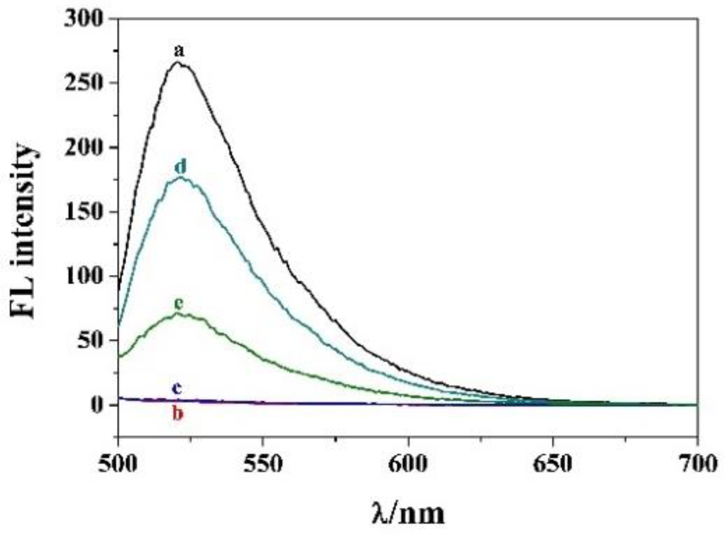
To probe the feasibility of the method, miRNA-21 was tested as a target model because its expression level is closely related to many cancers. As shown in Figure 1, the Bio-DNA-FAM probe emitted a strong fluorescence at 521 nm (curve a). When it was incubated with MB-SA under magnetic force (curve b), the fluorescence peak disappeared, indicating that the probe could be captured and removed by MB-SA. When the Bio-DNA-FAM probe was mixed with miRNA-21 in the absence of DSN, the fluorescence signal was close to the background value (curve c), indicating that the hybridization of miRNA-21 with Bio-DNA-FAM did not limit the capture of the probe by MB-SA. However, when the DNA-miRNA duplexes were incubated with DSN for a given time and then treated by MB-SA under the magnetic force, the fluorescence signal was intensified greatly (curve d). The result suggested that the Bio-DNA-FAM probes were enzymatically digested by DSN, thus releasing a large number of FAM-labeled fragments into the solution. To study the effect of steric hindrance on the hybridization/cleavage efficiency, the DNA-miRNA duplexes were pre-immobilized onto the MB-SA surface, and then DSN was added to the suspension for enzymatic digestion under the same reaction conditions. As a result, a smaller signal (curve e) was observed in contrast to the immobilization-free strategy (curve d). The result implied that the cleavage efficiency was reduced by the steric hindrance, which is consistent with that in the previous works [27,41]. Thus, our proposal could improve the detection sensitivity without increasing the operation complexity.

3.3. Optimization of Experimental Conditions

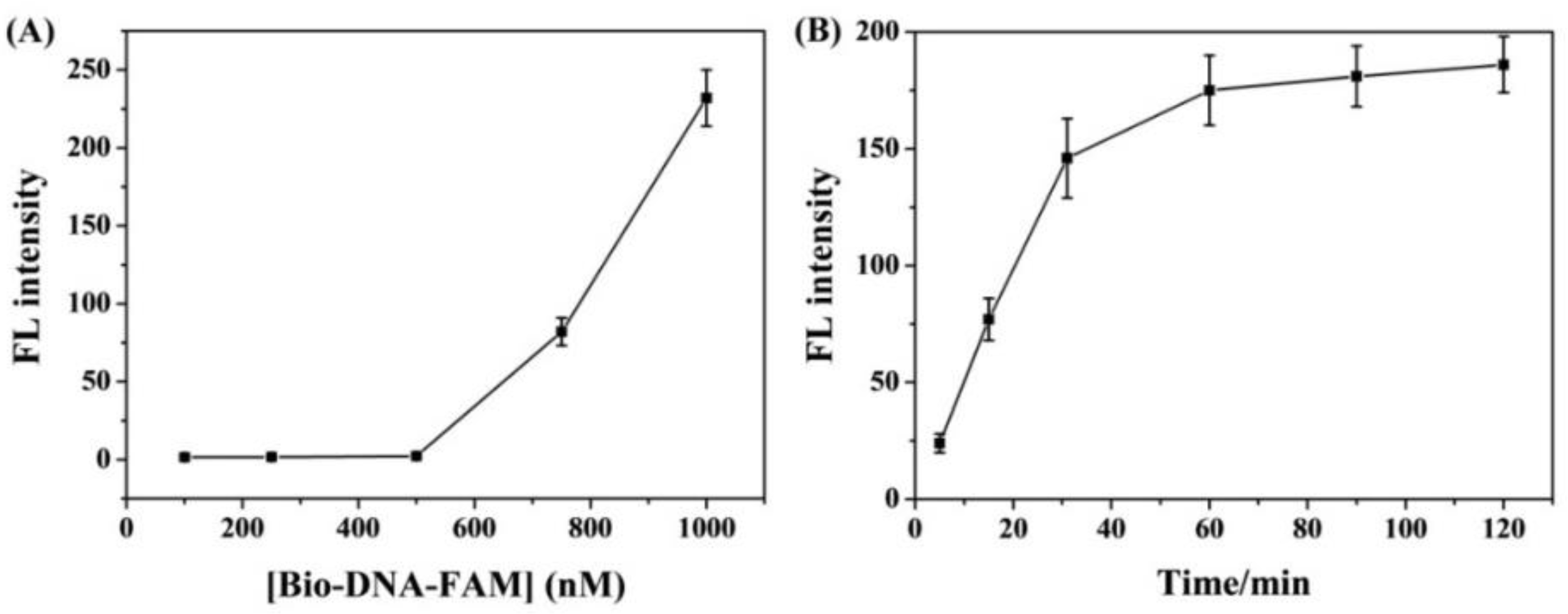
To achieve optimum analytical performance, the experimental conditions, such as the probe concentration and cleavage time, were investigated. Because the background signal would be dependent upon the probe concentration, we first measured the fluorescence signals of different concentrations of Bio-DNA-FAM after treatment by MB-SA under a magnetic force. As shown in Figure 2A, when the probe concentration was lower than 500 nM, the fluorescence intensity was negligible. Over the value, the signal began to increase, indicating that the surface of MB-SA was saturated by the probe through the SA-biotin interaction. The abundance of miRNAs in biological samples is extremely low (usually in the range of femtomolar to picomolar), and the background signal can affect the sensitivity of the analytical method. Thus, a low concentration of probe was used in the following trials. When the incubation time for the mixture of DNA/miRNA-21 and DSN was increased, the fluorescence intensity was gradually intensified and began to level off beyond 60 min for 100 pM miRNA-21 (Figure 2B). To attain high sensitivity and save detection time, the samples were incubated for 60 min to achieve hybridization and cleavage recycling.

3.4. Sensitivity for miRNA-21 Detection

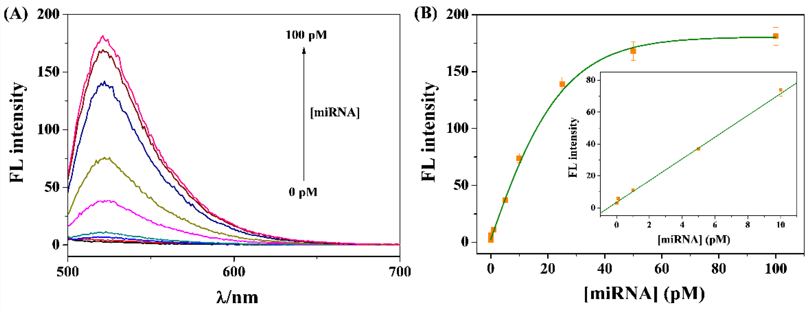
Under the optimal conditions, various concentrations of miRNA-21 were determined to indicate the analytical performance. As shown in Figure 3A, the fluorescence signals were intensified with the increase in the miRNA concentration in the range of 0~100 pM. A good linear relationship was found at the concentration range of 0.01~10 pM (Figure 3B). The linear equation can be expressed as FL = 3.47 + 6.23 [miRNA-21] (pM). The detection limit as the minimum detectable concentration (0.01 pM) [42] is lower than that achieved by other DSN-based fluorescence methods with nanomaterials as the quenchers, including gold nanoparticles, graphene oxide, molybdenum disulfide, and polydopamine-coated magnetic nanoparticles (Table 1). The sensitivity was also comparable to that achieved by DSN plus other signal-amplified techniques [4,6]. The high sensitivity can be attributed to the immobilization-free strategy, the low background signal, and the high hybridization and cleavage efficiency.

3.5. Selectivity

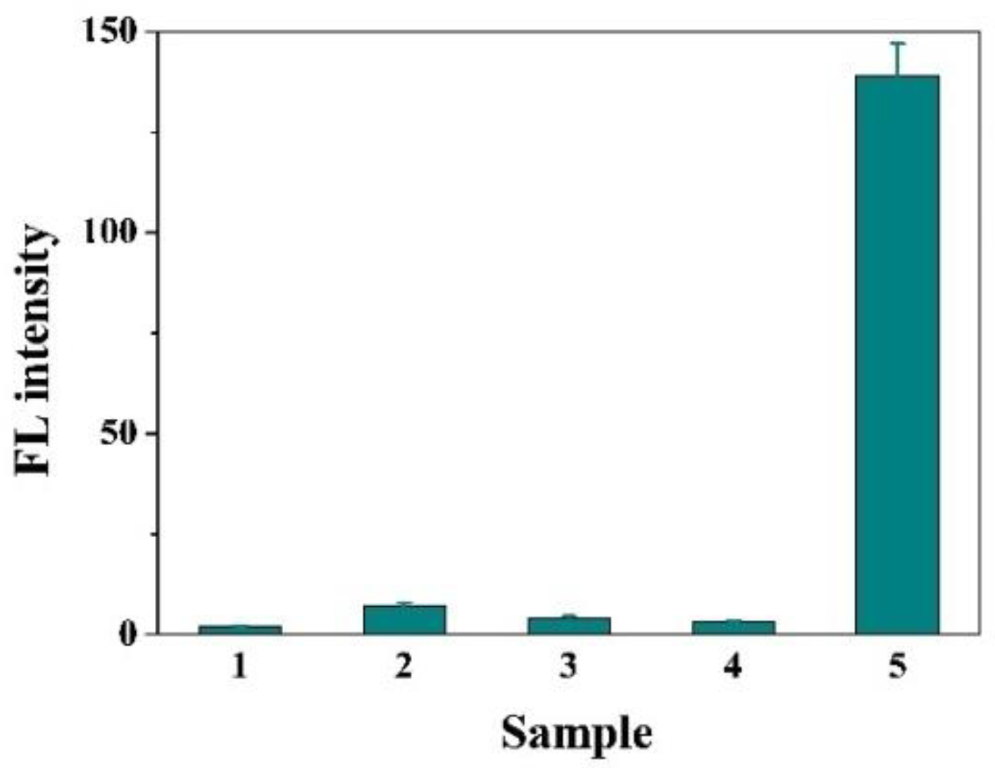
To indicate the selectivity of the strategy, a series of miRNA strands were tested, including miRNA-21 and its base-mismatched and non-complementary sequences. As shown in Figure 4, only the target miRNA-21 caused a significant increase in the fluorescence signal. Although the single-based mismatched sequence may hybridize with the probe at room temperature, it did not induce an obvious enhancement in the fluorescence signal. The result should benefit from the high specificity of DSN and the high reaction temperature where the mismatched sequences could not form DNA-RNA duplexes. In addition, the probability and abundance are very low for the existence of the miRNA-21 variant with a single mismatch in the real sample. By labeling the DNA probes with different fluorophores, we believe that the immobilization-free method could be used to simultaneously determine multiple types of miRNAs with a simple operation procedure and high detection throughput.

3.6. Assays of miRNA-21 in Cellular Lysates

To evaluate the validity of our method for real sample assays, the levels of miRNA-21 extracted from two types of cancer cell lines were determined. As depicted in Figure 5, the signals increased significantly with an increasing concentration of MCF-7 cells, while no obvious change was found for Hela cells even at a concentration higher than 10,000 cells. This indicated that the expression level of miRNA-21 in MCF-7 is higher than that in Hela cells. The result is in agreement with that found by the heterogeneous biosensors [39,40,56,57], indicating that our method can be used to measure the expression levels of miRNA in biological samples. The detectable cell number is higher than that of the heterogeneous assays, which may be attributed to the differences in the used sample volume and the expression level as well as extraction method for miRNA-21. We believe that the sensitivity would be further improved by the signal amplification of enzymes or nanomaterials in combination with the use of a more sensitive fluorescence spectrometer.

4. Conclusions

In summary, an immobilization-free fluorescence method with DSN-assisted signal amplification was developed for the sensitive detection of miRNAs. The effect of steric hindrance on the hybridization/cleavage efficiency was also investigated. The detection limit of this method is lower than that of other DSN-based fluorescence assays and even comparable to that achieved by multiple signal amplification strategies. It was successfully employed to monitor the expression levels of miRNA-21 in two cancer cell lines. In addition, the strategy does not require the pre-immobilization of a DNA probe on a solid surface for hybridization/cleavage recycling, thus eliminating the effect of steric hindrance and improving the detection efficiency. We believe that the proposal shows great potential for the simultaneous detection of multiple miRNAs by matching the DNA probes with different fluorophores.

Author Contributions

Conceptualization, G.L. and L.L.; methodology, M.L. and Y.H.; investigation, G.L., J.W. and J.L.; writing—original draft preparation, G.L. and M.L.; writing—review and editing, L.L.; project administration, Y.H.; funding acquisition, M.L. and L.L. All authors have read and agreed to the published version of the manuscript.

Funding

This research was funded by the fund project for Young Scholar sponsored by Henan province (2020JS227) and the Program for Innovative Research Team of Science and Technology in the University of Henan Province (21IRTSTHN005).

Institutional Review Board Statement

Not applicable.

Informed Consent Statement

Not applicable.

Data Availability Statement

Not applicable.

Conflicts of Interest

The authors declare no conflict of interest.

References

  1. Dong, H.; Lei, J.; Ding, L.; Wen, Y.; Ju, H.; Zhang, X. MicroRNA: Function, detection, and bioanalysis. Chem. Rev. 2013, 113, 6207–6233. [Google Scholar] [CrossRef] [PubMed]
  2. Imas, J.J.; Zamarreño, C.R.; Zubiate, P.; Sanchez-Martín, L.; Campión, J.; Matías, I.R. Optical biosensors for the detection of Rheumatoid Arthritis (RA) biomarkers: A comprehensive review. Sensors 2020, 20, 6289. [Google Scholar] [CrossRef] [PubMed]
  3. Chai, H.; Tang, Y.; Miao, P. Tetrahedral DNA supported walking nanomachine for ultrasensitive miRNA detection in cancer cells and serums. Anal. Chem. 2022, 94, 9975–9980. [Google Scholar] [CrossRef] [PubMed]
  4. Wu, Y.; Cui, S.; Li, Q.; Zhang, R.; Song, Z.; Gao, Y.; Chen, W.; Xing, D. Recent advances in duplex-specific nuclease-based signal amplification strategies for microRNA detection. Biosens. Bioelectron. 2020, 165, 112449. [Google Scholar] [CrossRef]
  5. Ashraf, G.; Zhong, Z.T.; Asif, M.; Aziz, A.; Iftikhar, T.; Chen, W.; Zhao, Y.D. State-of-the-art fluorescent probes: Duplex-specific nuclease-based strategies for early disease diagnostics. Biosensors 2022, 12, 1172. [Google Scholar] [CrossRef]
  6. Wu, D.; Yi, X.; Xia, N. Electrochemical biosensors for microRNA detection using duplex-specific nuclease based signal amplification strategies. Int. J. Electrochem. Sci. 2020, 15, 12136–12148. [Google Scholar] [CrossRef]
  7. Qiu, X.; Liu, X.; Zhang, W.; Zhang, H.; Jiang, T.; Fan, D.; Luo, Y. Dynamic monitoring of microRNA-DNA hybridization using DNAase-triggered signal amplification. Anal. Chem. 2015, 87, 6303–6310. [Google Scholar] [CrossRef]
  8. Ashraf, G.; Zhong, Z.-T.; Asif, M.; Aziz, A.; Song, L.; Zhang, S.; Liu, B.; Chen, W.; Zhao, Y.-D. Extension of duplex specific nuclease sensing application with RNA aptamer. Talanta 2022, 242, 123314. [Google Scholar] [CrossRef]
  9. Zhu, J.; Guo, Z.; Cui, J.; Miao, P. Partial collapse of DNA tetrahedron for miRNA assay with duplex-specific nuclease-assisted amplification. Analyst 2023, 148, 512–515. [Google Scholar] [CrossRef]
  10. Castañeda, A.D.; Brenes, N.J.; Kondajji, A.; Crooks, R.M. Detection of microRNA by electrocatalytic amplification: A general approach for aingle-particle biosensing. J. Am. Chem. Soc. 2017, 139, 7657–7664. [Google Scholar] [CrossRef]
  11. Yin, B.-C.; Liu, Y.-Q.; Ye, B.-C. One-step, multiplexed fluorescence detection of microRNAs based on duplex-specific nuclease signal amplification. J. Am. Chem. Soc. 2012, 134, 5064–5067. [Google Scholar] [CrossRef]
  12. Zhou, H.; Yang, C.; Chen, H.; Li, X.; Li, Y.; Fan, X. A simple G-quadruplex molecular beacon-based biosensor for highly selective detection of microRNA. Biosens. Bioelectron. 2017, 87, 552–557. [Google Scholar] [CrossRef]
  13. Hu, Z.; Chen, J.; Li, W.; Wang, Y.; Li, Y.; Sang, L.; Li, J.; Zhang, Q.; Ibupoto, Z.H.; Yu, C. Label-free fluorescence turn-on detection of microRNA based on duplex-specific nuclease and a perylene probe. Anal. Chim. Acta 2015, 895, 89–94. [Google Scholar] [CrossRef]
  14. Lu, W.; Chen, Y.; Liu, Z.; Tang, W.; Feng, Q.; Sun, J.; Jiang, X. Quantitative detection of microRNA in one step via next generation magnetic relaxation switch sensing. ACS Nano 2016, 10, 6685–6692. [Google Scholar] [CrossRef]
  15. Pang, Y.; Wang, C.; Wang, J.; Sun, Z.; Xiao, R.; Wang, S. Fe3O4@Ag magnetic nanoparticles for microRNA capture and duplex-specific nuclease signal amplification based SERS detection in cancer cells. Biosens. Bioelectron. 2016, 79, 574–580. [Google Scholar] [CrossRef]
  16. Li, M.; Xiong, C.; Zheng, Y.; Liang, W.; Yuan, R.; Chai, Y. Ultrasensitive photoelectrochemical biosensor based on DNA tetrahedron as nanocarrier for efficient immobilization of CdTe QDs-methylene blue as signal probe with near-zero background noise. Anal. Chem. 2018, 90, 8211–8216. [Google Scholar] [CrossRef]
  17. Bai, Y.-Y.; Wu, Z.; Xu, C.-M.; Zhang, L.; Feng, J.; Pang, D.-W.; Zhang, Z.-L. One-to-many single entity electrochemistry biosensing for ultrasensitive detection of microRNA. Anal. Chem. 2020, 92, 853–858. [Google Scholar] [CrossRef]
  18. Tian, B.; Ma, J.; Qiu, Z.; Zardán Gómez de la Torre, T.; Donolato, M.; Hansen, M.F.; Svedlindh, P.; Strömberg, M. Optomagnetic detection of microRNA based on duplex-specific nuclease-assisted target recycling and multilayer core-satellite magnetic superstructures. ACS Nano 2017, 11, 1798–1806. [Google Scholar] [CrossRef]
  19. Kim, E.; Howes, P.D.; Crowder, S.W.; Stevens, M.M. Multi-amplified sensing of microRNA by a small DNA fragment-driven enzymatic cascade reaction. ACS Sens. 2017, 2, 111–118. [Google Scholar] [CrossRef]
  20. Deng, H.; Ren, Y.; Shen, W.; Gao, Z. An ultrasensitive homogeneous chemiluminescent assay for microRNAs. Chem. Commun. 2013, 49, 9401–9403. [Google Scholar] [CrossRef]
  21. Zhang, J.; Wu, D.; Chen, Q.; Chen, M.; Xia, Y.; Cai, S.; Zhang, X.; Wu, F.; Chen, J. Label-free microRNA detection based on terbium and duplex-specific nuclease assisted target recycling. Analyst 2015, 140, 5082–5089. [Google Scholar] [CrossRef] [PubMed]
  22. Huang, X.; Xu, Z.; Liu, J.-H.; Yu, B.-Y.; Tian, J. Dual signal amplification for microRNA-21 detection based on duplex-specific nuclease and invertase. RSC Adv. 2020, 10, 11257–11262. [Google Scholar] [CrossRef] [PubMed]
  23. Li, J.; Tong, Y.; Sun, Z.; Chen, Y.; Wang, Y.; Zhou, L.; Jiang, Y.; Li, P.; Wang, C.; Du, L. A duplex-specific nuclease assisted photoelectrochemical biosensor based on MoS2@ReS2/Ti3C2 hybrid for ultrasensitive detection of colorectal cancer-related piRNA-31,143. Acta Biomater. 2022, 149, 287–296. [Google Scholar] [CrossRef] [PubMed]
  24. Shen, W.; Yeo, K.H.; Gao, Z. A simple and highly sensitive fluorescence assay for microRNAs. Analyst 2015, 140, 1932–1938. [Google Scholar] [CrossRef] [PubMed]
  25. Deng, H.; Shen, W.; Ren, Y.; Gao, Z. A highly sensitive and selective homogenous assay for profiling microRNA expression. Biosens. Bioelectron. 2014, 54, 650–655. [Google Scholar] [CrossRef]
  26. Fu, C.; Liu, C.; Wang, S.; Luo, F.; Lin, Z.; Chen, G. A signal-on homogeneous electrochemical biosensor for sequence-specific microRNA based on duplex-specific nuclease-assisted target recycling amplification. Anal. Methods 2016, 8, 7034–7039. [Google Scholar] [CrossRef]
  27. Djebbi, K.; Shi, B.; Weng, T.; Bahri, M.; Elaguech, M.A.; Liu, J.; Tlili, C.; Wang, D. Highly sensitive fluorescence assay for miRNA detection: Investigation of the DNA spacer effect on the DSN enzyme activity toward magnetic-bead-tethered probes. ACS Omega 2022, 7, 2224–2233. [Google Scholar] [CrossRef]
  28. Zhang, J.; Wu, D.Z.; Cai, S.X.; Chen, M.; Xia, Y.K.; Wu, F.; Chen, J.H. An immobilization-free electrochemical impedance biosensor based on duplex-specific nuclease assisted target recycling for amplified detection of microRNA. Biosens. Bioelectron. 2016, 75, 452–457. [Google Scholar] [CrossRef]
  29. Chang, J.F.; Wang, X.; Wang, J.; Li, H.Y.; Li, F. Nucleic acid functionalized MOFs-based homogeneous electrochemical biosensor for simultaneous detection of multiple tumor biomarkers. Anal. Chem. 2019, 91, 3604–3610. [Google Scholar] [CrossRef]
  30. Seferos, D.S.; Prigodich, A.; Giljohann, D.A.; Patel, P.C.; Mirkin, C.A. Polyvalent DNA nanoparticle conjugates stabilize nucleic acids. Nano Lett. 2009, 9, 308–311. [Google Scholar] [CrossRef]
  31. Prigodich, A.E.; Alhasan, A.H.; Mirkin, C.A. Selective enhancement of nucleases by polyvalent DNA-functionalized gold nanoparticles. J. Am. Chem. Soc. 2011, 133, 2120–2123. [Google Scholar] [CrossRef]
  32. Zhao, G.; Yan, X.; Zhang, Y.; Deng, J.; Liang, X. Sensitive detection of miRNA and circRNA through DSN enzyme cooperating NEase assisted dual signal amplification. Anal. Biochem. 2022, 654, 114744. [Google Scholar] [CrossRef]
  33. Ma, F.; Liu, W.J.; Zhang, Q.; Zhang, C.Y. Sensitive detection of microRNAs by duplex specific nuclease-assisted target recycling and pyrene excimer switching. Chem. Commun. 2017, 53, 10596–10599. [Google Scholar] [CrossRef]
  34. Qi, T.; Song, C.; He, J.; Shen, W.; Kong, D.; Shi, H.; Tan, L.; Pan, R.; Tang, S.; Lee, H.K. Highly sensitive detection of multiple microRNAs by high-performance liquid chromatography coupled with long and short probe-based recycling amplification. Anal. Chem. 2020, 92, 5033–5040. [Google Scholar] [CrossRef]
  35. Yang, L.M.; Yin, X.H.; An, B.; Li, F. Precise capture and direct quantification of tumor exosomes via highly efficient dual-aptamer recognition-assisted ratiometric immobilization-free electrochemical strategy. Anal. Chem. 2021, 93, 1709–1716. [Google Scholar] [CrossRef]
  36. Sukla, S.; Mondal, P.; Biswas, S.; Ghosh, S. A rapid and easy-to-perform method of nucleic-acid based dengue virus diagnosis using fluorescence-based molecular beacons. Biosensors 2021, 11, 479. [Google Scholar] [CrossRef]
  37. Wang, L.; Lin, J. Recent advances on magnetic nanobead based biosensors: From separation to detection. TrAC-Trend. Anal. Chem. 2020, 128, 115915. [Google Scholar] [CrossRef]
  38. Xia, N.; Sun, T.; Liu, L.; Tian, L.; Sun, Z. Heterogeneous sensing of post-translational modification enzymes by integrating the advantage of homogeneous analysis. Talanta 2022, 237, 122949. [Google Scholar] [CrossRef]
  39. Liu, L.; Deng, D.; Wu, D.; Hou, W.; Wang, L.; Li, N.; Sun, Z. Duplex-specific nuclease-based electrochemical biosensor for the detection of microRNAs by conversion of homogeneous assay into surface-tethered electrochemical analysis. Anal. Chim. Acta 2021, 1149, 338199. [Google Scholar] [CrossRef]
  40. Huang, Y.; Sun, T.; Liu, L.; Xia, N.; Zhao, Y.; Yi, X. Surface plasmon resonance biosensor for the detection of miRNAs by combining the advantages of homogeneous reaction and heterogeneous detection. Talanta 2021, 234, 122622. [Google Scholar] [CrossRef]
  41. Degliangeli, F.; Kshirsagar, P.; Brunetti, V.; Pompa, P.P.; Fiammengo, R. Absolute and direct microRNA quantification using DNA—Gold nanoparticle probes. J. Am. Chem. Soc. 2014, 136, 2264–2267. [Google Scholar] [CrossRef] [PubMed]
  42. Daniels, J.S.; Pourmand, N. Label-free impedance biosensors: Opportunities and challenges. Electroanalysis 2007, 19, 1239–1257. [Google Scholar] [CrossRef] [PubMed]
  43. Miao, X.; Cheng, Z.; Ma, H.; Li, Z.; Xue, N.; Wang, P. Label-free platform for microRNA detection based on the fluorescence quenching of positively charged gold nanoparticles to silver nanoclusters. Anal. Chem. 2018, 90, 1098–1103. [Google Scholar] [CrossRef] [PubMed]
  44. Sun, Z.; Li, J.; Yang, Y.; Tong, Y.; Li, H.; Wang, C.; Du, L.; Jiang, Y. Ratiometric fluorescent biosensor based on self-assembled fluorescent gold nanoparticles and duplex-specific nuclease-assisted signal amplification for sensitive detection of exosomal miRNA. Bioconjug. Chem. 2022, 33, 1698–1706. [Google Scholar] [CrossRef]
  45. Zhao, Q.; Piao, J.; Peng, W.; Wang, Y.; Zhang, B.; Gong, X.; Chang, J. Simple and sensitive quantification of microRNAs via PS@Au microspheres-based DNA probes and DSN-assisted signal amplification platform. ACS Appl. Mater. Interfaces 2018, 10, 3324–3332. [Google Scholar] [CrossRef]
  46. Ma, H.; Xue, N.; Li, Z.; Xing, K.; Miao, X. Ultrasensitive detection of miRNA-155 using multi-walled carbon nanotube-gold nanocomposites as a novel fluorescence quenching platform. Sens. Actuat. B Chem. 2018, 266, 221–227. [Google Scholar] [CrossRef]
  47. Guo, S.; Yang, F.; Zhang, Y.; Ning, Y.; Yao, Q.; Zhang, G.-J. Amplified fluorescence sensing of miRNA by combination of graphene oxide with duplex-specific nuclease. Anal. Methods 2014, 6, 3598–3603. [Google Scholar] [CrossRef]
  48. Yang, Z.; Qin, L.; Yang, D.; Chen, W.; Qian, Y.; Jin, J. Signal amplification method for miR-205 assay through combining graphene oxide with duplex-specific nuclease. RSC Adv. 2019, 9, 27341–27346. [Google Scholar] [CrossRef]
  49. Xiao, M.; Man, T.; Zhu, C.; Pei, H.; Shi, J.; Li, L.; Qu, X.; Shen, X.; Li, J. MoS2 nanoprobe for microRNA quantification based on duplex-specific nuclease signal amplification. ACS Appl. Mater. Interfaces 2018, 10, 7852–7858. [Google Scholar] [CrossRef]
  50. Gao, Z.; Yuan, H.; Mao, Y.; Ding, L.; Effah, C.Y.; He, S.; He, L.; Liu, L.-E.; Yu, S.; Wang, Y.; et al. In situ detection of plasma exosomal microRNA for lung cancer diagnosis using duplex-specific nuclease and MoS2 nanosheets. Analyst 2021, 146, 1924–1931. [Google Scholar] [CrossRef]
  51. Zhou, Y.; Zhang, J.; Zhao, L.; Li, Y.; Chen, H.; Li, S.; Cheng, Y. Visual detection of multiplex microRNAs using cationic conjugated polymer materials. ACS Appl. Mater. Interfaces 2016, 8, 1520–1526. [Google Scholar] [CrossRef]
  52. Deng, X.; Wu, S.; Zang, S.; Liu, X.; Ma, Y. PDA—PEI-copolymerized nanodots with tailorable fluorescence emission and quenching properties for the sensitive ratiometric fluorescence sensing of miRNA in serum. Anal. Chem. 2022, 94, 14546–14553. [Google Scholar] [CrossRef]
  53. Sun, Y.; Wang, C.; Tang, L.; Zhang, Y.; Zhang, G.-J. Magnetic-enhanced fluorescence sensing of tumor miRNA by combination of MNPs@PDA with duplex specific nuclease. RSC Adv. 2021, 11, 2968–2975. [Google Scholar] [CrossRef]
  54. Wang, J.-J.; Zheng, C.; Jiang, Y.-Z.; Zheng, Z.; Lin, M.; Lin, Y.; Zhang, Z.-L.; Wang, H.; Pang, D.-W. One-step monitoring of multiple enterovirus 71 infection-related microRNAs using core-satellite structure of magnetic nanobeads and multicolor quantum dots. Anal. Chem. 2020, 92, 830–837. [Google Scholar] [CrossRef]
  55. Yan, T.; Zhang, S.; Yang, Y.; Li, Y.; Xu, L. Biomineralizationinspired magnetic nanoflowers for sensitive miRNA detection based on exonuclease assisted target recycling amplification. Microchim. Acta 2022, 189, 260. [Google Scholar] [CrossRef]
  56. Kaplan, M.; Kilic, T.; Guler, G.; Mandli, J.; Amine, A.; Ozsoz, M. A novel method for sensitive microRNA detection: Electropolymerization based doping. Biosens. Bioelectron. 2017, 92, 770–778. [Google Scholar] [CrossRef]
  57. Zhang, X.; Yang, Z.; Chang, Y.; Qing, M.; Yuan, R.; Chai, Y. A novel 2D DNA nanoprobe mediated enzyme-free target recycling amplification for ultrasensitive electrochemical detection of microRNA. Anal. Chem. 2018, 90, 9538–9544. [Google Scholar] [CrossRef]
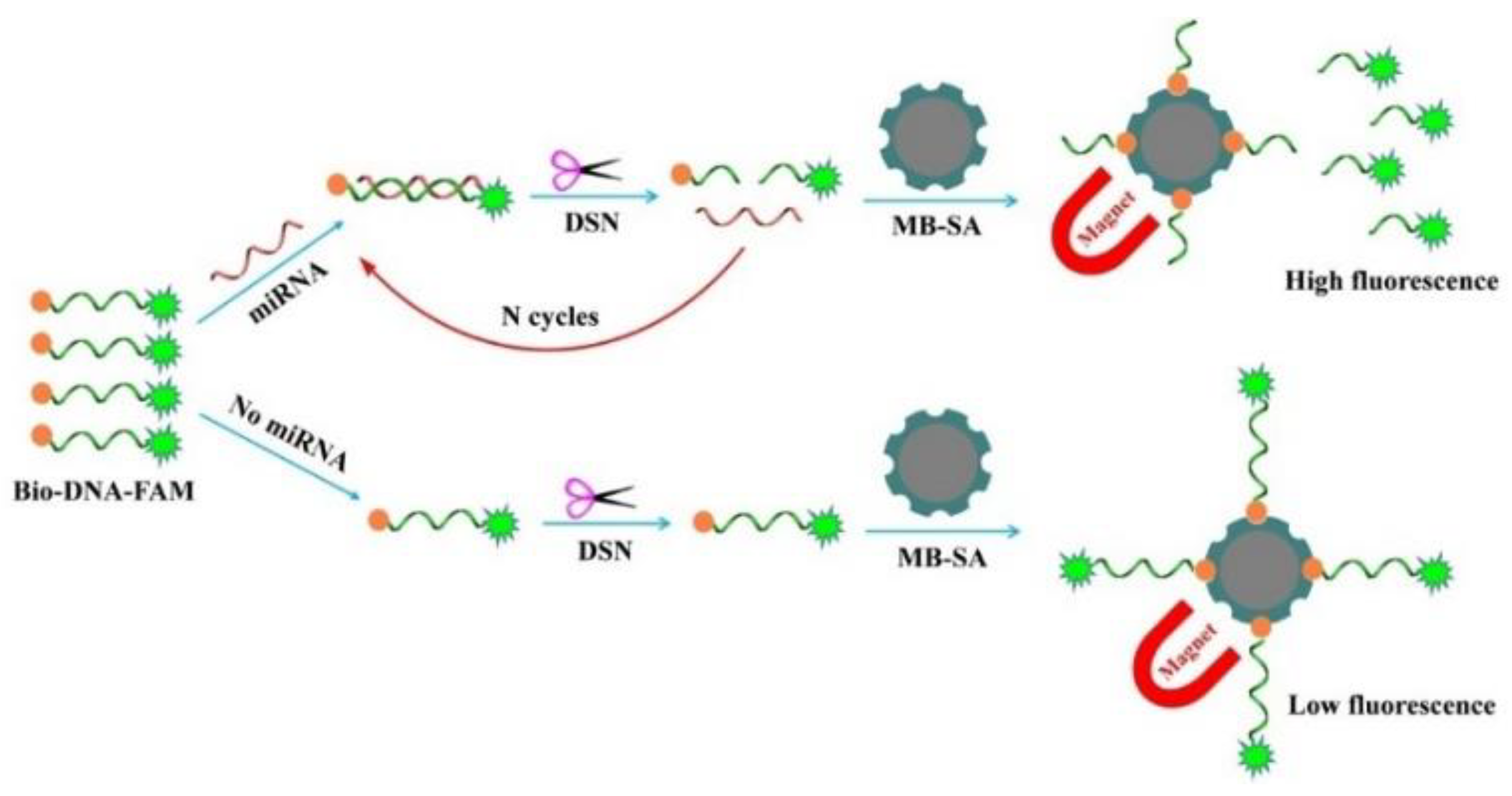
Scheme 1. Schematic representation of the magnetically assisted immobilization-free method for miRNA detection.
Biosensors 13 00699 sch001
Figure 1. Fluorescence spectra of Bio-DNA-FAM before (curve a) and after (curve b) incubation with MB-SA, as well as the mixture of Bio-DNA-FAM/miRNA-21 (curve c) and Bio-DNA-FAM/miRNA-21/DSN (curve d) after treatment by MB-SA. Curve corresponds to that of incubating the MB-SA-Bio-DNA-FAM conjugates with the mixture of miRNA/DSN. The used concentrations of Bio-DNA-FAM, DSN, and miRNA-21 were 500 nM, 0.1 U, and 100 pM.
Biosensors 13 00699 g001
Figure 2. Effect of probe concentration (A) and incubation time (B) on fluorescence intensity. The concentrations of Bio-DNA-FAM, miRNA-21, and DSN were 500 nM, 100 pM, and 0.1 U, respectively.
Biosensors 13 00699 g002
Figure 3. (A) Fluorescence spectra for the detection of different concentrations of miRNA-21 (from to bottom to top: 0, 0.01, 0.1, 1, 5, 10, 25, 50, and 100 pM). (B) Calibration curve between fluorescence intensity and miRNA-21 concentration. The errors were deduced from three replicate measurements. The inset shows the linear portion of the calibration curve.
Biosensors 13 00699 g003
Figure 4. Selectivity of the method. Bar 1, buffer blank; bar 2, single-base mismatch; bar 3, three-base mismatch; bar 4, non-complementary; bar 5, miRNA-21.
Biosensors 13 00699 g004
Figure 5. Results for the assays of miRNA-21 in lysates extracted from MCF-7 and Hela cells.
Biosensors 13 00699 g005
Table 1. Analytical performances of DSN-based fluorescence methods for miRNA detection.
Materials/ReportersLinear RangeDetection LimitRef.
AuNPs/dye5–200 pM5 pM[41]
AuNPs/dye0.1–1000 pM33.4 fM[43]
AuNPs/dye0.1–10 pM45 fM[44]
PS@Au/dye1–1000 pM50 fM[45]
MWCNT-AuNCs/dye0.1–1000 pM33.4 fM[46]
GO/dye0.5–1000 pM160 fM[47]
GO/dye5–40 nM132 pM[48]
MoS2/dye1–10,000 pM10 fM[49]
MoS2/dye0.5–50 nM426 pM[50]
CCP/dye10–200 pM4.6 pM[51]
PDA-PEI/dye0.8–50 nM0.52 nM[52]
MNPs@PDA/dye5–5000 pM0.42 pM[53]
MB/QDs0.5–10 pM0.5 pM[54]
Cu3(PO4)2 NFs/dye1–100,000 pM0.23 pM[55]
MB-SA/dye0.01–10 pM0.01 pMthis work
Abbreviations: AuNPs, gold nanoparticles; PS@Au, gold nanoparticle-coated polystyrene microbeads; MWCNT-AuNCs, multi-walled carbon nanotube–gold nanocomposites; GO, graphene oxide; MoS2, molybdenum disulfide; CCP, cationic conjugated polymer; PDA-PEI, dopamine and polyethyleneimine (PEI) copolymerized nanodots; MNPs@PDA, magnetic nanoparticles coated with polydopamine; MB, magnetic bead; QDs, quantum dots; Cu3(PO4)2 NFs, Cu3(PO4)2 hybrid nanoflowers.
Disclaimer/Publisher’s Note: The statements, opinions and data contained in all publications are solely those of the individual author(s) and contributor(s) and not of MDPI and/or the editor(s). MDPI and/or the editor(s) disclaim responsibility for any injury to people or property resulting from any ideas, methods, instructions or products referred to in the content.