Rational Truncation of Aptamer for Ultrasensitive Aptasensing of Chloramphenicol: Studies Using Bio-Layer Interferometry


Abstract
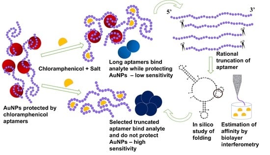
1. Introduction
2. Materials and Methods
2.1. Materials
2.2. Methods
2.2.1. Truncation of the Original Aptamer
2.2.2. Structure, Energetics, and Stability of Truncated Oligonucleotides
2.2.3. Biolayer Interferometry Studies on Binding Affinity
2.2.4. Aptasensing of CAP
Performance of the Assay in Buffer
Validation in Real Samples
Reproducibility and Specificity
3. Results and Discussion
3.1. Truncation of the Original Aptamer
3.2. Determination of Secondary Structures of Truncated Oligonucleotides
3.3. Computational Prediction of Stability of Truncated Oligonucleotides
3.4. Binding Affinity with Analyte
3.5. Aptasensing of CAP
3.5.1. Performance of the Assay
3.5.2. Validation in Real Samples
3.5.3. Specificity and Reproducibility
3.5.4. Comparison with Reported Literature
4. Conclusions
Supplementary Materials
Author Contributions
Funding
Institutional Review Board Statement
Informed Consent Statement
Data Availability Statement
Acknowledgments
Conflicts of Interest
References
- Li, J.; Shao, B.; Shen, J.; Wang, S.; Wu, Y. Occurrence of chloramphenicol-resistance genes as environmental pollutants from swine feedlots. Environ. Sci. Technol. 2013, 47, 2892–2897. [Google Scholar] [CrossRef]
- Extralabel Drug Use in Animals. Fed. Regist. 1994, 61, 57732–57745.
- Durham, M. A bitter taste of honey. In The Guardian (International Edition); The Guardian: London, UK, 2004. [Google Scholar]
- Moudgil, P.; Bedi, J.S.; Aulakh, R.S.; Gill, J.P.S.; Kumar, A. Validation of HPLC Multi-residue Method for Determination of Fluoroquinolones, Tetracycline, Sulphonamides and Chloramphenicol Residues in Bovine Milk. Food Anal. Methods 2019, 12, 338–346. [Google Scholar] [CrossRef]
- FSSAI. Manual of Methods of Analysis Food; FSSAI: New Delhi, India, 2012. [Google Scholar]
- Panel, E.; Chain, F. Scientific Opinion on Chloramphenicol in food and feed. EFSA J. 2014, 12, 3907. [Google Scholar] [CrossRef]
- Storey, J.; Pfenning, A.; Turnipseed, S.; Nandrea, G.; Lee, R.; Burns, C.; Madson, M. Determination of chloramphenicol residues in shrimp and crab tissues by electrospray triple quadrupole LC/MS/MS. Laboratory Information Bulletin 4306 Chloramphenicol Residues Shrimp Crab Tissues, Volume 2004, no. 26/03/2004. 2003. Available online: https://citeseerx.ist.psu.edu/document?repid=rep1&type=pdf&doi=a7afb82606d443adda9924fd503250125105e4fa (accessed on 28 March 2023).
- Sharma, R.; Raghavarao, K.S.M.S. Nanoparticle-Based Aptasensors for Food Contaminant Detection. In Nanomaterials for Food Applications; Elsevier: Amsterdam, The Netherlands, 2019; pp. 123–145. [Google Scholar] [CrossRef]
- Gao, H.; Gan, N.; Pan, D.; Chen, Y.; Li, T.; Cao, Y.; Fu, T. A sensitive colorimetric aptasensor for chloramphenicol detection in fish and pork based on the amplification of a nano-peroxidase-polymer. Anal. Methods 2015, 7, 6528–6536. [Google Scholar] [CrossRef]
- Gao, H.; Pan, D.; Gan, N.; Cao, J.; Sun, Y.; Wu, Z.; Zeng, X. An aptamer-based colorimetric assay for chloramphenicol using a polymeric HRP-antibody conjugate for signal amplification. Microchim. Acta 2015, 182, 2551–2559. [Google Scholar] [CrossRef]
- Huang, W.; Zhang, H.; Lai, G.; Liu, S.; Li, B.; Yu, A. Sensitive and rapid aptasensing of chloramphenicol by colorimetric signal transduction with a DNAzyme-functionalized gold nanoprobe. Food Chem. 2019, 270, 287–292. [Google Scholar] [CrossRef] [PubMed]
- Li, J.; Yu, C.; Wu, Y.N.; Zhu, Y.; Xu, J.; Wang, Y.; Wang, H.; Guo, M.; Li, F. Novel sensing platform based on gold nanoparticle-aptamer and Fe-metal-organic framework for multiple antibiotic detection and signal amplification. Environ. Int. 2019, 125, 135–141. [Google Scholar] [CrossRef]
- Wu, Y.y.; Liu, B.w.; Huang, P.; Wu, F.Y. A novel colorimetric aptasensor for detection of chloramphenicol based on lanthanum ion–assisted gold nanoparticle aggregation and smartphone imaging. Anal. Bioanal. Chem. 2019, 411, 7511–7518. [Google Scholar] [CrossRef]
- Miao, Y.; Gan, N.; Li, T.; Zhang, H.; Cao, Y.; Jiang, Q. A colorimetric aptasensor for chloramphenicol in fish based on double-stranded DNA antibody labeled enzyme-linked polymer nanotracers for signal amplification. Sens. Actuators B Chem. 2015, 220, 679–687. [Google Scholar] [CrossRef]
- Miao, Y.; Gan, N.; Ren, H.X.; Li, T.; Cao, Y.; Hu, F.; Yan, Z.; Chen, Y. A triple-amplification colorimetric assay for antibiotics based on magnetic aptamer-enzyme co-immobilized platinum nanoprobes and exonuclease-assisted target recycling. Analyst 2015, 140, 7663–7671. [Google Scholar] [CrossRef]
- Abnous, K.; Danesh, N.M.; Ramezani, M.; Emrani, A.S.; Taghdisi, S.M. A novel colorimetric sandwich aptasensor based on an indirect competitive enzyme-free method for ultrasensitive detection of chloramphenicol. Biosens. Bioelectron. 2016, 78, 80–86. [Google Scholar] [CrossRef]
- Chang, C.C.; Wang, G.; Takarada, T.; Maeda, M. Iodine-mediated etching of triangular gold nanoplates for colorimetric sensing of copper ion and aptasensing of chloramphenicol. ACS Appl. Mater. Interfaces 2017, 9, 34518–34525. [Google Scholar] [CrossRef] [PubMed]
- Luan, Q.; Xi, Y.; Gan, N.; Cao, Y.; Li, T.; Chen, Y. A facile colorimetric aptamer assay for small molecule detection in food based on a magnetic single-stranded DNA binding protein-linked composite probe. Sens. Actuators B Chem. 2017, 239, 979–987. [Google Scholar] [CrossRef]
- Javidi, M.; Housaindokht, M.R.; Verdian, A.; Razavizadeh, B.M. Detection of chloramphenicol using a novel apta-sensing platform based on aptamer terminal-lock in milk samples. Anal. Chim. Acta 2018, 1039, 116–123. [Google Scholar] [CrossRef] [PubMed]
- Xie, Y.; Huang, Y.; Tang, D.; Cui, H.; Cao, H. A competitive colorimetric chloramphenicol assay based on the non-cross-linking deaggregation of gold nanoparticles coated with a polyadenine-modified aptamer. Microchim. Acta 2018, 185, 534. [Google Scholar] [CrossRef]
- Yan, C.; Zhang, J.; Yao, L.; Xue, F.; Lu, J.; Li, B.; Chen, W. Aptamer-mediated colorimetric method for rapid and sensitive detection of chloramphenicol in food. Food Chem. 2018, 260, 208–212. [Google Scholar] [CrossRef]
- Sharma, R.; Ragavan, K.V.; Raghavarao, K.S.M.S.; Thakur, M.S. Nano-aptamer Based Quantitative Detection of Chloramphenicol. In Biotechnology and Biochemical Engineering; Springer: Singapore, 2016; pp. 187–195. [Google Scholar] [CrossRef]
- Thakur, M.S.; Ranjan, R.; Vinayaka, A.C.; Abhijith, K.S.; Sharma, R. Nanoparticles and Biophotonics as Efficient Tools in Resonance Energy Transfer-Based Biosensing for Monitoring Food Toxins and Pesticides. Adv. Appl. Nanotechnol. Agric. 2013, 1143, 55–84. [Google Scholar] [CrossRef]
- Mittal, R.; Sharma, R. Phyconanofabrication-algae as bio- templates for commercially applicable nanomaterials. In Synthesis of Bionanomaterials for Biomedical Applications, 1st ed.; Ozturk, M., Roy, A., Bhat, R.A., Vardar-Sukan, F., Tonelli, F.M.P., Eds.; Elsevier: Amsterdam, The Netherlands, 2023; pp. 95–130. [Google Scholar] [CrossRef]
- Mckeague, M.; Calzada, V.; Cerchia, L.; Derosa, M.; Heemstra, J.M.; Janjic, N.; Johnson, P.E.; Kraus, L.; Limson, J.; Mayer, G.; et al. The minimum aptamer publication standards (MAPS guidelines) for de novo aptamer selection. Aptamers 2022, 6, 10–18. [Google Scholar]
- Duan, Y.; Gao, Z.; Wang, L.; Wang, H.; Zhang, H.; Li, H. Selection and Identification of Chloramphenicol-Specific DNA Aptamers by Mag-SELEX. Appl. Biochem. Biotechnol. 2016, 180, 1644–1656. [Google Scholar] [CrossRef]
- Mehta, J.; Van Dorst, B.; Rouah-Martin, E.; Herrebout, W.; Scippo, M.L.; Blust, R.; Robbens, J. In vitro selection and characterization of DNA aptamers recognizing chloramphenicol. J. Biotechnol. 2011, 155, 361–369. [Google Scholar] [CrossRef] [PubMed]
- Zuker, M. Mfold web server for nucleic acid folding and hybridization prediction. Nucleic Acids Res. 2003, 31, 3406–3415. [Google Scholar] [CrossRef]
- Searle, M.S.; Williams, D.H. On the stability of nucleic acid structures in solution: Enthalpy-entropy compensations, internal rotations and reversibility. Nucleic Acids Res. 1993, 21, 2051–2056. [Google Scholar] [CrossRef]
- Owczarzy, R.; You, Y.; Groth, C.L.; Tataurov, A.V. Stability and mismatch discrimination of locked nucleic acid-DNA duplexes. Biochemistry 2011, 50, 9352–9367. [Google Scholar] [CrossRef]
- Gao, Y.; Torrente-Murciano, L. Mechanistic insights of the reduction of gold salts in the Turkevich protocol. Nanoscale 2020, 12, 2740–2751. [Google Scholar] [CrossRef]
- Yang, Y.; Yin, Y.; Li, X.; Wang, S.; Dong, Y. Development of a chimeric aptamer and an AuNPs aptasensor for highly sensitive and specific identification of Aflatoxin B1. Sens. Actuators B Chem. 2020, 319, 128250. [Google Scholar] [CrossRef]
- Alawad, A.; Istamboulié, G.; Calas-Blanchard, C.; Noguer, T. A reagentless aptasensor based on intrinsic aptamer redox activity for the detection of tetracycline in water. Sens. Actuators B Chem. 2019, 288, 141–146. [Google Scholar] [CrossRef]
- Sharma, R.; Akshath, U.S.; Bhatt, P.; Raghavarao, K. Fluorescent aptaswitch for chloramphenicol detection–Quantification enabled by immobilization of aptamer. Sens. Actuators B Chem. 2019, 290, 110–117. [Google Scholar] [CrossRef]
- Abnous, K.; Danesh, N.M.; Ramezani, M.; Taghdisi, S.M.; Emrani, A.S. A novel electrochemical aptasensor based on H-shape structure of aptamer-complimentary strands conjugate for ultrasensitive detection of cocaine. Sens. Actuators B Chem. 2016, 224, 351–355. [Google Scholar] [CrossRef]
- Auer, S.; Koho, T.; Uusi-Kerttula, H.; Vesikari, T.; Blazevic, V.; Hytönen, V.P. Rapid and sensitive detection of norovirus antibodies in human serum with a biolayer interferometry biosensor. Sens. Actuators B Chem. 2015, 221, 507–514. [Google Scholar] [CrossRef]
- Gandhi, I.; Narendran, K.; Jackson, G.W. Rapid DNA Aptamer Binding Characterization and ELASA Development Using Biolayer Interferometry (BLI). 2011. Available online: https://www.basepairbio.com (accessed on 20 July 2022).
- Tao, X.; He, F.; Liu, X.; Zhang, F.; Wang, X.; Peng, Y.; Liu, J. Detection of chloramphenicol with an aptamer-based colorimetric assay: Critical evaluation of specific and unspecific binding of analyte molecules. Mikrochim. Acta 2020, 187, 668. [Google Scholar] [CrossRef] [PubMed]
- Dayne, D.; Gandhi, I.; Jackson, G.W.; Kumaraswamy, S.; Silva, C. Fortebio Interactions. 2012; 5, 1. [Google Scholar]
- Haiss, W.; Thanh, N.T.K.; Aveyard, J.; Fernig, D.G. Determination of Size and Concentration of Gold Nanoparticles from UV−Vis Spectra. Anal. Chem 2007, 79, 4215–4221. [Google Scholar] [CrossRef] [PubMed]


| Seq. No | Sequence (5′ 🡪 3′) | No. of Bases Removed | Terminal/Seq Base Numbers |
|---|---|---|---|
| 1 | AGCAGCACAGAGGTCAGATGACTTCAGTGAGTTGTCCCACGGTCGGCGAGTCGGTGGTAGCCTATGCGTGCTACCGTGAA (original) | 0 | - 1 to 80 |
| 2 | CACAGAGGTCAGATGACTTCAGTGAGTTGTCCCACGGTCGGCGAGTCGGTGGTAGCCTATGCGTGCTACCGTGAA | 5 | 5′ 6 to 80 |
| 3 | AGCAGCACAGAGGTCAGATGACTTCAGTGAGTTGTCCCACGGTCGGCGAGTCGGTGGTAGCCTATGCGTGCTACC | 5 | 3′ 1 to 75 |
| 4 | CACAGAGGTCAGATGACTTCAGTGAGTTGTCCCACGGTCGGCGAGTCGGTGGTAGCCTATGCGTGCTACC | 10 | 5′and 3′ 6 to 75 |
| 5 | ACTTCAGTGAGTTGTCCCACGGTCGGCGAGTCGGTGGTAGCCTATGCGTGCTACCGTGAA | 20 | 5′ 21 to 80 |
| 6 | AGCAGCACAGAGGTCAGATGACTTCAGTGAGTTGTCCCACGGTCGGCGAGTCGGTGGTAG | 20 | 3′ 1 to 60 |
| 7 | ACTTCAGTGAGTTGTCCCACGGTCGGCGAGTCGGTGGTAG | 40 | 5′ and 3′ 21 to 60 |
| 8 | GTTGTCCCACGGTCGGCGAGTCGGTGGTAGCCTATGCGTGCTACCGTGAA | 30 | 5′ 31 to 80 |
| 9 | AGCAGCACAGAGGTCAGATGACTTCAGTGAGTTGTCCCACGGTCGGCGAG | 30 | 3′ 1 to 50 |
| 10 | GTTGTCCCACGGTCGGCGAG | 60 | 5′ and 3′ 31 to 50 |
| 11 | GGTCGGCGAGTCGGTGGTAGCCTATGCGTGCTACCGTGAA | 40 | 5′ 41 to 80 |
| Sequence Number | Kd (μM) | R2 |
|---|---|---|
| 1 | 0.704 (previously reported value in literature is 0.766) | 0.993 |
| 2 | 0.877 | 0.973 |
| 3 | 0.645 | 0.946 |
| 4 | 0.661 | 0.945 |
| 5 | 1.712 | 0.924 |
| 6 | 4.014 | 0.905 |
| 7 | 0.370 | 0.882 |
| 8 | 0.153 | 0.853 |
| 9 | 0.092 | 0.914 |
| 10 | 3.109 | 0.699 |
| 11 | 0.426 | 0.757 |
| Spiked Amount (pg mL−1) | Aptasensing (pg mL−1)/(pg g−1) | HPLC (pg mL−1)/(pg g−1) | Recovery (%) ± SD (with Respect to Spiked Amount) |
|---|---|---|---|
| 0 | Not detected | Not detected | - |
| 10 | 9.7/28.9 | Not detected | 96.6 ± 2.7 |
| 100 | 94.8/284.3 | Not detected | 94.7 ± 3.8 |
| 1000 | 1004.3/3012.8 | Not detected | 100.4 ± 6.9 |
| 10,000 | 9812.5/29,437.5 | 9766.7/29,300.0 | 98.12 ± 1.1 |
| 50,000 | 49,605.7/148,817.1 | 48,942.9/148,828.8 | 97.88 ± 8.6 |
Disclaimer/Publisher’s Note: The statements, opinions and data contained in all publications are solely those of the individual author(s) and contributor(s) and not of MDPI and/or the editor(s). MDPI and/or the editor(s) disclaim responsibility for any injury to people or property resulting from any ideas, methods, instructions or products referred to in the content. |
© 2023 by the authors. Licensee MDPI, Basel, Switzerland. This article is an open access article distributed under the terms and conditions of the Creative Commons Attribution (CC BY) license (https://creativecommons.org/licenses/by/4.0/).
Share and Cite
Sharma, R.; Mukherjee, M.; Bhatt, P.; Raghavarao, K.S.M.S. Rational Truncation of Aptamer for Ultrasensitive Aptasensing of Chloramphenicol: Studies Using Bio-Layer Interferometry. Biosensors 2023, 13, 660. https://doi.org/10.3390/bios13060660
Sharma R, Mukherjee M, Bhatt P, Raghavarao KSMS. Rational Truncation of Aptamer for Ultrasensitive Aptasensing of Chloramphenicol: Studies Using Bio-Layer Interferometry. Biosensors. 2023; 13(6):660. https://doi.org/10.3390/bios13060660
Chicago/Turabian StyleSharma, Richa, Monali Mukherjee, Praveena Bhatt, and K. S. M. S. Raghavarao. 2023. "Rational Truncation of Aptamer for Ultrasensitive Aptasensing of Chloramphenicol: Studies Using Bio-Layer Interferometry" Biosensors 13, no. 6: 660. https://doi.org/10.3390/bios13060660
APA StyleSharma, R., Mukherjee, M., Bhatt, P., & Raghavarao, K. S. M. S. (2023). Rational Truncation of Aptamer for Ultrasensitive Aptasensing of Chloramphenicol: Studies Using Bio-Layer Interferometry. Biosensors, 13(6), 660. https://doi.org/10.3390/bios13060660