Trends in Paper-Based Sensing Devices for Clinical and Environmental Monitoring

 , , ,
, , , 

Abstract
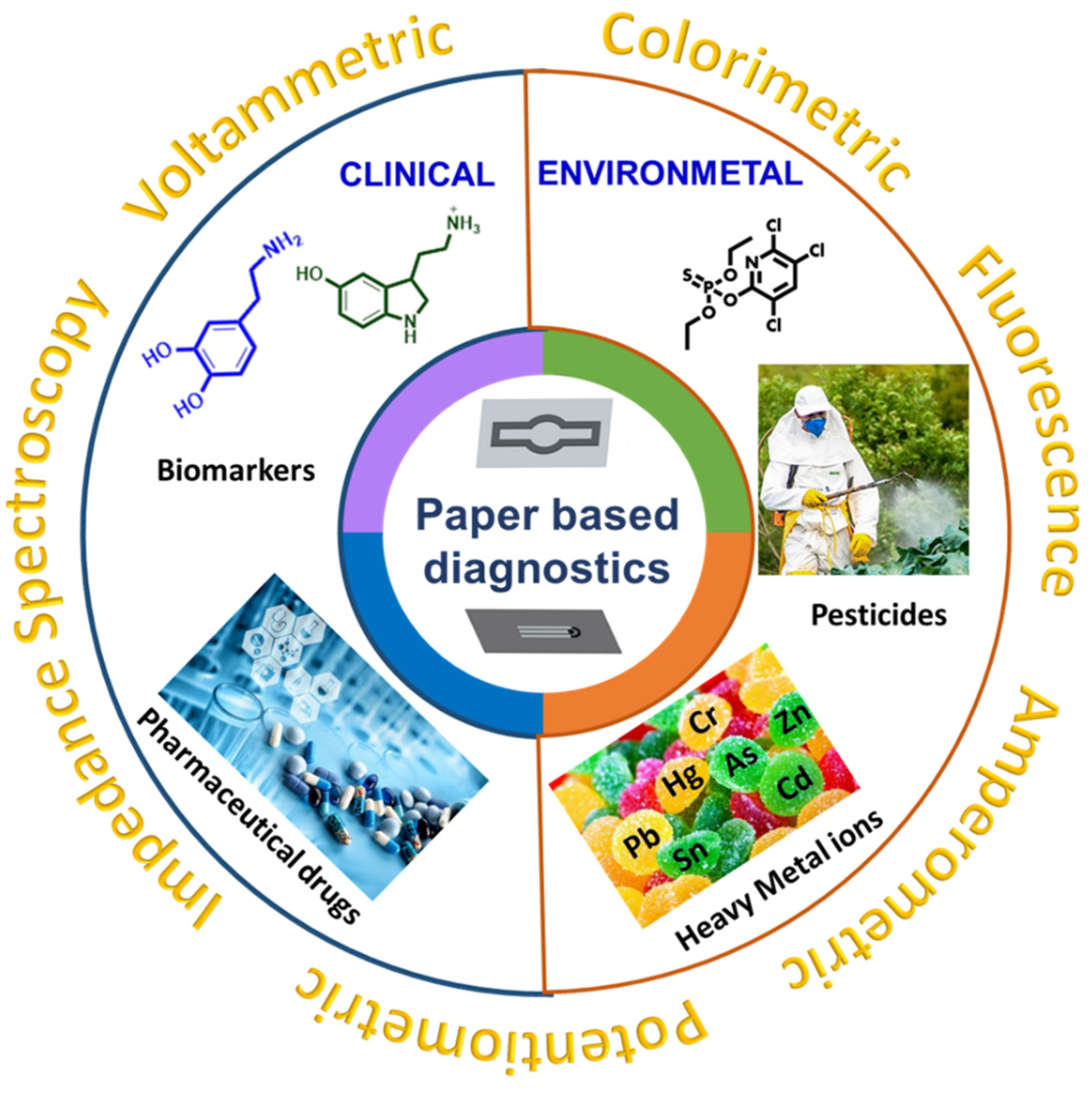
1. Introduction
2. Optical Paper-Based Sensors
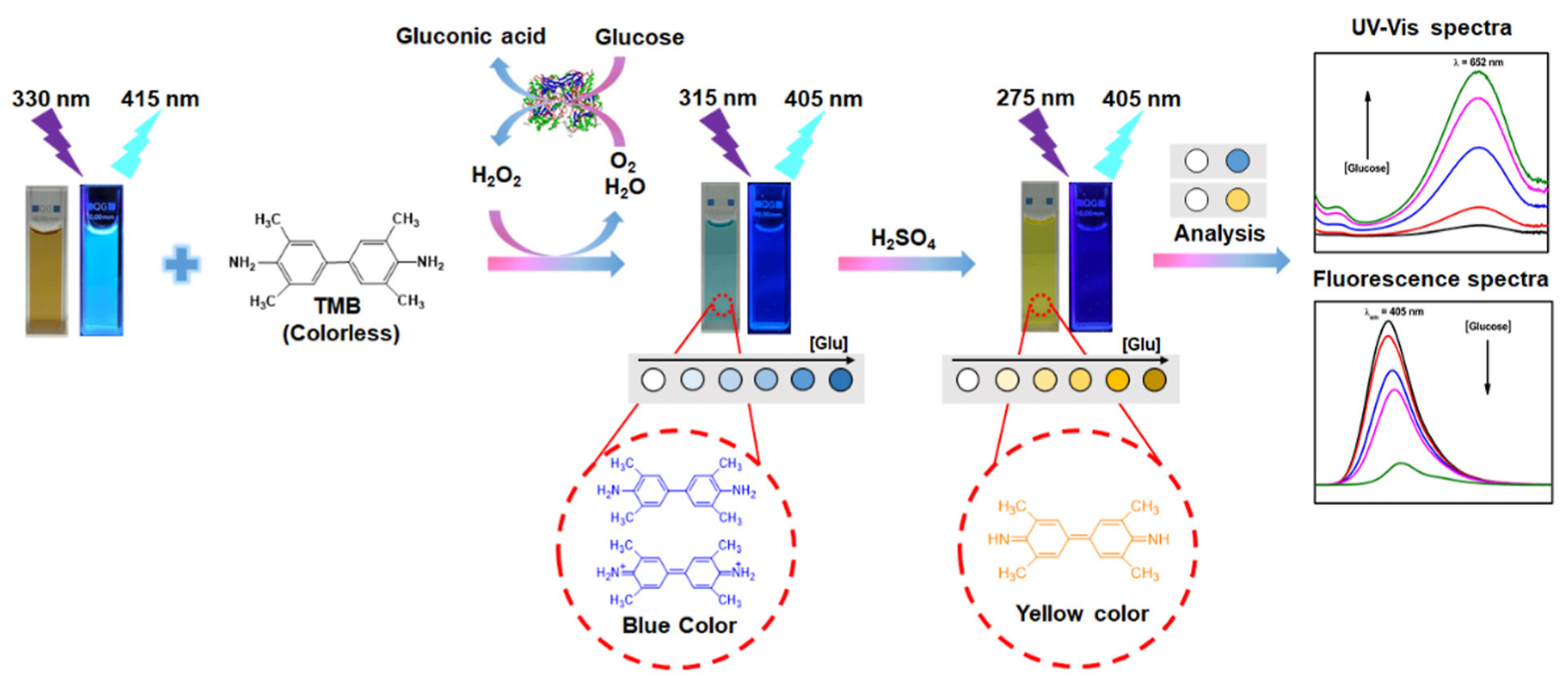
2.1. Fluorescence
2.2. Absorbance and Colorimetric Sensors
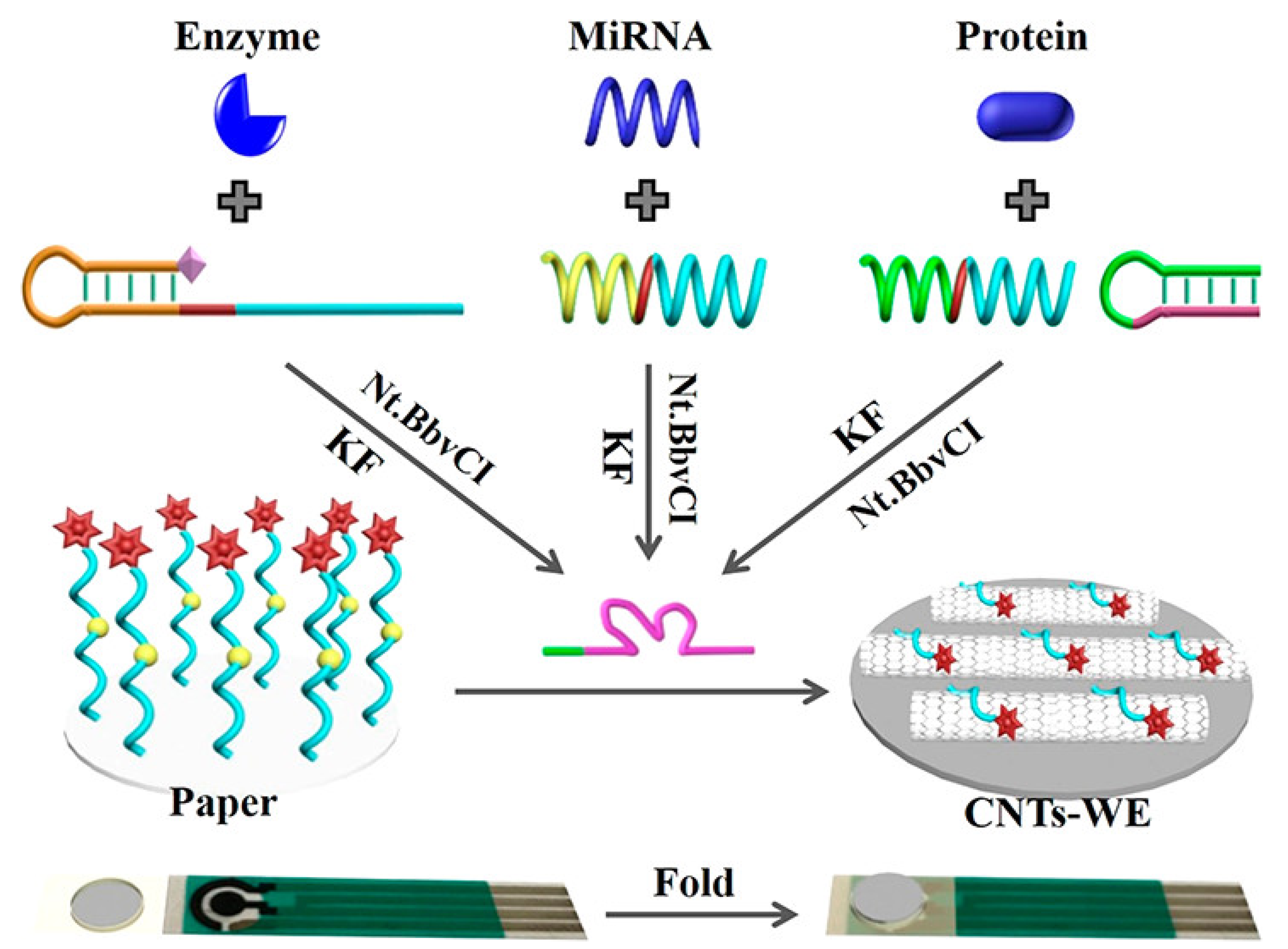
3. Electrochemical Paper-Based Sensors
4. Summary, Conclusions, and Future Perspectives
Author Contributions
Funding
Institutional Review Board Statement
Informed Consent Statement
Data Availability Statement
Acknowledgments
Conflicts of Interest
References
- Kudr, J.; Zitka, O.; Klimanek, M.; Vrba, R.; Adam, V. Microfluidic Electrochemical Devices for Pollution Analysis–A Review. Sens. Actuators B Chem. 2017, 246, 578–590. [Google Scholar] [CrossRef]
- Azzouz, A.; Goud, K.Y.; Raza, N.; Ballesteros, E.; Lee, S.E.; Hong, J.; Deep, A.; Kim, K.H. Nanomaterial-Based Electrochemical Sensors for the Detection of Neurochemicals in Biological Matrices. TrAC Trends Anal. Chem. 2019, 110, 15–34. [Google Scholar] [CrossRef]
- Reddy, K.K.; Bandal, H.; Satyanarayana, M.; Goud, K.Y.; Gobi, K.V.; Jayaramudu, T.; Amalraj, J.; Kim, H. Recent Trends in Electrochemical Sensors for Vital Biomedical Markers Using Hybrid Nanostructured Materials. Adv. Sci. 2020, 7, 1902980. [Google Scholar] [CrossRef]
- Kummari, S.; Sunil Kumar, V.; Vengatajalabathy Gobi, K. Facile Electrochemically Reduced Graphene Oxide-Multi-walled Carbon Nanotube Nanocomposite as Sensitive Probe for In-vitro Determination of Nitrofurantoin in Biological Fluids. Electroanalysis 2020, 32, 2452–2462. [Google Scholar] [CrossRef]
- Kummari, S.; Sunil Kumar, V.; Yugender Goud, K.; Vengatajalabathy Gobi, K. Nano-Au Particle Decorated Poly-(3-Amino-5-Hydroxypyrazole) Coated Carbon Paste Electrode for in-Vitro Detection of Valacyclovir. J. Electroanal. Chem. 2022, 904, 115859. [Google Scholar] [CrossRef]
- Shekher, K.; Sampath, K.; Vandini, S.; Satyanarayana, M.; Vengatajalabathy Gobi, K. Gold Nanoparticle Assimilated Polymer Layer on Carbon Nanotube Matrices for Sensitive Detection of Serotonin in Presence of Dopamine In-Vitro. Inorg. Chim. Acta 2023, 549, 121399. [Google Scholar] [CrossRef]
- Goud, K.Y.; Satyanarayana, M.; Hayat, A.; Gobi, K.V.; Marty, J.L. Nanomaterial-Based Electrochemical Sensors in Pharmaceutical Applications. In Nanoparticles in Pharmacotherapy; Elsevier: Amsterdam, The Netherlands, 2019; pp. 195–216. ISBN 9780128165041. [Google Scholar]
- Yugender Goud, K.; Hayat, A.; Satyanarayana, M.; Sunil Kumar, V.; Catanante, G.; Vengatajalabathy Gobi, K.; Marty, J.L. Aptamer-Based Zearalenone Assay Based on the Use of a Fluorescein Label and a Functional Graphene Oxide as a Quencher. Microchim. Acta 2017, 184, 4401–4408. [Google Scholar] [CrossRef]
- Hu, X.; Goud, K.Y.; Kumar, V.S.; Catanante, G.; Li, Z.; Zhu, Z.; Louis, J. Sensors and Actuators B: Chemical Disposable Electrochemical Aptasensor Based on Carbon Nanotubes-V 2 O 5 -Chitosan Nanocomposite for Detection of Ciprofloxacin. Sens. Actuators B Chem. 2018, 268, 278–286. [Google Scholar] [CrossRef]
- Goud, K.Y.; Kumar, S.; Gobi, K.V.; Kim, K. Biosensors and Bioelectronics Progress on Nanostructured Electrochemical Sensors and Their Recognition Elements for Detection of Mycotoxins: A Review. Biosens. Bioelectron. 2018, 121, 205–222. [Google Scholar] [CrossRef] [PubMed]
- Goud, K.Y.; Teymourian, H.; Sandhu, S.S.; Tostado, N.; Mishra, R.K.; Moore, L.C.; Harvey, S.P.; Wang, J. OPAA/Fluoride Biosensor Chip towards Field Detection of G-Type Nerve Agents. Sens. Actuators B Chem. 2020, 320, 128344. [Google Scholar] [CrossRef]
- Zejli, H.; Goud, K.Y.; Marty, J.L. An Electrochemical Aptasensor Based on Polythiophene-3-Carboxylic Acid Assisted Methylene Blue for Aflatoxin B1 Detection. Sens. Bio-Sens. Res. 2019, 25, 100290. [Google Scholar] [CrossRef]
- Goud, K.Y.; Kumar, V.S.; Hayat, A.; Catanante, G.; Gobi, K.V.; Marty, J.L. Polymer Scaffold Layers of Screen-Printed Electrodes for Homogeneous Deposition of Silver Nanoparticles: Application to the Amperometric Detection of Hydrogen Peroxide. Microchim. Acta 2019, 186, 1–10. [Google Scholar] [CrossRef]
- Sandhu, S.S.; Kotagiri, Y.G.; Fernando, I.P.A.I.; Kalaj, M.; Tostado, N.; Teymourian, H.; Alberts, E.M.; Thornell, T.L.; Jenness, G.R.; Harvey, S.P.; et al. Green MIP-202(Zr) Catalyst: Degradation and Thermally Robust Biomimetic Sensing of Nerve Agents. J. Am. Chem. Soc. 2021, 143, 18261–18271. [Google Scholar] [CrossRef] [PubMed]
- Goud, K.Y.; Sandhu, S.S.; Teymourian, H.; Yin, L.; Tostado, N.; Raushel, F.M.; Harvey, S.P.; Moores, L.C.; Wang, J. Textile-Based Wearable Solid-Contact Flexible Fluoride Sensor: Toward Biodetection of G-Type Nerve Agents. Biosens. Bioelectron. 2021, 182, 113172. [Google Scholar] [CrossRef]
- Mahato, K.; Srivastava, A.; Chandra, P. Paper Based Diagnostics for Personalized Health Care: Emerging Technologies and Commercial Aspects. Biosens. Bioelectron. 2017, 96, 246–259. [Google Scholar] [CrossRef] [PubMed]
- Sempionatto, J.R.; Khorshed, A.A.; Ahmed, A.; De Loyola E Silva, A.N.; Barfidokht, A.; Yin, L.; Goud, K.Y.; Mohamed, M.A.; Bailey, E.; May, J.; et al. Epidermal Enzymatic Biosensors for Sweat Vitamin C: Toward Personalized Nutrition. ACS Sens. 2020, 5, 1804–1813. [Google Scholar] [CrossRef]
- Niroula, J.; Premaratne, G.; Krishnan, S. Lab-on-Paper Aptasensor for Label-Free Picomolar Detection of a Pancreatic Hormone in Serum. Biosens. Bioelectron. X 2022, 10, 100114. [Google Scholar] [CrossRef]
- Krishnan, S.; Syed, Z.u.Q. Colorimetric Visual Sensors for Point-of-Needs Testing. Sens. Actuators Rep. 2022, 4, 100078. [Google Scholar] [CrossRef]
- Ge, S.; Zhang, Y.; Zhang, L.; Liang, L.; Liu, H.; Yan, M.; Huang, J.; Yu, J. Ultrasensitive Electrochemical Cancer Cells Sensor Based on Trimetallic Dendritic Au@PtPd Nanoparticles for Signal Amplification on Lab-on-Paper Device. Sens. Actuators B Chem. 2015, 220, 665–672. [Google Scholar] [CrossRef]
- Kuswandi, B.; Ensafi, A.A. Perspective—Paper-Based Biosensors: Trending Topic in Clinical Diagnostics Developments and Commercialization. J. Electrochem. Soc. 2020, 167, 037509. [Google Scholar] [CrossRef]
- Bordbar, M.M.; Sheini, A.; Hashemi, P.; Hajian, A.; Bagheri, H. Disposable Paper-Based Biosensors for the Point-of-Care Detection of Hazardous Contaminations—A Review. Biosensors 2021, 11, 316. [Google Scholar] [CrossRef] [PubMed]
- Baharfar, M.; Rahbar, M.; Tajik, M.; Liu, G. Engineering Strategies for Enhancing the Performance of Electrochemical Paper-Based Analytical Devices. Biosens. Bioelectron. 2020, 167, 112506. [Google Scholar] [CrossRef]
- Deroco, P.B.; Wachholz Junior, D.; Kubota, L.T. Paper-based Wearable Electrochemical Sensors: A New Generation of Analytical Devices. Electroanalysis 2023, 35, e202200177. [Google Scholar] [CrossRef]
- Mettakoonpitak, J.; Boehle, K.; Nantaphol, S.; Teengam, P.; Adkins, J.A.; Srisa-Art, M.; Henry, C.S. Electrochemistry on Paper-Based Analytical Devices: A Review. Electroanalysis 2016, 28, 1420–1436. [Google Scholar] [CrossRef]
- Fu, L.-M.; Wang, Y.-N. Detection Methods and Applications of Microfluidic Paper-Based Analytical Devices. TrAC Trends Anal. Chem. 2018, 107, 196–211. [Google Scholar] [CrossRef]
- Ataide, V.N.; Mendes, L.F.; Gama, L.I.L.M.; de Araujo, W.R.; Paixão, T.R.L.C. Electrochemical Paper-Based Analytical Devices: Ten Years of Development. Anal. Methods 2020, 12, 1030–1054. [Google Scholar] [CrossRef]
- Zhang, T.; Ding, F.; Yang, Y.; Zhao, G.; Zhang, C.; Wang, R.; Huang, X. Research Progress and Future Trends of Microfluidic Paper-Based Analytical Devices in In-Vitro Diagnosis. Biosensors 2022, 12, 485. [Google Scholar] [CrossRef]
- Wang, M.; Cui, J.; Wang, Y.; Yang, L.; Jia, Z.; Gao, C.; Zhang, H. Microfluidic Paper-Based Analytical Devices for the Determination of Food Contaminants: Developments and Applications. J. Agric. Food Chem. 2022, 70, 8188–8206. [Google Scholar] [CrossRef]
- Kim, Y.; Jang, G.; Lee, T.S. New Fluorescent Metal-Ion Detection Using a Paper-Based Sensor Strip Containing Tethered Rhodamine Carbon Nanodots. ACS Appl. Mater. Interfaces 2015, 7, 15649–15657. [Google Scholar] [CrossRef]
- Khoshbin, Z.; Housaindokht, M.R.; Izadyar, M.; Verdian, A.; Bozorgmehr, M.R. A Simple Paper-Based Aptasensor for Ultrasensitive Detection of Lead (II) Ion. Anal. Chim. Acta 2019, 1071, 70–77. [Google Scholar] [CrossRef]
- Liang, L.; Su, M.; Li, L.; Lan, F.; Yang, G.; Ge, S.; Yu, J.; Song, X. Aptamer-Based Fluorescent and Visual Biosensor for Multiplexed Monitoring of Cancer Cells in Microfluidic Paper-Based Analytical Devices. Sens. Actuators B Chem. 2016, 229, 347–354. [Google Scholar] [CrossRef]
- Van Tam, T.; Hur, S.H.; Chung, J.S.; Choi, W.M. Novel Paper- and Fiber Optic-Based Fluorescent Sensor for Glucose Detection Using Aniline-Functionalized Graphene Quantum Dots. Sens. Actuators B Chem. 2021, 329, 129250. [Google Scholar] [CrossRef]
- Xiao, G.; He, J.; Chen, X.; Qiao, Y.; Wang, F.; Xia, Q.; Yu, L.; Lu, Z. A Wearable, Cotton Thread/Paper-Based Microfluidic Device Coupled with Smartphone for Sweat Glucose Sensing. Cellulose 2019, 26, 4553–4562. [Google Scholar] [CrossRef]
- Ngo, Y.-L.T.; Nguyen, P.L.; Jana, J.; Choi, W.M.; Chung, J.S.; Hur, S.H. Simple Paper-Based Colorimetric and Fluorescent Glucose Sensor Using N-Doped Carbon Dots and Metal Oxide Hybrid Structures. Anal. Chim. Acta 2021, 1147, 187–198. [Google Scholar] [CrossRef] [PubMed]
- Li, D.; Duan, H.; Ma, Y.; Deng, W. Headspace-Sampling Paper-Based Analytical Device for Colorimetric/Surface-Enhanced Raman Scattering Dual Sensing of Sulfur Dioxide in Wine. Anal. Chem. 2018, 90, 5719–5727. [Google Scholar] [CrossRef]
- Chaiyo, S.; Siangproh, W.; Apilux, A.; Chailapakul, O. Highly Selective and Sensitive Paper-Based Colorimetric Sensor Using Thiosulfate Catalytic Etching of Silver Nanoplates for Trace Determination of Copper Ions. Anal. Chim. Acta 2015, 866, 75–83. [Google Scholar] [CrossRef] [PubMed]
- Lookadoo, D.B.; Schonhorn, J.E.; Harpaldas, H.; Uherek, C.M.; Schatz, P.; Lindgren, A.; Depa, M.; Kumar, A.A. Paper-Based Optode Devices (PODs) for Selective Quantification of Potassium in Biological Fluids. Anal. Chem. 2021, 93, 9383–9389. [Google Scholar] [CrossRef]
- Qin, Y.; Pan, S.; Howlader, M.M.R.; Ghosh, R.; Hu, N.-X.; Deen, M.J. Paper-Based, Hand-Drawn Free Chlorine Sensor with Poly(3,4-Ethylenedioxythiophene):Poly(Styrenesulfonate). Anal. Chem. 2016, 88, 10384–10389. [Google Scholar] [CrossRef]
- Tsai, T.-T.; Huang, C.-Y.; Chen, C.-A.; Shen, S.-W.; Wang, M.-C.; Cheng, C.-M.; Chen, C.-F. Diagnosis of Tuberculosis Using Colorimetric Gold Nanoparticles on a Paper-Based Analytical Device. ACS Sens. 2017, 2, 1345–1354. [Google Scholar] [CrossRef]
- Bhattacharjee, M.; Nemade, H.B.; Bandyopadhyay, D. Nano-Enabled Paper Humidity Sensor for Mobile Based Point-of-Care Lung Function Monitoring. Biosens. Bioelectron. 2017, 94, 544–551. [Google Scholar] [CrossRef] [PubMed]
- Mustafa, F.; Andreescu, S. Paper-Based Enzyme Biosensor for One-Step Detection of Hypoxanthine in Fresh and Degraded Fish. ACS Sens. 2020, 5, 4092–4100. [Google Scholar] [CrossRef]
- Alizadeh, N.; Salimi, A.; Hallaj, R. Mimicking Peroxidase Activity of Co2(OH)2CO3-CeO2 Nanocomposite for Smartphone Based Detection of Tumor Marker Using Paper-Based Microfluidic Immunodevice. Talanta 2018, 189, 100–110. [Google Scholar] [CrossRef] [PubMed]
- Kitchawengkul, N.; Prakobkij, A.; Anutrasakda, W.; Yodsin, N.; Jungsuttiwong, S.; Chunta, S.; Amatatongchai, M.; Jarujamrus, P. Mimicking Peroxidase-Like Activity of Nitrogen-Doped Carbon Dots (N-CDs) Coupled with a Laminated Three-Dimensional Microfluidic Paper-Based Analytical Device (Laminated 3D-ΜPAD) for Smart Sensing of Total Cholesterol from Whole Blood. Anal. Chem. 2021, 93, 6989–6999. [Google Scholar] [CrossRef] [PubMed]
- Li, H.; Wang, W.; Wang, Z.; Lv, Q.; Bai, H.; Zhang, Q. Analyte-Enhanced Photocatalytic Activity of CdSe/ZnS Quantum Dots for Paper-Based Colorimetric Sensing of Hg2+ under Visible Light. Microchem. J. 2021, 164, 106037. [Google Scholar] [CrossRef]
- Liu, Y.-C.; Hsu, C.-H.; Lu, B.-J.; Lin, P.-Y.; Ho, M.-L. Determination of Nitrite Ions in Environment Analysis with a Paper-Based Microfluidic Device. Dalt. Trans. 2018, 47, 14799–14807. [Google Scholar] [CrossRef]
- Al Yahyai, I.; Al-Lawati, H.A.J.; Hassanzadeh, J. A Paper-Based Chemiluminescence Detection Device Based on S,N-Doped Carbon Quantum Dots for the Selective and Highly Sensitive Recognition of Bendiocarb. Anal. Methods 2021, 13, 3461–3470. [Google Scholar] [CrossRef] [PubMed]
- Grazioli, C.; Faura, G.; Dossi, N.; Toniolo, R.; Tubaro, F.; Terzi, F.; Bontempelli, G. A Colorimetric Paper-Based Smart Label Soaked with a Deep-Eutectic Solvent for the Detection of Malondialdehyde. Sens. Actuators B Chem. 2021, 329, 129174. [Google Scholar] [CrossRef]
- He, R.-Y.; Tseng, H.-Y.; Lee, H.-A.; Liu, Y.-C.; Koshevoy, I.O.; Pan, S.-W.; Ho, M.-L. Paper-Based Microfluidic Devices Based on 3D Network Polymer Hydrogel for the Determination of Glucose in Human Whole Blood. RSC Adv. 2019, 9, 32367–32374. [Google Scholar] [CrossRef]
- Arcot, L.R.; Uddin, K.M.A.; Chen, X.; Wenchao, X.; Xianming, K.; Johansson, L.S.; Ras, R.H.A.; Rojas, O.J. Paper-Based Plasmon-Enhanced Protein Sensing by Controlled Nucleation of Silver Nanoparticles on Cellulose. Cellulose 2015, 22, 4027–4034. [Google Scholar] [CrossRef]
- Tan, W.; Zhang, L.; Doery, J.C.G.; Shen, W. Three-Dimensional Microfluidic Tape-Paper-Based Sensing Device for Blood Total Bilirubin Measurement in Jaundiced Neonates. Lab Chip 2020, 20, 394–404. [Google Scholar] [CrossRef]
- Liu, F.; Zhang, C. A Novel Paper-Based Microfluidic Enhanced Chemiluminescence Biosensor for Facile, Reliable and Highly-Sensitive Gene Detection of Listeria Monocytogenes. Sens. Actuators B Chem. 2015, 209, 399–406. [Google Scholar] [CrossRef]
- Pena-Pereira, F.; Lavilla, I.; Bendicho, C. Paper-Based Analytical Device for Instrumental-Free Detection of Thiocyanate in Saliva as a Biomarker of Tobacco Smoke Exposure. Talanta 2016, 147, 390–396. [Google Scholar] [CrossRef] [PubMed]
- Choi, Y.-S.; Lee, M.R.; Yang, K.-Y.; Kim, C.S.; Lee, K.-H. Paper-Based Colorimetric Sensor for Easy and Simple Detection of Polygalacturonase Activity Aiming for Diagnosis of Allium White Rot Disease. Anal. Chim. Acta 2020, 1113, 1–8. [Google Scholar] [CrossRef] [PubMed]
- Wirojsaengthong, S.; Aryuwananon, D.; Aeungmaitrepirom, W.; Pulpoka, B.; Tuntulani, T. A Colorimetric Paper-Based Optode Sensor for Highly Sensitive and Selective Determination of Thiocyanate in Urine Sample Using Cobalt Porphyrin Derivative. Talanta 2021, 231, 122371. [Google Scholar] [CrossRef] [PubMed]
- Jang, H.; Park, J.-H.; Oh, J.; Kim, K.; Kim, M.-G. Advanced Colorimetric Paper Sensors Using Color Focusing Effect Based on Asymmetric Flow of Fluid. ACS Sens. 2019, 4, 1103–1108. [Google Scholar] [CrossRef]
- Chen, C.-A.; Wang, P.-W.; Yen, Y.-C.; Lin, H.-L.; Fan, Y.-C.; Wu, S.-M.; Chen, C.-F. Fast Analysis of Ketamine Using a Colorimetric Immunosorbent Assay on a Paper-Based Analytical Device. Sens. Actuators B Chem. 2019, 282, 251–258. [Google Scholar] [CrossRef]
- Ali, M.; Khalid, M.A.U.; Shah, I.; Kim, S.W.; Kim, Y.S.; Lim, J.H.; Choi, K.H. Paper-Based Selective and Quantitative Detection of Uric Acid Using Citrate-Capped Pt Nanoparticles (PtNPs) as a Colorimetric Sensing Probe through a Simple and Remote-Based Device. New J. Chem. 2019, 43, 7636–7645. [Google Scholar] [CrossRef]
- Khachornsakkul, K.; Chang, J.J.; Lin, P.H.; Lin, Y.H.; Dungchai, W.; Chen, C.H. Highly Sensitive Distance-Based Liquid Crystalline Visualization for Paper-Based Analytical Devices. Anal. Chim. Acta 2021, 1154, 338328. [Google Scholar] [CrossRef]
- Ahmadi, A.; Khoshfetrat, S.M.; Kabiri, S.; Dorraji, P.S.; Larijani, B.; Omidfar, K. Electrochemiluminescence Paper-Based Screen-Printed Electrode for HbA1c Detection Using Two-Dimensional Zirconium Metal-Organic Framework/Fe3O4 Nanosheet Composites Decorated with Au Nanoclusters. Microchim. Acta 2021, 188, 296. [Google Scholar] [CrossRef]
- Wu, L.; Ma, C.; Ge, L.; Kong, Q.; Yan, M.; Ge, S.; Yu, J. Paper-Based Electrochemiluminescence Origami Cyto-Device for Multiple Cancer Cells Detection Using Porous AuPd Alloy as Catalytically Promoted Nanolabels. Biosens. Bioelectron. 2015, 63, 450–457. [Google Scholar] [CrossRef]
- Shibata, H.; Ikeda, Y.; Hiruta, Y.; Citterio, D. Inkjet-Printed PH-Independent Paper-Based Calcium Sensor with Fluorescence Signal Readout Relying on a Solvatochromic Dye. Anal. Bioanal. Chem. 2020, 412, 3489–3497. [Google Scholar] [CrossRef] [PubMed]
- Rafatmah, E.; Hemmateenejad, B. Colorimetric and Visual Determination of Hydrogen Peroxide and Glucose by Applying Paper-Based Closed Bipolar Electrochemistry. Microchim. Acta 2019, 186, 684. [Google Scholar] [CrossRef] [PubMed]
- Guo, X.; Liu, R.; Li, H.; Wang, J.; Yuan, Z.; Zhang, W.; Sang, S. A Novel NiFe2O4/Paper-Based Magnetoelastic Biosensor to Detect Human Serum Albumin. Sensors 2020, 20, 5286. [Google Scholar] [CrossRef] [PubMed]
- Wang, Y.; Zhang, L.; Kong, Q.; Ge, S.; Yu, J. Time-Resolution Addressable Photoelectrochemical Strategy Based on Hollow-Channel Paper Analytical Devices. Biosens. Bioelectron. 2018, 120, 64–70. [Google Scholar] [CrossRef]
- Kappi, F.A.; Tsogas, G.Z.; Routsi, A.-M.; Christodouleas, D.C.; Giokas, D.L. Paper-Based Devices for Biothiols Sensing Using the Photochemical Reduction of Silver Halides. Anal. Chim. Acta 2018, 1036, 89–96. [Google Scholar] [CrossRef]
- Mabbott, S.; Fernandes, S.C.; Schechinger, M.; Cote, G.L.; Faulds, K.; Mace, C.R.; Graham, D. Detection of Cardiovascular Disease Associated MiR-29a Using Paper-Based Microfluidics and Surface Enhanced Raman Scattering. Analyst 2020, 145, 983–991. [Google Scholar] [CrossRef]
- Zhou, M.; Yang, M.; Zhou, F. Paper Based Colorimetric Biosensing Platform Utilizing Cross-Linked Siloxane as Probe. Biosens. Bioelectron. 2014, 55, 39–43. [Google Scholar] [CrossRef]
- Demirel, G.; Babur, E. Vapor-Phase Deposition of Polymers as a Simple and Versatile Technique to Generate Paper-Based Microfluidic Platforms for Bioassay Applications. Analyst 2014, 139, 2326–2331. [Google Scholar] [CrossRef]
- Li, M.; Wang, Y.; Zhang, Y.; Yu, J.; Ge, S.; Yan, M. Graphene Functionalized Porous Au-Paper Based Electrochemiluminescence Device for Detection of DNA Using Luminescent Silver Nanoparticles Coated Calcium Carbonate/Carboxymethyl Chitosan Hybrid Microspheres as Labels. Biosens. Bioelectron. 2014, 59, 307–313. [Google Scholar] [CrossRef]
- Zhang, W.; Niu, X.; Li, X.; He, Y.; Song, H.; Peng, Y.; Pan, J.; Qiu, F.; Zhao, H.; Lan, M. A Smartphone-Integrated Ready-to-Use Paper-Based Sensor with Mesoporous Carbon-Dispersed Pd Nanoparticles as a Highly Active Peroxidase Mimic for H2O2 Detection. Sens. Actuators B Chem. 2018, 265, 412–420. [Google Scholar] [CrossRef]
- Wu, Y.; Sun, Y.; Xiao, F.; Wu, Z.; Yu, R. Sensitive Inkjet Printing Paper-Based Colormetric Strips for Acetylcholinesterase Inhibitors with Indoxyl Acetate Substrate. Talanta 2017, 162, 174–179. [Google Scholar] [CrossRef] [PubMed]
- Yen, Y.-K.; Lee, K.-Y.; Lin, C.-Y.; Zhang, S.-T.; Wang, C.-W.; Liu, T.-Y. Portable Nanohybrid Paper-Based Chemiresistive Sensor for Free Chlorine Detection. ACS Omega 2020, 5, 25209–25215. [Google Scholar] [CrossRef] [PubMed]
- Pourreza, N.; Golmohammadi, H.; Rastegarzadeh, S. Highly Selective and Portable Chemosensor for Mercury Determination in Water Samples Using Curcumin Nanoparticles in a Paper Based Analytical Device. RSC Adv. 2016, 6, 69060–69066. [Google Scholar] [CrossRef]
- Abdollahiyan, P.; Hasanzadeh, M.; Pashazadeh-Panahi, P.; Seidi, F. Application of Cys A@AuNPs Supported Amino Acids towards Rapid and Selective Identification of Hg(II) and Cu(II) Ions in Aqueous Solution: An Innovative Microfluidic Paper-Based (ΜPADs) Colorimetric Sensing Platform. J. Mol. Liq. 2021, 338, 117020. [Google Scholar] [CrossRef]
- Shrivas, K.; Sahu, B.; Deb, M.K.; Thakur, S.S.; Sahu, S.; Kurrey, R.; Kant, T.; Patle, T.K.; Jangde, R. Colorimetric and Paper-Based Detection of Lead Using PVA Capped Silver Nanoparticles: Experimental and Theoretical Approach. Microchem. J. 2019, 150, 104156. [Google Scholar] [CrossRef]
- Pena-Pereira, F.; Velázquez, A.; Lavilla, I.; Bendicho, C. A Paper-Based Colorimetric Assay with Non-Instrumental Detection for Determination of Boron in Water Samples. Talanta 2020, 208, 120365. [Google Scholar] [CrossRef]
- Firdaus, M.; Aprian, A.; Meileza, N.; Hitsmi, M.; Elvia, R.; Rahmidar, L.; Khaydarov, R. Smartphone Coupled with a Paper-Based Colorimetric Device for Sensitive and Portable Mercury Ion Sensing. Chemosensors 2019, 7, 25. [Google Scholar] [CrossRef]
- Bordbar, M.M.; Nguyen, T.A.; Arduini, F.; Bagheri, H. A Paper-Based Colorimetric Sensor Array for Discrimination and Simultaneous Determination of Organophosphate and Carbamate Pesticides in Tap Water, Apple Juice, and Rice. Microchim. Acta 2020, 187, 621. [Google Scholar] [CrossRef] [PubMed]
- Rull-Barrull, J.; D’Halluin, M.; Le Grognec, E.; Felpin, F.-X. A Paper-Based Biomimetic Device for the Reduction of Cu(II) to Cu(I)—Application to the Sensing of Cu(II). Chem. Commun. 2016, 52, 6569–6572. [Google Scholar] [CrossRef]
- Idros, N.; Chu, D. Triple-Indicator-Based Multidimensional Colorimetric Sensing Platform for Heavy Metal Ion Detections. ACS Sens. 2018, 3, 1756–1764. [Google Scholar] [CrossRef]
- Chen, Y.; Zilberman, Y.; Mostafalu, P.; Sonkusale, S.R. Paper Based Platform for Colorimetric Sensing of Dissolved NH3 and CO2. Biosens. Bioelectron. 2015, 67, 477–484. [Google Scholar] [CrossRef] [PubMed]
- Lo, S.-J.; Chen, K.-H.; Yao, D.-J. An Approach to Enhance Self-Compensation Capability in Paper-Based Devices for Chemical Sensing. Talanta 2015, 145, 29–34. [Google Scholar] [CrossRef]
- Xu, J.; Zhang, Y.; Li, L.; Kong, Q.; Zhang, L.; Ge, S.; Yu, J. Colorimetric and Electrochemiluminescence Dual-Mode Sensing of Lead Ion Based on Integrated Lab-on-Paper Device. ACS Appl. Mater. Interfaces 2018, 10, 3431–3440. [Google Scholar] [CrossRef]
- Kim, H.J.; Kim, Y.; Park, S.J.; Kwon, C.; Noh, H. Development of Colorimetric Paper Sensor for Pesticide Detection Using Competitive-Inhibiting Reaction. BioChip J. 2018, 12, 326–331. [Google Scholar] [CrossRef]
- Zhang, X.; Wang, Z.; Huang, X.; Huang, Q.; Wen, Y.; Li, B.; Holmes, M.; Shi, J.; Zou, X. Uniform stain pattern of robust MOF-mediated probe for flexible paper-based colorimetric sensing toward environmental pesticide exposure. Chem. Eng. J. 2023, 451, 138928. [Google Scholar] [CrossRef]
- Sutariya, P.G.; Soni, H.; Gandhi, S.A.; Pandya, A. Single-Step Fluorescence Recognition of As 3+, Nd 3+ and Br − Using Pyrene-Linked Calix[4]Arene: Application to Real Samples, Computational Modelling and Paper-Based Device. New J. Chem. 2019, 43, 737–747. [Google Scholar] [CrossRef]
- İncel, A.; Akın, O.; Çağır, A.; Yıldız, Ü.H.; Demir, M.M. Smart Phone Assisted Detection and Quantification of Cyanide in Drinking Water by Paper Based Sensing Platform. Sens. Actuators B Chem. 2017, 252, 886–893. [Google Scholar] [CrossRef]
- Zhu, Q.-H.; Zhang, G.-H.; Yuan, W.-L.; Wang, S.-L.; He, L.; Yong, F.; Tao, G.-H. Handy Fluorescent Paper Device Based on a Curcumin Derivative for Ultrafast Detection of Peroxide-Based Explosives. Chem. Commun. 2019, 55, 13661–13664. [Google Scholar] [CrossRef]
- Sutariya, P.G.; Soni, H.; Gandhi, S.A.; Pandya, A. Novel Tritopic Calix[4]Arene CHEF-PET Fluorescence Paper Based Probe for La3+, Cu2+, and Br−: Its Computational Investigation and Application to Real Samples. J. Lumin. 2019, 212, 171–179. [Google Scholar] [CrossRef]
- Kassal, P.; Steinberg, M.D.; Horak, E.; Steinberg, I.M. Wireless Fluorimeter for Mobile and Low Cost Chemical Sensing: A Paper Based Chloride Assay. Sens. Actuators B Chem. 2018, 275, 230–236. [Google Scholar] [CrossRef]
- Petruci, J.F.D.S.; Cardoso, A.A. Portable and Disposable Paper-Based Fluorescent Sensor for In Situ Gaseous Hydrogen Sulfide Determination in Near Real-Time. Anal. Chem. 2016, 88, 11714–11719. [Google Scholar] [CrossRef]
- El-Shaheny, R.; Yoshida, S.; Fuchigami, T. Graphene Quantum Dots as a Nanoprobe for Analysis of O- and p-Nitrophenols in Environmental Water Adopting Conventional Fluorometry and Smartphone Image Processing-Assisted Paper-Based Analytical Device. In-Depth Study of Sensing Mechanisms. Microchem. J. 2020, 158, 105241. [Google Scholar] [CrossRef]
- Sutariya, P.G.; Soni, H.; Gandhi, S.A.; Pandya, A. Novel Luminescent Paper Based Calix[4]Arene Chelation Enhanced Fluorescence- Photoinduced Electron Transfer Probe for Mn2+, Cr3+ and F-. J. Lumin. 2019, 208, 6–17. [Google Scholar] [CrossRef]
- Li, L.; Li, G.; Zhang, W.; She, C.; Lin, J.; Liu, S.; Yue, F.; Jing, C.; Cheng, Y.; Chu, J. A Flexible Paper Sensor Based on Polyaniline/Germanium Film for NH3 Detection. Mater. Lett. 2020, 278, 128438. [Google Scholar] [CrossRef]
- Chen, P.-C.; Li, Y.-C.; Ma, J.-Y.; Huang, J.-Y.; Chen, C.-F.; Chang, H.-T. Size-Tunable Copper Nanocluster Aggregates and Their Application in Hydrogen Sulfide Sensing on Paper-Based Devices. Sci. Rep. 2016, 6, 24882. [Google Scholar] [CrossRef] [PubMed]
- Alkasir, R.S.J.; Rossner, A.; Andreescu, S. Portable Colorimetric Paper-Based Biosensing Device for the Assessment of Bisphenol A in Indoor Dust. Environ. Sci. Technol. 2015, 49, 9889–9897. [Google Scholar] [CrossRef] [PubMed]
- Jayawardane, B.M.; Coo, L.D.L.C.; Cattrall, R.W.; Kolev, S.D. The Use of a Polymer Inclusion Membrane in a Paper-Based Sensor for the Selective Determination of Cu(II). Anal. Chim. Acta 2013, 803, 106–112. [Google Scholar] [CrossRef] [PubMed]
- Chen, G.-H.; Chen, W.-Y.; Yen, Y.-C.; Wang, C.-W.; Chang, H.-T.; Chen, C.-F. Detection of Mercury(II) Ions Using Colorimetric Gold Nanoparticles on Paper-Based Analytical Devices. Anal. Chem. 2014, 86, 6843–6849. [Google Scholar] [CrossRef]
- Hassinen, J.; Kauppila, J.; Leiro, J.; Määttänen, A.; Ihalainen, P.; Peltonen, J.; Lukkari, J. Low-Cost Reduced Graphene Oxide-Based Conductometric Nitrogen Dioxide-Sensitive Sensor on Paper. Anal. Bioanal. Chem. 2013, 405, 3611–3617. [Google Scholar] [CrossRef]
- Wang, X.R.; Li, B.W.; You, H.Y.; Chen, L.X. An Ion Imprinted Polymers Grafted Paper-Based Fluorescent Sensor Based on Quantum Dots for Detection of Cu2+ Ions. Chin. J. Anal. Chem. 2015, 43, 1499–1504. [Google Scholar] [CrossRef]
- Qi, Y.-X.; Zhang, M.; Zhu, A.; Shi, G. Terbium(III)/Gold Nanocluster Conjugates: The Development of a Novel Ratiometric Fluorescent Probe for Mercury(II) and a Paper-Based Visual Sensor. Analyst 2015, 140, 5656–5661. [Google Scholar] [CrossRef]
- Petruci, J.F.D.S.; Cardoso, A.A. Sensitive Luminescent Paper-Based Sensor for the Determination of Gaseous Hydrogen Sulfide. Anal. Methods 2015, 7, 2687–2692. [Google Scholar] [CrossRef]
- Huang, L.; Jiang, P.; Wang, D.; Luo, Y.; Li, M.; Lee, H.; Gerhardt, R.A. A Novel Paper-Based Flexible Ammonia Gas Sensor via Silver and SWNT-PABS Inkjet Printing. Sens. Actuators B Chem. 2014, 197, 308–313. [Google Scholar] [CrossRef]
- Zhang, J.; Huang, L.; Lin, Y.; Chen, L.; Zeng, Z.; Shen, L.; Chen, Q.; Shi, W. Pencil-Trace on Printed Silver Interdigitated Electrodes for Paper-Based NO 2 Gas Sensors. Appl. Phys. Lett. 2015, 106, 143101. [Google Scholar] [CrossRef]
- Sun, G.; Wang, P.; Ge, S.; Ge, L.; Yu, J.; Yan, M. Photoelectrochemical Sensor for Pentachlorophenol on Microfluidic Paper-Based Analytical Device Based on the Molecular Imprinting Technique. Biosens. Bioelectron. 2014, 56, 97–103. [Google Scholar] [CrossRef] [PubMed]
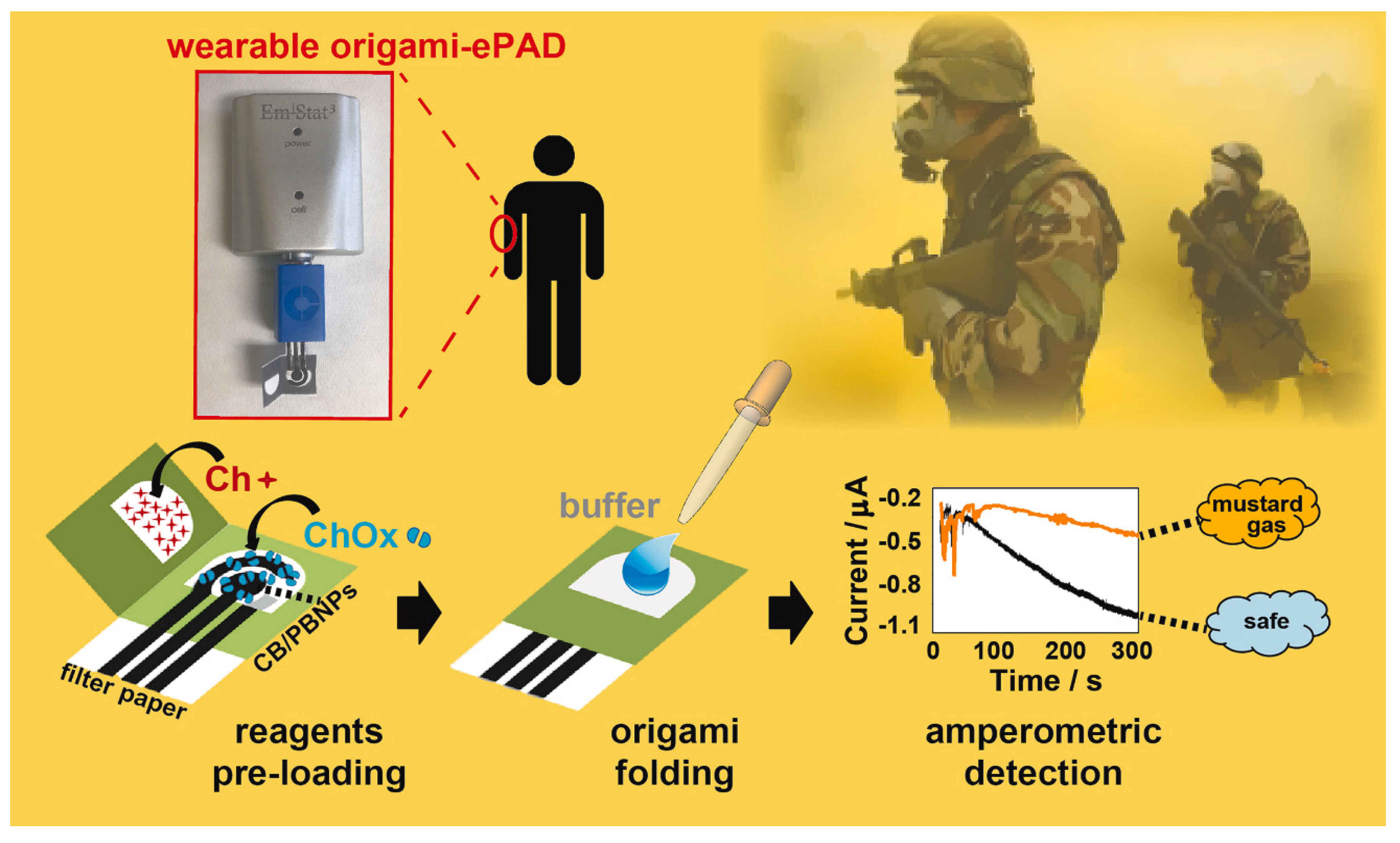
- Colozza, N.; Kehe, K.; Dionisi, G.; Popp, T.; Tsoutsoulopoulos, A.; Steinritz, D.; Moscone, D.; Arduini, F. A Wearable Origami-like Paper-Based Electrochemical Biosensor for Sulfur Mustard Detection. Biosens. Bioelectron. 2019, 129, 15–23. [Google Scholar] [CrossRef] [PubMed]
- Li, M.; Wang, L.; Liu, R.; Li, J.; Zhang, Q.; Shi, G.; Li, Y.; Hou, C.; Wang, H. A Highly Integrated Sensing Paper for Wearable Electrochemical Sweat Analysis. Biosens. Bioelectron. 2021, 174, 112828. [Google Scholar] [CrossRef] [PubMed]
- de França, C.C.L.; Meneses, D.; Silva, A.C.A.; Dantas, N.O.; de Abreu, F.C.; Petroni, J.M.; Lucca, B.G. Development of Novel Paper-Based Electrochemical Device Modified with CdSe/CdS Magic-Sized Quantum Dots and Application for the Sensing of Dopamine. Electrochim. Acta 2021, 367, 137486. [Google Scholar] [CrossRef]
- Fava, E.L.; Martimiano do Prado, T.; Almeida Silva, T.; Cruz de Moraes, F.; Censi Faria, R.; Fatibello-Filho, O. New Disposable Electrochemical Paper-based Microfluidic Device with Multiplexed Electrodes for Biomarkers Determination in Urine Sample. Electroanalysis 2020, 32, 1075–1083. [Google Scholar] [CrossRef]
- Cunningham, J.C.; Brenes, N.J.; Crooks, R.M. Paper Electrochemical Device for Detection of DNA and Thrombin by Target-Induced Conformational Switching. Anal. Chem. 2014, 86, 6166–6170. [Google Scholar] [CrossRef]
- Moazeni, M.; Karimzadeh, F.; Kermanpur, A. Peptide Modified Paper Based Impedimetric Immunoassay with Nanocomposite Electrodes as a Point-of-Care Testing of Alpha-Fetoprotein in Human Serum. Biosens. Bioelectron. 2018, 117, 748–757. [Google Scholar] [CrossRef] [PubMed]
- Li, X.; Qin, Z.; Fu, H.; Li, T.; Peng, R.; Li, Z.; Rini, J.M.; Liu, X. Enhancing the Performance of Paper-Based Electrochemical Impedance Spectroscopy Nanobiosensors: An Experimental Approach. Biosens. Bioelectron. 2021, 177, 112672. [Google Scholar] [CrossRef] [PubMed]
- Boonkaew, S.; Jang, I.; Noviana, E.; Siangproh, W.; Chailapakul, O.; Henry, C.S. Electrochemical Paper-Based Analytical Device for Multiplexed, Point-of-Care Detection of Cardiovascular Disease Biomarkers. Sens. Actuators B Chem. 2021, 330, 129336. [Google Scholar] [CrossRef]
- Liu, X.; Li, X.; Gao, X.; Ge, L.; Sun, X.; Li, F. A Universal Paper-Based Electrochemical Sensor for Zero-Background Assay of Diverse Biomarkers. ACS Appl. Mater. Interfaces 2019, 11, 15381–15388. [Google Scholar] [CrossRef] [PubMed]
- Wang, H.; Jian, Y.; Kong, Q.; Liu, H.; Lan, F.; Liang, L.; Ge, S.; Yu, J. Ultrasensitive Electrochemical Paper-Based Biosensor for MicroRNA via Strand Displacement Reaction and Metal-Organic Frameworks. Sens. Actuators B Chem. 2018, 257, 561–569. [Google Scholar] [CrossRef]
- Cinti, S.; Cusenza, R.; Moscone, D.; Arduini, F. Paper-Based Synthesis of Prussian Blue Nanoparticles for the Development of Whole Blood Glucose Electrochemical Biosensor. Talanta 2018, 187, 59–64. [Google Scholar] [CrossRef] [PubMed]
- Li, Z.-H.; Zhao, X.-L.; Song, R.-M.; Chen, C.; Wei, P.-J.; Zhu, Z.-G. Free-Standing Palladium Modified Reduced Graphene Oxide Paper Based on One-Pot Co-Reduction and Its Sensing Application. Chem. Phys. Lett. 2018, 712, 71–77. [Google Scholar] [CrossRef]
- Narang, J.; Malhotra, N.; Singhal, C.; Mathur, A.; Chakraborty, D.; Anil, A.; Ingle, A.; Pundir, C.S. Point of Care with Micro Fluidic Paper Based Device Integrated with Nano Zeolite–Graphene Oxide Nanoflakes for Electrochemical Sensing of Ketamine. Biosens. Bioelectron. 2017, 88, 249–257. [Google Scholar] [CrossRef]
- Boobphahom, S.; Ruecha, N.; Rodthongkum, N.; Chailapakul, O.; Remcho, V.T. A Copper Oxide-Ionic Liquid/Reduced Graphene Oxide Composite Sensor Enabled by Digital Dispensing: Non-Enzymatic Paper-Based Microfluidic Determination of Creatinine in Human Blood Serum. Anal. Chim. Acta 2019, 1083, 110–118. [Google Scholar] [CrossRef]
- Svigelj, R.; Dossi, N.; Grazioli, C.; Toniolo, R. Paper-Based Aptamer-Antibody Biosensor for Gluten Detection in a Deep Eutectic Solvent (DES). Anal. Bioanal. Chem. 2021, 414, 3341–3348. [Google Scholar] [CrossRef]
- Nantaphol, S.; Chailapakul, O.; Siangproh, W. A Novel Paper-Based Device Coupled with a Silver Nanoparticle-Modified Boron-Doped Diamond Electrode for Cholesterol Detection. Anal. Chim. Acta 2015, 891, 136–143. [Google Scholar] [CrossRef] [PubMed]
- Li, W.; Qian, D.; Wang, Q.; Li, Y.; Bao, N.; Gu, H.; Yu, C. Fully-Drawn Origami Paper Analytical Device for Electrochemical Detection of Glucose. Sens. Actuators B Chem. 2016, 231, 230–238. [Google Scholar] [CrossRef]
- Li, X.; Zhao, C.; Liu, X. A Paper-Based Microfluidic Biosensor Integrating Zinc Oxide Nanowires for Electrochemical Glucose Detection. Microsyst. Nanoeng. 2015, 1, 15014. [Google Scholar] [CrossRef]
- He, X.; Chang, S.J.; Settu, K.; Chen, C.-J.; Liu, J.-T. An Anti-HCT-Interference Glucose Sensor Based on a Fiber Paper-Based Screen-Printed Carbon Electrode. Sens. Actuators B Chem. 2019, 297, 126763. [Google Scholar] [CrossRef]
- Amor-Gutiérrez, O.; Costa-Rama, E.; Fernández-Abedul, M.T. Fully Integrated Sampler and Dilutor in an Electrochemical Paper-Based Device for Glucose Sensing. Microchim. Acta 2021, 188, 302. [Google Scholar] [CrossRef]
- Ge, S.; Zhang, L.; Zhang, Y.; Liu, H.; Huang, J.; Yan, M.; Yu, J. Electrochemical K-562 Cells Sensor Based on Origami Paper Device for Point-of-Care Testing. Talanta 2015, 145, 12–19. [Google Scholar] [CrossRef]
- Vishnu, N.; Sihorwala, A.Z.; Sharma, C.S. Paper Based Low-Cost and Portable Ultrasensitive Electroanalytical Devicefor The Detection of Uric Acid in Human Urine. ChemistrySelect 2021, 6, 8426–8434. [Google Scholar] [CrossRef]
- Martins, G.V.; Tavares, A.P.M.; Fortunato, E.; Sales, M.G.F. Paper-Based Sensing Device for Electrochemical Detection of Oxidative Stress Biomarker 8-Hydroxy-2′-Deoxyguanosine (8-OHdG) in Point-of-Care. Sci. Rep. 2017, 7, 14558. [Google Scholar] [CrossRef]
- Yen, Y.-K.; Chao, C.-H.; Yeh, Y.-S. A Graphene-PEDOT:PSS Modified Paper-Based Aptasensor for Electrochemical Impedance Spectroscopy Detection of Tumor Marker. Sensors 2020, 20, 1372. [Google Scholar] [CrossRef]
- Ahmadi, A.; Khoshfetrat, S.M.; Kabiri, S.; Fotouhi, L.; Dorraji, P.S.; Omidfar, K. Impedimetric Paper-Based Enzymatic Biosensor Using Electrospun Cellulose Acetate Nanofiber and Reduced Graphene Oxide for Detection of Glucose From Whole Blood. IEEE Sens. J. 2021, 21, 9210–9217. [Google Scholar] [CrossRef]
- Tian, R.; Li, Y.; Bai, J. Hierarchical Assembled Nanomaterial Paper Based Analytical Devices for Simultaneously Electrochemical Detection of MicroRNAs. Anal. Chim. Acta 2019, 1058, 89–96. [Google Scholar] [CrossRef]
- Zhu, W.; Zhang, Y.; Gong, J.; Ma, Y.; Sun, J.; Li, T.; Wang, J. Surface Engineering of Carbon Fiber Paper toward Exceptionally High-Performance and Stable Electrochemical Nitrite Sensing. ACS Sens. 2019, 4, 2980–2987. [Google Scholar] [CrossRef] [PubMed]
- Hu, H.; Wu, S.; Wang, C.; Wang, X.; Shi, X. Electrochemical behaviour of cellulose/reduced graphene oxide/carbon fiber paper electrodes towards the highly sensitive detection of amitrole. RSC Adv. 2023, 13, 1867–1876. [Google Scholar] [CrossRef] [PubMed]
- Pokpas, K.; Jahed, N.; McDonald, E.; Bezuidenhout, P.; Smith, S.; Land, K.; Iwuoha, E. Graphene-AuNP Enhanced Inkjet-printed Silver Nanoparticle Paper Electrodes for the Detection of Nickel(II)-Dimethylglyoxime [Ni(DmgH 2 )] Complexes by Adsorptive Cathodic Stripping Voltammetry (AdCSV). Electroanalysis 2020, 32, 3017–3031. [Google Scholar] [CrossRef]
- Cinti, S.; Talarico, D.; Palleschi, G.; Moscone, D.; Arduini, F. Novel Reagentless Paper-Based Screen-Printed Electrochemical Sensor to Detect Phosphate. Anal. Chim. Acta 2016, 919, 78–84. [Google Scholar] [CrossRef]
- Wang, P.; Wang, M.; Zhou, F.; Yang, G.; Qu, L.; Miao, X. Development of a Paper-Based, Inexpensive, and Disposable Electrochemical Sensing Platform for Nitrite Detection. Electrochem. Commun. 2017, 81, 74–78. [Google Scholar] [CrossRef]
- Pungjunun, K.; Chaiyo, S.; Praphairaksit, N.; Siangproh, W.; Ortner, A.; Kalcher, K.; Chailapakul, O.; Mehmeti, E. Electrochemical Detection of NOx Gas Based on Disposable Paper-Based Analytical Device Using a Copper Nanoparticles-Modified Screen-Printed Graphene Electrode. Biosens. Bioelectron. 2019, 143, 111606. [Google Scholar] [CrossRef]
- Martins, G.V.; Marques, A.C.; Fortunato, E.; Sales, M.G.F. Wax-Printed Paper-Based Device for Direct Electrochemical Detection of 3-Nitrotyrosine. Electrochim. Acta 2018, 284, 60–68. [Google Scholar] [CrossRef]
- Guo, Z.; Zhou, S.; Li, J.; Guo, X.; Cui, J.; Wu, D. Development of a Paper-Based Microanalysis Device Doped with Multi-Walled Carbon Nanotubes for in Vitro Evaluation of Fluorene Cytotoxicity. Bioelectrochemistry 2020, 135, 107552. [Google Scholar] [CrossRef]
- Liu, Y.; Alkhamis, O.; Liu, X.; Yu, H.; Canoura, J.; Xiao, Y. Aptamer-Integrated Multianalyte-Detecting Paper Electrochemical Device. ACS Appl. Mater. Interfaces 2021, 13, 17330–17339. [Google Scholar] [CrossRef]
- Mohan, J.M.; Amreen, K.; Kulkarni, M.B.; Javed, A.; Dubey, S.K.; Goel, S. Optimized Ink Jetted Paper Device for Electroanalytical Detection of Picric Acid. Colloids Surf. B Biointerfaces 2021, 208, 112056. [Google Scholar] [CrossRef]
- Metters, J.P.; Houssein, S.M.; Kampouris, D.K.; Banks, C.E. Paper-Based Electroanalytical Sensing Platforms. Anal. Methods 2013, 5, 103–110. [Google Scholar] [CrossRef]
- Santhiago, M.; Henry, C.S.; Kubota, L.T. Low Cost, Simple Three Dimensional Electrochemical Paper-Based Analytical Device for Determination of p-Nitrophenol. Electrochim. Acta 2014, 130, 771–777. [Google Scholar] [CrossRef]
- Arduini, F.; Cinti, S.; Caratelli, V.; Amendola, L.; Palleschi, G.; Moscone, D. Origami Multiple Paper-Based Electrochemical Biosensors for Pesticide Detection. Biosens. Bioelectron. 2019, 126, 346–354. [Google Scholar] [CrossRef] [PubMed]
- Scala-Benuzzi, M.L.; Raba, J.; Soler-Illia, G.J.A.A.; Schneider, R.J.; Messina, G.A. Novel Electrochemical Paper-Based Immunocapture Assay for the Quantitative Determination of Ethinylestradiol in Water Samples. Anal. Chem. 2018, 90, 4104–4111. [Google Scholar] [CrossRef] [PubMed]
- Kokkinos, C.; Economou, A.; Giokas, D. Paper-Based Device with a Sputtered Tin-Film Electrode for the Voltammetric Determination of Cd(II) and Zn(II). Sens. Actuators B Chem. 2018, 260, 223–226. [Google Scholar] [CrossRef]
- Sánchez-Calvo, A.; Fernández-Abedul, M.T.; Blanco-López, M.C.; Costa-García, A. Paper-Based Electrochemical Transducer Modified with Nanomaterials for Mercury Determination in Environmental Waters. Sens. Actuators B Chem. 2019, 290, 87–92. [Google Scholar] [CrossRef]
- Cinti, S.; Colozza, N.; Cacciotti, I.; Moscone, D.; Polomoshnov, M.; Sowade, E.; Baumann, R.R.; Arduini, F. Electroanalysis Moves towards Paper-Based Printed Electronics: Carbon Black Nanomodified Inkjet-Printed Sensor for Ascorbic Acid Detection as a Case Study. Sens. Actuators B Chem. 2018, 265, 155–160. [Google Scholar] [CrossRef]
- Lamas-Ardisana, P.J.; Martínez-Paredes, G.; Añorga, L.; Grande, H.J. Glucose Biosensor Based on Disposable Electrochemical Paper-Based Transducers Fully Fabricated by Screen-Printing. Biosens. Bioelectron. 2018, 109, 8–12. [Google Scholar] [CrossRef]
- Ding, J.; Li, B.; Chen, L.; Qin, W. A Three-Dimensional Origami Paper-Based Device for Potentiometric Biosensing. Angew. Chem. Int. Ed. 2016, 55, 13033–13037. [Google Scholar] [CrossRef]
- Bell, J.G.; Mousavi, M.P.S.; Abd El-Rahman, M.K.; Tan, E.K.W.; Homer-Vanniasinkam, S.; Whitesides, G.M. Paper-Based Potentiometric Sensing of Free Bilirubin in Blood Serum. Biosens. Bioelectron. 2019, 126, 115–121. [Google Scholar] [CrossRef] [PubMed]
- Yao, Y.; Jiang, C.; Ping, J. Flexible Freestanding Graphene Paper-Based Potentiometric Enzymatic Aptasensor for Ultrasensitive Wireless Detection of Kanamycin. Biosens. Bioelectron. 2019, 123, 178–184. [Google Scholar] [CrossRef] [PubMed]
- Ding, R.; Fiedoruk-Pogrebniak, M.; Pokrzywnicka, M.; Koncki, R.; Bobacka, J.; Lisak, G. Solid Reference Electrode Integrated with Paper-Based Microfluidics for Potentiometric Ion Sensing. Sens. Actuators B Chem. 2020, 323, 128680. [Google Scholar] [CrossRef]
- Yehia, A.M.; Farag, M.A.; Tantawy, M.A. A Novel Trimodal System on a Paper-Based Microfluidic Device for on-Site Detection of the Date Rape Drug “Ketamine”. Anal. Chim. Acta 2020, 1104, 95–104. [Google Scholar] [CrossRef] [PubMed]
- Hu, J.; Ho, K.T.; Zou, X.U.; Smyrl, W.H.; Stein, A.; Bühlmann, P. All-Solid-State Reference Electrodes Based on Colloid-Imprinted Mesoporous Carbon and Their Application in Disposable Paper-Based Potentiometric Sensing Devices. Anal. Chem. 2015, 87, 2981–2987. [Google Scholar] [CrossRef]
- Ruecha, N.; Chailapakul, O.; Suzuki, K.; Citterio, D. Fully Inkjet-Printed Paper-Based Potentiometric Ion-Sensing Devices. Anal. Chem. 2017, 89, 10608–10616. [Google Scholar] [CrossRef]


| No. | Nanocomposite|Electrode | Analyte | Method | Linear Range | LOD | Interferents | Real Sample | Ref. |
|---|---|---|---|---|---|---|---|---|
| Clinical Samples | ||||||||
| 1 | Cellulose-paper | BSA | SERS | 10–60 mg mL−1 | 10 mg mL−1 | - | - | [50] |
| 2 | PODs | K+ | Colorimetric | 2–7 mM | 0.089 mM | Ca2+, Li+, Mg2+ | plasma | [38] |
| 3 | Caffeine benzoate paper | Bilirubin | Colorimetric | 0 to 25 mg dL−1 | 1.2 mg dL−1 | spiked hemoglobin | jaundiced whole human blood samples | [51] |
| 4 | Wax screen printing | 198-bp DNA | Chemiluminescent | 1.94 × 10⁻¹ pmol/L–1.94 × 104 pmol/L | 6.3 × 10⁻² pmol/L | S1 + S2 + S3, S4 + S2 + S3, S1 + S2 + S5, S4 + S2 + S5 and S1 + E. coli + S3 | - | [52] |
| 5 | Co2(OH)2CO3-CeO2 | CEA | Colorimetric | 0.002–75.0 ng mL−1 | 0.51 pg mL−1 | AFP, CA 125, CA 15-3 | human serum samples | [43] |
| 6 | Iron(III)-thiocyanate | Thiocyanate | Colorimetric | 0.25 and 20 mM | 0.06 mM | - | human saliva | [53] |
| 7 | PGA-RR | Polygalacturonase | Colorimetric | 0.02–0.1 unit | 0.02 unit | - | - | [54] |
| 8 | Tetrakis (4-octyloxyphenyl)porphyrin cobalt(II) | Thiocyanate | Colorimetric | 0.001–5 mM | 1.26 μM | SCN−, NO2−, AsO3 3−, AsO4 3−, ClO4−, NO3−, Br−, Cl−, H2PO4−, I−, F−, SO42−. | urine samples | [55] |
| 9 | N-CDs | Cholesterol | Colorimetric | 2.5–7.5 mM | 0.676 mM | - | whole blood samples | [44] |
| 10 | AuNPs | Double-stranded TB DNA | Colorimetric | 1.95 × 10⁻²–1.95 × 101 ng/mL | 1.95 × 10⁻² ng/mL | - | - | [40] |
| 11 | Chitosan oligosaccharide lactate | Glucose Uric acid | Colorimetric | 0–500 mg/dL 0–200 mg/dL | 0.6 mg/dL 0.03 mg/dL | - | human urine | [56] |
| 12 | Paper-based ELISA | Ketamine | Colorimetric | 10−4–10−1 mg/mL | 0.03 ng/mL | - | oral fluid sample | [57] |
| 13 | Citrate-capped PtNPs | Uric acid | Colorimetric | 0–8 mM | 4.2 ± 5 µM | K+, Na+, Mg2+, Ca2+, Zn2+, Glu, DA, UA | human urine | [58] |
| 14 | 5CB/LCs | Bilirubin | Distance-based | 2.0–30.0 pmol/L | 0.80 pmol/L | glucose, fruc- tose, lactose, sucrose, galactose, hemoglobin, bovine serum albumin, biotin, trypsin, cholesterol, glutathione, cysteine, glutamic acid, salicylic acid, ascorbic acid. | human control urine human serum | [59] |
| 15 | Zr-MOF/Fe3O4(TMC)/ AuNCs | HbA1c | ECL | 2–18 0.072% | 0.072% | - | human blood sample | [60] |
| 16 | Porous AuPd alloy | MCF-7 | ECL | 450–1.0 × 10⁷ cells mL−1 | 250 cells mL−1 | HepG2, SK-BR-3 | - | [61] |
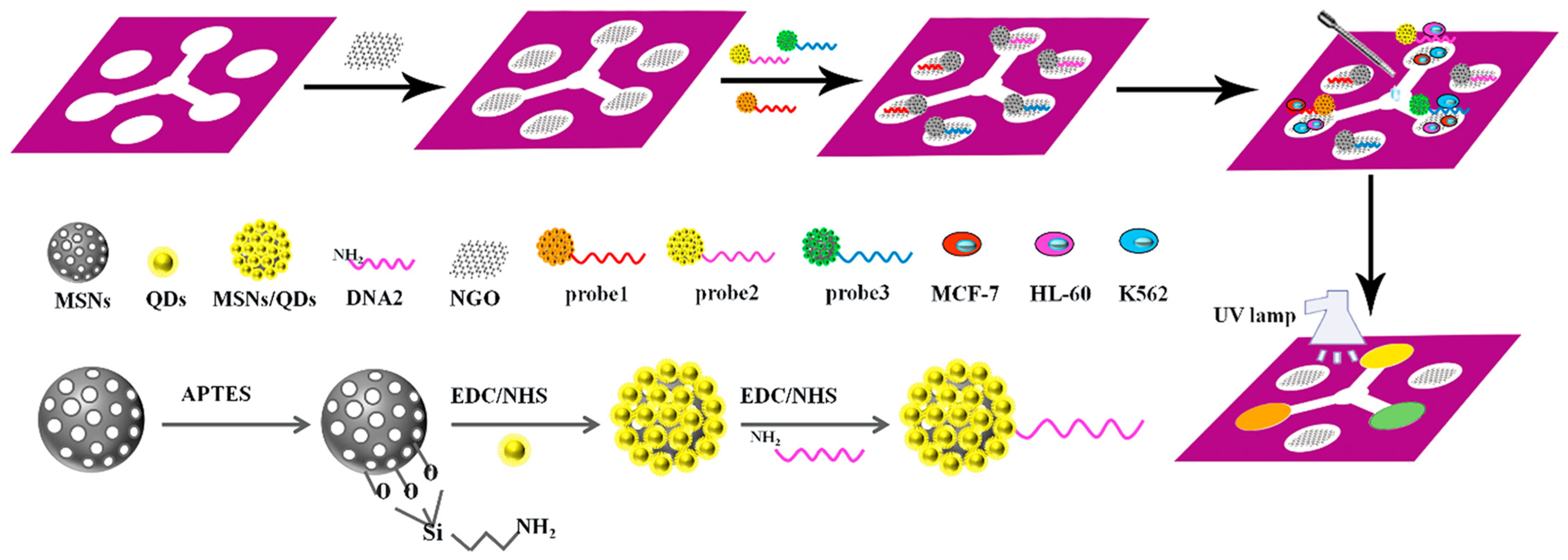
| 17 | GO-aptamer | MCF-7 HL-60 K562 | Fluorescence | 180–8 × 10⁷ cells mL−1 210–7 × 10⁷ cells mL−1 200–7 × 10⁷ cells mL−1 | 6270 cells mL−1 65 cells mL−1 | - | - | [32] |
| 18 | MFNCDs | H2O2 Glucose | Fluorescent Colorimetric | 0–15 mM 500 nM–15 mM | 97 nM 0.85 μM 84 nM 0.41 μM | ascorbic acid, uric acid, L-cysteine, dopamine, glycine, sorbitol, glutathione | real serum | [35] |
| 19 | ISOs | Ca2+ | Fluorescent | 10−5–1 mol L−1 | 19.3 μmol L−1 | Mg+2, Na+, K+ | mineral water | [62] |
| 20 | a-GQDs | Glucose | Fluorescent | 0.05–20 mM | 2.1 μM | maltose, lactose, fructose, sucrose | live cells | [33] |
| 21 | P-BPE | H2O2 Glucose | Colorimetric | 0.1 mmol L−1–4.0 mol L−1 0.1–50 mmol L−1 | 4.9 μmol L−1 70 μmol L−1 | AA, UA | human serum samples | [63] |
| 22 | Cu complex/polyacrylamide | Glucose | Luminescent | 3–200 mM | 0.44 mM | - | whole blood samples | [49] |
| 23 | μTPAD | Glucose | Colorimetric | 50–250 μM | 35 μM | - | human sweat | [34] |
| 24 | NiFe2O4/Paper-Based ME | HSA | Gauss meter | 10–200 µg mL−1 | 0.43 µg mL−1 | UA, CRE, HGB, BSA, CEA | - | [64] |
| 25 | CdS/RGO/ZnO | miRNA-21 miRNA-122b miRNA-7f | PEC | 0.5 fM–100 pM 0.5 fM–100 pM 10 fM–100 pM | 0.32 fM 0.37 fM 3.8 fM | - | human serum | [65] |
| 26 | Silver halide particles | Cysteine Glutathione Homocysteine | Photoreduction | 10–100 μM 10–100 μM 20–100 μM | 10.0 μM 10.0 μM 7.5 μM | glutamine, glutamic acid, cystine, asparagine, aspartic acid, glycine, histidine, lysine, valine, alanine, and arginine | human blood plasma | [66] |
| 27 | DNA-AuNPs | miR-29a | Colorimetric SERS | 18–360 pg μL−1 | 47 pg μL−1 | - | - | [67] |
| 28 | APTMS-GA | H2O2, Glucose anti-PSA | Colorimetric | 2.5–500 mM 0.5–30 mM 0.1–10 ng/mL | - | IgG, IgM, CEA, TNF-α | human serum | [68] |
| 29 | PPX-chromatography paper | Glucose Protein ALP ALT Uric acid | Colorimetric | - | 25 mg dL−1 1.04 g L−1 7.81 unit per L 1.6 nmol L−1 0.13 mmol L−1 | - | - | [69] |
| 30 | GR/Au-PWE | DNA | ECL | 4.0 × 10⁻¹⁷–5.0 × 10⁻¹¹ M | 8.5 × 10⁻¹⁸ M | - | human serum sample | [70] |
| Environmental monitoring | ||||||||
| 31 | Pd NPs/meso-C | H2O2 | Colorimetric | 5–300 μM | - | - | milk matrices | [71] |
| 32 | CdSe/ZnS quantum dots | Hg2+ | Colorimetric | 0.1–100 μM | 0.09 µM | Ca2+, K+, Mn2+, Co2+, Cr3+ and Ni2+ | tap water | [45] |
| 33 | Au NPs/CdS NPs | Humidity | - | - | - | - | - | [41] |
| 34 | Ag-μPAD | Nitrite | Colorimetric | 10–3200 μM | 6.2 × 10⁻⁵ µM | H+, Na+, K+, Ca2+, NH4+, F−, Cl−, Br−, BrO3−, IO3−, NO3 and SO42− | tap, river and lake water samples. | [46] |
| 35 | Piezoelectric inkjet printer | AChE | Colorimetric | - | 0.01 ng mL−1, 0.04 ng mL−1 | - | - | [72] |
| 36 | S,N-doped carbon quantum dots | Bendiocarb | Chemiluminescence | 0.1–10 µgmL−1 | 0.02 µgmL−1 | - | water and juice samples | [47] |
| 37 | PEDOT:PSS/graphene | Chlorine | Chemiresistive | 0.1−500 ppm | 0.18 ppm | - | on-site water | [73] |
| 38 | CURN | Mercury | Colorimetric | 0.5–20 µg mL−1 | 0.17 µg mL−1 | NH4+, C2O42−, CO32−, HCO3−, Br−, F−, Ni2+, Cd2+, Mn2+Bi3+, Cr3+, Cu2+ and Zn2+ | water samples | [74] |
| 39 | Cys A@AuNPs | Hg(II) Cu(II) ions | Fluorescence | 0.001–1 ppm 0.001–1 ppm | 0.001 ppm 0.1 ppm | Mn(II), Zr(IV), Ba(II), Mg(II), Sr(II), As(III), B(III), Al(III), V(III), K (I), Na(I), | - | [75] |
| 40 | AgNPs/PVA | Lead | Colorimetric | 20–1000 μgL−1 | 8 μgL−1 | Mg(II), Ca(II), Ba(II), Hg(II), Ni(II), Mn(II), As(III), Al(III), Cl− and CO3– | industrial waste water | [76] |
| 41 | chromatographic paper foils | MDA | Colorimetric | 2.5–20 μM | - | - | - | [48] |
| 42 | rGO/MPy-GNRs | SO2 | Colorimetric/ SERS | 1–2000 μM | 1.45 μM | - | wine | [36] |
| 43 | Curcumin-based PADs | Boron | Colorimetric | 0.2–0.8 mg/L | - | Cl−, PO43−, SO42−, K+, Na+, HCO3−, NO3− and Fe3+ | - | [77] |
| 44 | NBT | Hypoxanthine (HX), | Colorimetric | - | 3.7 μM | D-glucose, L-lactic acid, and dopamine | fish samples | [42] |
| 45 | AgNPs | Mercury | Colorimetric | 1 to 4 ppb | 0.86 ppb | - | drinking water | [78] |
| 46 | Quercetin-capped AgNPs | Carbaryl, paraoxon, parathion, malathion, diazinon, and chlorpyrifos | Colorimetric | - | 29.0, 22.0, 32.0, 17.0, 45.0, and 36.0 ng mL−1 | K+, Na+, Ca2+, Mg2+, SO42−, NO3−, I−, Br−, Cl−, S2− | tap water, apple juice, and rice | [79] |
| 47 | Paper-grafted thioglycolic acid Cu NP | Cu(II) | Colorimetric | 0.1–10 mM | 33 μM | Zn(II), Sn(II), Ni(II) Li(I), Pb(II), Fe(III), Ag(I), Cr(I), Cr(II), | - | [80] |
| 48 | µPADs | Cu2+, Fe3+, Cr2+, Ni2+, Hg2+, and Pb2+ | Colorimetric | - | 15 µM, 3.58 µM 0.5 µM, 0.8 µM 0.1 µM and 0.3 µM | - | tap water | [81] |
| 49 | Zn-TPP | NH3 and CO2 | Colorimetric | - | - | HCl, NaOH, NaHCO3 | - | [82] |
| 50 | Wax printing | Nitrite | Colorimetric | 10–500 μM | - | - | - | [83] |
| 51 | (rGO)-PdAu | Pb2+ | Colorimetric | 0.5–2000 nM | 0.14 nM | K+, Mn2+, Zn2+, Cu2+, Ca2+, Ni2+, Cr2+ and Cd2+ | tap water and river water | [84] |
| 52 | Wax printer | Chlorpyrifos | Colorimetric | 8.60 ppm | 0 to 100 ppm | - | - | [85] |
| 53 | MIP@MOF | thiacloprid | Colorimetric | 0.1–1.2 μM, and 1.2–10 μM | 0.04 μM | acetamiprid, imidacloprid and dinotefuran | Lettuce, water and soil | [86] |
| 54 | Cellulose paper | As3+, Nd3+ and Br- | Fluorescence | 11.53 nM, 0.65 nM and 11.25 nM | As3+ (0–150 nM), Nd3+ and Br− (0–120 nM) | - | industrial waste water | [87] |
| 55 | EuD4TEA-Au NPs | Cyanide | Fluorescence | - | 10−2–10−12 M | F−, N3−, H2PO4−, CH3COO−, Cl−, Br−, SO42− and NO3− | drinking water | [88] |
| 56 | [Ch][Cur]-coated cellulose paper | TATP, DADP | Fluorescence | - | 0.0–1000.0 ppm | - | - | [89] |
| 57 | Calix[4]arene | La3+,Cu2+, and Br− | Fluorescence | 0.88 nM for La3+, 0.19 nM for Cu2+ 0.15 nM for Br− | - | Zn2+, Cd2+, Fe2+, Fe3+, La3+, As3+, Nd3+, Zr4+, Ca2+, Ce3+, Li+, Ag+, Ba2+, Co2+, Hg2+, Na+ | soil samples for La3+, blood serum for Cu2+ industrial water for Br− | [90] |
| 58 | RFID | Chloride | Fluorescent | 10−7–10−5 M | 2.35 × 10⁻⁷ M | Na+, K+, Ca2+ | sweat | [91] |
| 59 | FMA | H2S | Fluorescent | 17–67 ppb | 3 ppb | - | - | [92] |
| 60 | GQDs | o- and p-nitrophenols | Fluorescent | 0.30–60.0 and 0.20–40.0 µg/mL | 0.07 and 0.03 µg/mL | - | tap water, river water, | [93] |
| 61 | Calix[4]arene | Mn2+, Cr3+ and F- | Fluorescence | 0–120 nM | 11 nM for Mn2+, 4 nM for Cr3+, 19 nM for F- | - | blood serum, industrial waste water | [94] |
| 62 | PANI/Ge | NH3 | Transient | - | 20 ppm | Ethanol, acetone, DMF and 2-propnal | - | [95] |
| 63 | CuNCs | H2S | Photoluminescent | 2–10 μM | 1 μM | SO42−, SO32−, PO43−, CO32−, Ac−, NO3−, NO2−, ClO4−, | springwater samples | [96] |
| 64 | Tyrosinase immobilized | BPA | Colorimetric | 0.05–3.87 μg/g | 0.28 μg/g | - | - | [97] |
| 65 | PEDOT:PSS | Chlorine | Colorimetric | 0.5–500 ppm | - | NaOCl, CaCl2, MgCl2, ZnCl2, K2SO4, CuSO4, (NH4)2SO4, NaHCO3, Na2CO3 | tap water and swimming pool water | [39] |
| 66 | PAN | Cu(II) | Colorimetric | 0.1–30.0 mg L−1 | 0.06 mg L−1 | Fe(III), Al(III), Zn(II), Cd(II), Pb(II), Ca(II), Mg(II), and Ni(II) | hot water | [98] |
| 67 | AuNPs) | Hg2+ | Colorimetric | 25–750 nM | 50 nM | Co2+, Mn2+, Pb2+, Ca2+, Cd2+, Cu2+, Ni2+, Zn2+, Cr3+ | pond and river water | [99] |
| 68 | AgNPls | Cu(II) | Colorimetric | 0.5–200 ng mL−1 | 1.0 ng mL−1 | K+, Cr3+, Cd2+, Zn2+, As3+, Mn2+, Co2+, Pb2+, Al3+, Ni2+, Fe3+, Mg2+, and Hg2+ | water food and blood | [37] |
| 69 | H-rGO | NO2 | Conductometric | 1–10 ppm | - | O2,O3,CO2, CO, SO2, NOx andNH3 | - | [100] |
| 70 | paper@QDs@Cu-IIP | Cu2+ | Fluorescent | 0.032–3.2 mg L−1 | 0.012 mg L−1 | Na+, K+, Ca2+, Mg2+, Zn2+, Ni2+, Co2+, Cd2+, Hg2+, Pb2+ and Fe3+ | lake water and sea water | [101] |
| 71 | Rhodamine-appended C-dots | Al3+ | Fluorescent | 1 × 10⁻⁴–1× 10⁻² M | 3.89 × 10⁻⁵ M | Mg2+, Cu2+, Co2+, Ni2+, Mg2+, Zn2+, Fe3+, and Fe2+ | - | [30] |
| 72 | Tb3+/BSA–AuNC | Hg2+ | Fluorescent | 0, 0.005–7 μM | 1 nM | Ag+, Ca2+, Co2+, Cu2+, Cd2+, K+, Na+, Ni2+, Fe2+, Hg2+, Mn2+, Mg2+ | - | [102] |
| 73 | PdA2 | H2S | Luminescent | 8–110 ppb | 2 ppb | NO2 and SO2 | - | [103] |
| 74 | SWNT-PABS | NH3 | Resistance | 10–250 ppm | - | - | - | [104] |
| 75 | Pencil-drawn | NO2 | Chemiresistor | 0.05–5 ppm | - | C2H5OH, CH3OH, NH3, CH3COCH3 | - | [105] |
| 76 | ZnO/AuNPs | PCP | Photoelectrochemical | 0.01–100 ng mL−1 | 4 pg mL−1 | aldrin, heptachlor, chlopyrifos | - | [106] |
| No. | Nanocomposite|Electrode | Analyte | Method | Linear Range | LOD | Interferents | Real Sample | Ref. |
|---|---|---|---|---|---|---|---|---|
| Clinical Samples | ||||||||
| 1 | MB/Ti3C2Tx/SPCE | Glucose and lactate | Amperometric | 0.08–1.25 mM 0.3–20.3 mM | 17.05 μM 3.73 μM | uric acid ascorbic acid | sweat | [108] |
| 2 | CuO/IL/ERGO | Creatinine | Amperometric | 0.01–2.0 mM | 0.22 μM | glucose, uric acid, urea, ascorbic acid | human blood serum | [120] |
| 3 | PBNPs | Glucose | Amperometric | up to 5 mM | 0.17 mM | - | blood glucose | [117] |
| 4 | Aptamer-antibody | Gluten | Amperometric | 0.2 and 20 mg L−1 | 0.2 mg L−1 | - | corn flakes, chickpea flour signal | [121] |
| 5 | AgNP/BDD | Cholesterol | Chronoamperometric | 0.01–7 mM | 6.5 µM | glucose (Glu), ascorbic acid (AA), and uric acid (UA) | bovine serum | [122] |
| 6 | FCA/GOx/PAD | Glucose | Chronoamperometric | 1–12 mM | 0.05 mM | AA, DA, UA | blood samples | [123] |
| 7 | Pd/rGOP | Glucose | Chronoamperometry | 0.5–8 mM | 30 µM | UA, AA | - | [118] |
| 8 | ZnO NWs | Glucose | CV | 0–15 mM | 59.5 μM | - | human serum | [124] |
| 9 | anti-HCT-interference | Glucose | CV | 3.7–13.8 mM | 0.88 mM | - | whole blood samples | [125] |
| 10 | Carbon ink | Glucose | CV | 4.4–6.6 mM | - | - | orange juice cola beverage | [126] |
| 11 | Zeo–GO | Ketamine | CV | 0.001–5 nM/mL | 0.001 nM/mL | whiskey urine organic juice | - | [119] |
| 12 | 3D Au NPs/GN | K-562 cells | DPV | 1.0 × 10³–5.0 × 10⁶ cells/mL | 200 cells/mL | - | - | [127] |
| 13 | Au@PdPt NPs | K-562 cell | DPV | 1.0 × 10²–2.0 × 10⁷ cells mL−1 | 31 cells mL−1 | MCF7, H9c2, normal cells | human serum samples | [20] |
| 14 | Cu-MOFs/uNPs | miRNA | DPV | 1.0 fM–10 nM | 0.35 fM | nDNA, nRNA, tRNA, sRNA | serum samples | [116] |
| 15 | PAD | Uric Acid | DPV | 0.1–1 mM | 8 μM | urea, AA, glucose | human urine | [128] |
| 16 | CNTs-WE | miR-21 | DPV | 1 fM–1 μM | - | miR-141, miR-155, miR-199a, miR-143 | spiked serum samples | [115] |
| 17 | CdSe/CdS MSQDs | Dopamine | DPV | 0.5–15 μmol L−1 | 96 nmol L−1 | AA, UA, AC | human blood serum | [109] |
| 18 | FCA carbon black | Glucose Uric acid | DPV Chronoamperometry | 0.63–20.0 mmol L−1 0.05–3.00 mmol L−1 | 0.12 ± 0.03 mmol L−1 0.012 ± 0.002 mmol L−1 | - | urine sample | [110] |
| 19 | Carbon ink/PEDOT | 8-OHdG | DPV | 50–1000 ng/ml | 14.4 ng/ml | UA, AA | diluted serum samples | [129] |
| 20 | Graphene-Ag | AFP | EIS | 1–104 ng/ml | - | ascorbic acid, glucose | human serum | [112] |
| 21 | ZnO NWs | p24 antigen IgG antibody | EIS | - | 0.4 pg mL−1 10 ng mL−1 | - | human serum samples | [113] |
| 22 | Graphene-PEDOT:PSS | CEA | EIS | 0.77–14 ng mL−1 | 0.45 ng mL−1 | BSA, PSA and insulin | serum sample | [130] |
| 23 | rGO-Au-SPEs | Glucose | EIS | 3.3–27.7 mM | - | - | whole blood | [131] |
| 24 | CdS/RGO/ZnO | miRNA-21 miRNA-122b miRNA-7f | PEC | 0.5 fM-100 pM 0.5 fM–100 pM 10 fM–100 pM | 0.32 fM 0.37 fM 3.8 fM | - | human serum | [65] |
| 25 | ptCuMOFs/DNA | miR-141 miR-21 | SWV | 1 fM–1 nM | 0.1 fM | uric acid, dopamine, miR-210 | serum samples | [132] |
| 26 | BSA/rGO/antibody/GO/G-SPCE | CRP cTnI PCT | SWV | 1–100,000 ng mL−1 0.001–250 ng mL−1 0.0005–250 ng mL−1 | 0.38 ng mL−1 0.16 pg mL−1 0.27 pg mL−1 | glycine, creatinine, L-cysteine, homocysteine, albumin, hemoglobin, myoglobin | serum sample | [114] |
| 27 | Au-carbon | DNA thrombin | SWV | - | 30 nM 16 nM | - | - | [111] |
| Environmental monitoring | ||||||||
| 28 | CFP | Nitrite | Amperometric | 0.1–3838.5 μM | 0.07 μM | NaNO2, KNO3, NH4Ac, MgSO4, Na2SiO3 and ZnSO4, | mineral water | [133] |
| 29 | CF/GO/cellulose | amitrole | Amperometric | 0 mM to 0.4 mM | 2.44 × 10⁻⁷ mol L−1 | Cl, NaNO3, NaAc, K2SO4, urea, and glucose - | Tap and lake water | [134] |
| 30 | ERGO-AuNP-CC-Ag-PPPE | Ni2+ | ACSV | 50–500 µg L−1 | 32.19 µg L−1 | - | drinking water | [135] |
| 31 | Paper CB-SPE | Phosphate | Chronoamperometry | 4 μM | 10–300 μM | - | river water | [136] |
| 32 | GNPs/graphene/MCE | Nitrite | DPV | 0.1 µM | 0.3–720 µM | Na+, Ca2+, Mg2+, K+, Zn2+, Cl−, NO3−, CO32− | Lake water, river water and milk | [137] |
| 33 | (CuNP/SPGE) | NOx gas | DPV | 0.23 vppm and 0.76 vppm | - | SO2, CO, N2O and O3 | - | [138] |
| 34 | Carbon Ag ink | 3-nitrotyrosine | SWV | 49.2 nM | 500 nM–1 mM | tyrosine, ascorbic acid, uric acid and creatinine | saliva, blood and urine | [139] |
| 35 | MWCNTs/l-PAD | Fluorene | LSV | 0–100 µM | 0.0500 µM | - | - | [140] |
| 36 | Aptamer-Modified mPEDs | Cocaine | SWV | 1−100 μM | 1 μM | - | urine, saliva | [141] |
| 37 | ZnO NPs | Picric acid | SWV | 4 μM–60 μM | 4.04 μM | zinc, lead, copper and mercury | lake water | [142] |
| 38 | IP-SPE | NADH and nitrite | CV | 10–100 μM 100–1000 μM | 1.8 μM and 15.1 μM | - | - | [143] |
| 39 | Pencil graphite | p-nitrophenol | DPV | 10–200 μM | 1.1 μM | 4-aminophenol, 1,4 dihydroxy benzene and phenol | - | [144] |
| 40 | Prussian blue nanoparticles | Atrazine 2,4-D paraoxon | Chronoamperometric | 10–100 ppb 100–1000 ppb 20–100 ppb | 50 ppb 2 ppb | - | surface water | [145] |
| 41 | RG-SPCE | Ethinylestradiol | 0.5–120 ng L−1 | 0.1 ng L−1 | humic acid | spiked water sample | [146] | |
| 42 | Wax-print Ag, pt, Sn sputtered | Cd(II) Zn(II) | SWASV | 5–40 μg L−1 | 0.9 μg L−1 1.1 μg L−1 | Mn(II), Ni(II), Mg(II), Fe(III), Pb((II) | - | [147] |
| 43 | CNFs/AuNPs | Mercury | LSV | 0.1–1.2 μM | 0.03 μM | Cd (II), Pb (II), Cu (II) and Zn (II) | - | [148] |
| 44 | Carbon black | AA | CV | - | sodium chloride, sodium bicarbonate, sucrose, citric acid, and sorbitol | dietary supplement | [149] | |
| 45 | graphite ink | Glucose | - | 0.5–50 mM | 0.33 mM | - | soft drinks | [150] |
| 46 | Silver halide particles | Cysteine Glutathione Homocysteine | Photoreduction | 10–100 μM 10–100 μM 20–100 μM | 10.0 μM 10.0 μM 7.5 μM | glutamine, glutamic acid, cystine, asparagine, aspartic acid, glycine, histidine, lysine, valine, alanine, and arginine | human blood plasma | [66] |
| 47 | 3D origami paper-based device | Methyl parathion | Potentiometric | 0.1–1.0 nM | 0.06 nM | - | - | [151] |
| 48 | BR ISE | Bilirubin | Potentiometric | (0.10 μM–1.0 mM | - | - | blood serum | [152] |
| 49 | Graphene paper | Kanamycin | Potentiometric | 0.05–30 pM | 0.05 pM | amoxicillin, ciprofloxacin, tetracycline, and chloramphenicol | milk | [153] |
| 50 | DPS containing solid KCl | K+ Na+ Cl− | Potentiometric | - | 10−4.1 ± 0.1 mol dm−3 10−3.3 ± 0.1 mol dm−3 10−4.1 ± 0.1 mol dm−3 | - | sweat | [154] |
| 51 | C-dots-AuNPs | Ketamine | Potentiometric | 2 × 10⁻⁴–1 × 10⁻³ mol L−1 | - | caffeine, glu-curonolactone, histamine, tyramine B-vitamins. | - | [155] |
| 52 | Gold/CIM | Cl - | Potentiometric | 10−7 M–10−1 M | - | - | - | [156] |
| 53 | G/PEDOT:PSS | Na+, K+ | Potentiometric | 10−4–1 M 10−4–1 M | 32 µM for Na+ 101 µM for K+ | - | human urine samples | [157] |
Disclaimer/Publisher’s Note: The statements, opinions and data contained in all publications are solely those of the individual author(s) and contributor(s) and not of MDPI and/or the editor(s). MDPI and/or the editor(s) disclaim responsibility for any injury to people or property resulting from any ideas, methods, instructions or products referred to in the content. |
© 2023 by the authors. Licensee MDPI, Basel, Switzerland. This article is an open access article distributed under the terms and conditions of the Creative Commons Attribution (CC BY) license (https://creativecommons.org/licenses/by/4.0/).
Share and Cite
Kummari, S.; Panicker, L.R.; Rao Bommi, J.; Karingula, S.; Sunil Kumar, V.; Mahato, K.; Goud, K.Y. Trends in Paper-Based Sensing Devices for Clinical and Environmental Monitoring. Biosensors 2023, 13, 420. https://doi.org/10.3390/bios13040420
Kummari S, Panicker LR, Rao Bommi J, Karingula S, Sunil Kumar V, Mahato K, Goud KY. Trends in Paper-Based Sensing Devices for Clinical and Environmental Monitoring. Biosensors. 2023; 13(4):420. https://doi.org/10.3390/bios13040420
Chicago/Turabian StyleKummari, Shekher, Lakshmi R. Panicker, Jagadeeswara Rao Bommi, Sampath Karingula, Venisheety Sunil Kumar, Kuldeep Mahato, and Kotagiri Yugender Goud. 2023. "Trends in Paper-Based Sensing Devices for Clinical and Environmental Monitoring" Biosensors 13, no. 4: 420. https://doi.org/10.3390/bios13040420
APA StyleKummari, S., Panicker, L. R., Rao Bommi, J., Karingula, S., Sunil Kumar, V., Mahato, K., & Goud, K. Y. (2023). Trends in Paper-Based Sensing Devices for Clinical and Environmental Monitoring. Biosensors, 13(4), 420. https://doi.org/10.3390/bios13040420