A Review of Magnetic Nanoparticle-Based Surface-Enhanced Raman Scattering Substrates for Bioanalysis: Morphology, Function and Detection Application


Abstract
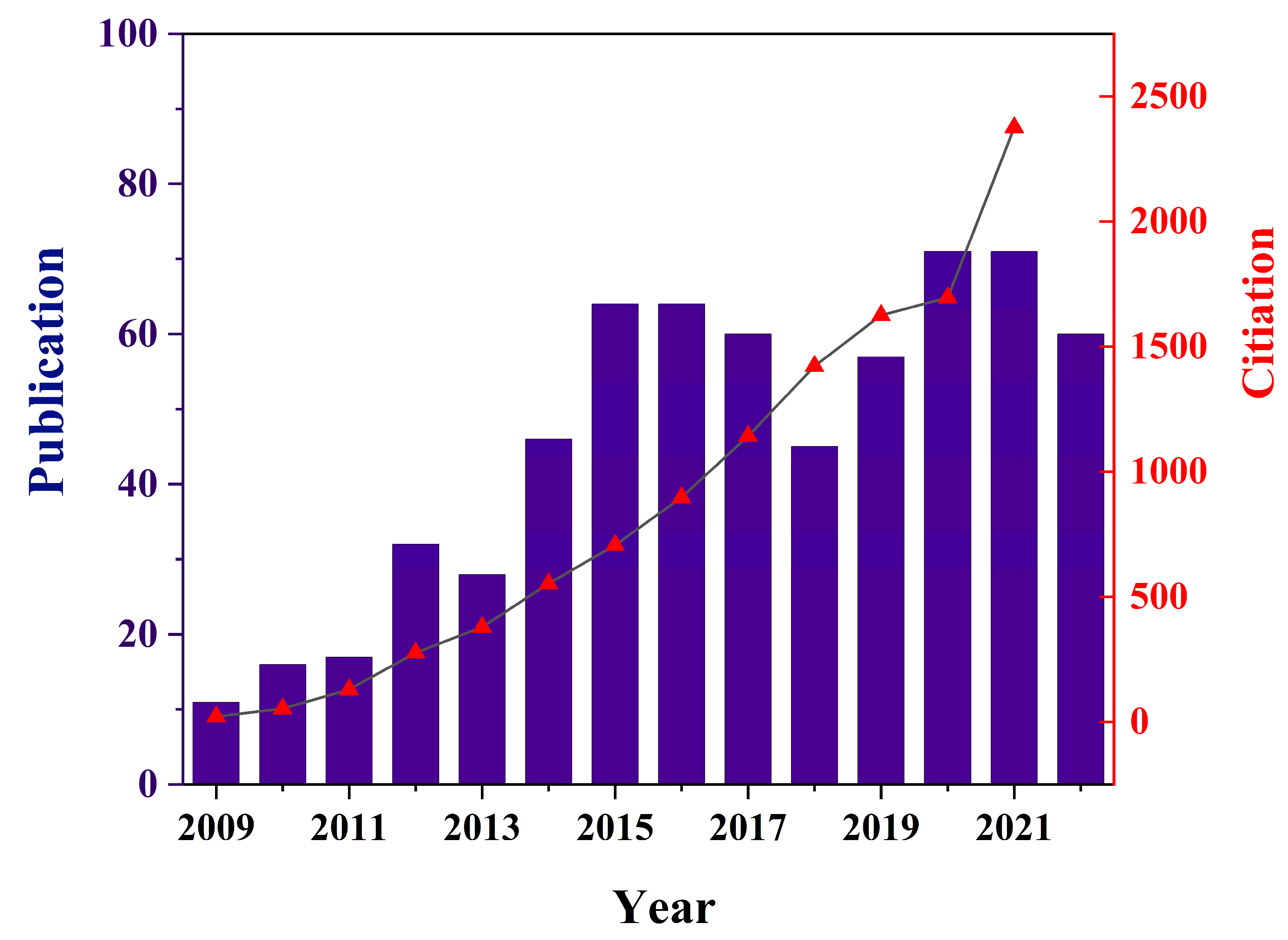
1. Introduction
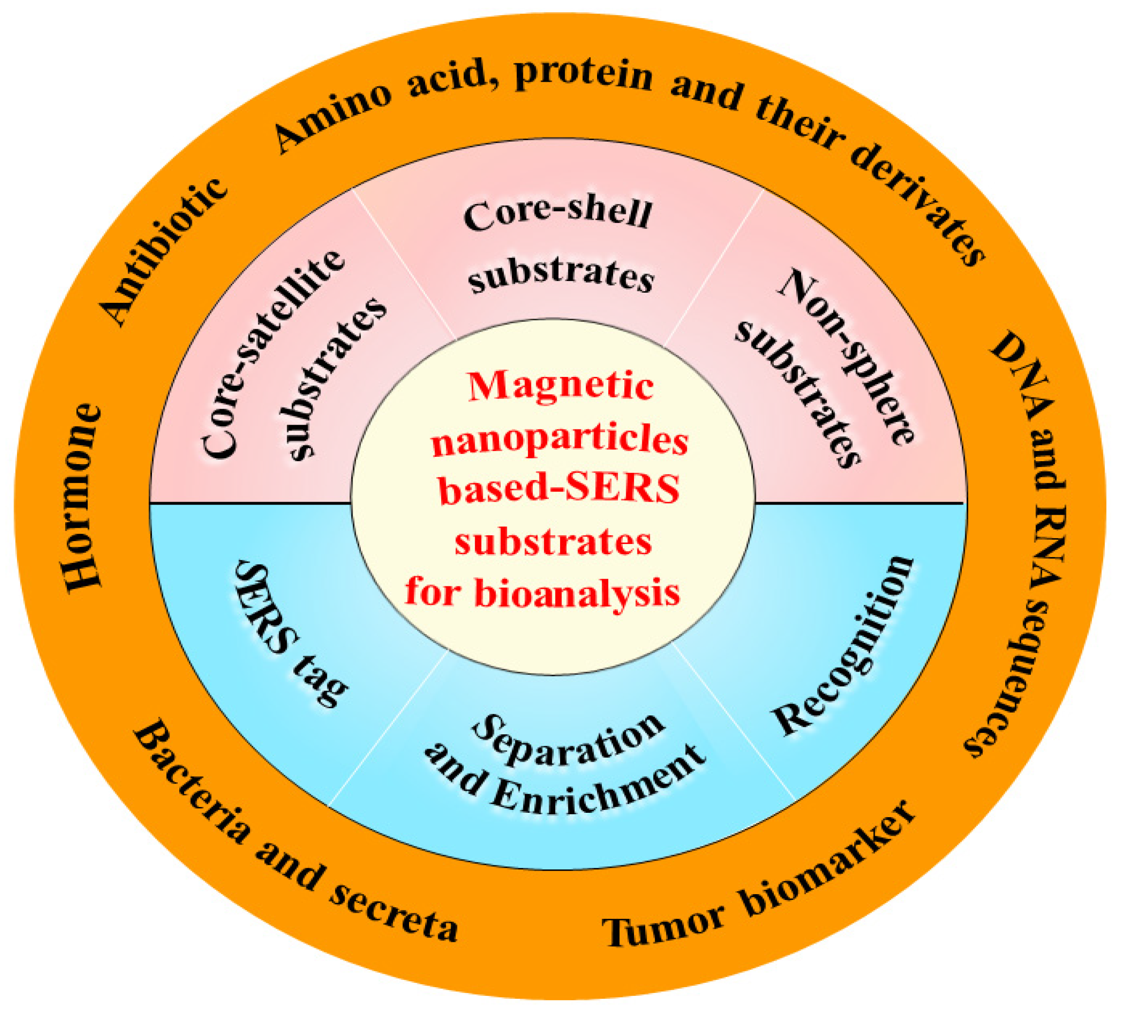
2. Morphologies of Magnetic Nanoparticle-Based SERS Substrates
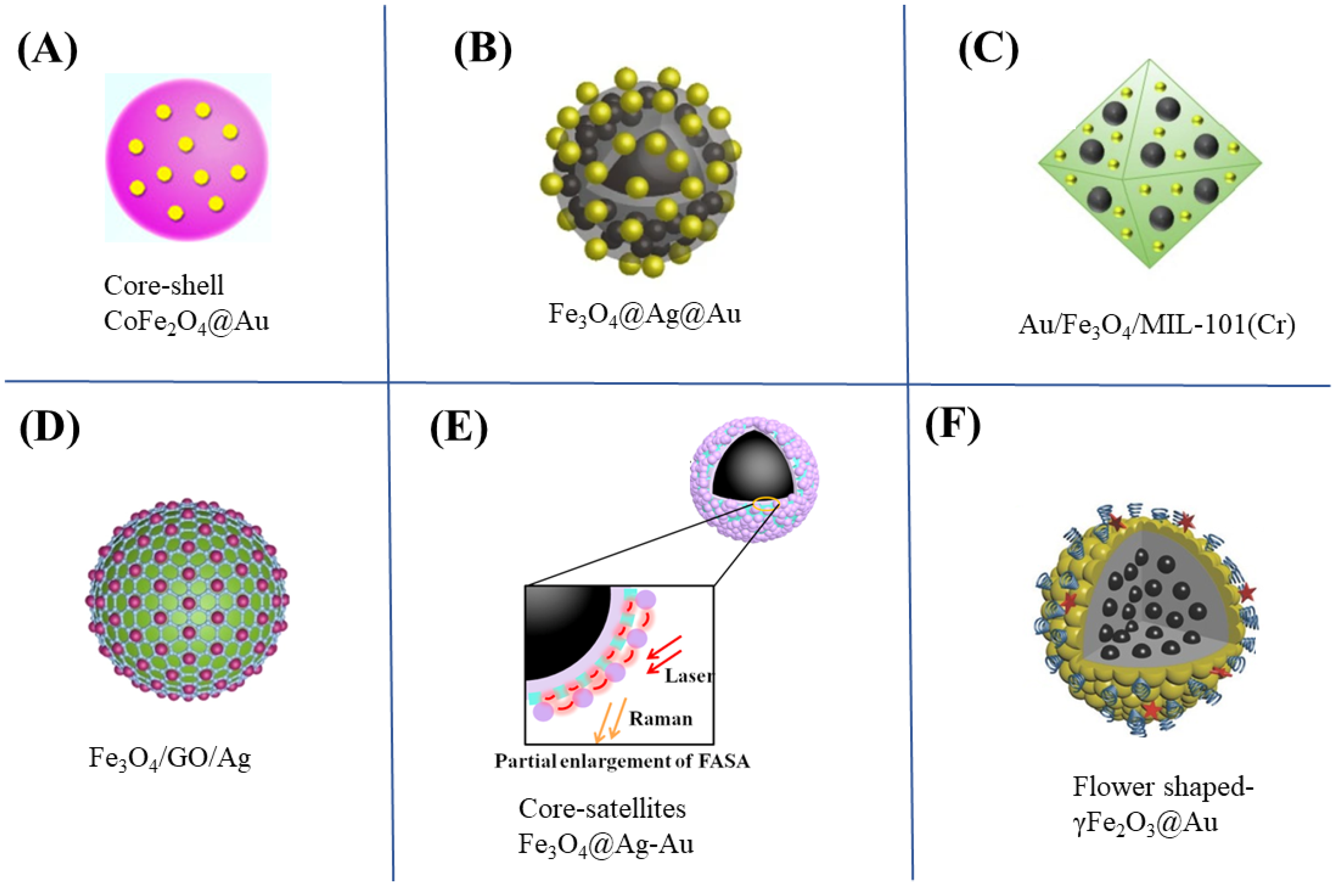
2.1. Magnetic Core–Shell Nanoparticles
2.2. Magnetic Core–Satellite Nanoparticles
2.3. Non-Spherical Magnetic Nanoparticles
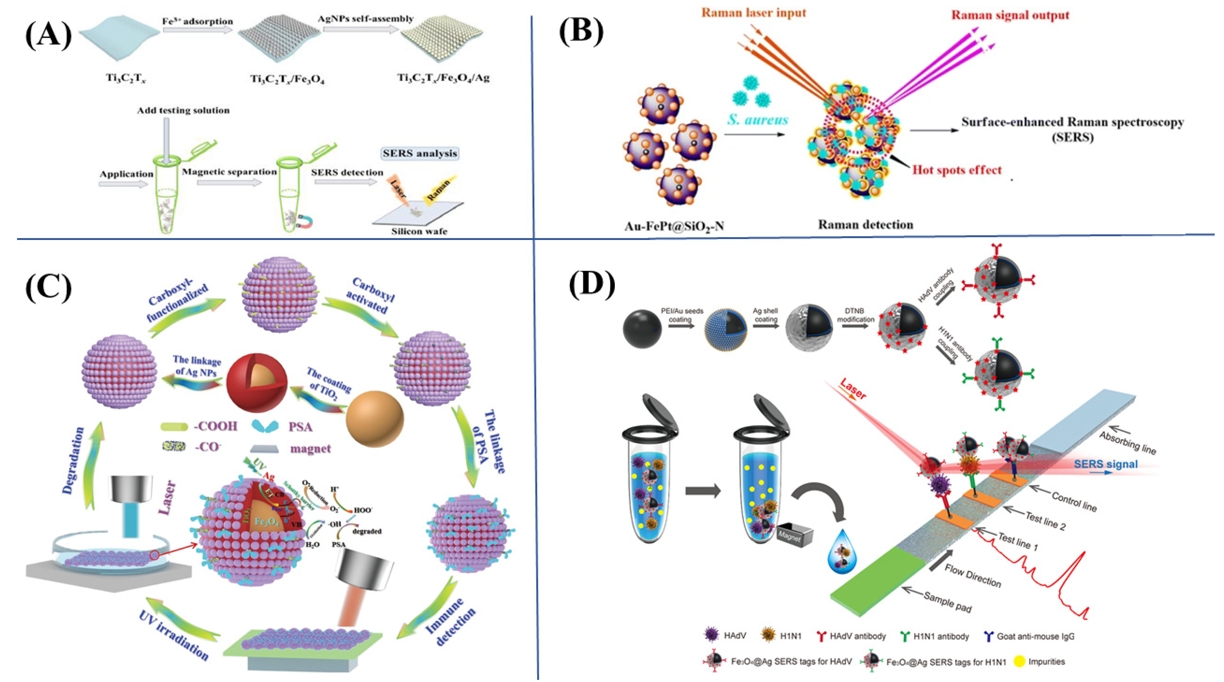
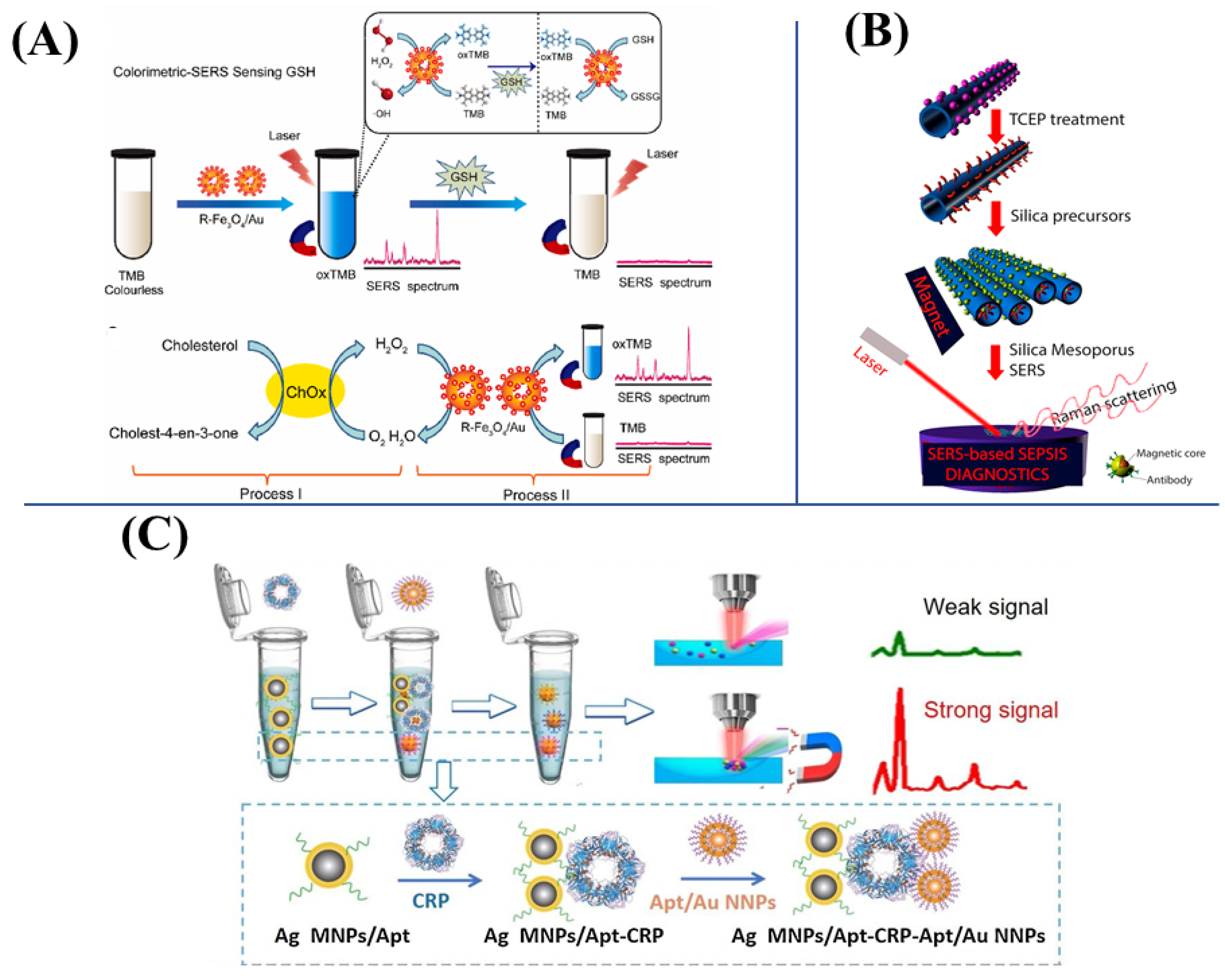
3. Functions of Magnetic SERS Substrates for Bioanalysis
3.1. Separation and Enrichment
3.2. Recognition
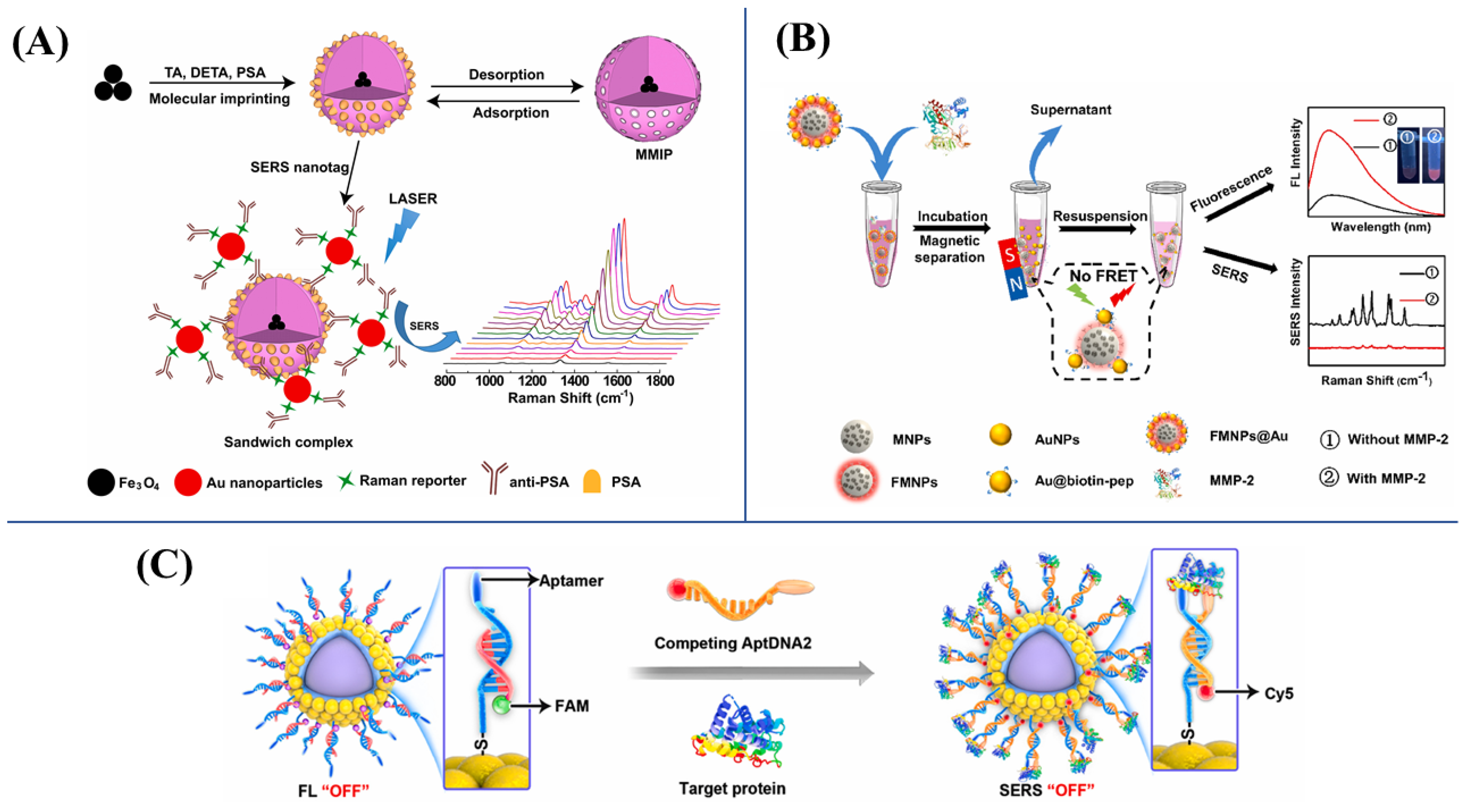
3.3. SERS Tags
4. Applications of Magnetic SERS Substrates for Bioanalysis
4.1. Amino Acids, Protein and Their Derivatives

4.2. DNA and RNA Sequences
4.3. Cancer Cells and Related Tumor Biomarkers
4.4. Other Biomolecules
5. Conclusions and Perspective
Author Contributions
Funding
Institutional Review Board Statement
Informed Consent Statement
Data Availability Statement
Conflicts of Interest
References
- Liu, H.; Lei, Y. A critical review: Recent advances in “digital” biomolecule detection with single copy sensitivity. Biosens. Bioelectron. 2021, 177, 112901. [Google Scholar] [CrossRef]
- Elwakeel, K.; Shathat, A.; Al-Bogami, A.; Wujesiri, B.; Goonetilleke, A. The synergistic effect of ultrasound power and magnetite incorporation on the sorption/desorption behavior of Cr(VI) and As(V) oxoanions in an aqueous system. J. Colloid Interface Sci. 2020, 569, 76–88. [Google Scholar] [CrossRef] [PubMed]
- Manmana, Y.; Kubo, T.; Otsuka, K. Recent developments of point-of-care (POC) testing platform for biomolecules. TrAC Trends Anal. Chem. 2021, 135, 116160. [Google Scholar] [CrossRef]
- Pashazadeh-Panahi, P.; Belali, S.; Sohrabi, H.; Oroojalian, F.; Hashemzaei, M.; Mokhtarzadeh, A.; de la Guardia, M. Metal-organic frameworks conjugated with biomolecules as efficient platforms for development of biosensors. TrAC Trends Anal. Chem. 2021, 141, 116285. [Google Scholar] [CrossRef]
- Alsalameh, S.; Alnajjar, K.; Makhzoum, T.; Al Eman, N.; Shakir, I.; Mir, T.A.; Alkattan, K.; Chinnappan, R.; Yaqinuddin, A. Advances in biosensing technologies for diagnosis of COVID-19. Biosensors 2022, 12, 898. [Google Scholar] [CrossRef] [PubMed]
- Li, W.; Zhou, J.; Maccaferri, N.; Krahne, R.; Wang, K.; Garoli, D. Enhanced optical spectroscopy for multiplexed DNA and protein-sequencing with plasmonic nanopores: Challenges and prospects. Anal. Chem. 2022, 94, 503–514. [Google Scholar] [CrossRef] [PubMed]
- Yao, D.; Li, C.; Wang, H.; Wen, G.; Liang, A.; Jiang, Z. A new dual-mode SERS and RRS aptasensor for detecting trace organic molecules based on gold nanocluster-doped covalent-organic framework catalyst. Sens. Actuators B Chem. 2020, 319, 128308. [Google Scholar] [CrossRef]
- Pahlow, S.; Mayerhöfer, T.; van der Loh, M.; Hübner, U.; Dellith, J.; Weber, K.; Popp, J. Interference-enhanced Raman spectroscopy as a promising tool for the detection of biomolecules on Raman-compatible Surfaces. Anal. Chem. 2018, 90, 9025–9032. [Google Scholar] [CrossRef]
- Wang, H.; Zhao, Y.; Shi, J.; Wen, G.; Liang, A.; Jiang, Z. A novel aptamer RRS assay platform for ultratrace melamine based on COF-loaded Pd nanocluster catalytic amplification. J. Hazard. Mater. 2022, 423, 127263. [Google Scholar] [CrossRef]
- Nan, X.; Jia, W.; Zhang, Y.; Wang, H.; Lin, Z.; Chen, S. An on-line detection system for screening small molecule inhibitors of α-amylase and α-glucosidase in prunus mume. J. Chromatogr. A 2022, 1663, 462754. [Google Scholar] [CrossRef]
- Wu, Q.; Li, Y.; Wang, Y.; Lu, H. Quantitative mass spectrometry imaging of amino acids with isomer differentiation in brain tissue via exhaustive liquid microjunction surface sampling–tandem mass tags labeling–ultra performance liquid chromatography–mass spectrometry. J. Chromatogr. A 2020, 1621, 461086. [Google Scholar] [CrossRef] [PubMed]
- Parker, E.T.; Karki, M.; Glavin, D.P.; Dworkin, J.P.; Krishnamurthy, R. A sensitive quantitative analysis of abiotically synthesized short homopeptides using ultraperformance liquid chromatography and time-of-flight mass spectrometry. J. Chromatogr. A 2020, 1630, 461509. [Google Scholar] [CrossRef] [PubMed]
- Ghosh, J.; Ghosh, R.; Giri, P.K. Tuning the visible photoluminescence in Al doped ZnO thin film and its application in label-free glucose detection. Sens. Actuators B Chem. 2018, 254, 681–689. [Google Scholar] [CrossRef]
- Chen, S.; Zhao, J.; Xu, C.; Sakharov, I.Y.; Zhao, S. Absolute quantification of microRNAs in a single cell with chemiluminescence detection based on rolling circle amplification on a microchip platform. Anal. Chem. 2021, 93, 9218–9225. [Google Scholar] [CrossRef]
- Hossain, M.B.; Islam, M.M.; Abdulrazak, L.F.; Rana, M.M.; Akib, T.B.A.; Hassan, M. Graphene-coated optical fiber SPR biosensor for BRCA1 and BRCA2 breast cancer biomarker detection: A numerical design-based analysis. Photonic Sens. 2020, 10, 67–79. [Google Scholar] [CrossRef]
- Huang, S.; Wu, C.; Wang, Y.; Yang, X.; Yuan, R.; Chai, Y. Ag/TiO2 nanocomposites as a novel SERS substrate for construction of sensitive biosensor. Sens. Actuators B Chem. 2021, 339, 129843. [Google Scholar] [CrossRef]
- Lai, H.; Chen, Z.; Li, G.; Zhang, Z. All-in-one preparation strategy integrated in a miniaturized device for fast analyses of biomarkers in biofluids by surface enhanced Raman scattering. Anal. Chem. 2022, 94, 16275–16281. [Google Scholar] [CrossRef]
- Yu, Z.; Huang, L.; Zhang, Z.; Li, G. Simultaneous and accurate quantification of multiple antibiotics in aquatic samples by surface-enhanced Raman scattering using a Ti3C2Tx/DNA/Ag membrane substrate. Anal. Chem. 2021, 93, 13072–13079. [Google Scholar] [CrossRef]
- Dong, J.; Wang, S.; Wang, Y.; Hu, K.; Qiu, L.; Lin, L.; Chen, X. In situ homogeneous formation of Au@AgNPs for the rapid determination of formaldehyde residues by surface-enhanced Raman spectroscopy coupled with microhydrodistillation. Microchim. Acta 2020, 187, 353. [Google Scholar] [CrossRef]
- Li, D.; Yao, D.; Li, C.; Luo, Y.; Liang, A.; Wen, G.; Jiang, Z. Nanosol SERS quantitative analytical method: A review. TrAC Trends Anal. Chem. 2020, 127, 115885. [Google Scholar] [CrossRef]
- Restaino, S.M.; White, I.M. A critical review of flexible and porous SERS sensors for analytical chemistry at the point-of-sample. Anal. Chim. Acta 2019, 1060, 17–29. [Google Scholar] [CrossRef] [PubMed]
- Xu, K.; Zhou, R.; Takei, K.; Hong, M. Toward flexible surface-enhanced Raman scattering (SERS) sensors for point-of-care diagnostics. Adv. Sci. 2019, 6, 1900925. [Google Scholar] [CrossRef] [PubMed]
- Garcia-Rico, E.; Alvarez-Puebla, R.A.; Guerrini, L. Direct surface-enhanced Raman scattering (SERS) spectroscopy of nucleic acids: From fundamental studies to real-life applications. Chem. Soc. Rev. 2018, 47, 4909–4923. [Google Scholar] [CrossRef] [PubMed]
- Zeng, Y.; Koo, K.M.; Trau, M.; Shen, A.-G.; Hu, J.-M. Watching SERS glow for multiplex biomolecular analysis in the clinic: A review. Appl. Mater. Today 2019, 15, 431–444. [Google Scholar] [CrossRef]
- Plou, J.; Valera, P.S.; García, I.; de Albuquerque, C.D.L.; Carracedo, A.; Liz-Marzán, L.M. Prospects of surface-enhanced Raman spectroscopy for biomarker monitoring toward precision medicine. ACS Photonics 2022, 9, 333–350. [Google Scholar] [CrossRef]
- Wang, Y.; Li, B.; Tian, T.; Liu, Y.; Zhang, J.; Qian, K. Advanced on-site and in vitro signal amplification biosensors for biomolecule analysis. TrAC Trends Anal. Chem. 2022, 149, 116565. [Google Scholar] [CrossRef]
- Ali, A.; Nettey-Oppong, E.E.; Effah, E.; Yu, C.Y.; Muhammad, R.; Soomro, T.A.; Byun, K.M.; Choi, S.H. Miniaturized Raman instruments for SERS-based point-of-care testing on respiratory viruses. Biosensors 2022, 12, 590. [Google Scholar] [CrossRef]
- Li, H.; Li, Y.; Wang, D.; Wang, J.; Zhang, J.; Jiang, W.; Zhou, T.; Liu, C.; Che, G. Synthesis of hydrophilic SERS-imprinted membrane based on graft polymerization for selective detection of L-tyrosine. Sens. Actuators B Chem. 2021, 340, 129955. [Google Scholar] [CrossRef]
- Barucci, A.; D’Andrea, C.; Farnesi, E.; Banchelli, M.; Amicucci, C.; de Angelis, M.; Hwang, B.; Matteini, P. Label-free SERS detection of proteins based on machine learning classification of chemo-structural determinants. Analyst 2021, 146, 674–682. [Google Scholar] [CrossRef]
- Madzharova, F.; Heiner, Z.; Gühlke, M.; Kneipp, J. Surface-enhanced hyper-Raman spectra of adenine, guanine, cytosine, thymine, and uracil. J. Phys. Chem. C 2016, 120, 15415–15423. [Google Scholar] [CrossRef]
- Morla-Folch, J.; Alvarez-Puebla, R.A.; Guerrini, L. Direct quantification of DNA base composition by surface-enhanced Raman scattering spectroscopy. J. Phys. Chem. Lett. 2016, 7, 3037–3041. [Google Scholar] [CrossRef] [PubMed]
- Liu, C.; Xu, T.; Cheng, G.; Zhang, X. Target-triggered regioselective assembly of nanoprobes for Raman imaging of dual cancer biomarkers in living cells. Sens. Actuators B Chem. 2021, 330, 129319. [Google Scholar] [CrossRef]
- Wang, X.; Zeng, J.; Sun, Q.; Yang, J.; Xiao, Y.; Zhu, Z.; Yan, B.; Li, Y. An effective method towards label-free detection of antibiotics by surface-enhanced Raman spectroscopy in human serum. Sens. Actuators B Chem. 2021, 343, 130084. [Google Scholar] [CrossRef]
- Göksel, Y.; Zor, K.; Rindzevicius, T.; Thorhauge Als-Nielsen, B.E.; Schmiegelow, K.; Boisen, A. Quantification of methotrexate in human serum using surface-enhanced Raman scattering—Toward therapeutic drug monitoring. ACS Sens. 2021, 6, 2664–2673. [Google Scholar] [CrossRef]
- Li, J.-F.; Zhang, Y.-J.; Ding, S.-Y.; Panneerselvam, R.; Tian, Z.-Q. Core–shell nanoparticle-enhanced Raman spectroscopy. Chem. Rev. 2017, 117, 5002–5069. [Google Scholar] [CrossRef]
- Lin, J.-S.; Radjenovic, P.M.; Jin, H.; Li, J.-F. Plasmonic core–shell nanoparticle enhanced spectroscopies for surface analysis. Anal. Chem. 2021, 93, 6573–6582. [Google Scholar] [CrossRef] [PubMed]
- Wu, K.; Su, D.; Liu, J.; Saha, R.; Wang, J.-P. Magnetic nanoparticles in nanomedicine: A review of recent advances. Nanotechnology 2019, 30, 502003. [Google Scholar] [CrossRef] [PubMed]
- Wu, T.; Li, J.; Zheng, S.; Yu, Q.; Qi, K.; Shao, Y.; Wang, C.; Tu, J.; Xiao, R. Magnetic nanotag-based colorimetric/SERS dual-readout immunochromatography for ultrasensitive detection of clenbuterol hydrochloride and ractopamine in food samples. Biosensors 2022, 12, 709. [Google Scholar] [CrossRef] [PubMed]
- Yang, M.-C.; Hardiansyah, A.; Cheng, Y.-W.; Liao, H.-L.; Wang, K.-S.; Randy, A.; Harito, C.; Chen, J.-S.; Jeng, R.-J.; Liu, T.-Y. Reduced graphene oxide nanosheets decorated with core-shell of Fe3O4-Au nanoparticles for rapid SERS detection and hyperthermia treatment of bacteria. Spectrochim. Acta A Mol. Biomol. Spectrosc. 2022, 281, 121578. [Google Scholar] [CrossRef]
- Huang, K.; Li, Z.; Lin, J.; Han, G.; Huang, P. Two-dimensional transition metal carbides and nitrides (MXenes) for biomedical applications. Chem. Soc. Rev. 2018, 47, 5109–5124. [Google Scholar] [CrossRef]
- Karthick Kannan, P.; Shankar, P.; Blackman, C.; Chung, C.-H. Recent advances in 2D inorganic nanomaterials for SERS sensing. Adv. Mater. 2019, 31, 1803432. [Google Scholar] [CrossRef] [PubMed]
- Huyh, K.; Hahm, E.; Noh, M.; Lee, J.; Pham, X.; Lee, S.; Kim, J.; Rho, W.; Chang, H.; Kim, D.; et al. Recent advances in surface-enhanced Raman scattering magnetic plasmonic particles for bioapplications. Nanomarterials 2021, 11, 1215. [Google Scholar]
- Lai, H.; Xu, F.; Li, W. A review of the preparation and application of magnetic nanoparticles for surface-enhanced Raman scattering. J. Mater. Sci. 2018, 53, 8677–8698. [Google Scholar] [CrossRef]
- Zhng, C.; Huang, L.; Pu, H.; Sun, D. Magnetic surface-enhanced Raman scattering (MagSERS) biosensors for microbial food safety: Fundamentals and applications. Trends Food. Sci. Technol. 2021, 113, 366–381. [Google Scholar] [CrossRef]
- Zhang, H.; Yi, Y.; Zhou, C.; Ying, G.; Zhou, X.; Fu, C.; Zhu, Y.; Shen, Y. SERS detection of microRNA biomarkers for cancer diagnosis using gold-coated paramagnetic nanoparticles to capture SERS-active gold nanoparticles. RSC Adv. 2017, 7, 52782–52793. [Google Scholar] [CrossRef]
- He, Y.; Wang, Y.; Yang, X.; Xie, S.; Yuan, R.; Chai, Y. Metal organic frameworks combining CoFe2O4 magnetic nanoparticles as highly efficient SERS sensing platform for ultrasensitive detection of N-terminal Pro-brain natriuretic peptide. ACS Appl. Mater. Interfaces 2016, 8, 7683–7690. [Google Scholar] [CrossRef]
- Lin, C.; Li, L.; Feng, J.; Zhang, Y.; Lin, X.; Guo, H.; Li, R. Aptamer-modified magnetic SERS substrate for label-based determination of cardiac troponin I. Microchim. Acta 2021, 189, 22. [Google Scholar] [CrossRef]
- Shao, Q.; Zhang, X.; Liang, P.; Chen, Q.; Qi, X.; Zou, M. Fabrication of magnetic Au/Fe3O4/MIL-101(Cr) (AF-MIL) as sensitive surface-enhanced Raman spectroscopy (SERS) platform for trace detection of antibiotics residue. Appl. Surf. Sci. 2022, 596, 153550. [Google Scholar] [CrossRef]
- He, J.; Song, G.; Wang, X.; Zhou, L.; Li, J. Multifunctional magnetic Fe3O4/GO/Ag composite microspheres for SERS detection and catalytic degradation of methylene blue and ciprofloxacin. J. Alloys Compd. 2022, 893, 162226. [Google Scholar] [CrossRef]
- Chen, M.; Luo, W.; Zhang, Z.; Zhu, F.; Liao, S.; Yang, H.; Chen, X. Sensitive surface enhanced Raman spectroscopy (SERS) detection of methotrexate by core-shell-satellite magnetic microspheres. Talanta 2017, 171, 152–158. [Google Scholar] [CrossRef]
- Huang, J.; Guo, M.; Ke, H.; Zong, C.; Ren, B.; Liu, G.; Shen, H.; Ma, Y.; Wang, X.; Zhang, H.; et al. Rational design and synthesis of γFe2O3@Au magnetic gold nanoflowers for efficient cancer theranostics. Adv. Mater. 2015, 27, 5049–5056. [Google Scholar] [CrossRef] [PubMed]
- Liu, X.; Yang, X.; Li, K.; Liu, H.; Xiao, R.; Wang, W.; Wang, C.; Wang, S. Fe3O4@Au SERS tags-based lateral flow assay for simultaneous detection of serum amyloid A and C-reactive protein in unprocessed blood sample. Sens. Actuators B Chem. 2020, 320, 128350. [Google Scholar] [CrossRef]
- Wang, C.; Rong, Z.; Wang, J.; Jiang, N.; Pang, Y.; Xiao, R.; Wang, S. Seed-mediated synthesis of high-performance silver-coated magnetic nanoparticles and their use as effective SERS substrates. Colloids Surf. Physicochem. Eng. Asp. 2016, 506, 393–401. [Google Scholar] [CrossRef]
- Xu, Y.; Yan, X.; Fang, W.; Daniele, S.; Zhang, J.; Wang, L. SERS self-monitoring of Ag-catalyzed reaction by magnetically separable mesoporous Fe3O4@Ag@mSiO2. Microporous Mesoporous Mater. 2018, 263, 113–119. [Google Scholar] [CrossRef]
- Su, Y.; Xu, S.; Zhang, J.; Chen, X.; Jiang, L.-P.; Zheng, T.; Zhu, J.-J. Plasmon near-field coupling of bimetallic nanostars and a hierarchical bimetallic SERS “hot field”: Toward ultrasensitive simultaneous detection of multiple cardiorenal syndrome biomarkers. Anal. Chem. 2019, 91, 864–872. [Google Scholar] [CrossRef]
- Aarthi, A.; Bindhu, M.R.; Umadevi, M.; Parimaladevi, R.; Sathe, G.V.; Al-Mohaimeed, A.M.; Elshikh, M.S.; Balasubramanian, B. Evaluating the detection efficacy of advanced bimetallic plasmonic nanoparticles for heavy metals, hazardous materials and pesticides of leachate in contaminated groundwater. Environ. Res. 2021, 201, 111590. [Google Scholar] [CrossRef]
- Pham, X.-H.; Hahm, E.; Kim, T.H.; Kim, H.-M.; Lee, S.H.; Lee, S.C.; Kang, H.; Lee, H.-Y.; Jeong, D.H.; Choi, H.S.; et al. Enzyme-amplified SERS immunoassay with Ag-Au bimetallic SERS hot spots. Nano Res. 2020, 13, 3338–3346. [Google Scholar] [CrossRef]
- Kaja, S.; Nag, A. Bimetallic Ag–Cu alloy microflowers as SERS substrates with single-molecule detection limit. Langmuir 2021, 37, 13027–13037. [Google Scholar] [CrossRef]
- Pham, T.T.H.; Vu, X.H.; Dien, N.D.; Trang, T.T.; Van Truong, N.; Thanh, T.D.; Tan, P.M.; Ca, N.X. The structural transition of bimetallic Ag–Au from core/shell to alloy and SERS application. RSC Adv. 2020, 10, 24577–24594. [Google Scholar] [CrossRef]
- Shen, J.; Zhou, Y.; Huang, J.; Zhu, Y.; Zhu, J.; Yang, X.; Chen, W.; Yao, Y.; Qian, S.; Jiang, H.; et al. In-situ SERS monitoring of reaction catalyzed by multifunctional Fe3O4@TiO2@Ag-Au microspheres. Appl. Catal. B Environ. 2017, 205, 11–18. [Google Scholar] [CrossRef]
- Sun, Y.; Yu, X.; Hu, J.; Zhuang, X.; Wang, J.; Qiu, H.; Ren, H.; Zhang, S.; Zhang, Y.; Hu, Y. Constructing a highly sensitivity SERS sensor based on a magnetic metal–organic framework (MOF) to detect the trace of thiabendazole in fruit juice. ACS Sustain. Chem. Eng. 2022, 10, 8400–8410. [Google Scholar] [CrossRef]
- Duan, N.; Shen, M.; Wu, S.; Zhao, C.; Ma, X.; Wang, Z. Graphene oxide wrapped Fe3O4@Au nanostructures as substrates for aptamer-based detection of vibrio parahaemolyticus by surface-enhanced Raman spectroscopy. Microchim. Acta 2017, 184, 2653–2660. [Google Scholar] [CrossRef]
- Prasiwi, O.D.I.; Saraswati, T.E.; Anwar, M.; Masykur, A. Magnetic carbon nanofibers prepared with Ni and Ni/graphitic carbon nanoparticle catalysts for glycine detection using surface-enhanced Raman spectroscopy. ACS Appl. Nano Mater. 2021, 4, 6594–6608. [Google Scholar] [CrossRef]
- Zheng, Y.; Thai, T.; Reineck, P.; Qiu, L.; Guo, Y.; Bach, U. DNA-directed self-assembly of core-satellite plasmonic nanostructures: A highly sensitive and reproducible near-IR SERS sensor. Adv. Funct. Mater. 2013, 23, 1519–1526. [Google Scholar] [CrossRef]
- Pu, H.; Zhu, H.; Xu, F.; Sun, D.-W. Development of core-satellite-shell structured MNP@Au@MIL-100(Fe) substrates for surface-enhanced Raman spectroscopy and their applications in trace level determination of malachite green in prawn. J. Raman Spectrosc. 2022, 53, 682–693. [Google Scholar] [CrossRef]
- Zhao, B.; Liu, H.; Wang, H.; Zhang, Y.; Wang, X.; Zhou, N. Bilayer magnetic-plasmonic satellite nanoassemblies for SERS detection of tobramycin with exonuclease amplification. Biosens. Bioelectron. 2022, 218, 114789. [Google Scholar] [CrossRef]
- Han, C.; Zhai, W.; Wang, Y.; Cao, J.; Wang, M. A SERS aptasensor for rapid detection of aflatoxin B1 in coix seed using satellite structured Fe3O4@Au nanocomposites. Food Control 2022, 142, 109228. [Google Scholar] [CrossRef]
- Achadu, O.J.; Abe, F.; Hossain, F.; Nasrin, F.; Yamazaki, M.; Suzuki, T.; Park, E.Y. Sulfur-doped carbon dots@polydopamine-functionalized magnetic silver nanocubes for dual-modality detection of norovirus. Biosens. Bioelectron. 2021, 193, 113540. [Google Scholar] [CrossRef]
- Xie, J.; Zhang, Q.; Lee, J.Y.; Wang, D.I.C. The synthesis of SERS-active gold nanoflower tags for in vivo applications. ACS Nano 2008, 2, 2473–2480. [Google Scholar] [CrossRef]
- Xuan, S.; Wang, F.; Gong, X.; Kong, S.-K.; Jimmy, C.Y.; Leung, K.C.F. Hierarchical core/shell Fe3O4 @SiO2@γ-AlOOH@Au micro/nanoflowers for protein immobilization. Chem. Commun. 2011, 47, 2514–2516. [Google Scholar] [CrossRef]
- Ding, Q.; Zhou, H.; Zhang, H.; Zhang, Y.; Wang, G.; Zhao, H. 3D Fe3O4@Au@Ag nanoflowers assembled magnetoplasmonic chains for in situ SERS monitoring of plasmon-assisted catalytic reactions. J. Mater. Chem. A 2016, 4, 8866–8874. [Google Scholar] [CrossRef]
- Aldeanueva-Potel, P.; Carbó-Argibay, E.; Pazos-Pérez, N.; Barbosa, S.; Pastoriza-Santos, I.; Alvarez-Puebla, R.A.; Liz-Marzán, L.M. Spiked gold beads as substrates for single-particle SERS. ChemPhysChem 2012, 13, 2561–2565. [Google Scholar] [CrossRef] [PubMed]
- Burda, C.; Chen, X.; Narayanan, R.; El-Sayed, M.A. Chemistry and properties of nanocrystals of different shapes. Chem. Rev. 2005, 105, 1025–1102. [Google Scholar] [CrossRef] [PubMed]
- Quaresma, P.; Osório, I.; Dória, G.; Carvalho, P.A.; Pereira, A.; Langer, J.; Araújo, J.P.; Pastoriza-Santos, I.; Liz-Marzán, L.M.; Franco, R.; et al. Star-shaped magnetite@gold nanoparticles for protein magnetic separation and SERS detection. RSC Adv. 2013, 4, 3659–3667. [Google Scholar] [CrossRef]
- Reguera, J.; de Aberasturi, D.J.; Winckelmans, N.; Langer, J.; Bals, S.; Liz-Marzán, L.M. Synthesis of janus plasmonic–magnetic, star–sphere nanoparticles, and their application in SERS detection. Faraday Discuss. 2016, 191, 47–59. [Google Scholar] [CrossRef] [PubMed]
- Litti, L.; Trivini, S.; Ferraro, D.; Reguera, J. 3D printed microfluidic device for magnetic trapping and SERS quantitative evaluation of environmental and biomedical analytes. ACS Appl. Mater. Interfaces 2021, 13, 34752–34761. [Google Scholar] [CrossRef]
- Yu, Z.; Huang, L.; Zhang, Z.; Li, G. Magnetic Ti3C2Tx/Fe3O4/Ag substrate for rapid quantification of trace sulfonamides in aquatic products by surface enhanced Raman spectroscopy. Chin. Chem. Lett. 2022, 33, 3853–3858. [Google Scholar] [CrossRef]
- Xiang, Y.; Yang, H.; Guo, X.; Wu, Y.; Ying, Y.; Wen, Y.; Yang, H. Surface enhanced Raman detection of the colon cancer biomarker cytidine by using magnetized nanoparticles of the type Fe3O4/Au/Ag. Microchim. Acta 2018, 185, 195. [Google Scholar] [CrossRef]
- Yu, S.; Liu, Z.; Li, H.; Zhang, J.; Yuan, X.; Jia, X.; Wu, Y. Combination of a graphene SERS substrate and magnetic solid phase micro-extraction used for the rapid detection of trace illegal additives. Analyst 2018, 143, 883–890. [Google Scholar] [CrossRef]
- Hardiansyah, A.; Chen, A.-Y.; Liao, H.-L.; Yang, M.-C.; Liu, T.-Y.; Chan, T.-Y.; Tsou, H.-M.; Kuo, C.-Y.; Wang, J.-K.; Wang, Y.-L. Core-shell of FePt@SiO2-Au magnetic nanoparticles for rapid SERS detection. Nanoscale Res. Lett. 2015, 10, 412. [Google Scholar] [CrossRef]
- Du, Y.; Liu, H.; Chen, Y.; Tian, Y.; Zhang, X.; Gu, C.; Jiang, T.; Zhou, J. Recyclable label-free SERS-based immunoassay of PSA in human serum mediated by enhanced photocatalysis arising from Ag nanoparticles and external magnetic field. Appl. Surf. Sci. 2020, 528, 146953. [Google Scholar] [CrossRef]
- Wang, C.; Wang, C.; Wang, X.; Wang, K.; Zhu, Y.; Rong, Z.; Wang, W.; Xiao, R.; Wang, S. Magnetic SERS strip for sensitive and simultaneous detection of respiratory viruses. ACS Appl. Mater. Interfaces 2019, 11, 19495–19505. [Google Scholar] [CrossRef] [PubMed]
- Wang, C.; Wang, J.; Li, M.; Qu, X.; Zhang, K.; Rong, Z.; Xiao, R.; Wang, S. A rapid SERS method for label-free bacteria detection using polyethylenimine-modified Au-coated magnetic microspheres and Au@Ag nanoparticles. Analyst 2016, 141, 6226–6238. [Google Scholar] [CrossRef] [PubMed]
- Juang, R.-S.; Chen, W.-T.; Cheng, Y.-W.; Wang, K.-S.; Jeng, R.-J.; Zeng, Z.-L.; Liu, S.-H.; Liu, T.-Y. Fabrication of in situ magnetic capturing and Raman enhancing nanoplatelets for detection of bacteria and biomolecules. Colloids Surf. Physicochem. Eng. Asp. 2022, 648, 129189. [Google Scholar] [CrossRef]
- Tamer, U.; Cetin, D.; Suludere, Z.; Boyaci, I.H.; Temiz, H.T.; Yegenoglu, H.; Daniel, P.; Dinçer, İ.; Elerman, Y. Gold-coated iron composite nanospheres targeted the detection of Escherichia coli. Int. J. Mol. Sci. 2013, 14, 6223–6240. [Google Scholar] [CrossRef] [PubMed]
- Zhang, P.; Chen, G.; Wang, Z.; Ma, J.; Jia, Q. Design and synthesis of Fe3O4@Au@cyclodextrin-molecularly imprinted polymers labeled with SERS nanotags for ultrasensitive detection of transferrin. Sens. Actuators B Chem. 2022, 361, 131669. [Google Scholar] [CrossRef]
- Shin, M.H.; Hong, W.; Sa, Y.; Chen, L.; Jung, Y.-J.; Wang, X.; Zhao, B.; Jung, Y.M. Multiple detection of proteins by SERS-based immunoassay with core shell magnetic gold nanoparticles. Vib. Spectrosc. 2014, 72, 44–49. [Google Scholar] [CrossRef]
- Du, Y.; Liu, H.; Tian, Y.; Gu, C.; Zhao, Z.; Zeng, S.; Jiang, T. Recyclable SERS-based immunoassay guided by photocatalytic performance of Fe3O4@TiO2@Au nanocomposites. Biosensors 2020, 10, 25. [Google Scholar] [CrossRef]
- Zhao, W.; Zhang, D.; Zhou, T.; Huang, J.; Wang, Y.; Li, B.; Chen, L.; Yang, J.; Liu, Y. Aptamer-conjugated magnetic Fe3O4@Au core-shell multifunctional nanoprobe: A three-in-one aptasensor for selective capture, sensitive SERS detection and efficient near-infrared light triggered photothermal therapy of staphylococcus aureus. Sens. Actuators B Chem. 2022, 350, 130879. [Google Scholar] [CrossRef]
- Wang, J.; Wu, X.; Wang, C.; Shao, N.; Dong, P.; Xiao, R.; Wang, S. Magnetically assisted surface-enhanced Raman Spectroscopy for the detection of staphylococcus aureus based on aptamer recognition. ACS Appl. Mater. Interfaces 2015, 7, 20919–20929. [Google Scholar] [CrossRef]
- Li, J.; Wu, T.; Wang, C.; Tu, J.; Song, X.; Shao, Y.; Wang, C.; Qi, K.; Xiao, R. Nanogapped Fe3O4@Au surface-enhanced Raman scattering tags for the multiplex detection of bacteria on an immunochromatographic strip. ACS Appl. Nano Mater. 2022, 5, 12679–12689. [Google Scholar] [CrossRef]
- Jun, B.-H.; Noh, M.S.; Kim, J.; Kim, G.; Kang, H.; Kim, M.-S.; Seo, Y.-T.; Baek, J.; Kim, J.-H.; Park, J.; et al. Multifunctional silver-embedded magnetic nanoparticles as SERS nanoprobes and their applications. Small 2010, 6, 119–125. [Google Scholar] [CrossRef]
- Qiu, Y.; Deng, D.; Deng, Q.; Wu, P.; Zhang, H.; Cai, C. Synthesis of magnetic Fe3O4–Au hybrids for sensitive SERS detection of cancer cells at low abundance. J. Mater. Chem. B 2015, 3, 4487–4495. [Google Scholar] [CrossRef] [PubMed]
- He, Y.; Yang, X.; Yuan, R.; Chai, Y. “Off” to “On” surface-enhanced Raman spectroscopy platform with padlock probe-based exponential rolling circle amplification for ultrasensitive detection of microRNA 155. Anal. Chem. 2017, 89, 2866–2872. [Google Scholar] [CrossRef] [PubMed]
- Lu, S.; Du, J.; Sun, Z.; Jing, C. Hairpin-structured magnetic SERS sensor for tetracycline resistance gene TetA detection. Anal. Chem. 2020, 92, 16229–16235. [Google Scholar] [CrossRef]
- Bedford, E.E.; Boujday, S.; Pradier, C.-M.; Gu, F.X. Spiky gold shells on magnetic particles for DNA biosensors. Talanta 2018, 182, 259–266. [Google Scholar] [CrossRef]
- Alula, M.T.; Lemmens, P.; Bo, L.; Wulferding, D.; Yang, J.; Spende, H. Preparation of silver nanoparticles coated ZnO/Fe3O4 composites using chemical reduction method for sensitive detection of uric acid via surface-enhanced Raman spectroscopy. Anal. Chim. Acta 2019, 1073, 62–71. [Google Scholar] [CrossRef]
- Usta, D.D.; Salimi, K.; Pinar, A.; Coban, İ.; Tekinay, T.; Tuncel, A. A boronate affinity-assisted SERS tag equipped with a sandwich system for detection of glycated hemoglobin in the hemolysate of human erythrocytes. ACS Appl. Mater. Interfaces 2016, 8, 11934–11944. [Google Scholar] [CrossRef]
- Feng, J.; Xu, Y.; Huang, W.; Kong, H.; Li, Y.; Cheng, H.; Li, L. A magnetic SERS immunosensor for highly sensitive and selective detection of human carboxylesterase 1 in human serum samples. Anal. Chim. Acta 2020, 1097, 176–185. [Google Scholar] [CrossRef]
- Yu, Z.; Grasso, M.F.; Cui, X.; Silva, R.N.; Zhang, P. Sensitive and label-free SERS detection of single-stranded DNA assisted by silver nanoparticles and gold-coated magnetic nanoparticles. ACS Appl. Bio Mater. 2020, 3, 2626–2632. [Google Scholar] [CrossRef]
- Donnelly, T.; Smith, W.E.; Faulds, K.; Graham, D. Silver and magnetic nanoparticles for sensitive DNA detection by SERS. Chem. Commun. 2014, 50, 12907–12910. [Google Scholar] [CrossRef] [PubMed]
- Shen, W.; Wang, C.; Yang, X.; Wang, C.; Zhou, Z.; Liu, X.; Xiao, R.; Gu, B.; Wang, S. Synthesis of raspberry-like nanogapped Fe3O4@Au nanocomposites for SERS-based lateral flow detection of multiple tumor biomarkers. J. Mater. Chem. C 2020, 8, 12854–12864. [Google Scholar] [CrossRef]
- Zhao, X.; Shen, H.; Huo, B.; Wang, Y.; Gao, Z. A novel bionic magnetic SERS aptasensor for the ultrasensitive detection of deoxynivalenol based on “dual antennae” nano-silver. Biosens. Bioelectron. 2022, 211, 114383. [Google Scholar] [CrossRef] [PubMed]
- Chen, R.; Sun, Y.; Huo, B.; Mao, Z.; Wang, X.; Li, S.; Lu, R.; Li, S.; Liang, J.; Gao, Z. Development of Fe3O4@Au nanoparticles coupled to Au@Ag core-shell nanoparticles for the sensitive detection of zearalenone. Anal. Chim. Acta 2021, 1180, 338888. [Google Scholar] [CrossRef] [PubMed]
- Zhou, Z.; Xiao, R.; Cheng, S.; Wang, S.; Shi, L.; Wang, C.; Qi, K.; Wang, S. A universal SERS-label immunoassay for pathogen bacteria detection based on Fe3O4@Au-aptamer separation and antibody-protein a orientation recognition. Anal. Chim. Acta 2021, 1160, 338421. [Google Scholar] [CrossRef] [PubMed]
- Xu, Y.; Hassan, M.M.; Zhu, A.; Li, H.; Chen, Q. Dual-mode of magnetic assisted Au@Ag SERS tags and cationic conjugated UCNPs for qualitative and quantitative analysis of multiple foodborne pathogens. Sens. Actuators B Chem. 2021, 344, 130305. [Google Scholar] [CrossRef]
- Gjergjizi, B.; Çoğun, F.; Yıldırım, E.; Eryılmaz, M.; Selbes, Y.; Sağlam, N.; Tamer, U. SERS-based ultrafast and sensitive detection of luteinizing hormone in human serum using a passive microchip. Sens. Actuators B Chem. 2018, 269, 314–321. [Google Scholar] [CrossRef]
- Yang, K.; Hu, Y.; Dong, N. A novel biosensor based on competitive SERS immunoassay and magnetic separation for accurate and sensitive detection of chloramphenicol. Biosens. Bioelectron. 2016, 80, 373–377. [Google Scholar] [CrossRef]
- Wu, H.-C.; Chen, T.-C.; Tsai, H.-J.; Chen, C.-S. Au nanoparticles deposited on magnetic carbon nanofibers as the ultrahigh sensitive substrate for surface-enhanced Raman scattering: Detections of rhodamine 6G and aromatic amino acids. Langmuir 2018, 34, 14158–14168. [Google Scholar] [CrossRef]
- Zhou, T.; Fan, M.; You, R.; Lu, Y.; Huang, L.; Xu, Y.; Feng, S.; Wu, Y.; Shen, H.; Zhu, L. Fabrication of Fe3O4/Au@ATP@Ag nanorod sandwich structure for sensitive SERS quantitative detection of histamine. Anal. Chim. Acta 2020, 1104, 199–206. [Google Scholar] [CrossRef]
- Saha, A.; Jana, N.R. Detection of cellular glutathione and oxidized glutathione using magnetic–plasmonic nanocomposite-based “turn-off” surface enhanced Raman scattering. Anal. Chem. 2013, 85, 9221–9228. [Google Scholar] [CrossRef] [PubMed]
- Ouyang, L.; Zhu, L.; Jiang, J.; Tang, H. A surface-enhanced Raman scattering method for detection of trace glutathione on the basis of immobilized silver nanoparticles and crystal violet probe. Anal. Chim. Acta 2014, 816, 41–49. [Google Scholar] [CrossRef] [PubMed]
- Huang, Y.; Gu, Y.; Liu, X.; Deng, T.; Dai, S.; Qu, J.; Yang, G.; Qu, L. Reusable ring-like Fe3O4/Au nanozymes with enhanced peroxidase-like activities for colorimetric-SERS dual-mode sensing of biomolecules in human blood. Biosens. Bioelectron. 2022, 209, 114253. [Google Scholar] [CrossRef]
- Yap, L.W.; Chen, H.; Gao, Y.; Petkovic, K.; Liang, Y.; Si, K.J.; Wang, H.; Tang, Z.; Zhu, Y.; Cheng, W. Bifunctional plasmonic-magnetic particles for an enhanced microfluidic SERS immunoassay. Nanoscale 2017, 9, 7822–7829. [Google Scholar] [CrossRef] [PubMed]
- Zhao, P.; Li, H.; Li, D.; Hou, Y.; Mao, L.; Yang, M.; Wang, Y. A SERS nano-tag-based magnetic-separation strategy for highly sensitive immunoassay in unprocessed whole blood. Talanta 2019, 198, 527–533. [Google Scholar] [CrossRef] [PubMed]
- Wei, C.; Sun, R.; Jiang, Y.; Guo, X.; Ying, Y.; Wen, Y.; Yang, H.; Wu, Y. Protease-protection strategy Combined with the SERS tags for detection of o-glcnac transferase activity. Sens. Actuators B Chem. 2021, 345, 130410. [Google Scholar] [CrossRef]
- Lin, C.; Li, L.; Feng, J.; Zhang, Y.; Guo, H.; Lin, X.; Li, R. A novel Apt-SERS platform for the determination of cardiac troponin I based on coral-like silver-modified magnetic substrate and BCA method. Anal. Chim. Acta 2022, 1225, 340253. [Google Scholar] [CrossRef] [PubMed]
- Alves, R.S.; Sigoli, F.A.; Mazali, I.O. Aptasensor based on a flower-shaped silver magnetic nanocomposite enables the sensitive and label-free detection of troponin I (CTnI) by SERS. Nanotechnology 2020, 31, 505505. [Google Scholar] [CrossRef] [PubMed]
- Selbes, Y.S.; Caglayan, M.G.; Eryilmaz, M.; Boyaci, I.H.; Saglam, N.; Basaran, A.A.; Tamer, U. Surface-enhanced Raman probe for rapid nanoextraction and detection of erythropoietin in urine. Anal. Bioanal. Chem. 2016, 408, 8447–8456. [Google Scholar] [CrossRef]
- Nguyen, A.H.; Shin, Y.; Sim, S.J. Development of SERS substrate using phage-based magnetic template for triplex assay in sepsis diagnosis. Biosens. Bioelectron. 2016, 85, 522–528. [Google Scholar] [CrossRef]
- Hu, Z.; Zhou, X.; Duan, J.; Wu, X.; Wu, J.; Zhang, P.; Liang, W.; Guo, J.; Cai, H.; Sun, P.; et al. Aptamer-based novel Ag-coated magnetic recognition and SERS nanotags with interior nanogap biosensor for ultrasensitive detection of protein biomarker. Sens. Actuators B Chem. 2021, 334, 129640. [Google Scholar] [CrossRef]
- Liang, Y.; Gong, J.-L.; Huang, Y.; Zheng, Y.; Jiang, J.-H.; Shen, G.-L.; Yu, R.-Q. Biocompatible core-shell nanoparticle-based surface-enhanced Raman scattering probes for detection of DNA related to hiv gene using silica-coated magnetic nanoparticles as separation tools. Talanta 2007, 72, 443–449. [Google Scholar] [CrossRef] [PubMed]
- Ngo, H.T.; Gandra, N.; Fales, A.M.; Taylor, S.M.; Vo-Dinh, T. Sensitive DNA detection and SNP discrimination using ultrabright SERS nanorattles and magnetic beads for malaria diagnostics. Biosens. Bioelectron. 2016, 81, 8–14. [Google Scholar] [CrossRef] [PubMed]
- Strelau, K.K.; Brinker, A.; Schnee, C.; Weber, K.; Möller, R.; Popp, J. Detection of PCR products amplified from DNA of epizootic pathogens using magnetic nanoparticles and SERS. J. Raman Spectrosc. 2011, 42, 243–250. [Google Scholar] [CrossRef]
- Lin, L.; Crew, E.; Yan, H.; Shan, S.; Skeete, Z.; Mott, D.; Krentsel, T.; Yin, J.; Chernova, N.A.; Luo, J.; et al. Bifunctional nanoparticles for SERS monitoring and magnetic intervention of assembly and enzyme cutting of DNAs. J. Mater. Chem. B 2013, 1, 4320–4330. [Google Scholar] [CrossRef]
- Wu, L.; Xiao, X.; Chen, K.; Yin, W.; Li, Q.; Wang, P.; Lu, Z.; Ma, J.; Han, H. Ultrasensitive SERS detection of bacillus thuringiensis special gene based on Au@Ag NRs and magnetic beads. Biosens. Bioelectron. 2017, 92, 321–327. [Google Scholar] [CrossRef]
- Yang, Y.; Jiang, X.; Chao, J.; Song, C.; Liu, B.; Zhu, D.; Sun, Y.; Yang, B.; Zhang, Q.; Chen, Y.; et al. Synthesis of magnetic core-branched Au shell nanostructures and their application in cancer-related miRNA detection via SERS. Sci. China Mater. 2017, 60, 1129–1144. [Google Scholar] [CrossRef]
- Yao, Y.; Zhang, H.; Tian, T.; Liu, Y.; Zhu, R.; Ji, J.; Liu, B. Iodide-modified Ag nanoparticles coupled with DSN-assisted cycling amplification for label-free and ultrasensitive SERS detection of microRNA-21. Talanta 2021, 235, 122728. [Google Scholar] [CrossRef]
- Xue, T.; Wang, S.; Ou, G.; Li, Y.; Ruan, H.; Li, Z.; Ma, Y.; Zou, R.; Qiu, J.; Shen, Z.; et al. Detection of circulating tumor cells based on improved SERS-active magnetic nanoparticles. Anal. Methods 2019, 11, 2918–2928. [Google Scholar] [CrossRef]
- Zong, S.; Wang, L.; Chen, C.; Lu, J.; Zhu, D.; Zhang, Y.; Wang, Z.; Cui, Y. Facile detection of tumor-derived exosomes using magnetic nanobeads and SERS nanoprobes. Anal. Methods 2016, 8, 5001–5008. [Google Scholar] [CrossRef]
- Turan, E.; Zengin, A.; Suludere, Z.; Kalkan, N.Ö.; Tamer, U. Construction of a sensitive and selective plasmonic biosensor for prostate specific antigen by combining magnetic molecularly-imprinted polymer and surface-enhanced Raman spectroscopy. Talanta 2022, 237, 122926. [Google Scholar] [CrossRef] [PubMed]
- Medetalibeyoglu, H.; Kotan, G.; Atar, N.; Yola, M.L. A novel sandwich-type SERS immunosensor for selective and sensitive carcinoembryonic antigen (CEA) detection. Anal. Chim. Acta 2020, 1139, 100–110. [Google Scholar] [CrossRef] [PubMed]
- Song, C.; Yang, Y.; Yang, B.; Min, L.; Wang, L. Combination assay of lung cancer associated serum markers using surface-enhanced Raman spectroscopy. J. Mater. Chem. B 2016, 4, 1811–1817. [Google Scholar] [CrossRef] [PubMed]
- Liu, L.; Chu, H.; Yang, J.; Sun, Y.; Ma, P.; Song, D. Construction of a magnetic-fluorescent-plasmonic nanosensor for the determination of MMP-2 activity based on SERS-fluorescence dual-mode signals. Biosens. Bioelectron. 2022, 212, 114389. [Google Scholar] [CrossRef] [PubMed]
- Huang, L.; Zhang, Z.; Li, G. DNA strand displacement based surface-enhanced Raman scattering-fluorescence dual-mode nanoprobes for quantification and imaging of vascular endothelial growth factor in living cells. Biosens. Bioelectron. 2022, 204, 114069. [Google Scholar] [CrossRef]
- Villa, J.E.L.; Garcia, I.; Jimenez de Aberasturi, D.; Pavlov, V.; Sotomayor, M.D.P.T.; Liz-Marzán, L.M. SERS-based immunoassay for monitoring cortisol-related disorders. Biosens. Bioelectron. 2020, 165, 112418. [Google Scholar] [CrossRef]
- Liu, B.; Zheng, S.; Tang, H.; Liu, Q.; Li, H.; Gao, B.; Zhao, X.; Sun, F. Highly sensitive detection of free testosterone assisted by magnetic nanobeads and gap-enhanced SERS nanotags. Colloids Surf. B Biointerfaces 2022, 214, 112460. [Google Scholar] [CrossRef]
- Ahi, E.E.; Torul, H.; Zengin, A.; Sucularlı, F.; Yıldırım, E.; Selbes, Y.; Suludere, Z.; Tamer, U. A capillary driven microfluidic chip for SERS based HCG detection. Biosens. Bioelectron. 2022, 195, 113660. [Google Scholar] [CrossRef]
- Weng, Y.-W.; Hu, X.-D.; Jiang, L.; Shi, Q.-L.; Wei, X.-L. An All-in-one magnetic SERS nanosensor for ratiometric detection of escherichia coli in foods. Anal. Bioanal. Chem. 2021, 413, 5419–5426. [Google Scholar] [CrossRef]
- Chattopadhyay, S.; Sabharwal, P.K.; Jain, S.; Kaur, A.; Singh, H. Functionalized polymeric magnetic nanoparticle assisted SERS immunosensor for the sensitive detection of S. typhimurium. Anal. Chim. Acta 2019, 1067, 98–106. [Google Scholar] [CrossRef]


| Category | Biotargets | Magnetic Substrates | LOD | Ref. |
|---|---|---|---|---|
| Amino acid and protein | Uric acid | Ag/ZnO/Fe3O4 | 365 nmol/L | [97] |
| Glycated hemoglobin | Ag-coated magnetic polymethacrylate microspheres | 50 ng/L | [98] | |
| Human carboxylesterase 1 | Fe3O4@SiO2@Ag | 0.1 ng/L | [99] | |
| DNA and RNA sequences | ssDNA associated with BRAF V600E mutation | MNP@SiO2@Au | 5.15 × 10⁻¹¹ mol/L | [100] |
| C. krusei and C. albicans target DNA | Ag@MNP | 20 fmol/L | [101] | |
| Tumor biomarker | CEA | Raspberry-like Fe3O4@Au | 1.43 pg/mL | [102] |
| Bacteria and secretions | Deoxynivalenol | Complementary DNA modified-Fe3O4@Au | 0.032 pg /mL | [103] |
| Zearalenone | Fe3O4@Au | 0.001 ng/mL | [104] | |
| E. coli, L. mono and S. typhi | Aptamer modified-Fe3O4@Au | 10 cells/mL (E. coli), 10 cells/mL (L. mono) and 25 cells/mL (S. typhi) | [105] | |
| E. coli, S. aureus and Salmonella | Magnetic Au@Ag | 20 cells/mL (E. coli), 13 cells/mL (S. aureus) and 19 cells/mL (Salmonella) | [106] | |
| Hormones and antibiotics | Luteinizing hormone | Antibody modified-Fe3O4@AuNP | 0.036 IU/L | [107] |
| CAP | CAP antibody modified-MNPs | 1.0 pg/mL | [108] |
Disclaimer/Publisher’s Note: The statements, opinions and data contained in all publications are solely those of the individual author(s) and contributor(s) and not of MDPI and/or the editor(s). MDPI and/or the editor(s) disclaim responsibility for any injury to people or property resulting from any ideas, methods, instructions or products referred to in the content. |
© 2022 by the authors. Licensee MDPI, Basel, Switzerland. This article is an open access article distributed under the terms and conditions of the Creative Commons Attribution (CC BY) license (https://creativecommons.org/licenses/by/4.0/).
Share and Cite
Huang, H.; Zhang, Z.; Li, G. A Review of Magnetic Nanoparticle-Based Surface-Enhanced Raman Scattering Substrates for Bioanalysis: Morphology, Function and Detection Application. Biosensors 2023, 13, 30. https://doi.org/10.3390/bios13010030
Huang H, Zhang Z, Li G. A Review of Magnetic Nanoparticle-Based Surface-Enhanced Raman Scattering Substrates for Bioanalysis: Morphology, Function and Detection Application. Biosensors. 2023; 13(1):30. https://doi.org/10.3390/bios13010030
Chicago/Turabian StyleHuang, Hanbing, Zhuomin Zhang, and Gongke Li. 2023. "A Review of Magnetic Nanoparticle-Based Surface-Enhanced Raman Scattering Substrates for Bioanalysis: Morphology, Function and Detection Application" Biosensors 13, no. 1: 30. https://doi.org/10.3390/bios13010030
APA StyleHuang, H., Zhang, Z., & Li, G. (2023). A Review of Magnetic Nanoparticle-Based Surface-Enhanced Raman Scattering Substrates for Bioanalysis: Morphology, Function and Detection Application. Biosensors, 13(1), 30. https://doi.org/10.3390/bios13010030