Living Sample Viability Measurement Methods from Traditional Assays to Nanomotion

 , , ,
, , ,  and
and
Abstract
1. Introduction
2. Living Sample Viability Measurement Methods
2.1. Chemical Viability Assays
2.2. Optical Measurement Methods
2.2.1. Raman Spectroscopy
2.2.2. Flow Imaging Microscopy
2.2.3. Holography
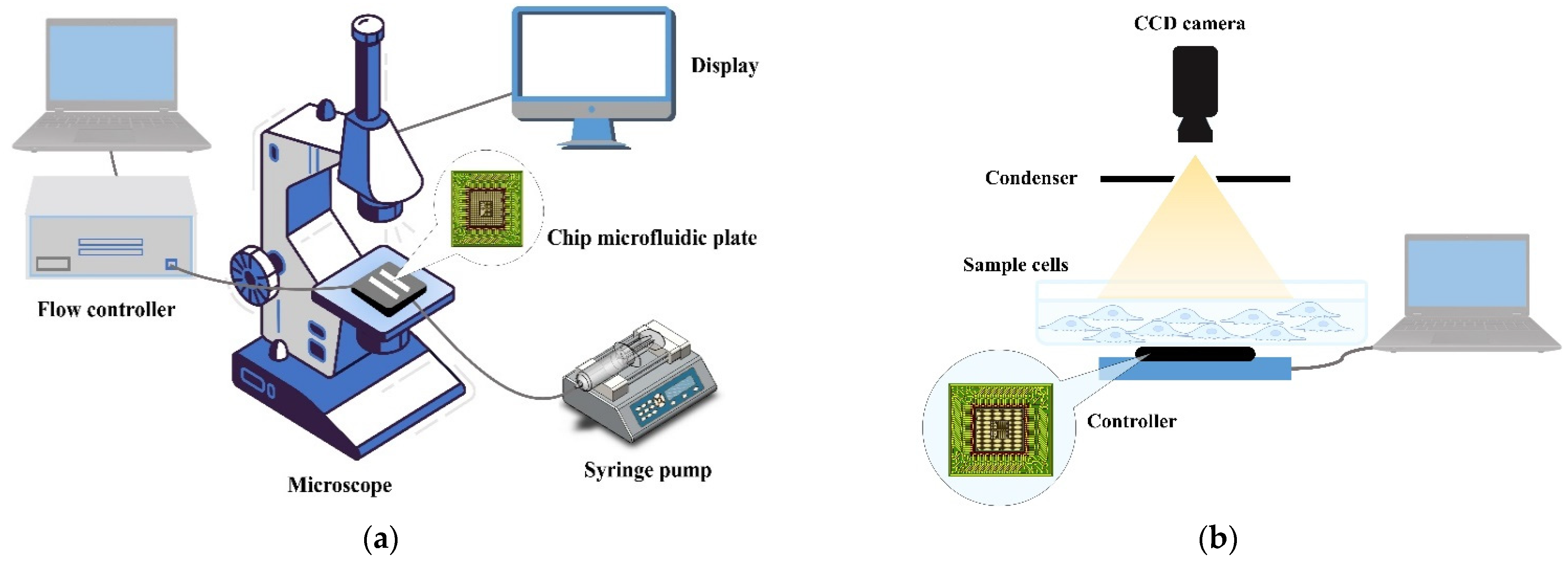
2.2.4. On-Chip, Lensless Video Microscopy Technology
2.3. Mechanical Measuring Methods
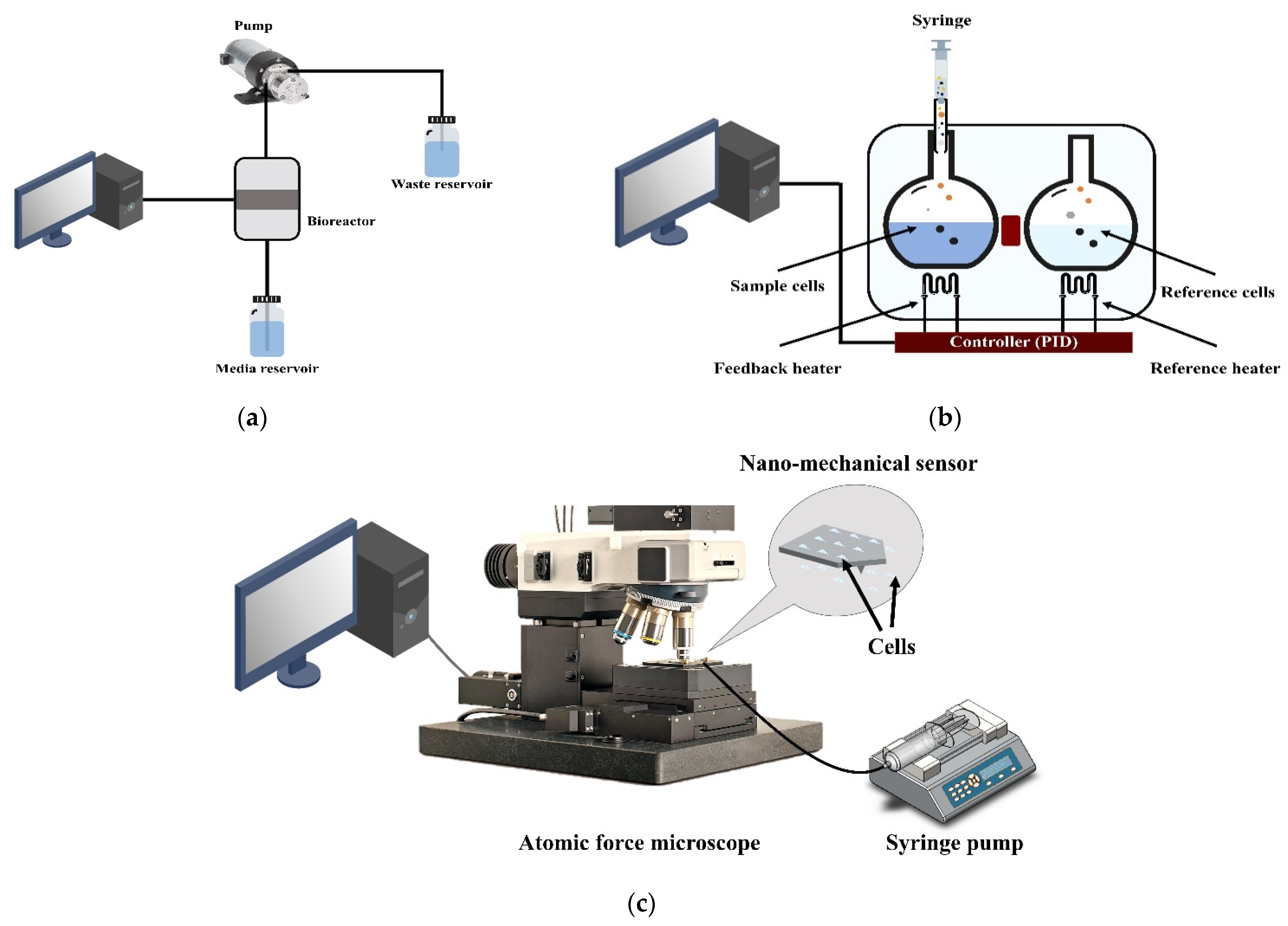
2.3.1. Respiratory Measuring Methods
2.3.2. Microcalorimeter Measurement Methods
2.3.3. Micro-Nanomechanical Oscillator Sensors
3. The AFM Oscillating Sensor Mode (Nanomotion)
3.1. Nanomotion Introduction
3.2. Nanomotion Application

| Attachment Protocol | Results Display | Application | Cell Type | Time | Agent | Cantilever Type | Cantilever Functionalization | Ref. |
|---|---|---|---|---|---|---|---|---|
| Inject sample medium inside AFM test room | Variance value | Antibiotic resistance | E. coli and S. aureus | 60–90 min | Ampicillin | DNP-10, Bruker | APTES (0.2%, 1.5 min) | [126] |
| Cantilever incubates in sample medium outside of the AFM test room | Variance value | Antibiotic resistance | E. Coli | 2 h | Ampicillin | DNP-10, Bruker | Glutaraldehyde (0.5%, 7 min) | [154] |
| Cantilever incubates in sample medium outside of the AFM test room | Variance value; power spectral density | Protein conformational changes | Ligands, such as ATP | <10 min | Topo II enzymes with Pbr322 DNA (200 nm) | DNP-10, Bruker | APTES (0.1%, 1 min) | [153] |
| Cantilever incubates in sample medium outside of the AFM test room and Micrometric motors of the AFM (AFM single-cell force spectroscopy) | Variance value | Life-searching experiments on Earth and interplanetary missions | E. coli | >190 min | Bactericidal dose (10 μg/mL) | DNP-10, Bruker | Glutaraldehyde (0.5%, 7 min) | [120] |
| S. aureus | >190 min | Bactericidal dose (2 μg/mL) | Glutaraldehyde (0.5%, 7 min) | |||||
| C. albicans | >190 min | Fungicidal dose (20 μg/mL) | Glutaraldehyde (0.5%, 7 min) | |||||
| MC3T3-E1 | >190 min | 5% glutaraldehyde | Fibronection (10 μg/mL, 15 min) | |||||
| M17 | >190 min | Salt concentration increasing | Poly-L-lysine (10%, 30 min) | |||||
| Cantilever incubates in sample medium outside of the AFM test room | Variance value | Cell viability | MCF7 | 7 h | Paclitaxel | DNP-10, Bruker | APTES (10%, 30 min) | [144] |
| Inject sample medium inside AFM test room | Damping value | Cell viability | Hela and MCF7 | 4–5 h | Au NPs | SNL-10, Bruker | - | [127] |
| Micrometric motors of the AFM (AFM single-cell force spectroscopy) | Variance value | Single-cell cytotoxicity assays | M17 | 7 h | Extracellular monomeric and amyloid α-synuclein species | DNP-10, Bruker | Poly-L-lysine (10%, 30 min) | [152] |
| Cantilever incubates in sample medium outside of the AFM test room | Variance value | Bloodstream infection | E. coli | 90 min | Ceftriaxone, ciprofloxacin and ampicillin | NP-O10, Bruker | Glutaraldehyde (0.5%, 7 min) | [149] |
| Cantilever incubates in sample medium outside of the AFM test room | Variance value | Mitochondrial activity detected | Mitochondria- embryonic kidney cells | 110 min | Malate, pyruvate, ADP, sodium azide, and rotenone | NP-O10, Bruker | Glutaraldehyde (5%, 10 min) | [145] |
| Inject sample medium inside AFM test room | Variance value | Sperm motility | Semen | - | Alcohol, spermagic | - | APTES (10%, 15 min) | [150] |
| Cantilever incubates in sample medium outside of the AFM test room | Variance value | Antibiotic resistance | B. pertussis | 100 min | Erythromycin (Sigma- E6376); clarithromycin (Sigma -A3487), trimthoprim-sulfamethoxazole | - | Glutaraldehyde (0.5%, 10 min) | [148] |
| Cantilever incubates in sample medium outside of the AFM test room | Variance value | Antibiotic resistance | Bacillus Calmette-Guérin (BCG) and M. abscessus | 200 min | BCG vs. Isoniazid and rifampicin M. abscessus vs. Amikacin | DNP-10, Bruker and SD-qp-CONT, NanoandMore | Glutaraldehyde (0.5%, 15 min) | [155] |
| The micrometric motors of the AFM (AFM single-cell force spectroscopy) | Variance value | Cell metabolic changes | HEK293 | 40 min | Frataxin overexpression | DNP-10, Bruker | Poly-D-lysine (20 μg/mL, 15 min) | [151] |
| Inject sample medium inside AFM test room | Variance value | Antibiotic resistance | E. coli | 120 min | Bacteriophage T7 | RC800PSA, Olympus | Poly-L-lysine (0.01%, 15 min) | [156] |
| Cantilever incubates in sample medium outside of the AFM test room | Variance value | Yeast resistance to antifungal drugs | C. albicans | >2 h | Fibronectin | Qp-CONT, nanoandmore | Con A (2 mg/mL, 30 min) | [157] |
| Cantilever incubates in sample medium outside of the AFM test room | Violin plots | Bacterial virulence | B. pertussis | 5 min | Mgso4 | SD-qp-CONT, nanoandmore | Poly-L-lysine (0.1%, 5 min) | [158] |
| Cantilever incubates in sample medium outside of the AFM test room | Variance value | Viability and susceptibility of microorganisms | E. coli and S. aureus | 4 h | Ampicillin, glutaraldehyde | SD-qp-CONT, nanoandmore | Glutaraldehyde (0.5%, 10 min) | [159] |
3.3. Attachment Protocol
3.4. Results Display
3.5. Challenges and Future Perspectives
4. Conclusions
Author Contributions
Funding
Institutional Review Board Statement
Informed Consent Statement
Acknowledgments
Conflicts of Interest
References
- Mahto, S.K.; Chandra, P.; Rhee, S.W. In vitro models, endpoints and assessment methods for the measurement of cytotoxicity. Toxicol. Environ. Health Sci. 2010, 2, 87–93. [Google Scholar] [CrossRef]
- Hu, C.; He, S.; Lee, Y.J.; He, Y.R.; Anastasio, M.; Popescu, G. Label-free cell viability assay using phase imaging with computational specificity (PICS). Quant. Phase Imaging VII 2021, 11653, 48. [Google Scholar] [CrossRef]
- Kroemer, G.; Galluzzi, L.; Vandenabeele, P.; Abrams, J.; Alnemri, E.S.; Baehrecke, E.H.; Blagosklonny, M.V.; El-Deiry, W.S.; Golstein, P.; Green, D.R.; et al. Classification of cell death: Recommendations of the Nomenclature Committee on Cell Death 2009. Cell Death Differ. 2009, 16, 3–11. [Google Scholar] [CrossRef] [PubMed]
- Single, A.; Beetham, H.; Telford, B.J.; Guilford, P.; Chen, A. A comparison of real-time and endpoint cell viability assays for improved synthetic lethal drug validation. J. Biomol. Screen. 2015, 20, 1286–1293. [Google Scholar] [CrossRef] [PubMed]
- Wei, M.; Zhang, R.; Zhang, F.; Zhang, Y.; Li, G.; Miao, R.; Shao, S. An Evaluation Approach of Cell Viability Based on Cell Detachment Assay in a Single-Channel Integrated Microfluidic Chip. ACS Sens. 2019, 4, 2654–2661. [Google Scholar] [CrossRef]
- Wei, M.; Zhang, R.; Zhang, F.; Zhang, Y. Evaluating cell viability heterogeneity based on information fusion of multiple adhesion strengths. Biotechnol. Bioeng. 2021, 118, 2360–2367. [Google Scholar] [CrossRef]
- Venturelli, L.; Kohler, A.C.; Stupar, P.; Villalba, M.I.; Kalauzi, A.; Radotic, K.; Bertacchi, M.; Dinarelli, S.; Girasole, M.; Pešić, M.; et al. A perspective view on the nanomotion detection of living organisms and its features. J. Mol. Recognit. 2020, 33, e2849. [Google Scholar] [CrossRef]
- Gilbert, D.F. Cell Viability Assays; Springer: New York, NY, USA, 2017. [Google Scholar]
- Kamiloglu, S.; Sari, G.; Ozdal, T.; Capanoglu, E. Guidelines for cell viability assays. Food Front. 2020, 1, 332–349. [Google Scholar] [CrossRef]
- Duellman, S.J.; Zhou, W.; Meisenheimer, P.; Vidugiris, G.; Cali, J.J.; Gautam, P.; Wennerberg, K.; Vidugiriene, J. Bioluminescent, Nonlytic, Real-Time Cell Viability Assay and Use in Inhibitor Screening. Assay Drug Dev. Technol. 2015, 13, 456–465. [Google Scholar] [CrossRef]
- Kerschbaum, H.H.; Tasa, B.A.; Schürz, M.; Oberascher, K.; Bresgen, N. Trypan blue—Adapting a dye used for labelling dead cells to visualize pinocytosis in viable cells. Cell. Physiol. Biochem. 2021, 55, 171–184. [Google Scholar] [CrossRef]
- Kim, S.I.; Kim, H.J.; Lee, H.J.; Lee, K.; Hong, D.; Lim, H.; Cho, K.; Jung, N.; Yi, Y.W. Application of a non-hazardous vital dye for cell counting with automated cell counters. Anal. Biochem. 2016, 492, 8–12. [Google Scholar] [CrossRef] [PubMed]
- Lippman, M.E. Comparison of dye exclusion assays with a clonogenic assay in the determination of drug-Induced cytotoxicity. Cancer Res. 1983, 43, 258–264. [Google Scholar]
- Tolnai, S. A method for viable cell count. Tissue Cult. Assoc. Man. 1975, 1, 37–38. [Google Scholar] [CrossRef]
- Dooley, M.P. The use of eosin B to assess the viability and developmental potential of rat embryos. Retrosp. Theses Diss. 1988, 8839, 1–256. [Google Scholar]
- Nakayama, Y.; Tsujinaka, T. Acceleration of robust ‘biotube’ vascular graft fabrication by in-body tissue architecture technology using a novel eosin Y-releasing mold. J. Biomed. Mater. Res. Part B Appl. Biomater. 2014, 102, 231–238. [Google Scholar] [CrossRef]
- Kay, A.B. Paul ehrlich and the early history of granulocytes. Myeloid Cells Health Dis. A Synth. 2017, 4, 3–15. [Google Scholar] [CrossRef]
- Kan, A.; Birnbaum, D.P.; Praveschotinunt, P.; Joshi, N.S. Congo red fluorescence for rapid in situ characterization of synthetic curli systems. Appl. Environ. Microbiol. 2019, 85, e00434-19. [Google Scholar] [CrossRef]
- Kuo, C.T.; Chen, Y.L.; Hsu, W.T.; How, S.C.; Cheng, Y.H.; Hsueh, S.S.; Liu, H.S.; Lin, T.H.; Wu, J.W.; Wang, S.S.S. Investigating the effects of erythrosine B on amyloid fibril formation derived from lysozyme. Int. J. Biol. Macromol. 2017, 98, 159–168. [Google Scholar] [CrossRef]
- Franke, J.D.; Braverman, A.L.; Cunningham, A.M.; Eberhard, E.E.; Perry, G.A. Erythrosin B: A versatile colorimetric and fluorescent vital dye for bacteria. Biotechnol. J. 2020, 68, 7–13. [Google Scholar] [CrossRef]
- Kumar, P.; Nagarajan, A.; Uchil, P.D. Analysis of cell viability by the lactate dehydrogenase assay. Cold Spring Harb. Protoc. 2018, 2018, 465–468. [Google Scholar] [CrossRef]
- Rotman, B.; Papermaster, B.W. Membrane properties of living mammalian cells as studied by enzymatic hydrolysis of fluorogenic esters. Proc. Natl. Acad. Sci. USA 1966, 55, 134–141. [Google Scholar] [CrossRef] [PubMed]
- Larson, E.M.; Doughman, D.J.; Gregerson, D.S.; Obritsch, W.F. A new, simple, nonradioactive, nontoxic in vitro assay to monitor corneal endothelial cell viability. Investig. Ophthalmol. Vis. Sci. 1997, 38, 1929–1933. [Google Scholar]
- Schirmer, K.; Chan, A.G.J.; Greenberg, B.M.; Dixon, D.G.; Bols, N.C. Methodology for demonstrating and measuring the photocytotoxicity of fluoranthene to fish cells in culture. Toxicol. In Vitro 1997, 11, 107–113. [Google Scholar] [CrossRef]
- Ganassin, R.C.; Bols, N.C. Growth of rainbow trout hemopoietic cells in methylcellulose and methods of monitoring their proliferative response in this matrix. Methods Cell Sci. 2000, 22, 147–152. [Google Scholar] [CrossRef] [PubMed]
- Jiajia, L.; Shinghung, M.; Jiacheng, Z.; Jialing, W.; Dilin, X.; Shengquan, H.; Zaijun, Z.; Qinwen, W.; Yifan, H.; Wei, C. Assessment of neuronal viability using fluorescein diacetate-propidium iodide double staining in cerebellar granule neuron culture. J. Vis. Exp. 2017, 2017, e55442. [Google Scholar] [CrossRef]
- Jones, K.H.; Senft, J.A. An improved method to determine cell viability by simultaneous staining with fluorescein diacetate-propidium iodide. J. Histochem. Cytochem. 1985, 33, 77–79. [Google Scholar] [CrossRef]
- Mecelroy, W.D. The energy source for bioluminescence in an isolated system. Zoology 1947, 33, 342–345. [Google Scholar] [CrossRef]
- Bajerski, F.; Stock, J.; Hanf, B.; Darienko, T.; Heine-Dobbernack, E.; Lorenz, M.; Naujox, L.; Keller, E.R.J.; Schumacher, H.M.; Friedl, T.; et al. ATP content and cell viability as indicators for cryostress across the diversity of life. Front. Physiol. 2018, 9, 921. [Google Scholar] [CrossRef]
- Kijanska, M.; Kelm, J. In vitro 3D Spheroids and Microtissues: ATP-based Cell Viability and Toxicity Assays. Assay Guid. Man. 2004, 1, 1–13. [Google Scholar]
- Smale, S.T. Luciferase assay. Cold Spring Harb. Protoc. 2010, 5, 2008–2011. [Google Scholar] [CrossRef]
- Nguyen, V.T.; Morange, M.; Bensaude, O. Firefly luciferase luminescence assays using scintillation counters for quantitation in transfected mammalian cells. Anal. Biochem. 1988, 171, 404–408. [Google Scholar] [CrossRef]
- de Wet, J.R.; Wood, K.V.; DeLuca, M.; Helinski, D.R.; Subramani, S. Firefly luciferase gene: Structure and expression in mammalian cells. Mol. Cell. Biol. 1987, 7, 725–737. [Google Scholar] [CrossRef] [PubMed]
- Zhou, W.; Valley, M.P.; Shultz, J.; Hawkins, E.M.; Bernad, L.; Good, T.; Good, D.; Riss, T.L.; Klaubert, D.H.; Wood, K.V. New bioluminogenic substrates for monoamine oxidase assays. J. Am. Chem. Soc. 2006, 128, 3122–3123. [Google Scholar] [CrossRef] [PubMed]
- van Engeland, M.; Ramaekers, F.C.S.; Schutte, B.; Reutelingsperger, C.P.M. A novel assay to measure loss of plasma membrane asymmetry during apoptosis of adherent cells in culture. Cytometry 1996, 24, 131–139. [Google Scholar] [CrossRef]
- Darzynkiewicz, Z.; Bruno, S.; Del Bino, G.; Gorczyca, W.; Hotz, M.A.; Lassota, P.; Traganos, F. Features of apoptotic cells measured by flow cytometry. Cytometry 1992, 13, 795–808. [Google Scholar] [CrossRef] [PubMed]
- da Silveira, M.G.; Romão, M.V.S.; Loureiro-Dias, M.C.; Rombouts, F.M.; Abee, T. Flow cytometric assessment of membrane integrity of ethanol-stressed Oenococcus oeni cells. Appl. Environ. Microbiol. 2002, 68, 6087–6093. [Google Scholar] [CrossRef]
- Gillissen, M.A.; Yasuda, E.; De Jong, G.; Levie, S.E.; Go, D.; Spits, H.; van Helden, P.M.; Hazenberg, M.D. The modified FACS calcein AM retention assay: A high throughput flow cytometer based method to measure cytotoxicity. J. Immunol. Methods 2016, 434, 16–23. [Google Scholar] [CrossRef]
- Davey, H.; Guyot, S. Estimation of Microbial Viability Using Flow Cytometry. Curr. Protoc. Cytom. 2020, 93, e72. [Google Scholar] [CrossRef]
- Sedlackova, L.; Korolchuk, V.I. Mitochondrial quality control as a key determinant of cell survival. Biochim. Biophys. Acta Mol. Cell Res. 2019, 1866, 575–587. [Google Scholar] [CrossRef]
- Cole, S.P.C. Rapid chemosensitivity testing of human lung tumor cells using the MTT assay. Cancer Chemother. Pharmacol. 1986, 17, 259–263. [Google Scholar] [CrossRef]
- Goodwin, C.J.; Holt, S.J.; Downes, S.; Marshall, N.J. Microculture tetrazolium assays: A comparison between two new tetrazolium salts, XTT and MTS. J. Immunol. Methods 1995, 179, 95–103. [Google Scholar] [CrossRef]
- Kazaks, A.; Collier, M.; Conley, M. Cytotoxicity of Caffeine on MCF-7 Cells Measured by XTT Cell Proliferation Assay (P06-038-19). Curr. Dev. Nutr. 2019, 3, 548. [Google Scholar] [CrossRef]
- Scudiero, D.A.; Shoemaker, R.H.; Paull, K.D.; Monks, A.; Tierney, S.; Nofziger, T.H.; Currens, M.J.; Seniff, D.; Boyd, M.R. Evaluation of a Soluble Tetrazolium/Formazan Assay for Cell Growth and Drug Sensitivity in Culture Using Human and Other Tumor Cell Lines. Cancer Res. 1988, 48, 4827–4833. [Google Scholar] [PubMed]
- Scarcello, E.; Lambremont, A.; Vanbever, R.; Jacques, P.J.; Lison, D. Mind your assays: Misleading cytotoxicity with the WST-1 assay in the presence of manganese. PLoS ONE 2020, 15, e0231634. [Google Scholar] [CrossRef] [PubMed]
- Tominaga, H.; Ishiyama, M.; Ohseto, F.; Sasamoto, K.; Hamamoto, T.; Suzuki, K.; Watanabe, M. A water-soluble tetrazolium salt useful for colorimetric cell viability assay. Anal. Commun. 1999, 36, 47–50. [Google Scholar] [CrossRef]
- Seifabadi, Z.S.; Rezaei-Tazangi, F.; Azarbarz, N.; Nejad, D.B.; Mohammadiasl, J.; Darabi, H.; Pezhmanlarki-Tork, S. Assessment of viability of wharton’s jelly mesenchymal stem cells encapsulated in alginate scaffold by WST-8 assay kit. Med. J. Cell Biol. 2021, 9, 42–47. [Google Scholar] [CrossRef]
- Skehan, P.; Storeng, R.; Scudiero, D.; Monks, A.; McMahon, J.; Vistica, D.; Warren, J.T.; Bokesch, H.; Kenney, S.; Boyd, M.R. New colorimetric cytotoxicity assay for anticancer-drug screening. J. Natl. Cancer Inst. 1990, 82, 1107–1112. [Google Scholar] [CrossRef]
- Vajrabhaya, L.o.; Korsuwannawong, S. Cytotoxicity evaluation of a Thai herb using tetrazolium (MTT) and sulforhodamine B (SRB) assays. J. Anal. Sci. Technol. 2018, 9, 1–6. [Google Scholar] [CrossRef]
- Ates, G.; Vanhaecke, T.; Rogiers, V.; Rodrigues, R.M. Assaying cellular viability using the neutral red uptake assay. Methods Mol. Biol. 2017, 1601, 19–26. [Google Scholar] [CrossRef]
- Borenfreund, E.; Puerner, J.A. A simple quantitative procedure using monolayer cultures for cytotoxicity assays (HTD/NR-90). J. Tissue Cult. Methods 1985, 9, 7–9. [Google Scholar] [CrossRef]
- Saotome, K.; Morita, H.; Umeda, M. Cytotoxicity test with simplified crystal violet staining method using microtitre plates and its application to injection drugs. Toxicol. In Vitro 1989, 3, 317–321. [Google Scholar] [CrossRef]
- Puck, T.T. Quantitaive Studies on Mammalian Cells in Vitro. Rev. Moderen Phys. 1993, 46, 177–188. [Google Scholar]
- Pegg, D.E. Viability assays for preserved cells, tissues, and organs. Cryobiology 1989, 26, 212–231. [Google Scholar] [CrossRef]
- Galindo, C.C.; Lozano, D.M.V.; Rodríguez, B.C.; Perdomo-Arciniegas, A.M. Improved cord blood thawing procedure enhances the reproducibility and correlation between flow cytometry CD34+ cell viability and clonogenicity assays. Cytotherapy 2018, 20, 891–894. [Google Scholar] [CrossRef]
- Decker, T.; Lohmann-Matthes, M.L. A quick and simple method for the quantitation of lactate dehydrogenase release in measurements of cellular cytotoxicity and tumor necrosis factor (TNF) activity. J. Immunol. Methods 1988, 115, 61–69. [Google Scholar] [CrossRef]
- Chan, F.K.M.; Moriwaki, K.; de Rosa, M.J. Detection of necrosis by release of lactate dehydrogenase activity. Methods Mol. Biol. 2013, 979, 65–70. [Google Scholar] [CrossRef]
- Ahmad, T.; Aggarwal, K.; Pattnaik, B.; Mukherjee, S.; Sethi, T.; Tiwari, B.K.; Kumar, M.; Micheal, A.; Mabalirajan, U.; Ghosh, B.; et al. Computational classification of mitochondrial shapes reflects stress and redox state. Cell Death Dis. 2013, 4, e461. [Google Scholar] [CrossRef]
- Karbowski, M.; Youle, R.J. Dynamics of mitochondrial morphology in healthy cells and during apoptosis. Cell Death Differ. 2003, 10, 870–880. [Google Scholar] [CrossRef]
- Arnoult, D. Mitochondrial fragmentation in apoptosis. Trends Cell Biol. 2006, 17, 6–12. [Google Scholar] [CrossRef]
- Liu, X.; Hajnoczky, G. Altered fusion dynamics underlie unique morphological changes in mitochondria during hypoxia—Reoxygenation stress. Cell Death Differ. 2011, 18, 1561–1572. [Google Scholar] [CrossRef] [PubMed]
- Mondol, A.S.; Töpfer, N.; Rüger, J.; Neugebauer, U.; Popp, J.; Schie, I.W. New perspectives for viability studies with high-content analysis Raman spectroscopy (HCA-RS). Sci. Rep. 2019, 9, 1–12. [Google Scholar] [CrossRef] [PubMed]
- Wang, J.; Lin, K.; Hu, H.; Qie, X.; Huang, W.E.; Cui, Z.; Gong, Y.; Song, Y. In vitro anticancer drug sensitivity sensing through single-cell raman spectroscopy. Biosensors 2021, 11, 286. [Google Scholar] [CrossRef] [PubMed]
- Wen, X.; Ou, Y.C.; Bogatcheva, G.; Thomas, G.; Mahadevan-Jansen, A.; Singh, B.; Lin, E.C.; Bardhan, R. Probing metabolic alterations in breast cancer in response to molecular inhibitors with Raman spectroscopy and validated with mass spectrometry. Chem. Sci. 2020, 11, 9863–9874. [Google Scholar] [CrossRef] [PubMed]
- Botelho, C.M.; Gonçalves, O.; Marques, R.; Thiagarajan, V.; Vorum, H.; Gomes, A.C.; Neves-Petersen, M.T. Photonic modulation of epidermal growth factor receptor halts receptor activation and cancer cell migration. J. Biophotonics 2018, 11, e201700323. [Google Scholar] [CrossRef]
- Czamara, K.; Petko, F.; Baranska, M.; Kaczor, A. Raman microscopy at the subcellular level: Study on early apoptosis in endothelial cells induced by Fas ligand and cycloheximide. Analyst 2016, 141, 1390–1397. [Google Scholar] [CrossRef]
- Abramczyk, H. Double face of cytochrome c in cancers by Raman imaging. Sci. Rep. 2022, 12, 1–11. [Google Scholar] [CrossRef]
- Pansare, K.; Singh, S.R.; Chakravarthy, V.; Gupta, N.; Hole, A.; Gera, P.; Sarin, R.; Krishna, C.M. Raman Spectroscopy: An Exploratory Study to Identify Post Radiation Cell Survival. Appl Spectrosc 2020, 2, 553–562. [Google Scholar] [CrossRef]
- Schie, I.W.; Rüger, J.; Mondol, A.S.; Ramoji, A.; Neugebauer, U.; Krafft, C.; Popp, J. High-Throughput Screening Raman Spectroscopy Platform for Label-Free Cellomics. Anal. Chem. 2018, 90, 2023–2030. [Google Scholar] [CrossRef]
- Jayan, H.; Pu, H.; Sun, D. Recent developments in Raman spectral analysis of microbial single cells: Techniques and applications. Crit. Rev. Food Sci. Nutr. 2021, 62, 4294–4308. [Google Scholar] [CrossRef]
- Goldrick, S.; Umprecht, A.; Tang, A.; Zakrzewski, R.; Cheeks, M.; Turner, R.; Charles, A.; Les, K.; Hulley, M.; Spencer, C.; et al. High-throughput raman spectroscopy combined with innovate data analysis workflow to enhance biopharmaceutical process development. Processes 2020, 8, 1179. [Google Scholar] [CrossRef]
- Verrier, S.; Zoladek, A.; Notingher, I. Raman Micro-Spectroscopy as a Non-invasive Cell Viability Test. In Mammalian Cell Viability. Methods in Molecular Biology (Methods and Protocols); Stoddart, M., Ed.; Humana Press, Springer: New York, NY, USA, 2011; Volume 740, pp. 179–189. [Google Scholar] [CrossRef]
- Grabarek, A.D.; Senel, E.; Menzen, T.; Hoogendoorn, K.H.; Pike-Overzet, K.; Hawe, A.; Jiskoot, W. Particulate impurities in cell-based medicinal products traced by flow imaging microscopy combined with deep learning for image analysis. Cytotherapy 2021, 23, 339–347. [Google Scholar] [CrossRef] [PubMed]
- Farrell, C.J.; Cicalese, S.M.; Davis, H.B.; Dogdas, B.; Shah, T.; Culp, T.; Hoang, V.M. Cell confluency analysis on microcarriers by micro-flow imaging. Cytotechnology 2016, 68, 2469–2478. [Google Scholar] [CrossRef]
- Sediq, A.S.; Klem, R.; Nejadnik, M.R.; Meij, P.; Jiskoot, W. Label-Free, Flow-Imaging Methods for Determination of Cell Concentration and Viability. Pharm. Res. 2018, 35, 1–10. [Google Scholar] [CrossRef] [PubMed]
- Wu, L.; Martin, T.; Li, Y.; Yang, L.; Halpenny, M.; Giulivi, A.; Allan, D.S. Cell aggregation in thawed haematopoietic stem cell products visualised using micro-flow imaging. Transfus. Med. 2012, 22, 218–220. [Google Scholar] [CrossRef] [PubMed]
- Grabarek, A.D.; Jiskoot, W.; Hawe, A.; Pike-overzet, K.; Menzen, T. Forced degradation of cell-based medicinal products guided by flow imaging microscopy: Explorative studies with Jurkat cells. Eur. J. Pharm. Biopharm. 2021, 167, 38–47. [Google Scholar] [CrossRef] [PubMed]
- Gambe-gilbuena, A.; Shibano, Y.; Krayukhina, E.; Torisu, T.; Uchiyama, S. Automatic Identi fi cation of the Stress Sources of Protein Aggregates Using Flow Imaging Microscopy Images. J. Pharm. Sci. 2020, 109, 614–623. [Google Scholar] [CrossRef]
- Kühn, J. Digital holographic microscopy real-time monitoring of cytoarchitectural alterations during simulated microgravity. J. Biomed. Opt. 2010, 15, 026021. [Google Scholar] [CrossRef]
- Pais, D.A.M.; Galrão, P.R.S.; Kryzhanska, A.; Barbau, J.; Isidro, I.A.; Alves, P.M. Holographic imaging of insect cell cultures: Online non-invasive monitoring of adeno-associated virus production and cell concentration. Processes 2020, 8, 487. [Google Scholar] [CrossRef]
- Kemper, B.; Carl, D.D.; Schnekenburger, J.; Bredebusch, I.; Schäfer, M.; Domschke, W.; von Bally, G. Investigation of living pancreas tumor cells by digital holographic microscopy. J. Biomed. Opt. 2006, 11, 034005. [Google Scholar] [CrossRef]
- Odete, M.A.; Philips, L. Label-free Viability Assay using Holographic Video Microscopy Label-free Viability Assay using Holographic Video Microscopy. Res. Sq. preprint. 2021. [Google Scholar] [CrossRef]
- Pala, M.A.; Çimen, M.E.; Akgül, A.; Yıldız, M.Z.; Boz, A.F. Fractal dimension-based viability analysis of cancer cell lines in lens-free holographic microscopy via machine. Eur. Phys. J. 2021, 123, 1–12. [Google Scholar] [CrossRef]
- Dubois, F.; Yourassowsky, C.; Monnom, O.; Legros, J.C.; Debeir IV, O.; Van Ham, P.; Kiss, R.; Decaestecker, C. Digital holographic microscopy for the three-dimensional dynamic analysis of in vitro cancer cell migration. J. Biomed. Opt. 2006, 11, 054032. [Google Scholar] [CrossRef]
- Moon, I.; Daneshpanah, M.; Javidi, B.; Stern, A. Automated three-dimensional identification and tracking of micro/nanobiological organisms by computational holographic microscopy. Proc. IEEE 2009, 97, 990–1010. [Google Scholar] [CrossRef]
- Pushkarsky, I.; Liu, Y.; Weaver, W.; Su, T.W.; Mudanyali, O.; Ozcan, A.; Di Carlo, D. Automated single-cell motility analysis on a chip using lensfree microscopy. Sci. Rep. 2014, 4, 1–9. [Google Scholar] [CrossRef]
- Jin, G.; Yoo, I.; Pil, S.; Yang, J.; Ha, U. Biosensors and Bioelectronics Lens-free shadow image based high-throughput continuous cell monitoring technique. Biosens. Bioelectron. 2012, 38, 126–131. [Google Scholar] [CrossRef] [PubMed]
- Kim, S.B.; Bae, H.; Cha, J.M.; Moon, S.J.; Dokmeci, M.R.; Cropek, D.M.; Khademhosseini, A. A cell-based biosensor for real-time detection of cardiotoxicity using lensfree imaging. Lab Chip 2011, 11, 1801–1807. [Google Scholar] [CrossRef] [PubMed]
- Zheng, G.; Lee, S.A.; Yang, S.; Yang, C. Sub-pixel resolving optofluidic microscope for on-chip cell imaging. Lab Chip 2010, 10, 3125–3129. [Google Scholar] [CrossRef]
- Cui, X.; Lee, L.M.; Heng, X.; Zhong, W.; Sternberg, P.W.; Psaltis, D.; Yang, C. Lensless high-resolution on-chip optofluidic microscopes for Caenorhabditis elegans and cell imaging. Proc. Natl. Acad. Sci. USA 2008, 105, 10670–10675. [Google Scholar] [CrossRef]
- Ozcan, A.; Demirci, U. Ultra wide-field lens-free monitoring of cells on-chip. Lab Chip 2007, 8, 98–106. [Google Scholar] [CrossRef]
- Kesavan, S.V.; Momey, F.; Cioni, O.; David-Watine, B.; Dubrulle, N.; Shorte, S.; Sulpice, E.; Freida, D.; Chalmond, B.; Dinten, J.M.; et al. High-throughput monitoring of major cell functions by means of lensfree video microscopy. Sci. Rep. 2014, 4, 1–11. [Google Scholar] [CrossRef]
- Nablo, B.J.; Ahn, J.J.; Bhadriraju, K.; Lee, J.M.; Reyes, D.R. Lens-Free Imaging as a Sensor for Dynamic Cell Viability Detection Using the Neutral Red Uptake Assay. ACS Appl. Bio Mater. 2020, 3, 6633–6638. [Google Scholar] [CrossRef] [PubMed]
- Huang, X.; Li, Y.; Xu, X.; Wang, R.; Yao, J.; Han, W.; Wei, M.; Chen, J.; Xuan, W.; Sun, L. High-precision lensless microscope on a chip based on in-line holographic imaging. Sensors 2021, 21, 720. [Google Scholar] [CrossRef] [PubMed]
- Rothbauer, M.; Ertl, P.; Mayr, T. Measurement of respiration and acidification rates of mammalian cells in thermoplastic microfluidic devices. Sens. Actuators B Chem. 2021, 334, 129664. [Google Scholar] [CrossRef]
- O’Riordan, T.C.; Buckley, D.; Ogurtsov, V.; O’Connor, R.; Papkovsky, D.B. A cell viability assay based on monitoring respiration by optical oxygen sensing. Anal. Biochem. 2000, 278, 221–227. [Google Scholar] [CrossRef]
- Bäckman, P.; Wadsö, I. Cell growth experiments using a microcalorimetric vessel equipped with oxygen and pH electrodes. J. Biochem. Biophys. Methods 1991, 23, 283–293. [Google Scholar] [CrossRef]
- Halpern, H.J.; Yu, C.; Peric, M.; Barth, E.D.; Karczmar, G.S.; River, J.N.; Grdina, D.J.; Teicher, B.A. Measurement of differences in pO2 in response to perfluorocarbon/carbogen in FSa and NFSa murine fibrosarcomas with low-frequency electron paramagnetic resonance oximetry. Radiat. Res. 1996, 145, 610–618. [Google Scholar] [CrossRef]
- Braissant, O.; Astasov-frauenhoffer, M.; Waltimo, T. A Review of Methods to Determine Viability, Vitality, and Metabolic Rates in Microbiology. Front. Microbiol. 2020, 11, 547458. [Google Scholar] [CrossRef]
- Randers-Eichhorn, L.; Bartlett, R.A.; Frey, D.D.; Rao, G. Noninvasive oxygen measurements and mass transfer considerations in tissue culture flasks. Biotechnol. Bioeng. 1996, 51, 466–478. [Google Scholar] [CrossRef]
- Wodnicka, M.; Guarino, R.D.; Hemperly, J.J.; Timmins, M.R.; Stitt, D.; Pitner, J.B. Novel fluorescent technology platform for high throughput cytotoxicity and proliferation assays. J. Biomol. Screen. 2000, 5, 141–150. [Google Scholar] [CrossRef]
- Guarino, R.D.; Dike, L.E.; Haq, T.A.; Rowley, J.A.; Pitner, J.B.; Timmins, M.R. Method for determining oxygen consumption rates of static cultures from microplate measurements of pericellular dissolved oxygen concentration. Biotechnol. Bioeng. 2004, 86, 775–787. [Google Scholar] [CrossRef]
- Mishra, A.; Starly, B. Real time in vitro measurement of oxygen uptake rates for HEPG2 liver cells encapsulated in alginate matrices. Microfluid. Nanofluidics 2009, 6, 373–381. [Google Scholar] [CrossRef]
- Super, A.; Jaccard, N.; Marques, M.P.C.; Macown, R.J.; Griffin, L.D.; Veraitch, F.S.; Szita, N. Real-time monitoring of specific oxygen uptake rates of embryonic stem cells in a microfluidic cell culture device. Biotechnol. J. 2016, 11, 1179–1189. [Google Scholar] [CrossRef]
- Mahfouzi, S.H.; Amoabediny, G.; Doryab, A.; Safiabadi-Tali, S.H.; Ghanei, M. Noninvasive Real-Time Assessment of Cell Viability in a Three-Dimensional Tissue. Tissue Eng. Part C Methods 2018, 24, 197–204. [Google Scholar] [CrossRef] [PubMed]
- Xue, Y.; Lei, J.; Xu, X.; Ding, L.; Zhai, C.; Yan, F.; Ju, H. Real-time monitoring of cell viability by its nanoscale height change with oxygen as endogenous indicator. Chem. Commun. 2010, 46, 7388–7390. [Google Scholar] [CrossRef] [PubMed]
- Wadsö, I. Microcalorimetric techniques for characterization of living cellular systems. Will there be any important practical applications? Thermochim. Acta 1995, 269–270, 337–350. [Google Scholar] [CrossRef]
- Braissant, O.; Wirz, D.; Göpfert, B.; Daniels, A.U. Use of isothermal microcalorimetry to monitor microbial activities. FEMS Microbiol. Lett. 2010, 303, 1–8. [Google Scholar] [CrossRef]
- Yang, N.; Shi, Q.; Zhu, X.; Wei, M.; Ullah, I.; Kwabena, P.O.; Kulik, E.; Mao, H.; Zhang, R. A Cell Viability Evaluation Method Based on Respiratory Thermodynamic Feature Detected by Microscopic Infrared Thermal Imaging Sensor. IEEE Sens. J. 2020, 20, 637–647. [Google Scholar] [CrossRef]
- Tan, A.M.; Lu, J.H. Microcalorimetric study of antiviral effect of drug. J. Biochem. Biophys. Methods 1999, 38, 225–228. [Google Scholar] [CrossRef]
- Spaepen, P.; de Boodt, S.; Aerts, J.; Sloten, J.V. Chapter 21 Digital Image Processing of Live/Dead Staining. Mamm. Cell Viability Methods Protoc. Methods Mol. Biol. 2011, 740, 209–230. [Google Scholar] [CrossRef]
- Lemos, D.; Oliveira, T.; Martins, L.; De Azevedo, V.R.; Rodrigues, M.F.; Ketzer, L.A.; Rumjanek, F.D. Isothermal Microcalorimetry of Tumor Cells: Enhanced Thermogenesis by Metastatic Cells. Front. Oncol. 2019, 9, 1430. [Google Scholar] [CrossRef]
- Wang, F.; Han, Y.; Gu, N. Cell Temperature Measurement for Biometabolism Monitoring. ACS Sens. 2021, 6, 290–302. [Google Scholar] [CrossRef] [PubMed]
- Wang, Y.; Zhu, H.; Feng, J.; Neuzil, P. Recent advances of microcalorimetry for studying cellular metabolic heat. Trends Anal. Chem. 2021, 143, 116353. [Google Scholar] [CrossRef]
- Ilic, B.; Czaplewski, D.; Craighead, H.G.; Neuzil, P.; Campagnolo, C.; Batt, C. Mechanical resonant immunospecific biological detector. Appl. Phys. Lett. 2000, 77, 450–452. [Google Scholar] [CrossRef]
- Zheng, G.; Patolsky, F.; Cui, Y.; Wang, W.U.; Lieber, C.M. Multiplexed electrical detection of cancer markers with nanowire sensor arrays. Nat. Biotechnol. 2005, 23, 1294–1301. [Google Scholar] [CrossRef]
- Ramos, D.; Tamayo, J.; Mertens, J.; Calleja, M.; Villanueva, L.G.; Zaballos, A. Detection of bacteria based on the thermomechanical noise of a nanomechanical resonator: Origin of the response and detection limits. Nanotechnology 2008, 19, 035503. [Google Scholar] [CrossRef]
- Ahmad, M.R.; Nakajima, M.; Kojima, M.; Kojima, S.; Homma, M.; Fukuda, T. Instantaneous and quantitative single cells viability determination using dual nanoprobe inside ESEM. IEEE Trans. Nanotechnol. 2012, 11, 298–306. [Google Scholar] [CrossRef]
- Shen, Y.; Nakajima, M.; Kojima, S.; Homma, M.; Kojima, M.; Fukuda, T. Single cell adhesion force measurement for cell viability identification using an AFM cantilever-based micro putter. Meas. Sci. Technol. 2011, 22, 944–947. [Google Scholar] [CrossRef]
- Kasas, S.; Ruggeri, F.S.; Benadiba, C.; Maillard, C.; Stupar, P.; Tournu, H.; Dietler, G.; Longo, G. Detecting nanoscale vibrations as signature of life. Proc. Natl. Acad. Sci. USA 2015, 112, 378–381. [Google Scholar] [CrossRef]
- Mader, A.; Gruber, K.; Castelli, R.; Hermann, B.A.; Seeberger, P.H.; Rädler, J.O.; Leisner, M. Discrimination of Escherichia coli strains using glycan cantilever array sensors. Nano Lett. 2012, 12, 420–423. [Google Scholar] [CrossRef]
- Sharma, H.; Mutharasan, R. Rapid and sensitive immunodetection of Listeria monocytogenes in milk using a novel piezoelectric cantilever sensor. Biosens. Bioelectron. 2013, 45, 158–162. [Google Scholar] [CrossRef]
- Ndieyira, J.W.; Kappeler, N.; Logan, S.; Cooper, M.A.; Abell, C.; McKendry, R.A.; Aeppli, G. Surface-stress sensors for rapid and ultrasensitive detection of active free drugs in human serum. Nat. Nanotechnol. 2014, 9, 225–232. [Google Scholar] [CrossRef] [PubMed]
- Maciaszek, J.L.; Andemariam, B.; Abiraman, K.; Lykotrafitis, G. AKAP-dependent modulation of BCAM/Lu adhesion on normal and sickle cell disease RBCs revealed by force nanoscopy. Biophys. J. 2014, 106, 1258–1267. [Google Scholar] [CrossRef]
- Liu, Y.; Schweizer, L.M.; Wang, W.; Reuben, R.L.; Schweizer, M.; Shu, W. Chemical Label-free and real-time monitoring of yeast cell growth by the bending of polymer microcantilever biosensors. Sens. Actuators B. Chem. 2013, 178, 621–626. [Google Scholar] [CrossRef]
- Longo, G.; Alonso-Sarduy, L.; Rio, L.M.; Bizzini, A.; Trampuz, A.; Notz, J.; Dietler, G.; Kasas, S. Rapid detection of bacterial resistance to antibiotics using AFM cantilevers as nanomechanical sensors. Nat. Nanotechnol. 2013, 8, 522–526. [Google Scholar] [CrossRef] [PubMed]
- Yang, F.; Riedel, R.; Del Pino, P.; Pelaz, B.; Said, A.H.; Soliman, M.; Pinnapireddy, S.R.; Feliu, N.; Parak, W.J.; Bakowsky, U.; et al. Real-time, label-free monitoring of cell viability based on cell adhesion measurements with an atomic force microscope. J. Nanobiotechnol. 2017, 15, 1–10. [Google Scholar] [CrossRef] [PubMed]
- Bennett, I.; Pyne, A.L.B.; McKendry, R.A. Cantilever Sensors for Rapid Optical Antimicrobial Sensitivity Testing. ACS Sensors 2020, 5, 3133–3139. [Google Scholar] [CrossRef]
- Linna, E.; BinAhmed, S.; Stottrup, B.L.; Castrill, S.R.V. Effect of Graphene Oxide Packing on Bacterial Adhesion using Single Cell Force Spectroscopy. Biophys. J. 2018, 114, 352a–353a. [Google Scholar] [CrossRef]
- Evans, E.A.; Calderwood, D.A. Forces and bond dynamics in cell adhesion. Science 2007, 316, 1148–1153. [Google Scholar] [CrossRef]
- Huang, H.; Dai, C.; Shen, H.; Gu, M.; Wang, Y.; Liu, J.; Chen, L.; Sun, L. Recent advances on the model, measurement technique, and application of single cell mechanics. Int. J. Mol. Sci. 2020, 21, 6248. [Google Scholar] [CrossRef]
- Müller, D.J.; Dufrêne, Y.F. Atomic force microscopy: A nanoscopic window on the cell surface. Trends Cell Biol. 2011, 21, 461–469. [Google Scholar] [CrossRef]
- Ungai-Salánki, R.; Peter, B.; Gerecsei, T.; Orgovan, N.; Horvath, R.; Szabó, B. A practical review on the measurement tools for cellular adhesion force. Adv. Colloid Interface Sci. 2019, 269, 309–333. [Google Scholar] [CrossRef] [PubMed]
- Stewart, M.P.; Hodel, A.W.; Spielhofer, A.; Cattin, C.J.; Müller, D.J.; Helenius, J. Wedged AFM-cantilevers for parallel plate cell mechanics. Methods 2013, 60, 186–194. [Google Scholar] [CrossRef] [PubMed]
- Mathur, A.B.; Collinsworth, A.M.; Reichert, W.M.; Kraus, W.E.; Truskey, G.A. Endothelial, cardiac muscle and skeletal muscle exhibit different viscous and elastic properties as determined by atomic force microscopy. J. Biomech. 2001, 34, 1545–1553. [Google Scholar] [CrossRef]
- Yang, S.P.; Yang, C.Y.; Lee, T.M.; Lui, T.S. Effects of calcium-phosphate topography on osteoblast mechanobiology determined using a cytodetacher. Mater. Sci. Eng. C 2012, 32, 254–262. [Google Scholar] [CrossRef]
- Sagvolden, G.; Giaever, I.; Pettersen, E.O.; Feder, J. Cell adhesion force microscopy. Proc. Natl. Acad. Sci. USA 1999, 96, 471–476. [Google Scholar] [CrossRef]
- Yamamoto, A.; Mishima, S.; Maruyama, N.; Sumita, M. Quantitative evaluation of cell attachment to glass, polystyrene, and fibronectin- or collagen-coated polystyrene by measurement of cell adhesive shear force and cell detachment energy. J. Biomed. Mater. Res. 2000, 50, 114–124. [Google Scholar] [CrossRef]
- Lee, C.C.; Wu, C.C.; Su, F.C. The Technique for Measurement of Cell Adhesion Force. J. Med. Biol. Eng. 2004, 24, 51–56. [Google Scholar]
- Marcotte, L.; Tabrizian, M. Sensing surfaces: Challenges in studying the cell adhesion process and the cell adhesion forces on biomaterials. Itbm-Rbm 2008, 29, 77–88. [Google Scholar] [CrossRef]
- Elbourne, A.; Chapman, J.; Gelmi, A.; Cozzolino, D.; Crawford, R.J.; Truong, V.K. Bacterial-nanostructure interactions: The role of cell elasticity and adhesion forces. J. Colloid Interface Sci. 2019, 546, 192–210. [Google Scholar] [CrossRef]
- Friedrichs, J.; Legate, K.R.; Schubert, R.; Bharadwaj, M.; Werner, C.; Müller, D.J.; Benoit, M. A practical guide to quantify cell adhesion using single-cell force spectroscopy. Methods 2013, 60, 169–178. [Google Scholar] [CrossRef]
- Ramos, D.; Tamayo, J.; Mertens, J.; Calleja, M.; Zaballos, A. Origin of the response of nanomechanical resonators to bacteria adsorption. J. Appl. Phys. 2006, 100, 106105. [Google Scholar] [CrossRef]
- Wu, S.; Liu, X.; Zhou, X.; Liang, X.M.; Gao, D.; Liu, H.; Zhao, G.; Zhang, Q.; Wu, X. Quantification of cell viability and rapid screening anti-cancer drug utilizing nanomechanical fluctuation. Biosens. Bioelectron. 2016, 77, 164–173. [Google Scholar] [CrossRef] [PubMed]
- Stupar, P.; Chomicki, W.; Maillard, C.; Mikeladze, D.; Kalauzi, A.; Radotić, K.; Dietler, G.; Kasas, S. Mitochondrial activity detected by cantilever based sensor. Mech. Sci. 2017, 8, 23–28. [Google Scholar] [CrossRef]
- Kohler, A.C.; Venturelli, L.; Longo, G.; Dietler, G.; Kasas, S. Nanomotion detection based on atomic force microscopy cantilevers. Cell Surf. 2019, 5, 100021. [Google Scholar] [CrossRef] [PubMed]
- Lissandrello, C.; Inci, F.; Francom, M.; Paul, M.R.; Demirci, U.; Ekinci, K.L. Nanomechanical motion of Escherichia coli adhered to a surface. Appl. Phys. Lett. 2014, 105, 113701. [Google Scholar] [CrossRef]
- Villalba, M.I.; Stupar, P.; Chomicki, W.; Bertacchi, M.; Dietler, G.; Arnal, L.; Vela, M.E.; Yantorno, O.; Kasas, S. Nanomotion Detection Method for Testing Antibiotic Resistance and Susceptibility of Slow-Growing Bacteria. Small 2018, 14, 1702671. [Google Scholar] [CrossRef]
- Stupar, P.; Opota, O.; Longo, G.; Prod’Hom, G.; Dietler, G.; Greub, G.; Kasas, S. Nanomechanical sensor applied to blood culture pellets: A fast approach to determine the antibiotic susceptibility against agents of bloodstream infections. Clin. Microbiol. Infect. 2017, 23, 400–405. [Google Scholar] [CrossRef]
- Wu, S.; Zhang, Z.; Zhou, X.; Liu, H.; Xue, C.; Zhao, G.; Cao, Y.; Zhang, Q.; Wu, X. Nanomechanical sensors for direct and rapid characterization of sperm motility based on nanoscale vibrations. Nanoscale 2017, 9, 18258–18267. [Google Scholar] [CrossRef]
- Vannocci, T.; Dinarelli, S.; Girasole, M.; Pastore, A.; Longo, G. A new tool to determine the cellular metabolic landscape: Nanotechnology to the study of Friedreich’s ataxia. Sci. Rep. 2019, 9, 1–9. [Google Scholar] [CrossRef]
- Ruggeri, F.S.; Mahul-Mellier, A.L.; Kasas, S.; Lashuel, H.A.; Longo, G.; Dietler, G. Amyloid single-cell cytotoxicity assays by nanomotion detection. Cell Death Discov. 2017, 3, 1–8. [Google Scholar] [CrossRef]
- Alonso-Sarduy, L.; De Los Rios, P.; Benedetti, F.; Vobornik, D.; Dietler, G.; Kasas, S.; Longo, G. Real-time monitoring of protein conformational changes using a nano-mechanical sensor. PLoS ONE 2014, 9, e103674. [Google Scholar] [CrossRef] [PubMed]
- Aghayee, S.; Benadiba, C.; Notz, J.; Kasas, S.; Dietler, G.; Longo, G. Combination of fluorescence microscopy and nanomotion detection to characterize bacteria. J. Mol. Recognit. 2013, 26, 590–595. [Google Scholar] [CrossRef] [PubMed]
- Mustazzolu, A.; Venturelli, L.; Dinarelli, S.; Brown, K.; Floto, R.A.; Dietler, G.; Fattorini, L.; Kasas, S.; Girasole, M.; Longo, G. A rapid unraveling of the activity and antibiotic susceptibility of mycobacteria. Antimicrob. Agents Chemother. 2019, 63, e02194-18. [Google Scholar] [CrossRef] [PubMed]
- Mertens, J.; Cuervo, A.; Carrascosa, J.L. Nanomechanical detection of: Escherichia coli infection by bacteriophage T7 using cantilever sensors. Nanoscale 2019, 11, 17689–17698. [Google Scholar] [CrossRef] [PubMed]
- Kohler, A.C.; Venturelli, L.; Kannan, A.; Sanglard, D.; Dietler, G.; Willaert, R.; Kasas, S. Yeast nanometric scale oscillations highlights fibronectin induced changes in C. Albicans. Fermentation 2020, 6, 28. [Google Scholar] [CrossRef]
- Villalba, M.I.; Venturelli, L.; Willaert, R.; Vela, M.E.; Yantorno, O.; Dietler, G.; Longo, G.; Kasas, S. Nanomotion spectroscopy as a new approach to characterize bacterial virulence. Microorganisms 2021, 9, 1545. [Google Scholar] [CrossRef]
- Venturelli, L.; Harrold, Z.R.; Murray, A.E.; Villalba, M.I.; Lundin, E.M.; Dietler, G.; Kasas, S.; Foschia, R. Nanomechanical bio-sensing for fast and reliable detection of viability and susceptibility of microorganisms. Sensors Actuators B Chem. 2021, 348, 130650. [Google Scholar] [CrossRef]
- Stupar, P. Atomic Force Microscopy of Biological Systems: Quantitative Imaging and Nanomotion Detection. EPFL 2018, 8334, 1–133. [Google Scholar]
- Lukacs, G.; Maloney, N.; Hegner, M. Ink-jet printing: Perfect tool for cantilever array sensor preparation for microbial growth detection. J. Sens. 2012, 3, 276–283. [Google Scholar] [CrossRef]
- Maciaszek, J.L.; Partola, K.; Zhang, J.; Andemariam, B.; Lykotrafitis, G. Single-cell force spectroscopy as a technique to quantify human red blood cell adhesion to subendothelial laminin. J. Biomech. 2014, 47, 3855–3861. [Google Scholar] [CrossRef]
- Zanetti, M.; Chen, S.N.; Conti, M.; Taylor, M.R.; Sbaizero, O.; Mestroni, L.; Lazzarino, M. Microfabricated cantilevers for parallelized cell—cell adhesion measurements. Eur. Biophys. J. 2021, 51, 147–156. [Google Scholar] [CrossRef] [PubMed]
- Nelson, S.L.; Proctor, D.T.; Ghasemloonia, A.; Lama, S.; Zareinia, K.; Ahn, Y.; Al-Saiedy, M.R.; Green, F.H.; Amrein, M.W.; Sutherland, G.R. Vibrational profiling of brain tumors and cells. Theranostics 2017, 7, 2417–2430. [Google Scholar] [CrossRef] [PubMed][Green Version]
- Braun, T.; Ghatkesar, M.K.; Backmann, N.; Grange, W.; Boulanger, P.; Letellier, L.; Lang, H.P.; Bietsch, A.; Gerber, C.; Hegner, M. Quantitative time-resolved measurement of membrane protein-ligand interactions using microcantilever array sensors. Nat. Nanotechnol. 2009, 4, 179–185. [Google Scholar] [CrossRef] [PubMed]
- Pelling, A.E.; Sehati, S.; Gralla, E.B.; Gimzewski, J.K. Time dependence of the frequency and amplitude of the local nanomechanical motion of yeast. Nanomed. Nanotechnol. Biol. Med. 2005, 1, 178–183. [Google Scholar] [CrossRef] [PubMed]


| Attachment Protocol | Incubation Condition | Advantages | Drawbacks | Ref. |
|---|---|---|---|---|
| Cantilever incubated in sample medium outside of the AFM test room | The adhesion process is carried out under different conditions of the chemical effect process | Easy and no need for expensive equipment | The location and number of cells or bacteria cannot be controlled; When handling and installing the cantilever, there is a risk of contamination, sample death, or cantilever damage | [143,148,149,151,153,154,155,157,158,159] |
| Inject sample medium inside the test room | The adhesion and chemical effect processes are carried out in the same test room and under the same conditions | All measurement processes are carried out under the same conditions; There is no risk of contamination or death of cells or bacteria | The location and number of cells or bacteria cannot be controlled; Requires high sample concentration | [126,127,150,156] |
| The micrometric motors of the AFM—AFM single-cell force spectroscopy | The adhesion and chemical effect processes are carried out in the same test room and under the same conditions | The location and number of cells or bacteria can be controlled; It is a single-cell and multi-cell measurement process | Complex and expensive equipment; There is a risk of cell injury during the adhesion process; A sample is limited by its size and by cantilever size | [120,151,152] |
| Ink-jet printing | The adhesion and chemical effect processes are carried out in the same test room and under the same conditions | The location of cells or bacteria can be controlled; There is no risk of contamination or death of cells or bacteria | Complex and expensive equipment is needed; The number of cells or bacteria cannot be controlled | [161,165] |
| Measurement Method | Principle | Features |
|---|---|---|
| Chemical viability assays | Injection of chemical compound(s) into living samples and evaluation of sample interaction with these compound(s) |
|
| Raman spectroscopy | Detection of morphological changes |
|
| Flow imaging microscopy | Detection of morphological changes of living samples while the sample fluid is in a continuous flow |
|
| Holography | Detection of rapid changes in living sample structure parameters resulting from mechanical or morphological changes |
|
| On-chip, lensless video microscopy technology | Detection and evaluation of changes in the shadows of living samples |
|
| Respiratory measuring methods | Detection of the oxygen absorbed and consumed by a living sample |
|
| Microcalorimeter measuring methods | Detection of the resulting heat from a living sample |
|
| Nanomotion | Take advantage of the AFM cantilever’s high sensitivity to changes in mass caused by sample adherence to the cantilever surface |
|
Publisher’s Note: MDPI stays neutral with regard to jurisdictional claims in published maps and institutional affiliations. |
© 2022 by the authors. Licensee MDPI, Basel, Switzerland. This article is an open access article distributed under the terms and conditions of the Creative Commons Attribution (CC BY) license (https://creativecommons.org/licenses/by/4.0/).
Share and Cite
Al-madani, H.; Du, H.; Yao, J.; Peng, H.; Yao, C.; Jiang, B.; Wu, A.; Yang, F. Living Sample Viability Measurement Methods from Traditional Assays to Nanomotion. Biosensors 2022, 12, 453. https://doi.org/10.3390/bios12070453
Al-madani H, Du H, Yao J, Peng H, Yao C, Jiang B, Wu A, Yang F. Living Sample Viability Measurement Methods from Traditional Assays to Nanomotion. Biosensors. 2022; 12(7):453. https://doi.org/10.3390/bios12070453
Chicago/Turabian StyleAl-madani, Hamzah, Hui Du, Junlie Yao, Hao Peng, Chenyang Yao, Bo Jiang, Aiguo Wu, and Fang Yang. 2022. "Living Sample Viability Measurement Methods from Traditional Assays to Nanomotion" Biosensors 12, no. 7: 453. https://doi.org/10.3390/bios12070453
APA StyleAl-madani, H., Du, H., Yao, J., Peng, H., Yao, C., Jiang, B., Wu, A., & Yang, F. (2022). Living Sample Viability Measurement Methods from Traditional Assays to Nanomotion. Biosensors, 12(7), 453. https://doi.org/10.3390/bios12070453