Review

Ir(III) Complexes with AIE Characteristics for Biological Applications

1
Center for AIE Research, College of Materials Science and Engineering, Shenzhen University, Shenzhen 518060, China
2
Key Laboratory of Nanobiosensing and Nanobioanalysis at Universities of Jilin Province, Department of Chemistry, Northeast Normal University, 5268 Renmin Street, Changchun 130024, China
3
Shenzhen Research Institute of Hunan University, Shenzhen 518000, China
4
School of Science and Engineering, Shenzhen Institute of Aggregate Science and Technology, The Chinese University of Hong Kong, Shenzhen 518172, China
*
Authors to whom correspondence should be addressed.
These authors contributed equally to this work.
Biosensors 2022, 12(12), 1104; https://doi.org/10.3390/bios12121104
Submission received: 31 October 2022 / Revised: 21 November 2022 / Accepted: 23 November 2022 / Published: 1 December 2022

Abstract

:
Both biological process detection and disease diagnosis on the basis of luminescence technology can provide comprehensive insights into the mechanisms of life and disease pathogenesis and also accurately guide therapeutics. As a family of prominent luminescent materials, Ir(III) complexes with aggregation-induced emission (AIE) tendency have been recently explored at a tremendous pace for biological applications, by virtue of their various distinct advantages, such as great stability in biological media, excellent fluorescence properties and distinctive photosensitizing features. Significant breakthroughs of AIE-active Ir(III) complexes have been achieved in the past few years and great progress has been witnessed in the construction of novel AIE-active Ir(III) complexes and their applications in organelle-specific targeting imaging, multiphoton imaging, biomarker-responsive bioimaging, as well as theranostics. This review systematically summarizes the basic concepts, seminal studies, recent trends and perspectives in this area.

1. Introduction

Iridium(III) complexes have attracted significant scientific interest in various fields, ranging from photoelectric devices, catalysis and chemical sensing to biological applications, benefitting from their prominent photophysical properties. Ir(III) has a higher spin-orbit coupling (SOC) constant (4430 cm1) than other transition metals (Ru(II): 990 cm1, Rh(III): 1425 cm1, Os(II): 3531 cm1, Pt(II): 4000 cm1) and the strong SOC effect can accelerate the intersystem crossing (ISC) process [1,2]. Moreover, Ir(III) can easily form multiple coordination compounds. The structures of auxiliary ligands that coordinate with the metal center are rich and diverse, resulting in efficient metal to ligand charge transfer (MLCT), ligand-centered (LC) and ligand to ligand charge transfer (LLCT) [3,4,5], which causes strong phosphorescent emission and long excited-state lifetimes [6]. Meanwhile, Ir(III) complexes possess tunable photophysical properties, satisfactory photostability and large Stokes shifts. On the basis of those features, in particular, Ir(III) complexes are receiving great attention as promising candidates for optical bioimaging and phototherapy [7,8,9].
Photodynamic therapy (PDT) has accurately eradicated tumors and reduced side effects in healthy tissues by controlling the area of light irradiation. In general, the tumor apoptosis caused by PDT goes through photochemical reactions between excited triplet states of photosensitizers (PSs) and cellular substrates or oxygen to generate cytotoxic reactive oxygen species (ROS). Typically, the activated PSs may migrate to their triplet excited state via ISC. After that, the triple-activated PSs further react with oxygen to afford ROS via electron transfer (type I) or energy transfer (type II) processes [10]. Of particular interest is the heavy atom effect of Ir atoms, which plays an important role in efficiently enhancing the ability of ISC and effectively improving the generation efficiency of ROS [11,12,13]. However, the aggregation-caused quenching (ACQ) effect caused by the aggregation of Ir(III) complexes in an aqueous physiological environment leads to a reduction in luminescence emission and the generation capacity of ROS, which further limits their practical applications in the biomedical field [14,15].
Fortunately, aggregation-induced emission (AIE) was proposed in 2001 to solve the problem of unsatisfactory emission of luminescent materials in the aggregated state [16]. The main mechanism of the AIE phenomenon is the restricted intramolecular motion (RIM), thereby blocking the nonradiative energy decay pathway and boosting the radiative channel [17]. In recent years, AIE luminogens (AIEgens) have attracted extensive attention in the field of bioimaging, sensing and therapy applications, thanks to their distinct properties, including excellent photobleaching resistance and reliable output signal in an aggregated state [18,19,20]. In particular, AIEgens have unique aggregation-enhanced theranostics (AET) properties, referring to the advantages of enhancements in ROS generation, nonlinear optical effects under aggregation-induced effects [21,22]. On the other hand, many cases of Ir(III) complexes with AIE properties have been reported to date [23,24,25,26] and the following design strategies are generally recommended: (i) introducing active units with AIE properties into Ir(III) complexes and (ii) restricting intramolecular motion of flexible rotating units (such as imine bonds of Schiff bases) through coordination with the central metal iridium. Ir(III) complexes are potential phosphorescent molecules for the construction of AIE materials via strategic modification of ligands [27]. Therefore, constructing AIE phosphorescent materials based on Ir(III) complexes is one of the most promising candidates in the field of photo-triggered theranostics.
For bioimaging applications, Ir(III) complexes with the AIE property encounter aggregation spontaneously in a physiological environment, providing good signal-to-noise ratio and high sensitivity in specific responses to analytes. In addition, general Ir(III) complex PSs mainly exhibit two challenges in the field of PDT: (i) the original short excitation wavelength (300–450 nm) causes poor tissue penetration and adverse to deep tumor treatment [28,29]; (ii) ROS have a limited area of action region (20~40 nm) and short lifetime (<40 ns), so organelle-targeting is a desirable strategy to further enhance the efficacy of PDT [30,31,32]. At present, AIE-active Ir(III) complexes are still under development in the field of biological applications. Nonetheless, considerable progress has been made on AIE materials based on Ir(III) complexes to address the above two issues. For example, the excellent optical properties of Ir(III) complexes can overcome the problem of penetration depth of PSs through two-photon excitation [33,34,35,36]. Meanwhile, the positive charge and lipophilicity of the cationic Ir(III) complexes exhibit potential affinity to mitochondria [37,38,39,40]. This mini-review summarizes and presents the development of AIE-active Ir(III) complexes (Scheme 1) for bioimaging, sensing and therapy applications, in order to stimulate more researchers to enter this field.

2. Improving Photophysical Property of Ir(III) Complexes with AIE Activity

The main drawback of Ir-based PSs is that they are usually excited by UV/Vis light, which causes shallower penetration and limits the treatment of thicker and larger tumors [2,41]. Therefore, many concerns have focused on improving excitation and emission spectra of Ir(III) complexes [42].

2.1. Red-Shift Emission

Making the emission red shift is a good way to reduce the interference of background antofluorescence and increase the depth of penetration [43]. Yang et al. [44] presented complex 1, featuring AIE for image-guided PDT for cancer (Figure 1A). Complex 1 exhibited an absorption peak at ~384 nm (Figure 1B), with phosphorescence peaking at ~630 nm (Figure 1B). When the EtOH fraction was more than 20%, 1 exhibited weak phosphorescence intensity. Furthermore, the solid of 1 also demonstrated bright-red phosphorescence (in the inset of Figure 1D) and the solid-state phosphorescence quantum yield was 5.5%. Hence, 1 showed typical AIE features. It was observed that the ROS genergation efficiency of 1 was much better than that of Rose Bengal (RB), which is a commercially available and popular PS (Figure 1E). MTT assessment revealed that 1 displayed both high phototoxicity and low dark cytotoxicity (Figure 1F). Moreover, a cell-imaging experiment was conducted via the incubation of HeLa cells with 1, as illustrated in Figure 1G. Cells can be clearly visualized with excellent image contrast to the cell background.
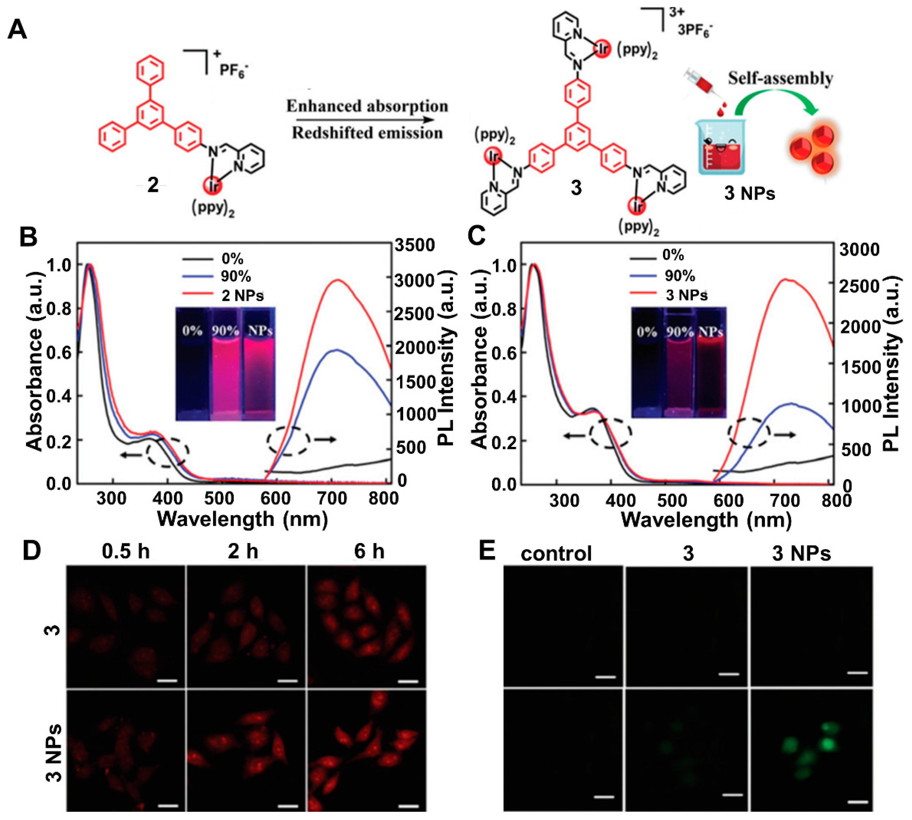
Compared with red emission, near-infrared (NIR) emission has better performance in overcoming autofluorescence interference, minimizing photodamage to cells and improving the signal-to-noise ratio (SNR) of imaging [45,46]. Li et al. designed and synthesized mono- and tri-nuclear NIR AIE cationic Ir(III) complexes and corresponding nanoparticles (NPs) 2 and 3 by self-assembling (Figure 2A) [47]. Further, 2 and 3 showed bright NIR emission at 710 nm and 730 nm when the water fraction reached 90%, showing typical AIE character (Figure 2B,C). The photoluminescence quantum yields (PLQYs) of 2 NPs and 3 NPs were 25% and 22% in water, which were higher than those of 2 and 3 (6% and 4%, respectively). Remarkably, the NPs possessed similar emission peaks while PL intensities were obviously enhanced. The greater biological NIR emission was valuable for efficient biological applications. Intracellular NIR emission was monitored, showing that 3 NPs exhibited excellent imaging performance (Figure 2D). The 1O2 production was evaluated by using 2,7-dichlorodihydrofluorescein diacetate (DCFH-DA) as an indicator and bright-green fluorescence was observed with 3 and 3 NPs upon light irradiation (Figure 2E). These results proved that 3 NPs could be applied as an efficient PS for imaging-guided PDT.

2.2. Enhancing Molar Absorption Coefficient

In general, the upper limit of the output signal depends on the absorption capacity of the PSs. Actually, high brightness can be achieved by increasing the molar absorption coefficients. Therefore, improving absorptive capacity has been the focus of many efforts [48]. Zhang et al. proposed, for the first time, that additional metal centers further resulted in an increase in the molar absorption coefficient by increasing 3MLCT [49]. They designed the first example of an AIE-active multinuclear Ir(III) complex. Adding Ir(III) metal centers, the molar absorption coefficient increased (Figure 3A,B). Thus, 4 NPs, 5 NPs and 6 NPs showed higher PLQYs (33%, 15% and 35%, respectively) in water than those of 4, 5 and 6 (11%, 5%, and 8%, respectively) in THF–water mixtures. Confocal laser scanning microscopy (CLSM) demonstrated the cellular uptake of PS and 6 NPs showed red fluorescence in cells (Figure 3C). The enhanced cellular uptake was highly advantageous to improve PDT. With light irradiation, green emission was observed from cells, indicating the presence of 1O2 generation (Figure 3D). This study offered a new method to improve the efficiency of PSs for clinical cancer treatments.

2.3. With the UCNPs

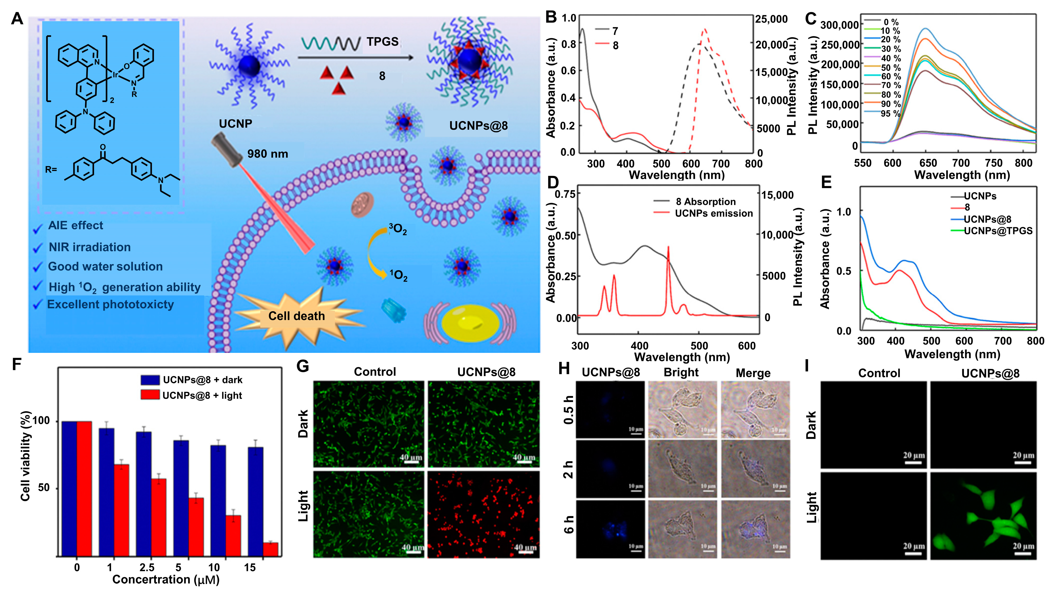
Upconversion nanoparticles (UCNPs) are desirable photoconversion materials for biosensing and biomedicine on account of their capability to transform the NIR photons to UV/visible photons [50,51]. Therefore, the conjugation of PSs to UCNPs presents an alternative to achieve NIR light-triggered generation of ROS. With this idea in mind, Liu et al. [52] employed AIE-active non-charged Ir(III) complexes integrated with UCNPs for the first time (Figure 4A). In UV-vis absorption spectra, 7 and 8 exhibited absorption peaks at ~425 nm. The PL spectra showed that 7 emitted at 640 nm and 8 emitted at 660 nm (Figure 4B), while the PLQYs were 16.6% and 18.9%, respectively. When the water fraction reached 50%, the emission intensity of 8 was greatly enhanced, which demonstrated that 8 had typical AIE features (Figure 4C). Further, 8 was then encapsulated with UCNPs within d-α-tocopherol polyethylene glycol 1000 succinate (TPGS) to construct UCNPs@8 as PSs for the ensuing in vitro experiments. The UV-vis absorption spectra of UCNPs@8 showed a significant absorption at about 425 nm, corresponding to that of 8 (Figure 4D). To ensure that the energy transferred between 8 and UCNPs can be realized and UCNPs@8 could be irradiated by NIR light, the PL spectrum of UCNPs was measured under laser irradiation (980 nm). The absorption band of 8 was in the UV and blue regions, overlapping with the emission bands of UCNPs (Figure 4E). Encouraged by the 1O2 generation ability of NIR-irradiated UCNPs@8, their ability to kill cancer cells through PDT was investigated. MTT assays indicated that UCNPs@8 had considerable cytotoxicity (Figure 4F). To further evaluate the therapeutic effect of UCNPs@8, Calcein AM and propidium iodide (PI) co-staining assay showed that a number of cells died under 980 nm irradiation (Figure 4G). The cells treated by UCNPs@8 exhibited strong green fluorescence (Figure 4I). All the results illustrated the feasible strategy that UCNPs and AIE-active 8 were encapsulated together to achieve effective energy transfer and exerted their respective advantages to construct NIR-irradiated PSs, UCNPs@8, which generated toxic ROS and thereby killed the cancer cells. This approach effectively overcame the problems of Ir(III) complexes, which own short-wavelength absorption [52].

2.4. Two-Photon Absorption (TPA)

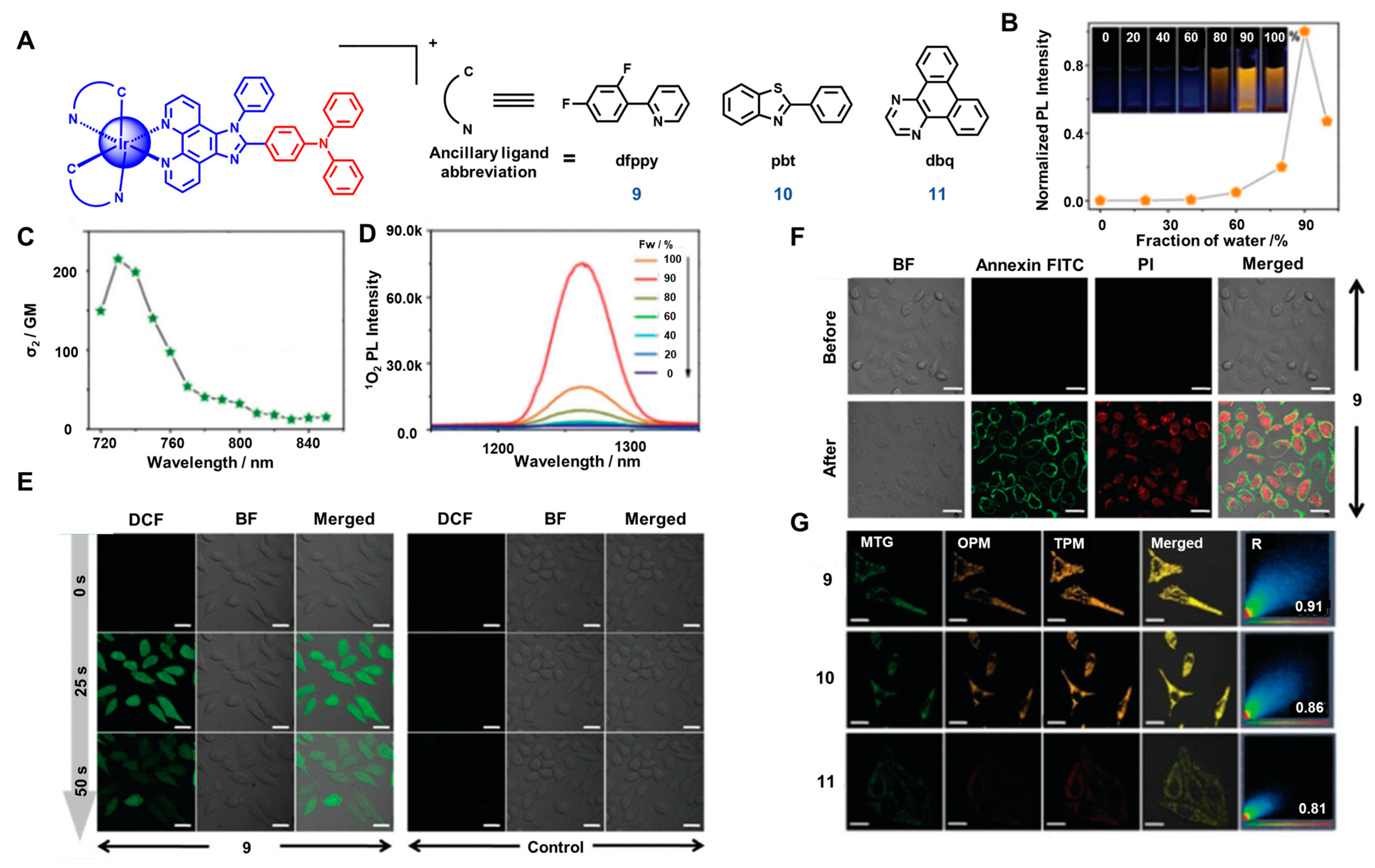
In recent years, two-photon absorption (TPA) has emerged as an attractive protocol in bioimaging and PDT. Two-photon photodynamic therapy (TP-PDT) activates PSs by contemporaneously absorbing two photons [53,54,55,56]. It provides photodamage and phototoxicity as well as deeper penetration, weaker autofluorescence and lower photobleaching [57,58]. Developing Ir(III) complexes with TPA is a new strategy to enhance the efficiency of Ir-PSs. However, conventional PSs suffer from ACQ and reduced photocytotoxicity, resulting in inferior imaging and PDT efficacy. In summary, it is very prospective to construct Ir(III) complexes as AIE-active TPA PSs [59]. Liu et al. [60] conjugated triphenylamine with Ir(III) complexes to achieve the goals of AIE and TPA (Figure 5A). The PLQYs of 9 were 0.1% in DMSO. Further, 9 performed obvious AIE behavior and the TPA cross-sections (ð2) were better than the previous reported TPA organometallic molecular structure (Figure 5B,C). As anticipated, the 1O2 emission intensity enhanced as the water ratio of the mixture gradually increased from 0 to 90%, i.e., the strongest AIE effect (Figure 5D). Under two-photon laser irradiation, for the majority of cells treated with 9 (Figure 5E,F), the fluorescence intensity enhanced and significant annexin V-FITC (AV) and PI signals were detected, revealing that 9 possessed the highest potency of being an efficient TPA–PDT agent.
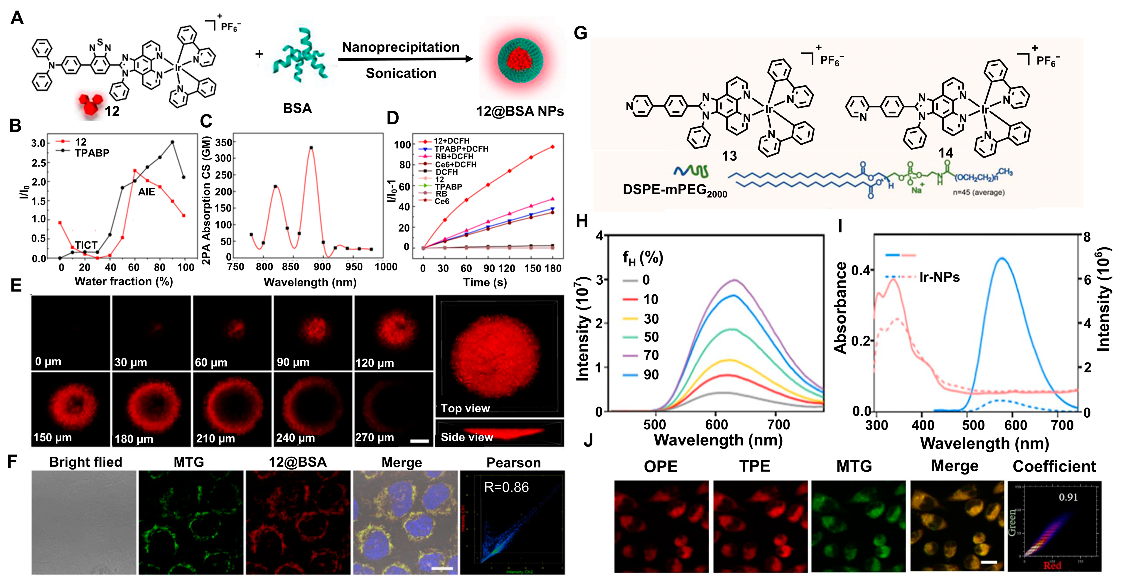
However, TP-AIE-active Ir(III) complexes still have two main disadvantages: (i) Most activated TP-PSs undergo type II photochemical reactions to generate ROS. Their therapeutic effect is still limited by hypoxia. (ii) Poor water solubility makes it difficult to use in the clinic [61]. To address these issues, Jiang et al. designed the first type-I AIE PSs with the TP-PDT strategy, named TPABP and 12 (Figure 6A). To enhance biocompatibility and structural stability of 12, bovine serum albumin (BSA) was added to formulate 12@BSA NPs. TPABP and 12 exhibited the balance between the AIE feature and the typical TICT effect. When the water fraction was 0%, the PLQYs of 12 were about 9%. σ2 of 12@BSA NPs was 340 GM at 880 nm (Figure 6B,C). The ROS generation of 12 was better than RB and Ce6 (Figure 6D). Under two-photon irradiation, a 3D tumor model was constructed to evaluate the penetrativity of 12@BSA NPs. A penetration depth of 270 μm was measured by z-scanning (Figure 6E,F), which showed 12@BSA NPs performed well [62]. Similarly, Cai et al. [63] synthesized two Ir(III) complexes (13 and 14) (Figure 6G) and 13 possessed the AIE feature (Figure 6H). Encapsulating 13 to DSPE-mPEG2000 to form nanoparticles (13 NPs) is a strategy to improve biocompatibility and the NP carrier did not influence the photophysical properties of 13 (Figure 6I). The cellular imaging capability of 13 NPs was investigated using CLSM. Upon two-photon excitation, the red phosphorescent signal was stonger than that upon one-photon excitation (Figure 6J).

2.5. Multiphoton Absorption (MPA)

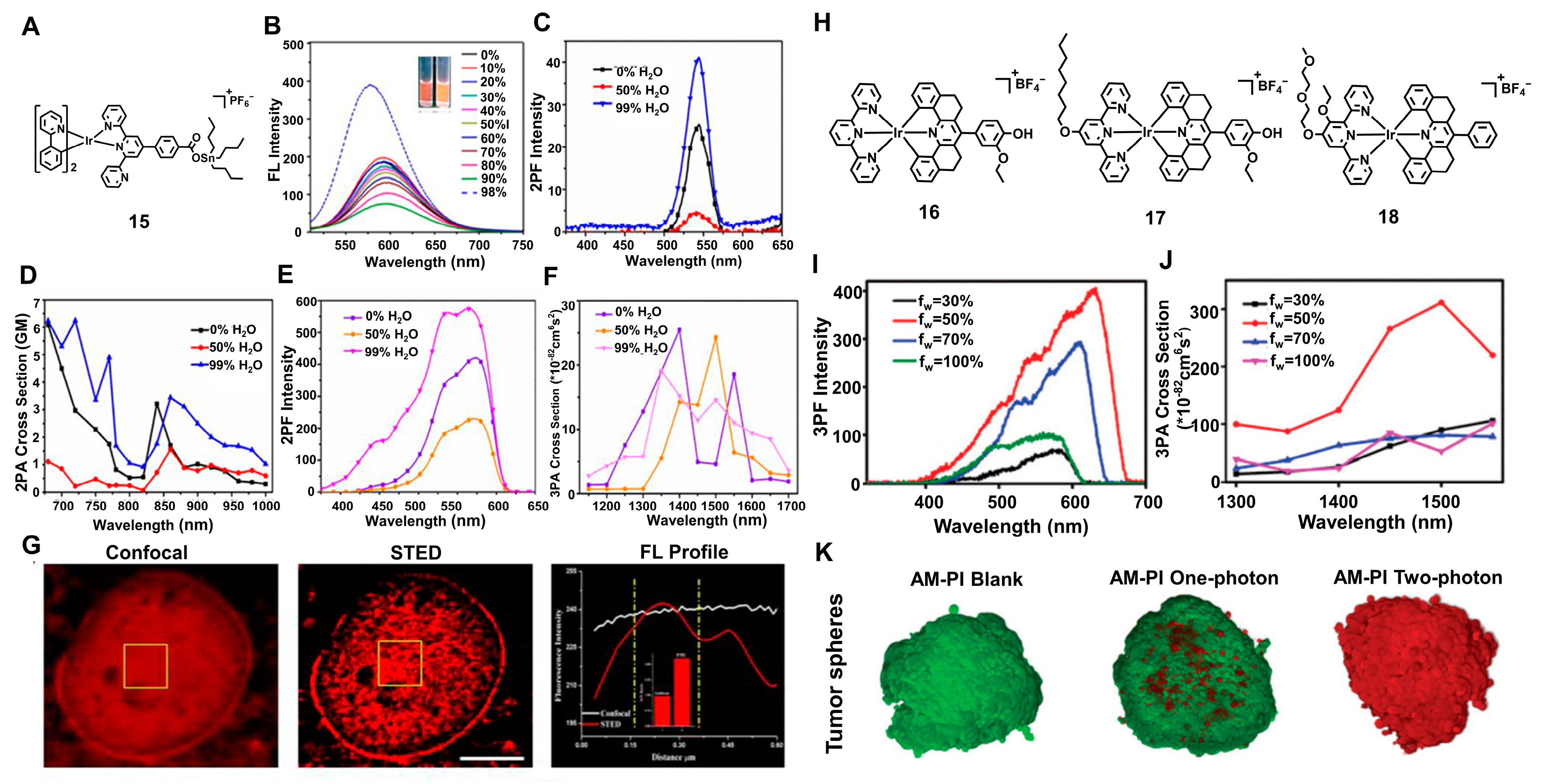
Compared with TPA, multiphoton absorption (MPA) improves the conversion efficiency through simultaneously absorbing several infrared photons. The two primary benefits of MPA have attracted attention: excitation wavelengths are in the near infrared (NIR) (700–900 nm) and the relationship between multiphoton absorption activity and excitation intensity is nonlinear [55]. Nevertheless, Ir(III) complexes activated by MPA are still very rare. Zhang et al. [64] combined the distinct merits of excellent photostability of infrared Ir(III) complexes, religious antibacterial ability of organotin(IV) compounds and MPA materials to design 15 and it showed obvious AIE behavior (Figure 7A,B). Using [Ru(byp)3]Cl2 as the standard, the PLQYs of 15 were 0.2 in water. The 2PA and 3PA cross-sections of 15 were 6.25 GM and 1.5 × 10−81 cm6 s2 per photon2 (Figure 7C–F). Under multiphoton confocal and STED microscopies, 15 showed superstability in nucleolus, which displayed that 15 had high signal-to-noise ratio (Figure 7G). Recently, Lu et al. [65] designed and synthesized a series of 3PA-Ir(III) complexes, named 16, 17 and 18 (Figure 7H). Only 16, which did not have a long alkoxy chain, showed AIE activity and excellent three-photon absorption properties (Figure 7I,J). Further, 16 also revealed the PLQYs, which were 1.4%. They displayed strong red emission under two-photon laser irradiation (820 nm), indicating the effectiveness of 16 in inducing cell death (Figure 7K).

3. Accurate Targeting of Ir(III) Complexes with AIE Activity

3.1. Targeting Mitochondria

It is well known that mitochondria are one of the most essential organelles. As the power workshop of cells, mitochondria provide most of the cellular ATP through pathways, such as pyruvate oxidation, fatty acid oxidation, tricarboxylic acid cycle (TCA cycle), and so on. On the other hand, mitochondria are the key to the apoptotic pathway. For instance, in extrinsic apoptosis, the mitochondrial outer membrane opens after signals, including intracellular Ca2+ overload and oxidation, which leads to the release of pro-apoptotic factors [66]. PSs act on mitochondria and make mitochondrial ROS burst, so mitochondria will be destroyed and then Cytochrome C, kinds of pro-apoptotic factors, will release, resulting in cancer cell death [67]. As a result, mitochondria-targeted PDT becomes an effective method to improve the treatment of cancer. The agents with delocalized positive charge are preferable to go through the mitochondrial lipid bilayer membrane due to the highly negative inner-membrane potential of mitochondria. Ir(III) complexes are demonstrated as mitochondrial targeting molecules applied in PDT, due to their lipophilic cationic properties.
Jiang et al. [62] proposed an Ir(III) complex with AIE characteristics, which was called 12. Due to the feature that 12 was a lipophilic cation, there was an electrostatic interaction between 12 NPs and mitochondria, holding a highly negative inner-membrane potential, which caused 12 NPs to penetrate lipid bilayers and accumulate inside mitochondria (Figure 6I). Cai et al. [63] reported two Ir(III) complexes and both of them possessed AIE properties, good photosensitivity and mitochondria-targeting ability. Further, 13 was encapsulated with DSPE-mPEG2000 so that nanoparticles called 13 NPs were formed. Taken up via caveolae-mediated endocytosis, 13 NPs targeted mitochondria, which benefited from damaging mitochondria and enhanced antitumor effect (Figure 6M). Liu et al. [60] reported three different structural Ir(III) complexes 9–11, which were PSs with AIE property and mitochondria-targeting capability, achieved though the endocytic pathway (Figure 5G). Interestingly, 9–11 exhibited more preferential photocytotoxicity towards HeLa cells than L02 cells.
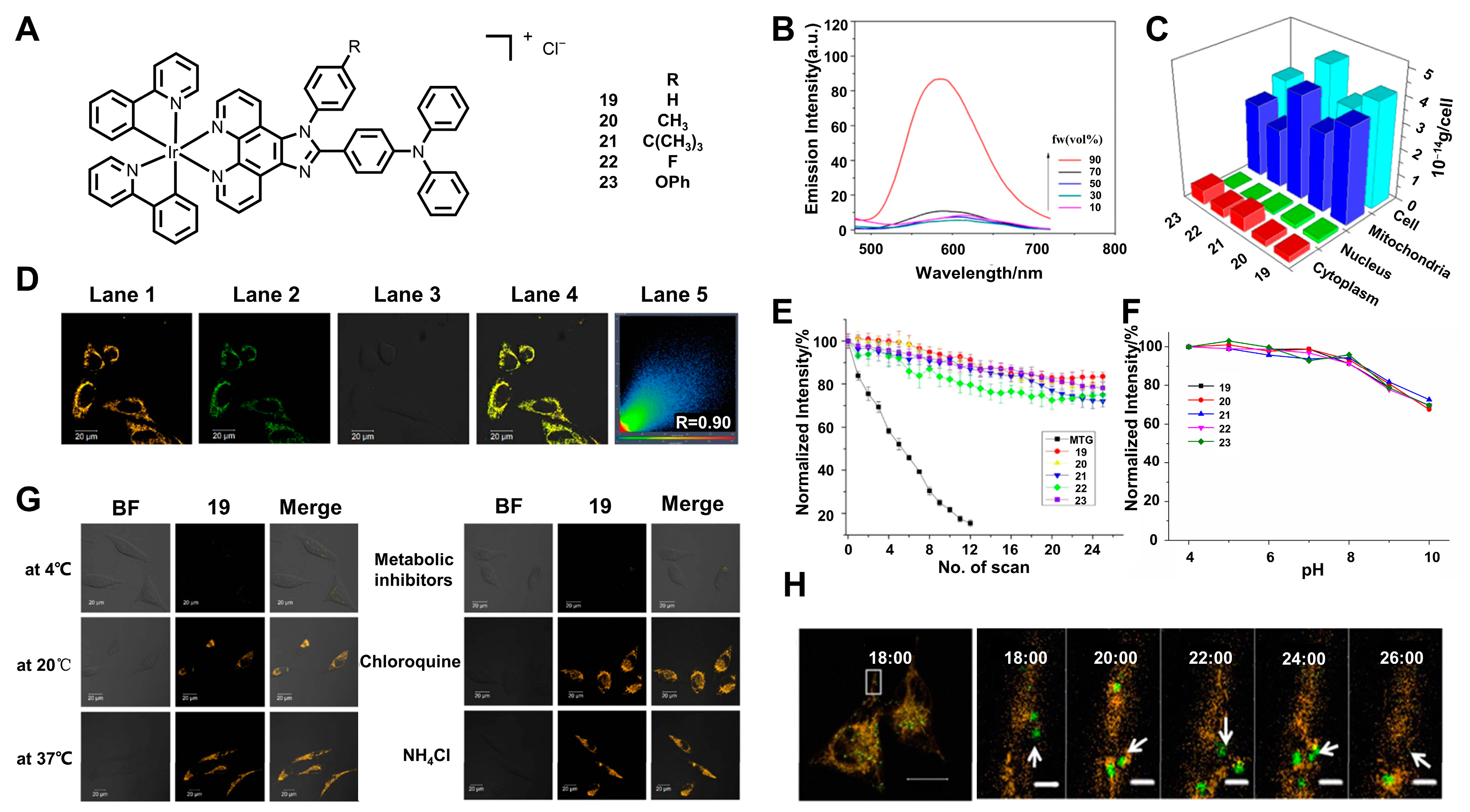
In addition, the damaged mitochondria can be removed via mitophagy, a form of autophagy, which provides healthy surroundings in cells. During the autophagy process, a membrane structure forms the phagophore, which gradually expands and engulfs a portion of the cytosol or specific cargos and delivers them to the vacuole/lysosome for degradation [68]. The physiological changes are related to several diseases, so it is indispensable to monitor the mitophagy process. Jin et al. [69] synthesized five Ir(III) complexes, which possessed the AIE property (Figure 8A,B). These Ir(III) complexes, which were named 19–23, were certified as specifically accumulated in mitochondria through mitochondrial co-localization imaging experiments with MitoTracker Green (MTG) and ICP-MS experiments (Figure 8C,D). The mechanisms of cellular uptake also proved that 19 used a non-endocytic energy-dependent active transport to cross the plasma membrane (Figure 8G). Thus, 19 successfully fulfilled the real-time monitoring of the mitophagy process, which was caused by CCCP (Figure 8H). In addition, 19–23 owned excellent stability during 10 scans, with a total irradiation time of ~150 s, as well as in a pH range from 4.0 to 8.0 (Figure 8E,F). Further, 19–23 were expected to be used as mitochondrion-specific probes in biological imaging and dynamic monitoring studies.
Ir complexes can be applied in mitochondrial staining simply. Alam et al. [70] proposed two AIE-active Ir(III) complexes (Figure 9A–C,E). The PLQYs for 25 were tested and found to be 6.03%. With poor ROS production capacity, 25 had lower cytotoxicity (Figure 9F) and 25 was useful for bioimaging. When 25 was incubated with HOS cells, the good fluorescence of 25 was only observed in mitochondria (Figure 9D). With AIE characteristics, low cytotoxicity and mitochondrion-targeted capability, 25 owned potential applications in bioimaging, particularly in mitochondrial staining.

3.2. Targeting Nucleolus

Ribonucleic acid (RNA) exists in biological cells and some viruses, as a viroid genetic information carrier. As the basis of life, RNA takes part in biological processes, such as gene transcription, post-transcriptional regulation of gene expression, protein synthesis based on gene information, etc. Thereinto, rRNA is responsible for protein synthesizing. rRNA gathers in the nucleolar region, which is a dark region under fluorescence microscopy. In recent years, the exploration of rRNA-selective probes for the nucleolar region becomes a meaningful topic. Holding more intense interaction with DNA than rRNA, small molecules are difficult to make use of as rRNA-selective probes. Ir(III) complexes have potential to construct a kind of rRNA-selective probe due to their greater interaction with intra-nuclear proteins [9].
Based on the previous work, Sheet et al. [71] synthesized the 26 Ir(III) complex with benzimidazole-substituted 1,2,3-triazole-pyridine ligand (BiPT), which showed green luminescence (Figure 10A). The mechanisms of the AIE property of 26 are that 26 formed into aggregated particles in a CH3CN–water mixture and were verified by fluorescence, DLS and SEM images (Figure 10B,E–G). The PLQYs of 26 were found to be 0.016 in CH3CN at the same time. Noteworthily, a large portion of 26 was found to enter the cell nucleoli region after being incubated with Hela cells in co-staining experiments while faint green luminescence was still observed in the cytoplasm, which meant that 26 selectively targeted the nucleoli where most RNA was assembled (Figure 10I). Compared with ctDNA, ssDNA, G-quadruplex DNA and BSA, there was stronger interaction with 26 and RNA (Figure 10C). The results of Autodock Tools suggested that 26 combined with the purine and pyrimidine bases of RNA via supramolecular N–H∙∙∙π and π–π interactions (Figure 10H). Exhilaratingly, PL intensity of 26 elevated gradually with an enhancement in rRNA concentration (Figure 10D). Thereout, 26 was the first Ir(Ⅲ) complex reported as an rRNA-selective probe.

3.3. Selective Toxicity of Cells

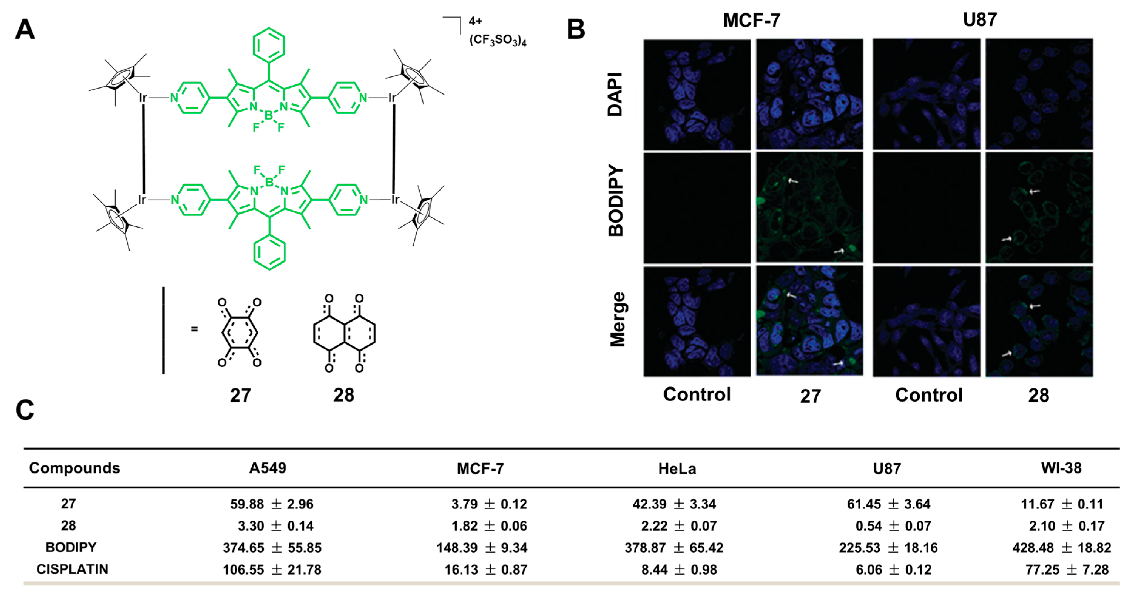
Selective anticancer activity is one of the standards to estimate anticancer drugs. Damaging cancer cells and keeping normal cells well simultaneously are what researchers are trying to achieve. Ir(III) metalla-rectangles 27 and 28, which consist of a BODIPY-based linker, were reported by Gupta et al. (Figure 11A). The white arrows showed 28 aggregated in the form of tiny granules, namely the AIE phenomenon (Figure 11B). Through initial biological screening, the selective toxicities of 27 against MCF-7 and 28 against U87 cells were proved (Figure 11B,C). The toxicity against cancer cell lines of 27 and 28 was stronger than that against normal cells. The complexes had different activities to different cell lines, attributed to various intracellular molecular targets, which demands further study [72].

4. Specific Response of Ir(III) Complexes with AIE Activity

In recent years, Ir(III) complexes with AIE properties have attracted more attention as photoluminescent probes in bioimaging, owing to their high photophysical performance (such as large Stokes shifts, tunable photophysical properties, long excited-states lifetime, satisfactory photostability, etc.). So far, several specific responsive probes based on Ir(III) complexes with AIE activity have been reported for bioimaging applications. The detailed structures and photophysical properties of these probes are summarized and discussed in the following sections.

4.1. Perchlorate (ClO4)

Perchlorate (ClO4) salts are widespread and relatively stable in foods and water samples [73]. ClO4 competitively disrupts thyroid uptake of iodine and accumulates in the human body due to its similar ionic radius with iodide, resulting in adverse consequences, such as iodide deficiency and goiter, even brain impairment [74]. Hence, it is necessary to detect ClO4 in water and monitor it in cells. An AIE-active complex 29 based on the Ir(III) complex for specific sensing of ClO4 in cells reported by Chao et al. in 2016 was rationally designed by introducing the thioacetal group [75]. The phosphorescence quantum yield of 29 in solution was 1%, which increased to 2% in the solid state due to the aggregation of nanoparticles (Figure 12B). In particular, 29 exhibited a specific response to ClO4 in HEPES buffer (Figure 12C,D) and the luminescence turn-on response was not interfered with by Hg2+, although the thioacetal group can undergo a thioacetal deprotection reaction [76]. Further, 29 and ClO4 lead to enhanced-emissive nanoparticles in water, while the reaction of 29 and Hg2+ generated weak emission (Figure 12E). Moreover, the binding rate of 29 and ClO4 was faster than the thioacetal deprotection reaction of Hg2+. The application of 29 in imaging ClO4 in living cells was further investigated, where a strong red phosphorescent emission was observed in the cytoplasm.
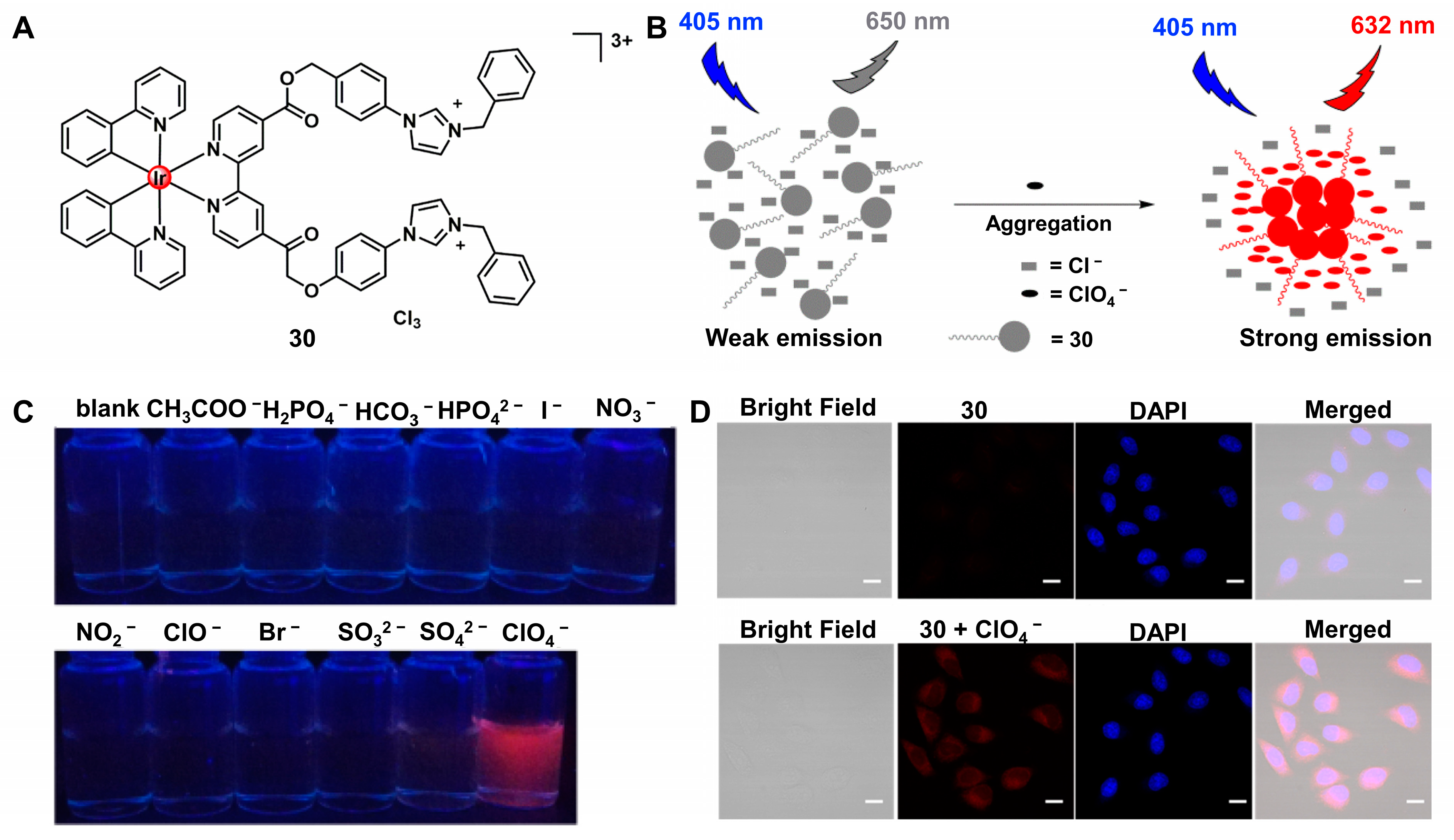
In 2017, Chao et al. [77] first reported a water-soluble Ir(III) complex as a probe of ClO4 based on the AIE phenomenon. Complex 30 had two imidazolium-like groups to improve positive charges and solubility (Figure 13A). A possible mechanism was presented that ClO4 induced 30 and aggregated in water due to the exchange between Cl and ClO4, resulting in an enhanced emission (Figure 13B). Furthermore, 30 was used for cell imaging and showed strong red emission in the presence of ClO4 in HeLa cells (Figure 13D).

4.2. BSA

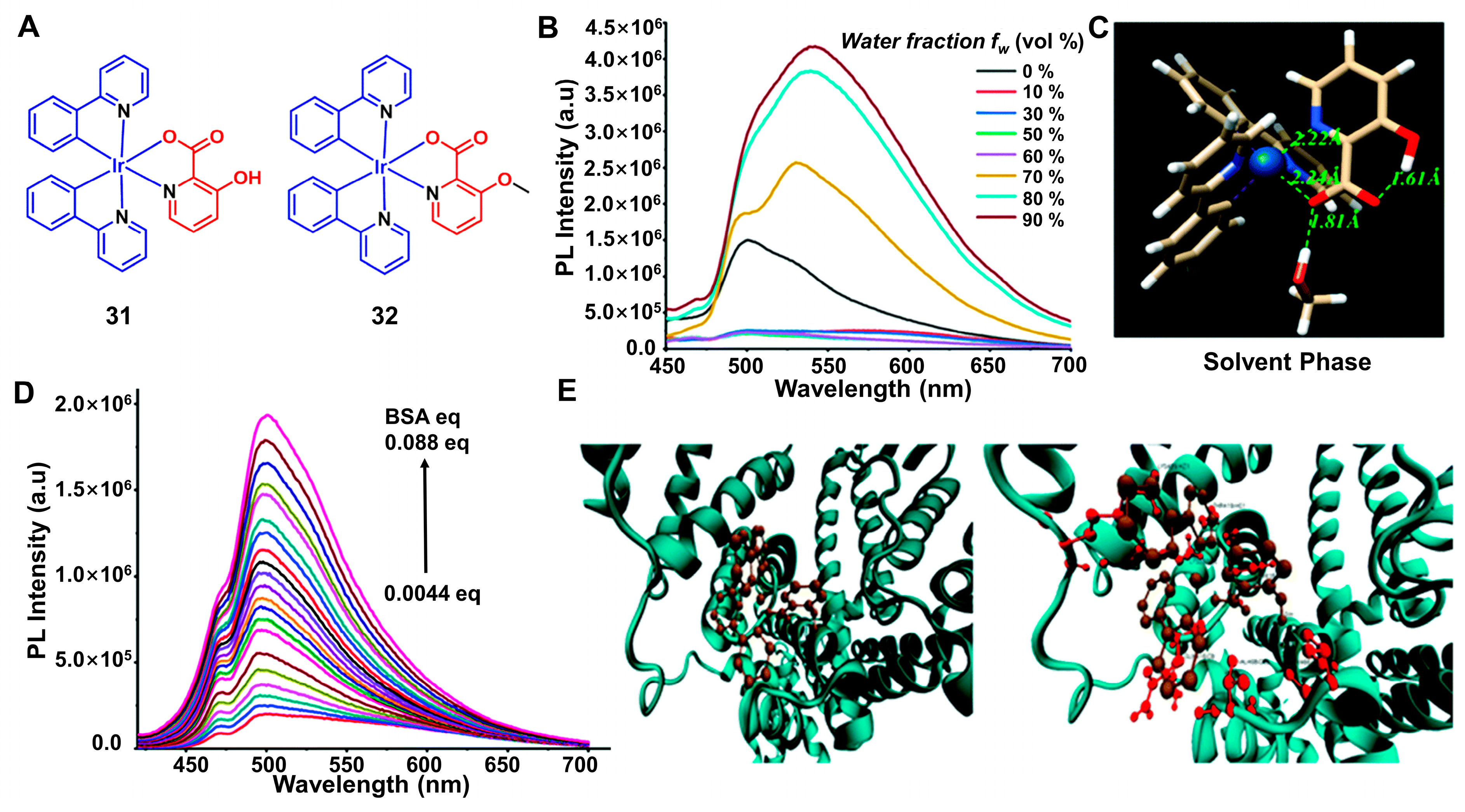
BSA is a globular protein and has an important effect on biophysical functions, such as blood circulation and drug binding [78,79]. Two Ir(III) complexes, namely 31 and 32 (Figure 14A), were reported by Laskar et al. [80]. The emission intensity of 31 raised with the increasing concentration of water (Figure 14B). Compared with 32, only 31 had AIE properties due to the restriction of intramolecular motion of the picolinate group (Figure 14C). Moreover, the interaction of 31 with BSA resulted in green emission (Figure 14D). The mechanism of 31 as a specific probe for sensing BSA proved that the auxiliary ligand may bind to the protein molecule through molecular docking studies (Figure 14E).

4.3. Drug-Resistant Bacteria

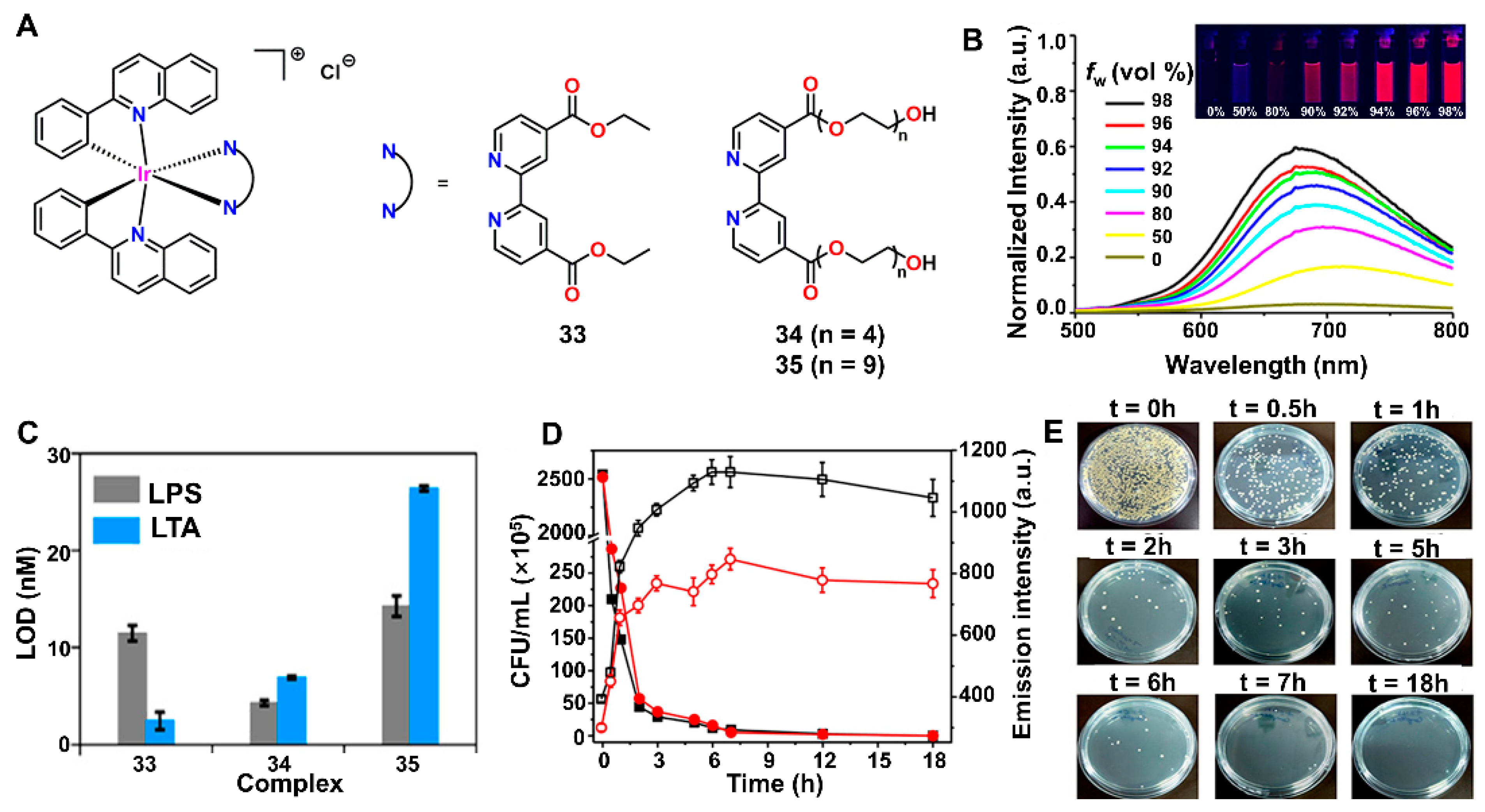
Antimicrobial resistance (AMR) has developed rapidly due to the overuse of medicines, leading to contaminating water bodies and posing a serious threat to public health [81,82]. Therefore, there is an urgent need for monitoring and eliminating drug-resistant bacteria to control waterborne diseases [83]. The work of Sasmal et al. is a good example of developing Ir(III) complexes with AIE properties for detecting and inhibiting drug-resistant bacteria [84]. Three cationic Ir(III) complexes (3335) were synthesized as bacterial theranostic agents (Figure 15A). These complexes contained the same cyclometalated ligands and different auxiliary ligands. Further, 33 had two ethyl ester moieties in the auxiliary ligand, whereas 34 and 35 had bis-polyethylene glycol (PEG) to improve the biocompatibility and aqueous solubility. Both 33 and 34 were found to possess AIE active and highly selective for detecting LPS/LTA (Figure 15D). The PLQYs for 33, 34 and 35 were determined to be 0.0072, 0.0091 and 0.0084, respectively. Moreover, the mechanisms of bacterial cells damaged by 33 and 34 suggest the disintegration of membrane integrity and generation of ROS (Figure 15E). This work is a platform that integrates the detection and elimination of bacteria into AIE-active Ir(III) complexes.

5. Conclusions

This review provides an overview of Ir(III) complexes with AIE properties for bioimaging, sensing and therapy applications in terms of extended absorption wavelength, precise targeting and specific response, respectively. By introducing various ligands into Ir(III) complexes, the photophysical properties could be improved to enhance the effect of bioimaging and therapeutics. These results are of great importance and promote the development of AIE materials based on Ir(III) complexes for biomedical applications.
However, there are still many limitations in the design strategy of efficient AIE-active Ir(III) complexes. Herein, we propose research directions where concerted efforts are required for AIEgens based on Ir(III) complexes in bioimaging and therapeutics. First, extending the absorption of Ir(III) complexes to NIR II will further improve the tissue-penetration depth and reduce side effects. Second, more attention should be placed on enhancing the targeting ability of AIE-active Ir(III) complexes and selectively releasing in the tumor microenvironment (TME) to improve the efficacy of tumor treatment. Third, the synergistic therapy of AIEgens could be further increased via exploiting the rational design of the Ir(III) complexes. We believe that AIE materials based on Ir(III) complexes would become a powerful tool for the biomedical field in the future.

Author Contributions

Conceptualization, Y.P., Y.S. and M.H.; writing—original draft preparation, Y.P., Y.S. and M.H.; writing—review and editing, Y.P., Y.S., M.H., Z.Z. (Zebing Zeng), Z.Z. (Zhijun Zhang), D.Y. and J.C.; supervision, D.W. and D.Z.; project administration, D.Z., D.W. and B.T.; funding acquisition, D.W. All authors have read and agreed to the published version of the manuscript.

Funding

This research was funded by the National Natural Science Foundation of China (52122317, 22175120), the Developmental Fund for Science and Technology of Shenzhen Government (RCYX20200714114525101, JCYJ20190808153415062), the Natural Science Foundation for Distinguished Young Scholars of Guangdong Province (2020B1515020011), MOE Key Laboratory of Macromolecular Synthesis and Functionalization, Zhejiang University (2021MSF04) and the Shenzhen Science and Technology Program (Grant No. RCJC20200714114434015 and JCYJ20210324141212031).

Institutional Review Board Statement

Not applicable.

Informed Consent Statement

Not applicable.

Data Availability Statement

Not applicable.

Conflicts of Interest

The authors declare no conflict of interest.

References

  1. Kober, E.M.; Meyer, T.J. Concerning the absorption spectra of the ions M(bpy)32+ (M = Fe, Ru, Os; bpy = 2,2′-bipyridine). Inorg. Chem. 1982, 21, 3967–3977. [Google Scholar] [CrossRef]
  2. You, Y.; Nam, W. Photofunctional triplet excited states of cyclometalated Ir(III) complexes: Beyond electroluminescence. Chem. Soc. Rev. 2012, 41, 7061–7084. [Google Scholar] [CrossRef] [PubMed]
  3. Flamigni, L.; Barbieri, A.; Sabatini, C.; Ventura, B.; Barigelletti, F. Photochemistry and photophysics of coordination compounds: Iridium. Top. Curr. Chem. 2007, 281, 143–203. [Google Scholar] [CrossRef]
  4. Zhao, Q.; Li, F.; Huang, C. Phosphorescent chemosensors based on heavy-metal complexes. Chem. Soc. Rev. 2010, 39, 3007–3030. [Google Scholar] [CrossRef] [PubMed]
  5. Alam, P.; Climent, C.; Alemany, P.; Laskar, I.R. “Aggregation-induced emission” of transition metal compounds: Design, mechanistic insights, and applications. J. Photoch. Photobio. C Photoch. Rev. 2019, 41. [Google Scholar] [CrossRef]
  6. Ulbricht, C.; Beyer, B.; Friebe, C.; Winter, A.; Schubert, U.S. Recent developments in the application of phosphorescent iridium(III) complex systems. Adv. Mater. 2009, 21, 4418–4441. [Google Scholar] [CrossRef]
  7. Lee, L.C.-C.; Lo, K.K.-W. Luminescent and photofunctional transition metal complexes: From molecular design to diagnostic and therapeutic applications. J. Am. Chem. Soc. 2022, 144, 14420–14440. [Google Scholar] [CrossRef]
  8. Guan, R.; Xie, L.; Ji, L.; Chao, H. Phosphorescent iridium(III) complexes for anticancer applications. Eur. J. Inorg. Chem. 2020, 2020, 3978–3986. [Google Scholar] [CrossRef]
  9. Caporale, C.; Massi, M. Cyclometalated iridium(III) complexes for life science. Coordin. Chem. Rev. 2018, 363, 71–91. [Google Scholar] [CrossRef] [Green Version]
  10. Gierlich, P.; Mata, A.I.; Donohoe, C.; Brito, R.M.M.; Senge, M.O.; Gomes-da-Silva, L.C. Ligand-targeted delivery of photosensitizers for cancer treatment. Molecules 2020, 25, 5317. [Google Scholar] [CrossRef]
  11. Huang, H.; Banerjee, S.; Qiu, K.; Zhang, P.; Blacque, O.; Malcomson, T.; Paterson, M.J.; Clarkson, G.J.; Staniforth, M.; Stavros, V.G.; et al. Targeted photoredox catalysis in cancer cells. Nat. Chem. 2019, 11, 1041–1048. [Google Scholar] [CrossRef] [PubMed]
  12. Shen, J.; Rees, T.W.; Ji, L.; Chao, H. Recent advances in ruthenium(II) and iridium(III) complexes containing nanosystems for cancer treatment and bioimaging. Coordi. Chem. Rev. 2021, 443, 214016. [Google Scholar] [CrossRef]
  13. Liu, Y.; Song, N.; Chen, L.; Xie, Z.-G. BODIPY@Ir(III) complexes assembling organic nanoparticles for enhanced photodynamic therapy. Chinese J. Polym. Sci. 2018, 36, 417–424. [Google Scholar] [CrossRef]
  14. Mei, J.; Hong, Y.; Lam, J.W.Y.; Qin, A.; Tang, Y.; Tang, B.Z. Aggregation-induced emission: The whole is more brilliant than the parts. Adv. Mater. 2014, 26, 5429–5479. [Google Scholar] [CrossRef]
  15. Yuan, Y.; Xu, S.; Cheng, X.; Cai, X.; Liu, B. Bioorthogonal turn-on probe based on aggregation-induced emission characteristics for cancer cell imaging and ablation. Angew. Chem. Inter. Ed. 2016, 55, 6457–6461. [Google Scholar] [CrossRef]
  16. Luo, J.; Xie, Z.; Lam, J.W.; Cheng, L.; Chen, H.; Qiu, C.; Kwok, H.S.; Zhan, X.; Liu, Y.; Zhu, D.; et al. Aggregation-induced emission of 1-methyl-1,2,3,4,5-pentaphenylsilole. Chem. Commun. 2001, 1740–1741. [Google Scholar] [CrossRef]
  17. Chen, J.; Law, C.C.W.; Lam, J.W.Y.; Dong, Y.; Lo, S.M.F.; Williams, I.D.; Zhu, D.; Tang, B.Z. Synthesis, light emission, nanoaggregation, and restricted intramolecular rotation of 1,1-substituted 2,3,4,5-tetraphenylsiloles. Chem. Mater. 2003, 15, 1535–1546. [Google Scholar] [CrossRef]
  18. Chen, C.; Ni, X.; Jia, S.; Liang, Y.; Wu, X.; Kong, D.; Ding, D. Cancer immunotherapy: Massively evoking immunogenic cell death by focused mitochondrial oxidative stress using an AIE luminogen with a twisted molecular structure. Adv. Mater. 2019, 31, 1970372. [Google Scholar] [CrossRef] [Green Version]
  19. Wang, D.; Tang, B.Z. Aggregation-induced emission luminogens for activity-based sensing. Acc. Chem. Res. 2019, 52, 2559–2570. [Google Scholar] [CrossRef]
  20. Kwok, R.T.K.; Leung, C.W.T.; Lam, J.W.Y.; Tang, B.Z. Biosensing by luminogens with aggregation-induced emission characteristics. Chem. Soc. Rev. 2015, 44, 4228–4238. [Google Scholar] [CrossRef]
  21. Zhang, Z.; Kang, M.; Tan, H.; Song, N.; Li, M.; Xiao, P.; Yan, D.; Zhang, L.; Wang, D.; Tang, B.Z. The fast-growing field of photo-driven theranostics based on aggregation-induced emission. Chem. Soc. Rev. 2022, 51, 1983–2030. [Google Scholar] [CrossRef] [PubMed]
  22. Song, N.; Zhang, Z.; Liu, P.; Yang, Y.W.; Wang, L.; Wang, D.; Tang, B.Z. Nanomaterials with supramolecular assembly based on AIE luminogens for theranostic applications. Adv. Mater. 2020, 32, e2004208. [Google Scholar] [CrossRef] [PubMed]
  23. Zhao, Q.; Li, L.; Li, F.; Yu, M.; Liu, Z.; Yi, T.; Huang, C. Aggregation-induced phosphorescent emission (AIPE) of iridium(III) complexes. Chem. Commun. 2008, 685–687. [Google Scholar] [CrossRef] [PubMed]
  24. Pei, Y.; Xie, J.; Cui, D.; Liu, S.; Li, G.; Zhu, D.; Su, Z. A mechanochromic cyclemetalated cationic Ir(III) complex with AIE activity by strategic modification of ligands. Dalton Trans. 2020, 49, 13066–13071. [Google Scholar] [CrossRef]
  25. Li, D.; Li, G.; Xie, J.; Zhu, D.; Su, Z.; Bryce, M.R. Strategic modification of ligands for remarkable piezochromic luminescence (PCL) based on a neutral Ir(III) phosphor. J. Mater. Chem. C 2019, 7, 10876–10880. [Google Scholar] [CrossRef]
  26. Di, L.; Xing, Y.; Yang, Z.; Qiao, C.; Xia, Z. Photostable aggregation-induced emission of iridium(III) complex realizing robust and high-resolution imaging of latent fingerprints. Sens. Actuat. B Chem. 2023, 375, 132898. [Google Scholar] [CrossRef]
  27. Li, G.; Ren, X.; Shan, G.; Che, W.; Zhu, D.; Yan, L.; Su, Z.; Bryce, M.R. New AIE-active dinuclear Ir(III) complexes with reversible piezochromic phosphorescence behaviour. Chem. Commun. 2015, 51, 13036–13039. [Google Scholar] [CrossRef] [Green Version]
  28. He, Z.; Zhao, L.; Zhang, Q.; Chang, M.; Li, C.; Zhang, H.; Lu, Y.; Chen, Y. An acceptor–donor–acceptor structured small molecule for effective NIR triggered dual phototherapy of cancer. Adv. Funct. Mater. 2020, 30, 1910301. [Google Scholar] [CrossRef]
  29. Chelushkin, P.S.; Shakirova, J.R.; Kritchenkov, I.S.; Baigildin, V.A.; Tunik, S.P. Phosphorescent NIR emitters for biomedicine: Applications, advances and challenges. Dalton Trans. 2022, 51, 1257–1280. [Google Scholar] [CrossRef]
  30. Zhang, P.; Wu, G.; Zhao, C.; Zhou, L.; Wang, X.; Wei, S. Magnetic stomatocyte-like nanomotor as photosensitizer carrier for photodynamic therapy based cancer treatment. Colloid. Surface. B Biointerfaces 2020, 194, 111204. [Google Scholar] [CrossRef]
  31. Li, B.; Zhou, Q.; Wang, H.; Zha, Y.; Zheng, P.; Yang, T.; Ma, D.; Qiu, L.; Xu, X.; Hu, Y.; et al. Mitochondria-targeted magnetic gold nanoheterostructure for multi-modal imaging guided photothermal and photodynamic therapy of triple-negative breast cancer. Chem. Eng. J. 2021, 403, 126364. [Google Scholar] [CrossRef]
  32. Smith, R.A.J.; Hartley, R.C.; Murphy, M.P. Mitochondria-targeted small molecule therapeutics and probes. Antioxid. Redox Signal. 2011, 15, 3021–3038. [Google Scholar] [CrossRef] [PubMed]
  33. McKenzie, L.K.; Sazanovich, I.V.; Baggaley, E.; Bonneau, M.; Guerchais, V.; Williams, J.A.G.; Weinstein, J.A.; Bryant, H.E. Metal complexes for two-photon photodynamic therapy: A cyclometallated iridium complex induces two-photon photosensitization of cancer cells under near-IR light. Chem. Eur. J. 2017, 23, 234–238. [Google Scholar] [CrossRef] [PubMed]
  34. Tian, X.; Zhu, Y.; Zhang, M.; Luo, L.; Wu, J.; Zhou, H.; Guan, L.; Battaglia, G.; Tian, Y. Localization matters: A nuclear targeting two-photon absorption iridium complex in photodynamic therapy. Chem. Commun. 2017, 53, 3303–3306. [Google Scholar] [CrossRef] [Green Version]
  35. Cho, S.; You, Y.; Nam, W. Lysosome-specific one-photon fluorescence staining and two-photon singlet oxygen generation by molecular dyad. RSC Adv. 2014, 4, 16913–16916. [Google Scholar] [CrossRef]
  36. Kuang, S.; Sun, L.; Zhang, X.; Liao, X.; Rees, T.W.; Zeng, L.; Chen, Y.; Zhang, X.; Ji, L.; Chao, H. A mitochondrion-localized two-photon photosensitizer generating carbon radicals against hypoxic tumors. Angew. Chem. Inter. Ed. 2020, 59, 20697–20703. [Google Scholar] [CrossRef]
  37. Cao, R.; Jia, J.; Ma, X.; Zhou, M.; Fei, H. Membrane localized iridium(III) complex induces endoplasmic reticulum stress and mitochondria-mediated apoptosis in human cancer cells. J. Med. Chem. 2013, 56, 3636–3644. [Google Scholar] [CrossRef]
  38. Zielonka, J.; Joseph, J.; Sikora, A.; Hardy, M.; Ouari, O.; Vasquez-Vivar, J.; Cheng, G.; Lopez, M.; Kalyanaraman, B. Mitochondria-targeted triphenylphosphonium-based compounds: Syntheses, mechanisms of action, and therapeutic and diagnostic applications. Chem. Rev. 2017, 117, 10043–10120. [Google Scholar] [CrossRef]
  39. Murase, T.; Yoshihara, T.; Tobita, S. Mitochondria-specific oxygen probe based on iridium complexes bearing triphenylphosphonium cation. Chem. Lett. 2012, 41, 262–263. [Google Scholar] [CrossRef]
  40. Chen, Y.; Rees, T.W.; Ji, L.; Chao, H. Mitochondrial dynamics tracking with iridium(III) complexes. Curr. Opin. Chem. Biol. 2018, 43, 51–57. [Google Scholar] [CrossRef]
  41. Li, G.; Zhu, D.; Wang, X.; Su, Z.; Bryce, M.R. Dinuclear metal complexes: Multifunctional properties and applications. Chem. Soc. Rev. 2020, 49, 765–838. [Google Scholar] [CrossRef] [Green Version]
  42. Wang, J.; Lu, Y.; McGoldrick, N.; Zhang, C.; Yang, W.; Zhao, J.; Draper, S.M. Dual phosphorescent dinuclear transition metal complexes, and their application as triplet photosensitizers for TTA upconversion and photodynamic therapy. J. Mater. Chem. C 2016, 4, 6131–6139. [Google Scholar] [CrossRef]
  43. Zhang, J.; Zheng, M.; Zhang, F.; Xu, B.; Tian, W.; Xie, Z. Supramolecular hybrids of AIEgen with carbon dots for noninvasive long-term bioimaging. Chem. Mater. 2016, 28, 8825–8833. [Google Scholar] [CrossRef]
  44. Yang, K.; Zhou, Y.; Wang, Y.; Zhao, S.; Wu, X.; Peng, X.; Huang, L.; Jiang, L.; Lan, M.; Yi, X.-Y. An iridium complex as an AIE-active photosensitizer for image-guided photodynamic therapy. Chem. Asian J. 2021, 16, 1780–1785. [Google Scholar] [CrossRef]
  45. Dai, J.; Li, Y.; Long, Z.; Jiang, R.; Zhuang, Z.; Wang, Z.; Zhao, Z.; Lou, X.; Xia, F.; Tang, B.Z. Efficient near-infrared photosensitizer with aggregation-induced emission for imaging-guided photodynamic therapy in multiple xenograft tumor models. ACS Nano. 2020, 14, 854–866. [Google Scholar] [CrossRef]
  46. Wang, D.; Su, H.; Kwok, R.T.K.; Shan, G.; Leung, A.C.S.; Lee, M.M.S.; Sung, H.H.Y.; Williams, I.D.; Lam, J.W.Y.; Tang, B.Z. Facile synthesis of red/NIR AIE luminogens with simple structures, bright emissions, and high photostabilities, and their applications for specific imaging of lipid droplets and image-guided photodynamic therapy. Adv. Funct. Mater. 2017, 27. [Google Scholar] [CrossRef]
  47. Li, L.; Zhang, L.; Tong, X.; Li, Y.; Yang, Z.; Zhu, D.; Su, Z.; Xie, Z. Near-infrared-emitting AIE multinuclear cationic Ir(III) complex-assembled nanoparticles for photodynamic therapy. Dalton Trans. 2020, 49, 15332–15338. [Google Scholar] [CrossRef]
  48. Li, Y.; Zhang, J.; Liu, S.; Zhang, C.; Chuah, C.; Tang, Y.; Kwok, R.T.K.; Lam, J.W.Y.; Ou, H.; Ding, D.; et al. Enlarging the reservoir: High absorption coefficient dyes enable synergetic near infrared-II fluorescence imaging and near infrared-I photothermal therapy. Adv. Funct. Mater. 2021, 31. [Google Scholar] [CrossRef]
  49. Zhang, L.; Li, Y.; Che, W.; Zhu, D.; Li, G.; Xie, Z.; Song, N.; Liu, S.; Tang, B.Z.; Liu, X.; et al. AIE multinuclear Ir(III) complexes for biocompatible organic nanoparticles with highly enhanced photodynamic performance. Adv. Sci. 2019, 6, 1802050. [Google Scholar] [CrossRef] [PubMed] [Green Version]
  50. Wang, Z.; Liu, B.; Sun, Q.; Feng, L.; He, F.; Yang, P.; Gai, S.; Quan, Z.; Lin, J. Upconverted metal-organic framework janus architecture for near-infrared and ultrasound co-enhanced high performance tumor therapy. ACS Nano. 2021. [Google Scholar] [CrossRef]
  51. Wang, Y.; Li, Y.; Zhang, Z.; Wang, L.; Wang, D.; Tang, B.Z. Triple-jump photodynamic theranostics: MnO2 combined upconversion nanoplatforms involving a type-I photosensitizer with aggregation-induced emission characteristics for potent cancer treatment. Adv. Mater. 2021, 33, e2103748. [Google Scholar] [CrossRef] [PubMed]
  52. Liu, S.; Han, J.; Chang, Y.; Wang, W.; Wang, R.; Wang, Z.; Li, G.; Zhu, D.; Bryce, M.R. AIE-active iridium(III) complex integrated with upconversion nanoparticles for NIR-irradiated photodynamic therapy. Chem. Commun. 2022, 58, 10056–10059. [Google Scholar] [CrossRef] [PubMed]
  53. Zhao, S.; Wu, S.; Jia, Q.; Huang, L.; Lan, M.; Wang, P.; Zhang, W. Lysosome-targetable carbon dots for highly efficient photothermal/photodynamic synergistic cancer therapy and photoacoustic/two-photon excited fluorescence imaging. Chem. Eng. J. 2020, 388. [Google Scholar] [CrossRef]
  54. Li, C.; Liu, J.; Hong, Y.; Lin, R.; Liu, Z.; Chen, M.; Lam, J.W.Y.; Ning, G.H.; Zheng, X.; Qin, A.; et al. Click synthesis enabled sulfur atom strategy for polymerization-enhanced and two-photon photosensitization. Angew. Chem. Int. Ed. 2022, 61, e202202005. [Google Scholar] [CrossRef]
  55. Lu, S.; Zhou, F.; Zhang, Q.; Eda, G.; Ji, W. Layered hybrid perovskites for highly efficient three-photon absorbers: Theory and experimental observation. Adv. Sci. 2019, 6, 1801626. [Google Scholar] [CrossRef] [Green Version]
  56. Xu, W.; Lee, M.M.S.; Nie, J.J.; Zhang, Z.; Kwok, R.T.K.; Lam, J.W.Y.; Xu, F.J.; Wang, D.; Tang, B.Z. Three-pronged attack by homologous far-red/NIR AIEgens to achieve 1+1+1>3 synergistic enhanced photodynamic therapy. Angew. Chem. Int. Ed. 2020, 59, 9610–9616. [Google Scholar] [CrossRef]
  57. Ma, H.; Zhao, C.; Meng, H.; Li, R.; Mao, L.; Hu, D.; Tian, M.; Yuan, J.; Wei, Y. Multifunctional organic fluorescent probe with aggregation-induced emission characteristics: Ultrafast tumor monitoring, two-photon imaging, and image-guide photodynamic therapy. ACS Appl. Mater. Interfaces 2021, 13, 7987–7996. [Google Scholar] [CrossRef]
  58. McKenzie, L.K.; Bryant, H.E.; Weinstein, J.A. Transition metal complexes as photosensitisers in one- and two-photon photodynamic therapy. Coordi. Chem. Rev. 2019, 379, 2–29. [Google Scholar] [CrossRef] [Green Version]
  59. Porrès, L.; Mongin, O.; Katan, C.; Charlot, M.; Pons, T.; Mertz, J.; Blanchard-Desce, M. Enhanced two-photon absorption with novel octupolar propeller-shaped fluorophores derived from triphenylamine. Org. Lett. 2004, 6, 47–50. [Google Scholar] [CrossRef] [Green Version]
  60. Liu, J.; Jin, C.; Yuan, B.; Liu, X.; Chen, Y.; Ji, L.; Chao, H. Selectively lighting up two-photon photodynamic activity in mitochondria with AIE-active iridium(Ⅲ) complexes. Chem. Commun. 2017, 53, 2052–2055. [Google Scholar] [CrossRef]
  61. Fang, F.; Yuan, Y.; Wan, Y.; Li, J.; Song, Y.; Chen, W.C.; Zhao, D.; Chi, Y.; Li, M.; Lee, C.S.; et al. Near-infrared thermally activated delayed fluorescence nanoparticle: A metal-free photosensitizer for two-photon-activated photodynamic therapy at the cell and small animal levels. Small 2022, 18, e2106215. [Google Scholar] [CrossRef]
  62. Jiang, Y.; Zhu, W.; Xu, Z.; Zhang, Z.; Tang, S.; Fan, M.; Li, Z.; Zhang, J.; Yang, C.; Law, W.-C.; et al. A mitochondrion-targeting two-photon photosensitizer with aggregation-induced emission characteristics for hypoxia-tolerant photodynamic therapy. Chem. Eng. J. 2022, 448. [Google Scholar] [CrossRef]
  63. Cai, X.; Wang, K.N.; Ma, W.; Yang, Y.; Chen, G.; Fu, H.; Cui, C.; Yu, Z.; Wang, X. Multifunctional AIE iridium (III) photosensitizer nanoparticles for two-photon-activated imaging and mitochondria targeting photodynamic therapy. J. Nanobiotechnol. 2021, 19, 254. [Google Scholar] [CrossRef] [PubMed]
  64. Zhang, Q.; Lu, X.; Cao, H.; Wang, H.; Zhu, T.; Tian, X.; Li, D.; Zhou, H.; Wu, J.; Tian, Y. Multiphoton absorption iridium(III)-organotin(IV) dimetal complex with AIE behavior for both sensitive detection of tyrosine and antibacterial activity. ACS Appl. Bio. Mater. 2020, 3, 8105–8112. [Google Scholar] [CrossRef]
  65. Lu, X.; Xiong, C.; Li, B.; Du, W.; Li, D.; Ma, W.; Tian, X.; Tian, Y.; Zhang, Q. Three-photon absorption iridium(Ⅲ) photosensitizers featuring aggregation induced emission. Inorg. Chem. Front. 2022, 9, 1890–1896. [Google Scholar] [CrossRef]
  66. Vakifahmetoglu-Norberg, H.; Ouchida, A.T.; Norberg, E. The role of mitochondria in metabolism and cell death. Biochem. Biophys. Res. Commun. 2017, 482, 426–431. [Google Scholar] [CrossRef] [PubMed]
  67. Li, X.; Zhao, Y.; Zhang, T.; Xing, D. Mitochondria-specific agents for photodynamic cancer therapy: A key determinant to boost the efficacy. Adv. Healthc. Mater. 2021, 10, e2001240. [Google Scholar] [CrossRef]
  68. Wang, K.; Klionsky, D.J. Mitochondria removal by autophagy. Autophagy 2011, 7, 297–300. [Google Scholar] [CrossRef] [Green Version]
  69. Jin, C.; Liu, J.; Chen, Y.; Guan, R.; Ouyang, C.; Zhu, Y.; Ji, L.; Chao, H. Cyclometalated iridium(III) complexes as AIE phosphorescent probes for real-time monitoring of mitophagy in living cells. Sci. Rep. 2016, 6, 22039. [Google Scholar] [CrossRef] [Green Version]
  70. Alam, P.; Dash, S.; Climent, C.; Kaur, G.; Choudhury, A.R.; Casanova, D.; Alemany, P.; Chowdhury, R.; Laskar, I.R. ‘Aggregation induced emission’ active iridium(Ⅲ) complexes with applications in mitochondrial staining. RSC Adv. 2017, 7, 5642–5648. [Google Scholar] [CrossRef]
  71. Sheet, S.K.; Sen, B.; Aguan, K.; Khatua, S. A cationic organoiridium(Ⅲ) complex-based AIEgen for selective light-up detection of rRNA and nucleolar staining. Dalton Trans. 2018, 47, 11477–11490. [Google Scholar] [CrossRef] [PubMed]
  72. Gupta, G.; Das, A.; Ghate, N.B.; Kim, T.; Ryu, J.Y.; Lee, J.; Mandal, N.; Lee, C.Y. Novel BODIPY-based Ru(II) and Ir(III) metalla-rectangles: Cellular localization of compounds and their antiproliferative activities. Chem. Commun. 2016, 52, 4274–4277. [Google Scholar] [CrossRef] [PubMed]
  73. Kucharzyk, K.H.; Crawford, R.L.; Cosens, B.; Hess, T.F. Development of drinking water standards for perchlorate in the United States. J Environ. Manage. 2009, 91, 303–310. [Google Scholar] [CrossRef] [PubMed]
  74. Capen, C.C. Mechanistic data and risk assessment of selected toxic end points of the thyroid gland. Toxicol. Pathol. 1997, 25, 39–48. [Google Scholar] [CrossRef] [PubMed]
  75. Chao, D.; Ni, S. Highly selective sensing of ClO4 in water with a simple cationic iridium(III) complex and its application in bioimaging. J. Photoch. Photobio. A Chem. 2016, 324, 1–7. [Google Scholar] [CrossRef]
  76. Lu, F.; Yamamura, M.; Nabeshima, T. A highly selective and sensitive ratiometric chemodosimeter for Hg2+ ions based on an iridium(Ⅲ) complex via thioacetal deprotection reaction. Dalton Trans. 2013, 42, 12093–12100. [Google Scholar] [CrossRef] [PubMed]
  77. Chao, D.; Zhang, Y. A water–soluble cationic Ir(III) complex for turn–on sensing of ClO4 based on aggregation–induced emission. Sensor. Actuat. B Chem. 2017, 245, 599–604. [Google Scholar] [CrossRef]
  78. Chakraborty, A.; Seth, D.; Setua, P.; Sarkar, N. Photoinduced electron transfer in a protein−surfactant complex:  Probing the interaction of SDS with BSA. J. Phys. Chem. B 2006, 110, 16607–16617. [Google Scholar] [CrossRef]
  79. Bayraktutan, T.; Onganer, Y. Spectral-luminescent study of coumarin 35 as fluorescent “light-up” probe for BSA and DNA monitoring. Dyes Pigments 2017, 142, 62–68. [Google Scholar] [CrossRef]
  80. Kachwal, V.; Sharma, P.K.; Sarmah, A.; Chowdhury, S.; Laskar, I.R. A multistimuli responsive heteroleptic iridium(Ⅲ) complex: Role of hydrogen bonding in probing solvent, pH and bovine serum albumin (BSA). J. Mater. Chem. C 2020, 8, 6605–6614. [Google Scholar] [CrossRef]
  81. Fair, R.J.; Tor, Y. Antibiotics and bacterial resistance in the 21st century. Perspect. Med. Chem. 2014, 6, PMC-S14459. [Google Scholar] [CrossRef] [PubMed] [Green Version]
  82. Ramírez-Castillo, F.Y.; Loera-Muro, A.; Jacques, M.; Garneau, P.; Avelar-González, F.J.; Harel, J.; Guerrero-Barrera, A.L. Waterborne pathogens: Detection methods and challenges. Pathogens 2015, 4, 307–334. [Google Scholar] [CrossRef] [PubMed]
  83. Bengtsson-Palme, J.; Kristiansson, E.; Larsson, D.G.J. Environmental factors influencing the development and spread of antibiotic resistance. FEMS Microbiol. Rev. 2017, 42. [Google Scholar] [CrossRef] [PubMed]
  84. Gupta, A.; Prasad, P.; Gupta, S.; Sasmal, P.K. Simultaneous ultrasensitive detection and elimination of drug-resistant bacteria by cyclometalated iridium(III) complexes. ACS Appl. Mater. Interfaces 2020, 12, 35967–35976. [Google Scholar] [CrossRef]
Scheme 1. Chemical structures of Ir(III) complexes with AIE properties in biological field.
Biosensors 12 01104 sch001
Figure 1. (A) The molecular structure of 1. (B) Solution UV-vis absorbance spectra of 1 in EtOH/water (fractions of EtOH 1%, 10% and 100%). (C) Phosphorescence spectra of 1 in EtOH/water (fractions of water ranged from 0 to 99%). (D) Variation in PL intensity in EtOH/water mixtures with fw. (E) The phosphorescence spectrum of 1O2. (F) The cytotoxicity and phototoxicity of 1, Ce6 and RB to Hela cells. (G) CLSM images of Hela cells in the (a) presence (the red arrows: blebs were emerged around the cytomembrane) and (b) absence of 1 and their laser irradiation time-dependent bright-field images. Reprinted with permission from [44], copyright 2021, Wiley-VCH.
Biosensors 12 01104 g001
Figure 2. (A) Molecular structure of 2 and 3. (B) UV–vis absorption spectra and emission spectra of 2, 3 (DMSO, DMSO/water = 1/9) and corresponding NPs in water. (The arrows indicate the corresponding ordinate, The circles represent the absorption or emission curve of the materials). (C) CLSM images of Hela cells in the presence of 3 and 3 NPs. (D) Cellular uptake of 3 and 3 NPs detected by CLSM. (E) Confocal fluorescence images for the detection of 1O2 generation in HeLa cells treated with 3 and 3 NPs under light. Reprinted with permission from [47], copyright 2020, Royal Society of Chemistry.
Biosensors 12 01104 g002
Figure 3. (A) Molecular structure of 4, 5 and 6. (B) UV–vis absorption spectra and emission spectra of 4, 5, 6 and corresponding NPs. (C) CLSM images of Hela cells in the presence of 6 and 6 NPs. (D) Confocal fluorescence images for the detection of 1O2 generation in Hela cells treated with 6 and 6 NPs. Reprinted with permission from [49], copyright 2019, Wiley-VCH.
Biosensors 12 01104 g003
Figure 4. (A) Chemical structures of UCNPs@8 and the application PDT. (B) Absorption and emission spectra of 7 and 8. (C) PL spectra of 8 in CH3CN:H2O = 1:9 v/v. (D) The UV-vis absorption spectra of 8 (black line) and the PL spectra of UCNPs (red line). (E) The UV-vis absorption spectra of 8, UCNPs, UCNPs@TPGS and UCNPs@8. (F) Cell viability of 4T1 cells treated with UCNPs@8. (G) Confocal fluorescence images of 4T1 cells co-stained with calcein-AM (live cells, green fluorescence) and propidium iodide (dead cells, red fluorescence) after treatment with UCNPs@8. (H) Cellular uptake of UCNPs@8 detected by CLSM. (I) Confocal fluorescence images for detecting 1O2 generation in 4T1 cells treated with UCNPs@8. Reprinted with permission from [52], copyright 2022, Royal Society of Chemistry.
Biosensors 12 01104 g004
Figure 5. (A) Chemical structures of 9–11. (B) PL spectra of 9 in DMSO/water mixed solvents with different water fraction. (C) TPA cross-sections of 9. (D) 1O2 emission spectra in the presence of 9 and irradiation (405 nm) in varying fractions of water–DMSO mixture. (E) Confocal images of HeLa before and after TPA–PDT. (F) Annexin V-FITC/PI co-staining on HeLa cells before and after TPA–PDT. (G) Annexin V-FITC/PI co-staining on HeLa cells before and after TPA–PDT. Reprinted with permission from [60], copyright 2017, Royal Society of Chemistry.
Biosensors 12 01104 g005
Figure 6. (A) Schematic illustration of the fabrication of 12@BSA NPs. (B) Plots of the relative emission intensity (I/I0) versus water fraction. (C) Two-photon absorption cross-section of 12@BSA NPs. (D) ROS detection in the presence of samples under laser illumination. (E) Two-photon images of 3D tumor cells incubated with 12@BSA NPs. (F) Confocal images of MCF-7 cells after co-culture with 12@BSA NPs. Reprinted with permission from [62], copyright 2022, Elsevier. (G) Chemical structures of 13 and 14. (H) Phosphorescence spectra of 13 in H2O/DMSO mixtures. (I) The absorption and PL spectra of 13 and 13 NPs. (J) The one-photon excitation (OPE)/two-photon excitation (TPE) confocal images of 13 NPs. Reprinted with permission from [63], copyright 2021, Springer Nature.
Biosensors 12 01104 g006
Figure 7. (A) The chemical structure of 15. (B) PL spectra of 15 in different H2O fractions. (C) Two-photon fluorescence spectra of 15. (D) Two-photon cross-section of 15. (E) Three-photon fluorescence spectra of 15. (F) Three-photon fluorescence spectra of 15. (G) Two-photon fluorescence confocal imaging and stimulated emission depletion (STED) microscopy of 15 nuclear staining in A549 cells. Reprinted with permission from [64], copyright 2020, American Chemical Society. (H) The chemical structure of 16, 17 and 18. (I) Three-photon fluorescence spectra and (J) three-photon absorption cross-section of 16. (K) 3D fluorescence images of 3D multicellular tumor spheroids under one-photon and two-photon laser irradiation. Reprinted with permission from [65], copyright 2022, Royal Society of Chemistry.
Biosensors 12 01104 g007
Figure 8. (A) Molecular structures of 19–23 complexes. (B) Emission spectra of 19 in DMSO-PBS mixtures with different water fractions. (C) Viability of HeLa cells incubated with 500 nM of 19–23. (D) Confocal images of living HeLa cells incubated with 19 and their images. (E) Quantitative photobleaching results of 19–23 in HeLa cells. (F) Emission intensity of 10 μM of 19–23 at 590 nm under different pH. (G) Confocal luminescence image and bright-field images of living HeLa cells incubated with 500 nM 19 in DMSO-PBS under different conditions. (H) Confocal images of HeLa cells stained with 19 (500 nM, orange) and LTG (100 nM, green) in the presence of CCCP (10 μM). The regions (right) indicated in white boxes are enlarged from the shown area of this cell (left). The location that the white arrow pointed out indicated the occurrence of mitophagy. Reprinted with permission from [69], copyright 2016, Springer Nature.
Biosensors 12 01104 g008
Figure 9. (A) Synthetic routes for complexes 24 and 25. (B) Absorption and photoluminescence spectra of 24 and 25. (The arrows indicate the corresponding ordinate, The circles represent the absorption or emission curve of the materials). (C) PL spectra of 24 and 25 in MeOH/water mixed solvents with different water fractions. (D) Intracellular compound localization of 25 in HOS cells. (E) Luminescent images of 24 and 25 in solutions with different water fractions and solid sample of 24 and 25 under irradiation with a 365 nm UV lamp. (F) Cytotoxicity assay for 25 in HOS cells. Reprinted with permission from [70], copyright 2017, Royal Society of Chemistry.
Biosensors 12 01104 g009
Figure 10. (A) Chemical structure of 26. (B) THF–water mixtures with different water fractions (0 to 90%). (C) PL spectra of 26 (10 μM) upon addition of RNA, ctDNA, ssDNA, BSA and G-quadruplex DNA (4-fold) in CH3CN/PBS buffer. (D) PL titration of 26 with RNA (0 to 4-fold) in CH3CN/PBS buffer. (E) DLS profile of 26. (F) Fluorescence and (G) SEM images of 26 (10 μM). (H) View of the interactions of 26 with nearby pyrimidine and purine bases of rRNA in docked structure. (I) Fluorescence microscopy images of HeLa cells stained with different concentrations of 26. Reprinted with permission from [71], copyright 2018, Royal Society of Chemistry.
Biosensors 12 01104 g010
Figure 11. (A) Chemical structures of complexes 27 and 28. (B) Left: MCF-7 cells treated with 1 mM 27. Right: U87 cells treated with 0.75 mM 28. (C) IC50 (μM) of 27–28, BODIPY and cisplatin in different cell lines. Reprinted with permission from [72], copyright 2016, Royal Society of Chemistry.
Biosensors 12 01104 g011
Figure 12. (A) Synthetic route of 29. (B) The emission spectrum of 29 in solid state. Inset: emission image of 29 in solid state under 365 nm UV illumination. (C) Emission spectrum of 29 upon addition of NaClO4 in HEPES buffer solution, λex = 388 nm. (D) PL spectrum of 29 towards other ions in HEPES buffer solution at room temperature. (E) The mechanics of 29 in presence of Hg(ClO4)2 in THF and in H2O. Reprinted with permission from [75], copyright 2016, Elsevier.
Biosensors 12 01104 g012
Figure 13. (A) The chemical structure of 30. (B) Proposed possible processes of ClO4 with 30 in H2O. (C) Corresponding images of ClO4 and other anions under 365 nm UV illumination. (D) CLSM photographs of HeLa cells. Reprinted with permission from [77], copyright 2017, Elsevier.
Biosensors 12 01104 g013
Figure 14. (A) The chemical structures of complexes 31 and 32. (B) PL spectrum of 31 in MeOH with different water concentration. (C) DFT optimized structure of 31 in MeOH. (D) PL intensity of 31 (1 × 10⁻⁵ M) with gradually increasing BSA. (E) Alpha helices are binding partners of the ligand in the binding pocket and hydrogen bonding interactions of 31 with the residues of the proteins. Reprinted with permission from [80], copyright 2020, Royal Society of Chemistry.
Biosensors 12 01104 g014
Figure 15. (A) The chemical structures of complexes 33–35. (B) Emission spectrum of 34 in H2O–THF mixtures with different THF fractions. (C) Limit of detection (LOD) of LPS and LTA by the 33–35 confirmed from the linear fit curve of emission titration using fluorescence spectroscopy. (D) Kinetics of bacterial detection and growth inhibition. (E) Kinetics of growth inhibition for CRAB treated with 34 (15 μg/mL) in water. Reprinted with permission from [84], copyright 2020, American Chemical Society.
Biosensors 12 01104 g015
Publisher’s Note: MDPI stays neutral with regard to jurisdictional claims in published maps and institutional affiliations.