Review

Advances in Nanotechnology-Based Biosensing of Immunoregulatory Cytokines

by
Warangkana Lohcharoenkal
1,
Zareen Abbas
2,* and
Yon Rojanasakul
3,4,*
1
Essity Hygiene and Health AB, Mölndals Bro 2, SE-405 03 Gothenburg, Sweden
2
Department of Chemistry and Molecular Biology, University of Gothenburg, Kemigården 4, SE-412 96 Gothenburg, Sweden
3
Department of Pharmaceutical Sciences, West Virginia University, Morgantown, WV 26505, USA
4
West Virginia University Cancer Institute, West Virginia University, Morgantown, WV 26505, USA
*
Authors to whom correspondence should be addressed.
Biosensors 2021, 11(10), 364; https://doi.org/10.3390/bios11100364
Submission received: 30 August 2021 / Revised: 22 September 2021 / Accepted: 26 September 2021 / Published: 30 September 2021
(This article belongs to the Special Issue Bio–Nano-Interfaces for Engineering and Biomedical Applications)

Abstract

:
Cytokines are a large group of small proteins secreted by immune and non-immune cells in response to external stimuli. Much attention has been given to the application of cytokines’ detection in early disease diagnosis/monitoring and therapeutic response assessment. To date, a wide range of assays are available for cytokines detection. However, in specific applications, multiplexed or continuous measurements of cytokines with wearable biosensing devices are highly desirable. For such efforts, various nanomaterials have been extensively investigated due to their extraordinary properties, such as high surface area and controllable particle size and shape, which leads to their tunable optical emission, electrical, and magnetic properties. Different types of nanomaterials such as noble metal, metal oxide, and carbon nanoparticles have been explored for various biosensing applications. Advances in nanomaterial synthesis and device development have led to significant progress in pushing the limit of cytokine detection. This article reviews currently used methods for cytokines detection and new nanotechnology-based biosensors for ultrasensitive cytokine detection.

1. Introduction

The term ‘cytokine’ defines a large group of signaling proteins that exert diverse biological functions critical to normal homeostasis and disease development including innate and acquired immunity, hematopoiesis, inflammation, and proliferation [1,2]. Cytokines are secreted by many cell types and predominantly function in a paracrine manner by having effects on adjacent cells. They may also act at a distance via the circulation (endocrine or systemic effect) and on the cell of origin (autocrine effect). Cytokines function by binding to specific receptors on target cells, thereby activating intracellular signaling cascades that result in gene transcription and other cellular processes. Cytokines can be broadly classified based on their functions into major classes which include pro-inflammatory cytokines (e.g., interleukin-1 alpha; IL-1α, IL-6, tumor necrosis factor alpha; and TNF-α), anti-inflammatory cytokines (e.g., IL-1 receptor antagonist; IL1ra, and IL-10), neutrophil recruitment and activation cytokines (e.g., IL-1α, IL-8, IL17A, and TNF-α), eosinophil recruitment and activation cytokines (e.g., IL-2, IL-3, IL-4, and IL-5), cytokines derived from T-cells (e.g., interferon gamma; IFN-γ, IL-2, IL-6, IL-9, IL-12, IL-19, and IL-25), T-cell recruitment cytokines (e.g., IL-10), and growth factors (e.g., transforming growth factor-β and TGF-β) [3]. They can also be classified into different families based on the structure as given in Table 1. The effect of a particular cytokine is often a result of more than one function, and not restricted to only one biological function that is implied in its name [4].
Although cytokines are studied currently in nearly every field in biology, cytokine-mediated effects are predominantly studied in the field of inflammation. During the past few decades, cytokines have been extensively explored as diagnostic, prognostic and therapeutic targets in many inflammatory diseases [1]. Cytokines are either undetectable or present at very low levels in body fluids and tissues under normal circumstances. Therefore, their elevated expression levels indicate the activation of some specific pathways associated with disease processes (e.g., inflammation). Furthermore, the assessment of cytokines in body fluids, cells, and tissues can provide important information on the disease progression and development of treatment strategies. To date, a wide range of cytokine assays are available for the assessment of biological functions and therapeutic responses [6]. Biosensors have increasingly become a preferred alternative to standard detection methods due to many advantages such as high selectivity and sensitivity, quick turnaround time, cost effectiveness, ease of fabrication, user friendliness, adaptable nature, and ability to miniaturize [7]. Much attention has been given to the development of cytokine biosensors for point-of-care (POC) to facilitate rapid diagnosis and disease management. Ideal POC features, according to World Health Organization, are affordability, sensitivity, specificity, user-friendliness, rapidity, equipment-free, and deliverable to end users [8]. With the challenges associated with cytokine detection such as complex cytokine signaling, dynamic secretion, low concentration, and low stability, there has been a continuous development of detection platforms as well as materials for cytokine biosensors fabrication. A number of review articles on the cytokine biosensing topic have been published during the past decade. Most of the reviews focused on biosensors for specific cytokines [9,10] or diseases [11,12,13,14]. Some discussed the detection approaches, e.g., a review on cytokine immunosensors by Liu et al. [15] and an article about the current situation on electrochemical biosensors for cytokine profiling by Dutta et al. [16]. With recent advances in nanoscience and technology, many emerging technologies with superior sensing capabilities have been developed. Nanomaterials have been used in these technologies based on their unique optical, electrical, and mechanical properties related to their reduced dimension. Novel sensing platforms based on nanomaterials have shown considerably improved performances in cytokines analysis [17]. The application of nanomaterials for cytokine detection has been extensively reviewed recently by Singh et al. [17]. Several nanomaterials, including metal nanoparticles, zinc oxide nanorod, photonic crystal, and semiconducting nanoparticles have been discussed. However, only the optical mode of detection was taken into consideration. This review article discusses various cytokine detection techniques, as well as recent advances in cytokine detection strategies exploiting the unique properties of nanomaterials. Nanomaterials included in this review are quantum dots, noble metal nanomaterials, metal oxide nanomaterials, carbon-based nanomaterials, polymer nanomaterials, and bionanomatrials. For each highlighted nanomaterial, its detection capabilities, advantages, and challenges are discussed.

2. Conventional Cytokines Measurements

Cytokines can be measured by a range of techniques, including the detection of soluble cytokines (e.g., ELISA), cytokines produced by a population of cells or single cell (e.g., flow cytometry, enzyme-linked immunospot (ELISPOT), intracytoplasmic cytokine staining (ICC), and mRNA-based assays), cytokines in tissues (e.g., immunostaining), and the simultaneous detection of multiple cytokines in multiplex manner (e.g., DNA and protein microarrays) [6]. Advantages and disadvantages of the listed techniques are summarized in Table 2.

2.1. Enzyme Linked Immunosorbent Assay (ELISA)

Immunoassay using enzyme-conjugated antibodies has been recognized as a gold standard for cytokine detection and there are numerous commercially available ELISA kits for various cytokines. The basic sandwich-type ELISA employs highly purified antibodies (capture antibodies) which are coated on microwell plates. The cytokine presented in the samples is specifically captured by the immobilized antibodies and detected by enzyme-conjugated antibodies (detection antibodies). A colored product is generated by the enzyme after the addition of a suitable chromogenic substrate. The intensity of the colored product correlates with the level of the cytokine being detected, which can be quantified by spectrophotometry. The most important factor for the success of this method is the quality of the antibodies. Some cytokines exist in multiple forms (e.g., monomeric or polymeric, precursor or degradation products) which bind differently in immunoassays. Non-specific binding of capture antibodies with other proteins in the sample is also an important factor influencing the accuracy of cytokine measurements [18].

2.2. Radioimmunoassay (RIA)

RIA is a method for cytokines detection in biological samples. This method requires a sample with an antigen of interest, a complementary antibody, and the radiolabeled version of antigen (usually with 125I label). In this assay, the sample and antibody are incubated together before the radiolabeled antigen is added to compete and displace the sample antigen. The antigen–antibody complex is then pelleted and radioactivity of the pellet can be quickly and easily counted on a beta scintillation counter (for plate format assay) or a gamma counter (for tube format assay) [19]. This assay does not use enzymes, resulting in a lower risk of interference from the sample [6].

2.3. Enzyme-Linked Immunospot (ELISPOT)

ELISPOT is currently the method of choice for the detection of cytokines produced by a single cell. It is commonly performed in a well plates format in the same way as ELISA, which is adaptable to high throughput assays. The results (e.g., number of spots per well) can be acquired by unbiased image analysis [20]. An increasing number of modified ELISPOT assays has been developed, e.g., the dual-color ELISPOT assay for the differentiation of three subtypes of cells based on their secretion of two different cytokines. However, this assay has some limitations related to the difficulties in interpreting mixed color spots from the cell that secretes both cytokines, so there has been an attempt to develop the Fluorospot assay using multiple antibodies [21]. With a specialized reader that employs a light microscope and fluorescence illumination, this assay provides an improved discrimination of the dual cytokine-producing cells [22].

2.4. mRNA Based Assays

Quantitative real-time PCR (qRT-PCR) is a powerful technique for quantitative assessment of a particular mRNA transcript relative to a housekeeping transcript [23]. To date, cytokine-specific mRNA assays using biotin-labeled capture probes and digoxigenin-conjugated detection probes in a microplate format are available commercially [24]. The main disadvantage of mRNA-based assays is the requirement for high quality mRNA, which is highly susceptible to degradation by endogenous RNases. It is also worth taking into consideration that the absence of mRNA could reflect their rapid processing and/or degradation than the lack of synthesis [20].

2.5. Immunostaining

This technique can be used to detect different cytokines in cells and tissues. It can be used for in situ detection of mRNA or proteins using suitable probes. Similar to other immunoassays, this assay is very much dependent on the selected antibodies. As the technique includes a fixation step, it might alter cytokine conformation or cause partial denaturation that makes the cytokine unrecognizable by the antibodies [25]. There is also a possibility to observe nonspecific staining which can be controlled by using isotype-matched control antibodies. Immunostaining can be detected either by fluorescence or light microscopy using fluorescent or enzyme labeled antibodies.

2.6. Intra-Cytoplasmic Cytokine Staining (ICC)

Cytokine detection by ICC can be performed by flow cytometry, typically by using detergent-permeabilized cells to allow intracellular entry of antibody probes. Both fixation and permeabilization steps can lead to some artifacts which can be avoided by carefully titrating the antibodies and using appropriate control antibodies. Data should be interpreted with caution, as the assay cannot distinguish the cells that produce cytokines from those that merely internalize them [26]. ICC has been widely used in determining cytokine-producing T cells in circulation or stimulated hematopoietic cells by simultaneously detecting cell surface markers and specific cytokines [27].

2.7. Cytokine Microarrays

Due to the complexity of cytokine signaling pathways, much attention has been given to the development of assays that can simultaneously detect multiple cytokines in a sample. Based on the ELISA principle, multiplex cytokines microarrays have been developed and are now commercially available by several vendors. Expression profiling of multiple cytokines by microarrays has a tremendous amount of applications as it can generate a vast amount of information that can aid the understanding of the disease process. However, reproducibility of the multiplex assays could be problematic as many commercial assays show poor precision, high variation, and impaired sensitivity of detection of the individual analytes. The involvement of a large number of analytes in miniaturized microarray formats makes it prone to cross reactivity and false positive results [28].
In addition to protein microarrays, DNA microarrays have been used to determine changes in cytokines’ gene expression in response to various stimuli and pathologic conditions [29]. This technique involves spotting thousands of nucleic acid probes at known locations on a slide called a gene chip. Complementary pairing between the sample’s DNA and specific nucleic acid sequences on the chip produces corresponding measurable signals (e.g., fluorescence or chemiluminescence) [30]. Although this method has been used to validate the involvement of cytokines in various inflammatory diseases and there has been an increase in the availability of DNA microarrays commercially, it is still largely restricted to research practices.

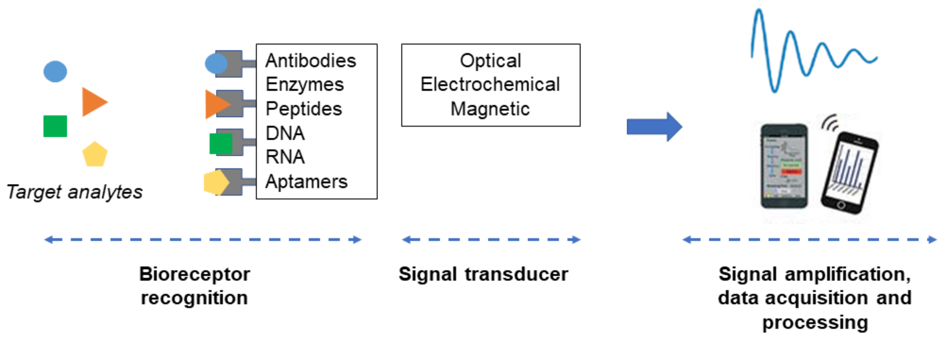
3. Biosensor (lab-on-a-Chip) Concept for Cytokines Detection

Cytokines are widely used for the monitoring and prediction of disease progression and therapeutic response. Currently, ELISA is the most routinely used cytokine assay for clinical samples analysis. Although relatively inexpensive and amenable to multiplexing, ELISA-based assays are not suitable for POC usage as it typically takes hours to prepare, process, and read the samples. It also requires a large sample size and relies on bulky equipment that is not suitable for POC applications [16,31]. With the limitations of conventional assays and the need for critical diagnostic cytokine measurements, many biosensor-based technologies have been developed as a simple, sensitive and inexpensive tool for cytokine detection. In particular, the development of portable handheld or wearable devices that can monitor health status continuously has received the most attention.
Biosensors are analytical devices that can convert specific biorecognition events into quantifiable signals. They are generally composed of the receptor (e.g., enzyme, antibody, or DNA) which specifically recognizes the analyte or biomarker (e.g., enzyme substrate, antigen, or DNA) and the transducer which converts the binding interaction into a measurable signal proportional to the analyte’s concentration (Figure 1). Biosensors are generally classified based on their signal detection mechanisms, e.g., electrochemical, optical, piezoelectric, or magnetic. These technologies have broad applications in health, environmental and forensic sciences [14]. In recent years, the emerging role of inflammation in many disease processes has led to the development of advanced biosensors for specific inflammatory cytokine detection in clinical samples (e.g., body fluids or tissue samples from patients). Key criteria for the development of these biosensors include sensitivity, selectivity, dynamic range, analysis time, accuracy, precision, and lifetime. Sensitivity and selectivity are the two most important factors for biosensor applications in research laboratory settings. Beyond the bench-top application, other factors, e.g., multiplexing, user friendliness, cost, and real-time monitoring become important. The configurations and detection approaches with the possibility to be integrated to the existing technologies or devices in clinical and POC settings are also much sought after [17].
An example of recently developed cytokine biosensors is the electrochemical biosensor for IL-1β and IL-10 detection in patients with heart failure after surgical implantation of left ventricular assist devices (LVADs). The non-biocompatibility nature of LVADs resulted in acute inflammation caused by the release of these two cytokines. The developed biosensor can detect both cytokines at a low level (e.g., 1–15 pg/mL), allowing early detection of the inflammation [32]. Another example is the electrochemical biosensor for the detection of IL-12 in multiple sclerosis patients based on the evidence that its high level in cerebrospinal fluid and serum correlates well with disease progression. It is also tested for the early identification of relapse and prediction of therapy outcome, which is of clinical importance for therapeutic intervention [33]. For multiplex cytokine detection, there has been an effort to develop a microfluidic surface plasmon resonance (SPR) biosensor for the detection of multiple analytes based on refractometry technique. This biosensor can perform multiplex analysis of six cytokines (IL-2, IL-4, IL-6, IL-10, IFN-γ, and TNF-α) within a linear detection range of 5–20 pg/mL, and only requires 1 μL of serum sample. The device was used for real-time monitoring of these inflammatory cytokines in two neonates after the bypass surgery [13]. In another study, a thumbnail-size patch was developed to detect seven cytokines from skin surface using self-assembly microdisks containing antibody arrays. This array-based patch was shown to provide sensitive detection of five cytokines, namely IL-1α, IL1RA, IL-17A, TNF-α, and IFN-γ [34].

4. Nanomaterials and Biosensing Application

As outlined in the previous section, several biosensors have been developed for the detection of various cytokines using different optical, electrical, electrochemical, and hybrid transduction methods. Among the various materials employed in these biosensors, nanomaterials have received the most attention due to their small size, ability to miniaturize, portability, and improved detection capabilities [17].
Nanomaterials are a group of materials that have nanoscale size (i.e., less than 100 nm in at least one dimension). They normally have different physicochemical properties than the same bulk materials. Nanomaterials can be classified into zero-dimensional (0D), 1D, and 2D. The shape of nanomaterials can vary from nanoparticles (NPs), nanorods (NRs), nanotubes (NTs), nanowires (NWs), and ultrathin films [35]. Nanomaterials are generally used as a transducer material, which is a key component of biosensors but can also be used for other functions such as immobilization support and signal amplifier [36]. Owing to the high reactive surface area and facile surface functionalization, improved performance of biosensors such as increased sensitivity of several orders of magnitude can be achieved. In some cases, the enhanced sensitivity is due to the fact that nanomaterials are of a similar size as the analyte, and thus are capable of probing previously unreachable matrices [37]. Biocompatibility is an important factor when designing a biosensor for biological applications [38]. Selectivity of the biosensors to specific analytes can be tailored by immobilizing biological recognition elements (e.g., enzymes, antibodies, and receptors) on the sensor substrate [39]. To date, many kinds of nanomaterials, such as metal NPs, metal oxide nanomaterials, and carbon NPs, have been used for biosensors fabrication. These nanomaterials can have different functions in different sensing systems [40].
Optical, electrochemical, and magnetic transduction methods are three major signal transduction methods in nanomaterial-based biosensors. The optical methods, particularly those that use colorimetric techniques to detect the signal in visible spectrum, are ideal for general users. Electrochemical methods are highly specific and can be simplistic to miniaturize [41]. Magnetic transduction methods exhibit minimal background signal as compared to optical and electrochemical methods, making them ideal for the detection of analytes at low concentration. In some applications, magnetic nanomaterials are used to pre-concentrate the analytes prior to their detection by optical or electrochemical methods.

4.1. Optical Transduction

Optical transduction is based on the emission or absorption of optical signal in a sample under irradiation (i.e., by visible, ultraviolet, or infrared light) [42]. To date, fluorescence and SPR-enabled spectroscopies are the two common optical transduction methods employed in the nanosensor field.
Fluorescence spectroscopy measures the emission of a fluorophore after excitation. In nanosensor applications, quantum dots (QDs), polymeric NP probes, or dye-doped silicon are frequently employed due to their superior photostability and robustness compared to traditional fluorescent dyes [43,44]. Changes in the fluorescence signal (i.e., quenching or restoration of the signal) can be interpreted as a result from the interaction between the analyte and NP or a conformational change of the sensor.
SPR-enabled spectroscopy is based on the localized SPR (LSPR) of noble metal nanomaterials [45]. The interaction of biomolecules with the nanomaterials results in the changes in LSPR and the optical response. Due to the sensitivity of LSPR band to the interparticle distance, it can be used to differentiate the dispersion and aggregation. It is commonly used in combination with secondary spectroscopic technique to generate a surface enhanced spectroscopy, i.e., surface-enhanced fluorescence (SEF) or surface-enhanced raman spectroscopy (SERS) [44].

4.2. Electrochemical Transduction

This method measures the change in electrical current or potential that results from the interaction between the analyte and the electrode. It can be classified into amperometric, potentiometric, and conductometric methods. Amperometric biosensors are based on the oxidation/reduction reaction that generates an electrochemical signal under a specific potential. The amperometric technique is generally used in the design of electrochemical biosensors by measuring current between the two electrodes when a potential is applied, or the analyte undergoes a redox reaction. For potentiometric biosensors, ion-selective electrodes are used to transduce biological reaction into electrical signal. The performance of this detection method is much dependent on the transducer surface properties, which determine the relative potential of the working electrode to reference electrode when zero current flows between them. This detection method is affected by the variations in ionic strength and pH of the detection media [46]. Conductometric biosensors measure the electrical current between the electrodes and reference. The current is generated due to the conductivity property of the analyte. It has mostly been used for enzyme detection to measure the change in ionic strength and conductivity of a solution between two electrodes as a result of enzymatic reactions [39]. Nanosensor designs can involve modifications of the electrodes (e.g., gold, silver, platinum, and graphite) with nanocarbons (e.g., carbon nanotubes and graphene) or functionalization with recognition elements (e.g., antibodies and aptamers) [41].
Due to the portability, self-containment, and low cost, electrochemical biosensors are dominant analytical tools for the biomarker detection. The application of functionalized nanomaterial for the fabrication of sensitive and selective electrochemical biosensors has been extensively explored in the biomedical field and other areas of analytical sciences. Numerous electrochemical biosensing techniques have been developed based on unique nanostructure designs for signal amplification and improved signal-to-noise ratios compared to the current electrochemical techniques [39,41,47]. The size and morphology of the analytes also affect sensor function. Small analytes are generally more diffusive and accessible, thereby better detection efficiency can be achieved compared to large molecules [48]. However, further knowledges on effective integration of nanomaterials, electrode microfabrication, and sensor engineering are crucial for the development of next-generation cytokine biosensors.

4.3. Magnetic Transduction

Magnetic NPs (MNPs) has been explored for the detection of analytes in biological samples. MNPs generally have low background magnetic signal. Under an applied magnetic field, magnetic signal from MNPs can be collected regardless of the optical properties of the solution [44,49]. MNPs has also been used to concentrate, separate, and purify analytes prior to the application of the secondary transduction method, such as electrochemical stripping [49].
An example of magnetic transduction is the magnetic-relaxation switches that incorporate superparamagnetic iron oxide NPs with the principle that the individual nanomagnetic probes will cluster upon their interaction with the target. The obtained NP clusters can enhance dephasing of the spins of the surrounding water protons. The change in spin–spin relaxation can be detected by magnetic resonance relaxometry [50]. Thus far, magnetic relaxation switches have been used for the detection of various analytes, e.g., DNA, mRNA, proteins, and viruses [51,52].

5. Nanomaterials as Biosensor for Inflammatory Cytokines

5.1. Quantum Dots (QDs)

QDs are semiconductor nanocrystals in a size range of 2–10 nm with unique optical and electronic properties. They are commonly synthesized from chemical elements in group II–VI or III–V of the periodic table, such as cadmium selenium (CdSe) or cadmium telluride (CdTe) [53]. Due to the quantum confinement effect, QDs show very different optoelectronic properties from bulk materials and have been extensively used in various research areas such as bioimaging, drug monitoring, diagnostics, and biomolecular interactions [54,55]. QDs typically have broad absorption spectra covering the ultraviolet to visible wavelength, depending on their particle size. The broad excitation and narrow size-tunable emission spectra of QDs, as well as their negligible photobleaching and high photochemical stability, make them superior alternatives to traditional fluorophores in many applications, including biosensor development [55]. The wide absorption spectrum of QDs offers great advantages in tracing multiple analytes simultaneously using simple instruments. QDs also offer some advantages in electrochemical applications with their semiconductor properties [56].
Recent studies demonstrated the value of QDs as a sensitive sensor for cytokines. Xu et al. reported a very low detection limit of 3.36 fg/mL for IL-8 when using DNA-templated QDs (DNA-QDs) as an electrochemical probe. In this application, samples were treated with a reducing agent, tris-(2-carboxyethyl) phosphine, and the obtained active thiols were then captured by the antibody-functionalized magnetic beads. DNA-QDs were then modified with maleimide, which can react to the active thiol. The amount of IL-8 in the samples can be determined from the electrochemical response of DNA-QDs [57]. In another study, graphene quantum dots (GQDs) were used to detect ultra-small amounts of cytokine intracellularly. The GQDs can be modified with cytokine aptamers (Ap-GQDs) or epitopes (Ep-GQDs). Both modified GQDs fluoresce in solutions at sufficient dilution; however, their fluorescence is quenched when conjugates of the modified GDQs are formed, i.e., due to aggregation between Ap-GQDs and Ep-GQDs. In the presence of cytokine-secreting cells, the secreted cytokines can compete with the epitope and cause the disassociation of the Ap-GQDs and Ep-GQDs aggregates, and therefore recover fluorescence signal. The Ap-GQDs and Ep-GQDs conjugates were successfully used to detect intracellular IFN-γ level at concentrations as low as 2 pg/mL [58].

5.2. Noble Metal Nanomaterials (NMNs)

Noble metals are metallic elements that are highly resistant to corrosion and oxidation, such as gold, silver, palladium, ruthenium, platinum, rhodium, osmium, and iridium. Currently, there is an unprecedented expansion of research to explore the application of NMNs in biotechnology and biomedicine due to their attractive properties, including a large surface area that enhances their biorecognition and receptor immobilization capabilities, the ability to catalyze electron transfer reactions, and good stability in biological systems [59]. NMNs have been used in sensors to amplify signal and improve sensitivity of the detection of biomolecules [60]. Optical, electrochemical, and piezoelectric sensing methods are the main sensing methods for NMN-based sensors. Among these biosensing methods, the colorimetric method has been the most frequently investigated due to its simplicity and strong prospect for applications at POC. For this reason, most biosensors to date are based on gold and silver NPs as they possess optical properties in the range of visible wavelength. In addition, they are easy to synthesize and functionalize. Other NMNs that have been used in biosensing applications include platinum NPs, albeit to a much lesser extent, due to their unique electrochemical properties [61].
• Gold Nanoparticles (AuNPs)
The majority of NP-based biosensing applications are based on AuNPs due to their optical properties [62,63]. AuNPs can transfer electrons efficiently, and the electron transfer rate in the presence of AuNPs is about 5000 per second compared to 700 per second in their absence [64]. The strong light-scattering and electromagnetic-enhancing properties of AuNPs allow them to be used as signal amplifier in diverse biosensor applications [65]. One interesting optical property of gold surfaces is that irradiation at a specific wavelength can cause oscillation of the electrons in the conduction band called resonant surface plasmons. This phenomenon is strongly dependent on size and shape of the particles, as well as dielectric constant of the environment. The change in oscillation frequency due to the recognition events provides unique advantage in the detection based on the color change of AuNPs, which is observable with the naked eye [66].
Research on the application of AuNPs as cytokine biosensors during the past decade can be divided into stages from proof of concept, attempts to improve existing detection methods, and development of new technologies or devices. For the proof of concept, there have been many studies investigating the SPR property of AuNPs for improved detection of several cytokines, including IL-1, IL-6, IFN-γ, and TNF-α [67,68,69]. One study demonstrated the detection of IL-6 based on visible color change caused by AuNPs aggregation (Figure 2a) [70]. Several other studies reported the use of AuNPs to improve existing cytokine detection methods, including immuno-polymerase chain reaction (iPCR)-based detection of IL-3 [71]. In this system, a DNA template and a target antigen-specific antibody are conjugated through AuNPs to form a new detection reagent with superior sensitivity and detection range than standard ELISA assay. The AuNP-iPCR assay is also easier and faster to perform with the possibility to measure low concentration and complex samples. New development of AuNP-based biosensors has focused on device portability. One study employed nanoimprinted gold strips with colloidal AuNPs for IL-10 detection. The sensor was fabricated via nanoimprinting process on polyethylene terephthalate (PET) film. AuNPs were deposited on the PET film to obtain the structures with a strong LSPR extinction peak. IL-10 detection antibody was subsequently immobilized on the strip surface. Colloidal Au nanocube (AuNC) was used to enhance the LSPR signal and the assay with AuNC crosslinked with IL-10 antibody demonstrates the sensitivity of detection at the nanomolar level [72]. In another study, a cytokine detection device using AuNP-modified silica optical fiber was developed to monitor locally variable concentrations of cytokine IL-6. The fiber was introduced into an intrathecal catheter. There were a micrometer-sized holes along the fiber length for fluid exchange. The optical fiber was modified with AuNPs layer and functionalized with IL-6 capture antibody. IL-6 detection antibody loaded on fluorescently labeled magnetic NPs was used for the detection (Figure 2b). The device managed to detect as low as 1 pg/mL of IL-6 from low volume sample (1 µL). It can also measure secreted IL-6 in real time, which is desirable for health condition monitoring [73].
• Silver Nanoparticles (AgNPs)
AgNPs have received enormous attention in the field of biosensors development due to their catalytic properties and enhanced detection of clinical markers [74,75]. Silver has many advantages compared to gold, such as sharper extinction bands, higher extinction coefficients and ratio of scattering to extinction, and extremely high field enhancements. However, it has been far less employed in sensors than gold due to its lower chemical stability in biological media and significant cytotoxicity compared to AuNPs [76,77]. Efforts have been made to improve the stability of AgNPs, i.e., by surface coating or modification. As a result, AgNPs are gaining popularity, and several research groups have begun to explore their use in optical sensors [76]. One of the most promising sensors developed from AgNPs is based on the enhancement of electromagnetic fields, which results in the so-called surface enhanced spectroscopies such as SERS and metal enhanced fluorescence (MEF). SERS allows ultrasensitive detection of various compounds, from chemical pollutants to biomolecules. It is sensitive enough to detect a single molecule of the analyte. In addition, this technique can give information about the structure of the analyte. Encapsulated SERS tags have been explored as alternative fluorescence tags. These SERS tags, compared to conventional fluorescence tag, demonstrate better stability and multiplexing capability. With this approach, Zhou et al. developed a 15-plexed silver-enhanced sandwich immunoassay (SENSIA). AgNPs linked to secondary antibodies are used, and the detection method was based on the quantification of the deposited Ag. This resulted in the affordable colorimetry-based detection for IL-1β, IL-10, IL-2, IL-4, and IL-6. The detection step can be performed using a simple flatbed scanner instead of the array readers that are normally used for multiplex cytokine detection with ELISA kits (Figure 3) [78]. Similar to SERS, MEF also has a great potential to overcome the drawbacks of traditional fluorescence tags [76]. In the vicinity of the fluorophores (4–10 nm distance), AgNPs can enhance the luminescence of fluorophores while other metals typically quench them. AgNPs, when deposited on a glass substrate, have shown to increase the emission intensity of a commonly used biological probe fluorescein by at least 3-fold [79]. Generally, MEF can lead to a 10–1000-fold intensity enhancement. The enhancement efficiency is based on the distance between AgNPs and the dye, the aggregation of AgNPs, and the overlap between the LSPR band of the AgNPs and the emission spectra of the fluorophore [76,80,81,82]. A study by Szmacinski et al. exemplified this approach for TNF-α detection. With a plasmonic nanostructure containing a unique composition of silver and silica layers, a more than 200-fold increase in fluorescence signal in the immunoassay was achieved. The system was used to image TNF-α secretion in primary murine macrophages after stimulation with lipopolysaccharide. The MEF assay also provided advanced imaging capability for the detection of cytokine secreted from a single cell without any complicated procedures required by standard methods [83].
• Palladium Nanoparticles (PdNPs)
PdNPs have captivated much attention in biomedical sensor development due to their exceptional catalytic activity. A variety of PdNPs with diverse composition and functionalization have been investigated for the detection of different biomarkers during the last decade. Higher abundance of Pd compared to other noble metals makes it a cheaper alternative for application in biosensing platforms [84]. Pd is a plasmonic material that possesses higher refractive index sensitivity than AuNPs and AgNPs. PdNPs are however not ideal in plasmonic sensing due to the broad extinction spectra and low scattering efficiency. They are therefore mostly investigated in combination with other NMNs for the optimization of sensor performance [85]. For example, PdNPs have been used with platinum (Pt) NPs to generate bimetallic Pd–Pt NPs for ultra-sensitive detection of IL-6 [86]. In this application, Pd–Pt NPs were synthesized and incorporated in an immunosensor to act as a functionalized bionanolabel. This immunosensor exhibited high sensitivity and selectivity for IL-6 detection with a broad linear response range of 0.1 to 2000 pg/mL and detection limit of 0.032 pg/mL in serum samples [86].

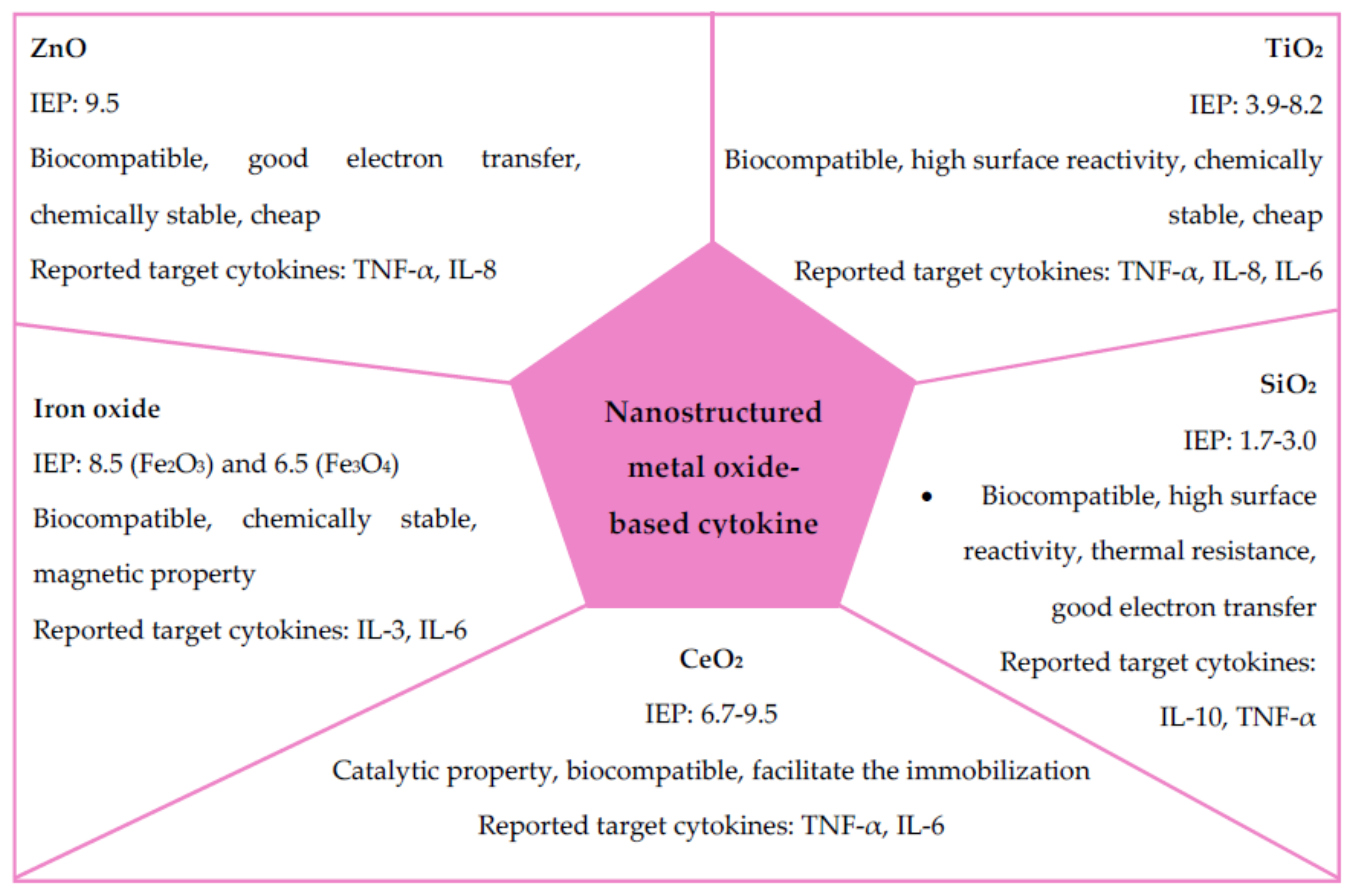
5.3. Metal Oxide Nanomaterials

Metals can form a large diversity of oxide compounds called metal oxides. Due to their exceptional electrical, magnetic, mechanical, optical, and catalytic properties, metal oxide-based nanomaterials and their polymeric composites have been used for many applications, including biosensing. Varieties of metal oxide NPs, including titanium dioxide (TiO2), silicon dioxide (SiO2), iron oxide (Fe2O3, Fe3O4), zinc oxide (ZnO), nickel oxide (NiO), gallium oxide (Ga2O3), and copper oxide (CuO), have been synthesized in different morphologies such as spherical, nanowires, triangular, nanotubes, and nanorods. With the reduced size and high-density corner or edge surface, oxide NPs display unique physical and chemical properties [87,88]. Their advantages include high stability; ease of preparation; customization to the desired size, shape, and porosity; swelling consistency; flexibility to incorporate into hydrophobic and hydrophilic systems; and ease of functionalization by various molecules due to the negatively or positively charged surface. Generally, metal oxide NPs are ionic particles, and the deposition of metal oxide NPs on the desired position with nanoscale resolution has a great potential for various applications in chemistry, electronics, optics, and biomedicine [89]. The main use of metal oxide NPs in electrochemical and biosensing applications is to strengthen the conductive sensing interface based on their catalytic property that allows electrical contact with the transducer surface. They can also work as electronic wires by facilitating fast electron transfer between the transducer and analyte molecule. Biocompatible metal oxide NPs have been used to fabricate immunosensors, enzyme sensors, and DNA sensors, while semiconducting NPs are primarily used as electrochemical markers and tracers. Nanoparticulated metal oxide can be coupled to the working electrode surface by several techniques such as physical adsorption, electrical deposition, chemical covalent bonding, and electro-polymerization [90]. Nanoparticulated metal oxide has been used in various applications, including small molecule electroanalysis, enzyme-based sensors, and immuno-and geno-sensors which are mainly used for cytokine detection. Genosensors utilize immobilized single-stranded DNA fragments on the electrode surface, while immunosensors are based on antigen–antibody interaction mostly in combination with electrochemical transduction. The performance of immunosensors depends on the density of immobilized antibodies/antigens, which is enabled by metal oxide nanostructures [89]. A few selected metal oxide NPs-based biosensors for cytokine detection are highlighted in this section. Key properties of these metal oxide NPs and their applications for cytokine sensing are summarized in Figure 4.
• Nanostructured zinc oxide (ZnO)
Recently, much attention has been given to the research on ZnO-based biosensors. Due to the distinctive electrical transport, high isoelectric point (IEP), low price, and high chemical stability, ZnO is a widely accepted candidate for biosensor applications [91]. Its high IEP allows better adsorption of enzymes, proteins, and DNA by electrostatic interactions. Its unique physicochemical properties have been exploited in a number of detection methods including electrochemical and optical methods. ZnO is known to be nontoxic and compatible with human skin, making it a promising material for use as a permanent biosensor in chronic diseases [91,92]. In the context of cytokine detection, some recent studies have shown good performance of biosensors based on ZnO nanorods. The ZnO nanorods allow enhanced fluorescence detection of DNA and protein samples and are therefore extensively investigated as an optical platform. The most recent development in this area is the multiplexed sensor for two biomarkers, TNF-α and IL-8, in urine to identify patients with the risk of having acute kidney injury. As compared to commercial ELISA-based sensors which have the detection limit of 5.5 pg/mL for TNF-α and 7.5 pg/mL for IL-8, the ZnO nanorod sensors have a lower detection limit of 4.2 fg/mL for TNF-α and 5.5 fg/mL for IL-8. In general, the ZnO nanorod sensors can detect TNF-α at the level well below the detection limit of ELISA. In addition to its superior sensitivity, the ZnO nanorod sensors offer the advantages of rapid analysis, minimal volume requirement, and reusability. The whole detection process can be completed within 90 min using only 60 µL of total sample volume. Furthermore, the biocompatible ZnO nanorod can survive at least 25 repeated assays with complex biological and chemical environments such as urine samples [17,93].
• Iron Oxide Nanoparticles
Iron oxide NPs including hematite (Fe2O3) and magnetite (Fe3O4) NPs have gained enormous attraction in biosensing due to their special magnetic, optical, and electrical properties. Their biocompatibility and durability make them highly desirable in many biomedical applications [94,95]. However, only a handful of iron oxide NPs-based biosensors have been reported, i.e., for the detection of IL-3 and IL-6. One of these studies utilized a nanocomposite of complexed longitudinal zeolite and iron oxide to improve surface characteristics of the biosensor designed for IL-3-mediated sepsis monitoring. The surface of zeolite-/iron oxide electrode were coupled to IL-3 antibody through an amine linker. The obtained sensor demonstrated the limit of IL-3 detection of 3 pg/mL due to the high degree of antibody immobilization on the sensing electrode [96]. Another study reported a new assay called “OnCELISA” for highly sensitive detection of trace cytokines secreted from individual live cells. This assay is the extension of ELISA by using cell surface to capture the secreted molecules that can be detected by fluorescent magnetic iron oxide NPs called Dragon Green superparamagnetic iron oxide (DG SPIO). This technique enhances the sensitivity of detection to 0.1 pg/mL, which is 10-fold more sensitive than standard fluorescence technique. This assay was implemented by surface biotinylation of cells followed by the attachment of IL-6 capture antibody. This secreted cytokine can then be captured on the cell surface immediately after their release instead of being diluted in the media. The captured cytokines are then labeled by DG SPIO-conjugated IL-6 detection antibodies and the fluorescence signals indicate the cytokine level. The labeling is stable after 12 h at 4 °C, and cell secretion and proliferation are not affected. However, the two antibodies required for OnCELISA should be produced against different epitopes of the same target cytokine [97].
• Titanium Dioxide (TiO2) Nanoparticles
TiO2 is a semiconductor material used in many applications such as photocatalysis, biosensors, and energy storage due to its properties such as high chemical stability, morphological versatility and biocompatibility. The wide applicability of recently developed TiO2 composites has accelerated the development and miniaturization of biosensing devices. The electrical characteristics of these nanostructures can be easily modified using structured shape and geometry to give higher surface reactivity and strong adsorption of biomolecules [98,99]. Sensitive nanostructured TiO2-based electrochemical biosensors for IL-6 detection have been developed based on surface-modified layers of TiO2 nanotubes. The nanotube layers are functionalized on the surface by direct immobilization of biological reagents. Strong antibody–antigen interaction has been used in two types of electrochemical biosensors: the impedimetric and amperometric. The impedimetric biosensor measures changes in capacitance caused by the formation of IL-6 antibody-antigen complex on the biosensor. For the amperometric biosensor, horseradish peroxidase (HRP) conjugated antibodies were used. The two electrochemical methods were reported to detect 5 pg/mL of IL-6, which is below the detection limit of commercially available ELISA kits [100]. The same technique was elaborated by the same research group to create an inexpensive, easy to prepare, and label-free cytokine array for the selective detection of IL-6, IL-8, and TNF-α. By immobilizing antibodies onto the TiO2 nanotube array via physical adsorption, IL-6, IL-8, and TNFα can be quantified simultaneously using electrochemical impedance spectroscopy (EIS). This impedimetric immunosensing technique shows good selectivity and high sensitivity against all analytes at the lower concentration than the normal concentration of these cytokines in the blood [101].
• Cerium Dioxide (CeO2) Nanoparticles
CeO2 is a nanostructured metal oxide commonly used in the biosensors field due to its easiness to immobilize enzymes or proteins on the electrode surface and its tremendous catalytic activity. In particular, immobilization of antibodies is crucial for the high sensitivity of the immunosensor [84]. CeO2 belongs to lanthanide metal oxide family and exhibits an excellent catalytic property due to the interconversion of oxidation states between Ce(III) and Ce(IV). In CeO2 nanostructures, Ce binds to oxygen to form fluorite crystalline structure. The transition of Ce(IV) to Ce(III) creates oxygen vacancies in the lattice. The presence of oxygen vacancies or defects in the lattice of each oxidation state improve electrocatalytic phenomena. Moreover, the high isoelectric point facilitates a strong binding interaction with biomolecules of low IEP via electrostatic interactions [102].
An ultrasensitive electrochemical immunosensor using Prussian Blue (PB) functionalized CeO2 NPs (PB-CeO2) has been developed for the detection of TNF-α. Preparation of PB-CeO2 started by coating CeO2 with positive charged chitosan (CS) layer. Negatively charged PB was then adsorbed on the CS layer. The synthesized NPs have a high sensitivity toward H2O2 detection. The electrochemical immunosensor was constructed on a conventional sandwich platform with immobilized primary anti-TNF-α antibody. Secondary anti-TNF-αantibody was crosslinked with PB-CeO2 through glutaraldehyde. The immunosensor was reported to have a low detection limit (2 pg/mL) for TNF-α over a wide linear range (0.005–5 ng/mL) [103]. CeO2 has also been used to improve the detection limit of ELISA-based cytokine assay kits together with magnetic Fe3O4 particles. Based on this principle, a magnetic colorimetric assay for IL-6 detection was developed by the immobilization of the capture antibody on magnetic Fe3O4 particles. The secondary antibody was coupled on CeO2 spheres. The labelled CeO2 catalyzed the oxidation of o-phenylenediamine to 2,3-diaminophenazine, a yellow product with absorption wavelength at 448 nm. The assay demonstrated the detection limit of IL-6 at 0.04 pg/mL with the linearity range of 0.0001–10 ng/mL [104].
• Silicon Dioxide (SiO2) Nanoparticles
SiO2, or silica, is a covalently bonded material consisting of silicon and oxygen. It is a component of sand that can be found naturally as quartz. SiO2 is usually white or colorless and not water-soluble. SiO2 NPs are important material for the optical fibers production and have been used for the construction of nanobiosensors due to their biocompatibility and good electron transfer properties. Their inherent resonance light scattering (RLS) characteristic can be used to develop a simple sensing method. SiO2 NPs possess a porous structure, high thermal resistance, and very high surface activity and adsorption properties. The structural features (e.g., size, shape, and porosity) of SiO2 NPs can be facilely modified to improve their performance. The silane chemistry enables surface engineering of SiO2 NPs with diverse molecules and polymers. Such surface functionalization was shown to influence the biological activities of SiO2 NPs in vivo, including blood circulation, biodistribution, and cellular internalization [105]. The refractive index of SiO2 NPs is similar to that of polymeric coatings, so it finds several applications in nanocomposites that improve the sensitivity and specificity of detection, and reduce the toxicity of biosensors, i.e., by encapsulation or sandwich formation [106,107,108,109,110]. To date, SiO2 NPs have been used in cytokine biosensors as an immobilization substrate or signal amplifier. Lee et al. developed a novel substrate based on hafnium oxide (HfO2) grown on SiO2 for IL-10 detection. The sensor measured IL-10 within the linear range of 0.1–20 pg/mL and sensitivity of 0.49 ng/mL [111]. As a signal amplifier for cytokines detection, QD-polymer-functionalized SiO2 nanospheres have been used for TNF-α measurement. Anti-TNF-α antibody was bonded to the functionalized nanospheres to obtain QD-polymer-functionalized SiO2 probes which were attached to the electrode surface. Enhanced sensitivity of detection can be accomplished by increasing the QD loading per immunoassay. With this approach, 10-and 5.5-fold increase in detection sensitivity from electrochemiluminescence (ECL) and square-wave voltammetry (SWV) measurements compared to unamplified method were observed, respectively. The detection limits of 7 and 3 pg/mL were achieved for the ECL and SWV detection of TNF-α, respectively [112].

5.4. Carbon-Based Nanomaterials

Due to their exceptional optical, electrical, mechanical, chemical, and thermal properties, extensive research has been carried out on carbon-based nanomaterials. Carbon is commonly used for the construction of electrodes based on its unique electrochemical properties, e.g., small background current and large potential window, as well as its low cost. Moreover, carbon nanomaterials are biocompatible. To date, different carbon allotropes such as graphene, carbon nanotubes (CNTs), fullerenes, carbon dots, carbon black, and nano-diamonds have been exploited to achieve specific properties or desirable features of biosensors [113,114,115]. Developed cytokine biosensors based on carbon nanomaterial are summarized in Table 3.
• Graphene
Graphene is the most prominent member of carbon nanomaterials for biosensor applications. The physical, optical, and electrochemical advantages of graphene have led to improved sensitivity and selectivity of biosensors [116]. However, there are some problems associated with graphene-based biosensors. For instance, graphene exhibits a high agglomeration tendency, making it difficult to prepare, and there are no standardized procedures for its biosensors. Moreover, the physical stability of graphene in biological media, as well as its potential toxicity, remain a concern [117,118]. Graphene-based multi-analyte electrochemical immunoassays have been developed for rapid and ultrasensitive detection of IL-6 and matrix metallopeptidase-9 (MMP-9) simultaneously. The sensor utilized a graphene nanoribbon (GNR)-modified electrode to immobilize the antibody and amplify the electrochemical signal. With sandwich-type immunoassay strategy, simultaneous detection without crosstalk between the sensing electrodes was achieved. The detection range of 10−5 to 103 ng/Ml with the detection limits of 5fg/mL for MMP-9 and 0.1pg/mL for IL-6 reported [119]. Several other studies investigated the benefit of graphene as a label-free sensor for IFN-γ. Ruecha et al. [120] reported a novel paper-based microfluidic device as a label-free electrochemical impedance immunosensor. The electrode was fabricated by depositing polyaniline (PANI) on a graphene screen-printed paper electrode. This modified electrode exhibited high electrochemical conductivity and surface area resulted in significant improved in sensor sensitivity due to better antibody immobilization. The developed sensor can detect IFN-γ at the lowest concentration of 3.4 pg/mL with linear relationship between the impedance and IFN-γ concentrations in the range of 5–1000 pg/mL [120]. In another study, IFN-γ aptamer was immobilized on a graphene surface to create a field-effect transistor (FET) sensor that measures the change in conductance due to binding of the molecules of interest. The graphene structure was incorporated onto a PDMS substrate. In the presence of analytes, a change in charge distribution occurs, which results in an increase in electron transfer that can be monitored via current measurements. Such current changes were found to correlate with IFN-γ concentrations in the nano to micromolar range with the detection limit of 83 pM [121].
• Nanodiamonds (NDs)
NDs are nanomaterials with the crystal structure and specific properties of diamond. They are small, but have large surface area. Their high adsorption capacity allows attachment of various chemical and biological molecules to their surface. NDs have low toxicity, with exceptional hardness, refractive index, thermal conductivity, chemical stability, and coefficient of friction. Due to the presence of a complex defect N-V, containing nitrogen (N) and a vacancy (V), they can also emit fluorescence signal which can be used for several applications [114,122]. Stochastic ND-based sensors for the detection of IL-1β, IL-6 and IL-12 have been reported. By creating nano-sized pores in a membrane, ionic transport through the pore in the presence of analytes was measured by the sensor. The magnitude, duration, and rates of occurrence of the resulting current blockade allow rapid analysis of analyte concentrations, as well as discrimination of the similar molecular species [123]. Reliable determination of the cytokine concentrations was demonstrated in whole blood, urine, and brain tumor samples [124]. NDs have also been explored for the application as insertable optical fibers for in vivo biosensing of cytokines. Captured antibodies were immobilized on the optic fiber surface. The functionalized fiber was placed in vivo for cytokine capturing, and it has successfully been used for the detection of IL-6. The sensor demonstrated the sensitivity of 0.4 pg/mL with spatial resolution of 200 µm. It has also been used to measure IL-1β release in discrete brain regions in animals via inserted fiber optic device [125,126].
• Fullerenes
Fullerenes are carbon-based hollow nanomaterials. The structure characteristic of fullerenes is the establishment of atomic Cn clusters (n > 20) on a spherical surface. C60 is the most extensively studied fullerene. It is a symmetric spherical molecule consist of 60 carbon atoms with the diameter of 0.7 nm. C60 offers several advantages, such as high symmetry, low cost, ease of production, inertness (under mild conditions), and non-toxicity. Fullerenes have unusual properties, such as multiple redox states, stability in different redox forms, and light source switching. They are also easy to be functionalized. Therefore, they have been used in many different applications, including superconductors, biosensors, catalysts, and optical and electronic devices [127,128]. Fullerenes have been used to increase the sensitivity of commercially available carbon electrodes for the detection of IL-8 via silver stripping voltammetry technique. The analyte concentration can be measured through current measurements over the scanned potential. The analyte is pre-concentrated at the electrode and then stripped by applying a potential. The nanostructured electrodes showed higher sensitivity than bare carbon electrodes with the detection limit of 0.61 ng/mL [129]. A nanocomposite containing fullerene and multi-walled carbon nanotubes (MWCNTs) was used to create a sensitive label-free electrochemical immunosensor for TNF-α detection. TNF-α antibodies were entrapped onto the fullerene-MWCNT nanocomposite and used as a sensing platform and electrocatalyst for oxidation. TNF-α was determined based on its obstruction of the electrocatalytic oxidation upon the binding to the electrode surface via an interaction with TNF-α antibody. Under optimal conditions, the electrochemical immunosensor can detect TNF-α with the dynamic range of 5–75 pg/mL and detection limit of 2.0 pg/mL [130].
• Carbon Nanotubes (CNTs)
CNTs are basically one layer of graphene rolled into a hollow tube with a diameter in nanoscale. Based on their structures, they can be simply classified into two groups: single-walled (SW) and multi-walled (MW) CNTs. Compared to other fibrous materials, CNTs possess excellent physical properties such as rigidity, tensile strength, and elasticity. They exhibit high aspect ratio and high thermal and electrical conductivities. CNTs have spurred much interdisciplinary research due to their unique structural, electronic, optoelectronic, mechanical, and chemical properties. CNT-based materials have increasingly been recognized as the next generation building block for ultra-sensitive and ultra-fast biosensors. Their strong asset is their constitute scaffolds, which accommodate functionalization by several recognition and signal transduction entities, thus allowing multiplexed testing of analytes. With greater ability to conduct electricity than copper wires, CNTs can potentially be a good material for the transduction of electric signals generated upon the biorecognition event of a target [131,132].
CNTs have been applied for the detection of several cytokines including IL-6, TGF-β, and IL-1β. Enzyme-labeled immunosensors for sensitive IL-6 detection have been developed based on dual amplification of AuNPs and CNTs. In this platform, the capture antibody was immobilized on the AuNP-modified electrode, while the detection antibody, which is conjugated with HRP, was coupled to CNTs for signal detection in the presence of H2O2. The system showed a linear response range of 4–800 pg/mL with the detection limit of 1 pg/mL for IL-6, consistent with standard ELISA results, demonstrating its reproducibility, selectivity, and reliability [133]. SWCNTs have been used as a nanotag in an amperometric biosensor for TGF-β. In this approach, the nanotubes were hybridized with viologen, an organic compound that can reversibly change its color through reduction and oxidation. The TGF-β antibody with HRP tag was then covalently linked to the viologen–SWCNT hybrid. Capture antibody was immobilized on the carbon electrodes and sandwich-type immunoassay was employed with signal amplification from the viologen–SWCNT-antibody-HRP as a carrier tag. The immunosensor yielded a linear detection range of 2.5–1000 pg/mL and detection limit of 0.95 pg/mL, which is better than other immunosensors or ELISA kits for TGF-β detection. Such high sensitivity was also observed with other proteins. Validation of the sensor was performed in saliva samples. Minimal sample preparation was required, and the results obtained were in good agreement with standard ELISA results [134].
In another study, an ultrasensitive electrochemical immunosensor based on CNTs forest electrodes was reported. SWCNT forests with captured IL-6 antibodies were used in an electrochemical sandwich immunoassay. The detection antibody was attached to HRP-labeled MWCNTs. This setup provided the most sensitive detection of IL-6 with the detection sensitivity of 0.5 pg/mL (25 fM) [135]. MWCNTs have also been used to fabricate electrochemical sensors for IL-1β detection using electro-click chemistry to immobilize ethynylated antibody onto azide-functionalized MWCNTs. The sensor was then used to perform sandwich-type immunoassay with biotinylated anti-IL-1β as a detection antibody. The electrochemical immunosensor exhibited two linear ranges from 10–200 pg/mL and 200–1200 pg/mL with the detection limit of 5.2 pg/mL, a significant improvement over standard ELISA assay. Outstanding performance of the sensor was observed when the sensor was used to determine the amount of IL-1β in saliva without the need of sample pre-treatment [136]. There has been an attempt to coat CNTs with cadmium telluride (CdTe) QDs and a single molecule of captured antibody to improve IL-6 detection. Sample IL-6 competed with surface-bound IL-6 for the binding sites on the captured antibody, which led to the depletion of CNT-QD-antibody on the sensor surface with increasing sample IL-6 concentrations [137].
CNTs have also been used for label-free impedimetric immunosensor development. Impedimetric immunosensors consist of a transducing electrode coated with a biorecognition element (antibody or aptamer) where binding of the target protein results in a change in the electrical impedance. There was a study to develop an impedimetric immunosensor for IL-6 detection by integrating SWCNT and AuNPs. The sensing platform was constructed by depositing AuNPs on SWCNT array on SiO2/Si substrate. The developed sensor can detect IL-6 in serum at a very low limit of 0.01 fg/mL, which is much lower than that the results from the sensor using CNTs forest or AuNPs alone. The combination sensor demonstrated wide linear response from 0.01–100 fg/mL with a low response to several interferences found in serum. The sensor also possesses a high stability with no change in sensitivity after being stored for a month at 4 °C [138]. Another example of label-free cytokines sensors was reported by Khosravi et al. [139] who employed aptamer-functionalized CNTs with aptamer as the recognition element. In this platform, SWCNTs were functionalized with 1-pyrenebutanoic acid succinimidyl ester (PASE) conjugated IL-6 aptamers. In the presence of IL-6, the conductivity of functionalized CNTs was decreased in the correlation to the concentrations of IL-6 [139].

5.5. Polymer Nanomaterials

Polymeric nanomaterial is a common term used for all polymer-based nanomaterials. They are obtained from synthetic or natural sources and can be classified based on their in vivo behavior as biodegradable or non-biodegradable. Polymer nanocomposites have a large surface area, high electrical conductivity, and fast electron transfer rate which make them a good transducer in electrochemical sensors. They can be synthesized with several nanoscale variations of fillers leading to various specific sensing applications. The fillers enable ion diffusion by intercalating into the polymer matrices resulting in improved stability of active electron transfer and detection limit of the sensor [140]. To date, several polymeric nanomaterials, including conducting polymers (CPs), dendrimers, and molecular-imprinted polymers (MIPs), have been developed for the detection of various analytes [84].
CPs are polyconjugated polymers consist of alternating single and double bonds. With this molecular structure, CPs exhibit electrical and optical properties that are suitable to be used as sensing transducer. Inclusion of CP nanostructures in the sensor transducer has been shown to improve sensing performance significantly [141]. Several CPs have been integrated into electrochemical immunosensors, including polypyrrole (PPy), PANI, poly-2,5-di-(2-thienyl(-1-pyrrole-1-(p-benzoic acid) (DPB), poly-(3,4-ethylene dioxythiophene) (PEDOT), and poly-pyrrolepropionic acid (PPA). PPy, a five-membered heterocyclic ring, is prominent for its tunable conductivity, ease of synthesis, reversible redox activity, and environmental stability. As PPy nanostructures can be prepared in both aqueous and non-aqueous solutions through electrochemical or chemical interaction, they are particularly useful for various applications, including biosensors [141,142]. Aptamer probes doped in PPy have been employed for the detection of platelet-derived growth factor (PDGF), an important inflammatory cytokine, by EIS with the reported sensitivity of 10 ng/mL [143]. PPy-modified silicon nitride (Si3N4) substrate has similarly been employed to develop a label-free capacitance impedimetric immunosensor for IL-10 detection. The immunosensor was created by chemically depositing the PPy conducting layer on Si/SiO2/Si3N4 substrate, followed by IL-10 antibody immobilization, and detection by EIS. The developed sensor showed a linear detection response in the range of 1–50 pg/mL with a high sensitivity of 0.1128 pg/mL and very low detection limit of 0.347 pg/mL [144].
PANI is a semiconducting polymer that contains repeating units of benzenoid diamine and quinoid diamine. PANI has high surface to volume ratio, as well as distinct redox couples based on its repeating units, resulting in effective electron transfer. To date, 2D PANI is one of the promising nanostructures in flexible biosensors development. However, diminished conductivity of PANI under physiological conditions tends to be an obstacle for its application. PANI has been employed in several studies to develop biosensors for IFN-γ. One study used magnetic NPs coated with PANI and captured antibody to magnetically capture IFN-γ from the sample. AuNPs conjugated with detection antibody and cadmium sulfide (CdS) NPs were then added to form a multifunctional complex and added on a screen-printed carbon electrode (SPCE) chip. The electrochemical signal from CdS-NPs was then measured by stripping voltammetry. The whole detection process with the developed immunosensor can be completed in about 1 h [145]. In another study, a screen-printed paper electrode (SPPE) was used to detect IFN-γ by microfluidic immunosensors. The electrode consists of graphene coated with PANI, which enables covalent immobilization of IFN-γ antibody. One unique advantage of this paper-based sensor is its ability to be mass produced at a low cost [120]. Another nanocomposite of graphene oxide, PANI and QDs has been used for electrochemiluminescence detection of IL-6 with the sensitivity range of 0.0005–10 ng/mL [146].
PEDOT is one of the most explored CPs. It is polythiophene derivative synthesized from 3,4-ethylenedioxythiophene (EDOT) monomer. It has exceptional conductivity, good optical properties, and electronic stability in physiological settings. Currently, the most studied PEDOT derivative is PEDOT:polystyrene sulfonate (PSS). It is the co-polymer mixture of PEDOT and sodium polystyrene sulfonate. PEDOT:PSS exhibits high conductivity and stable in oxidized state. It is also visible light transmissive and water dispersible. To date, PEDOT:PSS is commonly used as a flexible polymeric electrode based on its flexibility, tunable conductivity, biocompatibility, and low cost [141,147].
MIP-based biosensors have been investigated for the detection of various molecules. Although imprinting large or complex structures such as proteins is still a challenge, imprinting techniques is well demonstrated to reveal selective organization of functional groups in the imprinting biomolecules. Nanostructured MIPs can be produced by a variety of methods including dispersion, suspension, and emulsion-seeded polymerization [84]. A combination of MIP and PEDOT has been used for the detection of IL-1β via an electrochemical MIP sensor. To enhance the detection sensitivity and signal stability, the carbon backbone was functionalized with PEDOT/4-aminothiophenol and MIP film. Using EIS detection, a linear response range of 60 pM to 600 nM with a detection limit of 1.5 pM was obtained. Selectivity tests confirmed good selectivity of the developed biosensor [148].

5.6. Bionanomaterials

Bionanomaterials are biomaterials with a nanoscale dimension that comprise partially or totally of biological molecules such as antibodies, proteins, DNA, RNA, polysaccharides, and lipids. Numerous efforts have been made on the development of hybrid structures containing bionanomaterials and other functional NPs (e.g., CNTs, graphene, nanometal, and QDs) for applications in biosensors, bio-imaging, biomineralization, biocatalysts, and drug delivery [84]. The most widely used bionanomaterial for cytokines biosensors is aptamer. Aptamers are single-stranded oligonucleotides (DNA or RNA) that can form high-affinity complexes with their targets via specific conformational changes and can be used as an alternative small biomolecule recognition element [149]. Several aptamers have been incorporated in cytokine biosensors, e.g., fluorescence DNA/RNA probes. In these aptamer-based sensors, the release of quencher-labeled complementary strand or fluorophore-labeled aptamer upon the binding of analytes is detected [17]. Few studies reported IFN-γ detection using Förster resonance energy transfer (FRET)-based aptasensors. An aptamer variant was constructed by the hybridization of fluorophore-labeled aptamer with quencher-labeled complementary oligonucleotide strand. IFN-γ displaced the quenching strands, disrupting FRET and resulting in fluorescence signal. The signal can be observed directly upon binding of the IFN-γ. This aptasensor was reported to be able to detect IFN-γ in a linear range from 5 to 100 nM with the detection limit of 5 nM [150]. The sensor was further developed for real-time monitoring of cytokine secretion. Micro-patterned aptamer beacons were fabricated by micro-patterning glass substrate with polyethylene glycol (PEG) hydrogel microwells to define sites for attachment of aptamer molecules. In the presence of IFN-γ, the quencher displaced from fluorophore-labeled aptamer and fluorescence signal in the wells increased. The real-time changes in fluorescence signal was confined to microwells and depends on IFN-γ concentration [151].
In this section, some interesting applications of nanomaterial for the development of cytokine biosensors are presented. In general, several parameters should be considered before deciding which nanomaterials to use, including dynamic range, sensitivity, cost, ease of miniaturization, and biocompatibility. An increasing number of studies regarding the combination of these nanomaterials to produce new nanocomposites with enhanced performances have been reported in the literature. Table 4 compares the strengths and weaknesses of different nanomaterials for cytokine biosensor application. To date, there is no commercially available cytokine biosensors for POC application. Most of the discussed studies are still in the developmental stage at academic research laboratories. However, integration into real application has been accelerated, i.e., a wearable SWEATSENSER device for the detection of IL-6, IL-8, IL-10, and TNF-α fabricated with ZnO nanofilm. The device demonstrated analytical range of 0.2–200 pg/mL and had been shown in a clinical study to be able to distinguish healthy subjects from subjects with infection [152].

6. Conclusions and Future Perspectives

Advanced cytokine biosensors are being developed at a rapid pace due to the increasing realization of the crucial role of cytokines in disease pathogenesis and the need for early diagnosis, disease monitoring, and therapeutic response assessment. This article reviews the evolving nanomaterials-based biosensors for cytokine detection, as well as the strategies and limitations associated with these biosensors. Major signal transduction methods employed in nanomaterials-based biosensors including optical, electrochemical, and magnetic transduction methods were mentioned. Nanomaterials discussed are QDs, noble metal nanomaterials, metal oxide nanomaterials, carbon-based nanomaterials, and bionanomaterials. Several of these biosensors utilize hybrid nanomaterials to achieve the specific application needs. Examples of these biosensors and their integrated nanomaterials are described, along with their applications in cytokine measurements.
Most cytokines in the body function in concert with other cytokines to elicit their biological or pathological effects. Consequently, there is a need for multiplexed detection systems that can detect multiple cytokines simultaneously. Several multiplexed cytokine biosensors have been developed in recent years, largely through the incorporation of new nanomaterials and surface coating or functionalization of the nanomaterials with multiple biosensing moieties. Due to their increasing complexity, the issues of portability and ease of use should be carefully addressed to promote their applications. Hardware and software packages will also need to be developed to handle the complexities of future devices and to provide user-friendly readouts. Advances in nanomaterials science and bioengineering have paved the way for ultrasensitive detection and real-time monitoring of multiple cytokines that could not be achieved by conventional detection methods. Further development of these technologies will enable better and more sensitive cytokine biosensors. However, the current implementation of these technologies has been limited to some specialized research fields and analytical laboratories. Translation of these biosensor technologies into clinical and hospital settings remains a challenge. For POC applications, portability, sensitivity, specificity, stability, and real-time monitoring are critical. Wearable devices can be an exciting opportunity in healthcare due to their potential to change the status quo in patient care, with tools that the patients can use to track their own conditions and take responsibility for their own health. The unique properties of nanomaterials, such as high conductivity, large aspect ratio, high tensile strength, and fast electron transfer rate, make these materials well suited for wearable biosensor applications.
Currently, the information on large-scale fabrication of nanomaterial-based devices is still lacking, so their practical applications have not yet been fully realized. Power supply is another important issue in wearable sensors. With the trend toward miniaturization and flexibility to function at biological interfaces which may be soft, folded, or curvy, rigid batteries would be ineffective. Battery-free and wireless sensing systems, such as near-field communication (NFC), could be a good choice for future research in this field. NFC modules allow real time and wireless data transmission without the need for battery. One limitation of this technology is its short transmission range, which results in the need for a close distance between the sensor and data receiving unit.
Despite their many advantages, nanomaterials do pose human health risks and potential threat to the environment. Nanoscale materials, due to their exceptionally small size, can enter the systemic circulation and penetrate deeper tissues to cause unintended health effects. To date, several nanomaterials have been investigated for their biocompatibility and biodegradability to avoid such consequences. These studies should be conducted in parallel with their performance assessment to ensure safe and effective utilization of the technologies.

Author Contributions

W.L.: conceptualization, original draft preparation, and editing; Z.A.: discussed the idea of writing review, its content, and feedback on the written review; and Y.R.: conceptualization and feedback on the article content and original draft. All authors have read and agreed to the published version of the manuscript.

Funding

This work was supported by grants from the National Institutes of Health (NIH) (R01-ES022968 and R01-EB018857).

Acknowledgments

Z.A. acknowledge the financial support for a project in collaboration with Essity AB.

Conflicts of Interest

The authors declare no conflict of interest.

References

  1. Dinarello, C.A. Historical insights into cytokines. Eur. J. Immunol. 2007, 37, S34–S45. [Google Scholar] [CrossRef] [PubMed] [Green Version]
  2. Kelso, A. Cytokines: Principles and prospects. Immunol. Cell Biol. 1998, 76, 300–317. [Google Scholar] [CrossRef] [PubMed]
  3. Chung, K.F. Chapter 27—Cytokines. In Asthma and COPD, 2nd ed.; Barnes, P.J., Ed.; Academic Press: Oxford, UK, 2009; pp. 327–341. [Google Scholar]
  4. Luo, Y.; Zheng, S.G. Hall of Fame among Pro-inflammatory Cytokines: Interleukin-6 Gene and Its Transcriptional Regulation Mechanisms. Front. Immunol. 2016, 7, 604. [Google Scholar] [CrossRef] [PubMed]
  5. Kupsa, T.; Horacek, J.M.; Jebavy, L. The role of cytokines in acute myeloid leukemia: A systematic review. Biomed. Pap. Med. Fac.Univ. Palacky Olomouc Czech Repub. 2012, 156, 291–301. [Google Scholar] [CrossRef] [PubMed] [Green Version]
  6. Sachdeva, N.; Asthana, D. Cytokine quantitation: Technologies and applications. Front. Biosci. J. Virtual Libr. 2007, 12, 4682–4695. [Google Scholar] [CrossRef] [PubMed] [Green Version]
  7. Garg, M.; Sharma, A.L.; Singh, S. Advancement in biosensors for inflammatory biomarkers of SARS-CoV-2 during 2019–2020. Biosens. Bioelectron. 2021, 171, 112703. [Google Scholar] [CrossRef] [PubMed]
  8. Liu, G.; Jiang, C.; Lin, X.; Yang, Y. Point-of-care detection of cytokines in cytokine storm management and beyond: Significance and challenges. VIEW 2021, 2, 20210003. [Google Scholar] [CrossRef]
  9. Filik, H.; Avan, A.A. Electrochemical immunosensors for the detection of cytokine tumor necrosis factor alpha: A review. Talanta 2020, 211, 120758. [Google Scholar] [CrossRef]
  10. Khan, M.A.; Mujahid, M. Recent Advances in Electrochemical and Optical Biosensors Designed for Detection of Interleukin 6. Sensors 2020, 20, 646. [Google Scholar] [CrossRef] [Green Version]
  11. Campuzano, S.; Yáñez-Sedeño, P.; Pingarrón, J.M. Revisiting Electrochemical Biosensing in the 21st Century Society for Inflammatory Cytokines Involved in Autoimmune, Neurodegenerative, Cardiac, Viral and Cancer Diseases. Sensors 2020, 21, 189. [Google Scholar] [CrossRef]
  12. Tanak, A.S.; Muthukumar, S.; Krishnan, S.; Schully, K.L.; Clark, D.V.; Prasad, S. Multiplexed cytokine detection using electrochemical point-of-care sensing device towards rapid sepsis endotyping. Biosens. Bioelectron. 2021, 171, 112726. [Google Scholar] [CrossRef]
  13. Chen, P.; Chung, M.T.; McHugh, W.; Nidetz, R.; Li, Y.; Fu, J.; Cornell, T.T.; Shanley, T.P.; Kurabayashi, K. Multiplex Serum Cytokine Immunoassay Using Nanoplasmonic Biosensor Microarrays. ACS Nano 2015, 9, 4173–4181. [Google Scholar] [CrossRef] [Green Version]
  14. Abreu, C.M.; Soares-Dos-Reis, R.; Melo, P.; Relvas, J.; Guimarães, J.; Sá, M.J.; Cruz, A.P.; Pinto, I.M. Emerging Biosensing Technologies for Neuroinflammatory and Neurodegenerative Disease Diagnostics. Front. Mol. Neurosci. 2018, 11, 164. [Google Scholar] [CrossRef]
  15. Liu, G.; Qi, M.; Hutchinson, M.; Yang, G.; Goldys, E.M. Recent advances in cytokine detection by immunosensing. Biosens. Bioelectron. 2016, 79, 810–821. [Google Scholar] [CrossRef] [PubMed] [Green Version]
  16. Dutta, N.; Lillehoj, P.; Estrela, P.; Dutta, G. Electrochemical Biosensors for Cytokine Profiling: Recent Advancements and Possibilities in the Near Future. Biosensors 2021, 11, 94. [Google Scholar] [CrossRef] [PubMed]
  17. Singh, M.; Truong, J.; Reeves, W.B.; Hahm, J.-I. Emerging Cytokine Biosensors with Optical Detection Modalities and Nanomaterial-Enabled Signal Enhancement. Sensors 2017, 17, 428. [Google Scholar] [CrossRef]
  18. Crowther, J.R. Basic Immunology. In ELISA: Theory and Practice; Crowther, J.R., Ed.; Humana Press: Totowa, NJ, USA, 1995; pp. 1–34. [Google Scholar]
  19. Grange, R.; Thompson, J.; Lambert, D. Radioimmunoassay, enzyme and non-enzyme-based immunoassays. Br. J. Anaesth. 2014, 112, 213–216. [Google Scholar] [CrossRef] [Green Version]
  20. Whiteside, T.L. Cytokine assays. Biotechniques 2002, 10, 12–25. [Google Scholar] [CrossRef] [Green Version]
  21. Palzer, S.; Bailey, T.; Hartnett, C.; Grant, A.; Tsang, M.; Kalyuzhny, A.E. Simultaneous detection of multiple cytokines in ELISPOT assays. Methods Mol. Biol. 2005, 302, 273–288. [Google Scholar]
  22. Gazagne, A.; Malkusch, W.; Vingert, B.; Fridman, W.H.; Tartour, E.; Kalyuzhny, A.E. Fluorospot Assay: Methodological Analysis. Methods Mol. Biol. 2005, 302, 289–296. [Google Scholar] [CrossRef] [PubMed]
  23. Bustin, S.A.; Mueller, R. Real-time reverse transcription PCR (qRT-PCR) and its potential use in clinical diagnosis. Clin. Sci. 2005, 109, 365–379. [Google Scholar] [CrossRef] [Green Version]
  24. Gogos, C.A.; Drosou, E.; Bassaris, H.P.; Skoutelis, A. Pro- versus Anti-inflammatory Cytokine Profile in Patients with Severe Sepsis: A Marker for Prognosis and Future Therapeutic Options. J. Infect. Dis. 2000, 181, 176–180. [Google Scholar] [CrossRef]
  25. Thorpe, R.; Wadhwa, M.; Bird, C.; Mire-Sluis, A. Detection and measurement of cytokines. Blood Rev. 1992, 6, 133–148. [Google Scholar] [CrossRef]
  26. Arora, S.K. Analysis of intracellular cytokines using flowcytometry. Methods Cell Sci. 2002, 24, 37–40. [Google Scholar] [CrossRef]
  27. Picker, L.J.; Singh, M.K.; Zdraveski, Z.; Treer, J.R.; Waldrop, S.L.; Bergstresser, P.R.; Maino, V.C. Direct demonstration of cytokine synthesis heterogeneity among human memory/effector T cells by flow cytometry. Blood 1995, 86, 1408–1419. [Google Scholar] [CrossRef]
  28. Roberts, W.L. CDC/AHA Workshop on Markers of Inflammation and Cardiovascular Disease. Circulation 2004, 110, e572–e576. [Google Scholar] [CrossRef] [Green Version]
  29. Napoli, C.; De Nigris, F.; Sica, V. New Advances in Microarrays: Finding the Genes Causally Involved in Disease. Methods Mol. Med. 2005, 108, 215–234. [Google Scholar] [CrossRef] [PubMed]
  30. Gagna, C.E.; Lambert, W.C. Novel multistranded, alternative, plasmid and helical transitional DNA and RNA microarrays: Implications for therapeutics. Pharmacogenomics 2009, 10, 895–914. [Google Scholar] [CrossRef] [PubMed]
  31. Mobed, A.; Shakouri, S.K.; Dolati, S. Biosensors: A novel approach to and recent discovery in detection of cytokines. Cytokine 2020, 136, 155272. [Google Scholar] [CrossRef] [PubMed]
  32. Baraket, A.; Lee, M.; Zine, N.; Sigaud, M.; Bausells, J.; Errachid, A. A fully integrated electrochemical biosensor platform fabrication process for cytokines detection. Biosens. Bioelectron. 2017, 93, 170–175. [Google Scholar] [CrossRef]
  33. Bhavsar, K.; Fairchild, A.; Alonas, E.; Bishop, D.K.; La Belle, J.T.; Sweeney, J.; Alford, T.; Joshi, L. A cytokine immunosensor for Multiple Sclerosis detection based upon label-free electrochemical impedance spectroscopy using electroplated printed circuit board electrodes. Biosens. Bioelectron. 2009, 25, 506–509. [Google Scholar] [CrossRef]
  34. Oh, D.Y.; Na, H.; Song, S.W.; Kim, J.; In, H.; Lee, A.C.; Jeong, Y.; Lee, D.; Jang, J.; Kwon, S. ELIPatch, a thumbnail-size patch with immunospot array for multiplexed protein detection from human skin surface. Biomicrofluidics 2018, 12, 031101. [Google Scholar] [CrossRef] [PubMed]
  35. Cao, G.; Wang, Y. Nanostructures and Nanomaterials. In World Scientific Series in Nanoscience and Nanotechnology; World Scientific: Singapore, 2011; Volume 2, 596p. [Google Scholar]
  36. Batool, R.; Rhouati, A.; Nawaz, M.H.; Hayat, A.; Marty, J.L. A Review of the Construction of Nano-Hybrids for Electrochemical Biosensing of Glucose. Biosensors 2019, 9, 46. [Google Scholar] [CrossRef] [PubMed] [Green Version]
  37. Willner, M.R.; Vikesland, P.J. Nanomaterial enabled sensors for environmental contaminants. J. Nanobiotechnology 2018, 16, 1–16. [Google Scholar] [CrossRef] [PubMed]
  38. Malhotra, B.D.; Ali, M.A. Chapter 1—Nanomaterials for Biosensors: Fundamentals and Applications; Malhotra, B.D., Ali, M.A., Eds.; William Andrew Publishing: Norwich, NY, USA, 2018; pp. 1–74. [Google Scholar]
  39. Grieshaber, D.; MacKenzie, R.; Vörös, J.; Reimhult, E. Electrochemical Biosensors—Sensor Principles and Architectures. Sensors 2008, 8, 1400–1458. [Google Scholar] [CrossRef]
  40. Das, S.; Mitra, S.; Khurana, S.P.; Debnath, N. Nanomaterials for biomedical applications. Front. Life Sci. 2013, 7, 90–98. [Google Scholar] [CrossRef]
  41. Sadik, O.A.; Aluoch, A.; Zhou, A. Status of biomolecular recognition using electrochemical techniques. Biosens. Bioelectron. 2009, 24, 2749–2765. [Google Scholar] [CrossRef]
  42. Kimmel, D.W.; LeBlanc, G.; Meschievitz, M.E.; Cliffel, D.E. Electrochemical Sensors and Biosensors. Anal. Chem. 2012, 84, 685–707. [Google Scholar] [CrossRef] [PubMed] [Green Version]
  43. Bae, S.W.; Tan, W.; Hong, J.-I. Fluorescent dye-doped silica nanoparticles: New tools for bioapplications. Chem. Commun. 2012, 48, 2270–2282. [Google Scholar] [CrossRef]
  44. Vikesland, P.J.; Wigginton, K.R. Nanomaterial Enabled Biosensors for Pathogen Monitoring—A Review. Environ. Sci. Technol. 2010, 44, 3656–3669. [Google Scholar] [CrossRef]
  45. Wei, H.; Abtahi, S.M.H.; Vikesland, P.J. Plasmonic colorimetric and SERS sensors for environmental analysis. Environ. Sci. Nano 2015, 2, 120–135. [Google Scholar] [CrossRef] [Green Version]
  46. D’Orazio, P. Biosensors in clinical chemistry. Clin. Chim. Acta 2003, 334, 41–69. [Google Scholar] [CrossRef]
  47. Sanvicens, N.; Pastells, C.; Pascual, N.; Marco, M.-P. Nanoparticle-based biosensors for detection of pathogenic bacteria. TrAC Trends Anal. Chem. 2009, 28, 1243–1252. [Google Scholar] [CrossRef]
  48. García-Aljaro, C.; Cella, L.N.; Shirale, D.J.; Park, M.; Munoz, P.; Yates, M.V.; Mulchandani, A. Carbon nanotubes-based chemiresistive biosensors for detection of microorganisms. Biosens. Bioelectron. 2010, 26, 1437–1441. [Google Scholar] [CrossRef] [PubMed]
  49. Koets, M.; van der Wijk, T.; van Eemeren, J.; van Amerongen, A.; Prins, M. Rapid DNA multi-analyte immunoassay on a magneto-resistance biosensor. Biosens. Bioelectron. 2009, 24, 1893–1898. [Google Scholar] [CrossRef] [PubMed]
  50. Reddy, L.H.; Arias, J.L.; Nicolas, J.; Couvreur, P. Magnetic Nanoparticles: Design and Characterization, Toxicity and Biocompatibility, Pharmaceutical and Biomedical Applications. Chem. Rev. 2012, 112, 5818–5878. [Google Scholar] [CrossRef]
  51. Perez, J.M.; Simeone, F.J.; Saeki, Y.; Josephson, A.L.; Weissleder, R. Viral-Induced Self-Assembly of Magnetic Nanoparticles Allows the Detection of Viral Particles in Biological Media. J. Am. Chem. Soc. 2003, 125, 10192–10193. [Google Scholar] [CrossRef]
  52. Zhang, Y.; Yang, Y.; Ma, W.; Guo, J.; Lin, Y.; Wang, C. Uniform Magnetic Core/Shell Microspheres Functionalized with Ni2+–Iminodiacetic Acid for One Step Purification and Immobilization of His-Tagged Enzymes. ACS Appl. Mater. Interfaces 2013, 5, 2626–2633. [Google Scholar] [CrossRef]
  53. Gomes, S.A.; Vieira, C.S.; Almeida, D.B.; Santos-Mallet, J.R.; Menna-Barreto, R.F.S.; Cesar, C.L.; Feder, D. CdTe and CdSe Quantum Dots Cytotoxicity: A Comparative Study on Microorganisms. Sensors 2011, 11, 11664–11678. [Google Scholar] [CrossRef]
  54. Şahin, S.; Ünlü, C.; Trabzon, L. Affinity biosensors developed with quantum dots in microfluidic systems. Emergent Mater. 2021, 4, 187–209. [Google Scholar] [CrossRef]
  55. Frasco, M.F.; Chaniotakis, N. Semiconductor Quantum Dots in Chemical Sensors and Biosensors. Sensors 2009, 9, 7266–7286. [Google Scholar] [CrossRef] [Green Version]
  56. Amelia, M.; Lincheneau, C.; Silvi, S.; Credi, A. Electrochemical properties of CdSe and CdTe quantum dots. Chem. Soc. Rev. 2012, 41, 5728–5743. [Google Scholar] [CrossRef] [PubMed]
  57. Xu, J.; Yu, X.; Xie, L.; Shao, M. Facile incorporation of DNA-templated quantum dots for sensitive electrochemical detection of the oral cancer biomarker interleukin-8. Anal. Bioanal. Chem. 2020, 412, 2599–2606. [Google Scholar] [CrossRef]
  58. Liu, G.; Zhang, K.; Ma, K.; Care, A.; Hutchinson, M.; Goldys, E.M. Graphene quantum dot based “switch-on” nanosensors for intracellular cytokine monitoring. Nanoscale 2017, 9, 4934–4943. [Google Scholar] [CrossRef] [PubMed] [Green Version]
  59. Azharuddin, M.; Zhu, G.H.; Das, D.; Ozgur, E.; Uzun, L.; Turner, A.P.F.; Patra, H.K. A repertoire of biomedical applications of noble metal nanoparticles. Chem. Commun. 2019, 55, 6964–6996. [Google Scholar] [CrossRef] [PubMed]
  60. Malekzad, H.; Zangabad, P.S.; Mirshekari, H.; Karimi, M.; Hamblin, M.R. Noble metal nanoparticles in biosensors: Recent studies and applications. Nanotechnol. Rev. 2017, 6, 301–329. [Google Scholar] [CrossRef] [PubMed]
  61. Doria, G.; Conde, J.; Veigas, B.; Giestas, L.; Almeida, C.; Assunção, M.; Rosa, J.; Baptista, P.V. Noble Metal Nanoparticles for Biosensing Applications. Sensors 2012, 12, 1657–1687. [Google Scholar] [CrossRef]
  62. Baptista, P.; Pereira, E.; Eaton, P.; Doria, G.; Miranda, A.; Gomes, I.; Quaresma, P.; Franco, R. Gold nanoparticles for the development of clinical diagnosis methods. Anal. Bioanal. Chem. 2008, 391, 943–950. [Google Scholar] [CrossRef]
  63. Sperling, R.; Gil, P.R.; Zhang, F.; Zanella, M.; Parak, W.J. Biological applications of gold nanoparticles. Chem. Soc. Rev. 2008, 37, 1896–1908. [Google Scholar] [CrossRef]
  64. Xiao, Y.; Patolsky, F.; Katz, E.; Hainfeld, J.F.; Willner, I. “Plugging into Enzymes”: Nanowiring of Redox Enzymes by a Gold Nanoparticle. Science 2003, 299, 1877–1881. [Google Scholar] [CrossRef]
  65. Li, Y.; Schluesener, H.J.; Xu, S. Gold nanoparticle-based biosensors. Gold Bull. 2010, 43, 29–41. [Google Scholar] [CrossRef] [Green Version]
  66. Holzinger, M.; Le Goff, A.; Cosnier, S. Nanomaterials for biosensing applications: A review. Front. Chem. 2014, 2, 63. [Google Scholar] [CrossRef] [Green Version]
  67. Chou, T.-H.; Chuang, C.-Y.; Wu, C.-M. Quantification of Interleukin-6 in cell culture medium using surface plasmon resonance biosensors. Cytokine 2010, 51, 107–111. [Google Scholar] [CrossRef]
  68. Battaglia, T.M.; Masson, J.-F.; Sierks, M.R.; Beaudoin, S.P.; Rogers, J.; Foster, K.N.; Holloway, A.G.A.; Booksh, K.S. Quantification of Cytokines Involved in Wound Healing Using Surface Plasmon Resonance. Anal. Chem. 2005, 77, 7016–7023. [Google Scholar] [CrossRef]
  69. Stybayeva, G.; Kairova, M.; Ramanculov, E.; Simonian, A.L.; Revzin, A. Detecting interferon-gamma release from human CD4 T-cells using surface plasmon resonance. Colloids Surfaces B Biointerfaces 2010, 80, 251–255. [Google Scholar] [CrossRef]
  70. Giorgi-Coll, S.; Marín, M.J.; Sule, O.; Hutchinson, P.J.; Carpenter, K.L. Aptamer-modified gold nanoparticles for rapid aggregation-based detection of inflammation: An optical assay for interleukin-6. Microchim. Acta 2019, 187, 1–11. [Google Scholar] [CrossRef] [Green Version]
  71. Potůčková, L.; Franko, F.; Bambousková, M.; Dráber, P. Rapid and sensitive detection of cytokines using functionalized gold nanoparticle-based immuno-PCR, comparison with immuno-PCR and ELISA. J. Immunol. Methods 2011, 371, 38–47. [Google Scholar] [CrossRef] [PubMed] [Green Version]
  72. Baek, S.H.; Song, H.W.; Lee, S.; Kim, J.-E.; Kim, Y.H.; Wi, J.-S.; Ok, J.G.; Park, J.S.; Hong, S.; Kwak, M.K.; et al. Gold Nanoparticle-Enhanced and Roll-to-Roll Nanoimprinted LSPR Platform for Detecting Interleukin-10. Front. Chem. 2020, 8, 285. [Google Scholar] [CrossRef] [PubMed]
  73. Liu, G.; Zhang, K.; Nadort, A.; Hutchinson, M.R.; Goldys, E.M. Sensitive Cytokine Assay Based on Optical Fiber Allowing Localized and Spatially Resolved Detection of Interleukin-6. ACS Sensors 2017, 2, 218–226. [Google Scholar] [CrossRef] [Green Version]
  74. Sun, Y.; Xia, Y. Shape-Controlled Synthesis of Gold and Silver Nanoparticles. Science 2002, 298, 2176. [Google Scholar] [CrossRef] [PubMed] [Green Version]
  75. Tan, P.; Li, H.; Wang, J.; Gopinath, S.C. Silver nanoparticle in biosensor and bioimaging: Clinical perspectives. Biotechnol. Appl. Biochem. 2020. [Google Scholar] [CrossRef]
  76. Caro, C.; Castillo, P.M.; Klippstein, R.; Pozo, D.; Zaderenko, A.P. Silver Nanoparticles: Sensing and Imaging Applications. Silver Nanoparticles 2010. [Google Scholar] [CrossRef] [Green Version]
  77. Yaqoob, S.B.; Adnan, R.; Khan, R.M.R.; Rashid, M. Gold, Silver, and Palladium Nanoparticles: A Chemical Tool for Biomedical Applications. Front. Chem. 2020, 8, 376. [Google Scholar] [CrossRef] [PubMed]
  78. Zhou, G.; Bergeron, S.; Juncker, D. High-Performance Low-Cost Antibody Microarrays Using Enzyme-Mediated Silver Amplification. J. Proteome Res. 2015, 14, 1872–1879. [Google Scholar] [CrossRef] [PubMed]
  79. Pugh, V.J.; Szmacinski, H.; Moore, W.E.; Geddes, C.D.; Lakowicz, J.R. Submicrometer spatial resolution of metal-enhanced fluorescence. Appl. Spectrosc. 2003, 57, 1592–1598. [Google Scholar] [CrossRef] [PubMed]
  80. Chen, Y.; Munechika, K.; Ginger, D.S. Dependence of Fluorescence Intensity on the Spectral Overlap between Fluorophores and Plasmon Resonant Single Silver Nanoparticles. Nano Lett. 2007, 7, 690–696. [Google Scholar] [CrossRef]
  81. Anger, P.; Bharadwaj, P.; Novotny, L. Enhancement and Quenching of Single-Molecule Fluorescence. Phys. Rev. Lett. 2006, 96, 113002. [Google Scholar] [CrossRef] [Green Version]
  82. Zhang, J.; Fu, Y.; Chowdhury, M.H.; Lakowicz, J.R. Metal-Enhanced Single-Molecule Fluorescence on Silver Particle Monomer and Dimer: Coupling Effect between Metal Particles. Nano Lett. 2007, 7, 2101–2107. [Google Scholar] [CrossRef] [Green Version]
  83. Szmacinski, H.; Toshchakov, V.; Piao, W.; Lakowicz, J.R. Imaging of Protein Secretion from a Single Cell Using Plasmonic Substrates. BioNanoScience 2013, 3, 30–36. [Google Scholar] [CrossRef] [Green Version]
  84. Maduraiveeran, G.; Sasidharan, M.; Ganesan, V. Electrochemical sensor and biosensor platforms based on advanced nanomaterials for biological and biomedical applications. Biosens. Bioelectron. 2018, 103, 113–129. [Google Scholar] [CrossRef]
  85. Wang, C.; Cai, Y.; MacLachlan, A.; Chen, P. Novel Nanoplasmonic-Structure-Based Integrated Microfluidic Biosensors for Label-Free In Situ Immune Functional Analysis: A review of recent progress. IEEE Nanotechnol. Mag. 2020, 14, 46-C3. [Google Scholar] [CrossRef]
  86. Fan, D.; Xia, L. Development of Human Interleukin-6 electrochemical Immunosensor Based on Pt-Pd Nanocomposite for Evaluation of Intervertebral Disc Degeneration. Int. J. Electrochem. Sci. 2017, 12, 11646–11655. [Google Scholar]
  87. Prasanna, S.S.; Balaji, K.; Pandey, S.; Rana, S. Chapter 4—Metal Oxide Based Nanomaterials and Their Polymer Nanocomposites. In Nanomaterials and Polymer Nanocomposites; Karak, N., Ed.; Elsevier: Amsterdam, The Netherlands, 2019; pp. 123–144. [Google Scholar]
  88. Fernández-García, M.; Rodriguez, J.A. Metal Oxide Nanoparticles. In Encyclopedia of Inorganic and Bioinorganic Chemistry; John Wiley & Sons: Hoboken, NJ, USA, 2011. [Google Scholar]
  89. Nikolova, M.P.; Chavali, M.S. Metal Oxide Nanoparticles as Biomedical Materials. Biomimetics 2020, 5, 27. [Google Scholar] [CrossRef] [PubMed]
  90. George, J.M.; Antony, A.; Mathew, B. Metal oxide nanoparticles in electrochemical sensing and biosensing: A review. Microchim. Acta 2018, 185, 358. [Google Scholar] [CrossRef] [PubMed]
  91. Șerban, I.; Enesca, A. Metal Oxides-Based Semiconductors for Biosensors Applications. Front. Chem. 2020, 8, 354. [Google Scholar] [CrossRef]
  92. Napi, M.L.M.; Sultan, S.M.; Ismail, R.; How, K.W.; Ahmad, M.K. Electrochemical-Based Biosensors on Different Zinc Oxide Nanostructures: A Review. Materials 2019, 12, 2985. [Google Scholar] [CrossRef] [PubMed] [Green Version]
  93. Singh, M.; Alabanza, A.; Gonzalez, L.E.; Wang, W.; Reeves, W.B.; Hahm, J.-I. Ultratrace level determination and quantitative analysis of kidney injury biomarkers in patient samples attained by zinc oxide nanorods. Nanoscale 2016, 8, 4613–4622. [Google Scholar] [CrossRef] [Green Version]
  94. Wu, W.; Wu, Z.; Yu, T.; Jiang, C.; Kim, W.-S. Recent progress on magnetic iron oxide nanoparticles: Synthesis, surface functional strategies and biomedical applications. Sci. Technol. Adv. Mater. 2015, 16, 023501. [Google Scholar] [CrossRef]
  95. Rahman, S.S.U.; Qureshi, M.T.; Sultana, K.; Rehman, W.; Khan, M.Y.; Asif, M.H.; Farooq, M.; Sultana, N. Single step growth of iron oxide nanoparticles and their use as glucose biosensor. Results Phys. 2017, 7, 4451–4456. [Google Scholar] [CrossRef]
  96. Chen, C.; Gopinath, S.C.B.; Anbu, P. Longitudinal Zeolite-Iron Oxide Nanocomposite Deposited Capacitance Biosensor for Interleukin-3 in Sepsis Detection. Nanoscale Res. Lett. 2021, 16, 68. [Google Scholar] [CrossRef]
  97. Liu, G.; Bursill, C.; Cartland, S.; Anwer, A.G.; Parker, L.; Zhang, K.; Feng, S.; He, M.; Inglis, D.; Kavurma, M.M.; et al. A Nanoparticle-Based Affinity Sensor that Identifies and Selects Highly Cytokine-Secreting Cells. iScience 2019, 20, 137–147. [Google Scholar] [CrossRef] [Green Version]
  98. Shetti, N.P.; Bukkitgar, S.D.; Reddy, K.R.; Reddy, C.V.; Aminabhavi, T.M. Nanostructured titanium oxide hybrids-based electrochemical biosensors for healthcare applications. Colloids Surfaces B Biointerfaces 2019, 178, 385–394. [Google Scholar] [CrossRef]
  99. Nadzirah, S.; Azizah, N.; Hashim, U.; Gopinath, S.C.B.; Kashif, M. Titanium Dioxide Nanoparticle-Based Interdigitated Electrodes: A Novel Current to Voltage DNA Biosensor Recognizes E. coli O157:H7. PLoS ONE 2015, 10, e0139766. [Google Scholar] [CrossRef]
  100. Arkusz, K.A.; Nycz, M.A.; Paradowska, E.; Krasicka-Cydzik, E. Electrochemical detection method for interleukin-6 on titania nanotube platforms. Eng. Biomater. 2014, 17, 21–29. [Google Scholar]
  101. Arkusz, K.; Paradowska, E. Impedimetric Detection of Femtomolar Levels of Interleukin6, Interleukin 8, and Tumor Necrosis Factor Alpha Based on Thermally Modified Nanotubular Titanium Dioxide Arrays. Nanomaterials 2020, 10, 2399. [Google Scholar] [CrossRef]
  102. Kumar, R. Cerium oxide nanostructures for bio-sensing application. Sci. Lett. 2015, 4, 161. [Google Scholar]
  103. Li, T.; Si, Z.; Hu, L.; Qi, H.; Yang, M. Prussian Blue-functionalized ceria nanoparticles as label for ultrasensitive detection of tumor necrosis factor-α. Sens. Actuators B Chem. 2012, 171–172, 1060–1065. [Google Scholar] [CrossRef]
  104. Peng, J.; Guan, J.; Yao, H.; Jin, X. Magnetic colorimetric immunoassay for human interleukin-6 based on the oxidase activity of ceria spheres. Anal. Biochem. 2016, 492, 63–68. [Google Scholar] [CrossRef] [PubMed]
  105. Liberman, A.; Mendez, N.; Trogler, W.C.; Kummel, A.C. Synthesis and surface functionalization of silica nanoparticles for nanomedicine. Surf. Sci. Rep. 2014, 69, 132–158. [Google Scholar] [CrossRef] [Green Version]
  106. Lad, A.N.; Agrawal, Y.K. SiO2-based nanobiosensor monitoring toxicological behavior of Mitoxantrone in vitro. Appl. Nanosci. 2014, 4, 523–529. [Google Scholar] [CrossRef] [Green Version]
  107. Gomes, D.G.; Pieretti, J.C.; Rolim, W.R.; Seabra, A.B.; Oliveira, H.C. Advances in nano-based delivery systems of micronutrients for a greener agriculture. In Advances in Nano-Fertilizers and Nano-Pesticides in Agriculture; Jogaiah, S., Ed.; Woodhead Publishing: Sawston, UK, 2021; pp. 111–143. [Google Scholar]
  108. Roy, B.; Krishnan, S.P.; Chandrasekaran, N.; Mukherjee, A. Chapter Five—Toxic effects of engineered nanoparticles (metal/metal oxides) on plants using Allium cepa as a model system. In Comprehensive Analytical Chemistry; Verma, S.K., Das, A.K., Eds.; Elsevier: Amsterdam, The Netherlands, 2019; pp. 125–143. [Google Scholar]
  109. Soleymani, J.; Hasanzadeh, M.; Somi, M.H.; Jouyban, A. Nanomaterials based optical biosensing of hepatitis: Recent analytical advancements. TrAC Trends Anal. Chem. 2018, 107, 169–180. [Google Scholar] [CrossRef]
  110. Huang, H.; Feng, W.; Chen, Y.; Shi, J. Inorganic nanoparticles in clinical trials and translations. Nano Today 2020, 35, 100972. [Google Scholar] [CrossRef]
  111. Lee, M.; Zine, N.; Baraket, A.; Zabala, M.; Campabadal, F.; Caruso, R.; Trivella, M.G.; Jaffrezic-Renault, N.; Errachid, A. A novel biosensor based on hafnium oxide: Application for early stage detection of human interleukin-10. Sens. Actuators B Chem. 2012, 175, 201–207. [Google Scholar] [CrossRef]
  112. Yuan, L.; Hua, X.; Wu, Y.; Pan, X.; Liu, S. Polymer-Functionalized Silica Nanosphere Labels for Ultrasensitive Detection of Tumor Necrosis Factor-alpha. Anal. Chem. 2011, 83, 6800–6809. [Google Scholar] [CrossRef] [PubMed]
  113. Gergeroglu, H.; Yildirim, S.; Ebeoglugil, M.F. Nano-carbons in biosensor applications: An overview of carbon nanotubes (CNTs) and fullerenes (C60). SN Appl. Sci. 2020, 2, 603. [Google Scholar] [CrossRef] [Green Version]
  114. Kour, R.; Arya, S.; Young, S.-J.; Gupta, V.; Bandhoria, P.; Khosla, A. Review—Recent Advances in Carbon Nanomaterials as Electrochemical Biosensors. J. Electrochem. Soc. 2020, 167, 037555. [Google Scholar] [CrossRef]
  115. Pasinszki, T.; Krebsz, M.; Tung, T.T.; Losic, D. Carbon Nanomaterial Based Biosensors for Non-Invasive Detection of Cancer and Disease Biomarkers for Clinical Diagnosis. Sensors 2017, 17, 1919. [Google Scholar] [CrossRef] [Green Version]
  116. Tung, T.T.; Nine, J.; Krebsz, M.; Pasinszki, T.; Coghlan, C.J.; Tran, D.N.H.; Losic, D. Recent Advances in Sensing Applications of Graphene Assemblies and Their Composites. Adv. Funct. Mater. 2017, 27, 1702891. [Google Scholar] [CrossRef]
  117. Bollella, P.; Fusco, G.; Tortolini, C.; Sanzò, G.; Favero, G.; Gorton, L.; Antiochia, R. Beyond graphene: Electrochemical sensors and biosensors for biomarkers detection. Biosens. Bioelectron. 2016, 89, 152–166. [Google Scholar] [CrossRef]
  118. Reina, G.; González-Domínguez, J.M.; Criado, A.; Vazquez, E.; Bianco, A.; Prato, M. Promises, facts and challenges for graphene in biomedical applications. Chem. Soc. Rev. 2017, 46, 4400–4416. [Google Scholar] [CrossRef] [Green Version]
  119. Shi, J.-J.; He, T.-T.; Jiang, F.; Abdel-Halim, E.; Zhu, J.-J. Ultrasensitive multi-analyte electrochemical immunoassay based on GNR-modified heated screen-printed carbon electrodes and PS@PDA-metal labels for rapid detection of MMP-9 and IL-6. Biosens. Bioelectron. 2014, 55, 51–56. [Google Scholar] [CrossRef]
  120. Ruecha, N.; Shin, K.; Chailapakul, O.; Rodthongkum, N. Label-free paper-based electrochemical impedance immunosensor for human interferon gamma detection. Sens. Actuators B Chem. 2019, 279, 298–304. [Google Scholar] [CrossRef]
  121. Farid, S.; Meshik, X.; Choi, M.; Mukherjee, S.; Lan, Y.; Parikh, D.; Poduri, S.; Baterdene, U.; Huang, C.-E.; Wang, Y.Y.; et al. Detection of Interferon gamma using graphene and aptamer based FET-like electrochemical biosensor. Biosens. Bioelectron. 2015, 71, 294–299. [Google Scholar] [CrossRef]
  122. Mochalin, V.; Shenderova, O.; Ho, D.; Gogotsi, Y. The properties and applications of nanodiamonds. Nat. Nanotechnol. 2012, 7, 11–23. [Google Scholar] [CrossRef] [PubMed]
  123. Schmidt, J. Stochastic sensors. J. Mater. Chem. 2005, 15, 831–840. [Google Scholar] [CrossRef]
  124. Ilie-Mihai, R.M.; Gheorghe, S.S.; Staden, H.R.S.; Bratei, A. Electroanalysis of Interleukins 1β, 6, and 12 in Biological Samples Using a Needle Stochastic Sensor Based on Nanodiamond Paste. Electroanalysis 2021, 33, 6–10. [Google Scholar] [CrossRef]
  125. Li, J.; Ebendorff-Heidepriem, H.; Gibson, B.C.; Greentree, A.D.; Hutchinson, M.R.; Jia, P.; Kostecki, R.; Liu, G.; Orth, A.; Ploschner, M.; et al. Perspective: Biomedical sensing and imaging with optical fibers—Innovation through convergence of science disciplines. APL Photon. 2018, 3, 100902. [Google Scholar] [CrossRef]
  126. Zhang, K.; Liu, G.; Goldys, E.M. Robust immunosensing system based on biotin-streptavidin coupling for spatially localized femtogram mL−1 level detection of interleukin-6. Biosens. Bioelectron. 2018, 102, 80–86. [Google Scholar] [CrossRef]
  127. Pilehvar, S.; De Wael, K. Recent Advances in Electrochemical Biosensors Based on Fullerene-C60 Nano-Structured Platforms. Biosensors 2015, 5, 712–735. [Google Scholar] [CrossRef] [Green Version]
  128. Yadav, B.; Kumar, R. Structure, properties and applications of fullerenes. Int. J. Nanotechnol. Appl. 2008, 2, 15–24. [Google Scholar]
  129. Tonello, S.; Marziano, M.; Abate, G.; Kilic, T.; Memo, M.; Uberti, D.; Carrara, S.; Lopomo, N.F.; Serpelloni, M.; Sardini, E. Enhanced Sensing of Interleukin 8 by Stripping Voltammetry: Carbon Nanotubes versus Fullerene. In EMBEC & NBC 2017; Springer: Singapore, 2018. [Google Scholar]
  130. Mazloum-Ardakani, M.; Hosseinzadeh, L.; Khoshroo, A. Label-free electrochemical immunosensor for detection of tumor necrosis factor α based on fullerene-functionalized carbon nanotubes/ionic liquid. J. Electroanal. Chem. 2015, 757, 58–64. [Google Scholar] [CrossRef]
  131. Tîlmaciu, C.-M.; Morris, M.C. Carbon nanotube biosensors. Front. Chem. 2015, 3, 59. [Google Scholar] [CrossRef] [Green Version]
  132. Iijima, S. Carbon nanotubes: Past, present, and future. Phys. B Condens. Matter. 2002, 323, 1–5. [Google Scholar] [CrossRef]
  133. Wang, G.; Huang, H.; Zhang, G.; Zhang, X.; Fang, B.; Wang, L. Dual Amplification Strategy for the Fabrication of Highly Sensitive Interleukin-6 Amperometric Immunosensor Based on Poly-Dopamine. Langmuir 2011, 27, 1224–1231. [Google Scholar] [CrossRef]
  134. Sánchez-Tirado, E.; Arellano, L.M.; González-Cortés, A.; Yáñez-Sedeño, P.; Langa, F.; Pingarrón, J. Viologen-functionalized single-walled carbon nanotubes as carrier nanotags for electrochemical immunosensing. Application to TGF-β1 cytokine. Biosens. Bioelectron. 2017, 98, 240–247. [Google Scholar] [CrossRef]
  135. Malhotra, R.; Patel, V.; Vaqué, J.P.; Gutkind, J.S.; Rusling, J.F. Ultrasensitive Electrochemical Immunosensor for Oral Cancer Biomarker IL-6 Using Carbon Nanotube Forest Electrodes and Multilabel Amplification. Anal. Chem. 2010, 82, 3118–3123. [Google Scholar] [CrossRef] [Green Version]
  136. Guerrero, S.; Agüí, L.; Yáñez-Sedeño, P.; Pingarrón, J. Design of electrochemical immunosensors using electro-click chemistry. Application to the detection of IL-1β cytokine in saliva. Bioelectrochemistry 2020, 133, 107484. [Google Scholar] [CrossRef]
  137. Zhang, J.; Liu, Y.; Hu, L.-H.; Jiang, L.-P.; Zhu, J.-J. “Proof-of-principle” concept for ultrasensitive detection of cytokines based on the electrically heated carbon paste electrode. Chem. Commun. 2011, 47, 6551–6553. [Google Scholar] [CrossRef] [PubMed]
  138. Yang, T.; Wang, S.; Jin, H.; Bao, W.; Huang, S.; Wang, J. An electrochemical impedance sensor for the label-free ultrasensitive detection of interleukin-6 antigen. Sens. Actuators B Chem. 2013, 178, 310–315. [Google Scholar] [CrossRef]
  139. Khosravi, F.; Loeian, S.M.; Panchapakesan, B. Ultrasensitive Label-Free Sensing of IL-6 Based on PASE Functionalized Carbon Nanotube Micro-Arrays with RNA-Aptamers as Molecular Recognition Elements. Biosensors 2017, 7, 17. [Google Scholar] [CrossRef] [PubMed]
  140. Baiju, J. Polymer Nanocomposite-Based Electrochemical Sensors and Biosensors, Nanorods and Nanocomposites; IntechOpen: London, UK, 2020. [Google Scholar] [CrossRef] [Green Version]
  141. Cho, S.; Lee, J.S. Recent Development of Morphology Controlled Conducting Polymer Nanomaterial-Based Biosensor. Appl. Sci. 2020, 10, 5889. [Google Scholar] [CrossRef]
  142. Jang, J.; Yoon, H. Facile fabrication of polypyrrole nanotubes using reverse microemulsion. Chem. Commun. 2003, 6, 720–721. [Google Scholar] [CrossRef]
  143. Liao, W.; Randall, B.; Alba, N.; Cui, X.T. Conducting Polymer-based Aptamer Biosensor for in situ Monitoring of Cytokine. MRS Proc. 2011, 1065, 505. [Google Scholar] [CrossRef]
  144. Nessark, F.; Eissa, M.M.; Baraket, A.; Zine, N.; Nessark, B.; Zouaoui, A.; Bausells, J.; Errachid, A. Capacitance Polypyrrole-based Impedimetric Immunosensor for Interleukin-10 Cytokine Detection. Electroanalysis 2020, 32, 1795–1806. [Google Scholar] [CrossRef]
  145. Wang, Y.; Mazurek, G.H.; Alocilja, E.C. Measurement of Interferon Gamma Concentration Using an Electrochemical Immunosensor. J. Electrochem. Soc. 2016, 163, B140–B145. [Google Scholar] [CrossRef] [Green Version]
  146. Liu, P.-Z.; Hu, X.-W.; Mao, C.-J.; Niu, H.-L.; Song, J.-M.; Jin, B.-K.; Zhang, S.-Y. Electrochemiluminescence immunosensor based on graphene oxide nanosheets/polyaniline nanowires/CdSe quantum dots nanocomposites for ultrasensitive determination of human interleukin-6. Electrochim. Acta 2013, 113, 176–180. [Google Scholar] [CrossRef]
  147. Xu, M.; Obodo, D.; Yadavalli, V.K. The design, fabrication, and applications of flexible biosensing devices. Biosens. Bioelectron. 2019, 124–125, 96–114. [Google Scholar] [CrossRef] [PubMed]
  148. Cardoso, A.R.; de Sá, M.H.; Sales, M.G.F. An impedimetric molecularly-imprinted biosensor for Interleukin-1β determination, prepared by in-situ electropolymerization on carbon screen-printed electrodes. Bioelectrochemistry 2019, 130, 107287. [Google Scholar] [CrossRef] [PubMed] [Green Version]
  149. Guo, X.; Wen, F.; Zheng, N.; Saive, M.; Fauconnier, M.-L.; Wang, J. Aptamer-Based Biosensor for Detection of Mycotoxins. Front. Chem. 2020, 8, 195. [Google Scholar] [CrossRef] [PubMed] [Green Version]
  150. Tuleuova, N.; Jones, C.N.; Yan, J.; Ramanculov, E.; Yokobayashi, Y.; Revzin, A. Development of an Aptamer Beacon for Detection of Interferon-Gamma. Anal. Chem. 2010, 82, 1851–1857. [Google Scholar] [CrossRef]
  151. Tuleuova, N.; Revzin, A. Micropatterning of Aptamer Beacons to Create Cytokine-Sensing Surfaces. Cell. Mol. Bioeng. 2010, 3, 337–344. [Google Scholar] [CrossRef] [PubMed] [Green Version]
  152. Jagannath, B.; Lin, K.; Pali, M.; Sankhala, D.; Muthukumar, S.; Prasad, S. Temporal profiling of cytokines in passively expressed sweat for detection of infection using wearable device. Bioeng. Transl. Med. 2021, 6, e10220. [Google Scholar] [CrossRef] [PubMed]
Figure 1. Schematic diagram showing biosensor components: bioreceptor recognition, transducer, and data acquisition.
Biosensors 11 00364 g001
Figure 2. Illustration for some examples of the developed AuNPs-based cytokine biosensors. (a) A schematic of the aptamer-AuNPs-based aggregation assay for the IL-6 detection. Reproduced with permission from Ref [70]. Copyright 2019, Giorgi-Coll et al. (b) Scheme of the cytokine test strip preparation based on the optical fiber for IL-6 detection. Reproduced with permission from Ref [73]. Copyright 2016, American Chemical Society.
Biosensors 11 00364 g002
Figure 3. Schematic diagram of multiplex SENSIA. (a) Typical assay protocol. (b) Spotting pattern of capture antibodies against 27 proteins and an example assay result. Reproduced with permission from Ref [78]. Copyright 2015, American Chemical Society.
Biosensors 11 00364 g003
Figure 4. Representative metal oxide nanomaterials that have been used for the development of cytokines biosensors (IEP, isoelectric point).
Biosensors 11 00364 g004
Table 1. Classification of cytokines based on structure and function subgroups (modified from [5]).
Cytokine FamilyFunctionsExample
Interleukins-stimulate hematopoiesisIL-3, IL-7
-regulate pluripotency and inflammationIL-1, IL-6
-regulate T cells and B cellsIL-2, IL-4, IL-5, IL-12, and IL-13
Interferons-exert antiviral, anti-proliferative effectsINF-α, INF-β
-exert antitumor effectINF-γ
Tumor necrosis factors-stimulate inflammation, apoptosis, non-specific immune responseTNF-α, TNF-β
Chemokines-regulate migration of granulocytes and lymphocytes, promote angiogenesis and inflammationCXCL-1, CXCL-8
-regulate migration of monocytesCCL-3, CCL-5, CCL-7, and CCL-8
Colony stimulating factors-stimulate proliferation and maturation of myeloid precursorsG-CSF, GM-CSF
Transforming growth factor-stimulate fibroblast proliferation and extracellular matrix productionTGF-β
Table 2. Advantages and disadvantages of the discussed conventional techniques for cytokine detection.
MethodAdvantagesDisadvantages
ELISA-well-accepted and standardized protocol
-commercial kits available for a wide range of cytokines
-high sensitivity and specificity
-laborious assay procedure
-high cost
RIA-high sensitivity-exposure to radiations
-time consuming procedure
-costly equipment
ELISPOT-detection of cytokines from single cells-difficult to interpret the result
qRT-PCR-sensitive and well-developed -require proper handling of samples to avoid RNA degradation
-amount of detected RNA may not correlate with protein level
Immunostaining-provide physiologic and pathologic information-can be invasive to obtain tissue of interest
-time consuming procedure
-fixation step can denature cytokines
-need optimization for each antibody
ICC-multiple cytokines can be detected
-fast process compared to other methods
-procedure contains fixation step
-costly equipment
Microarray-multiplex-require extensive validation
Table 3. Recent studies on carbon nanomaterials-based cytokine biosensors mentioned in this review.
AllotropeSensor Platform/LabelAnalyteDetection MethodLinearity RangeLODRef
GrapheneGNR/HSPCE/PS@PDOPIL-6Electrochemical10−5–103 mg/mL0.1 pg/mL[119]
PANI/GrapheneIFN-γCV5–1000 pg/mL3.4 pg/mL[120]
Graphene/PDMS/aptamerIFN-γElectrochemicalNA83 pM[121]
NDsNDs/PIXIL-1β, IL-6, IL-12Stochastic electrochemicalIL-1β: 4 × 10⁻⁹–6.4 × 10⁻⁵ µg/mL, IL-6: 4 × 10⁻⁹–1 µg/mL,
IL-12: 5.1 × 10⁻⁷–8 × 10⁻³ µg/mL
IL-1β: 4 × 10⁻⁹ µg/mL
IL-6: 4 × 10⁻⁹ µg/mL
IL-12: 5.1 × 10⁻⁷ µg/mL
[124]
NDs optic fiber/magnetic NPsIL-6Fluorescence0.4–400 pg/mL0.1 pg/mL[126]
FullerenesSPES/FullerenesIL-8ASVNA0.61 g/mL[129]
Fullerenes/CNTs/Ionic liquidTNF-αElectrochemical5–75 pg/mL2 pg/mL[130]
CNTsAuNPs/PDOP/CNTsIL-6Amperometry4–800 pg/mL1 pg/mL[133]
HOOC-Phe-SWCNTs/Viologen/HRPTGF-βElectrochemical2.5–1000 pg/mL0.95 pg/mL[134]
CNTs/Iron oxide/HRPIL-6AmperometryNA0.5 pg/mL[135]
Cu catalyzed IgG/azide-MWCNTsIL-1βDPV10–1200 pg/mL5.2 pg/mL[136]
AuNPs/SWCNTs/SiO2IL-6EIS0.01–100 fg/mL0.01 fg/mL[138]
SWCNTs/aptamer-PASE complexIL-6ConductometryNA10 pg/mL[139]
HSPCE = heated screen-printed carbon electrode, PS@PDOP = polydopamine-coated polystyrene, CV = cyclic voltammetry, PDMS = polymethylsiloxane, PIX = protoporphyrin IX, ASV = anodic stripping voltammetry, SPES = screen printed electrochemical sensors, DPV = differential pulse voltammetry, EIS = electrochemical impedance sensor, and NA = not available.
Table 4. Comparison of important features of different groups of nanomaterials discussed in preceding paragraph for cytokine biosensors application.
NanomaterialsAdvantagesDisadvantages
QDsHigh photostability
Long lifetime
Toxicity in in vivo system
Interact with protein in biological fluid
NMNsCan be used in both biorecognition and signal amplification purposes
Several transduction methods can be applied
Difficult to control morphology during synthesis
Cost-ineffective for large scale production
Metal oxideCharge and size controllable
Fast response and recovery time
Several transduction methods can be applied
Questionable toxicity for some of them
Often need to be used with other nanomaterials as a hybrid nanocomposite for better sensitivity
Carbon-basedHigh degree of selectivity when functionalized
Several transduction methods can be applied
Toxicity in in vivo system
NanopolymerFlexibility
Wide range of polymers
Difficult to functionalize due to complex structure
BionanomaterialsSuperior specificityNeed other materials as transducer
Publisher’s Note: MDPI stays neutral with regard to jurisdictional claims in published maps and institutional affiliations.

