Carbon-Based Quantum Dots for Electrochemical Detection of Monoamine Neurotransmitters—Review

 ,
, 
 and
and 
Abstract
1. Introduction
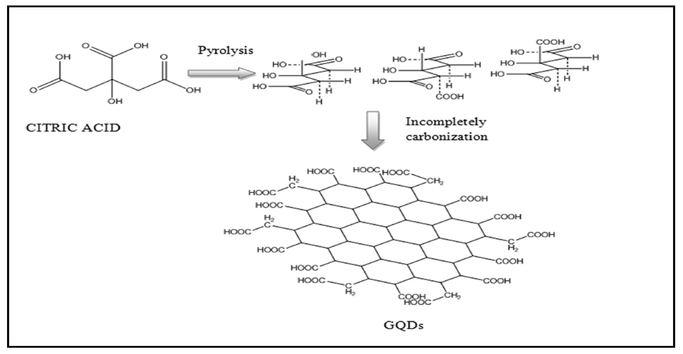
2. Graphene and Carbon Quantum Dots: Similarities and Disparities
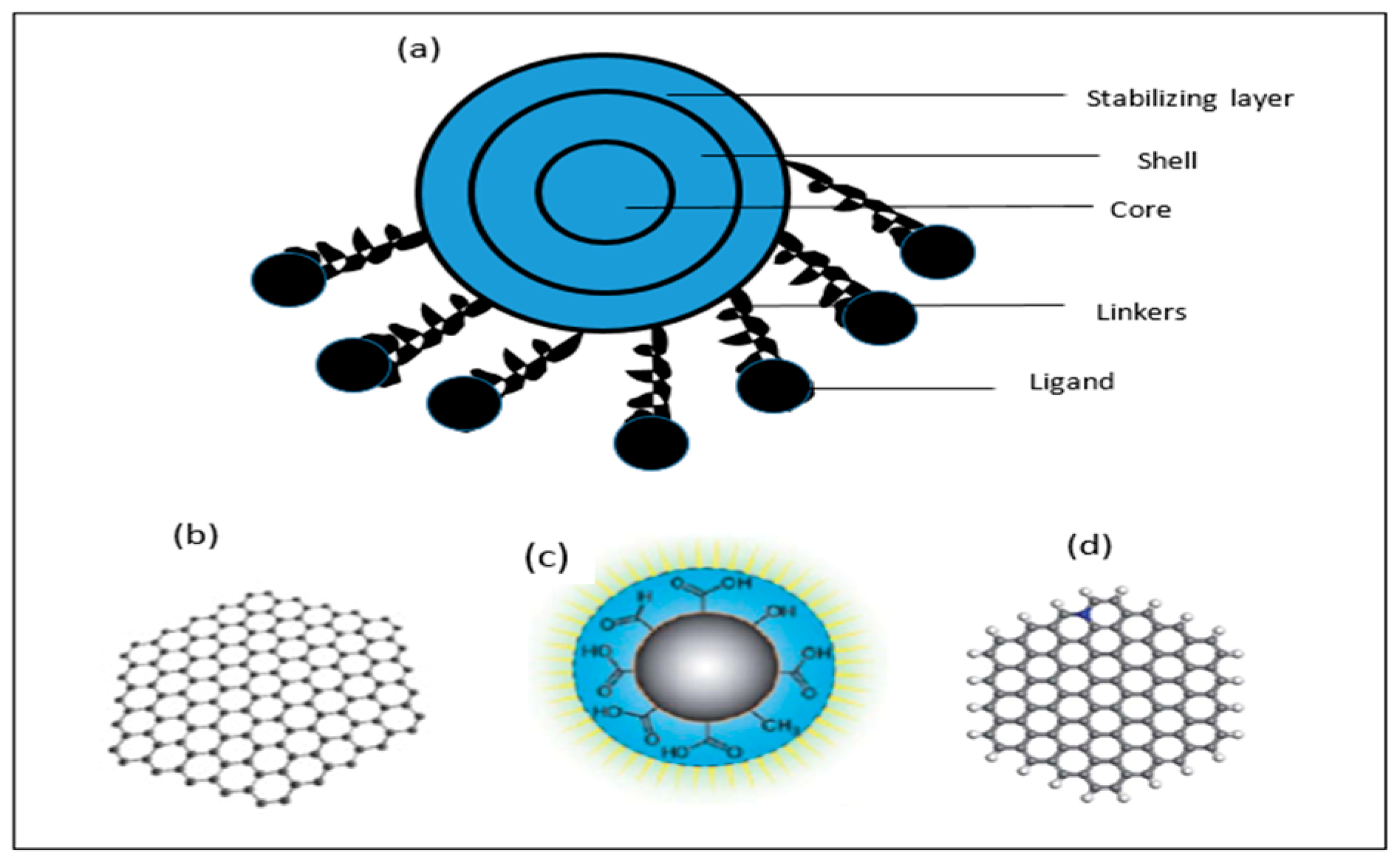
2.1. Similarities between CQDs and GQDs
2.2. Differences between GQDs and CQDs
3. Electrochemical Methods
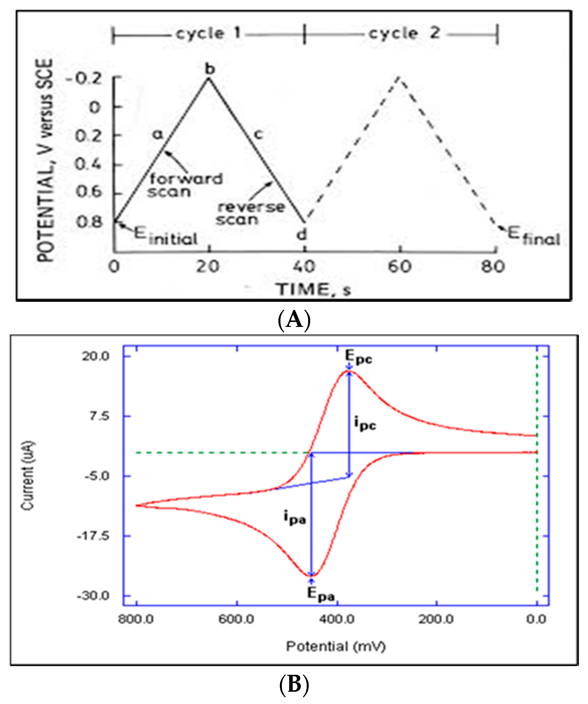
3.1. Cyclic Voltammetry
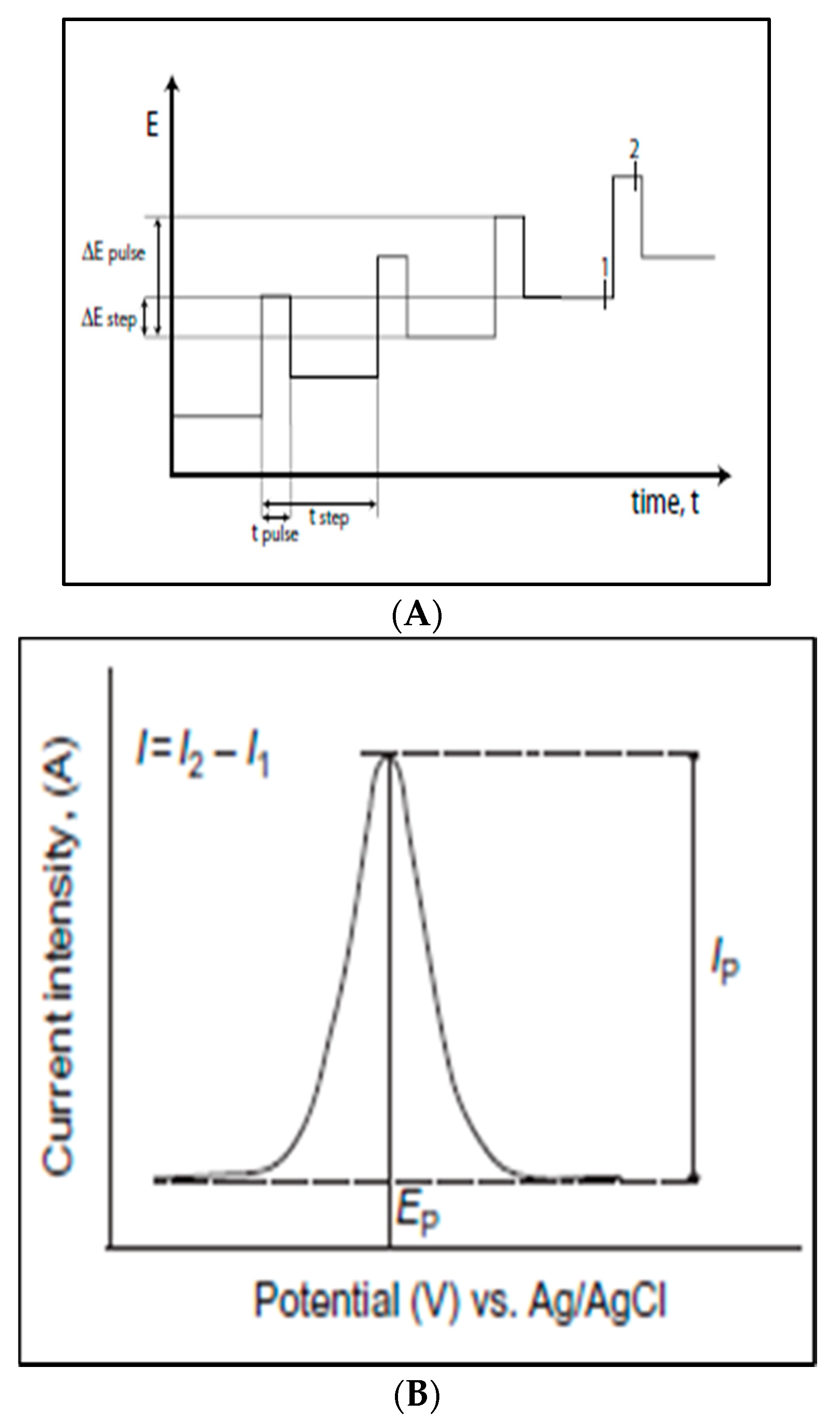
3.2. Differential Pulse Voltammetry (DPV)
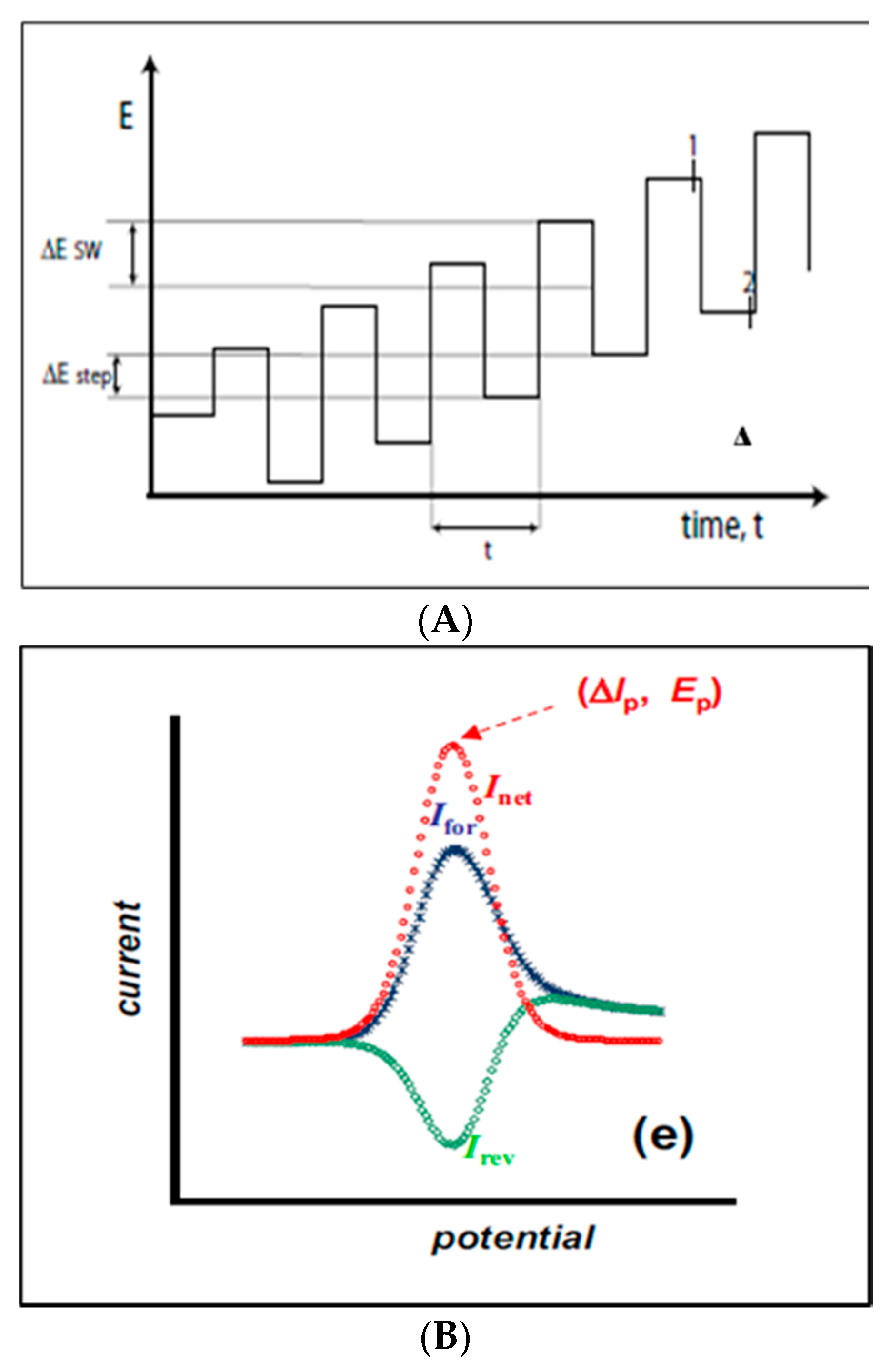
3.3. Square Wave Voltammetry
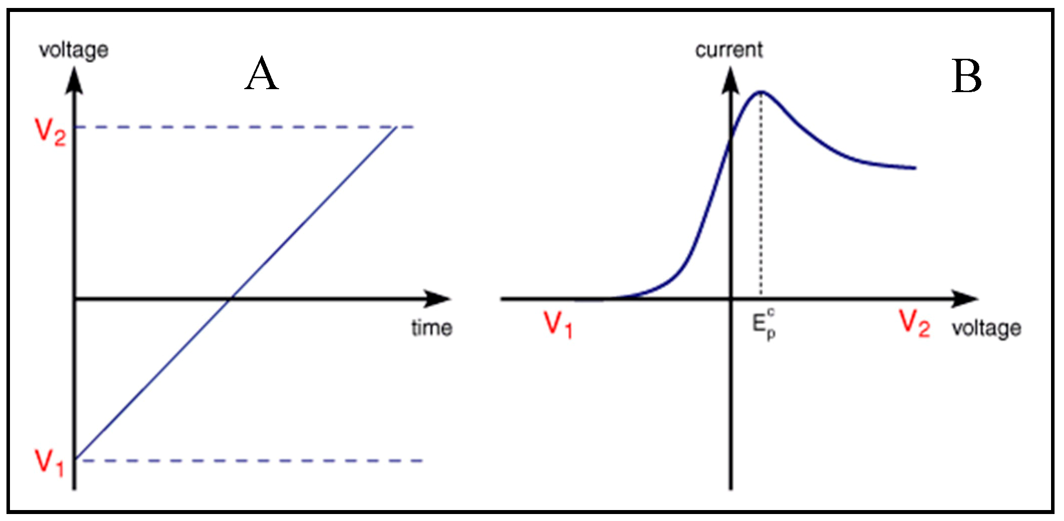
3.4. Linear Sweep Voltammetry
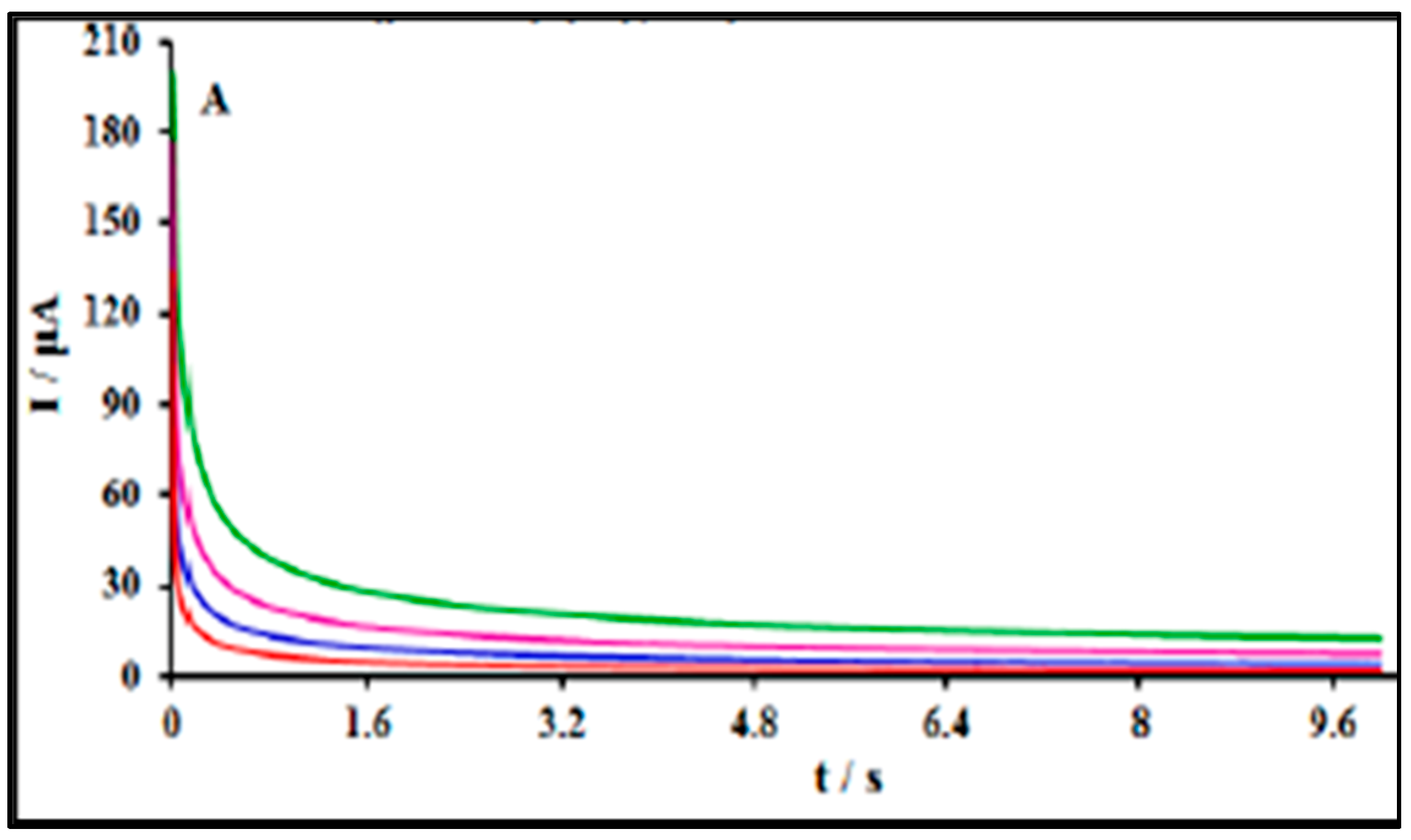
3.5. Chronoamperometry (CAP)
4. Electrode Fabrication
4.1. Modification of Glassy Carbon Electrodes (GCE)
4.2. Modification of Carbon Paste Electrode
4.3. Modification of Screen-Printed Electrodes
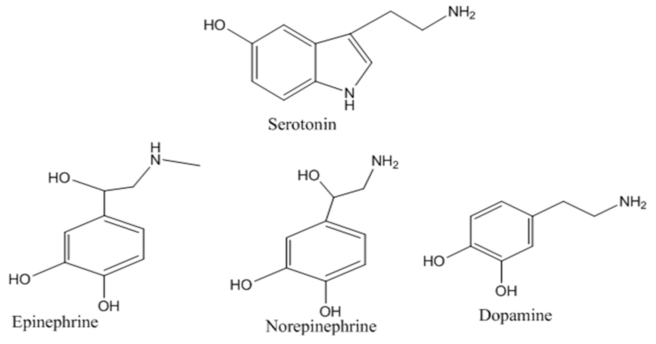
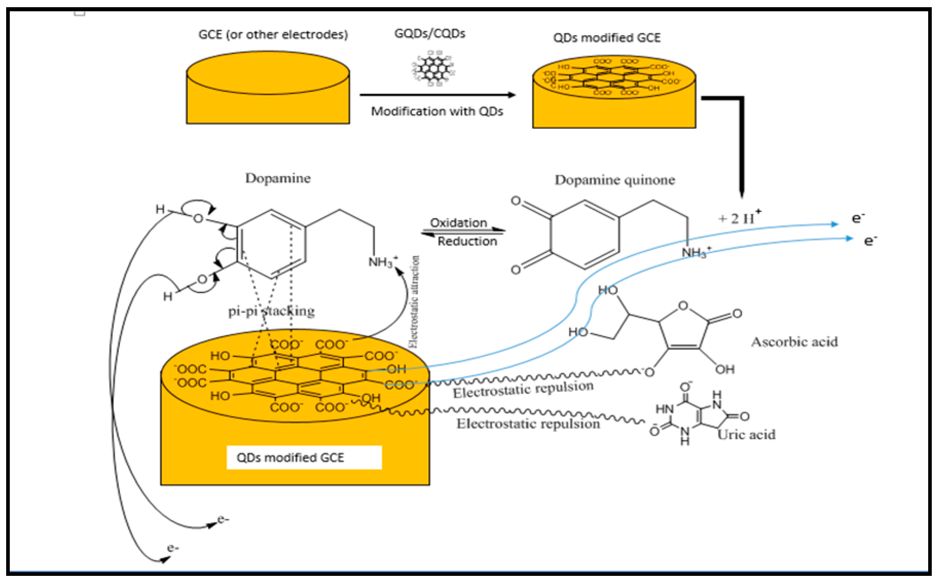
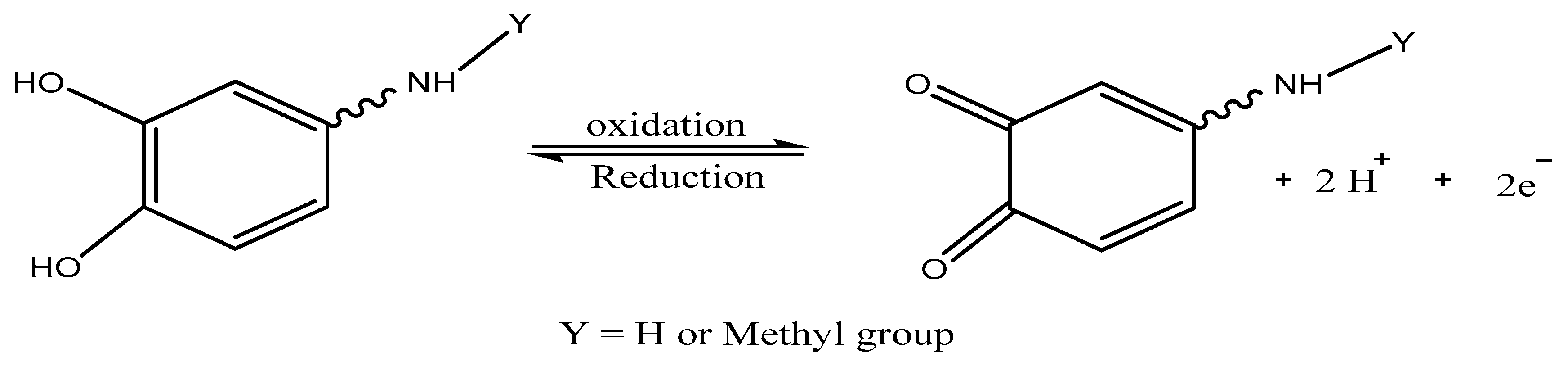
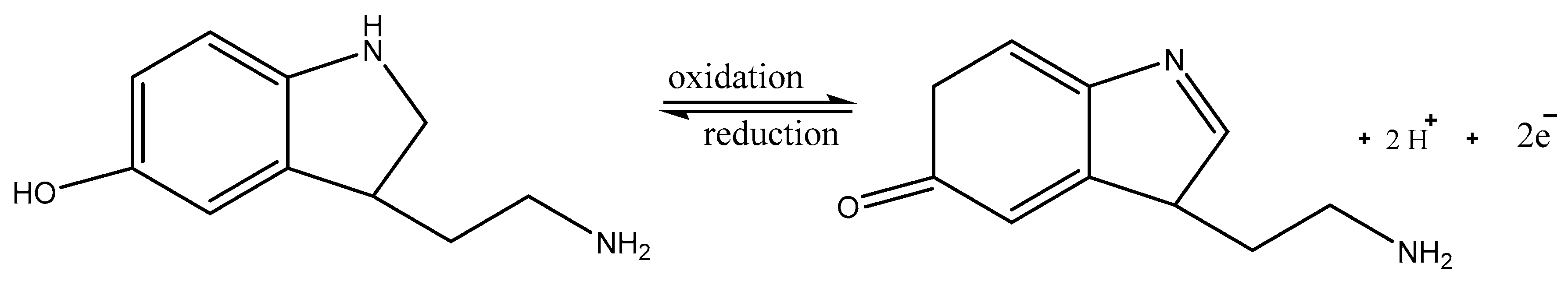
5. Mechanism of Monoamine Neurotransmitters Detection
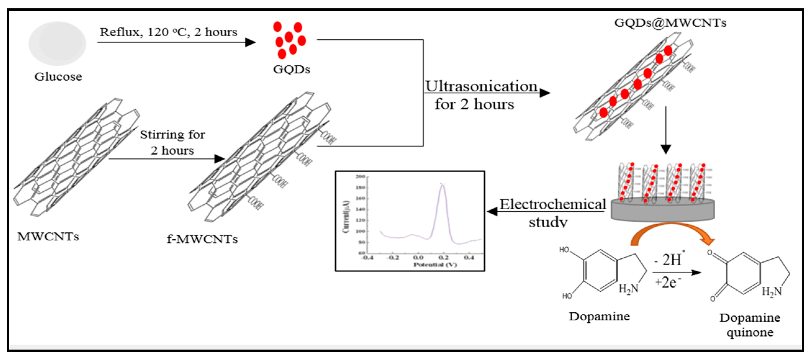
6. Electrochemical Performance of Carbon-Based QD-Modified Electrodes
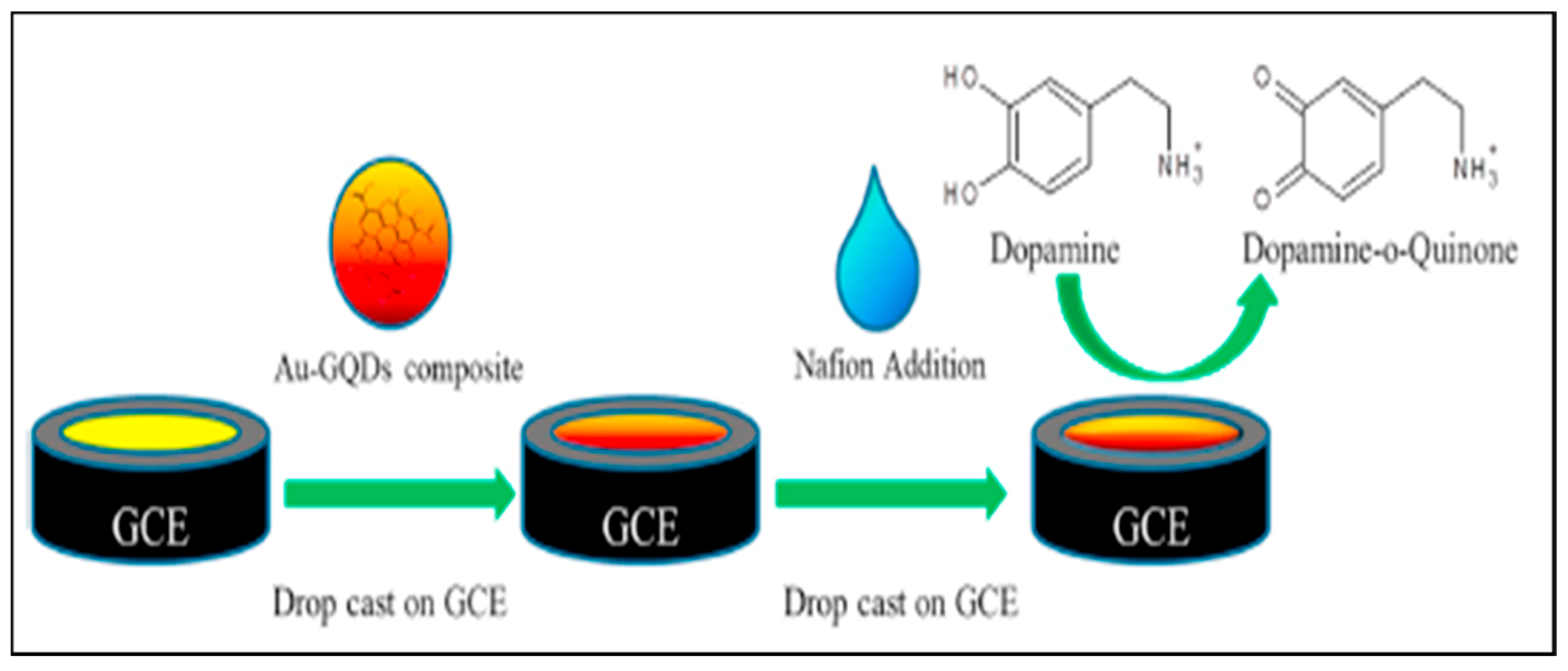
6.1. Electrochemical Performance in Dopamine Determination
6.2. Electrochemical Performance in Epinephrine (EP) Determination
6.3. Electrochemical Performance in Serotonin (SE) Determination
6.4. Electrochemical Performance in Norepinephrine (NE) Determination
7. Carbon-Based QDs and Monoamine Neurotransmitters: Now and Future Perspective
Author Contributions
Funding
Acknowledgments
Conflicts of Interest
Abbreviations
| DA | Dopamine |
| SE | Serotonin |
| NE | Norepinephrin |
| EP | Epinephrine |
| NTs | Neurotransmitters |
| GCE | Glassy carbon electrode |
| CPE | Carbon paste electrode |
| DPV | Differential pulse voltammetry |
| CV | Cyclic voltammetry |
| CAP | Chronoamperometry |
| GQDs | Graphene quantum dots |
| CQDs | Carbon quantum dots |
| MAO | Monoamine oxidase |
| CNTs | Carbon nanotubes |
| QDs | Quantum dots |
| N-CQDs | Nitrogen doped carbon quantum dots |
| N-GQDs | Nitrogen doped graphene quantum dots |
| MIPs | Molecularly imprinted polymers |
| AA | Amino acid |
| UA | Uric acid |
| IL | Ionic liquid |
| TEM | Transmission electron microscope |
| ITO | Indium tin oxide |
| CA | Citric acid |
| EIS | Electrochemical impedance spectroscopy |
| NPs | Nanoparticles |
| SPCE | screen printed carbon electrode |
| CS | Chitosan |
| SAM | self-enabled monolayers |
| eth | ethylene diamine |
| PANI | polyaniline |
| TMSPED | N-[3(trimethoxysilyl)propyl]ethylenediamine |
| AP | Amperometry |
| AuNCs | Gold nanocrystals |
| H-GQDs | Histidine functionalized GQDs |
| GMA | graphene micro aerogel |
| MWCNTs | Multiwalled carbon nanotubes |
| Cyse | Cystein |
| tyr | Tyrosine |
| trp | Tryptophan |
| asp | Aspartic acid |
| ala | Alanine |
| glu | Glutamic acid |
| his | Histidine |
| arg | Arginine |
| gly | Glycine |
| glc | Glucose |
| suc | Sucrose |
| fru | Fructose |
| SPE | screen printed electrode |
| LOD | Limit of detection |
| LDR | Linear dynamic range |
| DEA | diethanolamine |
| h-BN | Hezagonal boron nitride |
| OCP | obsessive compulsive disorder |
| PTSD | Post-traumatic stress disorder |
| SWSV | Square wave stripping voltammetry |
| ACh | Acetylcholine |
| HU | Human urine |
| HS | Human serum |
| LSV | Linear sweep voltammetry |
| PBS | Phosphate buffer saline |
| PCB | Phosphate-citrate buffer |
References
- Cooper, J.R.; Bloom, F.E.; Roth, R.H. The Biochemical Basis of Neuropharmacology; Oxford University Press: New York, NY, USA, 2003. [Google Scholar]
- Rutecki, P.A. Neuronal excitability: Voltage-dependent currents and synaptic transmission. J. Clin. Neurophysiol. 1992, 9, 195–211. [Google Scholar] [CrossRef]
- Purves, D.; Augustine, G.; Fitzpatrick, D.; Katz, L.; LaMantia, A.; McNamara, J.; Williams, S. Circuits within the basal ganglia system. In Neuroscience, 2nd ed.; Sinauer Associates Inc.: Sunderland, MA, USA, 2001. [Google Scholar]
- Atta, N.F.; El-Ads, E.H.; Ahmed, Y.M.; Galal, A. Determination of some neurotransmitters at cyclodextrin/ionic liquid crystal/graphene composite electrode. Electrochim. Acta 2016, 199, 319–331. [Google Scholar] [CrossRef]
- Kumar, K.H.; Elavarasi, P.; David, C.M. Definition of pain and classification of pain disorders. J. Adv. Clin. Res. Insights 2016, 3, 87–90. [Google Scholar] [CrossRef]
- Gutiérrez, A.; Primo, E.N.; Eguílaz, M.; Parrado, C.; Rubianes, M.D.; Rivas, G.A. Quantification of neurotransmitters and metabolically related compounds at glassy carbon electrodes modified with bamboo-like carbon nanotubes dispersed in double stranded DNA. Microchem. J. 2017, 130, 40–46. [Google Scholar] [CrossRef]
- Yu, D.; Zeng, Y.; Qi, Y.; Zhou, T.; Shi, G. A novel electrochemical sensor for determination of dopamine based on AuNPs@SiO2 core-shell imprinted composite. Biosens. Bioelectron. 2012, 38, 270–277. [Google Scholar] [CrossRef]
- Babaei, A.; Sohrabi, M.; Afrasiabi, M. A Sensitive Simultaneous Determination of Epinephrine and Piroxicam Using a Glassy Carbon Electrode Modified with a Nickel Hydroxide Nanoparticles/Multiwalled Carbon Nanotubes Composite. Electroanalysis 2012, 24, 2387–2394. [Google Scholar] [CrossRef]
- Sorouraddin, M.H.; Manzoori, J.L.; Kargarzadeh, E.; Shabani, A.M.H. Spectrophotometric determination of some catecholamine drugs using sodium bismuthate. J. Pharm. Biomed. Anal. 1998, 18, 877–881. [Google Scholar] [CrossRef]
- Gérard, M.; Chaubey, A.; Malhotra, B.D. Application of conducting polymers to biosensors. Biosens. Bioelectron. 2002, 17, 345–359. [Google Scholar] [CrossRef]
- Song, Y.Z.; Li, X.; Cheng, Z.P.; Zhong, H.; Xu, J.M.; Lu, J.S.; Wei, C.G.; Zhu, A.F.; Wu, F.Y.; Song, Y.; et al. Electrochemical synthesis of gold nanoparticles on the surface of multi-walled carbon nanotubes with glassy carbon electrode and their application. Russ. J. Phys. Chem. A 2012, 87, 74–79. [Google Scholar] [CrossRef]
- Kartsova, L.; Sidorova, A.A.; Kazakov, V.A.; Bessonova, E.A.; Yashin, A.Y. Determination of Catecholamines by Capillary Electrophoresis and Reversed-Phase High-Performance Liquid Chromatography. J. Anal. Chem. 2004, 59, 737–741. [Google Scholar] [CrossRef]
- Chicharro, M.; Zapardiel, A.; Bermejo, E.; A Perez, J.; Hernández, L. Direct determination of ephedrine and norephedrine in human urine by capillary zone electrophoresis. J. Chromatogr. 1993, 622, 103–108. [Google Scholar] [PubMed]
- Lövheim, H. A new three-dimensional model for emotions and monoamine neurotransmitters. Med. Hypotheses 2012, 78, 341–348. [Google Scholar] [CrossRef]
- Babaei, A.; Taheri, A. Nafion/Ni(OH)2 nanoparticles-carbon nanotube composite modified glassy carbon electrode as a sensor for simultaneous determination of dopamine and serotonin in the presence of ascorbic acid. Sens. Actuators B Chem. 2013, 176, 543–551. [Google Scholar] [CrossRef]
- Adekunle, A.S.; Agboola, B.O.; Pillay, J.; Ozoemena, K.I. Electrocatalytic detection of dopamine at single-walled carbon nanotubes–iron (III) oxide nanoparticles platform. Sens. Actuators B Chem. 2010, 148, 93–102. [Google Scholar] [CrossRef]
- Hernández, P.; Sánchez, I.; Patón, F.; Hernández, L. Cyclic voltammetry determination of epinephrine with a carbon fiber ultramicroelectrode. Talanta 1998, 46, 985–991. [Google Scholar] [CrossRef]
- Carlsson, A.; Hansson, L.O.; Waters, N.; Carlsson, M.L. Neurotransmitter aberrations in schizophrenia: New perspectives and therapeutic implications. Life Sci. 1997, 61, 75–94. [Google Scholar] [CrossRef]
- Mphuthi, N.G.; Adekunle, A.S.; Ebenso, E. Electrocatalytic oxidation of Epinephrine and Norepinephrine at metal oxide doped phthalocyanine/MWCNT composite sensor. Sci. Rep. 2016, 6, 26938. [Google Scholar] [CrossRef]
- Beitollahi, H.; Mohammadi, S. Selective voltammetric determination of norepinephrine in the presence of acetaminophen and tryptophan on the surface of a modified carbon nanotube paste electrode. Mater. Sci. Eng. C 2013, 33, 3214–3219. [Google Scholar] [CrossRef]
- Chandrashekar, B.; Swamy, B.E.K. Simultaneous cyclic voltammetric determination of norepinephrine, ascorbic acid and uric acid using TX-100 modified carbon paste electrode. Anal. Methods 2012, 4, 849. [Google Scholar] [CrossRef]
- Abbaspour, A.; Ghaffarinejad, A. Preparation of a sol–gel-derived carbon nanotube ceramic electrode by microwave irradiation and its application for the determination of adenine and guanine. Electrochim. Acta 2010, 55, 1090–1096. [Google Scholar] [CrossRef]
- Lin, M.T.; Tsay, H.J.; Su, W.H.; Chueh, F.Y. Changes in extracellular serotonin in rat hypothalamus affect thermoregulatory function. Am. J. Physiol. Content 1998, 274, R1260–R1267. [Google Scholar] [CrossRef] [PubMed]
- Fayemi, O.E.; Adekunle, A.S.; Ebenso, E.E. Electrochemical determination of serotonin in urine samples based on metal oxide nanoparticles/MWCNT on modified glassy carbon electrode. Sens. Biosens. Res. 2017, 13, 17–27. [Google Scholar] [CrossRef]
- Samie, H.A.; Arvand, M. RuO2 nanowires on electrospun CeO2-Au nanofibers/functionalized carbon nanotubes/graphite oxide nanocomposite modified screen-printed carbon electrode for simultaneous determination of serotonin, dopamine and ascorbic acid. J. Alloys Compd. 2019, 782, 824–836. [Google Scholar] [CrossRef]
- Isbister, G.K.; Bowe, S.J.; Dawson, A.H.; Whyte, I.M. Relative Toxicity of Selective Serotonin Reuptake Inhibitors (SSRIs) in Overdose. J. Toxicol. Clin. Toxicol. 2004, 42, 277–285. [Google Scholar] [CrossRef]
- Goyal, R.N.; Bishnoi, S. Simultaneous determination of epinephrine and norepinephrine in human blood plasma and urine samples using nanotubes modified edge plane pyrolytic graphite electrode. Talanta 2011, 84, 78–83. [Google Scholar] [CrossRef] [PubMed]
- Ghoreishi, S.M.; Behpour, M.; Ghoreishi, F.S.; Mousavi, S. Voltammetric determination of tryptophan in the presence of uric acid and dopamine using carbon paste electrode modified with multi-walled carbon nanotubes. Arab. J. Chem. 2017, 10, S1546–S1552. [Google Scholar] [CrossRef]
- Wang, S.; Wang, Y.; Min, Q.; Shu, T.; Zhu, X.; Peng, A.; Ding, H. Simultaneous electrochemical determination of dopamine and serotonin in rat cerebrospinal fluid using screen-printed electrode modified with MWNTs-SiO2-chitosan composites. Int. J. Electrochem. Sci. 2016, 11, 2360–2376. [Google Scholar]
- Tian, L.; Wang, X.; Wu, K.; Hu, Y.; Wang, Y.; Lu, J.; Li, T. Ultrasensitive electrochemiluminescence biosensor for dopamine based on ZnSe, graphene oxide@multi walled carbon nanotube and Ru (bpy) 32+. Sens. Actuators B Chem. 2019, 286, 266–271. [Google Scholar] [CrossRef]
- Salem, F. Spectrophotometric and titrimetric determination of catecholamines. Talanta 1987, 34, 810–812. [Google Scholar] [CrossRef]
- Shafi, N.; Midgley, J.; Watson, D.; Smail, G.; Strang, R.; Macfarlane, R. Analysis of biogenic amines in the brain of the American cockroach (Periplaneta Americana) by gas chromatography-negative ion chemical ionisation mass spectrometry. J. Chromatogr. B Biomed. Sci. Appl. 1989, 490, 9–19. [Google Scholar] [CrossRef]
- Mukdasai, S.; Langsi, V.; Pravda, M.; Srijaranai, S.; Glennon, J.D. A highly sensitive electrochemical determination of norepinephrine using l-cysteine self-assembled monolayers over gold nanoparticles/multi-walled carbon nanotubes electrode in the presence of sodium dodecyl sulfate. Sens. Actuators B Chem. 2016, 236, 126–135. [Google Scholar] [CrossRef]
- Yang, Y.J.; Li, W. CTAB functionalized graphene oxide/multiwalled carbon nanotube composite modified electrode for the simultaneous determination of ascorbic acid, dopamine, uric acid and nitrite. Biosens. Bioelectron. 2014, 56, 300–306. [Google Scholar] [CrossRef] [PubMed]
- Sun, D.; Li, H.; Li, M.; Li, C.; Dai, H.; Sun, D.; Yang, B. Electrodeposition synthesis of a NiO/CNT/PEDOT composite for simultaneous detection of dopamine, serotonin, and tryptophan. Sens. Actuators B Chem. 2018, 259, 433–442. [Google Scholar] [CrossRef]
- Sroysee, W.; Chairam, S.; Amatatongchai, M.; Jarujamrus, P.; Tamuang, S.; Pimmongkol, S.; Chaicharoenwimolkul, L.; Somsook, E. Poly (M-ferrocenylaniline) modified carbon nanotubes-paste electrode encapsulated in nafion film for selective and sensitive determination of dopamine and uric acid in the presence of ascorbic acid. J. Saudi Chem. Soc. 2018, 22, 173–182. [Google Scholar] [CrossRef]
- Zhou, H.; Xu, G.; Zhu, A.; Zhao, Z.; Ren, C.; Nie, L.; Kan, X. A multiporous electrochemical sensor for epinephrine recognition and detection based on molecularly imprinted polypyrrole. RSC Adv. 2012, 2, 7803–7808. [Google Scholar] [CrossRef]
- Tisdale, W.A.; Zhu, X.-Y. Artificial atoms on semiconductor surfaces. Proc. Natl. Acad. Sci. USA 2010, 108, 965–970. [Google Scholar] [CrossRef]
- Wang, Y.; Hu, A. Carbon quantum dots: Synthesis, properties and applications. J. Mater. Chem. C 2014, 2, 6921. [Google Scholar] [CrossRef]
- Baker, S.N.; Baker, G.A. Luminescent Carbon Nanodots: Emergent Nanolights. Angew. Chem. Int. Ed. 2010, 49, 6726–6744. [Google Scholar] [CrossRef]
- Geszke-Moritz, M.; Moritz, M. Quantum dots as versatile probes in medical sciences: Synthesis, modification and properties. Mater. Sci. Eng. C 2013, 33, 1008–1021. [Google Scholar] [CrossRef]
- Comparelli, R.; Zezza, F.; Striccoli, M.; Curri, M.L.; Tommasi, R.; Agostiano, A. Improved optical properties of CdS quantum dots by ligand exchange. Mater. Sci. Eng. C 2003, 23, 1083–1086. [Google Scholar] [CrossRef]
- Sellami, K.; Jaziri, S. Piezoelectric coupling effect on exciton–phonon scattering rates in CdSe quantum dots embedded in glass matrix. Mater. Sci. Eng. C 2006, 26, 555–558. [Google Scholar] [CrossRef]
- Algarra, M.; Campos, B.; Aguiar, F.; Rodriguez-Borges, J.E.; Da Silva, J.E.; Da Silva, J.C.E. Novel β-cyclodextrin modified CdTe quantum dots as fluorescence nanosensor for acetylsalicylic acid and metabolites. Mater. Sci. Eng. C 2012, 32, 799–803. [Google Scholar] [CrossRef]
- Dabbousi, B.O.; Rodríguez-Viejo, J.; Mikulec, F.V.; Heine, J.R.; Mattoussi, H.; Ober, R.; Jensen, K.F.; Bawendi, M.G. (CdSe)ZnS Core−Shell Quantum Dots: Synthesis and Characterization of a Size Series of Highly Luminescent Nanocrystallites. J. Phys. Chem. B 1997, 101, 9463–9475. [Google Scholar] [CrossRef]
- Reiss, P.; Carayon, S.; Bleuse, J.; Pron, A. Low polydispersity core/shell nanocrystals of CdSe/ZnSe and CdSe/ZnSe/ZnS type: Preparation and optical studies. Synth. Met. 2003, 139, 649–652. [Google Scholar] [CrossRef]
- Jiang, D.; Cao, L.; Liu, W.; Su, G.; Qu, H.; Sun, Y.; Dong, B. Synthesis and Luminescence Properties of Core/Shell ZnS:Mn/ZnO Nanoparticles. Nanoscale Res. Lett. 2008, 4, 78–83. [Google Scholar] [CrossRef] [PubMed]
- Li, R.; Tang, L.; Zhao, Q.; Teng, K.S.; Lau, S.P. Facile synthesis of ZnS quantum dots at room temperature for ultra-violet photodetector applications. Chem. Phys. Lett. 2020, 742, 137127. [Google Scholar] [CrossRef]
- Shen, Q.; Arae, D.; Toyoda, T. Photosensitization of nanostructured TiO2 with CdSe quantum dots: Effects of microstructure and electron transport in TiO2 substrates. J. Photochem. Photobiol. A Chem. 2004, 164, 75–80. [Google Scholar] [CrossRef]
- Du, J.; Xu, N.; Fan, J.; Sun, W.; Peng, X. Carbon dots for in vivo bioimaging and theranostics. Small 2019, 15, 1805087. [Google Scholar] [CrossRef]
- Canevari, T.C.; Nakamura, M.; Cincotto, F.H.; De Melo, F.M.; Toma, H.E. High performance electrochemical sensors for dopamine and epinephrine using nanocrystalline carbon quantum dots obtained under controlled chronoamperometric conditions. Electrochim. Acta 2016, 209, 464–470. [Google Scholar] [CrossRef]
- Vinoth, V.; Natarajan, L.N.; Mangalaraja, R.V.; Valdés, H.; Anandan, S. Simultaneous electrochemical determination of dopamine and epinephrine using gold nanocrystals capped with graphene quantum dots in a silica network. Microchim. Acta 2019, 186, 681. [Google Scholar] [CrossRef]
- Das, R.; Bandyopadhyay, R.; Pramanik, P. Carbon quantum dots from natural resource: A review. Mater. Today Chem. 2018, 8, 96–109. [Google Scholar] [CrossRef]
- Chandra, S.; Pathan, S.H.; Mitra, S.; Modha, B.H.; Goswami, A.; Pramanik, P. Tuning of photoluminescence on different surface functionalized carbon quantum dots. RSC Adv. 2012, 2, 3602. [Google Scholar] [CrossRef]
- Lu, W.; Qin, X.; Liu, S.; Chang, G.; Zhang, Y.; Luo, Y.; Asiri, A.M.; Al-Youbi, A.O.; Sun, X. Economical, Green Synthesis of Fluorescent Carbon Nanoparticles and Their Use as Probes for Sensitive and Selective Detection of Mercury(II) Ions. Anal. Chem. 2012, 84, 5351–5357. [Google Scholar] [CrossRef] [PubMed]
- Zhou, J.; Sheng, Z.; Han, H.; Zou, M.-Q.; Li, C. Facile synthesis of fluorescent carbon dots using watermelon peel as a carbon source. Mater. Lett. 2012, 66, 222–224. [Google Scholar] [CrossRef]
- Dong, Y.; Wang, R.; Li, H.; Shao, J.-W.; Chi, Y.; Lin, X.; Chen, G. Polyamine-functionalized carbon quantum dots for chemical sensing. Carbon 2012, 50, 2810–2815. [Google Scholar] [CrossRef]
- Sun, D.; Ban, R.; Zhang, P.-H.; Wu, G.-H.; Zhang, J.-R.; Zhu, J.-J. Hair fiber as a precursor for synthesizing of sulfur- and nitrogen-co-doped carbon dots with tunable luminescence properties. Carbon 2013, 64, 424–434. [Google Scholar] [CrossRef]
- Liu, S.; Tian, J.; Wang, L.; Zhang, Y.; Qin, X.; Luo, Y.; Asiri, A.M.; Al-Youbi, A.O.; Sun, X. Hydrothermal Treatment of Grass: A Low-Cost, Green Route to Nitrogen-Doped, Carbon-Rich, Photoluminescent Polymer Nanodots as an Effective Fluorescent Sensing Platform for Label-Free Detection of Cu(II) Ions. Adv. Mater. 2012, 24, 2037–2041. [Google Scholar] [CrossRef]
- Wang, N.; Wang, Y.; Guo, T.; Yang, T.; Chen, M.-L.; Wang, J.-H. Green preparation of carbon dots with papaya as carbon source for effective fluorescent sensing of Iron (III) and Escherichia coli. Biosens. Bioelectron. 2016, 85, 68–75. [Google Scholar] [CrossRef]
- Zhang, Y.-Q.; Ma, D.-K.; Zhuang, Y.; Zhang, X.; Chen, W.; Hong, L.-L.; Yan, Q.-X.; Yu, K.; Huang, S. One-pot synthesis of N-doped carbon dots with tunable luminescence properties. J. Mater. Chem. 2012, 22, 16714. [Google Scholar] [CrossRef]
- Jiang, G.; Jiang, T.; Li, X.; Wei, Z.; Du, X.; Wang, X. Boronic acid functionalized N-doped carbon quantum dots as fluorescent probe for selective and sensitive glucose determination. Mater. Res. Express 2014, 1, 025708. [Google Scholar] [CrossRef]
- Wang, C.; Shi, H.; Yang, M.; Yan, Y.; Liu, E.; Ji, Z.; Fan, J. A novel nitrogen-doped carbon quantum dots as effective fluorescent probes for detecting dopamine. J. Photochem. Photobiol. A Chem. 2020, 391, 112374. [Google Scholar] [CrossRef]
- Algarra, M.; González-Calabuig, A.; Radotić, K.; Mutavdzic, D.; Ania, C.O.; Martínez, J.M.L.; Jiménez-Jiménez, J.; Rodríguez-Castellón, E.; Del Valle, M. Enhanced electrochemical response of carbon quantum dot modified electrodes. Talanta 2018, 178, 679–685. [Google Scholar] [CrossRef] [PubMed]
- Chauhan, N.; Soni, S.; Agrawal, P.; Balhara, Y.P.S.; Jain, U. Recent advancement in nanosensors for neurotransmitters detection: Present and future perspective. Process Biochem. 2020, 91, 241–259. [Google Scholar] [CrossRef]
- Arumugasamy, S.K.; Chellasamy, G.; Gopi, S.; Govindaraju, S.; Yun, K. Current advances in the detection of neurotransmitters by nanomaterials: An update. TrAC Trends Anal. Chem. 2020, 123, 115766. [Google Scholar] [CrossRef]
- Sajid, M.; Baig, N.; Alhooshani, K. Chemically modified electrodes for electrochemical detection of dopamine: Challenges and opportunities. TrAC Trends Anal. Chem. 2019, 118, 368–385. [Google Scholar] [CrossRef]
- Ribeiro, J.A.; Fernandes, P.M.V.; Pereira, C.M.; Silva, F. Electrochemical sensors and biosensors for determination of catecholamine neurotransmitters: A review. Talanta 2016, 160, 653–679. [Google Scholar] [CrossRef]
- Tajik, S.; Dourandish, Z.; Zhang, K.; Beitollahi, H.; Van Le, Q.; Jang, H.W.; Shokouhimehr, M. Carbon and graphene quantum dots: A review on syntheses, characterization, biological and sensing applications for neurotransmitter determination. RSC Adv. 2020, 10, 15406–15429. [Google Scholar] [CrossRef]
- Li, M.; Chen, T.; Gooding, J.J.; Liu, J. Review of Carbon and Graphene Quantum Dots for Sensing. ACS Sens. 2019, 4, 1732–1748. [Google Scholar] [CrossRef]
- Campuzano, S.; Yáñez-Sedeño, P.; Pingarrón, J.M. Carbon Dots and Graphene Quantum Dots in Electrochemical Biosensing. Nanomaterials 2019, 9, 634. [Google Scholar] [CrossRef]
- Hola, K.; Bourlinos, A.B.; Kozák, O.; Berka, K.; Šišková, K.; Havrdova, M.; Tucekc, J.; Šafářová, K.; Otyepka, M.; Giannelis, E.P.; et al. Photoluminescence effects of graphitic core size and surface functional groups in carbon dots: COO− induced red-shift emission. Carbon 2014, 70, 279–286. [Google Scholar] [CrossRef]
- Paulo, S.; Palomares, E.; Martínez-Ferrero, E. Graphene and Carbon Quantum Dot-Based Materials in Photovoltaic Devices: From Synthesis to Applications. Nanomaterials 2016, 6, 157. [Google Scholar] [CrossRef] [PubMed]
- Dong, Y.; Lin, J.; Chen, Y.; Fu, F.; Chi, Y.; Chen, G. Graphene quantum dots, graphene oxide, carbon quantum dots and graphite nanocrystals in coals. Nanoscale 2014, 6, 7410–7415. [Google Scholar] [CrossRef] [PubMed]
- Lim, C.S.; Hola, K.; Ambrosi, A.; Zbořil, R.; Pumera, M. Graphene and carbon quantum dots electrochemistry. Electrochem. Commun. 2015, 52, 75–79. [Google Scholar] [CrossRef]
- Zhuang, X.; Wang, H.; He, T.; Chen, L. Enhanced voltammetric determination of dopamine using a glassy carbon electrode modified with ionic liquid-functionalized graphene and carbon dots. Microchim. Acta 2016, 183, 3177–3182. [Google Scholar] [CrossRef]
- Sanati, A.L.; Faridbod, F.; Ganjali, M.R. Synergic effect of graphene quantum dots and room temperature ionic liquid for the fabrication of highly sensitive voltammetric sensor for levodopa determination in the presence of serotonin. J. Mol. Liq. 2017, 241, 316–320. [Google Scholar] [CrossRef]
- Gonon, F.; Buda, M.; Cespuglio, R.; Jouvet, M.; Pujol, J.-F. In vivo electrochemical detection of catechols in the neostriatum of anaesthetized rats: Dopamine or DOPAC? Nature 1980, 286, 902–904. [Google Scholar] [CrossRef]
- Pang, P.; Yan, F.; Li, H.; Li, H.; Zhang, Y.; Wang, H.; Wu, Z.; Yang, W. Graphene quantum dots and Nafion composite as an ultrasensitive electrochemical sensor for the detection of dopamine. Anal. Methods 2016, 8, 4912–4918. [Google Scholar] [CrossRef]
- Huang, Q.; Hu, S.; Zhang, H.; Chen, J.; He, Y.; Li, F.; Weng, W.; Ni, J.; Bao, X.; Lin, Y. Carbon dots and chitosan composite film based biosensor for the sensitive and selective determination of dopamine. Analyst 2013, 138, 5417–5423. [Google Scholar] [CrossRef]
- Zheng, S. College of Chemistry and Material Engineering, Anhui Science and Technology University, 233100, Fengyang, Anhui Province, China A Highly Sensitive Dopamine Sensor Based on Graphene Quantum Dots Modified Glassy Carbon Electrode. Int. J. Electrochem. Sci. 2018, 5723–5735. [Google Scholar] [CrossRef]
- Huang, Q.; Wei, C.; Lin, X.; Zhang, Y.; Hu, S. A high performance electrochemical biosensor based on Cu2O–carbon dots for selective and sensitive determination of dopamine in human serum. RSC Adv. 2015, 5, 54102–54108. [Google Scholar] [CrossRef]
- Jiang, G.; Jiang, T.; Zhou, H.; Yao, J.; Kong, X. Preparation of N-doped carbon quantum dots for highly sensitive detection of dopamine by an electrochemical method. RSC Adv. 2015, 5, 9064–9068. [Google Scholar] [CrossRef]
- Hsu, W.-F.; Wu, T.-M. Electrochemical sensor based on conductive polyaniline coated hollow tin oxide nanoparticles and nitrogen doped graphene quantum dots for sensitively detecting dopamine. J. Mater. Sci. Mater. Electron. 2019, 30, 8449–8456. [Google Scholar] [CrossRef]
- Lai, E.P.C. Carbon, Graphene and Graphene Oxide Quantum Dots for Analytical Biochemistry Research. Biochem. Anal. Biochem. 2016, 5. [Google Scholar] [CrossRef]
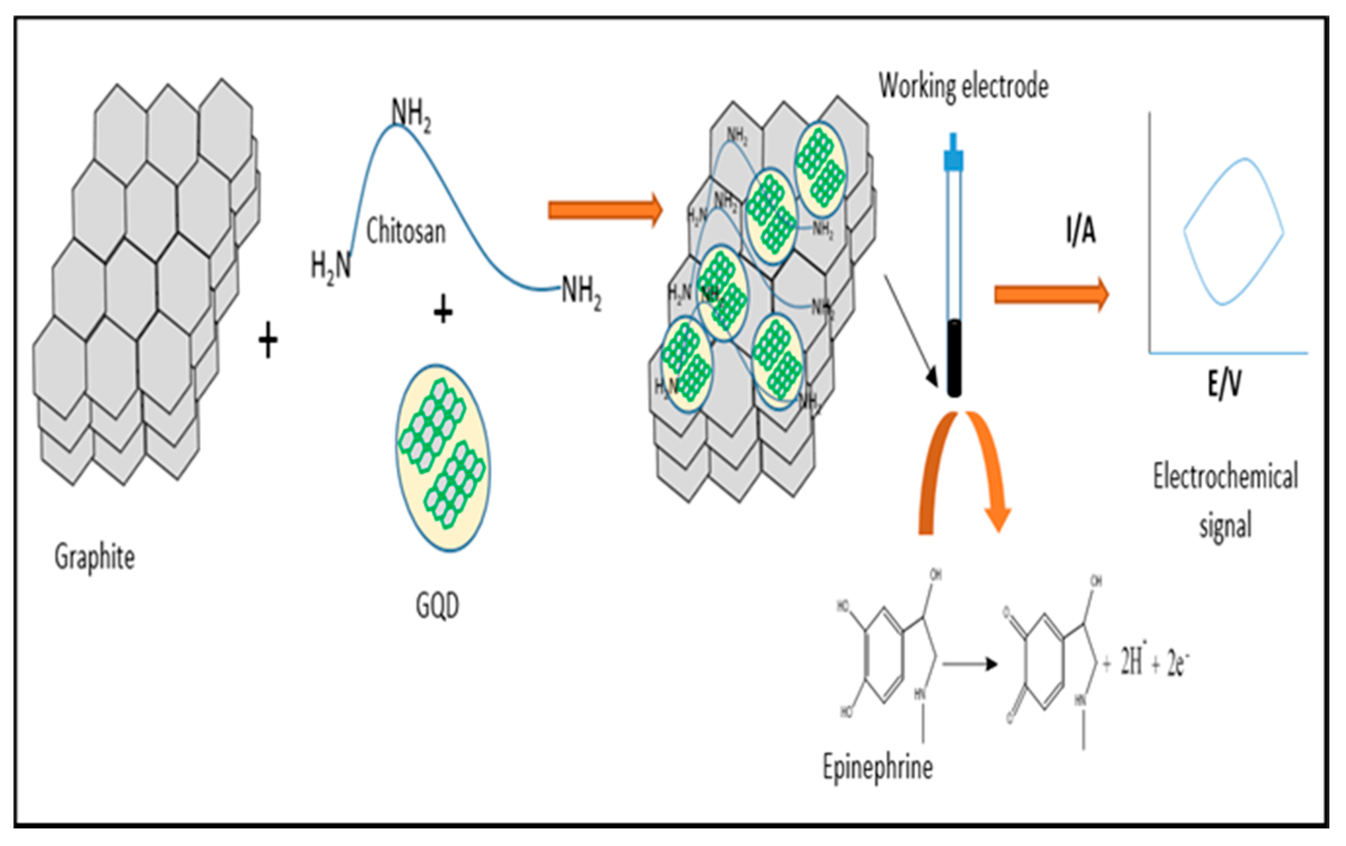
- Tashkhourian, J.; Nami-Ana, S.F.; Shamsipur, M. Designing a modified electrode based on graphene quantum dot-chitosan application to electrochemical detection of epinephrine. J. Mol. Liq. 2018, 266, 548–556. [Google Scholar] [CrossRef]
- Elgrishi, N.; Rountree, K.J.; McCarthy, B.D.; Rountree, E.; Eisenhart, T.T.; Dempsey, J.L. A Practical Beginner’s Guide to Cyclic Voltammetry. J. Chem. Educ. 2017, 95, 197–206. [Google Scholar] [CrossRef]
- Mabbott, G.A. An introduction to cyclic voltammetry. J. Chem. Educ. 1983, 60, 697. [Google Scholar] [CrossRef]
- Kissinger, P.T.; Heineman, W.R. Cyclic voltammetry. J. Chem. Educ. 1983, 60, 702. [Google Scholar] [CrossRef]
- Thomas, D.; Rasheed, Z.; Jagan, J.S.; Kumar, K.G. Study of kinetic parameters and development of a voltammetric sensor for the determination of butylated hydroxyanisole (BHA) in oil samples. J. Food Sci. Technol. 2015, 52, 6719–6726. [Google Scholar] [CrossRef]
- Laviron, E. General expression of the linear potential sweep voltammogram in the case of diffusionless electrochemical systems. J. Electroanal. Chem. 1979, 101, 19–28. [Google Scholar] [CrossRef]
- Ben Aoun, S. Nanostructured carbon electrode modified with N-doped graphene quantum dots–chitosan nanocomposite: A sensitive electrochemical dopamine sensor. R. Soc. Open Sci. 2017, 4, 171199. [Google Scholar] [CrossRef]
- Shankar, S.S.; Shereema, R.M.; Ramachandran, V.; Sruthi, T.V.; Kumar, V.S.; Rakhi, R.B. Carbon Quantum Dot-Modified Carbon Paste Electrode-Based Sensor for Selective and Sensitive Determination of Adrenaline. ACS Omega 2019, 4, 7903–7910. [Google Scholar] [CrossRef] [PubMed]
- Li, Q.; Xu, Z.; Tang, W.; Wu, Y. Determination of Dopamine with a Modified Carbon Dot Electrode. Anal. Lett. 2015, 48, 2040–2050. [Google Scholar] [CrossRef]
- Yola, M.L.; Atar, N. A novel detection approach for serotonin by graphene quantum dots/two-dimensional (2D) hexagonal boron nitride nanosheets with molecularly imprinted polymer. Appl. Surf. Sci. 2018, 458, 648–655. [Google Scholar] [CrossRef]
- Li, Y.; Jiang, Y.; Mo, T.; Zhou, H.; Li, Y.; Li, S. Highly selective dopamine sensor based on graphene quantum dots self-assembled monolayers modified electrode. J. Electroanal. Chem. 2016, 767, 84–90. [Google Scholar] [CrossRef]
- Ruiyi, L.; Sili, Q.; Zhangyi, L.; Ling, L.; Zaijun, L. Histidine-functionalized graphene quantum dot-graphene micro-aerogel based voltammetric sensing of dopamine. Sens. Actuators B Chem. 2017, 250, 372–382. [Google Scholar] [CrossRef]
- Jang, H.-S.; Kim, D.; Lee, C.; Yan, B.; Qin, X.; Piao, Y. Nafion coated Au nanoparticle-graphene quantum dot nanocomposite modified working electrode for voltammetric determination of dopamine. Inorg. Chem. Commun. 2019, 105, 174–181. [Google Scholar] [CrossRef]
- Huang, Q.; Zhang, H.; Hu, S.; Li, F.; Weng, W.; Chen, J.; Wang, Q.; He, Y.; Zhang, W.; Bao, X. A sensitive and reliable dopamine biosensor was developed based on the Au@carbon dots–chitosan composite film. Biosens. Bioelectron. 2014, 52, 277–280. [Google Scholar] [CrossRef]
- Chen, J.; He, P.; Bai, H.; He, S.; Zhang, T.; Zhang, X.; Dong, F. Poly(β-cyclodextrin)/carbon quantum dots modified glassy carbon electrode: Preparation, characterization and simultaneous electrochemical determination of dopamine, uric acid and tryptophan. Sens. Actuators B Chem. 2017, 252, 9–16. [Google Scholar] [CrossRef]
- Devi, N.R.; Kumar, T.H.V.; Sundramoorthy, A.K. Electrochemically Exfoliated Carbon Quantum Dots Modified Electrodes for Detection of Dopamine Neurotransmitter. J. Electrochem. Soc. 2018, 165, G3112–G3119. [Google Scholar] [CrossRef]
- Mirčeski, V.; Skrzypek, S.; Stojanov, L. Square-wave voltammetry. ChemTexts 2018, 4, 17. [Google Scholar] [CrossRef]
- Bard, A.J.; Faulkner, L.R. Fundamentals and applications. Electrochem. Methods 2001, 2, 580–632. [Google Scholar]
- Dias, L.G.; Meirinho, S.G.; Veloso, A.C.; Rodrigues, L.R.; Peres, A.M. Electronic tongues and aptasensors. In Bioinspired Materials for Medical Applications; Elsevier BV: Amsterdam, The Netherlands, 2017; pp. 371–402. [Google Scholar]
- Scholz, F. Electroanalytical Methods; Springer: Berlin, Germany, 2010; Volume 1. [Google Scholar]
- Fajardo, A.; Tapia, D.; Pizarro, J.; Segura, R.; Jara, P. Determination of norepinephrine using a glassy carbon electrode modified with graphene quantum dots and gold nanoparticles by square wave stripping voltammetry. J. Appl. Electrochem. 2019, 49, 423–432. [Google Scholar] [CrossRef]
- Hanlan, J.; Skoog, D.A.; West, D.M. Principles of Instrumental Analysis. Stud. Conserv. 1973, 18, 45. [Google Scholar] [CrossRef]
- Nahir, T.M.; Clark, R.A.; Bowden, E.F. Linear-Sweep Voltammetry of Irreversible Electron Transfer in Surface-Confined Species Using the Marcus Theory. Anal. Chem. 1994, 66, 2595–2598. [Google Scholar] [CrossRef]
- Fisher, A.C. “Electrochemistry Teaching Notes” in the Website of the Department of Chemical Engineering and Biotechnology, University of Cambridge. 2010. Available online: https://www.ceb.cam.ac.uk/research/groups/rg-eme/education-1/undergraduate-teaching-notes (accessed on 18 July 2020).
- Kreysa, G.; Ota, K.-I.; Savinell, R.F. Encyclopedia of Applied Electrochemistry; Springer: New York, NY, USA, 2014. [Google Scholar]
- Jahani, P.M. Graphene quantum dots/ionic liquid-Modified Carbon Paste Electrode-Based Sensor for Simultaneous voltammetric determination of norepinephrine and acetylcholine. Int. J. Electrochem. Sci. 2020, 15, 947–958. [Google Scholar] [CrossRef]
- Dekanski, A.; Stevanovic, J.; Stevanović, R.; Nikolić, B.Ž.; Jovanović, V.M. Glassy carbon electrodes. Carbon 2001, 39, 1195–1205. [Google Scholar] [CrossRef]
- Arumugasamy, S.K.; Govindaraju, S.; Yun, K. Electrochemical sensor for detecting dopamine using graphene quantum dots incorporated with multiwall carbon nanotubes. Appl. Surf. Sci. 2020, 508, 145294. [Google Scholar] [CrossRef]
- Baluta, S.; Lesiak, A.; Cabaj, J. Graphene Quantum Dots-based Electrochemical Biosensor for Catecholamine Neurotransmitters Detection. Electroanalysis 2018, 30, 1781–1790. [Google Scholar] [CrossRef]
- Jiang, Y.; Wang, B.; Meng, F.; Cheng, Y.; Zhu, C. Microwave-assisted preparation of N-doped carbon dots as a biosensor for electrochemical dopamine detection. J. Colloid Interface Sci. 2015, 452, 199–202. [Google Scholar] [CrossRef]
- Yola, M.L.; Atar, N. Development of molecular imprinted sensor including graphitic carbon nitride/N-doped carbon dots composite for novel recognition of epinephrine. Compos. Part B Eng. 2019, 175, 107113. [Google Scholar] [CrossRef]
- Ashrafi, A.M.; Richtera, L. Preparation and Characterization of Carbon Paste Electrode Bulk-Modified with Multiwalled Carbon Nanotubes and Its Application in a Sensitive Assay of Antihyperlipidemic Simvastatin in Biological Samples. Molecules 2019, 24, 2215. [Google Scholar] [CrossRef] [PubMed]
- De Faria, R.A.D.; Messaddeq, Y.; Heneine, G.D.; Matencio, T. Application of screen-printed carbon electrode as an electrochemical transducer in biosensors. Int. J. Biosens. Bioelectron. 2019, 5, 1. [Google Scholar] [CrossRef][Green Version]
- Beitollahi, H.; Dourandish, Z.; Ganjali, M.R.; Shakeri, S. Voltammetric determination of dopamine in the presence of tyrosine using graphite screen-printed electrode modified with graphene quantum dots. Ionics 2018, 24, 4023–4031. [Google Scholar] [CrossRef]
- Zhou, X.; Gao, X.; Song, F.; Wang, C.; Chu, F.; Wu, S. A sensing approach for dopamine determination by boronic acid-functionalized molecularly imprinted graphene quantum dots composite. Appl. Surf. Sci. 2017, 423, 810–816. [Google Scholar] [CrossRef]
- Kan, X.; Zhou, H.; Li, C.; Zhu, A.; Xing, Z.; Zhao, Z. Imprinted electrochemical sensor for dopamine recognition and determination based on a carbon nanotube/polypyrrole film. Electrochim. Acta 2012, 63, 69–75. [Google Scholar] [CrossRef]
- Liu, H.; Li, N.; Zhang, H.; Zhang, F.; Su, X. A simple and convenient fluorescent strategy for the highly sensitive detection of dopamine and ascorbic acid based on graphene quantum dots. Talanta 2018, 189, 190–195. [Google Scholar] [CrossRef]
- Zhu, H.; Wang, X.; Li, Y.; Wang, Z.; Yang, F.; Yang, X. Microwave synthesis of fluorescent carbon nanoparticles with electrochemiluminescence properties. Chem. Commun. 2009, 34, 5118–5120. [Google Scholar] [CrossRef]
- Liu, S.; Shi, F.; Zhao, X.; Chen, L.; Su, X. 3-Aminophenyl boronic acid-functionalized CuInS2 quantum dots as a near-infrared fluorescence probe for the determination of dopamine. Biosens. Bioelectron. 2013, 47, 379–384. [Google Scholar] [CrossRef]
- Dai, H.; Xu, G.; Gong, L.; Yang, C.; Lin, Y.; Tong, Y.; Chen, J.; Chen, G. Electrochemical detection of triclosan at a glassy carbon electrode modifies with carbon nanodots and chitosan. Electrochim. Acta 2012, 80, 362–367. [Google Scholar] [CrossRef]
- Yin, B.; Deng, J.; Peng, X.; Long, Q.; Zhao, J.; Lu, Q.; Chen, Q.; Li, H.; Tang, H.; Zhang, Y.; et al. Green synthesis of carbon dots with down- and up-conversion fluorescent properties for sensitive detection of hypochlorite with a dual-readout assay. Analyst 2013, 138, 6551–6557. [Google Scholar] [CrossRef]
- Prasad, B.B.; Prasad, A.; Tiwari, M.P.; Madhuri, R. Multiwalled carbon nanotubes bearing ‘terminal monomeric unit’ for the fabrication of epinephrine imprinted polymer-based electrochemical sensor. Biosens. Bioelectron. 2013, 45, 114–122. [Google Scholar] [CrossRef] [PubMed]
- Shahrokhian, S.; Khafaji, M. Application of pyrolytic graphite modified with nano-diamond/graphite film for simultaneous voltammetric determination of epinephrine and uric acid in the presence of ascorbic acid. Electrochim. Acta 2010, 55, 9090–9096. [Google Scholar] [CrossRef]
- Li, J.; Wang, X.; Duan, H.; Wang, Y.; Luo, C. Ultra-sensitive determination of epinephrine based on TiO2-Au nanoclusters supported on reduced graphene oxide and carbon nanotube hybrid nanocomposites. Mater. Sci. Eng. C 2016, 64, 391–398. [Google Scholar] [CrossRef] [PubMed]
- Reddy, K.K.; Satyanarayana, M.; Goud, K.Y.; Gobi, K.V.; Kim, H. Carbon nanotube ensembled hybrid nanocomposite electrode for direct electrochemical detection of epinephrine in pharmaceutical tablets and urine. Mater. Sci. Eng. C 2017, 79, 93–99. [Google Scholar] [CrossRef] [PubMed]
- Zhang, J.; Guo, X.-T.; Zhou, J.-P.; Liu, G.-Z.; Zhang, S.-Y. Electrochemical preparation of surface molecularly imprinted poly (3-aminophenylboronic acid)/MWCNTs nanocomposite for sensitive sensing of epinephrine. Mater. Sci. Eng. C 2018, 91, 696–704. [Google Scholar] [CrossRef]
- Young, S.N.; Leyton, M. The role of serotonin in human mood and social interaction. Insight from altered tryptophan levels. Pharmacol. Biochem. Behav. 2002, 71, 857–865. [Google Scholar] [CrossRef]
- Lin, S.-H.; Lee, L.-T.; Yang, Y.K. Serotonin and Mental Disorders: A Concise Review on Molecular Neuroimaging Evidence. Clin. Psychopharmacol. Neurosci. 2014, 12, 196–202. [Google Scholar] [CrossRef]
- Babaei, A.; Taheri, A.; Aminikhah, M. Nanomolar simultaneous determination of levodopa and serotonin at a novel carbon ionic liquid electrode modified with Co(OH)2 nanoparticles and multi-walled carbon nanotubes. Electrochim. Acta 2013, 90, 317–325. [Google Scholar] [CrossRef]
- Li, Y.; Huang, X.; Chen, Y.; Wang, L.; Lin, X. Simultaneous determination of dopamine and serotonin by use of covalent modification of 5-hydroxytryptophan on glassy carbon electrode. Microchim. Acta 2008, 164, 107–112. [Google Scholar] [CrossRef]
- Stansley, B.J.; Yamamoto, B.K. L-dopa-induced dopamine synthesis and oxidative stress in serotonergic cells. Neuropharmacology 2012, 67, 243–251. [Google Scholar] [CrossRef]
- Mendoza, J.; Foundas, A. Clinical Neuroanatomy: A Neurobehavioral Approach; Springer Science & Business Media: Berlin/Heidelberg, Germany, 2008. [Google Scholar]
- Arnsten, A.F.; Steere, J.C.; Hunt, R.D. The contribution of α2-noradrenergic mechanisms to prefrontal cortical cognitive function: Potential significance for attention-deficit hyperactivity disorder. Arch. Gen. Psychiatry 1996, 53, 448–455. [Google Scholar] [CrossRef] [PubMed]
- Salmanpour, S.; Tavana, T.; Pahlavan, A.; Khalilzadeh, M.A.; Ensafi, A.A.; Karimi-Maleh, H.; Beitollahi, H.; Kowsari, E.; Zareyee, D. Voltammetric determination of norepinephrine in the presence of acetaminophen using a novel ionic liquid/multiwall carbon nanotubes paste electrode. Mater. Sci. Eng. C 2012, 32, 1912–1918. [Google Scholar] [CrossRef]
- Fayemi, O.E.; Adekunle, A.S.; Ebenso, E.E. Metal Oxide Nanoparticles/Multi-walled Carbon Nanotube Nanocomposite Modified Electrode for the Detection of Dopamine: Comparative Electrochemical Study. J. Biosens. Bioelectron. 2015, 6. [Google Scholar] [CrossRef]
- Li, Y.; Song, H.; Zhang, L.; Zuo, P.; Ye, B.-C.; Yao, J.; Chen, W. Supportless electrochemical sensor based on molecularly imprinted polymer modified nanoporous microrod for determination of dopamine at trace level. Biosens. Bioelectron. 2016, 78, 308–314. [Google Scholar] [CrossRef] [PubMed]


| GQDs and CQDs Similarities | GQDs and CQDs Differences |
|---|---|
|
|
| Electrode Support | Electrode | Technique | LOD (nM) | LDR (µM) | Peak Difference (mV) | pH | Supporting Electrolyte | Validating Sample | Ref. |
|---|---|---|---|---|---|---|---|---|---|
| AA-DA UA-DA AA-UA a (DA-tyr) b (DA-EP) c (UA-trp) | |||||||||
| GQDs | GQDs/GCE | DPV | 50 | 0.4–100 | 148 - - | 7.0 | 0.1 M PBS | DA injection | [81] |
| Au-GQDs/Nafion/GCE | DPV | 840 | 2–50 | - - - | 7.4 | 0.1 M PBS | Human urine (HU) | [98] | |
| CS/N-GQDs/SPCE | DPV | 145 | 1–200 | 171 46 - | 7.0 | 0.1 M PBS | HU | [92] | |
| GQDs-Nafion/GCE | DPV | 0.45 | 0.005–100 | - - - | 7.0 | 0.1 M PBS | DA injection | [79] | |
| GQDs-eth/GCE | DPV | 115 | 1–150 | 288 194 - | 7.0 | 0.1 M PBS | DA injection | [96] | |
| SnO2/PANI/N-GQDs/GCE | DPV | 220 | 0.5–200 | 288 199 - | 7.0 | - | - | [84] | |
| GQDs-TMSPED-AuNCs/GCE | AP | 5 | 0.005–2.1 | - b 412 - | 7.0 | 0.1 M PBS | DA injection & HU | [52] | |
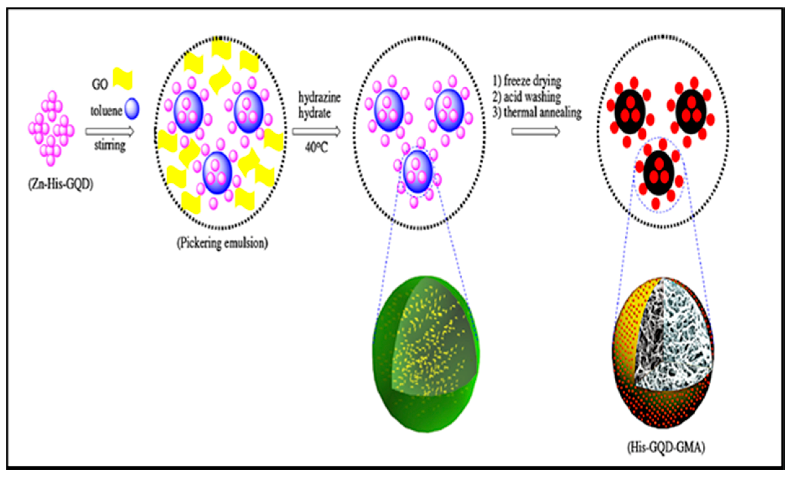
| H-GQDs-GMA | DPV | 0.29 | 0.001–80 | - - - | 7.0 | 0.01 M PBS | Rat brain | [97] | |
| GQDs/MWCNTs/GCE | DPV | 95 | 0.25–250 | - - - | 7.0 | 0.1 M PBS | Human serum (HS) | [113] | |
| GQDs/SPE | DPV | 500 | 1–900 | a 435 - - | 7.0 | 0.1 M PBS | DA ampoule & Urine | [119] | |
| CQDs | CD-CQDs/GCE | DPV | 140 | 4–220 | - b 150 c 420 | 7.0 | 0.1 M PBS | Urine | [100] |
| GCE/CQDs | DPV | 4.6 | 0.05–2 | - - - | 7.0 | 0.1M PBS | Urine | [51] | |
| GCE-CQDs | LSV | 2700 | 0.19–11.81 | - - - | 7.0 | 0.05 M PBS | - | [64] | |
| CQDs-CS/GCE | DPV | 11.2 | 0.1–30 | - - - | 7.0 | 0.5 mM PBS | DA injection | [80] | |
| IL-graphene/CQDs/GCE | DPV | 30 | 0.1–600 | - - - | 6.5 | PBS | Bovine serum | [76] | |
| NCQDs-nafion/GCE | DPV | 1.0 | 0.0–1000 | - 760 340 | 7.4 | 0.1 M PBS | - | [83] | |
| NCQDs/GCE | DPV | 1.2 | 0.05–800 | 580 - - | 6.5 | PBS | HS and HU | [115] | |
| CQDs/GCE | DPV | 26 | 0.15–150 | - - - | 6.0 | 0.1 M PBS | Human plasma | [94] | |
| AuNP/CQDs-CS/GCE | DPV | 1.0 | 0.1–30 | - - - | 7.0 | 0.5 mM PBS | DA injection | [99] | |
| Cu2O-CQDs/Nafion/GCE | DPV | 1.1 | 0.05–45 | - - - | 7.0 | 0.1 M PBS | HS | [82] | |
| CQDs/SPCE | CV | 99 | 1–7 | - - - | 7.4 | 0.1 M PBS | HU | [101] |
| Monoamine NTs | Electrode Support | Electrode | Technique | LOD (nM) | LDR (µM) | Interfering Species | pH | Supporting Electrolyte | Validating Sample | Ref. |
|---|---|---|---|---|---|---|---|---|---|---|
| Epinephrine | GQDs | GQDs/CS/CPE | CAP | 0.3 | 0.36–380 | Glc, suc, fru, cys, asp, ala, glu, his, arg, gly & trp | 7.0 | 0.1 M PBS | Blood serum & EP injection | [86] |
| GCE/GQDs/Lac | CV | 83.0 | 1–120 | AA, UA, trp, cys & glu | 5.2 | 0.1 M PCB | EP injection | [114] | ||
| CQDs | MIP/g-C3N4/NCQDs/GCE | DPV | 0.0003 | 0.001–1 | DA, AA, UA, tyr & trp | 7.5 | 0.1 M PBS | Human urine | [116] | |
| CQDs/CPE | DPV | 6.0 | 0.02–20 | SE & AA | 7.4 | 0.1 M PBS | EP injection | [93] | ||
| Serotonin | GQDs | MIP/h-BN/GQDs/GCE | DPV | 0.0002 | 0.001–10 | DA, NE & tyr | 7.0 | 0.1 M PBS | Human urine | [95] |
| CQDs | CQDs/CPE | DPV | 4.0 | 0.01–8 | AA & EP | 7.4 | 0.1 M PBS | - | [93] | |
| Norepinephrine | GQDs | GQDs-AuNPs/GCE | SWSV | 150 | 0.5–7.5 | AA & UA | 7.0 | 0.1 M PBS | NE injection & rat brain tissue | [106] |
| GQDs/IL/CPE | DPV | 60 | 0.2–400 | ACh | 7.0 | 0.1 M PBS | NE injection & human urine | [111] |
© 2020 by the authors. Licensee MDPI, Basel, Switzerland. This article is an open access article distributed under the terms and conditions of the Creative Commons Attribution (CC BY) license (http://creativecommons.org/licenses/by/4.0/).
Share and Cite
Elugoke, S.E.; Adekunle, A.S.; Fayemi, O.E.; Mamba, B.B.; Sherif, E.-S.M.; Ebenso, E.E. Carbon-Based Quantum Dots for Electrochemical Detection of Monoamine Neurotransmitters—Review. Biosensors 2020, 10, 162. https://doi.org/10.3390/bios10110162
Elugoke SE, Adekunle AS, Fayemi OE, Mamba BB, Sherif E-SM, Ebenso EE. Carbon-Based Quantum Dots for Electrochemical Detection of Monoamine Neurotransmitters—Review. Biosensors. 2020; 10(11):162. https://doi.org/10.3390/bios10110162
Chicago/Turabian StyleElugoke, Saheed E., Abolanle S. Adekunle, Omolola E. Fayemi, Bhekie B. Mamba, El-Sayed M. Sherif, and Eno E. Ebenso. 2020. "Carbon-Based Quantum Dots for Electrochemical Detection of Monoamine Neurotransmitters—Review" Biosensors 10, no. 11: 162. https://doi.org/10.3390/bios10110162
APA StyleElugoke, S. E., Adekunle, A. S., Fayemi, O. E., Mamba, B. B., Sherif, E.-S. M., & Ebenso, E. E. (2020). Carbon-Based Quantum Dots for Electrochemical Detection of Monoamine Neurotransmitters—Review. Biosensors, 10(11), 162. https://doi.org/10.3390/bios10110162