Article

Multilayer Core-Sheath Structured Nickel Wire/Copper Oxide/Cobalt Oxide Composite for Highly Sensitive Non-Enzymatic Glucose Sensor

School of Materials and Chemistry, University of Shanghai for Science and Technology, 516 Jungong Road, Shanghai 200093, China
*
Authors to whom correspondence should be addressed.
Nanomaterials 2025, 15(6), 411; https://doi.org/10.3390/nano15060411
Submission received: 21 January 2025 / Revised: 28 February 2025 / Accepted: 2 March 2025 / Published: 7 March 2025

Abstract

:
The development of micro glucose sensors plays a vital role in the management and monitoring of diabetes, facilitating real-time tracking of blood glucose levels. In this paper, we developed a three-layer core-sheath microwire (NW@CuO@Co3O4) with nickel wire as the core and copper oxide and cobalt oxide nanowires as the sheath. The unique core-sheath structure of microwire enables it to have both good conductivity and excellent electrochemical catalytic activity when used as an electrode for glucose detecting. The non-enzymatic glucose sensor base on a NW@CuO@Co3O4 core-sheath wire exhibits a high sensitivity of 4053.1 μA mM−1 cm−2, a low detection limit 0.89 μM, and a short response time of less than 2 s.

Graphical Abstract

1. Introduction

Glucose biosensors are essential in the medical field for monitoring blood glucose levels, as well as in the food industry for assessing sugar content given that high sugar intake is associated with increased cardiovascular disease risk [1,2,3]. In recent years, researchers have directed their efforts toward the development of non-enzymatic glucose sensors due to their faster response times, good stability, and lower costs [4,5]. Inorganic nanometals and metal oxides have been widely used in non-enzymatic electrochemical biosensors due to their long-term stability, high surface area, and excellent electrocatalytic activity [6,7]. However, studies involving metal oxide composites as active materials are relatively limited. Well-designed metal oxide composites can provide more active sites, significantly enhance electrocatalytic activity, facilitate electron transfer, and ultimately improve the sensor’s sensitivity.
Fiber-based sensors are ideally suited for the ongoing trend of miniaturization and smart applications, as their fiber-shaped and core-sheath structure design significantly enhances testing efficiency. Moreover, their remarkable flexibility and plasticity make them applicable across a diverse range of scenarios [8]. Zhao et al. reported a fiber-shaped biosensor based on gold fibers, which exhibited a high a sensitivity of 11.7 μA mM−1 cm−2 [9]. Carbon nanofibers have been reported for the development of an impedance sensor [10,11], and carbon nanofibers modified with aromatic diamine were used for glucose sensor with a high selectivity [12]. SnO2 nanofibers could be used for the development of a plasmonic resonance sensor [13]. The SnO2 nanofiber-based sensors could perform real-time monitoring of mammalian cells [14], and Cu2O-modified SnO2/N-C nanofibers could be used for NO2 detection [15]. Our group has grown gold nanomaterials on graphene fibers and used them for hydrogen peroxide and glucose detection, which showed excellent performance [16]. We also reported fiber-shaped glucose biosensors with F-doped nickel hydroxide nanorods on graphene fibers [17]. However, the preparation process of graphene fibers is relatively complicated, and the conductivity of graphene fibers is not very good.
Copper oxide (CuO) has found wide-ranging applications in photocatalysis, electrocatalysis, sensing, and antibacterial fields. This versatility is attributed to its high abundance, favorable redox potential, biocompatibility, and excellent stability. As the electrocatalytic activity of copper oxide depends on its surface properties and morphology, several researchers have employed various methods to synthesize copper oxide nanostructures that can be used for sensitive and selective detection of lactate and glucose [18,19]. The p-type copper oxide (CuO)-based photoelectric cathode, characterized by its substantial copper content, optimal bandgap, and advantageous band alignment, emerges as a promising candidate for large-scale photoelectrocatalytic water decomposition [20]. Its p-type conductivity can lead to limitations in performances under certain conditions, such as high glucose concentrations, which may cause sensitivity issues or interference from other substances. Composites of multiple metals, metal oxides, or carbon materials is one of the effective ways to solve this problem. For example, when using copper oxide/carbon layer core-shell composites as a non-enzymatic glucose sensor, they exhibited a good sensitivity of 272.6 μA mM−1 cm−2 [21]. Copper oxide/zinc oxide composites also displayed good sensitivity for glucose detection [22]. Co3O4 is a versatile inorganic compound known for its excellent electrochemical catalytic properties. Co3O4-based composites have been used as high-performance glucose sensors [23,24].
Herein, we have a rationale designed and prepared a three-layer core-sheath microwire (NW@CuO@Co3O4), with nickel wire as the core and copper oxide and cobalt oxide nanowires as the sheath, the latter of which is expected to demonstrate a synergistic effect for enhancing the performance for glucose detection. Nickel wire as the core has good conductivity and flexibility. The CuO and Co3O4 composite in the sheath layer has very good electrocatalytic activity, and the electrons generated by it can be quickly transferred to the nickel wire in the core. Finally, the NW@CuO@Co3O4 core-sheath wire was applied as the wire-shaped electrode for glucose sensing.

2. Experimental

2.1. Chemicals

Nickel sulphate (NiSO4·6H2O), boric acid (H3BO3), and copper sulphate (CuSO4·5H2O) were purchased from Shanghai Aladdin Reagent Company (Shanghai, China). Cobalt acetate tetrahydrate (C4H14CoO8), ammonium fluoride (NH4F), and urea (NH2CONH2) were purchased from Shanghai Titan Technology Co., Ltd. (Shanghai, China).

2.2. Preparation of Nickel Wire, Copper Oxide and Cobalt Oxide (NW@CuO@Co3O4) Composite Wire

A nickel wire with a diameter of 100 μm was first ultrasonically treated in acetone for 5 min to remove surface impurities and then washed with ionized water. The electrolyte was prepared by dissolving 0.2 M nickel sulfate (NiSO4·6H2O), 0.01 M copper sulfate (CuSO4·5H2O), and 0.1 M boric acid (H3BO3) in 50 mL DI water. The electrodeposition process took place in an electrochemical workstation with the nickel wire as the working electrode at a voltage of −0.8 V for 20 min. Then, the voltage was changed to +0.5 V and kept for another 20 min
The as-prepared wire was washed with DI water and transferred into a mixed aqueous solution containing 5 mM cobalt acetate tetrahydrate (C4H14CoO8), 10 mM ammonium fluoride (NH4F), and 25 mM urea (NH2CONH2). This aqueous solution was then placed in a Teflon-lined hydrothermal reactor and subjected to a hydrothermal reaction at 120 °C for 6 h in an oven. After completing the hydrothermal reaction, the sample was removed and washed with water. The dried wire was then placed in a tube furnace for annealing at 400 °C for 2 h to obtain the final product.

2.3. Characterization

Transmission electron microscope (TEM) images were captured with a JEOL 2000 microscope operating at 200 kV. Scanning electron microscopy (SEM) images were acquired using an FEI Quanta 450 FEG equipment. X-ray diffraction (XRD) analyses were carried out with an XPERT-PRO diffractometer featuring CuKα radiation (λ = 1.54 Å). X-ray photoelectron spectroscopy (XPS) spectra were recorded on a Thermo Fisher Scientific K-Alpha spectrometer with Al Ka X-ray radiation for excitation.
Prior to electrochemical testing, sensor electrode assembly was performed following the procedure illustrated in Figure S1. The NW@CuO@Co3O4 core-sheath wire was initially bonded to a copper wire using conductive silver adhesive. Subsequently, the integrated wire was encapsulated in a polyvinylidene fluoride (PVDF) tube (inner diameter of 2 mm), leaving 5 mm of the electrode outside the tube. Both ends of the PVDF tube were sealed with epoxy resin, while the copper wire could be connected to the electrochemical workstation via an electrode clamp.
Electrochemical testing was conducted on the CHI 660 Electrochemical Workstation with a three-electrode system in 0.1 M NaOH solution. An Ag/AgCl (4 M KCl) electrode and a platinum wire (diameter 0.5 mm) were used as the reference electrode and the counter electrode, respectively. The NW@CuO@Co3O4 electrode assembled as described above was used as the working electrode. For CV tests, the scan rate was 50 mV/s unless otherwise mentioned. For amperometric i–t tests, the electrolyte was stirred at 200 rpm to provide convective transport. All potentials were reported relative to the Ag/AgCl reference system.

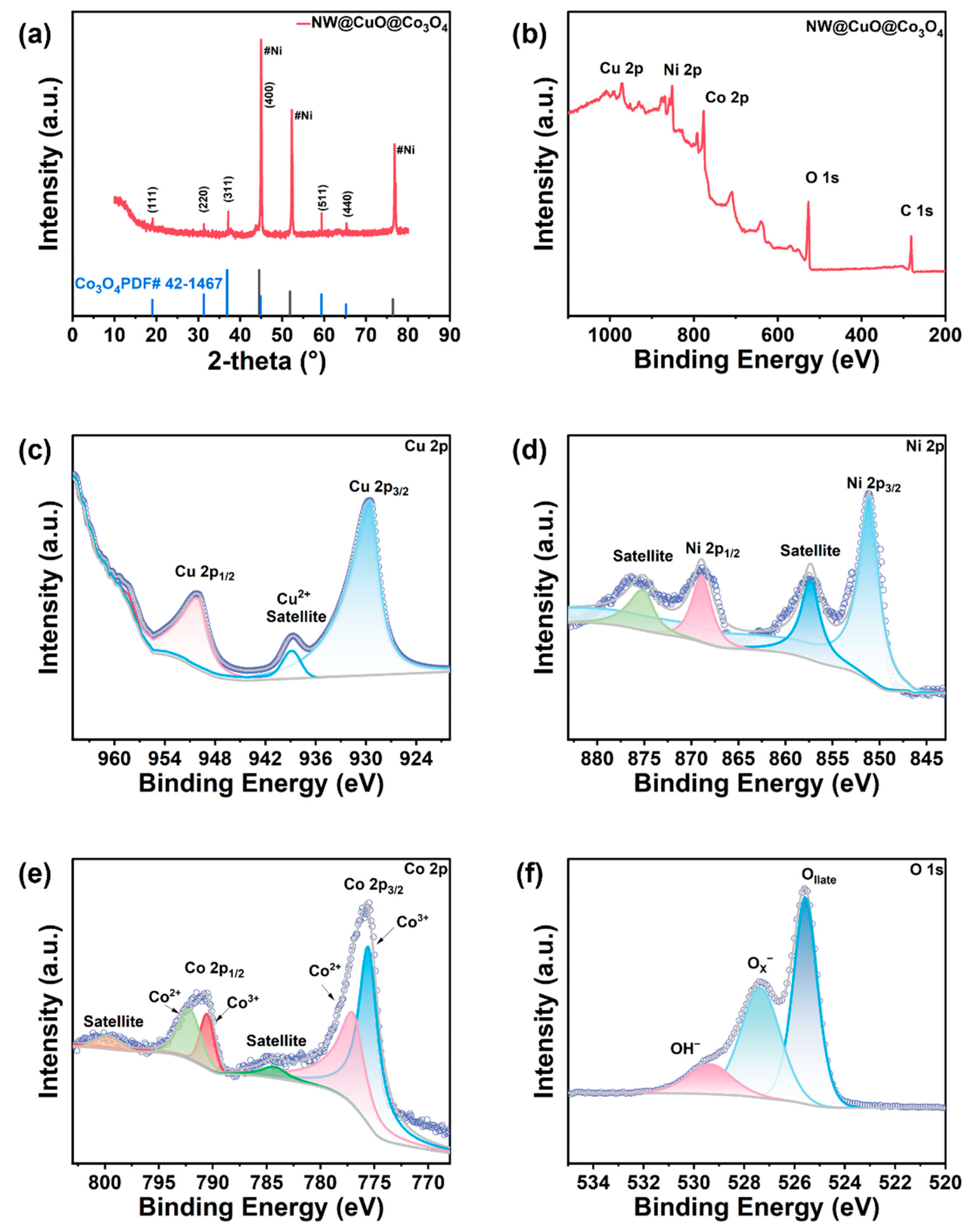
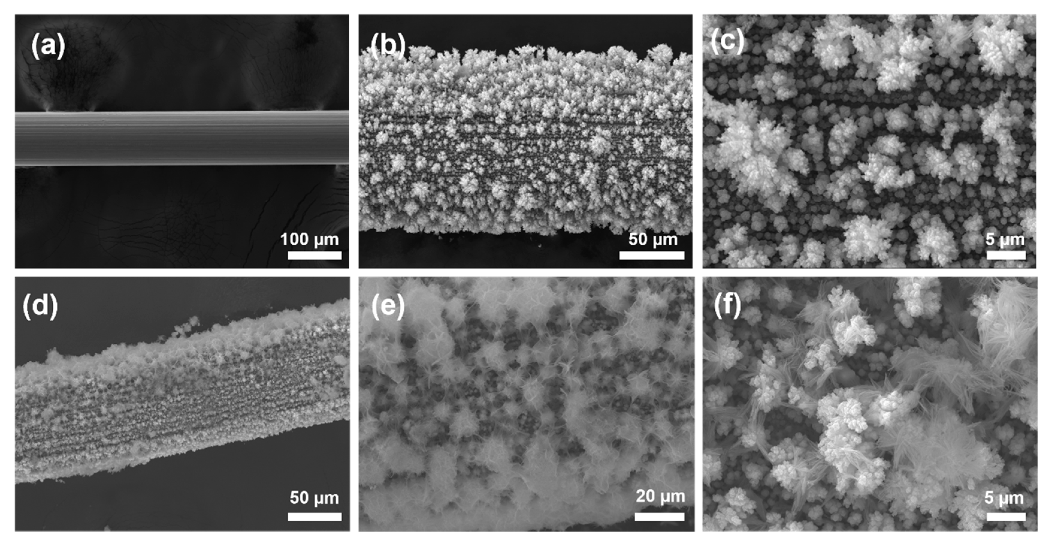
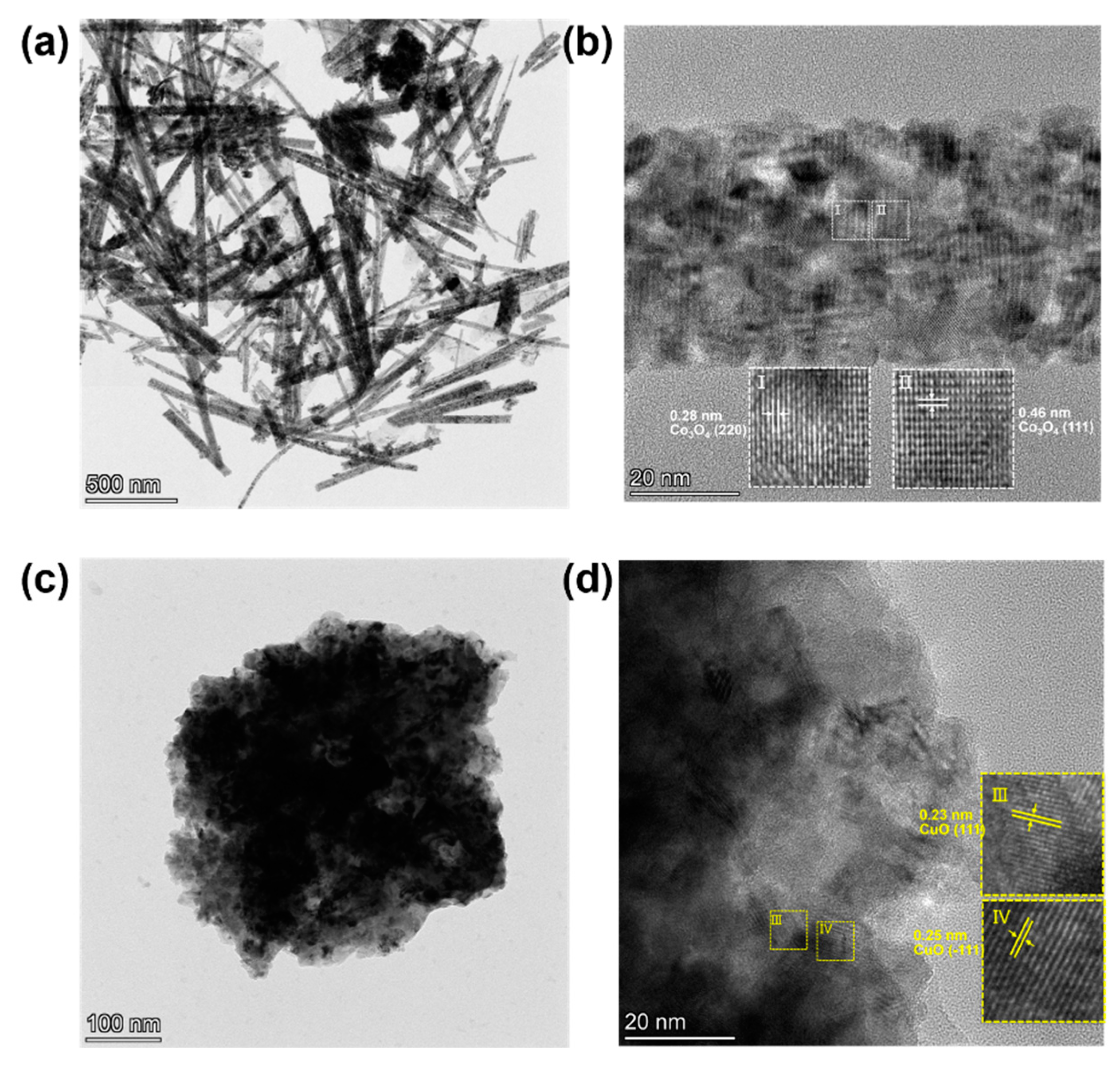
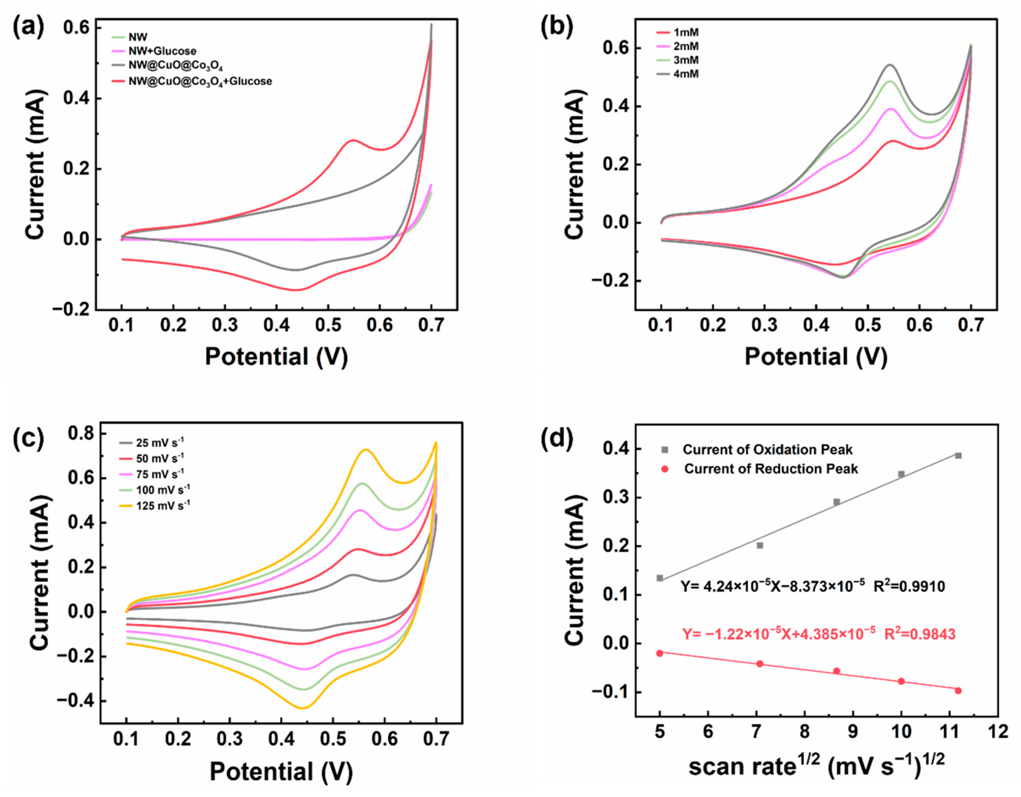
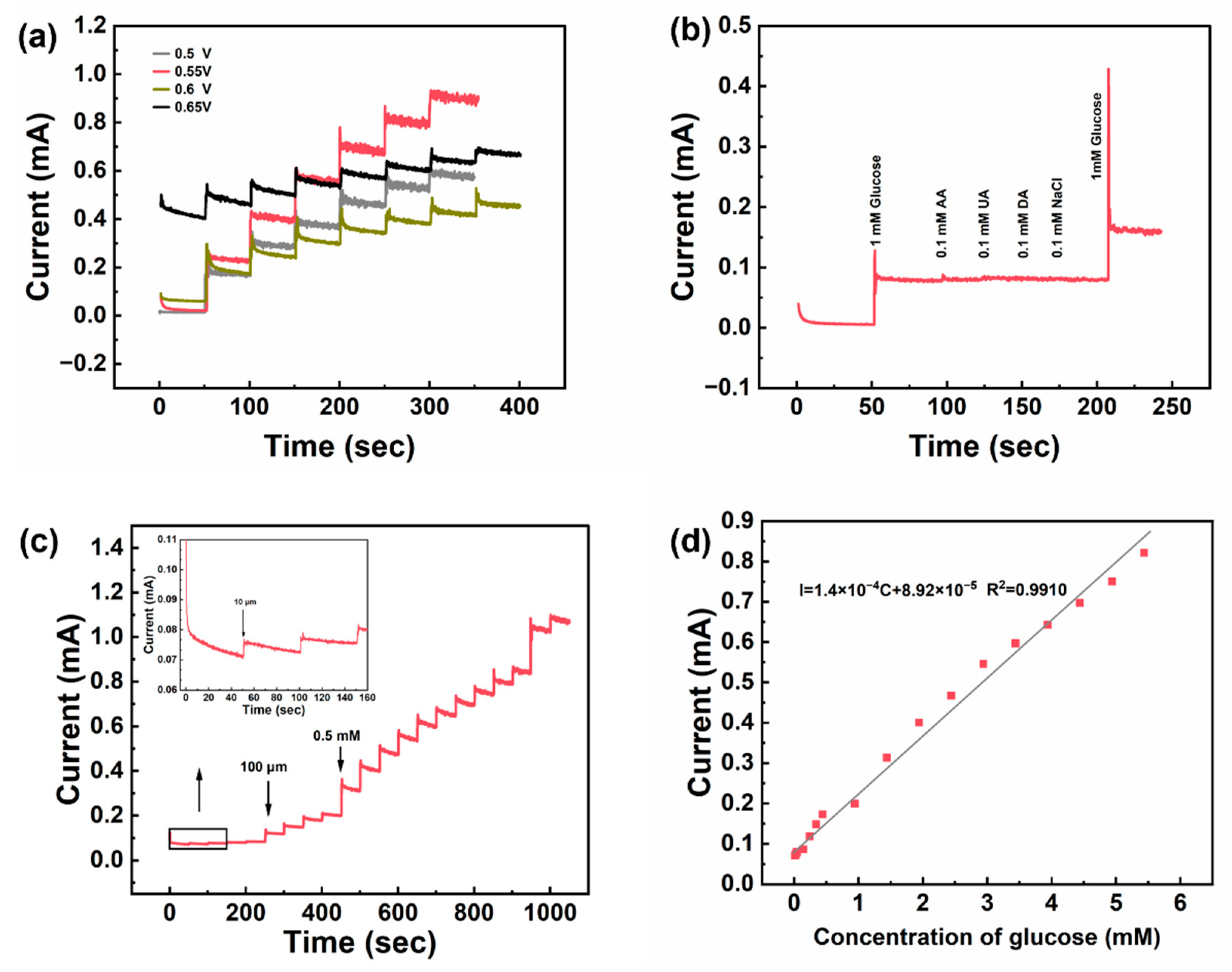
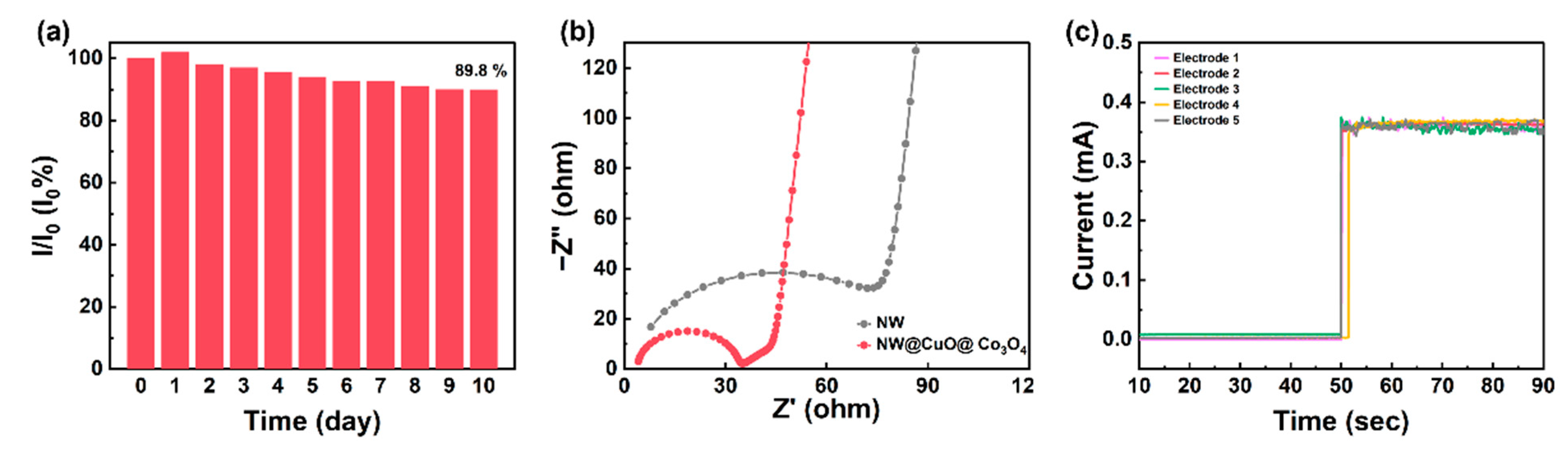
3. Results and Discussion

In order to understand the phase composition and crystal structure of the NW@CuO@Co3O4 electrode, XRD tests were conducted. As shown in Figure 1a, eight characteristic peaks were observed at 2θ values of 19.1°, 31.3°, 37.0°, 44.8°, 52.23°, 59.4°, 65.3°, and 78.01°. Based on PDF card JCPDS 42-1467, the peaks at 2θ = 19.1°, 31.3°, 37.0°, 44.8°, and 59.4° correspond to the (111), (220), (311), (400), and (511) crystal planes of Co3O4, respectively. Additionally, according to PDF card JCPDS 03-1051, the peaks at 2θ = 44.83°, 52.23°, and 78.01° correspond to the (111), (200), and (220) crystal planes of the nickel matrix, indicating that Co3O4 was successfully synthesized on the nickel wire substrate. It is worth noting that there is no obvious characteristic peak of copper oxide in the entire XRD spectrum. This is because copper oxide is covered by cobalt oxide and the content of copper oxide is relatively small, so its diffraction peak is not obvious.
Figure 1b displays the full spectrum of NW@CuO@Co3O4, where the signals for Ni, Co, O, and Cu arise from the nickel wire matrix as well as the loaded materials, CuO and Co3O4, respectively. In the high-resolution spectrum of Cu 2p, as depicted in Figure 1c, a peak at a binding energy of 929.5 eV is attributed to Cu 2p3/2, accompanied by a peak at 938.6 eV. The peak at 950.5 eV represents Cu 2p1/2, with a concomitant peak at 958.2 eV. Through the comprehensive analysis of the Cu spectrum, the difference in binding energy between Cu 2p1/2 and Cu 2p3/2 is found to be 21 eV, indicating that the copper in the NW@CuO@Co3O4 electrode is in the divalent state [25]. This indicated that the copper deposited on electrode after alloying and dealloying was oxidized to generate CuO during the subsequent hydrothermal process and high-temperature annealing process. Figure 1d presents the Ni 2p spectrum, where peaks 869.2 eV and 857.0 eV correspond to Ni 2p1/2 and Ni 2p3/2, respectively, along with characteristic peaks at 857.0 eV and 869.2 eV. These peaks are attributed to Ni2+, indicating the presence of divalent nickel in the NW@CuO@Co3O4 electrode. It is possible that part of the nickel was oxidized during the electrode preparation process.
Figure 1e shows the high-resolution spectrum of Co 2p. The peaks at 790.8 eV and 775.8 eV are associated with Co 2p1/2 and Co 2p3/2, respectively. Additionally, the binding energies at 800.2 eV and 784.3 eV are linked to the satellite peaks of Co 2p1/2 and Co 2p3/2. The Co 2p1/2 peak can be decomposed into two components at 792.3 eV and 790.6 eV, representing Co2+ and Co3+, respectively. Similarly, the Co 2p3/2 peak at 775.8 eV can be decomposed into two peaks at 777.3 eV and 775.5 eV, corresponding to Co2+ and Co3+, respectively. This confirms that the Co element in the NW@CuO@Co3O4 electrode exists as a mixture of divalent and trivalent states [26].
The O1s XPS spectrum (Figure 1f) is deconvoluted into three characteristic peaks at binding energies of 525.6 eV, 527.4 eV, and 529.4 eV. The peak at 525.6 eV corresponds to lattice oxygen within the NW@CuO@Co3O4 electrode, while the peak at 527.4 eV is associated with oxygen that occupies lattice vacancies in NW@CuO@Co3O4. The peak at 529.4 eV represents physically and chemically adsorbed oxygen on the electrode surface.
Figure 2a illustrates that the surface of the pure nickel wire is relatively smooth, with a diameter of approximately 100 μm. As shown in Figure 2b,c, after treating the nickel wire using the alloy/de-alloy method, a significant amount of copper nanoparticles were observed on its surface. These nanoparticles vary in size and are stacked upon one another, resulting in an uneven surface on the previously smooth nickel wire. Figure 2d reveals that a layer of Co3O4 was uniformly coated to the treated nickel wire through a hydrothermal process. The entire NW@CuO@Co3O4 composite wire has an approximate diameter of 130 μm, with a coating thickness of about 15 μm. As shown in Figure 2e, the Co3O4 particles are clustered and tightly connected to the copper nanoparticles formed during the initial step of alloy dealloying electrodeposition, which may facilitate the formation of a core–shell structure between these two metal oxides. The high-resolution SEM image in Figure 2f depicts a single cluster resembling a dandelion, consisting of multiple nanowires approximately 10 µm in length. This structure increases the specific surface area of the material, promoting redox reactions between the material and glucose.
Figure 3 presents the transmission electron microscope (TEM) image of NW@CuO@Co3O4. As shown in Figure 3a, the mixture of nanoparticles and nanowires is observed, which is consistent with the scanning electron microscope (SEM) image shown in Figure 2d. The high-magnification TEM image in Figure 3b reveals that the two lattice spacings correspond to the (220) and (111) crystal planes of Co3O4. Figure 3c shows the CuO nanoparticles with a diameter of approximately 500 nm. Figure 3d presents the high-resolution TEM image of a CuO nanoparticle, revealing two lattice spacings of 0.23 nm and 0.25 nm that are attributed to the (111) and (−111) crystal planes of CuO, respectively.
To assess the electrochemical performance of the NW@CuO@Co3O4 electrode, we tested both a pure nickel wire and the NW@CuO@Co3O4 electrode in a 0.1 M NaOH solution. Cyclic voltammetry tests were recorded in the electrolyte without and with the addition of 1 mM glucose. As depicted in Figure 4a, the results indicated that, regardless of glucose presence, the pure nickel wire did not exhibit significant redox peaks in the alkaline solution. This suggests that the nickel wire does not catalyze glucose in this system. Conversely, the NW@CuO@Co3O4 electrode demonstrated distinct redox peaks in alkaline solution. Notably, the peak current of these redox peaks increased significantly after the addition of glucose, indicating the occurrence of a redox reaction between the electrode and glucose, resulting in the generation of an additional current.
The NW@CuO@Co3O4 electrode participates in a redox reaction in alkaline solution, producing CoOOH. This CoOOH subsequently reacts further in the alkaline environment to form CoO2. Upon the addition of glucose, the equilibrium of this reaction is disrupted: CoO2 is consumed, glucose is oxidized, and CoO2 is reduced back to CoOOH. This process re-establishes a new equilibrium within the entire reaction system, resulting in an increase in the redox peak current. The specific reaction equations are as follows [27]:
C o 3 O 4 + O H + H 2 O 3 C o O O H + e
C o O O H + O H C o O 2 + H 2 O + e
2 C o O 2 + C 6 H 12 O 6 C 6 H 10 O 6 + 2 C o O O H
At the same time, copper oxide also participates in the catalysis of glucose in the following equations [28]:
C u O + O H C u O O H + 2 e
2 C u O O H + g l u c o s e ( C 6 H 12 O 6 ) 2 C u O + H 2 O + G l u c o l a c t o n e ( C 6 H 10 O 6 )
Figure 4b presents the CV curves at varying glucose concentrations. It is clear that in an alkaline solution, as glucose concentration increases, the oxidation peak current on the cyclic voltammogram rises accordingly, whereas the reduction peak current exhibits minimal variation. Furthermore, these results demonstrate that the NW@CuO@Co3O4 electrode possesses excellent catalytic oxidation properties toward glucose, generating distinct peak currents in response to varying concentrations of glucose in alkaline solutions.
Figure 4c displays CV curves at varying scan rates in a solution containing 1 mM glucose and 0.1 M NaOH. As the scan rate increases, the peak oxidation current rises and shifts to the right, while the peak reduction current simultaneously increases and shifts to the left. To clarify the relationship between peak currents and scan rates, a linear fit between the square root of half the scan rate and the peak current is provided in Figure 4d. The linear correlation coefficient for the peak oxidation current relative to the square root of half the scan rate is 0.9910, while for the peak reduction current, it is 0.9843. This linear relationship indicates that the entire reaction system is diffusion-controlled.
The operating voltage of the glucose sensor significantly influences its functional performance. Selecting an optimal operating voltage can enhance the efficiency of glucose catalytic oxidation, thereby ensuring optimal sensitivity and stability of the sensor. Cyclic voltammograms indicate that the oxidation peak occurs in a voltage range of 0.5 V to 0.65 V. A glucose solution was continuously added to a 0.1 M NaOH solution at voltages of 0.5 V, 0.55 V, 0.6 V, and 0.65 V, as illustrated in Figure 5a. An immediate response current was generated upon the addition of glucose, confirming that the NW@CuO@Co3O4 electrode exhibits a robust oxidative response to glucose. Within the voltage range of 0.5 V to 0.6 V, the response current increases with the rising voltage. However, at 0.65 V, the initial background current increases significantly. Following the addition of glucose, the increase in current does not surpass that observed at 0.55 V, and the response current becomes unstable with the gradual addition of glucose. Therefore, based on a comprehensive analysis of the data, 0.55 V is identified as the optimal operating voltage for this electrode.
Selectivity is a vital parameter in assessing the performance of the glucose sensor. Various other interfering substances may be present in the glucose solution environment, such as the structural analog of glucose, ascorbic acid (AA), dopamine hydrochloride (DA), and uric acid (UA). The principle of the glucose sensor is to convert oxidized glucose into a response current for characterizing glucose concentration. Consequently, it is essential for the sensor to selectively identify glucose molecules amidst the presence of other substances. To evaluate the sensor’s specificity, a series of tests were conducted with 0.5 mM glucose added to 0.1 M NaOH at 0.55 V, alongside AA, DA, UA, and NaCl. As shown in Figure 5b. The data reveal that a response current is generated upon the addition of glucose, while no significant response current is observed with other interfering substances. Moreover, the introduction of glucose after adding the interfering substances still elicits a response current, indicating that the glucose sensor does not adsorb these substances onto the electrode surface, thereby preserving its selective catalytic activity toward glucose. Based on these findings, the NW@CuO@Co3O4 electrode demonstrates excellent selectivity in glucose sensing.
To examine the relationship between glucose concentration and response current, glucose solutions of 0.01 mM, 0.1 mM, and 0.5 mM were sequentially added at an operating voltage of 0.55 V in 0.1 M NaOH solution, as shown in Figure 5c. The figure demonstrates that as the concentration of added glucose increases, the response current also escalates. Furthermore, as the concentration difference of the added glucose increases, the difference in response current correspondingly increases. With each addition of glucose, the response current exhibits a slight incremental rise until it becomes unstable, indicating that the sensor has reached its detection limit. A linear regression analysis of glucose concentration versus response current is presented in Figure 5d. The fitting relationship for glucose concentrations ranges from 0.01 mM to 5.44 mM. The linear correlation coefficient R2 is 0.991, the sensitivity of the NW@CuO@Co3O4 electrode is calculated to be 4053.1 μA·mM−1·cm−2, the current response time is less than 2 S, and the lowest detection limit is about 0.89 μM.
The long-term stability of the microelectrode was assessed by monitoring its current response in a mixed glucose solution containing 0.1 M NaOH and 2 mM glucose for 10 days. The testing method involves evaluating the current response of the same electrode after each day of its 100th use. As illustrated in Figure 6a, after 10 days (equating to 1000 reuses), the current response of the electrode maintained 89.8% of its initial value. Prior to testing, the electrode was stored in air at approximately 20 °C and 50% relative humidity.
Electrochemical impedance spectroscopy was conducted on the core-sheath microelectrode (NW@CuO@Co3O4) in a mixed glucose solution of 0.1 M NaOH and 2 mM glucose. As depicted in Figure 6b, the Nyquist plot features a semicircle in the high-frequency region and a linear component in the low-frequency region. The charge transfer resistance (Rct) can be determined by the diameter of the semicircle in the plot. The Rct for NW@CuO@Co3O4 microelectrode is 18.98 Ω and the series resistance (Rs) of this electrode is 3.458 Ω, which are much smaller than those of unmodified pure nickel wire (NW) electrode (Rct: 76.93 Ω and Rs: 4.29 Ω). The Rct decreases as the glucose concentration increases (Figure S2). The copper oxide and cobalt oxide in the sheath significantly reduce the resistance of the wire electrode and thus enhance the electron transmission efficiency of the electrode. The performance of the unmodified pure nickel wire is shown in Figure S3. Therefore, the performance of the NW@CuO@Co3O4 electrode for glucose detecting is much better than unmodified pure nickel wire and some glucose sensors in recent literature (Table 1).
In order to study its repeatability, the amperometric response of five electrodes towards 2 mM glucose was tested in 0.1 M NaOH (Figure 6c). A relative standard deviation (R.S.D.) of five electrodes was calculated to be 3.12%, confirming the good reproducibility of the NW@CuO@Co3O4 electrode.
A real sample recovery test was conducted using 50% (10 g/20 mL) glucose drinks sold on the market. The i–t method was utilized for testing and a linear fit was performed on the current density and glucose concentration. The glucose concentration was calculated and shown in Table S1 and Figure S4. The recoveries were in the range of 97–103.5%, illustrating the core-sheath wire electrode has outstanding reliability for glucose determination.

4. Conclusions

In summary, we designed and synthesized a three-layer core-sheath microwire (NW@CuO@Co3O4), with nickel wire as the core and copper oxide and cobalt oxide nanowires as the sheath. The synergistic effect of copper oxide nanoparticles and cobalt oxide nanowires enables it to have very high electrochemical activity, while the core-sheath structure gives the electrode very low resistances, including low Rct and Rs. The glucose sensor based on NW@CuO@Co3O4 electrode has good performance, with a linear range of 0.01~5.44 mM and a detection limit of 0.89 μM. The NW@CuO@Co3O4-based glucose sensor also exhibits good selectivity and stability.

Supplementary Materials

The following supporting information can be downloaded at: https://www.mdpi.com/article/10.3390/nano15060411/s1, Figure S1. (a) Schematic illustration of assembly for the NW@CuO@Co3O4 electrode. (b,c) Photographs of bare nickel microwire, NW@CuO@Co3O4 microwire and final sensor electrode. Figure S2. EIS of NW@CuO@Co3O4 microwire in 0.1 M NaOH with different concentration glucose. Figure S3. (a) Bare NW Amperometric response curve of adding different concentrations of glucose in 0.1 M NaOH solution in sequence at 0.55 V (b) Linear fitting diagram of current. Figure S4. Determination of glucose concentration in drinks by NW@CuO@Co3O4 electrode. Table S1. Determination of glucose concentration in drinks by NW@CuO@Co3O4 electrode.

Author Contributions

Methodology, Z.Z.; Investigation, Y.X.; Data curation, Z.Z.; Writing—original draft, Y.W.; Writing—review & editing, Y.X.; Supervision, X.L. and Y.X. All authors have read and agreed to the published version of the manuscript.

Funding

This work was supported by the National Natural Science Foundation of China (52172095), and Natural Science Foundation of Shanghai (24ZR1453900).

Data Availability Statement

No new data were created or analyzed in this study.

Conflicts of Interest

The authors declare no conflict of interest.

References

  1. Mohammadpour-Haratbar, A.; Mohammadpour-Haratbar, S.; Zare, Y.; Rhee, K.Y.; Park, S.J. A Review on Non-Enzymatic Electrochemical Biosensors of Glucose Using Carbon Nanofiber Nanocomposites. Biosensors 2022, 12, 1004. [Google Scholar] [CrossRef] [PubMed]
  2. Pohanka, M. Glucose Electrochemical Biosensors: The Past and Current Trends. Int. J. Electrochem. Sc. 2021, 16, 210719. [Google Scholar] [CrossRef]
  3. Lipinska, W.; Grochowska, K.; Siuzdak, K. Enzyme Immobilization on Gold Nanoparticles for Electrochemical Glucose Biosensors. Nanomaterials 2021, 11, 1156. [Google Scholar] [CrossRef]
  4. Jarnda, K.V.; Wang, D.Q.; Qurrat-Ul-Ain; Anaman, R.; Johnson, V.E.; Roberts, G.P.; Johnson, P.S..; Jallawide, B.J.R.; Kai, T.H.; Ding, P. Recent Advances in Electrochemical Non-Enzymatic Glucose Sensor for the Detection of Glucose in Tears and Saliva: A Review. Sensor Actuat A-Phys. 2023, 363, 114778. [Google Scholar] [CrossRef]
  5. Zohaa; Arif, D.; Hassan, M.; Abdullah, M.; Miran, W.; Nasir, M.A.; Batool, S.; Baig, M.A.; Liaqat, U. An Electrochemical Sensor Based on Copper Oxide Nanoparticles Loaded on a Mesoporous Mcm-41 for Non-Enzymatic Detection of Glucose. Ceram. Int. 2024, 50, 12614–12620. [Google Scholar] [CrossRef]
  6. Osuna, V.; Martinez, E.P.A.; Dominguez, R.B.; Rios, A.V. A Review on the Advances in Nanomaterials for Electrochemical Non-Enzymatic Glucose Sensors Working in Physiological Conditions. Chemosensors 2024, 12, 159. [Google Scholar] [CrossRef]
  7. Wang, Z.; Xu, J.; Yang, J.H.; Xue, Y.H.; Dai, L.M. Ultraviolet/Ozone Treatment for Boosting Oer Activity of Mof Nanoneedle Arrays. Chem. Eng. J. 2022, 427, 131498. [Google Scholar] [CrossRef]
  8. Jang, Y.; Kim, S.M.; Spinks, G.M.; Kim, S.J. Carbon Nanotube Yarn for Fiber-Shaped Electrical Sensors, Actuators, and Energy Storage for Smart Systems. Adv. Mater. 2020, 32, 1902670. [Google Scholar] [CrossRef]
  9. Zhao, Y.M.; Zhai, Q.F.; Dong, D.S.; An, T.C.; Gong, S.; Shi, Q.Q.; Cheng, W.L. Highly Stretchable and Strain-Insensitive Fiber-Based Wearable Electrochemical Biosensor to Monitor Glucose in the Sweat. Anal. Chem. 2019, 91, 6569–6576. [Google Scholar] [CrossRef]
  10. Joshi, B.; Wang, J.J.; Shan, X.N.; Mo, Y.L.; Hsu, T.T.C. Effects of Heat, Moisture/Water, and Compressive Force on Frequency Response Spectrum of Super Sensitive Carbon Nanofiber Aggregates (Sscnfa). Cement Concrete Comp. 2024, 152, 105638. [Google Scholar] [CrossRef]
  11. Li, W.G.; Guo, Y.P.; Zhang, X.R.; Dong, W.K.; Li, X.H.; Yu, T.; Wang, K.J. Development of Self-Sensing Ultra-High-Performance Concrete Using Hybrid Carbon Black and Carbon Nanofibers. Cement Concrete Comp. 2024, 148, 105466. [Google Scholar] [CrossRef]
  12. Ferlazzo, A.; Celesti, C.; Iannazzo, D.; Ampelli, C.; Giusi, D.; Costantino, V.; Neri, G. Functionalization of Carbon Nanofibers with an Aromatic Diamine: Toward a Simple Electrochemical-Based Sensing Platform for the Selective Sensing of Glucose. Acs Omega 2024, 9, 27085–27092. [Google Scholar] [CrossRef] [PubMed]
  13. Song, H.Y.; Wu, H.Y.; Xu, Y.P.; Ma, S.W.; Sun, M.; Wang, Q. Msm Fiber Optic Surface Plasmon Resonance Glucose Sensor Based on Sno2 Nanofibers/Au Structure. Photonic Sens. 2024, 15, 250119. [Google Scholar] [CrossRef]
  14. Kasiviswanathan, U.; Kumar, C.; Sahi, A.K.; Kumar, A.; Jit, S.; Sharma, N.; Mahto, S.K. Electrospun Stannic Oxide Nanofiber Thin-Film Based Sensing Device for Monitoring Functional Behaviors of Adherent Mammalian Cells. IEEE T Nanobiosci 2025, 24, 120–126. [Google Scholar] [CrossRef]
  15. Gu, L.Y.; Hu, J.; Zhu, D.C.; Zhang, W.X.; Cheng, M.; Wei, T.; Liu, Q.Q.; Wang, R.R.; Li, W.F.; Ling, Y.; et al. A Flexible Gas Sensor Based on One-Dimensional Cu2O-SnO2/N-C Nanofibers toward for NO2 Detection at Low Temperature. Sensor Actuat A-Phys. 2025, 383, 116257. [Google Scholar] [CrossRef]
  16. Peng, Y.; Lin, D.Q.; Gooding, J.J.; Xue, Y.H.; Dai, L.M. Flexible Fiber-Shaped Non-Enzymatic Sensors with a Graphene-Metal Heterostructure Based on Graphene Fibres Decorated with Gold Nanosheets. Carbon 2018, 136, 329–336. [Google Scholar] [CrossRef]
  17. Zhu, Z.W.; Wu, Y.T.; Yang, J.H.; Xue, Y.H. Core-Sheath Fibers Composed of F-Doped Nickel Hydroxide Nanorods and Graphene Fibers for Effective Fiber-Shaped Nonenzymatic Glucose Sensors. J. Alloys Compd. 2021, 889, 161608. [Google Scholar] [CrossRef]
  18. Khayyat, S.A.; Ansari, S.G.; Umar, A. Glucose Sensor Based on Copper Oxide Nanostructures. J. Nanosci. Nanotechnol. 2014, 14, 3569–3574. [Google Scholar] [CrossRef]
  19. Zhong, Y.; Shi, T.L.; Liu, Z.Y.; Cheng, S.Y.; Huang, Y.Y.; Tao, X.X.; Liao, G.L.; Tang, Z.R. Ultrasensitive Non-Enzymatic Glucose Sensors Based on Different Copper Oxide Nanostructures by in-Situ Growth. Sensor Actuat B-Chem. 2016, 236, 326–333. [Google Scholar] [CrossRef]
  20. Li, J.M.; Jin, X.; Li, R.G.; Zhao, Y.; Wang, X.Q.; Liu, X.D.; Jiao, H. Copper Oxide Nanowires for Efficient Photoelectrochemical Water Splitting. Appl. Catal. B-Environ. 2019, 240, 1–8. [Google Scholar] [CrossRef]
  21. Vediyappan, V.; Sivakumar, M.; Chen, S.M.; Lai, Q.W.; Madhu, R. Nanolayers of Carbon Protected Copper Oxide Nanocomposite for High Performance Energy Storage and Non-Enzymatic Glucose Sensor. J. Alloys Compd. 2021, 875, 160063. [Google Scholar] [CrossRef]
  22. Cheng, C.E.; Tangsuwanjinda, S.; Cheng, H.M.; Lee, P.H. Copper Oxide Decorated Zinc Oxide Nanostructures for the Production of a Non-Enzymatic Glucose Sensor. Coatings 2021, 11, 936. [Google Scholar] [CrossRef]
  23. Naderi, L.; Shahrokhian, S. Metal-Organic Framework-Assisted Co3o4/Cuo@Comnp with Core-Shell Nanostructured Architecture on Cu Fibers for Fabrication of Flexible Wire-Typed Enzyme-Free Micro-Sensors. Chem. Eng. J. 2023, 456, 141088. [Google Scholar] [CrossRef]
  24. Xiong, L.Y.; Kim, Y.J.; Seo, W.C.; Lee, H.K.; Yang, W.C.; Xie, W.F. High-Performance Non-Enzymatic Glucose Sensor Based on Co3o4/Rgo Nanohybrid. Rare Metals 2023, 42, 3046–3053. [Google Scholar] [CrossRef]
  25. Jiang, F.; Su, Q.M.; Li, H.J.; Yao, L.B.; Deng, H.H.; Du, G.H. Growth of Ultrafine Cuco2o4 Nanoparticle on Graphene with Enhanced Lithium Storage Properties. Chem. Eng. J. 2017, 314, 301–310. [Google Scholar] [CrossRef]
  26. Xiao, X.C.; Zhang, Z.Y.; Nan, F.; Zhao, Y.; Wang, P.K.; He, F.; Wang, Y.D. Mesoporous Cuco2o4 Rods Modified Glassy Carbon Electrode as a Novel Non-Enzymatic Amperometric Electrochemical Sensors with High-Sensitive Ascorbic Acid Recognition. J. Alloys Compd. 2021, 852, 157045. [Google Scholar] [CrossRef]
  27. Xu, J.N.; Li, F.H.; Wang, D.D.; Nawaz, M.H.; An, Q.B.; Han, D.X.; Niu, L. Co3o4 Nanostructures on Flexible Carbon Cloth for Crystal Plane Effect of Nonenzymatic Electrocatalysis for Glucose. Biosens. Bioelectron. 2019, 123, 25–29. [Google Scholar] [CrossRef] [PubMed]
  28. Cheng, S.Y.; DelaCruz, S.; Chen, C.; Tang, Z.R.; Shi, T.L.; Carraro, C.; Maboudian, R. Hierarchical Co3O4/CuO Nanorod Array Supported on Carbon Cloth for Highly Sensitive Non-Enzymatic Glucose Biosensing. Sensor Actuat B-Chem. 2019, 298, 126860. [Google Scholar] [CrossRef]
  29. Velmurugan, M.; Karikalan, N.; Chen, S.M. Synthesis and Characterizations of Biscuit-Like Copper Oxide for the Non-Enzymatic Glucose Sensor Applications. J. Colloid. Interf. Sci. 2017, 493, 349–355. [Google Scholar] [CrossRef]
  30. Cheng, D.; Wang, T.; Zhang, G.X.; Wu, H.M.; Mei, H. A Novel Nonenzymatic Electrochemical Sensor Based on Double-Shelled Cuco2o4 Hollow Microspheres for Glucose and H2O2. J. Alloys Compd. 2020, 819, 153014. [Google Scholar] [CrossRef]
  31. Heidari, H.; Habibi, E. Amperometric Enzyme-Free Glucose Sensor Based on the Use of a Reduced Graphene Oxide Paste Electrode Modified with Electrodeposited Cobalt Oxide Nanoparticles. Microchim. Acta 2016, 183, 2259–2266. [Google Scholar] [CrossRef]
  32. Tran, T.H.; Thi, M.L.N.; Son, N.T.; Bui, Q.B.; Ai-Le, P.H.; Nhac-Vu, H.T. Novel Nanoneedle Structures of Zinc-Doped Cobalt Hydroxide as a Self-Supported Sensor for Sensitive Glucose Detection. Mater. Res. Bull. 2019, 120, 110580. [Google Scholar] [CrossRef]
Figure 1. (a) XRD spectrum of the NW@CuO@Co3O4 electrode. XPS spectra of the NW@CuO@Co3O4 electrode, (b) full spectrum, (c) Cu 2p spectrum, (d) Ni 2p spectrum, (e) Co 2p spectrum, and (f) O 1s spectrum.
Nanomaterials 15 00411 g001
Figure 2. (a) SEM image of pure nickel wire. (b,c) SEM image of nickel wire after “alloy/de-alloy” treatment. (df) SEM image of NW@CuO@Co3O4 electrode.
Nanomaterials 15 00411 g002
Figure 3. (a) TEM image of CuO–Co3O4 composite. (b) TEM image of Co3O4 nanowires, (c,d) TEM images of CuO nanoparticles.
Nanomaterials 15 00411 g003
Figure 4. (a) Cyclic voltammograms of pure nickel wire electrode and NW@CuO@Co3O4 electrode at 50 mV s−1 in 0.1 M NaOH solution with and without 1mM glucose added. (b) Cyclic voltammograms of NW@CuO@Co3O4 electrode at 50 mV s−1 in different 0.1M NaOH solutions with different concentrations of glucose added. (c) Cyclic voltammograms of NW@CuO@Co3O4 electrode in 0.1 M NaOH at different scan rates (25~125 mv s−1). (d) Linear fitting diagram of cyclic voltammograms of oxidation peak current and reduction peak current at different scan rates and the half of the scan rate.
Nanomaterials 15 00411 g004
Figure 5. Glucose detection performance of NW@CuO@Co3O4 electrode (a) Current versus time curves of adding 0.5 mM glucose six times at 50 s intervals to 0.1 M NaOH solution at different voltages (0.5~0.65 V). (b) Selectivity curve after adding 1 mM glucose, ascorbic acid (AA), dopamine hydrochloride (UA), uric acid (DA), and glucose to 0.1 M NaOH solution in sequence. (c) Amperometric response curve of adding different concentrations of glucose in 0.1 M NaOH solution in sequence at 0.55 V. (d) Linear fitting diagram of current and glucose concentration in alkaline solution.
Nanomaterials 15 00411 g005
Figure 6. (a) Stability of NW@CuO@Co3O4 glucose sensors in 0.1 mM glucose solution. (b) EIS curves of NW and NW@CuO@Co3O4. (c) Current responses of five NW@CuO@Co3O4 electrodes in 0.1 M NaOH with 2 mM glucose at 0.55 V.
Nanomaterials 15 00411 g006
Table 1. Comparison of performances of our electrode with other metal oxide-flexible glucose sensors.
Sensitive MaterialsOperating PotentialLinear Interval (Up to, mM)Sensitivity
(μA·
mM−1·cm−2)
Detection Limit (μM)Ref
CuO biscuits/SPCE0.5 V4.03308.710.1[29]
Ni(OH)F@GF0.55 V7.66595.31[17]
CuCo2O40.55 V7.94000.75[30]
Co(NO3)2@rGO0.45 V412101.4[31]
Zn-Co-OH/3D NF0.6 V3.5361008.33[32]
NW0.55 V0.527045.2this work
NW@CuO@Co3O40.55 V5.4440530.89this work
Disclaimer/Publisher’s Note: The statements, opinions and data contained in all publications are solely those of the individual author(s) and contributor(s) and not of MDPI and/or the editor(s). MDPI and/or the editor(s) disclaim responsibility for any injury to people or property resulting from any ideas, methods, instructions or products referred to in the content.

