Article

Preparation and Optimization of Mn2+-Activated Na2ZnGeO4 Phosphors: Insights into Precursor Selection and Microwave-Assisted Solid-State Synthesis

International Joint Research Center for Photo-Responsive Molecules and Materials, School of Chemical and Material Engineering, Jiangnan University, Wuxi 214122, China
*
Authors to whom correspondence should be addressed.
These authors contributed equally to this work.
Nanomaterials 2025, 15(14), 1117; https://doi.org/10.3390/nano15141117
Submission received: 28 June 2025 / Revised: 16 July 2025 / Accepted: 16 July 2025 / Published: 18 July 2025
(This article belongs to the Section Inorganic Materials and Metal-Organic Frameworks)

Abstract

Mn2+-doped phosphors emitting green light have garnered significant interest due to their potential applications in display technologies and solid-state lighting. To facilitate the rapid synthesis of high-performance Mn2+-activated green phosphors, this research optimizes a microwave-assisted solid-state (MASS) method for the preparation of Na2ZnGeO4:Mn2+. Leveraging the unique attributes of the MASS technique, a systematic investigation into the applicability of various Mn-source precursors was conducted. Additionally, the integration of the MASS approach with traditional solid-state reaction (SSR) methods was assessed. The findings indicate that the MASS technique effectively incorporates Mn ions from diverse precursors (including higher oxidation states of manganese) into the crystal lattice, resulting in efficient green emission from Mn2+. Notably, the photoluminescence quantum yield (PLQY) of the sample utilizing MnCO3 as the manganese precursor was recorded at 2.67%, whereas the sample synthesized from MnO2 exhibited a remarkable PLQY of 17.69%. Moreover, the post-treatment of SSR-derived samples through the MASS process significantly enhanced the PLQY from 0.67% to 8.66%. These results underscore the promise of the MASS method as a novel and efficient synthesis strategy for the rapid and scalable production of Mn2+-doped green luminescent materials.

Graphical Abstract

1. Introduction

Green emission, as one of the three primary colors in luminescent systems, plays a critical role in applications such as display technologies, solid-state lighting, and safety signage [1,2,3,4]. In recent years, a variety of high-performance green phosphors have been developed. In addition to the conventional commercial green phosphor SiAlON:Eu2+ [5], several novel Eu2+-activated green-emitting materials have been reported, such as Ba2LiSi7AlN12:Eu2+ and Ba[Li2(Al2Si2)N6]:Eu2+ [6,7]. Although these materials exhibit excellent optical properties, they typically require Eu2+ as the luminescent center and the construction of complex oxynitride lattices, necessitating harsh synthesis conditions and highly reducing environments [8,9,10,11,12]. As a result, Mn-based green phosphors have attracted increasing attention as promising alternatives. These materials utilize transition metal Mn ions as activators and offer the advantage of simpler synthesis processes [13,14].
As a multivalent, non-rare-earth ion with accessible oxidation states ranging from +2 to +7, Mn ions are of particular interest [15,16,17,18,19,20]. Green emission from Mn2+ is typically attributed to the spin- and parity-forbidden 4T16A1 transition of the 3d5 electronic configuration in specific crystal field environments [21,22,23,24]. In recent years, numerous studies have demonstrated the potential of Mn2+-activated phosphors for green emission. For example, D. Zhang et al. synthesized ZnAl2O4:Mn phosphors via a sol–gel method, showing efficient green emission under blue LED excitation for potential white LED applications [25]. Subsequently, Menon et al. reported green emission at 523 nm from Mn2+ in Zn2SnO4 prepared via a solid-state reaction (SSR) [26]. In addition, Su et al. employed a co-precipitation precursor (COP) method to prepare γ-AlON:Mn2+,Mg2+ [27] phosphors exhibiting narrow-band green emission. Although significant progress has been made in the development of Mn2+-based green emitting phosphors, many of the reported synthesis routes still suffer from complex processing steps and high energy consumption. In this context, it is highly desirable to explore alternative synthesis strategies that are not only simple and environmentally friendly but also capable of achieving rapid reaction rates with low energy input. The microwave-assisted solid-state synthesis (MASS) method stands out in this regard, offering a time-efficient and energy-saving approach compared to conventional high-temperature solid-state techniques, thus providing a promising route for the scalable fabrication of Mn2+-doped green-emitting materials.
MASS has emerged in recent years as an efficient and convenient method for the preparation of inorganic luminescent materials [28,29]. This green technique utilizes a microwave susceptor to absorb microwave energy and convert it into heat, thereby rapidly raising the temperature of the precursor. As the reaction progresses, the dielectric properties of the precursor change, enabling it to directly absorb microwave energy and accelerate the reaction process [30,31,32,33]. In previous reports, Miranda de Carvalho et al. employed the MASS method to rapidly synthesize a series of photochromic compounds, Na8Al6Si6O24(Cl, S)2 [34]. Subsequently, Poelman and co-workers successfully prepared a range of luminescent materials, including aluminates, germanates, and gallates, using MASS [16,35]. Since then, numerous studies have demonstrated the versatility of this method for synthesizing various luminescent phosphors. However, there are still relatively few reports on the use of MASS for the synthesis of high-efficiency Mn2+-doped green-emitting materials. This is mainly because MASS, as a novel synthesis method for luminescent materials, relies heavily on the intrinsic microwave absorption characteristics of the precursors. Moreover, the process optimization strategies of MASS differ significantly from those of conventional SSR methods. In the SSR approach, the sintering temperature and reaction duration can be independently adjusted to regulate the nucleation and growth of crystals. In contrast, the MASS method involves a more complex interplay of parameters, as the microwave irradiation time simultaneously influences several critical factors, including the heating rate of the microwave susceptor, the energy absorption efficiency of the precursor, and the duration of crystal growth. This interdependence increases the complexity of optimizing synthesis conditions for achieving high-performance Mn2+-activated phosphors using the MASS technique. Therefore, it is necessary to systematically investigate the process parameters of MASS and explore suitable precursors for the efficient synthesis of Mn2+-doped luminescent materials using this method. Furthermore, in order to enhance the versatility and applicability of MASS, it is of great significance to explore the integration of MASS with conventional SSR, leveraging the microwave-driven energy absorption characteristics of the materials to improve luminescence performance and broaden the scope of synthesis.
In this work, a germanate-based system—a suitable host for the MASS technique—was selected as the doping matrix. Among them, Na2ZnGeO4 was chosen due to its abundant spatial lattice sites and relatively low structural symmetry, which can provide favorable sites for Mn2+ ions [36]. Accordingly, a series of Mn2+-doped Na2ZnGeO4 green-emitting phosphors were synthesized using three different strategies: the MASS, SSR, and a combined SSR + MASS approach. The process parameters (microwave irradiation duration and dopant contents) of the MASS method were systematically optimized, and the influence of different manganese precursors on the luminescence performance was also investigated. In addition, the compatibility of MASS with different manganese precursors was investigated. The results demonstrate that MnO2, Mn2O3, and MnCO3 can all serve as effective precursors for the successful synthesis of Mn2+-activated green phosphors via the MASS method, yielding photoluminescence quantum yields (PLQY) of 17.69%, 7.59%, and 2.67%, respectively. Furthermore, the integration of SSR with MASS markedly improved the PLQY from 0.67% to 8.66%, confirming the capability of MASS to enhance luminescence efficiency. These results demonstrate that MASS, either independently or in combination with SSR, offers a promising pathway for the rapid and efficient synthesis of Mn2+-activated green-emitting phosphors, providing valuable insights for the development of high-performance luminescent materials.

2. Materials and Methods

2.1. Materials Preparation

2.1.1. The MASS Samples

As illustrated in Figure 1a, the MASS synthesis process was employed using stoichiometric amounts of ZnO (99.9%), Na2CO3 (99.99%), GeO2 (99.99%), and different manganese sources—MnCO3 (99.99%), MnO2 (99.99%), and Mn2O3 (99.99%)—all purchased from Aladdin without further treatment. The microwave reaction setup consisted of two alumina crucibles and aluminosilicate insulation bricks. The larger crucible (50 mL) was filled with 7 g of activated carbon (10–20 mesh, Leyan, Shanghai, China), serving as the microwave susceptor. The smaller crucible (5 mL), containing 0.8 g of precursor powder, was covered with an alumina lid and embedded into the activated carbon bed inside the larger crucible. The entire crucible assembly was placed into a cavity made from high-temperature aluminosilicate insulation bricks. The reaction was carried out in a laboratory microwave oven operating at 2.45 GHz with an output power of 700 W for 10–30 min. The resulting powders were thoroughly ground for subsequent characterization.

2.1.2. The SSR Samples

Stoichiometric amounts of ZnO (99.9%, Aladdin), Na2CO3 (99.99%, Aladdin), GeO2 (99.99%, Aladdin), and one of the manganese sources—MnCO3 (99.99%, Aladdin), MnO2 (99.99%, Aladdin), or Mn2O3 (99.99%, Aladdin)—were carefully weighed for each synthesis batch. The resulting powder mixture was partially transferred into a 13 mm diameter die and pressed under a uniaxial pressure of 150 MPa to form the tablet precursor. These tablets, along with the remaining powder precursor, were then sintered in air at 1200 °C for 6 h in a muffle furnace, with a heating rate of 5 °C min−1. After sintering, the samples were furnace-cooled to room temperature, and the resulting powder was thoroughly ground into fine powders for further characterization.

2.1.3. The SSR + MASS Samples

The SSR-prepared samples, synthesized using the method described previously, were subsequently used as precursors for microwave-assisted treatment following the same MASS procedure outlined earlier. In this step, 7 g of activated carbon was used as the microwave susceptor, with a microwave power of 700 W and an irradiation time of 10 min. The resulting powders were thoroughly ground to ensure uniformity and used for further characterization.

2.2. Structural and Morphological Characterization

The crystallographic phase purity of the obtained phosphors was examined by X-ray diffraction (XRD) using an X-ray powder diffractometer (D8, Bruker AXS GmbH, Bruker, Germany), with Cu Kα1 (1.5406 Å) radiation (40 kV, 40 mA) at room temperature. X-ray photoelectron spectroscopy (XPS, Thermo Fisher Scientific K-Alpha, Thermo Scientific, Waltham, MA, USA) was employed to analyze the elemental composition of the samples. The microstructure and chemical composition of the samples were investigated by scanning electron microscopy (SEM; Zeiss Sigma 300, Zeiss, Oberkochen, Germany) equipped with an energy-dispersive X-ray spectroscopy system.

2.3. Luminescence Characterization

The room-temperature steady-state photoluminescence (PL) and photoluminescence excitation (PLE) spectra were measured by a fluorescence spectrometer (FS5, Edinburgh, UK), with a monochromated 450 W Xenon arc lamp as the excitation source. All spectra were automatically corrected for detector response. The PLQY was measured using a fluorescence spectrometer (FLS1000, Edinburg, UK) equipped with an integrating sphere coated with polytetrafluoroethylene (N-M01, Edinburgh, UK). Luminescent photos of the samples were taken with an SLR camera (T5i, Canon, Tokyo, Japan). Temperature-dependent emissions were collected from room temperature to 240 °C using a fiber connected portable spectrophotometer (Ocean Optics, Dunedin, FL, USA) and a programmed heating platform (SET450, Fanandair Co., Ltd., Shenzhen, China). The NIR spectra were measured by the NIR Quest + 1.7 900–1700 nm, equipped with the ISP-50-8-I 50MM integrating sphere (Ocean optics, Dunedin, FL, USA).

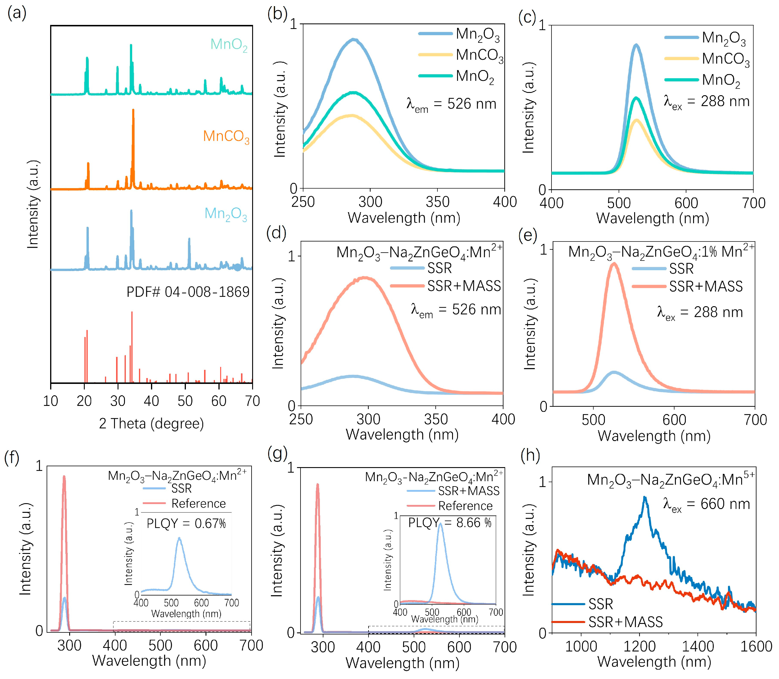
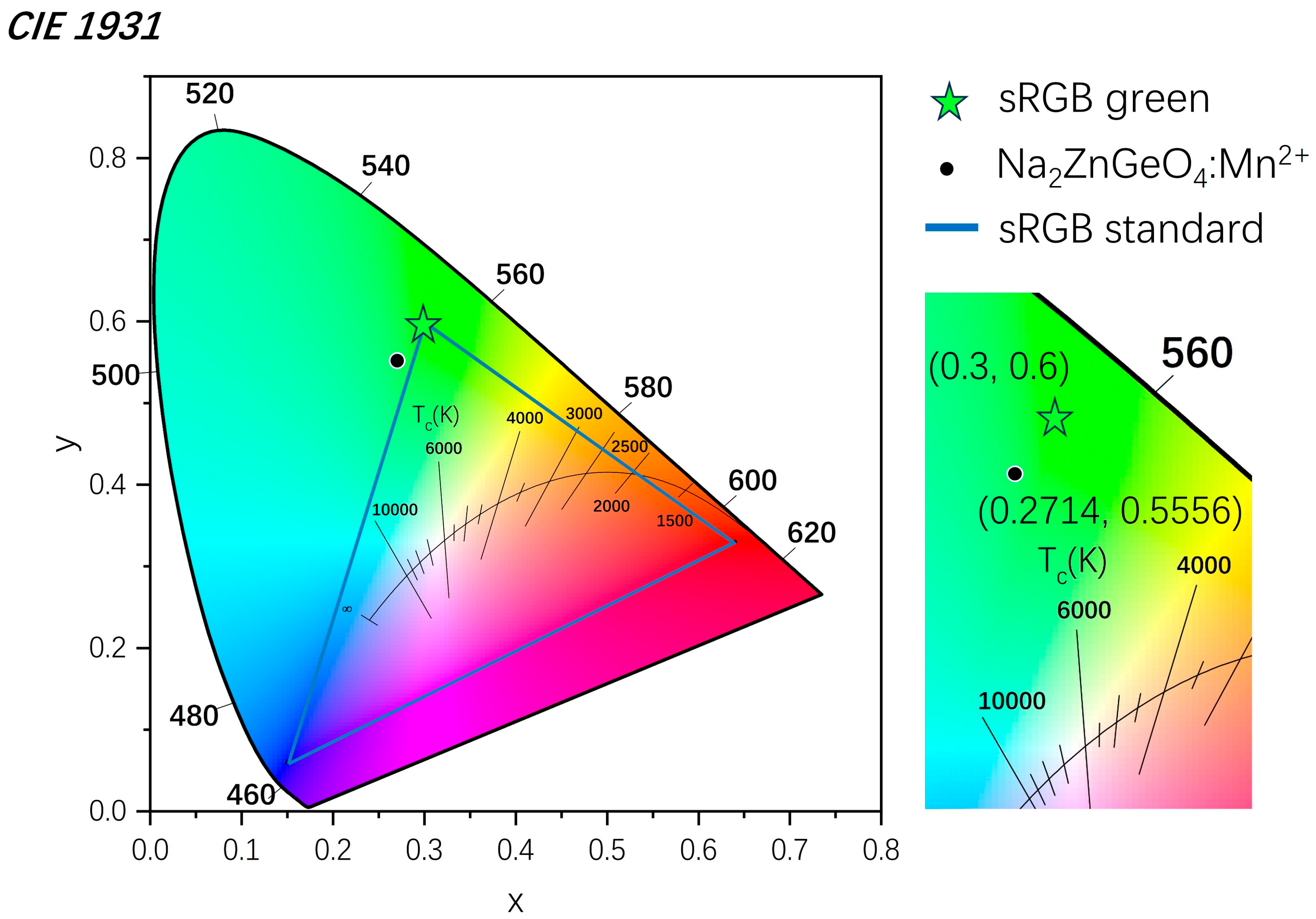
3. Results and Discussion

The crystal structure of Na2ZnGeO4 and the local coordination environments of Zn, Ge, and Na with four surrounding oxygen atoms are illustrated in Figure 1b. Na2ZnGeO4 belongs to the monoclinic crystal system (space group P1n1) and possesses abundant crystallographic sites. Within the crystal, three distinct coordination units are formed: Zn, Ge, and Na. Each coordinate has four oxygen atoms to form [ZnO4] (rZn = 0.59 Å), [GeO4] (rGe = 0.38 Å), and [NaO4] (rNa = 0.59 Å), respectively. These units are interconnected via oxygen atoms to establish a three-dimensional network structure [37]. Due to the identical valence state and similar ionic radius of Mn2+ (rMn = 0.65 Å, CN = 4) compared to Zn2+, with a radius difference of less than 30%, Mn2+ preferentially occupies the [ZnO4] site to form the [MnO4] structure [24].
The synthesis of Na2ZnGeO4 via MASS processing differs from the conventional SSR method [24].Traditional SSR involves the direct adjustment of the heating rate, sintering temperature, and duration to regulate crystal nucleation and growth, thus facilitating the formation of pure-phase Na2ZnGeO4. In contrast, MASS relies on a reaction medium that rapidly absorbs microwave energy to heat the precursor [38,39]. As the precursor temperature increases, its ability to absorb microwave energy is further enhanced, leading to an intense reaction and rapid nucleation and growth of Na2ZnGeO4. During this process, both the reaction medium and the precursor absorb microwave energy simultaneously. However, an excessive microwave energy input may result in the overgrowth of the crystal, potentially deteriorating its performance. Therefore, it is crucial to investigate the influence of the microwave irradiation time on the structural and luminescence properties of Na2ZnGeO4:Mn2+. The XRD patterns of Na2ZnGeO4:Mn2+ synthesized using MnCO3 as the manganese source under varying microwave durations are presented in Figure 1c. The results show that the XRD patterns of the samples obtained under microwave irradiation for 10–30 min match well with the standard Na2ZnGeO4 phase (PDF#04-008-1869), with no detectable impurity phases. In addition, no diffraction peaks corresponding to manganese-related compounds were observed, confirming that the Na2ZnGeO4 phase can be effectively formed within this microwave duration.
Room-temperature PL and PLE spectra were further recorded to analyze in detail the effect of different microwave irradiation times on the luminescence properties of the material. As Figure 2a shows, upon monitoring at an emission wavelength of 524 nm, the excitation spectrum spans the range of 200–350 nm, featuring both a broad excitation background and several sharp peaks. The broad band in the short-wavelength region is attributed to the Mn–O charge transfer transition, while the narrow excitation peaks originate from spin- and parity-forbidden d–d transitions of Mn2+ ions in a tetrahedral coordination environment [40,41,42]. The PL spectra excited at 296 nm are also displayed in Figure 2b. A green emission peak centered at 524 nm with a full width at half maximum (FWHM) of 42 nm is observed, which can be attributed to the spin- and parity-forbidden 4T16A1 transition of Mn2+ ions. The simultaneous observation of the PLE and PL spectra reveals that both the excitation and emission intensities gradually increase with an increasing microwave irradiation time, reaching a maximum at 25 min, followed by a decline at longer durations. This observation is consistent with the previous analysis. Under short microwave irradiation durations, increasing the irradiation time facilitates improved crystal growth, thereby enhancing the luminescence performance of the material. However, prolonged microwave exposure may lead to excessive energy absorption, which can potentially alter the local crystal field environment around the luminescent centers and subsequently degrade the emission intensity. These results suggest that, within the scope of this study, there exists an optimal range of microwave irradiation time that favors enhanced luminescent performance. Excessive irradiation beyond this point may lead to deterioration in luminescent performance. Photographs of the samples under 254 nm and 302 nm UV illumination are shown in Figure 2c. The visual observations are consistent with the spectral results. Specifically, the sample irradiated for 25 min exhibits the strongest emission under UV light, displaying a vivid green luminescence. Therefore, the microwave irradiation time in this work is applied for 25 min.
To further optimize the synthesis parameters of the MASS method, the effect of the Mn2+ doping concentration on the luminescence properties of the material was investigated using MnCO3 as the manganese source. The XRD patterns are presented in Figure 3a, and it is evident that the samples with varying doping concentrations synthesized via the microwave-assisted method exhibit a single-phase structure without detectable impurities. The elemental composition of the material was further examined through elemental mapping and XPS, as shown in Figures S1–S3. Elemental mapping (Figure S1) confirms that all target elements are successfully incorporated into the crystal structure and exhibit a uniform spatial distribution. Furthermore, the XPS spectra in Figures S2 and S3 verify that manganese ions are predominantly present in a +2 oxidation state (Mn2+), indicating their effective incorporation into the host lattice. This indicates that Mn2+ ions have been successfully incorporated into the crystal lattice across all doping levels. The PLE and PL spectra of samples with varying Mn2+ doping concentrations are presented in Figure 3b,c. The luminescence intensity reaches its maximum at a doping concentration of 0.5%. Notably, the MASS method differs significantly from the SSR approach. In the traditional method, when Mn2+ ions serve as luminescence centers, the luminescence intensity typically exhibits a systematic trend of enhancement followed by quenching as the doping concentration increases. However, in the case of MASS, the luminescence behavior appears less predictable, particularly at low doping levels, which may be attributed to local structural fluctuations induced by microwave energy absorption. Nevertheless, as the doping concentration continues to increase, concentration quenching effects begin to emerge, leading to a decline in emission intensity. Photographs of the samples with varying Mn2+ doping concentrations are shown in Figure 3d. All samples appear as white powders under ambient light. Under 254 nm and 302 nm UV irradiation, the samples emit bright green luminescence. Among them, the sample with a doping concentration of 0.5% exhibits the brightest luminescence, which is consistent with the photoluminescence results. Therefore, a doping concentration of 0.5%—which exhibited relatively strong luminescence performance in this study—was selected for the synthesis of subsequent samples.
The temperature-dependent PL spectra of the Na2ZnGeO4:Mn2+ upon 296 nm excitation are shown in Figure 4a. As the temperature increases, the luminescence intensity of Na2ZnGeO4:Mn2+ gradually decreases, as shown in Figure 4a. A quantitative analysis of the temperature-dependent emission intensity is presented in Figure 4b. It can be observed that at 150 °C, the material retains 34% of its initial emission intensity measured at 30 °C. To further investigate the underlying mechanism of thermal quenching, the thermal activation energy associated with the quenching process was calculated using the following equation [43,44]:
I = I 0 [ 1 + A e E a k T ] 1 ,
where I represents the emission intensity at a given temperature, I0 is the initial intensity at room temperature, k is the Boltzmann constant (8.629 × 10⁻⁵ eV·K−1), and T is the absolute temperature in Kelvin. The thermal activation energy (Ea) was determined from the slope of the fitted line, yielding a value of 0.44 eV. These results collectively indicate that the samples synthesized via the MASS method exhibit practically applicable thermal stability, thereby providing a foundation for the development of new materials using this approach.
Generally, since Mn2+ is a low-valence luminescent center, using high-valence manganese sources as precursors in air atmosphere often poses challenges. To achieve Mn2+ green emission during the synthesis process, high-valence Mn needs to be reduced and subsequently incorporated into the appropriate lattice sites [45,46,47,48]. For the MASS, the reaction process is more dependent on the microwave absorption characteristics of the precursors, making it more susceptible to their intrinsic properties. To evaluate the compatibility of the MASS method with different manganese sources, the luminescence properties of materials synthesized using various Mn precursors were systematically investigated. For Mn2+ ions, the local crystal environment has a significant influence on their emission behavior. During the MASS process, the microwave irradiation time simultaneously governs the heating of the susceptor, the energy absorption of the precursors, and the duration of crystal growth. To ensure that other synthesis parameters remained unchanged, samples were prepared using MnO2 and Mn2O3—serving as Mn4+ and Mn3+ sources, respectively—under the previously optimized MASS conditions and at identical doping concentrations.
The XRD patterns of the resulting samples are shown in Figure 5a. It is evident that all samples exhibit single-phase structures without detectable impurity peaks, indicating that the Mn ions have been successfully incorporated into the crystal lattice. PLQY measurements were further conducted to evaluate the influence of different manganese precursors on the luminescence performance of the samples. Figure 5b–d presents the emission spectra in the range of 220–700 nm, along with the corresponding magnified spectra in the 400–700 nm region. The specific quantum yield values were calculated using the following expression [49,50]:
η Q Y = L S E R E S
where LS represents the integrated emission spectrum of the sample, ER is the excitation signal measured without the sample (reference), and ES denotes the excitation signal measured with the sample placed in the integrating sphere. Based on this method, the calculated PLQY values for the samples prepared by MnCO3, Mn2O3, and MnO2 are 2.67%, 7.59%, and 17.69%, respectively. It can be seen that the selected Mn compounds with +2, +3, and +4 oxidation states can all effectively provide Mn2+ luminescent centers during the MASS process. Although the obtained PLQY value is relatively low, the previously presented luminescence photograph under UV light demonstrates that the sample prepared with MnCO3 still exhibits bright green emission. Therefore, MnCO3, Mn2O3, and MnO2 can all serve as viable precursors for the preparation of Na2ZnGeO4:Mn2+ via the MASS method.
To further explore the combination of MASS with the conventional SSR method, a series of samples using different manganese precursors were prepared via SSR for comparison. The XRD patterns of the resulting samples are shown in Figure 6a. All samples exhibit single-phase structures, indicating that Mn ions were successfully incorporated into the crystal lattice through the SSR process as expected. The PL and PLE spectra of samples prepared using different manganese sources via the SSR method are presented in Figure 6b,c. The sample synthesized with Mn2O3 via SSR displays the highest luminescence intensity. Figure S4 shows the appearance of the sample synthesized using Mn2O3 as the precursor under sunlight and 254 nm UV irradiation. Under UV light, the sample emits a bright green luminescence, demonstrating its effective emission behavior. Therefore, the sample synthesized using Mn2O3 as the precursor was subjected to the MASS process for further investigation.
Since the MASS process relies on the absorption of microwave energy by the precursor, the reaction is initiated through bond cleavage and reorganization once the energy threshold is reached. This principle can be extended by using SSR-prepared samples as new precursors [51]. By applying a shorter microwave irradiation time, the energy input can selectively perturb the local crystal environment around Mn2+ without disrupting the primary phase structure, thereby modulating the luminescence properties.
The PLE and PL spectra of samples prepared using this subsequent MASS processing strategy (SSR + MASS) are shown in Figure 6d,e. A significant enhancement in emission intensity is observed after MASS treatment. To further quantify this improvement, the PLQY values of the SSR and SSR + MASS samples were measured, as shown in Figure 6f,g. The results reveal a substantial increase in quantum efficiency from 0.67% to 8.66% following the MASS post-treatment.
This enhancement is likely attributed to the ability of the MASS process to perturb high-valent Mn ions originally incorporated into the crystal lattice, thereby facilitating their redistribution into Mn2+-active luminescent sites and consequently improving green emission performance. To further elucidate the influence of high-valent Mn species on Mn2+-based luminescence, Mn5+ was selected for analysis due to its distinct photoluminescent behavior. Specifically, Mn5+ can undergo a spin-allowed 3A23T1 transition upon red-light excitation, leading to near-infrared emission around 1200 nm, which is associated with the vibrational bending mode δ(Mn–O) of the [MnO4]3− group [52,53,54]. Moreover, previous reports have indicated a competitive relationship between Mn2+ and Mn5+ in certain host matrices [55]. As shown in Figures S5 and S6, the samples synthesized via SSR with Mn2O3 and MnO2 exhibit characteristic Mn5+ emission around 1200 nm under 660 nm excitation, confirming the formation of high-valent Mn5+ species. This phenomenon can be attributed to the fact that, although certain special crystal structures may facilitate the self-reduction of Mn valence states, the process is often incomplete, leading to the persistence of high-valence Mn5+ species, which in turn limits the optical efficiency [56,57,58,59].
Interestingly, as shown in Figure 6h, when comparing the PL spectra of SSR and SSR + MASS samples, a pronounced suppression of Mn5+ emission is observed following MASS treatment. This suppression may arise from two contributing factors. First, the localized structural distortion induced by microwave energy absorption during MASS may promote the occupation of Mn2+-favorable lattice sites. Second, the activated carbon used as a microwave susceptor generates a small amount of CO during microwave irradiation, thereby creating a mildly reducing atmosphere that facilitates the reduction of high-valent Mn5+ to Mn2+. Regardless of the exact mechanism, the net effect is a reduction in the population of Mn5+ occupying luminescent centers, with a corresponding increase in Mn2+ incorporation into optically active sites. This finding further confirms the effectiveness of the MASS technique in modulating the oxidation state and site occupancy of Mn ions to optimize the luminescence performance of phosphor materials.
To further demonstrate the application potential of the SSR + MASS method in the synthesis of green-emitting phosphors, the CIE chromaticity coordinates of the sample synthesized using Mn2O3 as the manganese source were calculated, as shown in Figure 7. The coordinates were determined (0.2714, 0.5556), which are slightly shifted from the standard green coordinates of the sRGB gamut (0.3000, 0.6000), indicating a broader green emission. This deviation suggests that the emission color of the SSR + MASS-derived phosphor extends beyond the conventional sRGB green region, thereby highlighting the feasibility and potential of the SSR + MASS strategy for the development of high-performance green luminescent materials.

4. Conclusions

In summary, this work systematically investigated the optimization of synthesis methods and the influence of manganese precursors on the luminescence performance of Na2ZnGeO4:Mn2+ green-emitting phosphors. Three synthesis strategies were employed: MASS, SSR, and a combined SSR + MASS approach. The results demonstrate that MnCO3, Mn2O3, and MnO2 can all serve as effective precursors for introducing Mn2+ into the Na2ZnGeO4 host lattice via MASS. Among them, the sample synthesized using MnO2 exhibited a PLQY of 17.69%. Furthermore, combining SSR with MASS treatment significantly enhanced the PLQY of Mn2O3-derived samples from 0.67% to 8.66%. These findings provide valuable insights into the precursor adaptability of MASS and its potential for fabricating Mn2+-activated green phosphors with improved luminescence performance.

Supplementary Materials

The following supplementary materials can be downloaded at: https://www.mdpi.com/article/10.3390/nano15141117/s1, Figure S1. The scanning electron microscope (SEM) image and elemental mapping of Na2ZnGeO4: Mn2+ prepared using MnCO3. Figure S2: The X-ray photoelectron spectroscopy (XPS) spectra of Na2ZnGeO4:Mn2+. Figure S3: High-resolution Mn XPS spectra of Na2ZnGeO4:Mn2+. Figure S4: The photographs of samples prepared by solid-state reaction (SSR) with Mn2O3 under daylight and 254 nm UV light. Figure S5: The photoluminescence (PL) spectra excited at 660 nm of the SSR Na2ZnGeO4: Mn2+ prepared with Mn2O3. Figure S6: The photoluminescence (PL) spectra excited at 660 nm of the SSR Na2ZnGeO4: Mn2+ prepared with MnO2.

Author Contributions

Conceptualization, X.W., J.Z., and J.D.; methodology, X.W., S.W., and J.D.; software, X.W., S.W., J.Z., and K.C.; validation X.W., S.W., Y.L., and J.Z.; formal analysis, X.W. and S.W.; investigation, X.W., S.W., and J.Z.; writing—original draft preparation, X.W.; writing—review and editing, J.D. and H.L.; visualization, X.W., S.W., and J.Z.; supervision, J.D. and H.L.; project administration, J.D. and H.L.; funding acquisition, J.D. and H.L. All authors have read and agreed to the published version of the manuscript.

Funding

This research was funded by National Natural Science Foundation of China (No. 12204197) and the Postgraduate Research & Practice Innovation Program of Jiangsu Province (KYCX24_2539).

Data Availability Statement

The data supporting the findings of this study are available from the corresponding author upon reasonable request.

Acknowledgments

The authors acknowledge the financial support from the National Natural Science Foundation of China (No. 12204197) and the Postgraduate Research & Practice Innovation Program of Jiangsu Province (KYCX24_2539).

Conflicts of Interest

The authors declare no conflicts of interest.

References

  1. Zhao, M.; Liao, H.; Ning, L.; Zhang, Q.; Liu, Q.; Xia, Z. Next-Generation Narrow-Band Green-Emitting RbLi(Li3SiO4)2:Eu2+ Phosphor for Backlight Display Application. Adv. Mater. 2018, 30, 1802489. [Google Scholar] [CrossRef] [PubMed]
  2. Su, B.; Zhou, G.; Huang, J.; Song, E.; Nag, A.; Xia, Z. Mn2+-Doped Metal Halide Perovskites: Structure, Photoluminescence, and Application. Laser Photonics Rev. 2020, 15, 2000334. [Google Scholar] [CrossRef]
  3. Träger, L.M.; Ekeya, J.I.; Liesenfeld, A.; Wieczorek, M.; Huppertz, H.; Suta, M. Mn2+-Activated Alkali Lithooxidosilicate Phosphors as Sustainable Alternative White-Light Emitters. Angew. Chem. Int. Ed. 2025, 64, e202504078. [Google Scholar] [CrossRef] [PubMed]
  4. Van der Heggen, D.; Joos, J.J.; Feng, A.; Fritz, V.; Delgado, T.; Gartmann, N.; Walfort, B.; Rytz, D.; Hagemann, H.; Poelman, D.; et al. Persistent Luminescence in Strontium Aluminate: A Roadmap to a Brighter Future. Adv. Funct. Mater. 2022, 32, 2208809. [Google Scholar] [CrossRef]
  5. Hirosaki, N.; Xie, R.J.; Kimoto, K.; Sekiguchi, T.; Yamamoto, Y.; Suehiro, T.; Mitomo, M. Characterization and properties of green-emitting—SiAlON: Eu2+ powder phosphors for white light-emitting diodes. Appl. Phys. Lett. 2006, 86, 211905. [Google Scholar] [CrossRef]
  6. Strobel, P.; Schmiechen, S.; Siegert, M.; Tücks, A.; Schmidt, P.J.; Schnick, W. Narrow-Band Green Emitting Nitridolithoalumosilicate Ba[Li2(Al2Si2)N6]:Eu2+ with Framework Topology whj for LED/LCD-Backlighting Applications. Chem. Mater. 2015, 27, 6109–6115. [Google Scholar] [CrossRef]
  7. Takeda, T.; Hirosaki, N.; Funahshi, S.; Xie, R.-J. Narrow-Band Green-Emitting Phosphor Ba2LiSi7AlN12:Eu2+ with High Thermal Stability Discovered by a Single Particle Diagnosis Approach. Chem. Mater. 2015, 27, 5892–5898. [Google Scholar] [CrossRef]
  8. Li, S.; Wang, L.; Hirosaki, N.; Xie, R.J. Color Conversion Materials for High-Brightness Laser-Driven Solid-State Lighting. Laser Photonics Rev. 2018, 12, 1800173. [Google Scholar] [CrossRef]
  9. Smet, P.F.; Botterman, J.; Van den Eeckhout, K.; Korthout, K.; Poelman, D. Persistent luminescence in nitride and oxynitride phosphors: A review. Opt. Mater. 2014, 36, 1913–1919. [Google Scholar] [CrossRef]
  10. You, S.; Li, S.; Zheng, P.; Zhou, T.; Wang, L.; Liu, L.; Horisaki, N.; Xu, F.; Xie, R.J. A Thermally Robust La3Si6N11:Ce-in-Glass Film for High-Brightness Blue-Laser-Driven Solid State Lighting. Laser Photonics Rev. 2018, 13, 1800216. [Google Scholar] [CrossRef]
  11. Zhuang, Y.; Lv, Y.; Wang, L.; Chen, W.; Zhou, T.-L.; Takeda, T.; Hirosaki, N.; Xie, R.-J. Trap Depth Engineering of SrSi2O2N2:Ln2+,Ln3+ (Ln2+ = Yb, Eu; Ln3+ = Dy, Ho, Er) Persistent Luminescence Materials for Information Storage Applications. ACS Appl. Mater. Interfaces 2018, 10, 1854–1864. [Google Scholar] [CrossRef] [PubMed]
  12. Zhuang, Y.; Wang, L.; Lv, Y.; Zhou, T.L.; Xie, R.J. Optical Data Storage and Multicolor Emission Readout on Flexible Films Using Deep-Trap Persistent Luminescence Materials. Adv. Funct. Mater. 2017, 28, 1705769. [Google Scholar] [CrossRef]
  13. Erasmus, L.J.B.; Smet, P.F.; Kroon, R.E.; Poelman, D.; Terblans, J.J.; Joos, J.J.; Van der Heggen, D.; Swart, H.C. Making Eu2+- and Sm2+-Doped Borates Fit for Solar Energy Applications. ACS Photonics 2023, 10, 609–622. [Google Scholar] [CrossRef]
  14. Xiao, Y.; Yang, X.; Zhao, H.-R.; Wu, D.; Chen, M.-X.; Zheng, T.; Zhang, R.; Sun, L.-D.; Yan, C.-H. Tracing the origin of near-infrared emissions emanating from manganese (II). Light Sci. Appl. 2025, 14, 194. [Google Scholar] [CrossRef] [PubMed]
  15. Du, J.; Li, K.; Van Deun, R.; Poelman, D.; Lin, H. Near-Infrared Persistent Luminescence and Trap Reshuffling in Mn4+ Doped Alkali-Earth Metal Tungstates. Adv. Opt. Mater. 2021, 10, 2101714. [Google Scholar] [CrossRef]
  16. Du, J.; Poelman, D.; Lin, H. Modulating trap distribution of persistent phosphors upon simple microwave-assisted solid-state reactions. Chem. Eng. J. 2022, 431, 133706. [Google Scholar] [CrossRef]
  17. Zhang, Q.; Yang, Z.; Hu, W.; Delaey, M.; Yin, L.; Jiang, Y.; Lei, S.; Vrielinck, H.; Poelman, D. Dual-Emitting Mn4+/Mn5+ in Ba6Na2Nb2P2O17 phosphors for enhanced NIR-I and NIR-II spectroscopy applications. Chem. Eng. J. 2025, 508, 161131. [Google Scholar] [CrossRef]
  18. Joos, J.J.; Lejaeghere, K.; Korthout, K.; Feng, A.; Poelman, D.; Smet, P.F. Charge transfer induced energy storage in CaZnOS:Mn—Insight from experimental and computational spectroscopy. Phys. Chem. Chem. Phys. 2017, 19, 9075–9085. [Google Scholar] [CrossRef] [PubMed]
  19. Tang, Y.; Cai, Y.; Dou, K.; Chang, J.; Li, W.; Wang, S.; Sun, M.; Huang, B.; Liu, X.; Qiu, J.; et al. Dynamic multicolor emissions of multimodal phosphors by Mn2+ trace doping in self-activated CaGa4O7. Nat. Commun. 2024, 15, 3209. [Google Scholar] [CrossRef] [PubMed]
  20. Verstraete, R.; Sijbom, H.F.; Joos, J.J.; Korthout, K.; Poelman, D.; Detavernier, C.; Smet, P.F. Red Mn4+-Doped Fluoride Phosphors: Why Purity Matters. ACS Appl. Mater. Interfaces 2018, 10, 18845–18856. [Google Scholar] [CrossRef] [PubMed]
  21. Bian, X.; Zhang, J. White-light emission in a single-phase Ca9.3Mg0.7K(PO4)7:Eu2+,Tb3+,Mn2+ phosphor for light-emitting diodes. J. Lumin. 2018, 194, 334–340. [Google Scholar] [CrossRef]
  22. Yu, S.; Gong, X.; Xiang, R.; Han, K.; Li, N.; Gao, K.; Zhang, P.; Zhang, Y.; Li, F.; Hou, G. Luminescence characteristics of SrZn2(PO4)2:Mn2+ and its anti-counterfeiting application. Opt. Mater. 2025, 161, 116788. [Google Scholar] [CrossRef]
  23. Zhu, Y.; Liang, Y.; Liu, S.; Li, H.; Chen, J. Narrow-Band Green-Emitting Sr2MgAl22O36:Mn2+ Phosphors with Superior Thermal Stability and Wide Color Gamut for Backlighting Display Applications. Adv. Opt. Mater. 2019, 7, 1801419. [Google Scholar] [CrossRef]
  24. Jin, Y.; Hu, Y.; Fu, Y.; Ju, G.; Mu, Z.; Chen, R.; Lin, J.; Wang, Z.; Ballato, J. Preparation, Design, and Characterization of the Novel Long Persistent Phosphors: Na2ZnGeO4 and Na2ZnGeO4:Mn2+. J. Am. Ceram. Soc. 2015, 98, 1555–1561. [Google Scholar] [CrossRef]
  25. Zhang, D.; Wang, C.; Liu, Y.; Shi, Q.; Wang, W.; Zhai, Y. Green and red photoluminescence from ZnAl2O4:Mn phosphors prepared by sol–gel method. J. Lumin. 2012, 132, 1529–1531. [Google Scholar] [CrossRef]
  26. Vien, L.T.T.; Tu, N.; Viet, D.X.; Anh, D.D.; Nguyen, D.H.; Huy, P.T. Mn2+- doped Zn2SnO4 green phosphor for WLED applications. J. Lumin. 2020, 227, 117522. [Google Scholar] [CrossRef]
  27. Su, Z.; Wang, Y.; Seto, T. Synthesis of efficient submicron γ-AlON Mn2+, Mg2+ phosphors for mini-LEDs by a coprecipitation precursor method. J. Mater. Chem. 2024, 12, 5587–5595. [Google Scholar] [CrossRef]
  28. Li, Y.; Du, J.; Wang, X.; Lin, H.; Suchocki, A. Facile construction of mechanoluminescence heterojunctions facilitating personalized anti-counterfeiting E-signature devices. Chem. Eng. J. 2025, 512, 162658. [Google Scholar] [CrossRef]
  29. Carvalho, J.M.; Heggen, D.V.; Martin, L.; Smet, P.F. Microwave-assisted synthesis followed by a reduction step: Making persistent phosphors with a large storage capacity. Dalton Trans. 2020, 49, 4518–4527. [Google Scholar] [CrossRef] [PubMed]
  30. Al-Obeidi, F.; Austin, R.E.; Okonya, J.F.; Bond, D.R.S. Microwave-Assisted Solid-phase Synthesis (MASS): Parallel and Combinatorial Chemical Library Synthesis. Mini Rev. Med. Chem. 2003, 3, 460. [Google Scholar] [CrossRef] [PubMed]
  31. Ishigaki, T.; Mizushina, H.; Uematsu, K.; Matsushita, N.; Yoshimura, M.; Toda, K.; Sato, M. Microwave synthesis technique for long phosphorescence phosphor SrAl2O4:Eu2+, Dy3+ using carbon reduction. Mater. Sci. Eng. B 2010, 173, 109–112. [Google Scholar] [CrossRef]
  32. Du, J.; Zhang, J.; Wang, T.; Zhou, P.; Cao, L.; Liu, Q.; Lin, H. Brighten strontium aluminate long-persistence materials via optimizing defect energy level distribution. Mater. Today Phys. 2023, 38, 101229. [Google Scholar] [CrossRef]
  33. Wang, X.; Du, J.; Lin, H. Facilitating Near-Infrared Persistent Luminescence in Cr3+-Doped Gadolinium Gallium Garnets. Small Methods 2023, 8, 2301001. [Google Scholar] [CrossRef] [PubMed]
  34. Carvalho, J.M.; Norrbo, I.; Ando, R.A.; Brito, H.F.; Fantini, M.C.A.; Lastusaari, M. Fast, low-cost preparation of hackmanite minerals with reversible photochromic behavior using a microwave-assisted structure-conversion method. Chem. Comm. 2018, 54, 7326–7329. [Google Scholar] [CrossRef] [PubMed]
  35. Du, J.; Poelman, D. Facile Synthesis of Mn4+-Activated Double Perovskite Germanate Phosphors with Near-Infrared Persistent Luminescence. Nanomaterials 2019, 9, 1759. [Google Scholar] [CrossRef] [PubMed]
  36. Gong, Y.; Chen, Y.; Zhao, Y.; Pang, H.; Zhang, Q.; Tian, J.; Zhang, C. Preparation and properties of Li2ZnGeO4:Mn2+ green light emitting materials. New J. Chem. 2025, 49, 11900–11905. [Google Scholar] [CrossRef]
  37. Zhang, J.C.; Gao, N.; Li, L.; Wang, S.; Shi, X.; Sun, M.; Yan, X.; He, H.W.; Ning, X.; Huang, B.; et al. Discovering and Dissecting Mechanically Excited Luminescence of Mn2+ Activators via Matrix Microstructure Evolution. Adv. Funct. Mater. 2021, 31, 2100221. [Google Scholar] [CrossRef]
  38. Kitchen, H.J.; Vallance, S.R.; Kennedy, J.L.; Tapia-Ruiz, N.; Carassiti, L.; Harrison, A.; Whittaker, A.G.; Drysdale, T.D.; Kingman, S.W.; Gregory, D.H. Modern Microwave Methods in Solid-State Inorganic Materials Chemistry: From Fundamentals to Manufacturing. Chem. Rev. 2013, 114, 1170–1206. [Google Scholar] [CrossRef] [PubMed]
  39. Miranda de Carvalho, J.; Pedroso, C.C.S.; Saula, M.S.d.N.; Felinto, M.C.F.C.; Brito, H.F.d. Microwave-Assisted Preparation of Luminescent Inorganic Materials: A Fast Route to Light Conversion and Storage Phosphors. Molecules 2021, 26, 2882. [Google Scholar] [CrossRef] [PubMed]
  40. Huang, Z.; Wang, X.; Du, P.; Feng, C.; Niu, P.; Xu, D.; Wang, L.; Gao, W.; Song, A. Tetracoordinated Manganese(II) Halide with Multimode Thermochromic Luminescence for Anticounterfeiting and Encryption. Inorg. Chem. 2025, 64, 11529–11538. [Google Scholar] [CrossRef] [PubMed]
  41. Wang, W.; Wei, Y.; Qiu, L.; Molokeev, M.S.; Yang, H.; Liu, H.; Gao, R.; Guan, M.; Tu, D.; Li, G. Mn–Mn Magnetic Coupling Interaction-Induced Red Emission in a Tetra-Coordinated Lattice. Chem. Mater. 2024, 36, 5574–5586. [Google Scholar] [CrossRef]
  42. Zheng, W.; Li, X.; Liu, N.; Yan, S.; Wang, X.; Zhang, X.; Liu, Y.; Liang, Y.; Zhang, Y.; Liu, H. Solution-Grown Chloride Perovskite Crystal of Red Afterglow. Angew. Chem. Int. Ed. 2021, 60, 24450–24455. [Google Scholar] [CrossRef] [PubMed]
  43. Xiao, Y.; Han, L.; Xiao, Z.; Song, J.; Li, T.; Liu, J.; Liu, T.; Huang, D.; You, W.; Han, X.; et al. Near-Unity and Near-Zero-Thermal-Quenching Luminescent GAGG–Al2O3:Cr3+ Ceramic via Containerless Solidification and Glass Crystallization Methods for NIR Spectroscopy Application. Laser Photonics Rev. 2025, 19, 2500165. [Google Scholar] [CrossRef]
  44. Liu, Y.; Zhang, J.; Zhang, C.; Xu, J.; Liu, G.; Jiang, J.; Jiang, H. Ba9Lu2Si6O24:Ce3+: An Efficient Green Phosphor with High Thermal and Radiation Stability for Solid-State Lighting. Adv. Opt. Mater. 2015, 3, 1096–1101. [Google Scholar] [CrossRef]
  45. Wang, H.; Mei, L.; Su, K.; Liu, J.; Wang, Q.; Wu, Z.; Guo, Q.; Liao, L. Structure Modulation and Charge Transfer in Self-Reduction Phosphors: A Review. Laser Photonics Rev. 2024, 19, 2400665. [Google Scholar] [CrossRef]
  46. Wu, L.; Bai, Y.; Chen, H.; Zheng, L.; Yi, H.; Wu, L.; Hu, Z.; Zhang, X.; Kong, Y.; Zhang, Y.; et al. Unique Self-Reduction of Transitional Metal Ion in a Borate with Planar [BO3]3− Groups. Adv. Opt. Mater. 2023, 11, 2300515. [Google Scholar] [CrossRef]
  47. Wu, L.; Sun, S.; Bai, Y.; Xia, Z.; Wu, L.; Chen, H.; Zheng, L.; Yi, H.; Sun, T.; Kong, Y.; et al. Defect-Induced Self-Reduction and Anti-Thermal Quenching in NaZn(PO3)3:Mn2+ Red Phosphor. Adv. Opt. Mater. 2021, 9, 2100870. [Google Scholar] [CrossRef]
  48. Zhang, P.; Zheng, Z.; Wu, L.; Kong, Y.; Zhang, Y.; Xu, J. Self-Reduction-Related Defects, Long Afterglow, and Mechanoluminescence in Centrosymmetric Li2ZnGeO4:Mn2+. Inorg. Chem. 2021, 60, 18432–18441. [Google Scholar] [CrossRef] [PubMed]
  49. Sun, Q.; Wang, S.; Sun, L.; Liang, J.; Devakumar, B.; Huang, X. Achieving full-visible-spectrum LED lighting via employing an efficient Ce3+-activated cyan phosphor. Mater. Today Energy 2020, 17, 100448. [Google Scholar] [CrossRef]
  50. Wang, S.; Sun, Q.; Devakumar, B.; Liang, J.; Sun, L.; Huang, X. Novel highly efficient and thermally stable Ca2GdTaO6:Eu3+ red-emitting phosphors with high color purity for UV/blue-excited WLEDs. J. Alloys Compd. 2019, 804, 93–99. [Google Scholar] [CrossRef]
  51. Wang, X.; Du, J.; Hreniak, D.; Stręk, W.; Jiang, K.; Lin, H. Construction of an Fe–Ni energy bridge for NIR-II luminescence enhancement and anti-thermal quenching via microwave-induced defect engineering. Mater. Horiz. 2025, 12, 5277–5286. [Google Scholar] [CrossRef] [PubMed]
  52. Huang, W.; Zhang, J.; Fan, J.; Zhang, J.; Huang, J.; Chen, P.; Zhou, L.; Zhang, X. Intense NIR-II luminescence through stabilization of tetrahedral (Mn5+O4) lumophore in apatites with high external quantum efficiency. Acta Mater. 2024, 273, 119981. [Google Scholar] [CrossRef]
  53. Kuzman, S.; Dramićanin, T.; Popov, A.I.; Brik, M.G.; Dramićanin, M.D. Pigments and Near-Infrared Phosphors Based on Mn5+. Nanomaterials 2025, 15, 275. [Google Scholar] [CrossRef] [PubMed]
  54. Tang, M.; Song, E.; Zhang, Q. Selective site occupancy engineering enabling Mn5+ activated highly efficient near infrared-II emission in Ba3BPO7:Mn5+. Appl. Phys. Lett. 2024, 124, 091102. [Google Scholar] [CrossRef]
  55. Zhang, Q.; Yang, Z.; Zhou, X.; Delaey, M.; Wang, M.; Fu, R.; Lei, S.; Vrielinck, H.; Poelman, D. Achieving High Quantum Efficiency in Mn5+ Activated Phosphors for NIR-II Deep Bioimaging Application. Laser Photonics Rev. 2024, 18, 2400781. [Google Scholar] [CrossRef]
  56. Gao, D.; Wang, P.; Gao, F.; Nguyen, W.; Chen, W. Tuning Multicolor Emission of Manganese-Activated Gallogermanate Nanophosphors by Regulating Mn Ions Occupying Sites for Multiple Anti-Counterfeiting Application. Nanomaterials 2022, 12, 2029. [Google Scholar] [CrossRef] [PubMed]
  57. Hu, J.Q.; Song, E.H.; Ye, S.; Zhou, B.; Zhang, Q.Y. Anomalous spontaneous-reduction of Mn7+/Mn4+to Mn2+and luminescence properties in Zn2GeO4:Mn. J. Mater. Chem. 2017, 5, 3343–3351. [Google Scholar] [CrossRef]
  58. Li, S.; Hu, W.; Brik, M.G.; Lian, S.; Qiu, Z. Achieving highly thermostable red emission in singly Mn2+-doped BaXP2O7 (X = Mg/Zn) via self-reduction. Inorg. Chem. Front. 2022, 9, 3224–3232. [Google Scholar] [CrossRef]
  59. Wu, S.; Hu, S.; Liu, Q.; Zhang, S.; Jiang, D.; Zhang, G.; Le, Y.; Xiao, Y.; Xiao, B.; Xiong, P.; et al. Self-reduction of Mn4+ to Mn2+: NaY9Si6O26:Mn2+ red phosphors with excellent thermal stability for NUV LEDs. J. Mater. Chem. 2023, 11, 3865–3874. [Google Scholar] [CrossRef]
Figure 1. (a) Diagrammatic sketch of MASS method for preparing Na2ZnGeO4:Mn2+; (b) crystal structure of Na2ZnGeO4 with [ZnO4], [GeO4] and [NaO4] units; (c) XRD patterns of Na2ZnGeO4:Mn2+ prepared with different microwave irradiation durations (from 10 to 30 min).
Nanomaterials 15 01117 g001
Figure 2. (a) PLE spectra of Na2ZnGeO4:Mn2+ samples prepared with microwave treatment (10–30 min) monitored at 524 nm; (b) PL spectra of the same samples under 296 nm excitation; (c) comparison of photographs of Na2ZnGeO4:Mn2+ phosphors prepared by different microwave durations upon 254 and 302 nm excitation.
Nanomaterials 15 01117 g002
Figure 3. (a) XRD patterns of Na2ZnGeO4:Mn2+ prepared with different Mn2+ doping concentrations; (b) PLE spectra monitored at 524 nm and (c) PL spectra of the same samples excited at 296 nm. (d) Comparison of photographs of Na2ZnGeO4:Mn2+ phosphors with varying the concentrations of Mn2+ ions upon daylight, 254 and 302 nm excitation.
Nanomaterials 15 01117 g003
Figure 4. (a) The temperature dependent PL spectra of the Na2ZnGeO4:Mn2+; (b) the normalized integrated emission intensity of Na2ZnGeO4:Mn2+; (c) the fitting of the ln(I0/It − 1) as a function of 1/kT for Na2ZnGeO4:Mn2+.
Nanomaterials 15 01117 g004
Figure 5. (a) XRD patterns of Na2ZnGeO4:Mn2+ prepared with precursors MnO2 and Mn2O3; the PLQY of Na2ZnGeO4:Mn2+ prepared with precursors of (b) MnCO3, (c) Mn2O3, and (d) MnO2.
Nanomaterials 15 01117 g005
Figure 6. (a) XRD patterns of samples prepared by the SSR method using MnCO3, MnO2, and Mn2O3 as manganese sources; (b) PLE spectra monitored at 526 nm emission and (c) PL spectra excited at 288 nm of the samples; (d) comparison of PLE and (e) PL spectra of SSR samples prepared by Mn2O3 before and after a subsequent MASS processing; the PLQY of (f) SSR sample and (g) samples after subsequent MASS processing (SSR + MASS); (h) comparison of NIR-PL spectra of SSR sample and samples after subsequent MASS processing. The excitation wavelength at 660 nm was employed.
Nanomaterials 15 01117 g006
Figure 7. The 1931 Commission International de l’Eclairage (CIE) coordinates of Na2ZnGeO4:Mn2+ prepared by SSR + MASS using Mn2O3.
Nanomaterials 15 01117 g007
Disclaimer/Publisher’s Note: The statements, opinions and data contained in all publications are solely those of the individual author(s) and contributor(s) and not of MDPI and/or the editor(s). MDPI and/or the editor(s) disclaim responsibility for any injury to people or property resulting from any ideas, methods, instructions or products referred to in the content.

