Article

Gated Nanosensor for Sulphate-Reducing Bacteria Detection

by
Alba López-Palacios
1,2,3,
Ángela Morellá-Aucejo
1,2,
Yolanda Moreno
4,
Román Ponz-Carcelén
5,
María Pedro-Monzonís
5,
M. Dolores Marcos
1,2,6,7,
Andrea Bernardos
1,2,6,7,
Félix Sancenón
1,2,6,7,
Elena Aznar
1,2,3,7,*,
Ramón Martínez-Máñez
1,2,3,6,7,* and
Andy Hernández-Montoto
1,2,3
1
Instituto Interuniversitario de Investigación de Reconocimiento Molecular y Desarrollo Tecnológico (IDM), Universitat Politècnica de València, Universitat de València, Camino de Vera s/n, 46022 Valencia, Spain
2
CIBER de Bioingeniería Biomateriales y Nanomedicina, Instituto de Salud Carlos III, 46022 Valencia, Spain
3
Unidad Mixta de Investigación en Nanomedicina y Sensores, Universitat Politècnica de València, Instituto de Investigación Sanitaria La Fe (IISLAFE), Av. Fernando Abril Martorell, 106, 46026 Valencia, Spain
4
Instituto Universitario del Agua y Medio Ambiente (IIAMA), Universitat Politècnica de València, Camí de Vera s/n, 46022 Valencia, Spain
5
Global Omnium Group, Gran Vía Marqués del Turia, 19, 46005 Valencia, Spain
6
Unidad Mixta UPV-CIPF de Investigación en Mecanismos de Enfermedades y Nanomedicina, Universitat Politècnica de València, Centro de Investigación Príncipe Felipe, Avenida Eduardo Primo Yúfera, 3, 46012 Valencia, Spain
7
Departamento de Química, Universitat Politècnica de València, Camí de Vera s/n, 46022 Valencia, Spain
*
Authors to whom correspondence should be addressed.
Nanomaterials 2025, 15(10), 774; https://doi.org/10.3390/nano15100774
Submission received: 14 April 2025 / Revised: 2 May 2025 / Accepted: 15 May 2025 / Published: 21 May 2025
(This article belongs to the Section Nanoelectronics, Nanosensors and Devices)

Abstract

Desulfovibrio vulgaris is an anaerobic microorganism belonging to the group of sulphate-reducing bacteria (SRB). SRB form biofilms on metal surfaces in water supply networks, producing a microbiologically influenced corrosion (MIC). This process produces the deterioration of metal surfaces, leading to high economic costs and different environmental safety and health problems related to its chemical treatment. For that reason, rapid and accurate detection methods of SRB are needed. In this work, a new detection system for Desulfovibrio has been developed using gated nanoporous materials. The probe is based on hybrid nanoporous alumina films encapsulating a fluorescent molecule (rhodamine B), whose release is controlled by an oligonucleotide gate. Upon exposure to Desulfovibrio’s genomic material, a movement of the oligonucleotide gatekeeper happens, resulting in the selective delivery of the entrapped rhodamine B. The developed material shows high selectivity and sensitivity for detecting Desulfovibrio DNA in aqueous buffer and biological media. The implementation of this technology for the detection of Desulfovibrio as a tool for monitoring water supply networks is innovative and allows real-time in situ monitoring, making it possible to detect the growth of Desulfovibrio inside of pipes at an early stage and perform timely interventions to reverse it.

1. Introduction

Microbiologically influenced corrosion (MIC) represents a significant deterioration process driven by arranged microorganisms forming a biofilm [1]. These microorganisms cause organic matter deposits on submerged metal surfaces, producing different corrosive gases that induce biocorrosion and several changes in the surface behaviour. As a result, metal surfaces degrade, leading to a marked reduction in their effectiveness and usable duration [2]. Furthermore, this process produces a new interface between the solution and the metal surface, potentially accelerating MIC. Thus, the economic costs and limited understanding of MIC make it an ongoing challenge. Moreover, the usual solutions to this problem involve the use of toxic and hazardous chemicals, which raise health and environmental safety concerns. MIC monitoring is challenging due to the diversity in biofilm composition and spatial variability. Hence, understanding the microorganisms involved in MIC processes and developing detection and treatment strategies are crucial [3].
Sulphate-reducing bacteria (SRB) are obligate anaerobes that stand out as the primary culprits in MIC. SRB derive electrons from hydrogen and various organic compounds (lactate, acetate, pyruvate, and malate), while employing sulphate as the terminal electron acceptor to produce hydrogen sulphide. This sulphate reduction makes SRB the principal group of microorganisms linked to the anaerobic corrosion of metal surfaces due to the generation of sulphides that are extremely corrosive, toxic, and reactive [4,5]. In fact, SRB are responsible for roughly half of all MIC cases, posing both environmental and industrial hazards [6]. Thus, there is a pressing necessity for ultra-sensitive, specific detection methods which would help in corrosion analysis and environmental monitoring.
Traditional microbiological tests for the identification of SRB are often time-consuming and challenging [2]. Considerable attention has been focused on developing rapid and reliable detection methods of SRB in both natural and industrial settings [7]. Different methods are employed to identify SRB in environmental samples, categorized into direct detection and culture methods. Traditional culture techniques are slow due to bacterial growth [8] and often fail to accurately characterize the diversity and abundance of bacteria present in the sample [9,10]. In contrast, direct detection methods employing biomarkers such as adenosine triphosphate (ATP), nucleic acids (RNA and DNA), proteins, and metabolic products offer insights into community structure [11], but these approaches remain under development and have to face different challenges when used in situ [12]. On the other hand, metagenomics, involving large-scale DNA sequencing directly from environmental samples, allows for the genomic investigation of microbial communities without prior cultivation in the laboratory [13]. Polymerase chain reaction (PCR) is an extensively employed approach for SRB detection, offering simplicity and sensitivity [14]. PCR allows highly sensitive quantification by targeting functional markers like the 16S rRNA gene [15]. However, PCR has drawbacks like a long time requirement and a need for sophisticated equipment to provide the results with selectivity and sensitivity [14].
Recognizing these limitations, the development of new biosensors is in progress [16]. Different types of biosensors have been used to detect sulphate-reducing bacteria, including electrochemical [16,17,18] and optical [19,20,21] biosensors. As an alternative to more classical systems, nanotechnology is a valuable avenue for biosensor development [22]. Particularly, nanomaterials have been employed in the design of gated systems for sensing applications [23,24]. In these sensors, a cargo (generally a dye or fluorophore) is enclosed within a porous support and is selectively released in the presence of a specific analyte [25,26,27,28]. Due to the possibility of using different porous supports such as mesoporous silica nanoparticles [29,30,31] and nanoporous anodic alumina [32,33,34], different gating mechanisms [35,36], and an array of indicator molecules [27,28], this approach is highly flexible. For instance, different oligonucleotide-capped mesoporous materials have been successfully designed in our laboratory for miRNA in patient samples [37,38] and Mycoplasma fermentans bacteria in cell culture media [39,40], in addition to Candida albicans [41], Staphylococcus aureus [42], and Candida auris [43] in blood sample detection. More recently, detection methods for SARS-CoV-2 [44,45] and human papilloma virus [46] in clinical samples have also been described using mesoporous gated materials.
Here, we describe a new approach for detecting SRB using gated porous supports. In particular, we are focused on the detection of Desulfovibrio vulgaris due to its relevance in the field of the metabolism and biochemistry of SRB [47]. Furthermore, it was the first SRB genome sequenced [48], which further emphasizes its relevance. There are several studies that highlight its important role in biocorrosion and use different genes as molecular markers for its identification [7,49,50].
In our work, the biosensor consists of rhodamine B, used as the fluorescent reporter, loaded into the pores of nanoporous anodic alumina (NAA) materials and coated with a complementary oligonucleotide sequence that specifically binds to a Desulfovibrio genome sequence. If Desulfovibrio DNA is present, it hybridizes with the capping oligonucleotide, unblocking the pores and releasing rhodamine B. This new approach to detect the presence of Desulfovibrio has been tested in aqueous solutions and biological media. Implementing this approach for the detection of Desulfovibrio and monitoring water supply networks will allow the real-time control of abnormal bacterial growth inside pipes.

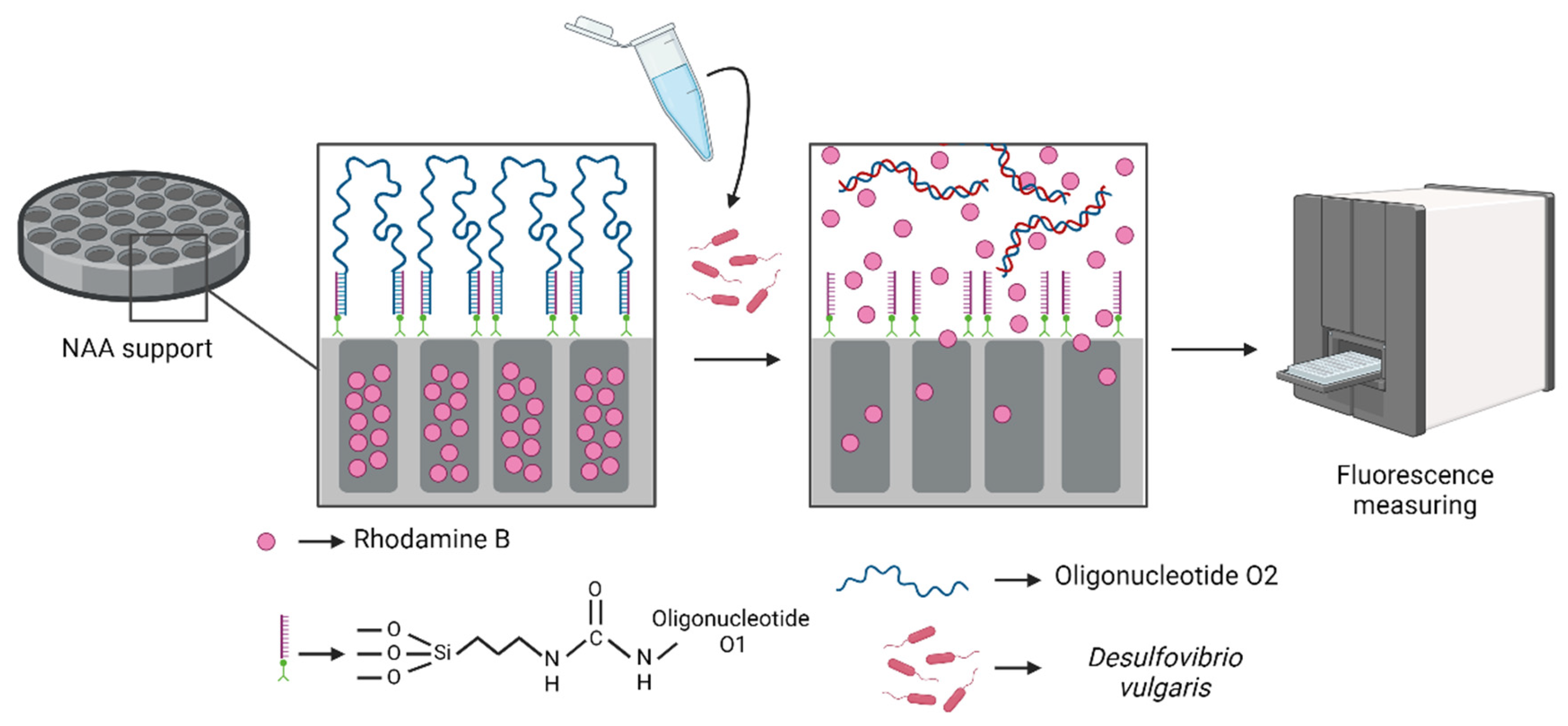
2. Materials and Methods

2.1. Probe Design

A comprehensive literature review was undertaken to identify effective strategies for DNA probe design targeting SRB (sulphate-reducing bacteria), with a particular focus on the genus Desulfovibrio within the class Deltaproteobacteria, due to its relevance in our study. A dataset of 57 sequences of the 16S rRNA gene from the Desulfovibrio genus was selected from the NCBI (National Center for Biotechnology Information) platform. Common alignment regions were identified within these sequences for probe construction, and a sequence was generated using the BioEdit Sequence Alignment Editor tool. Target regions from 25 to 60 bp were evaluated based on linearity and the absence of stable secondary structures, such as hairpin loops, that could interfere with the hybridization efficiency of the sensing mechanism. Specificity for Desulfovibrio was assessed using BLAST alignment [51], while structural stability was analyzed using the UNAFold folding algorithm [52].
Following these analyses, the sequence 5′-GAC AGG TGC ATG GCT GTC GTC AGC TCG TGC CGT G-3′ was selected for its high conservation and structural suitability. To help anchor it to the material surface, a poly-T/poly-G tail (TTTTTTGGGG) was added to this sequence for hybridization with a complementary linker strand O1 (5′-AAA AAA CCC C-3′) linked to the material (vide infra). Thus, the sequence O1 helps the anchoring of the sequence O2, which combines both the tail and the probe region (5′-TTT TTT GGG G GAC AGG TGC ATG GCT GTC GTC AGC TCG TGC CGT G G GGG TTT TTT-3′). The oligonucleotide sequences were obtained from Integrated DNA Technologies (Leuven, Belgium).

2.2. Chemical Reagents

The reagents utilized throughout this study include 3-(triethoxysilyl)propyl isocyanate, rhodamine B, tris(hydroxymethyl)aminomethane (TRIS), triethylamine (TEA), anhydrous acetonitrile ≥ 99.8% (CH3CN), magnesium chloride (MgCl2), and hydrochloric acid (HCl), acquired from Sigma-Aldrich (Madrid, Spain). These materials played essential roles in surface functionalization, fluorescent dye encapsulation, and buffer preparation for hybridization and detection assays.

2.3. Microscopy and Spectroscopy

A high resolution field emission scanning electron microscopy (HR-FESEM) analysis was conducted with a ZEISS Gemini SEM 500 microscope (Jena, Germany) operated at a voltage of 5 kV. Elemental composition and surface distribution were analyzed by energy-dispersive x-ray spectroscopy (EDXS) utilizing an X-ray detector integrated into the microscope. Fluorescence spectroscopy evaluation was carried out with a BioTek Synergy H1 microplate reader (Winooski, VT, USA).

2.4. Fabrication of Oligonucleotide-Capped Materials

The NAA materials were sourced from InRedox® (Longmont, CO, USA) and used as the solid supports designated S0 (NAA films of 2 mm in diameter, 5 nm in pore diameter, and 10 μm in alumina thickness). S0 supports were initially loaded with a rhodamine B solution in CH3CN (1.57 mM, 8 mL) and incubated for 24 h to facilitate dye encapsulation. The cargo-loading step was followed by the chemical functionalization of the surface via a reaction with 3-(triethoxysilyl)propyl isocyanate (1 mL, 4 mmol), under continuous stirring for 5.5 h to obtain functionalized supports labelled S1.
To proceed with probe attachment, S1 supports were maintained at room temperature during incubation for 3 h in a solution containing rhodamine B in CH3CN (1.57 mM, 2.1 mL), O1 (30 μL, 100 μM), and TEA (12 μL) to obtain intermediate product S2. Finally, hybridization with oligonucleotide O2 (10 μL, 100 μM) was carried out in a hybridization buffer (100 μL, 20 mM Tris-HCl, 37.5 mM MgCl2, pH 7.5) at room temperature under stirring for 2 h, leading to the final capped sensor, referred to as S3. To remove unbound sequences, the S3 materials were washed repeatedly with TRIS buffer and subsequently air-dried at 4 °C overnight.

2.5. Cargo Quantification

To evaluate dye release capacity, two separate S3 supports were individually immersed in 1 mL of TRIS buffer. One support was subjected to agitation at 90 °C for 1 h to induce complete pore opening and facilitate the highest dye release, while the other support continued at 37 °C under identical stirring conditions as a control. The concentration of released rhodamine B was determined through fluorescence spectroscopy at 575 nm (λexc = 555 nm), using a pre-established calibration curve. The experiment for cargo quantification was repeated three times to guarantee reproducibility.

2.6. Detection Procedure

To assess detection performance, two groups of S3 supports were incubated in 900 µL of TRIS buffer. One group received 100 μL of the complementary O2c strand (1 μM, 13.8 ng·μL−1), while the other was supplemented with an equivalent volume of TRIS buffer as a control. Fluorescence measurements at 575 nm (λexc = 555 nm) were taken every 15 min over a 1 h period at 37 °C to monitor dye release kinetics.
In a parallel experiment, the same procedure was repeated using genomic DNA (1.8 ng·μL−1) extracted from Desulfovibrio vulgaris (DSM 644, DSMZ, The Leibniz Institute DSMZ—German Collection of Microorganisms and Cell Cultures GmbH, Braunschweig, Germany), replacing the synthetic oligonucleotide O2c.
The next step was testing the system’s capacity to detect bacterial DNA from cell culture samples. Thus, a diluted culture of Desulfovibrio with a known concentration of 103 cells·μL−1 was prepared in TRIS buffer. As before, two sets of S3 materials were prepared for evaluation, and both were immersed in 900 μL of TRIS buffer. One was treated with 100 μL of the diluted bacterial sample, and 100 µL of TRIS buffer was added to the other. After 1 h of incubation at 37 °C, fluorescence emission from delivered rhodamine B was measured (λexc = 555 nm) to determine target-induced pore opening.

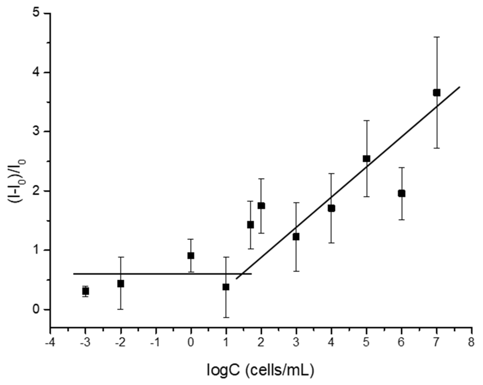
2.7. Sensitivity Analysis

The detection sensitivity of the system was evaluated by analyzing the reaction of individual S3 materials to serial dilutions of the Desulfovibrio culture, with concentrations ranging from 0.01 to 108 cells·mL−1. Each concentration was tested using a separate support, and 100 µL of bacterial suspension was introduced to 900 µL of TRIS buffer to reach a final volume of 1 mL. The mixtures were incubated at 37 °C for 1 h under gentle agitation. Fluorescence emission at 575 nm (λexc = 555 nm) was measured to quantify the rhodamine B released in response to each concentration.

2.8. Selectivity Analysis

To assess the selectivity of the sensor, the same protocol described previously was employed in the presence of different non-target bacterial species, including Escherichia coli (103 cells·mL−1), Staphylococcus aureus (103 cells·mL−1), Staphylococcus epidermidis (103 cells·mL−1), and Lactobacillus rhamnosus (103 cells·mL−1). For each species, 100 µL of bacterial suspension was added to 900 µL of TRIS buffer containing the S3 material. In addition, two groups were included: a positive control group with Desulfovibrio (103 cells·mL−1) and a negative control group containing only TRIS buffer. After 1 h of incubation at 37 °C, rhodamine B fluorescence was evaluated at 575 nm (λexc = 555 nm).

2.9. Testing System’s Performance in Water Samples

To evaluate the performance of the detection platform under realistic conditions, an assay was conducted using water samples collected from the metropolitan water network of Valencia. The performance of the S3 supports was evaluated in TRIS buffer solutions containing varying proportions of tap water. For each condition, two sets of S3 solids were submerged in 900 µL of the corresponding buffer/water mixture. One was treated with 100 μL of the complementary O2c strand (13.796 ng·μL−1), while the other received 100 µL of TRIS buffer only. After 1 h of stirring at 37 °C, the released rhodamine B was measured by fluorescence at 575 nm (λexc = 555 nm).
The same procedure was carried out again with water-diluted bacterial cultures at concentrations ranging from 10 to 104 cells·mL−1 to verify system functionality in complex aqueous environments.

3. Results and Discussion

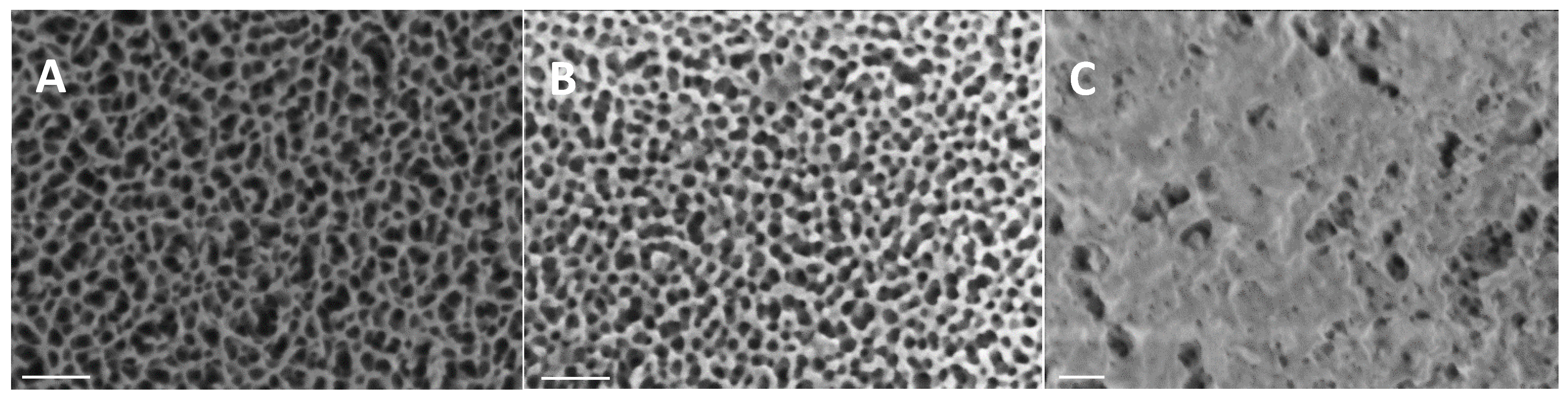
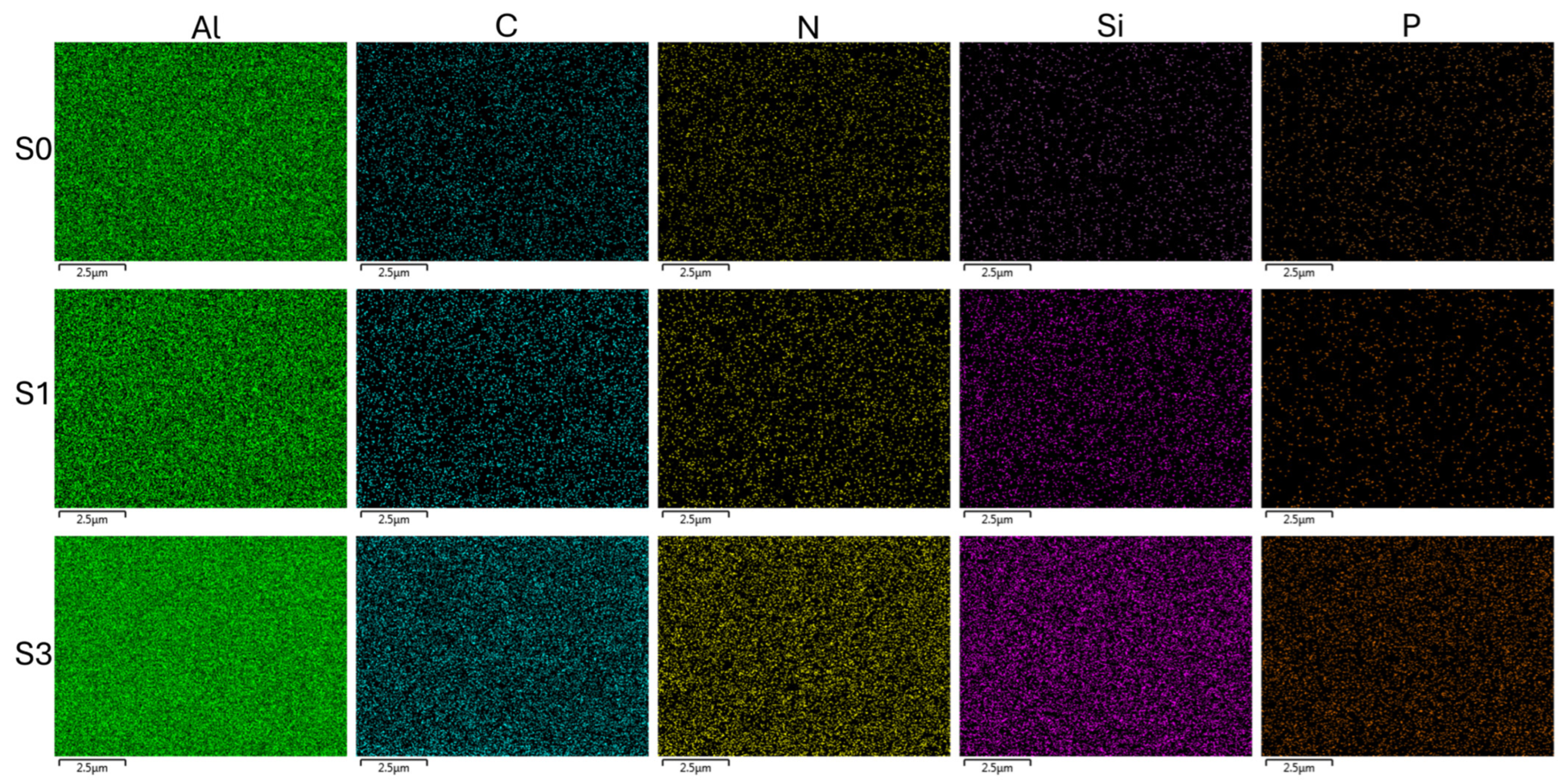
3.1. Fabrication and Characterization of Sensors

The production of the sensing platform involved a multi-step procedure. Initially, nanoporous anodic alumina materials (S0) were filled with rhodamine B through passive diffusion using a concentrated solution of the dye in acetonitrile. Subsequently, the surface underwent chemical modification with 3-(triethoxysilyl)propyl isocyanate, turning into an intermediate S1 solid. In the next step, an amine-functionalized oligonucleotide (O1) was covalently attached via urea bond formation, obtaining S2. Final pore capping was achieved by hybridization between linker O1 and sequence O2, resulting in the final sensor S3, where the pores were effectively sealed. Upon exposure to Desulfovibrio DNA, specific hybridization with the capping strand triggered pore opening and rhodamine B release to the medium (Figure 1).
The characterization of each stage (S0S3) was conducted by HR-FESEM and EDXS analysis to verify that the different steps were successfully modified. Solids S0, purchased from commercial sources, consisted of anodic aluminum oxide films atop a 0.1 mm thick aluminum base with a density of 9·1011 pores∙cm−2 (Figure 2). According to the supplier specifications, the pore morphology was conical, with diameters ranging from 20 to 30 nm at the opening and narrowing to 5 nm at the base. This structure achieved a higher level of uniformity along a 10 µm length at a depth of 15 nm. No significant morphological differences were observed between solids S0, S1, and S2, indicating that surface functionalization did not alter the structure’s integrity. However, S3 displayed a distinct surface coating, attributed to the immobilized O2 strands, which confirmed successful pore closure.
The elemental atomic analysis of the supports’ surfaces was determined employing energy-dispersive X-ray spectroscopy (EDXS) to complement HR-FESEM imaging and confirm changes in the surface composition throughout the sensor fabrication process. As shown in Table 1, a progressive increase in C and Si atomic content from supports S0 to S1 was detected, confirming the successful incorporation of rhodamine B and functionalization with the alkoxysilane groups, respectively. Moreover, phosphorous was identified in solids S3, resulting from the phosphate backbone of the capping oligonucleotide O2. In addition, representative elemental mapping images were obtained to further support these findings. As shown in Figure 3, the spatial distribution of key elements across the material surface can be observed. The phosphorus signal, which is lightly present in samples S0 and S1, becomes clearly detectable only in S3, confirming the successful immobilization of the oligonucleotide. This observation is consistent with the elemental analysis data presented in Table 1, as well as with the HR-FESEM images, which show a surface coating indicative of pore closure. Together, these results support the effective functionalization and chemical modification achieved in the final stage of material preparation.
Finally, quantification experiments revealed that the maximum concentration of rhodamine B contained in solids S3 was 26.6 ng per g of material.

3.2. Release Assays

After the fabrication of the supports, release assays in the presence of the DNA sequence of Desulfovibrio O2c complementary to the capping oligonucleotide were carried out. This study allowed for the evaluation of the specific release of the cargo due to the recognition between the complementary O2c and the capping oligonucleotide. For this assay, two different sets of gated supports were employed and submerged in TRIS buffer. In total, 100 μL of the complementary Desulfovibrio sequence O2c (1 μM) was added only to one of the solids, whereas 100 μL of TRIS buffer was added to the other one. The release of dye into the aqueous solution was observed by taking periodic aliquots every 15 min for 1 h at 37 °C, and the fluorescence was measured at 575 nm (λexc = 555 nm). The obtained results indicate that the S3 control supports incubated in TRIS buffer showed a negligible rhodamine B release, even at longer times, supporting the capping efficiency of oligonucleotide probe O2 that blocked the pores and avoided rhodamine B release. However, high fluorescence enhancement was observed from S3 when exposed to the complementary oligonucleotide that unblocked the pores, leading to dye delivery.
Once the functionality of the sensor platform with the specific Desulfovibrio DNA sequence was evaluated, the system behaviour was tested using the whole extracted genomic DNA. Like in the previous experiment, two independent sets of S3 solids were immersed in TRIS solution. In total, 100 μL of the gDNA of D. vulgaris (1.8 ng·μL−1) was added to one support, and 100 μL of TRIS buffer was added to the other one, which acted as a control assay. Supports were incubated in their respective solution for 1 h at 37 °C, and periodic aliquots of the supernatant were taken every 15 min to measure fluorescence at 575 nm (λexc = 555 nm). Results are summarized in Figure 4a. A higher rhodamine B release was observed from the supports that were in the presence of the bacterial gDNA compared to the delivery from the control supports in the presence of TRIS buffer solution. This procedure confirmed the system’s capacity to identify and react to the presence of D. vulgaris DNA, thereby verifying its effectiveness.
The subsequent step in the evaluation of the system involved an assessment of the capability to detect the bacteria’s DNA directly from a bacterial culture sample. To accomplish this test, a sample of D. vulgaris culture resuspended in TRIS solution with a known concentration of 103 cells·mL−1 was used. As in previous experiments, two sets of S3 supports were needed. Both sets were submerged in 900 µL of TRIS medium, and 100 µL of the diluted bacterial culture was added to one of the sets, while 100 µL of TRIS medium was added to the other set of supports. Following a 1 h incubation at 37 °C, the amount of free rhodamine B was determined by measuring fluorescence emission at 575 nm (λexc = 555 nm). The results obtained are shown in Figure 4b. A higher fluorescence emission was detected from the supports that were in medium with bacterial culture. These results point out that sample pretreatment is not necessary for the detection of the bacteria’s DNA. The effect of TRIS buffer to enhance cell membrane permeability has been described in several works [53,54,55,56] and could explain the direct detection of the bacteria’s DNA without the previous heating or lysis treatment of the samples.

3.3. Sensitivity Assay

The system’s sensitivity was assessed by studying the materials’ behaviour to a range of concentrations of D. vulgaris culture. Thus, eleven separated supports were immersed in TRIS buffer with different bacterial concentrations spanning from 0.001 to 107 cells·mL−1. After a 1 h incubation period at 37 °C, the fluorescence of the released rhodamine B from the pores was measured at 575 nm (λexc = 555 nm). The results shown in Figure 5 reveal that fluorescence emission exhibited a direct linear correlation with the concentrations (C) of D. vulgaris cells in the 10–107 cell mL−1 range. Thanks to the cargo quantification assay, we can calculate the percentage of rhodamine B released with the bacterial samples. We use the fluorescence signal obtained under forced pore-opening conditions at 90 °C as the reference for the 100% cargo release capacity of the sensor. Based on this, we determined that 42.6% of the total loaded dye was released after a 1 h incubation with D. vulgaris cells at the highest concentration used (107 cells·mL−1), while only 3.17% of the total amount of entrapped rhodamine B was released at lower concentrations of cells (50 cells·mL−1). Moreover, the limit of detection (LOD) of the system was established by locating the intersection point of the horizontal line and the positive slope line. Following this approach, the LOD was calculated to be 30 cells·mL−1. This LOD is similar to other values previously reported for SRB detection using different sensors, such as the plastic optical fibre immunosensor [19] and a CdS nanoparticle-based fluorogenic sensor [20]. This result shows high sensitivity of the oligonucleotide-gated nanoporous alumina-based sensors S3 for the detection of D. vulgaris.
Furthermore, it is interesting to note that the sensing system shows a remarkable amplification, as it releases a substantially greater amount of entrapped rhodamine B molecules in comparison to the quantity of D. vulgaris cells needed to produce this release. The number of dye molecules released compared to the number of cells at concentrations near the LOD (50 cells·mL−1) was calculated to be around 5·1010 RhB molecules released per cell, showing a great signal amplification that is similar to previously obtained data in other systems based on the same technology [42].

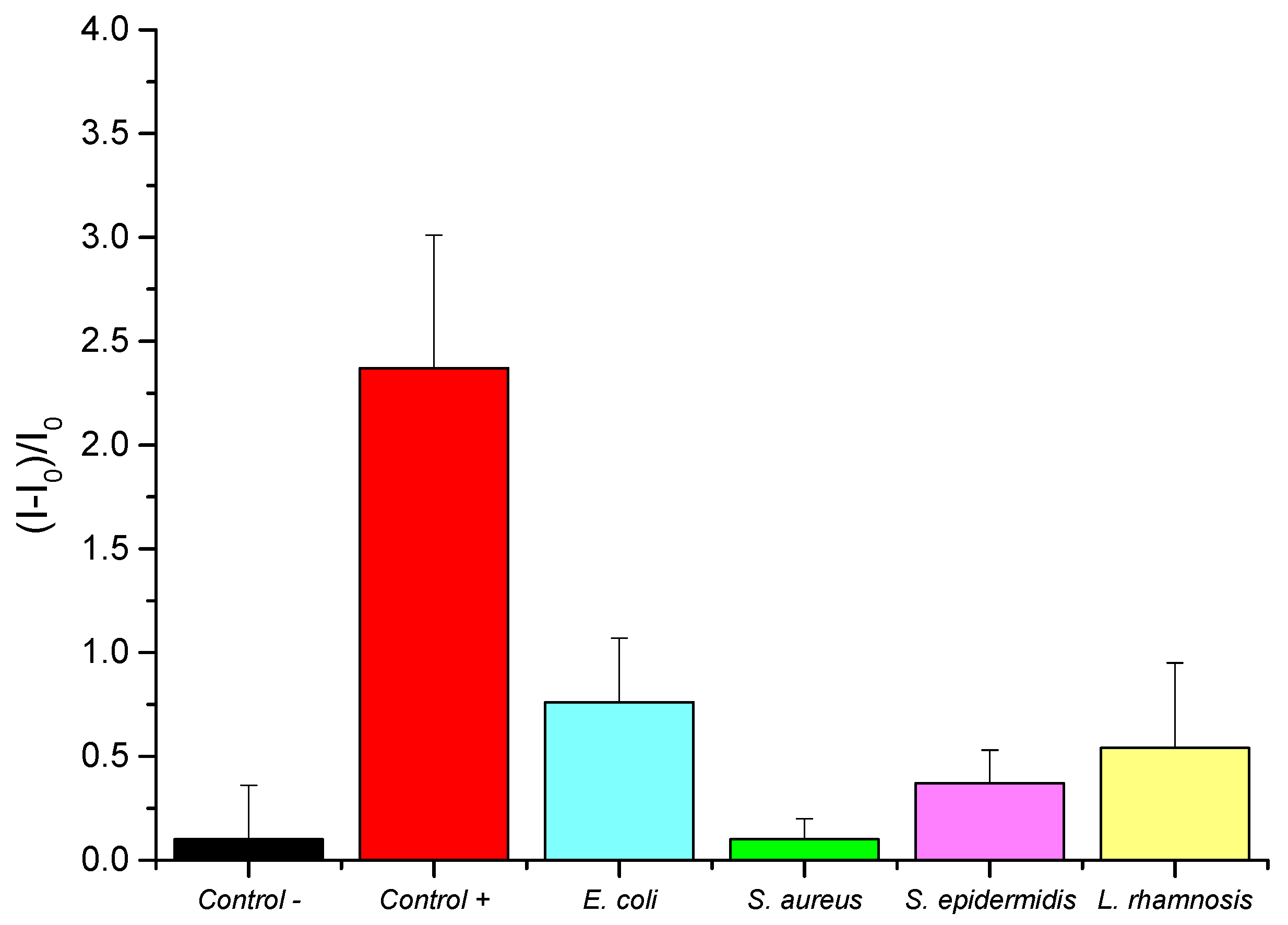
3.4. Selectivity Analysis

The selectivity of the biosensor was studied by evaluating the behaviour of S3 to different bacterial species, including E. coli, S. aureus, S. epidermidis, and L. rhamnosus. E. coli is the main interfering pathogen in Desulfovibrio detection because both are Gram-negative bacteria and could be found in the same samples. Selectivity studies involved testing independent S3 supports with each bacterial culture sample, each set diluted with TRIS buffer to the same final concentration (103 cells·mL−1). For each assay, 100 μL of the corresponding culture sample were introduced into 900 μL of hybridization buffer, where S3 solids were previously immersed. In addition, two other sets of supports were included in this study: a positive control group with a D. vulgaris culture sample and a negative control group with TRIS buffer. Then, supports were stirred at 37 °C for 1 h, and the release of rhodamine B was quantified by measuring the fluorescence emission at 575 nm (λexc = 555 nm). Results are summarized in Figure 6. The released rhodamine B from sensors in contact with other bacterial species was similar to the delivered dye from control sensors. Moreover, the highest fluorescence signal was obtained from S3 biosensors in the presence of the D. vulgaris cells, supporting the selectivity of the oligonucleotide-gated nanoporous sensor for the detection of the D. vulgaris target cell DNA.
The residual fluorescence from E. coli and L. rhamnosus may result from partial sequence similarity in the non-target genome or minor non-specific hybridization events, though these are not sufficient to generate a false-positive signal. Despite these background signals, the fluorescence emission from D. vulgaris remains at least three times higher than any of the fluorescence produced from non-target bacteria, demonstrating effective detection.

3.5. Performance Analysis in Inoculated Water Samples

After the characterization of the materials in control media (TRIS buffer), the system’s robustness for the detection of Desulfovibrio in real media was conducted using water samples obtained from the Valencia water network. First, the behaviour of the supports was evaluated by employing a range of percentages of TRIS buffer and water sample in the final mixtures. For each condition, two independent sets of S3 solids were submerged in 900 µL of TRIS/water mixture, and to one of them, 100 µL of the O2c sequence was added, and TRIS solution was added to the other one. The solution was agitated for 1 h at 37 °C. Subsequently, the released rhodamine B was quantified through fluorescence measurement at 575 nm (λexc = 555 nm). The best result was accomplished using a mixture with 25% water. A greater addition of water to the media affected the stability and the performance of the system.
In a further step, the biosensor’s response using different concentrations of Desulfovibrio bacterial culture in the optimal TRIS/water mixture using a 25% water sample was evaluated. Five sets of S3 solids were immersed in 900 µL of the TRIS/water mixture. In total, 100 µL of TRIS buffer was added to the control support, whereas 100 µL of bacterial culture was added to the other supports in the concentration (C) range of 102–105 cells·mL−1. After 1 h, the quantity of released rhodamine B was measured through the fluorescence emission at 575 nm (λexc = 555 nm). Results can be observed in Figure 7. The supports incubated with cell samples (red bars) exhibited enhanced fluorescence emission intensities in comparison to the control sample (black bar). These preliminary results prove the sensor’s capacity to detect D. vulgaris cell DNA in mixtures of water samples and TRIS buffer with high sensitivity and selectivity, supporting its potential use for the in situ control of MIC in water supply networks.

4. Conclusions

An effective sulphate-reducing bacteria detection platform integrating nanostructured hybrid organic–inorganic materials with the technology of molecular gates has been successfully developed. This system relies on nanoporous anodic alumina films with a fluorescent dye entrapped and gated with a specific oligonucleotide probe, creating a platform for the detection of Desulfovibrio DNA. In the system, the detachment of the oligonucleotide gate occurs when the complementary sequence of Desulfovibrio DNA is present. This event induces the release of rhodamine B from the porous structure, which was detected by a fluorescence read-out. To ensure the robustness and reliability of the detection system, the surface morphology of the sensors was meticulously characterized using high-resolution field emission scanning electron microscopy. Additionally, the surface atomic composition was calculated by energy-dispersive X-ray spectroscopy, providing a comprehensive understanding of the sensor’s structural integrity and chemical composition. The biosensor’s capacity to detect the genomic DNA of D. vulgaris and the DNA of bacterial cells in culture samples was demonstrated. Sensitivity was proven at different concentrations of the cell culture, achieving a limit of detection of 30 cells ml−1, and selectivity was demonstrated in the presence of different bacterial samples of Escherichia coli, Staphylococcus aureus, Staphylococcus epidermidis, and Lactobacillus rhamnosus. The sensor’s performance for the detection of D. vulgaris cell DNA was further validated in real media, using water samples from the Valencia water network. The best result to detect Desulfovibrio’s DNA in water samples was obtained using a mixture of 75% TRIS buffer and 25% water sample. This successful validation underscores the system’s potential use for the in situ control of microbiologically influenced corrosion in water supply networks. This technology represents a significant advance in the field of sulphate-reducing bacteria detection, offering a versatile and reliable solution for directly monitoring this bacterial group in water networks without any sample pretreatment. The integration of nanostructured materials and molecular detection techniques positions this system as a valuable tool for ensuring the safety and integrity of water supplies, particularly in regions susceptible to MIC.

Author Contributions

Conceptualization, Y.M., R.P.-C., M.P.-M., R.M.-M., E.A., M.D.M. and A.B.; methodology, A.L.-P., Á.M.-A., Y.M. and A.H.-M.; validation, A.L.-P., Á.M.-A. and A.B.; investigation, A.L.-P.; resources, R.M.-M., E.A. and F.S.; writing—original draft preparation, A.L.-P.; writing—review and editing, A.L.-P., A.H.-M., F.S., E.A. and R.M.-M.; supervision, M.D.M., R.M.-M. and E.A.; project administration, R.M.-M., E.A., R.P.-C. and M.P.-M.; funding acquisition, R.M.-M., E.A., R.P.-C. and M.D.M. All authors have read and agreed to the published version of the manuscript.

Funding

This research was supported by project PID2021-126304OB-C41 and PID2021-128141OB-C22 funded by MCIN/AEI/10.13039/501100011033/ and by European Regional Development Fund—A way of doing Europe. The work was also supported by Generalitat Valenciana (CIPROM/2021/007), Agència Valenciana de la Innovació (INNEST/2021/169, Universitat Politècnica de València; INNEST/2021/174, Global Omnium) and is susceptible to be co-founded by European Union. This research was supported by CIBER-Consorcio Centro de Investigación Biomédica en Red- (CB06/01/2012), Instituto de Salud Carlos III, Ministerio de Ciencia e Innovación. A.L. thanks to the Ministerio de Universidades for her predoctoral grant (FPU20/05297). Á.M.-A. thanks to the Universitat Politècnica de València (UPV) for her Ph.D. grant (DOCEMPR22).

Data Availability Statement

The original contributions presented in this study are included in the article. Further inquiries can be directed to the corresponding author.

Acknowledgments

The authors want to thank the Electron Microscopy Service at the UPV for support. Figure 1 was created with BioRender (www.biorender.com (accessed on 23 February 2025)).

Conflicts of Interest

Authors Román Ponz-Carcelén and María Pedro-Monzonís are employed by the company Global Omnium Group. The remaining authors declare that the research was conducted in the absence of any commercial or financial relationships that could be construed as a potential conflict of interest.

Abbreviations

The following abbreviations are used in this manuscript:
MICMicrobiologically influenced corrosion
SRBSulphate-reducing bacteria
ATPAdenosine triphosphate
RNARibonucleic acid
DNADeoxyribonucleic acid
PCRPolymerase chain reaction
NAANanoporous anodic alumina

References

  1. Kumar, M.A.; Kasirajan, K.T.; Parthiban, K. Production and characterization of exopolysaccharides (EPS) from biofilm forming marine bacterium. Braz. Arch. Biol. Technol. 2011, 54, 259–265. [Google Scholar] [CrossRef]
  2. Marschall, C.; Frenzel, P.; Cypionka, H. Influence of oxygen on sulfate reduction and growth of sulfate-reducing bacteria. Arch. Microbiol. 1993, 159, 168–173. [Google Scholar] [CrossRef]
  3. Eguía-López, E.; Trueba-Ruiz, A.; Río-Calonge, B.; Girón-Portilla, M.A.; Bielva-Tejera, C. Recent studies on antifouling systems to artificial structures in marine ecosystem. J. Marit. Res. 2006, 3, 73–89. [Google Scholar]
  4. Beech, I.B.; Sunner, J. Biocorrosion: Towards understanding interactions between biofilms and metals. Curr. Opin. Biotechnol. 2004, 15, 181–186. [Google Scholar] [CrossRef]
  5. Zadvorny, O.A.; Zorin, N.A.; Gogotoy, I.N. Transformation of metals and metal ions by hydrogenases from phototrophic bacteria. Arch. Microbiol. 2006, 184, 279–285. [Google Scholar] [CrossRef]
  6. Yuan, S.; Liang, B.; Zhao, Y.; Pehkonen, S.O. Surface chemistry and corrosion behavior of 304 stainless steel in simulated seawater containing inorganic sulphide and sulphate-reducing bacteria. Corros. Sci. 2013, 74, 353–366. [Google Scholar] [CrossRef]
  7. Ben-Dov, E.; Brenner, A.; Kushmaro, A. Quantification of Sulfate-reducing Bacteria in Industrial Wastewater, by Real-time Polymerase Chain Reaction (PCR) Using dsrA and apsA Genes. Microb. Ecol. 2007, 54, 439–451. [Google Scholar] [CrossRef]
  8. Gibson, G.R.; Cummings, J.H.; Macfarlane, G.T. Growth and activities of sulphate-reducing bacteria in gut contents of healthy subjects and patients with ulcerative colitis. FEMS Microbiol. Lett. 1991, 86, 103–111. [Google Scholar] [CrossRef]
  9. Klein, M.; Friedrich, M.; Roger, A.J.; Hugenholtz, P.; Fishbain, S.; Abicht, H.; Blackall, L.L.; Stahl, D.A.; Wagner, M. Multiple Lateral Transfers of Dissimilatory Sulfite Reductase Genes between Major Lineages of Sulfate-Reducing Prokaryotes. J. Bacteriol. 2001, 183, 6028–6035. [Google Scholar] [CrossRef]
  10. Tanner, R.S. Monitoring sulfate-reducing bacteria: Comparison of enumeration media. J. Microbiol. Methods 1989, 10, 83–90. [Google Scholar] [CrossRef]
  11. Daly, K.; Sharp, R.J.; McCarthy, A.J. Development of oligonucleotide probes and PCR primers for detecting phylogenetic subgroups of sulfate-reducing bacteria. Microbiology 2000, 146, 1693–1705. [Google Scholar] [CrossRef] [PubMed]
  12. Amann, R.I.; Ludwig, W.; Schleifer, K.H. Phylogenetic identification and in situ detection of individual microbial cells without cultivation. Microbiol. Rev. 1995, 59, 143–169. [Google Scholar] [CrossRef] [PubMed]
  13. Beale, D.J.; Karpe, A.V.; Jadhav, S.; Muster, T.H.; Palombo, E.A. Omics-based approaches and their use in the assessment of microbial-influenced corrosion of metals. Corros. Rev. 2015, 34, 1–15. [Google Scholar] [CrossRef]
  14. Kondo, R.; Nedwell, D.B.; Purdy, K.J.; Queiroz-Silva, S. Detection and Enumeration of Sulphate-Reducing Bacteria in Estuarine Sediments by Competitive PCR. Geomicrobiol. J. 2004, 21, 145–157. [Google Scholar] [CrossRef]
  15. Purdy, K.J.; Nedwell, D.; Embley, T.M.; Takii, S. Use of 16S rRNA-targeted oligonucleotide probes to investigate the occurrence and selection of sulfate-reducing bacteria in response to nutrient addition to sediment slurry microcosms from a Japanese estuary. FEMS Microbiol. Ecol. 1997, 24, 221–234. [Google Scholar] [CrossRef]
  16. Salimiyan rizi, K.; Ashrafi, A. Biosensors, mechatronics, & microfluidics for early detection & monitoring of microbial corrosion: A comprehensive critical review. Results Mater. 2023, 18, 100402. [Google Scholar] [CrossRef]
  17. Chen, S.; Cheng, Y.F.; Voordouw, G. Three-dimensional graphene nanosheet doped with gold nanoparticles as electrochemical DNA biosensor for bacterial detection. Sens. Actuators B Chem. 2018, 262, 860–868. [Google Scholar] [CrossRef]
  18. Asif, M.; Ashraf, G.; Aziz, A.; Iftikhar, T.; Wang, Z.; Xiao, F.; Sun, Y. Tuning the Redox Chemistry of Copper Oxide Nanoarchitectures Integrated with rGOP via Facet Engineering: Sensing H2S toward SRB Detection. ACS Appl. Mater. Interfaces 2022, 14, 19480–19490. [Google Scholar] [CrossRef]
  19. Lopes, R.N.; Rodrigues, D.M.C.; Allil, R.C.S.B.; Werneck, M.M. Plastic optical fiber immunosensor for fast detection of sulfate-reducing bacteria. Measurement 2018, 125, 377–385. [Google Scholar] [CrossRef]
  20. Qi, P.; Zhang, D.; Zeng, Y.; Wan, Y. Biosynthesis of CdS nanoparticles: A fluorescent sensor for sulfate-reducing bacteria detection. Talanta 2016, 147, 142–146. [Google Scholar] [CrossRef]
  21. Zheng, L.; Ye, X.; Qi, P.; Zhang, D.; Sun, Y. Fluorometric detection of sulfate-reducing bacteria via the aggregation-induced emission of glutathione-gold(I) complexe. Microchim. Acta 2019, 186, 382. [Google Scholar] [CrossRef] [PubMed]
  22. Vigneshvar, S.; Sudhakumari, C.C.; Septhilkumaran, B.; Prakash, H. Recent Advances in Biosensor Technology for Potential Applications—An Overview. Front. Bioeng. Biotechnol. 2016, 4, 11. [Google Scholar] [CrossRef]
  23. Aznar, E.; Oroval, M.; Pascual, L.; Murguía, J.R.; Martínez-Máñez, R.; Sancenón, F. Gated materials for on-command release of guest molecules. Chem. Rev. 2016, 116, 561–718. [Google Scholar] [CrossRef]
  24. Pascual, L.; Baroja, I.; Aznar, E.; Sancenón, F.; Marcos, M.D.; Murguía, J.R.; Amorós, P.; Rurack, K.; Martínez-Máñez, R. Oligonucleotide-capped mesoporous silica nanoparticles as DNA-responsive dye delivery systems for genomic DNA detection. Chem. Commun. 2015, 51, 1414–1416. [Google Scholar] [CrossRef]
  25. García-Fernández, A.; Aznar, E.; Martínez-Máñez, R.; Sancenón, F. New Advances in In Vivo Applications of Gated Mesoporous Silica as Drug Delivery Nanocarriers. Small 2020, 16, 1902242. [Google Scholar] [CrossRef]
  26. Llopis-Lorente, A.; Lozano-Torres, B.; Bernardos, A.; Martínez-Máñez, R.; Sancenón, F. Mesoporous silica materials for controlled delivery based on enzymes. J. Mater. Chem. B 2017, 5, 3069–3083. [Google Scholar] [CrossRef]
  27. Sancenón, F.; Pascual, L.; Oroval, M.; Aznar, E.; Martínez-Máñez, R. Gated silica mesoporous materials in sensing applications. ChemistryOpen 2015, 4, 418–437. [Google Scholar] [CrossRef]
  28. Pla, L.; Lozano-Torres, B.; Martínez-Máñez, R.; Sancenón, F.; Ros-Lis, J.V. Overview of the Evolution of Silica-Based Chromo-Fluorogenic Nanosensors. Sensors 2019, 19, 5138. [Google Scholar] [CrossRef]
  29. Castillo, R.R.; Baeza, A.; Vallet-Regí, M. Recent applications of the combination of mesoporous silica nanoparticles with nucleic acids: Development of bioresponsive devices, carriers and sensors. Biomater. Sci. 2017, 5, 353–377. [Google Scholar] [CrossRef]
  30. Ribes, À.; Santiago-Felipe, S.; Bernardos, A.; Marcos, M.D.; Pardo, T.; Sancenón, F.; Aznar, E. Two new fluorogenic aptasensors based on capped mesoporous silica nanoparticles to detect ochratoxin A. ChemistryOpen 2017, 6, 653–659. [Google Scholar] [CrossRef]
  31. Climent, E.; Martínez-Máñez, R.; Sancenón, F.; Marcos, M.D.; Soto, J.; Maquieira, A.; Amorós, P. Controlled Delivery Using Oligonucleotide-Capped Mesoporous Silica Nanoparticles. Angew. Chem. Int. Ed. 2010, 49, 7281–7283. [Google Scholar] [CrossRef] [PubMed]
  32. Baranowska, M.; Slota, A.J.; Eravuchira, P.J.; Macias, G.; Xifré-Pérez, E.; Pallares, J.; Marsal, L.F. Protein attachment to nanoporous anodic alumina for biotechnological applications: Influence of pore size, protein size and functionalization path. Colloids Surf. B Biointerfaces 2014, 122, 375–383. [Google Scholar] [CrossRef] [PubMed]
  33. Santos, A.; Kumeria, T.; Losic, D. Nanoporous Anodic Alumina: A Versatile Platform for Optical Biosensors. Materials 2014, 7, 4297–4320. [Google Scholar] [CrossRef]
  34. Rajeev, G.; Prieto Simon, B.; Marsal, L.F.; Voelcker, N.H. Advances in Nanoporous Anodic Alumina-Based Biosensors to Detect Biomarkers of Clinical Significance: A Review. Adv. Healthc. Mater. 2018, 7, 1700904. [Google Scholar] [CrossRef]
  35. Alberti, S.; Soler-Illi, G.J.A.A.; Azzaroni, O. Gated supramolecular chemistry in hybrid mesoporous silica nanoarchitectures: Controlled delivery and molecular transport in response to chemical, physical and biological stimuli. Chem. Commun. 2015, 51, 6050–6075. [Google Scholar] [CrossRef]
  36. Lee, W.; Park, S.J. Porous Anodic Aluminum Oxide: Anodization and Templated Synthesis of Functional Nanostructures. Chem. Rev. 2014, 114, 7487–7556. [Google Scholar] [CrossRef]
  37. Ribes, A.; Santiago-Felipe, S.; Avino, A.; Candela-Noguera, V.; Eritja, R.; Sancenón, F.; Aznar, E. Design of oligonucleotide-capped mesoporous silica nanoparticles for the detection of miRNA-145 by duplex and triplex formation. Sens. Actuators B Chem. 2018, 277, 598–603. [Google Scholar] [CrossRef]
  38. Garrido-Cano, I.; Pla, L.; Santiago-Felipe, S.; Simon, S.; Ortega, B.; Bermejo, B.; Martínez-Máñez, R. Nanoporous anodic alumina-based sensor for miR-99a-5p detection as an effective early breast cancer diagnostic tool. ACS Sens. 2021, 6, 1022–1029. [Google Scholar] [CrossRef]
  39. Climent, E.; Mondragón, L.; Martínez-Máñez, R.; Sancenón, F.; Marcos, M.D.; Murguía, J.R.; Amorós, P.; Rurack, K.; Pérez-Payá, E. Selective, highly sensitive, and rapid detection of genomic DNA by using gated materials: Mycoplasma detection. Angew. Chem. Int. Ed. 2013, 52, 8938–8942. [Google Scholar] [CrossRef]
  40. Pla, L.; Xifré-Pérez, E.; Ribes, À.; Aznar, E.; Marcos, M.D.; Marsal, L.F.; Sancenón, F. A mycoplasma genomic DNA probe using gated nanoporous anodic alumina. ChemPlusChem 2017, 82, 337–341. [Google Scholar] [CrossRef]
  41. Ribes, À.; Aznar, E.; Santiago-Felipe, S.; Xifre-Perez, E.; Tormo-Mas, M.À.; Peman, J.; Martínez-Máñez, R. Selective and sensitive probe based in oligonucleotide-capped nanoporous alumina for the rapid screening of infection produced by Candida albicans. ACS Sens. 2019, 4, 1291–1298. [Google Scholar] [CrossRef] [PubMed]
  42. Pla, L.; Santiago-Felipe, S.; Tormo-Mas, M.Á.; Pemán, J.; Sancenón, F.; Aznar, E.; Martínez-Máñez, R. Aptamer-Capped nanoporous anodic alumina for Staphylococcus aureus detection. Sens. Actuators B Chem. 2020, 320, 128281. [Google Scholar] [CrossRef]
  43. Pla, L.; Santiago-Felipe, S.; Tormo-Mas, M.À.; Ruiz-Gaitán, A.; Pemán, J.; Valentín, E.; Sancenón, F.; Aznar, E.; Martínez-Máñez, R. Oligonucleotide-capped nanoporous anodic alumina biosensor as diagnostic tool for rapid and accurate detection of Candida auris in clinical samples. Emerg. Microbes Infect. 2021, 10, 407–415. [Google Scholar] [CrossRef] [PubMed]
  44. Caballos, I.; Aranda, M.N.; López-Palacios, A.; Pla, L.; Santiago-Felipe, S.; Hernández-Montoto, A.; Tormo-Mas, M.À.; Pemán, J.; Gómez-Ruiz, M.D.; Calabuig, E.; et al. Aptamer-Capped Nanoporous Anodic Alumina for SARS-CoV-2 Spike Protein Detection. Adv. Mater. Technol. 2023, 8, 2201913. [Google Scholar] [CrossRef]
  45. López-Palacios, A.; Aranda, M.N.; Caballos, I.; Hernández-Montoto, A.; Calabuig, E.; Gómez-Ruiz, M.D.; Tormo-Mas, M.Á.; Pemán, J.; Sancenón, F.; Martínez-Máñez, R.; et al. SARS-COV-2 viral RNA detection through oligonucleotide-capped nanoporous anodic alumina supports. Sens Actuators Rep. 2025, 9, 100298. [Google Scholar] [CrossRef]
  46. Hernández-Montoto, A.; Aranda, M.N.; Caballos, I.; López-Palacios, A.; Tormo-Mas, M.À.; Pemán, J.; Prieto-Rodríguez, M.; Picornell, C.; Aznar, E.; Martínez-Máñez, R. Human Papilloma Virus DNA Detection in Clinical Samples Using Fluorogenic Probes Based on Oligonucleotide Gated Nanoporous Anodic Alumina Films. Adv. Healthc. Mater. 2023, 12, 2203326. [Google Scholar] [CrossRef]
  47. Hansen, T.A. Metabolism of sulfate-reducing prokaryotes. Antonie Van Leeuwenhoek 1994, 66, 165–185. [Google Scholar] [CrossRef]
  48. Heildelberg, J.F.; Seshadri, R.; Haveman, S.A.; Hemme, C.L.; Paulsen, I.T.; Kolonay, J.F.; Eisen, J.A.; Ward, N.; Methe, B.; Brinkac, L.M.; et al. The genoma sequence of the anaerobic, sulfate-reducing bacterium Desulfovibrio vulgaris Hildenborough. Nat. Biotechnol. 2004, 22, 554–559. [Google Scholar] [CrossRef]
  49. Marangoni, P.R.D.; Robl, D.; Berton, M.A.C.; Garcia, C.M.; Bozza, A.; Porsani, M.V.; Dalzoto, P.R.; Vicente, V.A.; Pimentel, I.C. Occurrence of sulphate reducing bacteria (SRB) associated with biocorrosion on metallic surfaces in a hydroelectric power station in Ibirama (SC)—Brazil. Braz. Arch. Biol. Technol. 2013, 56, 801–809. [Google Scholar] [CrossRef]
  50. Dar, S.A.; Yao, L.; Dongen, U.; Kuenen, J.G.; Muyzer, G. Analysis of diversity and activity of sulfate-reducing bacterial communities in sulfidogenic bioreactors using 16S rRNA and dsrB genes as molecular markers. Appl. Environ. Microbiol. 2007, 73, 594–604. [Google Scholar] [CrossRef]
  51. Altschul, S.F.; Gish, W.; Miller, W.; Myers, E.W.; Lipman, D.J. Basic local alignment search tool. J. Mol. Biol. 1990, 215, 403–410. [Google Scholar] [CrossRef] [PubMed]
  52. Zuker, M. Mfold web server for nucleic acid folding and hybridization prediction. Nucleic Acids Res. 2003, 31, 3406–3415. [Google Scholar] [CrossRef] [PubMed]
  53. Neu, H.C.; Ashman, D.F.; Price, T.D. Effect of ethylenediaminetetraacetic acid-Tris(hydroxymethyl)aminomethane on release of the acid-soluble nucleotide pool and on breakdown of ribosomal ribonucleic acid in Escherichia coli. J. Bacteriol. 1967, 93, 1360–1368. [Google Scholar] [CrossRef]
  54. Donohue, T.J.; Cain, B.D.; Kaplan, S. Alterations in the phospholipid composition of Rhodopseudomonas sphaeroides and other bacteria induced by Tris. J. Bacteriol. 1982, 152, 595–606. [Google Scholar] [CrossRef]
  55. Hancock, R.E.; Wong, P.G. Compounds which increase the permeability of the Pseudomonas aeruginosa outer membrane. Antimicrob. Agents Chemother. 1984, 26, 48–52. [Google Scholar] [CrossRef]
  56. Wurm, D.J.; Slouka, C.; Bosilj, T.; Herwig, C.; Spadiut, O. How to trigger periplasmic release in recombinant Escherichia coli: A comparative analysis. Eng. Life Sci. 2017, 17, 215–222. [Google Scholar] [CrossRef]
Figure 1. Scheme of the controlled release system. The sensor S3, loaded with rhodamine B and capped with the O2 sequence, delivers the encapsulated rhodamine B dye in response to the target DNA from D. vulgaris.
Nanomaterials 15 00774 g001
Figure 2. HR-FESEM images of NAA supports during the fabrication process (scale bars: 100 nm). (A) NAA plate without modification (S0). (B) Loaded and functionalized NAA plate with rhodamine B and isocyanate (S1). (C) Loaded, functionalized, and capped NAA plate with O2 (S3).
Nanomaterials 15 00774 g002
Figure 3. Elemental mapping images of the support surfaces for samples S0, S1, and S3, obtained by energy-dispersive X-ray spectroscopy (EDXS). The spatial distribution of key elements (Al, C, N, Si, and P) is shown to illustrate the progressive surface modification throughout the functionalization process.
Nanomaterials 15 00774 g003
Figure 4. Fluorescence emission kinetics of released RhB from supports S3 in control media TRIS solution with Desulfovibrio vulgaris (a) genomic DNA and (b) cell culture sample. The red line shows the release profile in the presence of D. Vulgaris gDNA or cells, and the black line represents the release in the absence of DNA or the bacteria. Data are expressed as mean values and standard deviations derived from measurements for 3 distinct supports.
Nanomaterials 15 00774 g004
Figure 5. Rhodamine B release in response to increasing concentrations of D. vulgaris cell culture.
Nanomaterials 15 00774 g005
Figure 6. Interference assay: dye release in the presence of different bacterial species, including E. coli, S. aureus, S. epidermidis, L. rhamnosus, and D. vulgaris cell cultures.
Nanomaterials 15 00774 g006
Figure 7. Fluorescence emission from support S3 in TRIS/water samples containing D. vulgaris cells. The red bars show the release in the presence of D. vulgaris cells from bacterial cultures at different concentrations, and the black bar represents the release in the absence of the bacterial cells.
Nanomaterials 15 00774 g007
Table 1. Elemental atomic composition of S0, S1, and S3.
C/AlN/AlSi/AlP/Al
S00.14 ± 0.01---
S10.48 ± 0.100.07 ± 0.020.10 ± 0.04-
S30.35 ± 0.010.05 ± 0.010.04 ± 0.020.04 ± 0.01
Disclaimer/Publisher’s Note: The statements, opinions and data contained in all publications are solely those of the individual author(s) and contributor(s) and not of MDPI and/or the editor(s). MDPI and/or the editor(s) disclaim responsibility for any injury to people or property resulting from any ideas, methods, instructions or products referred to in the content.

