

Computational Nanotoxicology Models for Environmental Risk Assessment of Engineered Nanomaterials

 ,
,  , and
, and
Abstract
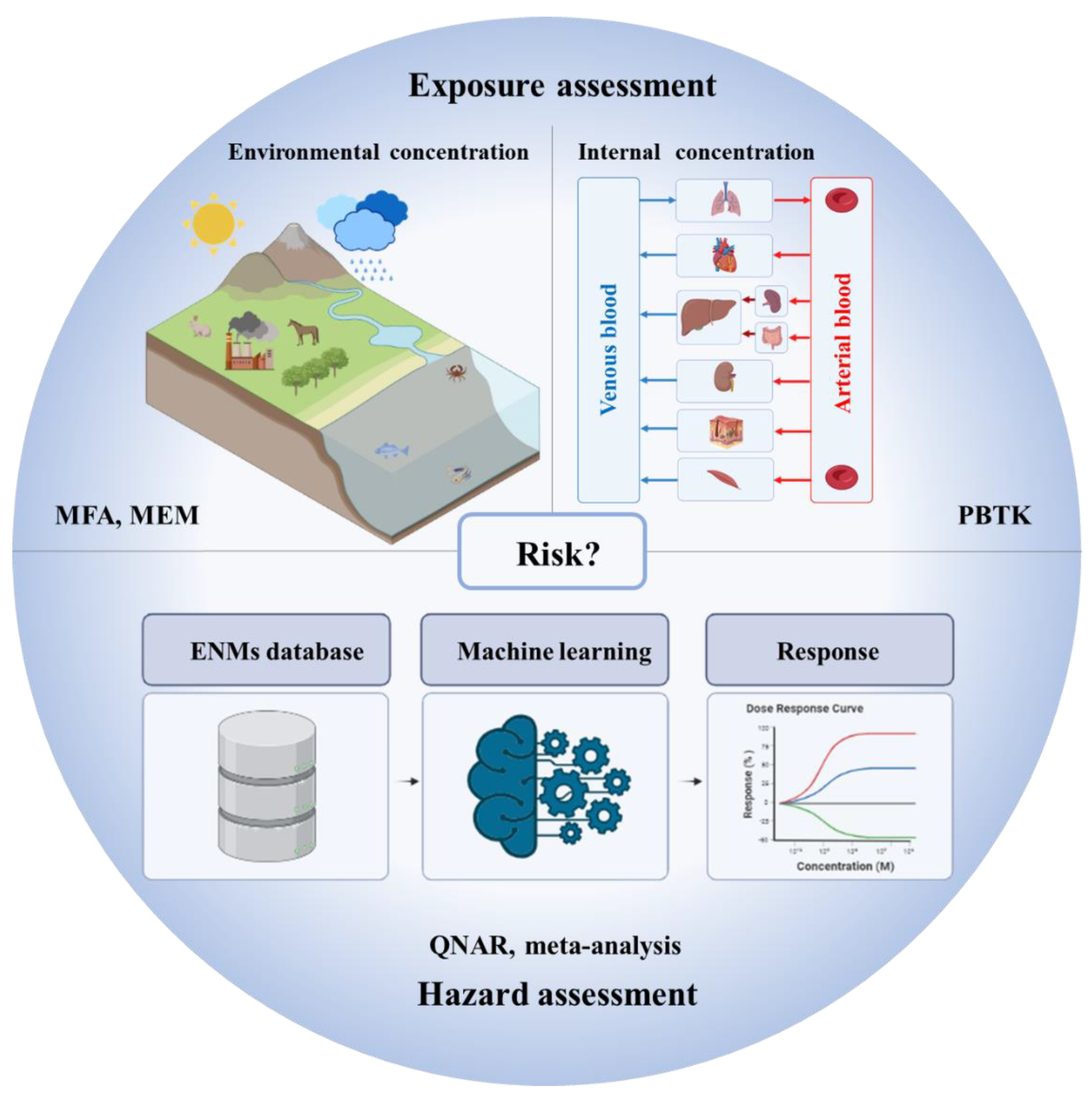
1. Introduction
2. Literature Survey
3. Material Flow Analysis and Multimedia Environmental Models
4. Physiologically Based Toxicokinetics Models
5. Quantitative Nanostructure–Activity Relationships
6. Meta-Analysis
7. Concluding Remarks, Challenges, and Perspectives
Funding
Acknowledgments
Conflicts of Interest
References
- European Commission. Third European Report on Science & Technology Indicators; EU Publications Office: Luxembourg, 2003. [Google Scholar]
- McWilliams, A. The Maturing Nanotechnology Market: Products and Applications; NAN031G, Global Markets; BBC Research: Wellesley, MA, USA, 2016. [Google Scholar]
- Global nanotechnology market (by component and applications), funding & investment, patent analysis and 27 companies profile & recent developments—Forecast to 2024. iGATE Res. 2018, 4520812.
- Haase, A.; Klaessig, F. EU US Roadmap Nanoinformatics 2030; EU Nanosafety Cluster, 2018. [Google Scholar] [CrossRef]
- Markiewicz, M.; Kumirska, J.; Lynch, I.; Matzke, M.; Köser, J.; Bemowsky, S.; Docter, D.; Stauber, R.; Westmeier, D.; Stolte, S. Changing environments and biomolecule coronas: Consequences and challenges for the design of environmentally acceptable engineered nanoparticles. Green Chem. 2018, 20, 4133–4168. [Google Scholar] [CrossRef]
- Abbas, Q.; Yousaf, B.; Ullah, H.; Ali, M.U.; Ok, Y.S.; Rinklebe, J. Environmental transformation and nano-toxicity of engineered nano-particles (ENPs) in aquatic and terrestrial organisms. Crit. Rev. Environ. Sci. Technol. 2020, 50, 2523–2581. [Google Scholar] [CrossRef]
- Domingues, C.; Santos, A.; Alvarez-Lorenzo, C.; Concheiro, A.; Jarak, I.; Veiga, F.; Barbosa, I.; Dourado, M.; Figueiras, A. Where is nano today and where is it headed? A review of nanomedicine and the dilemma of nanotoxicology. ACS Nano 2022, 16, 9994–10041. [Google Scholar] [CrossRef] [PubMed]
- Steffen, W.; Richardson, K.; Rockstrom, J.; Cornell, S.E.; Fetzer, I.; Bennett, E.M.; Biggs, R.; Carpenter, S.R.; de Vries, W.; de Wit, C.A.; et al. Planetary boundaries: Guiding human development on a changing planet. Science 2015, 347, 1259855. [Google Scholar] [CrossRef] [PubMed]
- Persson, L.; Almroth, B.M.C.; Collins, C.D.; Cornell, S.; de Wit, C.A.; Diamond, M.L.; Fantke, P.; Hassellov, M.; MacLeod, M.; Ryberg, M.W.; et al. Outside the safe operating space of the planetary boundary for novel entities. Environ. Sci. Technol. 2022, 56, 1510–1521. [Google Scholar] [CrossRef]
- Rockstrom, J.; Steffen, W.; Noone, K.; Persson, A.; Chapin, F.S.; Lambin, E.F.; Lenton, T.M.; Scheffer, M.; Folke, C.; Schellnhuber, H.J.; et al. A safe operating space for humanity. Nature 2009, 461, 472–475. [Google Scholar] [CrossRef]
- Nel, A.; Xia, T.; Madler, L.; Li, N. Toxic potential of materials at the nanolevel. Science 2006, 311, 622–627. [Google Scholar] [CrossRef]
- Valsami-Jones, E.; Lynch, I. How safe are nanomaterials? Science 2015, 350, 388–389. [Google Scholar] [CrossRef]
- Hochella, M.F.; Mogk, D.W.; Ranville, J.; Allen, I.C.; Luther, G.W.; Marr, L.C.; McGrail, B.P.; Murayama, M.; Qafoku, N.P.; Rosso, K.M.; et al. Natural, incidental, and engineered nanomaterials and their impacts on the Earth system. Science 2019, 363, eaau8299. [Google Scholar] [CrossRef]
- Zhang, T.T.; Zhu, X.; Guo, J.H.; Gu, A.Z.; Li, D.; Chen, J.M. Toxicity assessment of nano-zno exposure on the human intestinal microbiome, metabolic functions, and resistome using an in vitro colon simulator. Environ. Sci. Technol. 2021, 55, 6884–6896. [Google Scholar] [CrossRef] [PubMed]
- Yu, S.J.; Lai, Y.J.; Dong, L.J.; Lie, J.F. Intracellular dissolution of silver nanoparticles: Evidence from double stable isotope tracing. Environ. Sci. Technol. 2019, 53, 10218–10226. [Google Scholar] [CrossRef] [PubMed]
- Azimzada, A.; Jreije, I.; Hadioui, M.; Shaw, P.; Farner, J.M.; Wilkinson, K.J. Quantification and characterization of ti-, ce-, and ag-nanoparticles in global surface waters and precipitation. Environ. Sci. Technol. 2021, 55, 9836–9844. [Google Scholar] [CrossRef] [PubMed]
- Cohen, Y.; Rallo, R.; Liu, R.; Liu, H.H. In silico analysis of nanomaterials hazard and risk. Acc. Chem. Res. 2013, 46, 802–812. [Google Scholar] [CrossRef] [PubMed]
- Bottini, A.A.; Hartung, T. Food for thought... on the economics of animal testing. Altex-Altern. Zu Tierexp. 2009, 26, 3–16. [Google Scholar] [CrossRef] [PubMed]
- Sistare, F.D.; Morton, D.; Alden, C.; Christensen, J.; Keller, D.; De Jonghe, S.; Storer, R.D.; Reddy, M.V.; Kraynak, A.; Trela, B.; et al. An analysis of pharmaceutical experience with decades of rat carcinogenicity testing: Support for a proposal to modify current regulatory guidelines. Toxicol. Pathol. 2011, 39, 716–744. [Google Scholar] [CrossRef] [PubMed]
- Halappanavar, S.; Nymark, P.; Krug, H.F.; Clift, M.J.D.; Rothen-Rutishauser, B.; Vogel, U. Non-animal strategies for toxicity assessment of nanoscale materials: Role of adverse outcome pathways in the selection of endpoints. Small 2021, 17, e2007628. [Google Scholar] [CrossRef]
- Hartung, T.; Hoffmann, S. Food for thought... on in silico methods in toxicology. Altex-Altern. Zu Tierexp. 2009, 26, 155–166. [Google Scholar] [CrossRef]
- Kavlock, R.; Dix, D. Computational toxicology as implemented by the us epa: Providing high throughput decision support tools for screening and assessing chemical exposure, hazard and risk. J. Toxicol. Environ. Health-Part B-Crit. Rev. 2010, 13, 197–217. [Google Scholar] [CrossRef]
- Valerio, L.G. In silico toxicology for the pharmaceutical sciences. Toxicol. Appl. Pharmacol. 2009, 241, 356–370. [Google Scholar] [CrossRef]
- Tang, W.H.; Chen, J.W.; Wang, Z.Y.; Xie, H.B.; Hong, H.X. Deep learning for predicting toxicity of chemicals: A mini review. J. Environ. Sci. Health C 2018, 36, 252–271. [Google Scholar] [CrossRef] [PubMed]
- Suhendra, E.; Chang, C.H.; Hou, W.C.; Hsieh, Y.C. A Review on the environmental fate models for predicting the distribution of engineered nanomaterials in surface Waters. Int. J. Mol. Sci. 2020, 21, 4554. [Google Scholar] [CrossRef] [PubMed]
- Kostewicz, E.S.; Aarons, L.; Bergstrand, M.; Bolger, M.B.; Galetin, A.; Hatley, O.; Jamei, M.; Lloyd, R.; Pepin, X.; Rostami-Hodjegan, A.; et al. PBPK models for the prediction of in vivo performance of oral dosage forms. Eur. J. Pharm. Sci. 2014, 57, 300–321. [Google Scholar] [CrossRef] [PubMed]
- Sager, J.E.; Yu, J.J.; Ragueneau-Majlessi, I.; Isoherranen, N. Physiologically based pharmacokinetic (PBPK) modeling and simulation approaches: A systematic review of published models, applications, and model verification. Drug Metab. Dispos. 2015, 43, 1823–1837. [Google Scholar] [CrossRef] [PubMed]
- Cherkasov, A.; Muratov, E.N.; Fourches, D.; Varnek, A.; Baskin, I.I.; Cronin, M.; Dearden, J.; Gramatica, P.; Martin, Y.C.; Todeschini, R.; et al. QSAR modeling: Where have you been? where are you going to? J. Med. Chem. 2014, 57, 4977–5010. [Google Scholar] [CrossRef]
- Huang, Y.; Li, X.H.; Xu, S.J.; Zheng, H.Z.; Zhang, L.L.; Chen, J.W.; Hong, H.X.; Kusko, R.; Li, R.B. Quantitative structure-activity relationship models for predicting inflammatory potential of metal oxide nanoparticles. Environ. Health Perspect. 2020, 128, 67010. [Google Scholar] [CrossRef]
- Heo, S.; Safder, U.; Yoo, C. Deep learning driven QSAR model for environmental toxicology: Effects of endocrine disrupting chemicals on human health. Environ. Pollut. 2019, 253, 29–38. [Google Scholar] [CrossRef]
- Vamathevan, J.; Clark, D.; Czodrowski, P.; Dunham, I.; Ferran, E.; Lee, G.; Li, B.; Madabhushi, A.; Shah, P.; Spitzer, M.; et al. Applications of machine learning in drug discovery and development. Nat. Rev. Drug Discov. 2019, 18, 463–477. [Google Scholar] [CrossRef]
- Singh, A.V.; Ansari, M.H.D.; Rosenkranz, D.; Maharjan, R.S.; Kriegel, F.L.; Gandhi, K.; Kanase, A.; Singh, R.; Laux, P.; Luch, A. Artificial intelligence and machine learning in computational nanotoxicology: Unlocking and empowering nanomedicine. Adv. Healthc. Mater. 2020, 9, e1901862. [Google Scholar] [CrossRef]
- Hadrup, N.; Zhernovkov, V.; Jacobsen, N.R.; Voss, C.; Strunz, M.; Ansari, M.; Schiller, H.B.; Halappanavar, S.; Poulsen, S.S.; Kholodenko, B.; et al. Acute phase response as a biological mechanism-of-action of (nano)particle-induced cardiovascular disease. Small 2020, 16, e1907476. [Google Scholar] [CrossRef]
- Maynard, A.D.; Warheit, D.B.; Philbert, M.A. the new toxicology of sophisticated materials: Nanotoxicology and beyond. Toxicol. Sci. 2011, 120, S109–S129. [Google Scholar] [CrossRef] [PubMed]
- Gatoo, M.A.; Naseem, S.; Arfat, M.Y.; Dar, A.M.; Qasim, K.; Zubair, S. Physicochemical properties of nanomaterials: Implication in associated toxic manifestations. BioMed Res. Int. 2014, 2014, 498420. [Google Scholar] [CrossRef] [PubMed]
- Zhang, H.Y.; Ji, Z.X.; Xia, T.; Meng, H.; Low-Kam, C.; Liu, R.; Pokhrel, S.; Lin, S.J.; Wang, X.; Liao, Y.P.; et al. Use of metal oxide nanoparticle band gap to develop a predictive paradigm for oxidative stress and acute pulmonary inflammation. ACS Nano 2012, 6, 4349–4368. [Google Scholar] [CrossRef]
- Utembe, W.; Clewell, H.; Sanabria, N.; Doganis, P.; Gulumian, M. Current approaches and techniques in physiologically based pharmacokinetic (pbpk) modelling of nanomaterials. Nanomaterials 2020, 10, 1267. [Google Scholar] [CrossRef] [PubMed]
- Furxhi, I.; Murphy, F.; Mullins, M.; Arvanitis, A.; Poland, C.A. Practices and trends of machine learning application in nanotoxicology. Nanomaterials 2020, 10, 116. [Google Scholar] [CrossRef] [PubMed]
- Caballero-Guzman, A.; Nowack, B. A critical review of engineered nanomaterial release data: Are current data useful for material flow modeling? Environ. Pollut. 2016, 213, 502–517. [Google Scholar] [CrossRef]
- Dale, A.L.; Casman, E.A.; Lowry, G.V.; Lead, J.R.; Viparelli, E.; Baalousha, M. Modeling nanomaterial environmental fate in aquatic systems. Environ. Sci. Technol. 2015, 49, 2587–2593. [Google Scholar] [CrossRef] [PubMed]
- Li, M.; Zou, P.; Tyner, K.; Lee, S. Physiologically based pharmacokinetic (PBPK) modeling of pharmaceutical nanoparticles. AAPS J. 2017, 19, 26–42. [Google Scholar] [CrossRef]
- Yuan, D.F.; He, H.; Wu, Y.; Fan, J.H.; Cao, Y.G. Physiologically based pharmacokinetic modeling of nanoparticles. J. Pharm. Sci. 2019, 108, 58–72. [Google Scholar] [CrossRef]
- Chen, G.C.; Peijnenburg, W.; Xiao, Y.L.; Vijver, M.G. Current Knowledge on the use of computational toxicology in hazard assessment of metallic engineered nanomaterials. Int. J. Mol. Sci. 2017, 18, 1504. [Google Scholar] [CrossRef]
- Burello, E. Review of (Q)SAR models for regulatory assessment of nanomaterials risks. Nanoimpact 2017, 8, 48–58. [Google Scholar] [CrossRef]
- von der Kammer, F.; Ferguson, P.L.; Holden, P.A.; Masion, A.; Rogers, K.R.; Klaine, S.J.; Koelmans, A.A.; Horne, N.; Unrine, J.M. Analysis of engineered nanomaterials in complex matrices (environment and biota): General considerations and conceptual case studies. Environ. Toxicol. Chem. 2012, 31, 32–49. [Google Scholar] [CrossRef] [PubMed]
- Nowack, B. Evaluation of environmental exposure models for engineered nanomaterials in a regulatory context. Nanoimpact 2017, 8, 38–47. [Google Scholar] [CrossRef]
- Mueller, N.C.; Nowack, B. Exposure modeling of engineered nanoparticles in the environment. Environ. Sci. Technol. 2008, 42, 4447–4453. [Google Scholar] [CrossRef] [PubMed]
- Sun, T.Y.; Conroy, G.; Donner, E.; Hungerbuhler, K.; Lombi, E.; Nowack, B. Probabilistic modelling of engineered nanomaterial emissions to the environment: A spatio-temporal approach. Environ. Sci. Nano 2015, 2, 340–351. [Google Scholar] [CrossRef]
- Gottschalk, F.; Scholz, R.W.; Nowack, B. Probabilistic material flow modeling for assessing the environmental exposure to compounds: Methodology and an application to engineered nano-TiO2 particles. Environ. Model. Softw. 2010, 25, 320–332. [Google Scholar] [CrossRef]
- Kuenen, J.; Pomar-Portillo, V.; Vilchez, A.; Visschedijk, A.; van der Gon, H.D.; Vázquez-Campos, S.; Nowack, B.; Adam, V. Inventory of country-specific emissions of engineered nanomaterials throughout the life cycle. Environ. Sci. Nano 2020, 7, 3824–3839. [Google Scholar] [CrossRef]
- Adam, V.; Caballero-Guzman, A.; Nowack, B. Considering the forms of released engineered nanomaterials in probabilistic material flow analysis. Environ. Pollut. 2018, 243, 17–27. [Google Scholar] [CrossRef]
- Zheng, Y.F.; Nowack, B. Size-Specific, Dynamic, Probabilistic material flow analysis of titanium dioxide releases into the environment. Environ. Sci. Technol. 2021, 55, 2392–2402. [Google Scholar] [CrossRef]
- Muller, E.; Hilty, L.M.; Widmer, R.; Schluep, M.; Faulstich, M. Modeling metal stocks and flows: A review of dynamic material flow analysis methods. Environ. Sci. Technol. 2014, 48, 2102–2113. [Google Scholar] [CrossRef]
- Bornhoft, N.A.; Sun, T.Y.; Hilty, L.M.; Nowack, B. A dynamic probabilistic material flow modeling method. Environ. Model. Softw. 2016, 76, 69–80. [Google Scholar] [CrossRef]
- Sun, T.Y.; Bornhoft, N.A.; Hungerbuhler, K.; Nowack, B. Dynamic probabilistic modeling of environmental emissions of engineered nanomaterials. Environ. Sci. Technol. 2016, 50, 4701–4711. [Google Scholar] [CrossRef] [PubMed]
- Sun, T.Y.; Mitrano, D.M.; Bornhoft, N.A.; Scheringer, M.; Hungerbuhler, K.; Nowack, B. Envisioning nano release dynamics in a changing world: Using dynamic probabilistic modeling to assess future environmental emissions of engineered nanomaterials. Environ. Sci. Technol. 2017, 51, 2854–2863. [Google Scholar] [CrossRef] [PubMed]
- Rajkovic, S.; Bornhöft, N.A.; van der Weijden, R.; Nowack, B.; Adam, V. Dynamic probabilistic material flow analysis of engineered nanomaterials in European waste treatment systems. Waste Manag. 2020, 113, 118–131. [Google Scholar] [CrossRef] [PubMed]
- Wang, Y.; Nowack, B. Dynamic probabilistic material flow analysis of nano-SiO2, nano iron oxides, nano-CeO2, nano-Al2O3, and quantum dots in seven European regions. Environ. Pollut. 2018, 235, 589–601. [Google Scholar] [CrossRef] [PubMed]
- Garner, K.L.; Suh, S.; Keller, A.A. Assessing the risk of engineered nanomaterials in the environment: Development and application of the nanofate model. Environ. Sci. Technol. 2017, 51, 5541–5551. [Google Scholar] [CrossRef] [PubMed]
- Liu, H.H.; Cohen, Y. Multimedia environmental distribution of engineered nanomaterials. Environ. Sci. Technol. 2014, 48, 3281–3292. [Google Scholar] [CrossRef]
- Meesters, J.A.J.; Koelmans, A.A.; Quik, J.T.K.; Hendriks, A.J.; van de Meentt, D. Multimedia modeling of engineered nanoparticles with simplebox4nano: Model definition and evaluation. Environ. Sci. Technol. 2014, 48, 5726–5736. [Google Scholar] [CrossRef]
- Parker, N.; Keller, A.A. Variation in regional risk of engineered nanoparticles: NanoTiO as a case study. Environ. Sci. Nano 2019, 6, 444–455. [Google Scholar] [CrossRef]
- Meesters, J.A.J.; Quik, J.T.K.; Koelmans, A.A.; Hendriks, A.J.; van de Meent, D. Multimedia environmental fate and speciation of engineered nanoparticles: A probabilistic modeling approach. Environ. Sci. Nano 2016, 3, 715–727. [Google Scholar] [CrossRef]
- Khalil, F.; Laer, S. Physiologically based pharmacokinetic modeling: Methodology, applications, and limitations with a focus on its role in pediatric drug development. J. Biomed. Biotechnol. 2011, 2011, 907461. [Google Scholar] [CrossRef]
- Lu, M.G.; Al-Jamal, K.T.; Kostarelos, K.; Reineke, J. Physiologically based pharmacokinetic modeling of nanoparticles. ACS Nano 2010, 4, 6303–6317. [Google Scholar] [CrossRef]
- Lee, H.A.; Leavens, T.L.; Mason, S.E.; Monteiro-Riviere, N.A.; Riviere, J.E. Comparison of quantum dot biodistribution with a blood-flow-limited physiologically based pharmacokinetic model. Nano Lett. 2009, 9, 794–799. [Google Scholar] [CrossRef] [PubMed]
- Lankveld, D.P.K.; Oomen, A.G.; Krystek, P.; Neigh, A.; Troost-de Jong, A.; Noorlander, C.W.; Van Eijkeren, J.C.H.; Geertsma, R.E.; De Jong, W.H. The kinetics of the tissue distribution of silver nanoparticles of different sizes. Biomaterials 2010, 31, 8350–8361. [Google Scholar] [CrossRef] [PubMed]
- Aborig, M.; Malik, P.R.V.; Nambiar, S.; Chelle, P.; Darko, J.; Mutsaers, A.; Edginton, A.N.; Fleck, A.; Osei, E.; Wettig, S. Biodistribution and physiologically-based pharmacokinetic modeling of gold nanoparticles in mice with interspecies extrapolation. Pharmaceutics 2019, 11, 179. [Google Scholar] [CrossRef] [PubMed]
- Bachler, G.; von Goetz, N.; Hungerbuhler, K. Using physiologically based pharmacokinetic (PBPK) modeling for dietary risk assessment of titanium dioxide (TiO2) nanoparticles. Nanotoxicology 2015, 9, 373–380. [Google Scholar] [CrossRef] [PubMed]
- Carlander, U.; Moto, T.P.; Desalegn, A.A.; Yokel, R.A.; Johanson, G. Physiologically based pharmacokinetic modeling of nanoceria systemic distribution in rats suggests dose- and route-dependent biokinetics. Int. J. Nanomed. 2018, 13, 2631–2646. [Google Scholar] [CrossRef]
- Chen, W.Y.; Cheng, Y.H.; Hsieh, N.H.; Wu, B.C.; Chou, W.C.; Ho, C.C.; Chen, J.K.; Liao, C.M.; Lin, P. Physiologically based pharmacokinetic modeling of zinc oxide nanoparticles and zinc nitrate in mice. Int. J. Nanomed. 2015, 10, 6277–6292. [Google Scholar] [CrossRef]
- Kumar, M.; Kulkarni, P.; Liu, S.F.; Chemuturi, N.; Shah, D.K. Nanoparticle biodistribution coefficients: A quantitative approach for understanding the tissue distribution of nanoparticles. Adv. Drug Deliv. Rev. 2023, 194, 114708. [Google Scholar] [CrossRef]
- Kutumova, E.O.; Akberdin, I.R.; Kiselev, I.N.; Sharipov, R.N.; Egorova, V.S.; Syrocheva, A.O.; Parodi, A.; Zamyatnin, A.A.; Kolpakov, F.A. Physiologically based pharmacokinetic modeling of nanoparticle biodistribution: A review of existing models, simulation software, and data analysis tools. Int. J. Mol. Sci. 2022, 23, 12560. [Google Scholar] [CrossRef]
- Gakis, G.P.; Krikas, A.; Neofytou, P.; Tran, L.; Charitidis, C. Modelling the biodistribution of inhaled gold nanoparticles in rats with interspecies extrapolation to humans. Toxicol. Appl. Pharmacol. 2022, 457, 116322. [Google Scholar] [CrossRef] [PubMed]
- Dubaj, T.; Kozics, K.; Sramkova, M.; Manova, A.; Bastus, N.G.; Moriones, O.H.; Kohl, Y.; Dusinska, M.; Runden-Pran, E.; Puntes, V.; et al. Pharmacokinetics of PEGylated gold nanoparticles: In vitro-in vivo correlation. Nanomaterials 2022, 12, 511. [Google Scholar] [CrossRef] [PubMed]
- Cheng, Y.H.; Riviere, J.E.; Monteiro-Riviere, N.A.; Lin, Z.M. Probabilistic risk assessment of gold nanoparticles after intravenous administration by integrating and toxicity with physiologically based pharmacokinetic modeling. Nanotoxicology 2018, 12, 453–469. [Google Scholar] [CrossRef] [PubMed]
- Liang, X.W.; Wang, H.L.; Grice, J.E.; Li, L.; Liu, X.; Xu, Z.P.; Roberts, M.S. Physiologically based pharmacokinetic model for long-circulating inorganic nanoparticles. Nano Lett. 2016, 16, 939–945. [Google Scholar] [CrossRef] [PubMed]
- Deng, L.J.; Liu, H.; Ma, Y.S.; Miao, Y.F.; Fu, X.L.; Deng, Q.H. Endocytosis mechanism in physiologically-based pharmacokinetic modeling of nanoparticles. Toxicol. Appl. Pharmacol. 2019, 384, 114765. [Google Scholar] [CrossRef]
- Chou, W.C.; Cheng, Y.H.; Riviere, J.E.; Monteiro-Riviere, N.A.; Kreyling, W.G.; Lin, Z.M. Development of a multi-route physiologically based pharmacokinetic (PBPK) model for nanomaterials: A comparison between a traditional versus a new route-specific approach using gold nanoparticles in rats. Part. Fibre Toxicol. 2022, 19, 47. [Google Scholar] [CrossRef]
- Rosário, F.; Creylman, J.; Verheyen, G.; Van Miert, S.; Santos, C.; Hoet, P.; Oliveira, H. Impact of particle size on toxicity, tissue distribution and excretion kinetics of subchronic intratracheal instilled silver nanoparticles in mice. Toxics 2022, 10, 260. [Google Scholar] [CrossRef]
- Muratov, E.N.; Bajorath, J.; Sheridan, R.P.; Tetko, I.V.; Filimonov, D.; Poroikov, V.; Oprea, T.I.; Baskin, I.I.; Varnek, A.; Roitberg, A.; et al. QSAR without borders. Chem. Soc. Rev. 2020, 49, 3716. [Google Scholar] [CrossRef]
- Yan, X.L.; Yue, T.T.; Winkler, D.A.; Yin, Y.G.; Zhu, H.; Jiang, G.B.; Yan, B. Converting nanotoxicity data to information using artificial intelligence and simulation. Chem. Rev. 2023, 123, 8575–8637. [Google Scholar] [CrossRef]
- Weininger, D. Smiles, a chemical language and information-system.1. Introduction to methodology and encoding rules. J. Chem. Inf. Comp. Sci. 1988, 28, 31–36. [Google Scholar] [CrossRef]
- Toropova, A.P.; Toropov, A.A. Nanomaterials: Quasi-SMILES as a flexible basis for regulation and environmental risk assessment. Sci. Total Environ. 2022, 823, 153747. [Google Scholar] [CrossRef] [PubMed]
- Toropova, A.P.; Toropov, A.A. QSPR and nano-QSPR: What is the difference? J. Mol. Struct. 2019, 1182, 141–149. [Google Scholar] [CrossRef]
- Trinh, T.X.; Choi, J.S.; Jeon, H.; Byun, H.G.; Yoon, T.H.; Kim, J. Quasi-SMILES-based nano-quantitative structure-activity relationship model to predict the cytotoxicity of multiwalled carbon nanotubes to human lung cells. Chem. Res. Toxicol. 2018, 31, 183–190. [Google Scholar] [CrossRef] [PubMed]
- Choi, J.S.; Trinh, T.X.; Yoon, T.H.; Kim, J.; Byun, H.G. Quasi-QSAR for predicting the cell viability of human lung and skin cells exposed to different metal oxide nanomaterials. Chemosphere 2019, 217, 243–249. [Google Scholar] [CrossRef] [PubMed]
- Bunmahotama, W.; Vijver, M.G.; Peijnenburg, W. Development of a quasi-quantitative structure-activity relationship model for prediction of the immobilization response of exposed to metal-based nanomaterials. Environ. Toxicol. Chem. 2022, 41, 1439–1450. [Google Scholar] [CrossRef]
- Roy, J.; Roy, K. Evaluating metal oxide nanoparticle (MeOx NP) toxicity with different types of nano descriptors mainly focusing on simple periodic table-based descriptors: A mini-review. Environ. Sci. Nano 2023, 10, 2989–3011. [Google Scholar] [CrossRef]
- Roy, J.; Ojha, P.K.; Roy, K. Risk assessment of heterogeneous TiO2-based engineered nanoparticles (NPs): A QSTR approach using simple periodic table based descriptors. Nanotoxicology 2019, 13, 701–716. [Google Scholar] [CrossRef]
- De, P.; Kar, S.; Roy, K.; Leszczynski, J. Second generation periodic table-based descriptors to encode toxicity of metal oxide nanoparticles to multiple species: QSTR modeling for exploration of toxicity mechanisms. Environ. Sci. Nano 2018, 5, 2742–2760. [Google Scholar] [CrossRef]
- Wang, W.Y.; Sedykh, A.; Sun, H.N.; Zhao, L.L.; Russo, D.P.; Zhou, H.Y.; Yan, B.; Zhu, H. Predicting nano-bio interactions by integrating nanoparticle libraries and quantitative nanostructure activity relationship modeling. ACS Nano 2017, 11, 12641–12649. [Google Scholar] [CrossRef]
- Chew, A.K.; Pedersen, J.A.; Van Lehn, R.C. Predicting the physicochemical properties and biological activities of monolayer-protected gold nanoparticles using simulation-derived descriptors. ACS Nano 2022, 16, 6282–6292. [Google Scholar] [CrossRef]
- Yan, X.L.; Sedykh, A.; Wang, W.Y.; Zhao, X.L.; Yan, B.; Zhu, H. In silico profiling nanoparticles: Predictive nanomodeling using universal nanodescriptors and various machine learning approaches. Nanoscale 2019, 11, 8352–8362. [Google Scholar] [CrossRef] [PubMed]
- Yan, X.L.; Zhang, J.; Russo, D.P.; Zhu, H.; Yan, B. Prediction of nano-bio interactions through convolutional neural network analysis of nanostructure images. ACS Sustain. Chem. Eng. 2020, 8, 19096–19104. [Google Scholar] [CrossRef]
- Singh, K.P.; Gupta, S. Nano-QSAR modeling for predicting biological activity of diverse nanomaterials. RSC Adv. 2014, 4, 13215–13230. [Google Scholar] [CrossRef]
- Gurevitch, J.; Koricheva, J.; Nakagawa, S.; Stewart, G. Meta-analysis and the science of research synthesis. Nature 2018, 555, 175–182. [Google Scholar] [CrossRef] [PubMed]
- Gernand, J.M.; Casman, E.A. A meta-analysis of carbon nanotube pulmonary toxicity studies-how physical dimensions and impurities affect the toxicity of carbon nanotubes. Risk Anal. 2014, 34, 583–597. [Google Scholar] [CrossRef] [PubMed]
- Oh, E.; Liu, R.; Nel, A.; Gemill, K.B.; Bilal, M.; Cohen, Y.; Medintz, I.L. Meta-analysis of cellular toxicity for cadmium-containing quantum dots. Nat. Nanotechnol. 2016, 11, 479–486. [Google Scholar] [CrossRef] [PubMed]
- Bilal, M.; Oh, E.; Liu, R.; Breger, J.C.; Medintz, I.L.; Cohen, Y. Bayesian network resource for meta-analysis: Cellular toxicity of quantum dots. Small 2019, 15, e1900510. [Google Scholar] [CrossRef] [PubMed]
- Labouta, H.I.; Asgarian, N.; Rinker, K.; Cramb, D.T. Meta-analysis of nanoparticle cytotoxicity via data-mining the literature. ACS Nano 2019, 13, 1583–1594. [Google Scholar] [CrossRef]
- Gul, G.; Yildirim, R.; Ileri-Ercan, N. Cytotoxicity analysis of nanoparticles by association rule mining. Environ. Sci. Nano 2021, 8, 937–949. [Google Scholar] [CrossRef]
- Cui, Y.H.; Chen, J.W.; Wang, Z.Y.; Wang, J.Y.; Allen, D.T. Coupled dynamic material flow, multimedia environmental model, and ecological risk analysis for chemical management: A Di(2-ethylhexhyl) phthalate case in China. Environ. Sci. Technol. 2022, 56, 11006–11016. [Google Scholar] [CrossRef]
- Nowack, B.; Baalousha, M.; Bornhöft, N.; Chaudhry, Q.; Cornelis, G.; Cotterill, J.; Gondikas, A.; Hassellöv, M.; Lead, J.; Mitrano, D.M.; et al. Progress towards the validation of modeled environmental concentrations of engineered nanomaterials by analytical measurements. Environ. Sci. Nano 2015, 2, 421–428. [Google Scholar] [CrossRef]
- Svendsen, C.; Walker, L.A.; Matzke, M.; Lahive, E.; Harrison, S.; Crossley, A.; Park, B.; Lofts, S.; Lynch, I.; Vázquez-Campos, S.; et al. Key principles and operational practices for improved nanotechnology environmental exposure assessment. Nat. Nanotechnol. 2020, 15, 731–742. [Google Scholar] [CrossRef] [PubMed]
- van den Brink, N.W.; Kokalj, A.J.; Silva, P.V.; Lahive, E.; Norrfors, K.; Baccaro, M.; Khodaparast, Z.; Loureiro, S.; Drobne, D.; Cornelis, G.; et al. Tools and rules for modelling uptake and bioaccumulation of nanomaterials in invertebrate organisms. Environ. Sci. Nano 2019, 6, 1985–2001. [Google Scholar] [CrossRef]
- Tenzer, S.; Docter, D.; Kuharev, J.; Musyanovych, A.; Fetz, V.; Hecht, R.; Schlenk, F.; Fischer, D.; Kiouptsi, K.; Reinhardt, C.; et al. Rapid formation of plasma protein corona critically affects nanoparticle pathophysiology. Nat. Nanotechnol. 2013, 8, 772–781. [Google Scholar] [CrossRef] [PubMed]
- Chou, W.C.; Chen, Q.R.; Yuan, L.; Cheng, Y.H.; He, C.L.; Monteiro-Riviere, N.A.; Riviere, J.E.; Lin, Z.M. An artificial intelligence-assisted physiologically-based pharmacokinetic model to predict nanoparticle delivery to tumors in mice. J. Control. Release 2023, 361, 53–63. [Google Scholar] [CrossRef] [PubMed]
- Li, J.; Wang, C.X.; Yue, L.; Chen, F.R.; Cao, X.S.; Wang, Z.Y. Nano-QSAR modeling for predicting the cytotoxicity of metallic and metal oxide nanoparticles: A review. Ecotoxicol. Environ. Saf. 2022, 243, 113955. [Google Scholar] [CrossRef] [PubMed]
- Ji, Z.W.; Guo, W.J.; Wood, E.L.; Liu, J.; Sakkiah, S.; Xu, X.M.; Patterson, T.A.; Hong, H.X. Machine Learning Models for Predicting Cytotoxicity of Nanomaterials. Chem. Res. Toxicol. 2022, 35, 125–139. [Google Scholar] [CrossRef]
- Yan, X.L.; Sedykh, A.; Wang, W.Y.; Yan, B.; Zhu, H. Construction of a web-based nanomaterial database by big data curation and modeling friendly nanostructure annotations. Nat. Commun. 2020, 11, 2519. [Google Scholar] [CrossRef]
- Kim, S.; Chen, J.; Cheng, T.J.; Gindulyte, A.; He, J.; He, S.Q.; Li, Q.L.; Shoemaker, B.A.; Thiessen, P.A.; Yu, B.; et al. PubChem in 2021: New data content and improved web interfaces. Nucleic Acids Res. 2021, 49, D1388–D1395. [Google Scholar] [CrossRef]
- Wyrzykowska, E.; Mikolajczyk, A.; Lynch, I.; Jeliazkova, N.; Kochev, N.; Sarimveis, H.; Doganis, P.; Karatzas, P.; Afantitis, A.; Melagraki, G.; et al. Representing and describing nanomaterials in predictive nanoinformatics. Nat. Nanotechnol. 2022, 17, 924–932. [Google Scholar] [CrossRef]


| Model Categories | Methods or Name | ENMs | Environmental Compartments | Technological Compartments | Ref. |
|---|---|---|---|---|---|
| Material flow analysis | Deterministic | Ag, TiO2, CNT | Air, water, and soil | Waste incineration plants, landfills, and sewage treatment plants | [47] |
| Probabilistic | Ag, TiO2, ZnO | Air, natural and urban soil, sludge-treated soil, surface water | Production and manufacturing, consumption, recycling, landfill, waste incineration plant | [50] | |
| Probabilistic | Ag, TiO2 | Surface water, soil, air | Production, manufacturing, use, wastewater system, solid waste management, export | [51] | |
| Probabilistic, dynamic | TiO2 | Air, natural and urban soil, subsurface, sludge-treated soil, surface soil | Production, manufacturing, consumption, in-use stock, release, wastewater management, solid waste management | [52] | |
| Probabilistic, dynamic | TiO2, ZnO, Ag, CNT | Atmosphere, natural and urban soil, sewage sludge treated soil, surface waters, and sediment | Landfills, sewage treatment plants, waste incineration plants, recycling and export | [55] | |
| Probabilistic, dynamic | TiO2, ZnO, Ag, CNT | Air, soil, surface water, and sediment | Production/manufacturing, wastewater treatment, waste incineration, landfill, and recycling | [56] | |
| Probabilistic, dynamic | CNT, Ag, TiO2, ZnO | Air, soil, surface water, and sediment | Production/manufacturing, wastewater treatment, waste incineration, landfill, and recycling | [57] | |
| Probabilistic, dynamic | SiO2, iron oxides, CeO2, Al2O3, quantum dots | Air, soil, water, sediment | Production, manufacturing, sewage treatment plants, waste incineration plants, landfills, export and recycling | [58] | |
| Multimedia environmental models | MendNano | Al2O3, CeO2, CuO, Fe3O4, TiO2, ZnO, Ag, nanoclays, SiO2, CNT | Air, water, soil, sediment, biota | - | [60] |
| SimpleBox4Nano | TiO2 | Air, rain, surface waters, soil, and sediment | - | [61] | |
| nanoFate | CeO2, CuO, TiO2, and ZnO | Atmosphere, soil, water, sediment | - | [59] | |
| nanoFate | TiO2 | Air, freshwater, marine, natural soil, urban soil, agricultural soil, biosolids soil | [62] | ||
| SimpleBox4Nano | CeO2, TiO2, ZnO | Atmosphere, water, sediment, soil | [63] |
| ENMs | Species | Exposure Routes | Model Structures | Ref. |
|---|---|---|---|---|
| Gold | Rats | Intraperitoneal injection | Heart, kidneys, muscle, skin, brain, adipose, gonads, liver, stomach, spleen, pancreas, small intestine, large intestine, bone, and lungs (each organ includes 4 sub-compartments: plasma, vascular endothelium, macrophages, and interstitial space) | [68] |
| Cerium dioxide | Rats | Intravenous administration | Blood, liver, spleen, lung, kidney, heart, brain, bone marrow, and other tissues | [70] |
| Gold | Rats | Inhalation | Blood, liver, kidney, heart, brain, lymph nodes, spleen, gastrointestinal tract, olfactory, and tracheobronchial | [74] |
| Gold | Human | Intravenous injection | Liver, venous plasma, kidney, and skin | [76] |
| Quantum dot | Mice | Intravenous injection | Lung, liver, kidney, spleen, and the rest of the body, blood (each organ includes 3 subcompartments: vascular space, phagocytic cells, and tissue) | [77] |
| Glycol-coated gold | Mice | Intravenous injection | Blood, liver, spleen, kidneys, lungs, brain, and rest of the body tissues | [78] |
| Gold | Rats | Intravenous, oral gavage, intratracheal instillation, and endotracheal inhalation | Blood, lungs, liver, spleen, gastrointestinal tract, kidneys, and remaining tissues | [79] |
| Silver | Mice | Intratracheal instilled | Lung, spleen, kidney, liver, brain, heart, and blood | [80] |
| Model Categories | ENMs | Endpoints | Algorithms | Data Size | Descriptors | Performance | Ref. |
|---|---|---|---|---|---|---|---|
| TiO2 | Cytotoxicity | Linear regression | 34 | First generation of simple periodic table-based descriptors | R2 = 0.922–0.926 | [90] | |
| Metal oxide | Cytotoxicity | Multiple linear regression | 12 | Second generation of simple periodic table-based descriptors | R2 = 0.88 | [91] | |
| Gold | Cell uptake | k nearest neighbors | 34 | Theoretical descriptors based on virtual structures | R2 > 0.918 | [92] | |
| Gold | Cell uptake | LASSO and random forest regression | 154 | Simulation-derived descriptors | r = 0.9 | [93] | |
| Gold | (1) Enzyme binding affinities; (2) cellular uptake; (3) cellular uptake. | Random forest and k nearest neighbor | (1) 47; (2) 41; (3) 71 | Universal nanodescriptors | (1) R2cv = 0.9; (2) R2cv = 0.92; (3) R2cv = 0.84. | [94] | |
| Gold; platinum; palladium. | Cellular uptake and protein adsorption | Convolutional neural network | 147 | Nanoparticle images | R2 > 0.68 | [95] | |
| (1) Metal cores; (2) metal; (3) metal oxide; (4) carbon nanotubes. | (1) ATP content, apoptosis, mitochondrial membrane potential; (2) cell uptake; (3) bacteria cytotoxicity; (4) cell cytotoxicity. | Ensemble learning | (1) 51; (2) 109; (3) 17; (4) 80 | Chemistry Development Kit, ChemSpider | (1) CCC = 0.961; (2) CCC = 0.932; (3) CCC = 0.974; (4) CCC = 0.932. | [96] | |
| Meta-analysis | Carbon nanotube | Pulmonary toxicity | Random forest | 136 | 20 experimental conditions; 17 nanoparticle properties; 4 experimental endpoints. | No data | [98] |
| Cadmium quantum dots | Cellular toxicity | Random forest | 1741 | 24 qualitative/quantitative attributes on material properties and experimental conditions | R2 = 0.92 | [99] | |
| Cadmium quantum dots | Cellular toxicity | Bayesian network | 3028 | 15 categorical and 3 quantitative attributes, including quantum dot properties, surface properties, experimental conditions, and biological conditions | R2 > 0.81 | [100] | |
| Organic and inorganic nanoparticles | Cytotoxicity | Decision trees | 2896 | Nanoparticle-related features, cell-related features, methodological parameters | ACC > 87.9% | [101] | |
| Inorganic, organic, and carbon based | Cytotoxicity | Association rule mining | 4111 | 15 qualitative and 10 quantitative attributes | No data | [102] |
Disclaimer/Publisher’s Note: The statements, opinions and data contained in all publications are solely those of the individual author(s) and contributor(s) and not of MDPI and/or the editor(s). MDPI and/or the editor(s) disclaim responsibility for any injury to people or property resulting from any ideas, methods, instructions or products referred to in the content. |
© 2024 by the authors. Licensee MDPI, Basel, Switzerland. This article is an open access article distributed under the terms and conditions of the Creative Commons Attribution (CC BY) license (https://creativecommons.org/licenses/by/4.0/).
Share and Cite
Tang, W.; Zhang, X.; Hong, H.; Chen, J.; Zhao, Q.; Wu, F. Computational Nanotoxicology Models for Environmental Risk Assessment of Engineered Nanomaterials. Nanomaterials 2024, 14, 155. https://doi.org/10.3390/nano14020155
Tang W, Zhang X, Hong H, Chen J, Zhao Q, Wu F. Computational Nanotoxicology Models for Environmental Risk Assessment of Engineered Nanomaterials. Nanomaterials. 2024; 14(2):155. https://doi.org/10.3390/nano14020155
Chicago/Turabian StyleTang, Weihao, Xuejiao Zhang, Huixiao Hong, Jingwen Chen, Qing Zhao, and Fengchang Wu. 2024. "Computational Nanotoxicology Models for Environmental Risk Assessment of Engineered Nanomaterials" Nanomaterials 14, no. 2: 155. https://doi.org/10.3390/nano14020155
APA StyleTang, W., Zhang, X., Hong, H., Chen, J., Zhao, Q., & Wu, F. (2024). Computational Nanotoxicology Models for Environmental Risk Assessment of Engineered Nanomaterials. Nanomaterials, 14(2), 155. https://doi.org/10.3390/nano14020155