Article

Effects of Cation Exchange in Rhodamine B Photocatalytic Degradation Using Peroxo-Titanate Nanotubes

1
Department of Advanced Hard Materials, SANKEN, Osaka University, 8-1 Mihogaoka, Ibaraki 567-0047, Japan
2
Institute for Advanced Co-Creation Studies, Osaka University, 1-1 Yamadaoka, Suita 565-0871, Japan
*
Author to whom correspondence should be addressed.
Nanomaterials 2024, 14(14), 1170; https://doi.org/10.3390/nano14141170
Submission received: 15 June 2024 / Revised: 5 July 2024 / Accepted: 6 July 2024 / Published: 9 July 2024

Abstract

Lepidocrocite-type layered sodium titanate (NaxH2−xTi2O5) is widely used in environmental remediation because of its large specific surface area, formed by anisotropic crystal growth, and its ability to store and exchange cations between layers. Additionally, peroxo-titanate nanotubes (PTNTs), which are tubular titanates with peroxy groups, exhibit visible-light absorption capabilities, rendering them suitable for photocatalytic applications under visible light irradiation. However, because of cation exchange reactions, the Na+ concentration and pH of the solution can fluctuate under aqueous conditions, affecting the photocatalytic performance of the PTNTs. Herein, we evaluated the impact of cation exchange reactions on the photocatalytic degradation of Rhodamine B (Rh B) by PTNTs at controlled Na+ ratios. The observed pH of Rh B solutions increases due to the cation exchange reaction with Na+ and H3O+, leading to the formation of zwitter-ionic Rh B molecules, eventually weakening their adsorption and photodegradation performance. Moreover, the results indicate that inhibiting the pH increase of the Rh B solution can prevent the weakening of both the adsorption and photodegradation performance of PTNTs. This study highlights the significance of regulating the sodium ion content in layered titanate materials, emphasizing their importance in optimizing these materials’ photocatalytic efficacy for environmental purification applications.

1. Introduction

Among semiconductor materials, titanium oxide stands out for its high chemical stability and non-toxicity, making it a widely studied substance for the environmental purification of water and air pollution [1,2,3,4]. In particular, research on the photocatalytic degradation of pollutants such as dyes and pigments, which contribute to water contamination, has been ongoing for decades [5,6,7]. Among the series of titanium-based compounds, alkaline titanate materials have also been studied because of their varied crystal structures and excellent properties, such as photocatalytic reactions and cation exchange [8,9,10]. In particular, lepidocrocite-type sodium titanate (Na2TinO2n+1) shows a layered structure, in which TiO6 octahedra are shared at the edges and corners and have Na+ between interlayers [11,12,13]. By removing the Na+ between the layers of this material, titanate nanotubes, which possess a large specific surface area derived from a unique nanotubular structure formed by the scrolling of an anisotropic crystal-grown layer structure, can be obtained [14,15,16,17]. Focusing on the photocatalytic properties, the large specific surface area of the material can enhance photocatalytic performance by increasing the contact area with the pollutants [18,19,20]. Furthermore, the cations present in the interlayer of the layered crystal structure are exchangeable, making them suitable for the environmental purification of radioactive substances or heavy metal pollutants [21,22]. However, the bandgap energy of titanate nanotubes is 3.5 eV, limiting their photocatalytic application to conditions where ultraviolet light exceeds the bandgap energy, thus hindering their ability to harness visible light [23,24].
To solve this problem, our research group reported the synthesis of layered peroxo-titanate nanotubes (PTNTs) using ion precursors and a bottom-up process [25,26]. In previously reported synthesis methods, peroxo titanates were often synthesized by adding H2O2 after reacting with raw titanium materials [27,28]. Park et al. [25] introduced a method for the synthesis of peroxo titanates via the self-assembly of peroxo-titanium complex ions (PTC ion, Ti[(OH)3O2]). This method enables their utilization as visible light-responsive photocatalytic materials owing to the inclusion of peroxo bonding within their structure, yielding a bandgap energy of approximately 2.5 eV [25,26]. However, titanate materials with layered crystal structures undergo various cation exchange reactions in aqueous solutions, leading to unpredictable interactions with different ions. In particular, as the cation exchange reaction of the Na+ remaining between the layers progresses in the solution, it can result in changes in the solution’s pH, depending on the concentration of H3O+. Moreover, changes in the solution pH can alter the characteristics of a material, affecting its photocatalytic performance [29]. For instance, the surface charge of TiO2 in solution exhibits an isoelectric point at pH 6–7, with a positive charge under acidic conditions and a negative charge under alkaline conditions [30]. These changes can affect the adsorption characteristics of TiO2 for other materials (such as pollutants, in the case of photocatalysts), thereby affecting the photocatalytic performance. Therefore, it is important to understand the phenomena occurring in solution due to the ion-exchange properties of PTNT materials and simultaneously study their effects on the photocatalytic performance.
In this study, PTNT materials with different compositions of Na+ in the interlayer structure were prepared by adjusting the synthesis conditions, and their crystal structure, chemical composition, morphology, and optical properties were evaluated to understand their relationship with their photocatalytic properties. Furthermore, the photocatalytic performances of PTNT materials with different ion compositions were evaluated using Rhodamine B (Rh B) organic dye as a model material for organic pollutants to assess the effects of ion-exchange reactions on changes in the Rh B solution and their influence on photocatalytic performance.

2. Materials and Methods

2.1. Synthesis of Peroxo-Titanium Complex (PTC) Ion Precursors

The PTC ion precursors were synthesized following the procedures outlined in a previous study [19]. A solution comprising 62.5 mL of H2O2 (30%, FUJIFILM Wako Pure Chemical Laboratory Corporation, Osaka, Japan) and 15.29 mL of 10 mol/L NaOH (97%, FUJIFILM Wako Pure Chemical Laboratory Corporation, Osaka, Japan) was prepared to achieve a NaOH concentration of 1.5 mol/L and pH = 10. Subsequently, 1.87 g of TiH2 powder (>99%, Kojundo Chemical Laboratory Co., Ltd., Saitama, Japan) was added to the prepared mixture (NaOH and H2O2) and allowed to ionize for 24 h.

2.2. Synthesis of Layered Peroxo-Titanate Nanotube (PTNT)

Figure 1 shows a schematic of the preparation process of the PTNTs samples in this study. The PTC ion solution was subjected to heating at 100 °C for 24 h with stirring at 200 rpm in a refluxing vessel at atmospheric pressure. Upon completion of the reaction, the resulting precipitates were subjected to multiple rounds of washing with ultrapure water and filtration using a vacuum pump (MDA-020C, ULVAC, Inc., Kanagawa, Japan) until the ionic conductivity of the filtered solution reached approximately 5 μS/cm. Subsequently, the washed samples were dried using a freeze dryer (EYELA FDU-2200, Tokyo Rikakikai Co., Ltd., Tokyo, Japan) and labeled as ‘PTNT-w1’ (Figure 1a). Furthermore, to further remove the Na+ ions present in the PTNT-w1 sample, the dried PTNT-w1 sample was immersed in ultrapure water. The PTNT-w1 sample underwent a ‘rewashing process’, additional rounds of washing with ultrapure water and filtration using a vacuum pump, until the ionic conductivity of the filtered solution reached approximately 5 μS/cm. Following this rewashing process, the collected samples were dried using a freeze dryer using the same method as described above and labeled as ‘PTNT-w2’ (Figure 1b). To remove more Na+ ions from the PTNT-w2 sample, the dried PTNT-w2 sample was mixed with ultrapure water, followed by the addition of 0.01 mol/L HCl (hydrochloric acid, 35–37%, FUJIFILM Wako Pure Chemical Laboratory Corporation, Osaka, Japan) solution until the solution reached pH 4. The mixture was then stirred at 200 rpm for 2 h. Subsequently, the mixture was repeatedly washed with ethanol (99.5%, FUJIFILM Wako Pure Chemical Laboratory Corporation, Osaka, Japan), filtered using a vacuum pump, and the washing process was continued until the ionic conductivity of the filtered solution reached 5 μS/cm. The collected sample after additional acid treatment was dried using a freeze dryer by the same method above and labeled as ‘PTNT-w3’ (Figure 1c).

2.3. Characterization of PTNT Samples

The crystal phases of the samples were identified by X-ray diffraction (XRD; D8 ADVANCE, Bruker AXS Co. Ltd., Karlsruhe, Germany). XRD patterns were obtained using a Scintag diffractometer, operated in the Bragg configuration with Cu Kα radiation (λ = 1.54 Å) from 5.0 to 85.0°, at a scanning rate of 0.02°. X-ray fluorescence (XRF) spectroscopy (ZSX100e, Rigaku Co., Tokyo, Japan) was employed to determine the molar ratios of Na and Ti in the titanate samples in the EZ scan mode. For the morphological analysis, field-emission scanning electron microscopy (FE–SEM, SU-9000, Hitachi High-Tech Corp., Tokyo, Japan) and scanning transmission electron microscopy (STEM) were performed at an acceleration voltage of 30 kV. The specific surface area was calculated using the Brunauer–Emmett–Teller (BET) method based on the adsorption isotherm obtained in the P/P0 range of 0.1–0.3, measured using an N2 adsorption–desorption instrument (NOVA 4200e, Quantachrome Instruments, Boynton Beach, FL, USA). Optical properties such as reflectance and bandgap energies were analyzed using ultraviolet–visible (UV–vis.) spectroscopy (V-650, JASCO Corp., Tokyo, Japan). The zeta potential was evaluated using a dynamic light scattering analyzer coupled using a laser Doppler microelectrophoresis method (Zetasizer, Nano-ZS MALVERN Panalytical, Worcestershire, UK), with each measurement repeated three times to ensure accuracy. The pH of the solution was adjusted with a buffer solution and monitored using a pH meter (D-52; HORIBA Ltd., Kyoto, Japan).

2.4. Photocatalytic Degradation Study of PTNT Samples

The photocatalytic properties of the PTNT samples were assessed through a photodegradation reaction using Rh B (Sigma-Aldrich Japan, Tokyo, Japan) and Tetracycline (TC, FUJIFILM Wako Pure Chemical Laboratory Corporation, Osaka, Japan) as organic compounds. Rh B and TC solutions with concentrations of 10 mg/L were prepared, and each test was conducted in an aqueous phase in a stirred batch reactor containing 2 g/L of the photocatalyst, suspended in 50 mL of the substrate solution. The solution was stirred at 300 rpm in the dark for 30 min to achieve the adsorption–desorption equilibrium of the organic molecules on the PTNT surface. The solution was then stirred and irradiated with controlled UV and visible light. UV light was applied using a UV irradiator (TOSCURE 100, Toshiba Light-Tech. Co. Ltd., Yokosuka, Japan). The wavelength was limited to 280–380 nm by using a U340 bandpass filter (Hoya Corporation, Tokyo, Japan). By contrast, visible light from a solar simulator (OTENTO-SUN III, Bunkoh-Keiki Co. Ltd., Tokyo, Japan) was calibrated at a standard air mass of 1.5 and 1000 W/m2. Wavelengths below 420 nm were eliminated using an L-42 bandpass filter (<420 ± 5 nm, Hoya Corporation, Tokyo, Japan). The reacted solution was sampled at time intervals of 60 min and filtered using a 0.2 μm pore-sized filter to remove the powder, and their UV−vis. absorption spectra were recorded. The concentrations of Rh B and TC were determined from their characteristic absorbance peaks at 553 and 357 nm, respectively. The degradation rate (%) of organic compounds was calculated using Equation (1).
Degradation   % = C 0 C t C 0 × 100 ,
where C0 and Ct are the absorbances of the Rh B and TC solutions before and after the photocatalytic reaction, respectively.
To know the influence of the solution pH under the test, the concentration of Rh B and TC solutions and the amount of the added PTNT-w2 sample were kept consistent. After mixing the PTNT-w2 sample, 0.01 mol/L HCl solution was added to adjust the pH to 4, and the solution was stirred at 300 rpm in darkness for 30 min to achieve adsorption–desorption equilibrium. The solution was stirred and irradiated with controlled UV or visible light.

2.5. Kinetic Study of Rh B Photodegradation Test

The photodegradation kinetics of Rh B were studied using the Langmuir–Hinshelwood model [31,32]. The rate constants (k) were calculated using the Rh B concentration after 30 min of adsorption–desorption equilibrium under dark conditions.
ln C t = ln C 0 k t
ln C t C 0 = k t
where Ct is the Rh B concentration in the sample collected after time t, C0 is the initial Rh B concentration, and k is the photodegradation rate constant.

3. Results and Discussion

3.1. Crystallographic Properties of PTNT Samples

Figure 2a shows the XRD pattern of the PTNT-w1 and PTNT-w2 samples. The XRD patterns showed that all PTNT samples were identified as the layered titanate phase (H2Ti2O5) (PDF Card #00-047-0124). In the XRD pattern of the titanate phase, the 2θ and d-spacing values of the 200 diffraction peak are associated with the interlayer distance, which is determined by the type and amount of cations (such as Li+, Na+, and K+) between the layers. The d-spacing value of the 200 diffraction peak (d200) can be calculated using Bragg’s law (Equations (4) and (5)):
n λ = 2 d sin θ
d 200 = λ 2 sin θ
where n is the order of reflection, λ is the wavelength of X-ray radiation, d is the inter-planar spacing distance, and θ is the diffraction angle. Figure 2b shows an enlarged view of the 200 diffraction peak region, and Table 1 summarizes the 2θ and d-spacing values obtained by XRD results and the chemical composition of the each PTNT sample calculated based on the results of the XRF analysis. As shown in Figure 2b and Table 1, the PTNT-w2 sample, subjected to additional washing steps compared to PTNT-w1, exhibited a smaller interlayer distance. This phenomenon indicates that the exchange of Na+ ions with H+ (H3O+) ions occurs upon washing with ultrapure water. Moreover, from the XRF results, the chemical composition was calculated to have changed from Na0.5H1.5Ti2O5 for PTNT-w1 to Na0.2H1.8Ti2O5 after the ‘rewashing process’. That is, within the interlayer spaces of PTNT, the number of larger Na+ ions probably decreased, as they were exchanged with smaller H+, ions based on their hydrated ionic radius.

3.2. Morphological Properties of the PTNT Samples

Figure 3 shows SEM and STEM images and the N2 adsorption–desorption isotherms of PTNT-w1 (a, b, and e) and PTNT-w2 (c, d, and f). From the SEM and STEM images (Figure 3a–d), both samples demonstrated tubule-like structures formed by agglomerated or scrolled sheets, consistent with previously reported findings on PTNT [25]. The isotherms in Figure 3e,f show that both PTNT samples exhibit type IV behavior with H3-type hysteresis loops, according to the International Union of Pure and Applied Chemistry (IUPAC) classification. Table 2 presents the specific surface area of each sample, calculated using the Brunauer–Emmett–Teller (BET) method based on the N2 adsorption–desorption isotherm. These results revealed no significant observable difference in the morphological characteristics between PTNT-w1 and PTNT-w2. Next, Rh B photocatalytic degradation tests were conducted using two PTNT materials with different amounts of Na+.

3.3. Photocatalytic Degradation of Rhodamine B

3.3.1. Effects of Sodium Ion Contents on Photocatalytic Properties

Figure 4 shows the results of the photocatalytic degradation test of Rh B using the PTNT-w1 and PTNT-w2 samples. Both samples exhibited an adsorption of approximately 4–5% of Rh B during the initial 30 min under dark conditions. Subsequently, under UV light, the 3 h photodegradation reactions using PTNT-w1 and PTNT-w2 resulted in the decomposition of 52% and 66% of Rh B, respectively, while under visible (vis.) light, 14% and 33% of Rh B were decomposed, respectively. PTNT-w2 exhibited more pronounced degradation regardless of the UV or vis. light source. Furthermore, photodegradation occurred faster under UV irradiation than under visible light, regardless of the number of Na+ ions present. As mentioned in the introduction, the photocatalytic performance of a catalyst is significantly influenced by its specific surface area and photocatalytic reactivity. However, as shown in Figure 3, both samples show approximately the same surface area, and therefore, PTNT-w1 and PTNT-w2, with different amounts of Na+ ions, had their optical characteristics evaluated.
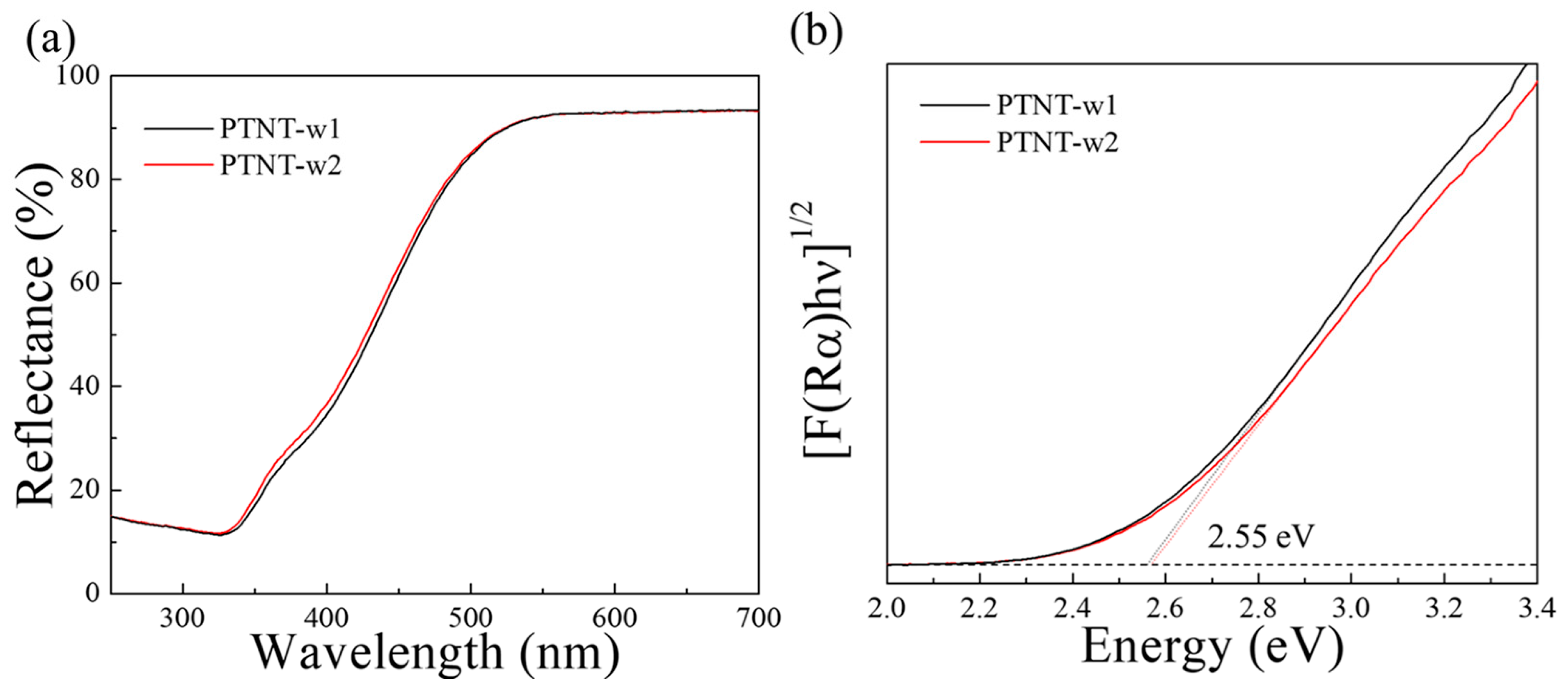
Figure 5 shows the UV–vis. reflectance spectra and Tauc plots of PTNT-w1 and PTNT-w2 samples calculated by considering indirectly allowed transition. Both samples absorbed light below 550 nm, with a band gap energy, calculated based on the Kubelka–Munk theory, of 2.55 eV [33,34]. Similar to their morphological characteristics, there is no significant difference in UV–vis. reflectance spectra between PTNT-w1 and PTNT-w2, consistent with previous reports on the characteristics of PTNT materials [25]. These results suggest that even with variations in the ratio of Na+ ion contents from 0.5 to 0.2 in PTNTs, although differences arise in the photodegradation performance of Rh B, there is no significant difference in the morphological and optical properties of the PTNT.

3.3.2. Effect of Cation Exchange on Rh B Solution

In general, layered titanate possesses cation exchange ability, which can affect the solution pH, potentially impacting the photodegradation performance. Thus, the pH values of the Rh B solutions containing the PTNT-w1 and PTNT-w2 samples were measured. The Rh B solution (10 mg/L) had an acidic pH value of 4.0. However, when the PTNT-w1 or PTNT-w2 samples were added, each of the resulting solutions had pH values of 7.9 and 7.1, respectively, showing a slight increase. These changes were probably due to cation exchange occurring when the PTNT containing Na+ ions and an acidic Rh B solution were mixed. The exchange of H+ ions from the acidic Rh B solution with Na+ ions from PTNT increased the solution’s pH. Moreover, a higher Na content in PTNT material correlates with a greater increase in pH. Therefore, in the case of PTNT-w2, which showed a smaller pH increase, the effect of ion exchange on the photodegradation performance was assumed to be small. To investigate this hypothesis, the PTNT-w3 sample, which had the lowest Na+ content, was prepared, and its photodegradation performance was evaluated.

3.4. Methods for Minimizing the Influence of Cation Exchange

3.4.1. Minimizing Sodium Ion Contents in PTNT Samples

Figure 6 shows the Rh B photodegradation results using the PTNT-w3 sample. Under dark conditions, the adsorption capacity after 30 min was 6–7%. The Rh B degradation reached 71% and 90% over 3 h under UV and vis. light sources, respectively. Table 3 presents the chemical composition and specific surface area of the PTNT-w3 sample. The chemical composition of PTNT-w3 is Na0.1H1.9Ti2O5, indicating a Na+ ion content approximately half of that in PTNT-w2. Upon mixing with the Rh B solution, the pH was 5.0. Comparing the Rh B adsorption of PTNT-w3 with those of PTNT-w1 and PTNT-w2, there was no significant difference in specific surface area; however, the adsorption capacity for 30 min under dark conditions was approximately doubled. Furthermore, the Rh B photodegradation reaction over 3 h under both UV and vis. light sources proceeded more effectively with PTNT-w3. Therefore, to examine the influence of the solution’s pH on the adsorption and photodegradation reactions in more detail, conditions with reduced pH were also tested. In this approach, the solution’s pH was adjusted using an HCl solution rather than adjusting the Na+ ion content of PTNTs, and this sample was designated as ‘PTNT-w2 + HCl’.

3.4.2. Controlling the pH of the Rhodamine B Solution

Figure 7a shows the changes in Rh B concentration (C/C0) when the PTNT-w2 and HCl solutions were added to the Rh B solution under dark conditions. At the beginning of the reaction, as indicated by the rate of decrease in the Rh B concentration upon adding PTNT-w2, an approximate 5% reduction was observed. This reduction tripled to about 15% after adding the HCl solution. Subsequently, the Rh B concentration remained constant for 3 h, with no significant differences, due to the addition of HCl. Figure 7b shows the Rh B photodegradation result using the PTNT-w2 + HCl condition. Similar to Figure 7a, an approximately 15% decrease in the Rh B concentration was observed in the dark for 30 min. Under UV and visible light irradiation for 3 h, the Rh B degradation reached approximately 92% and 97%, respectively. Comparing these results with those obtained under different conditions (PTNT-w1, PTNT-w2, and PTNT-w3), the most superior adsorption and photodegradation performance were observed under the PTNT-w2 + HCl condition. Of particular interest are two key points: first, the enhancement in the adsorption reaction of Rh B and second, the increase in the photocatalytic degradation rate of PTNT.
Regarding the enhancement in adsorption capability, it is necessary to investigate the phenomena arising from the changes in the PTNT surface properties, as well as those occurring owing to variations in the Rh B solution characteristics. Figure 8 shows the results of the zeta potential measurements for the PTNT material (PTNT-w2), which are relevant to the adsorption capacity concerning pH variations. In general, adsorption performance is influenced by the surface charge of the photocatalyst, as explained by the zeta potential [22]. For PTNT-w2, it was found that there was no significant change in the zeta potential, ranging from −30 to −25, across the pH range (pH 3–8) tested in this study. Therefore, it was concluded that the variation in the adsorption properties between the Rh B molecules and PTNT was likely due to factors other than changes in the surface properties of the PTNT material.
Therefore, the variation in the ionic form of Rh B in the solution with respect to pH was considered. Figure 9 shows the two ionic forms of the Rh B molecule: (a) cationic form and (b) zwitter-ionic form. The Rh B solution inherently possesses an acidic form, wherein the ionic form of the Rh B molecules resembles that shown in Figure 9a, with abundant H+ ions attached to the carboxyl group (–COOH), and the nitrogen of the amino group carrying a positive charge, resulting in an overall positive charge on the Rh B molecule [35]. However, as the pH increases, the Rh B molecules transform into their zwitter-ionic forms, as shown in Figure 9b. In the zwitter-ionic form, the carboxyl group (–COO) is negatively charged, whereas the amino group is positively charged. Inyinbor et al. and Zamouche et al. reported that Rh B molecules in zwitter-ionic form tend to aggregate, hindering adsorption [35,36]. Therefore, in this study, it was anticipated that the Rh B molecules would be transformed into the zwitter-ionic form by increasing the pH, owing to the addition of the PTNT sample into the Rh B solution. In the case of PTNT-w1 and PTNT-w2, the limited adsorption of Rh B resulted in a poor photocatalytic reaction. However, by adjusting the amount of Na+ ions present in PTNT, as in the case of the PTNT-w3, or by controlling pH, as in the PTNT-w2 + HCl condition, the formation of the zwitter-ionic form of Rh B could be reduced, thereby enhancing adsorption and photodegradation performance.

3.5. Kinetic Study of Rh B Photodegradation

In addition, the kinetics of all the photocatalytic performances evaluated in this study were analyzed according to the Langmuir–Hinshelwood model described in Equation (3). Figure 10 and Table 4 present the kinetics and calculated rate constant (k) values for all photocatalytic reactions under UV and visible light for the PTNT-w1, PTNT-w2, PTNT-w3, and PTNT-w2 + HCl conditions. The rate constants were calculated based on the Rh B concentration after photocatalytic degradation over time, following 30 min in the dark, and showed a linear relationship (R-squared value > 0.95). In the case of PTNT-w2 + HCl, the rate constants for UV and vis. light sources were 2.19 × 10⁻⁴ s−1 and 3.20 × 10⁻⁴ s−1, respectively, showing that the fastest degradation of Rh B is independent of the type of light source. Particularly noteworthy is that for PTNT-w1 and PTNT-w2, the rate constants under UV were faster than those under the vis. light source. Conversely, for PTNT-w3 and PTNT-w2 + HCl, the rate constants under vis. light were higher than those under the UV light source. Consequently, although the adsorption of Rh B on PTNT was poor (in PTNT-w1 and PTNT-w2), the photocatalytic reaction was faster under UV compared to vis. light. However, in cases where adsorption was enhanced (in PTNT-w3 and PTNT-w2 + HCl), the photocatalytic reaction was faster under vis. light compared to UV. According to Chen et al. [30], under vis. light, active oxygen species are generated around the adsorption sites of the photocatalyst material, facilitating photocatalytic degradation. Conversely, under UV light, the availability of higher energy with short wavelengths leads to the generation of active oxygen species, regardless of the adsorption sites on the photocatalyst [30]. From these results, it can be inferred that, depending on the type of light source, the relationship between the photocatalytic reaction rates of PTNTs, the adsorption state, and the amount of Rh B is reversed.

3.6. Effects of Cation Exchange in TC Photodegradation Test

Finally, to assess whether factors other than the pH-induced ionic form-change of Rh B affected the photodegradation performance observed in this study, TC organic pollutants were evaluated instead of Rh B in the photodegradation test. Initially, the TC solution had a pH of 3.5, similar to that of the Rh B solution. However, similar to the Rh B solution, due to cation exchange reactions induced by the PTNT-w2 addition, the pH increased to 5.6. Figure 11 shows the photocatalytic degradation results of the TC solution after adding the PTNT-w2 sample, leading to an increase in pH, and after the adjustment of pH by adding HCl. There were no significant differences in the adsorption and photodegradation reaction rates with or without HCl addition after 30 min in the dark. Consequently, the decrease in the adsorption amount and photodegradation rate due to the increase in pH caused by the Rh B solution and PTNT addition is primarily attributed to the variation in the ionic form of Rh B molecules with pH.

4. Conclusions

In this study, the adsorption and photodegradation performance of PTNT samples was evaluated using Rh B and TC solutions. In particular, focusing on the influence of the cation exchange ability of the PTNT material, Rh B photodegradation tests were conducted while observing the pH changes in the Rh B solution based on the amount of Na+ ions present in PTNTs. Even with variations in the Na+ ion content of the PTNT materials ranging from Na0.5H1.5Ti2O5 to Na0.1H1.9Ti2O5, there were no significant differences in their morphological and optical properties. However, the adsorption and photodegradation rates of Rh B varied for each sample, primarily because of the change in the ionic form of Rh B with pH. When the PTNT materials containing Na+ ions were mixed with an acidic Rh B solution, a cation exchange reaction occurred, causing an increase in the pH of the Rh B solution. Consequently, it was estimated that the Rh B molecules transformed from a cationic form to a zwitter-ionic form, leading to poor adsorption due to the aggregation of the Rh B molecules. Additionally, tests using TC instead of Rh B confirmed that the phenomenon was primarily caused by the change in the form of the Rh B molecules rather than by the variation in the Na+ ion content of the PTNT materials. This study suggests that the issue of poor adsorption due to pH changes in the Rh B solution can be addressed by reducing the amount of Na+ ions in the PTNT materials or by adjusting the pH by adding acidic solutions (such as HCl) to the Rh B solution, within a range that does not affect its properties.

Author Contributions

Conceptualization, D.H.H., H.P. and T.S.; methodology, D.H.H., H.P., T.G., Y.K. and T.S.; validation, D.H.H., H.P., S.C., Y.S., H.N. and T.S.; formal analysis, D.H.H., H.P., T.G., S.C. and T.S.; investigation, D.H.H., H.P. and T.S.; resources, T.G., S.C., Y.S., Y.K. and H.N.; data curation, D.H.H., H.P. and T.S.; writing—original draft preparation, D.H.H.; writing—review and editing, H.P., T.G., S.C. and T.S.; visualization, D.H.H., H.P. and T.G.; supervision, T.S.; project administration, T.S.; funding acquisition, T.S. All authors have read and agreed to the published version of the manuscript.

Funding

This research was funded by the Japan Science and Technology Agency (JST) under the Adaptable and Seamless Technology Transfer Program through Target-Driven R&D (A-STEP), grant number JPMJTR22T8.

Data Availability Statement

The datasets analyzed or generated during this study are available from the corresponding author upon reasonable request.

Acknowledgments

This research was supported by the “Dynamic Alliance for Open Innovation Bridging Human, Environment and Materials” in the “Network Joint Research Center for Materials and Devices” (MEXT, Tokyo, Japan). The authors acknowledge Masato Kakihana of SANKEN, Osaka University, for the valuable comments and suggestions. XRF analyses were performed at the Comprehensive Analysis Center, SANKEN, Osaka University, Japan.

Conflicts of Interest

The authors declare no conflicts of interest.

References

  1. Linsebigler, A.L.; Lu, G.; Yates, J.T. Photocatalysis on TiO2 Surfaces: Principles, Mechanisms, and Selected Results. Chem. Rev. 1995, 95, 735–758. [Google Scholar] [CrossRef]
  2. Xiong, Z.; Ma, J.; Ng, W.J.; Waite, T.D.; Zhao, X.S. Silver-Modified Mesoporous TiO2 Photocatalyst for Water Purification. Water Res. 2011, 45, 2095–2103. [Google Scholar] [CrossRef] [PubMed]
  3. Sweet, C.W.; Vermette, S.J. Toxic Volatile Organic Compounds in Urban Air in Illinois. Environ. Sci. Technol. 1992, 26, 165–173. [Google Scholar] [CrossRef]
  4. Mamaghani, A.H.; Haghighat, F.; Lee, C.S. Role of Titanium Dioxide (TiO2) Structural Design/Morphology in Photocatalytic Air Purification. Appl. Catal. B 2020, 269, 118735. [Google Scholar] [CrossRef]
  5. Lei, X.; Li, X.; Ruan, Z.; Zhang, T.; Pan, F.; Li, Q.; Xia, D.; Fu, J. Adsorption-Photocatalytic Degradation of Dye Pollutant in Water by Graphite Oxide Grafted Titanate Nanotubes. J. Mol. Liq. 2018, 266, 122–131. [Google Scholar] [CrossRef]
  6. Kocijan, M.; Ćurković, L.; Ljubas, D.; Mužina, K.; Bačić, I.; Radošević, T.; Podlogar, M.; Bdikin, I.; Otero-Irurueta, G.; Hortigüela, M.J.; et al. Graphene-Based TiO2 Nanocomposite for Photocatalytic Degradation of Dyes in Aqueous Solution under Solar-like Radiation. Appl. Sci. 2021, 11, 3966. [Google Scholar] [CrossRef]
  7. Mohamed Mukthar Ali, S.M.Y.; Sandhya, K.Y. A Novel Approach for P25-Carbon Dot Composite and the Reactive Oxygen Species Involved in the Visible Light Photocatalytic Mineralization of Rhodamine B. ChemistrySelect 2017, 2, 11840–11845. [Google Scholar] [CrossRef]
  8. Saothayanun, T.K.; Sirinakorn, T.T.; Ogawa, M. Layered Alkali Titanates (A2TinO2n+1): Possible Uses for Energy/Environment Issues. Front. Energy 2021, 15, 631–655. [Google Scholar] [CrossRef]
  9. Wang, Q.; Sohn, J.H.; Chung, J.S. Thermally Stable Pt/K2Ti2O5 as High-Temperature NOx Storage and Reduction Catalyst. Appl. Catal. B 2009, 89, 97–103. [Google Scholar] [CrossRef]
  10. Zhao, B.; Lin, L.; He, D. Phase and Morphological Transitions of Titania/Titanate Nanostructures from an Acid to an Alkali Hydrothermal Environment. J. Mater. Chem. A Mater. 2013, 1, 1659–1668. [Google Scholar] [CrossRef]
  11. Shirpour, M.; Cabana, J.; Doeff, M. Lepidocrocite-Type Layered Titanate Structures: New Lithium and Sodium Ion Intercalation Anode Materials. Chem. Mater. 2014, 26, 2502–2512. [Google Scholar] [CrossRef]
  12. Kim, M.; Hwang, S.H.; Lim, S.K.; Kim, S. Effects of Ion Exchange and Calcinations on the Structure and Photocatalytic Activity of Hydrothermally Prepared Titanate Nanotubes. Cryst. Res. Technol. 2012, 47, 1190–1194. [Google Scholar] [CrossRef]
  13. Kang, S.; Durand-Vidal, S.; Badot, J.C.; Legein, C.; Body, M.; Borkiewicz, O.J.; Dubrunfaut, O.; Dambournet, D. Intercalation–Exfoliation Processes during Ionic Exchange Reactions from Sodium Lepidocrocite-Type Titanate toward a Proton-Based Trititanate Structure. Phys. Chem. Chem. Phys. 2021, 23, 10498–10508. [Google Scholar] [CrossRef] [PubMed]
  14. Hu, W.; Li, L.; Li, G.; Liu, Y.; Withers, R.L. Atomic-Scale Control of TiO6 Octahedra through Solution Chemistry towards Giant Dielectric Response. Sci. Rep. 2014, 4, 6582. [Google Scholar] [CrossRef] [PubMed]
  15. Mao, Y.; Kanungo, M.; Hemraj-Benny, T.; Wong, S.S. Synthesis and Growth Mechanism of Titanate and Titania One-Dimensional Nanostructures Self-Assembled into Hollow Micrometer-Scale Spherical Aggregates. J. Phys. Chem. B 2005, 110, 702–710. [Google Scholar] [CrossRef] [PubMed]
  16. Barim, G.; Dhall, R.; Arca, E.; Kuykendall, T.R.; Yin, W.; Takeuchi, K.J.; Takeuchi, E.S.; Marschilok, A.C.; Doeff, M.M. Heterostructured Lepidocrocite Titanate-Carbon Nanosheets for Electrochemical Applications. ACS Appl. Nano Mater. 2022, 5, 678–690. [Google Scholar] [CrossRef]
  17. Qamar, M.; Yoon, C.R.; Oh, H.J.; Lee, N.H.; Park, K.; Kim, D.H.; Lee, K.S.; Lee, W.J.; Kim, S.J. Preparation and Photocatalytic Activity of Nanotubes Obtained from Titanium Dioxide. Catal. Today 2008, 131, 3–14. [Google Scholar] [CrossRef]
  18. Yu, H.; Yu, J.; Cheng, B.; Lin, J. Synthesis, Characterization and Photocatalytic Activity of Mesoporous Titania Nanorod/Titanate Nanotube Composites. J. Hazard. Mater. 2007, 147, 581–587. [Google Scholar] [CrossRef] [PubMed]
  19. Kim, D.S.; Kwak, S.Y. The Hydrothermal Synthesis of Mesoporous TiO2 with High Crystallinity, Thermal Stability, Large Surface Area, and Enhanced Photocatalytic Activity. Appl. Catal. A Gen. 2007, 323, 110–118. [Google Scholar] [CrossRef]
  20. Guohui, T.; Honggang, F.; Liqiang, J.; Baifu, X.; Kai, P. Preparation and Characterization of Stable Biphase TiO2 Photocatalyst with High Crystallinity, Large Surface Area, and Enhanced Photoactivity. J. Phys. Chem. C 2008, 112, 3083–3308. [Google Scholar]
  21. Goto, T.; Cho, S.H.; Lee, S.W.; Sekino, T. Sorption Capacity of Cs+ on Titania Nanotubes Synthesized by Solution Processing. J. Ceram. Soc. Jpn. 2018, 126, 801–807. [Google Scholar] [CrossRef]
  22. Goto, T.; Kondo, Y.; Cho, S.H.; Seino, S.; Sekino, T. Comparative Study of Divalent Cation Sorption on Titania Nanotubes Using Co2+, Ni2+, Zn2+, and Sr2+. Chem. Eng. J. Adv. 2022, 12, 100388. [Google Scholar] [CrossRef]
  23. Qianqian, Z.; Tang, B.; Guoxin, H. High Photoactive and Visible-Light Responsive Graphene/Titanate Nanotubes Photocatalysts: Preparation and Characterization. J. Hazard. Mater. 2011, 198, 78–86. [Google Scholar] [CrossRef]
  24. Doong, R.A.; Liao, C.Y. Enhanced Photocatalytic Activity of Cu-Deposited N-TiO2/Titanate Nanotubes under UV and Visible Light Irradiations. Sep. Purif. Technol. 2017, 179, 403–411. [Google Scholar] [CrossRef]
  25. Park, H.; Goto, T.; Han, D.H.; Cho, S.; Nishida, H.; Sekino, T. Low Alkali Bottom-Up Synthesis of Titanate Nanotubes Using a Peroxo Titanium Complex Ion Precursor for Photocatalysis. ACS Appl. Nano Mater. 2020, 3, 7795–7803. [Google Scholar] [CrossRef]
  26. Park, H.; Goto, T.; Cho, S.; Nishida, H.; Sekino, T. Enhancing Visible Light Absorption of Yellow-Colored Peroxo-Titanate Nanotubes Prepared Using Peroxo Titanium Complex Ions. ACS Omega 2020, 5, 21753–21761. [Google Scholar] [CrossRef] [PubMed]
  27. Lorençon, E.; Brandão, F.D.; Krambrock, K.; Alves, D.C.B.; Silva, J.C.C.; Ferlauto, A.S.; Lago, R.M. Generation of Reactive Oxygen Species in Titanates Nanotubes Induced by Hydrogen Peroxide and Their Application in Catalytic Degradation of Methylene Blue Dye. J. Mol. Catal. A Chem. 2014, 394, 316–323. [Google Scholar] [CrossRef]
  28. Erjavec, B.; Kaplan, R.; Pintar, A. Effects of Heat and Peroxide Treatment on Photocatalytic Activity of Titanate Nanotubes. Catal. Today 2015, 241, 15–24. [Google Scholar] [CrossRef]
  29. Kinsinger, N.M.; Dudchenko, A.; Wong, A.; Kisailus, D. Synergistic Effect of pH and Phase in a Nanocrystalline Titania Photocatalyst. ACS Appl. Mater. Interfaces 2013, 5, 6247–6254. [Google Scholar] [CrossRef]
  30. Chen, F.; Zhao, J.; Hidaka, H. Highly Selective Deethylation of Rhodamine B: Adsorption and Photooxidation Pathways of the Dye on the TiO2/SiO2 Composite Photocatalyst. Int. J. Photoenergy 2003, 5, 209–217. [Google Scholar] [CrossRef]
  31. Zhang, W.; Li, Y.; Wang, C.; Wang, P. Kinetics of Heterogeneous Photocatalytic Degradation of Rhodamine B by TiO2-Coated Activated Carbon: Roles of TiO2 Content and Light Intensity. Desalination 2011, 266, 40–45. [Google Scholar] [CrossRef]
  32. Sharma, M.; Mandal, M.K.; Pandey, S.; Kumar, R.; Dubey, K.K. Visible-Light-Driven Photocatalytic Degradation of Tetracycline Using Heterostructured Cu2O-TiO2 Nanotubes, Kinetics, and Toxicity Evaluation of Degraded Products on Cell Lines. ACS Omega 2022, 7, 33572–33586. [Google Scholar] [CrossRef] [PubMed]
  33. Yaemsunthorn, K.; Tatarchuk, T.; Danyliuk, N.; Shyichuk, A.; Macyk, W. Yellow TiO2 from Titanium Peroxo Complexes: Verification of the Visible Light Activity and a Rational Enhancement of Its Photocatalytic Efficiency. J. Environ. Chem. Eng. 2023, 11, 111520. [Google Scholar] [CrossRef]
  34. Makuła, P.; Pacia, M.; Macyk, W. How To Correctly Determine the Band Gap Energy of Modified Semiconductor Photocatalysts Based on UV-Vis Spectra. J. Phys. Chem. Lett. 2018, 9, 6814–6817. [Google Scholar] [CrossRef]
  35. Inyinbor, A.A.; Adekola, F.A.; Olatunji, G.A. Adsorption of Rhodamine B Dye from Aqueous Solution on Irvingia Gabonensis Biomass: Kinetics and Thermodynamics Studies. S. Afr. J. Chem. 2015, 68, 115–125. [Google Scholar] [CrossRef]
  36. Zamouche, M.; Hamdaoui, O. Sorption of Rhodamine B by Cedar Cone: Effect of pH and Ionic Strength. Energy Procedia 2012, 18, 1228–1239. [Google Scholar] [CrossRef]
Figure 1. Schematics diagram of preparation process for (a) PTNT-w1, (b) PTNT-w2, and (c) PTNT-w3.
Nanomaterials 14 01170 g001
Figure 2. (a) XRD pattern of PTNT-w1 and PTNT-w2 samples and (b) enlarged graph of the 200 diffraction peak in (a).
Nanomaterials 14 01170 g002
Figure 3. (ad) SEM and STEM images and (e,f) N2 adsorption–desorption isotherm graphs of PTNT-w1 (a,b,e) and PTNT-w2 (c,d,f) samples.
Nanomaterials 14 01170 g003
Figure 4. Photocatalytic degradation results of Rh B using PTNT-w1 and PTNT-w2 samples UV and vis. light irradiation. The gray area corresponds to dark condition prior to light irradiation.
Nanomaterials 14 01170 g004
Figure 5. (a) UV–vis. reflectance spectra and (b) Tauc plots of PTNT-w1 and PTNT-w2 samples.
Nanomaterials 14 01170 g005
Figure 6. Photocatalytic degradation results of Rh B using PTNT-w3 sample under the UV and vis. light irradiation. The gray area corresponds to dark condition prior to light irradiation.
Nanomaterials 14 01170 g006
Figure 7. (a) Changes in Rh B concentration (C/C0) under the dark conditions when PTNT-w2 was inserted, before and after the addition of HCl solution, and (b) photocatalytic degradation results of Rh B under the PTNT-w2 + HCl condition, where the gray area corresponds to dark condition before UV and vis. light irradiation.
Nanomaterials 14 01170 g007
Figure 8. Changes in zeta potential against the solution pH of the PTNT-w2 sample.
Nanomaterials 14 01170 g008
Figure 9. Ionic forms of Rh B: (a) cationic form and (b) zwitter-ionic form.
Nanomaterials 14 01170 g009
Figure 10. Kinetics graph of the photodegradation using all PTNT samples.
Nanomaterials 14 01170 g010
Figure 11. Photocatalytic degradation of TC using PTNT-w2 sample under the UV and vis. light irradiation, where the test was carried out under the normal (square) and pH adjusted using HCl (circle) conditions. The gray area corresponds to the dark condition without light irradiation.
Nanomaterials 14 01170 g011
Table 1. The 2theta and d200 values in the XRD pattern and chemical composition of the each PTNT samples calculated based on XRF analysis results.
Sample2thata (Degree)d200 (Å)Chemical Composition
PTNT-w19.39.5Na0.5H1.5Ti2O5
PTNT-w29.69.2Na0.2H1.8Ti2O5
Table 2. The specific surface area of PTNT-w1 and PTNT-w2 samples.
SampleSpecific Surface Area (m2/g)
PTNT-w1140.90
PTNT-w2130.01
Table 3. The specific surface area and chemical composition of PTNT-w3 sample.
SampleSpecific Surface Area (m2/g)Chemical Composition
PTNT-w3145.49Na0.1H1.9Ti2O5
Table 4. The rate constant of Rh B photodegradation with PTNT-w1, PTNT-w2, PTNT-w3, and PTNT-w2 + HCl conditions and R-squared value of each kinetic graph in Figure 11.
SampleIrradiation Lightk (s−1)R-Squared Value
PTNT-w1UV6.47 × 10−50.99
Vis1.13 × 10−50.96
PTNT-w2UV9.80 × 10−50.98
Vis3.54 × 10−50.98
PTNT-w3UV1.06 × 10−50.95
Vis2.04 × 10−50.99
PTNT-w2 + HClUV2.19 × 10−40.99
Vis3.20 × 10−40.99
Disclaimer/Publisher’s Note: The statements, opinions and data contained in all publications are solely those of the individual author(s) and contributor(s) and not of MDPI and/or the editor(s). MDPI and/or the editor(s) disclaim responsibility for any injury to people or property resulting from any ideas, methods, instructions or products referred to in the content.