Article

Studying the Process of Phosphogypsum Recycling into a Calcium Sulphide-Based Luminophor

by
Oleg A. Medennikov
1,
Marina A. Egorova
1,
Nina P. Shabelskaya
1,
Asatullo Rajabov
1,
Sergey I. Sulima
2,*,
Elena V. Sulima
2,
Zlatislava D. Khliyan
1 and
Daniil I. Monastyrskiy
1
1
Department of Ecology and Industrial Safety, Faculty of Technology, Platov South-Russian State Polytechnic University (NPI), Novocherkassk 346428, Russia
2
Department of Chemical Technologies, Faculty of Technology, Platov South-Russian State Polytechnic University (NPI), Novocherkassk 346428, Russia
*
Author to whom correspondence should be addressed.
Nanomaterials 2024, 14(11), 904; https://doi.org/10.3390/nano14110904
Submission received: 17 April 2024 / Revised: 16 May 2024 / Accepted: 20 May 2024 / Published: 22 May 2024

Abstract

:
Currently, one of the most important problems of environmental protection is the deep and complex processing of mineral raw materials. This problem is especially relevant when processing substandard ores and production waste, one of which is phosphogypsum. This study examines the process of CaSO4/CaS composite material formation during the reduction of phosphogypsum with citric acid. The composite structure formation mechanism is proposed. The resulting materials are characterized using various methods, including X-ray diffraction (XRD), transmission electron microscopy, the Scherrer method, thermogravimetric analysis (TGA), and FT-IR spectroscopy. The reduced sample emits orange radiation in the range of 500–750 nm with a quantum yield of 0.17. Experimental results showed that the sample decomposition process in the solid state consisted of two components with a predominant contribution from the long-lived component (~46 ns). The optimal conditions for producing luminescent materials by reducing phosphogypsum with citric acid were determined: a heat treatment temperature of 1073 K, a holding time of 60 min, and a reducing agent mole fraction of 37%. It was found that an increase in temperature with a simultaneous decrease in heat treatment time, as well as a decrease in temperature with a simultaneous increase in heat treatment time, led to a decrease in the luminescent properties of the synthesized material compared to optimal values. The results can be used to develop technology for recycling large-tonnage waste from the chemical industry into luminescent materials.

1. Introduction

The current level of production throughout the world is directly related to the global use of natural resources and the accumulation of large-tonnage technogenic waste [1,2,3]. This is a reality that must be considered; even an essential technically advanced industrial complex, if its impact on nature extends beyond environmentally acceptable limits or becomes destructive, may become undesirable for society, if not today, then in the nearest future.
Currently, one of the most important problems of environmental protection is the deep and complex processing of mineral raw materials. Several studies are devoted to ore materials’ complex processing issues and increasing the environmental safety of ore-related production [4,5,6]. This problem is especially relevant when processing substandard ores and production waste, including phosphogypsum.
Phosphogypsum is a product formed during the decomposition of raw phosphate materials with sulfuric acid using an extraction method. As a rule, extraction of phosphorus from raw materials is carried out with mineral acids (phosphoric, sulfuric, or, less often, nitric), which produce large quantities of waste. In recent years, many attempts have been made to recycle phosphogypsum. Among them, several areas can be noted. The authors in [7] proposed a technology for the complex processing of phosphogypsum into a chemical ameliorant. The use of phosphogypsum has been shown to improve the growth quality of direct-seeded rice. Several articles [2,8,9,10,11] discuss the possibilities of using phosphogypsum as a material to produce gypsum binders for further use for various purposes and propose a new effective technology for its utilization. The authors of [12] worked on obtaining a durable composite material based on phosphogypsum for construction use. Research [13] discusses the prospects of using phosphogypsum in road surface production. In studies [14,15,16,17], it was proposed to use phosphogypsum in chemical ring gasification processes, including synthesis gas production. In addition, developments in the field of extraction of rare earth elements from phosphogypsum are gaining widespread use [18]. The synthesis of organic–inorganic composite materials based on biochars and phosphogypsum [19,20] is accompanied with the production of effective adsorbents for antimonate ions and lead cations.
One of the possible applications of phosphogypsum is its use as a raw material for calcium sulfide synthesis [21,22,23,24,25]. Luminescent materials are produced based on CaS [26].
Several radiation phenomena that sharply deviate from the laws of thermal radiation have long been known. In some cases, substances begin to emit light at temperatures so low that the radiation cannot be explained by the transition of thermal energy into light. This includes the phenomena of the so-called “cold glow”, for example, paint covering the dials of glow-in-the-dark watches, etc. Such a glow is called luminescence. Luminescence is usually divided into the following classes: Triboluminescence is a glow arising from mechanical action, which includes numerous cases of glow from impact, friction, splitting, grinding, etc. The glow of sugar can serve as an example. Thermoluminescence is a glow caused by heating at relatively low temperatures (100–200 °C) at which there is no presence of visible thermal radiation. The glow of fluorite when heated is an example of that. Photoluminescence is a glow caused by light. There are usually two cases here: the first is when the glow continues after the end of irradiation (phosphorescence), and the second is when the glow is practically observed only during the continuation of the illumination process itself (fluorescence). For the case of illumination by X-rays, the term X-ray luminescence is used. Chemiluminescence is the result of chemical processes accompanied by light emittance without a particular increase in the reaction temperature. The glow during the oxidation of white phosphorus can serve as an example here.
Silicates, phosphates, sulfides, and other compounds are used as luminescent materials. In industry, zinc, barium, calcium, and cadmium sulfides activated with copper, silver, and rare earth elements (REE) are used. Calcium sulfide is one of the widely used luminophores [26,27].
There are direct and indirect methods for obtaining sulfide matrices. The essence of the direct synthesis method is to carry out a high-temperature reaction between two finely dispersed substances placed in a sealed ampoule filled with an inert gas. The ampoule is placed in a one- or two-zone oven. Heating is carried out gradually, but the higher the synthesis temperature, the faster the reaction proceeds and better homogenization of the final compound occurs. In general, the process of the direct synthesis of sulfides is represented by Equation (1):
xMe + yS = MexSy
The indirect method of calcium sulfide synthesis consists of a simpler way of obtaining sulfides from salts and oxides when they react with the vapors of a sulfonating agent.
According to the studies presented in [28], alkaline earth metal sulfides were synthesized by calcining a mixture of corresponding carbonates, sulfur, and a certain amount of a reducing agent. Starch was used as a reducing agent, since, when decomposing at high temperatures, it is a source of pure carbon and does not pollute the resulting sulfide with its decomposition products.
As a rule, the synthesis of sulfide luminescent materials requires the use of analytical reagents.
The goal of this study was to obtain luminescent materials based on a calcium sulfide matrix. To obtain CaS, we used phosphogypsum.

2. Experiment

2.1. Materials

To obtain calcium sulfide, agricultural phosphogypsum with a calcium sulfate dihydrate CaSO4∙2H2O content of 99% (wt.) was used. Citric acid (С6Н8О7) was used as a reducing agent, in which the content of the main component was 99.5% (wt.).

2.2. Composite Materials Synthesis

The synthesis was carried out according to the procedure described in detail in [29]. Phosphogypsum was pre-dried to a constant weight at a temperature of 373 K. To prepare the samples, 14 g of phosphogypsum and 4 g of a reducing agent were used, which were weighed with an accuracy of 0.01 g on an electronic balance and homogenized for 30 s. in a 0.45 kW mixer at 1500 rpm, placed in alundum crucibles in the working space of a muffle furnace, and subjected to heat treatment according to the temperature–time regimen, including heating to 140 °C for 30 min, holding for 30 min, heating to an isothermal temperature exposure at the speed of 13 K/min, exposure for 60 min, and slow cooling in the oven to room temperature.

2.3. Characteristics

Various techniques were used to characterize the resulting composite materials, including X-ray diffraction (XRD), transmission electron microscopy, the Scherrer method, thermogravimetric analysis (TGA), and FT-IR spectroscopy.
The phase composition was studied using an ARL X’TRA X-ray diffractometer (monochromatized Cu-Kα radiation was used) by point-by-point scanning (0.01° step, 2 s. accumulation time at a point) in the range of 2θ from 5 °C to 90 °C. The crystallite size was calculated using the Scherrer Equation (2)
D = 0.94·λ/(B∙cosθ)
where D is the average crystal size (nm), λ is the X-ray wavelength (nm), B is the peak linewidth at half height (rad), and cosθ is the cosine angle value for the peak.
To calculate the crystallite sizes, the following lines were used: line 020 for CaSO4·2H2O, CaSO4; line 200 for CaSO4·0.5H2O; and line 220 for CaS. The lines for calculation were chosen to avoid the phases’ reflections overlap.
The unit cell volume V (nm3) was calculated using Formulas (3) and (4):
-
for monoclinic and orthorhombic phases:
V = a·b·c·sinβ,
-
for the cubic phase:
V = a3
where a, b, c are unit cell parameters (nm).
The change in sample mass ms after heat treatment was determined using Formula (5)
ms = (m1m2) · 100/m1
where m1 is the sample mass after heat treatment, calculated theoretically (g), and m2 is the practical value of the sample mass (g).
A Quanta 200 scanning microscope (FEI Company, Hilsboro, OR, USA) was used to obtain micrographs.
Elemental analysis was carried out using a Quanta 200 scanning microscope combined with an EDAX Genesis XVS 30 X-ray microanalysis system (FEI Company).
The sample’s IR spectrum was recorded on a Spectrum Two Fourier IR spectrometer (PerkinElmer, Waltham, MA, USA) equipped with a universal device for measuring total reflectance (UATR) in the frequency range of 4000–400 cm−1. Spectrum processing and band intensity determination were carried out using special software (Spectrum 10.6) supplied with the spectrometer.

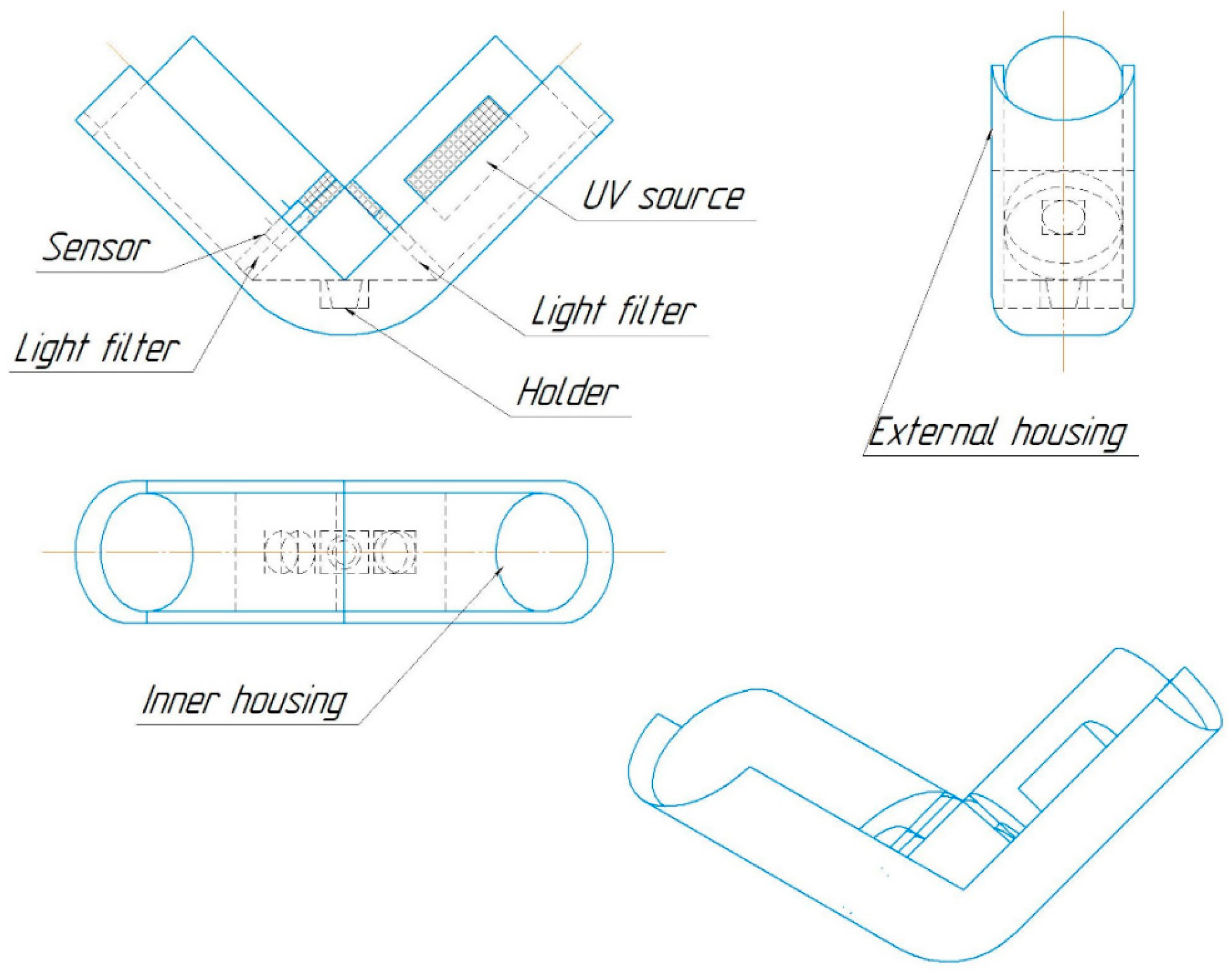
2.4. Luminescent Properties Study

For each sample, the relative luminous flux emitted by a fixed area of the sample surface was measured using the original setup [30] (Figure 1) consisting of an ultraviolet (UV) radiation source, light filters, and a recording sensor. The sample and the reference sample of a yellow YAG:Ce luminophore were placed in the installation, illuminated with radiation at a wavelength of 380 nm, then the sensor voltage drop was recorded, which occurred due to the sensor resistance change when the level of light flux illuminating its surface changed. The relative luminous flux was obtained as the ratio of the sensor voltage drop when the test sample was placed in the installation to the sensor voltage drop when the reference sample was placed in the installation.
Luminescence spectra were recorded using an FS5 spectrofluorimeter (Edinburgh Instruments, Edinburgh, UK). The absolute quantum yield was measured with an FS5 spectrofluorimeter using an SC-30 integrating sphere at λEx = 390 nm. The emission lifetime was measured using the TCSPC option of the FS5 spectrofluorimeter. The sample was excited with an EPL-375 picosecond pulsed diode laser centered at 375 nm. The instrument response function (IRF) was recorded under the described conditions by replacing the sample with a silica diffuser. Time decay data were analyzed by nonlinear curve fitting with IRF deconvolution using the Fluoracle software package F980.

3. Results and Discussion

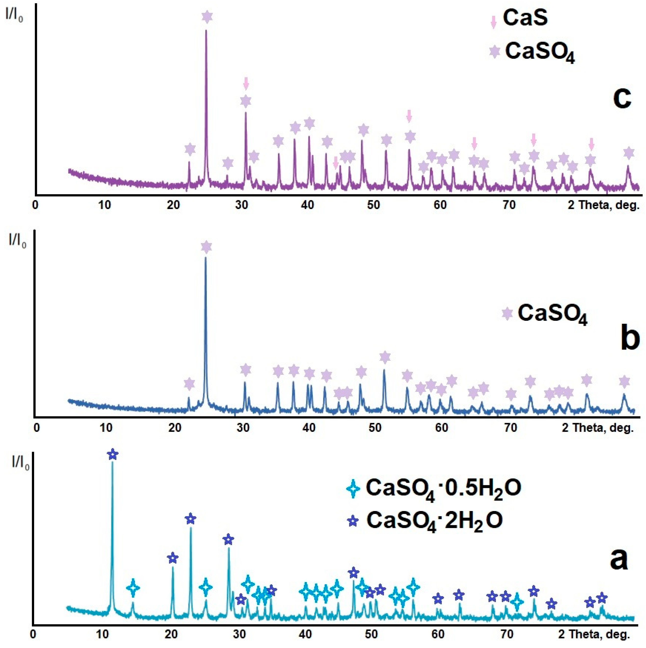
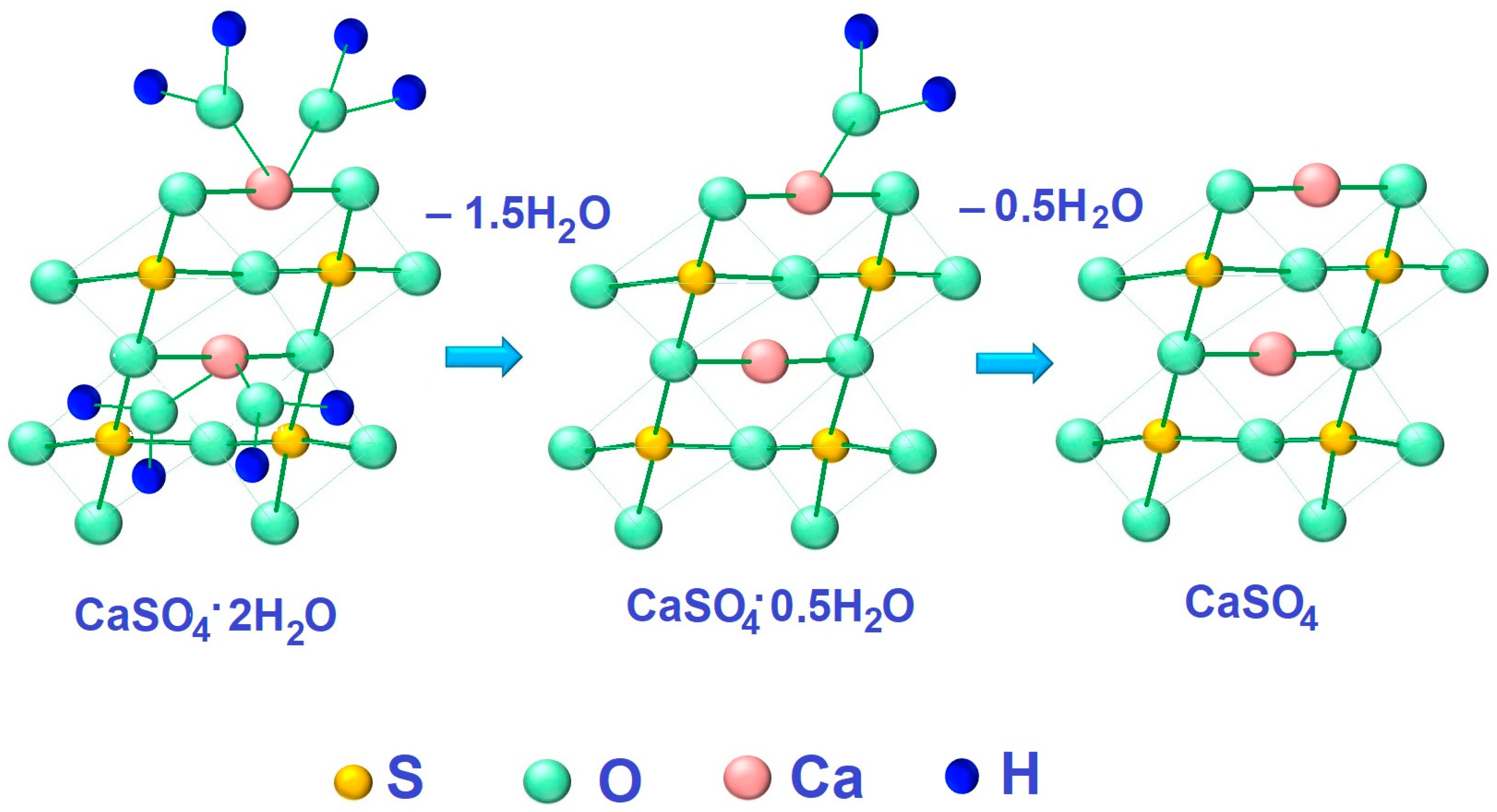
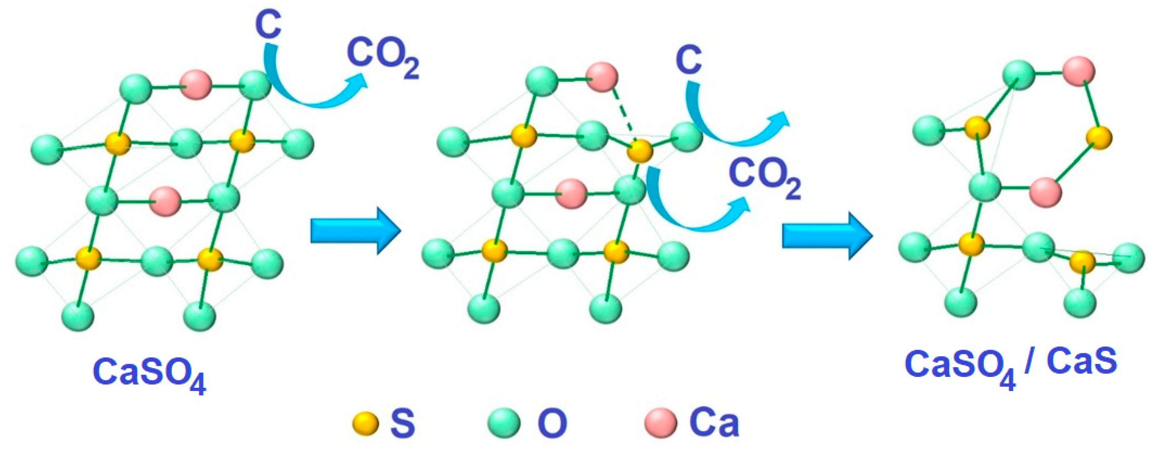
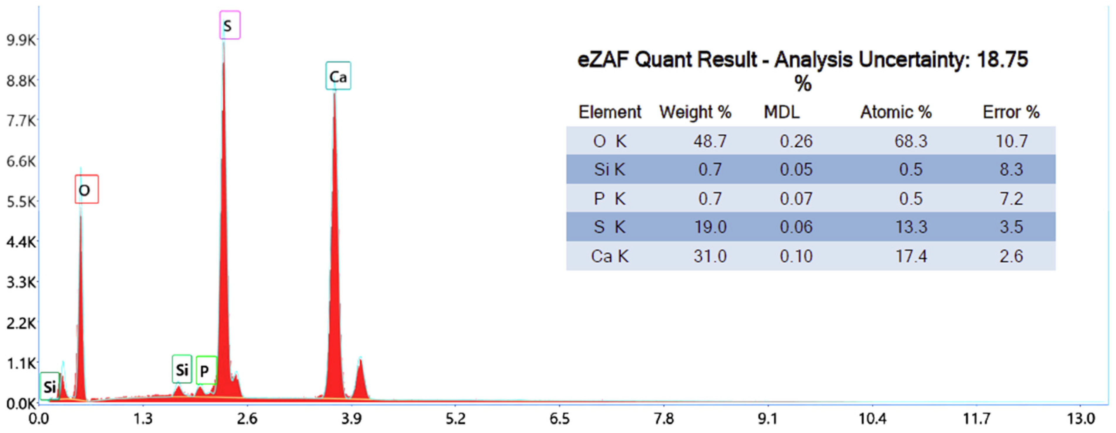
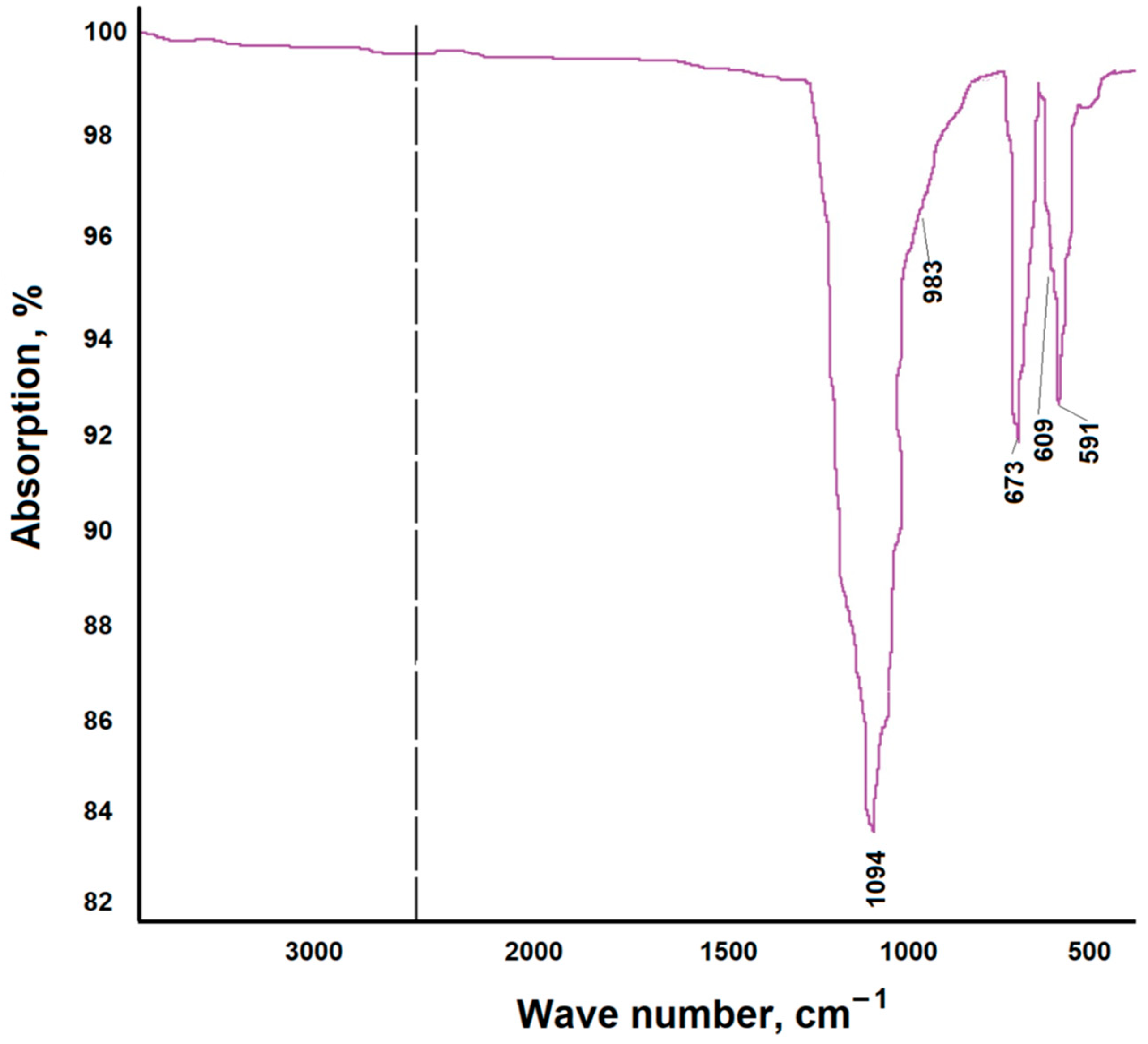
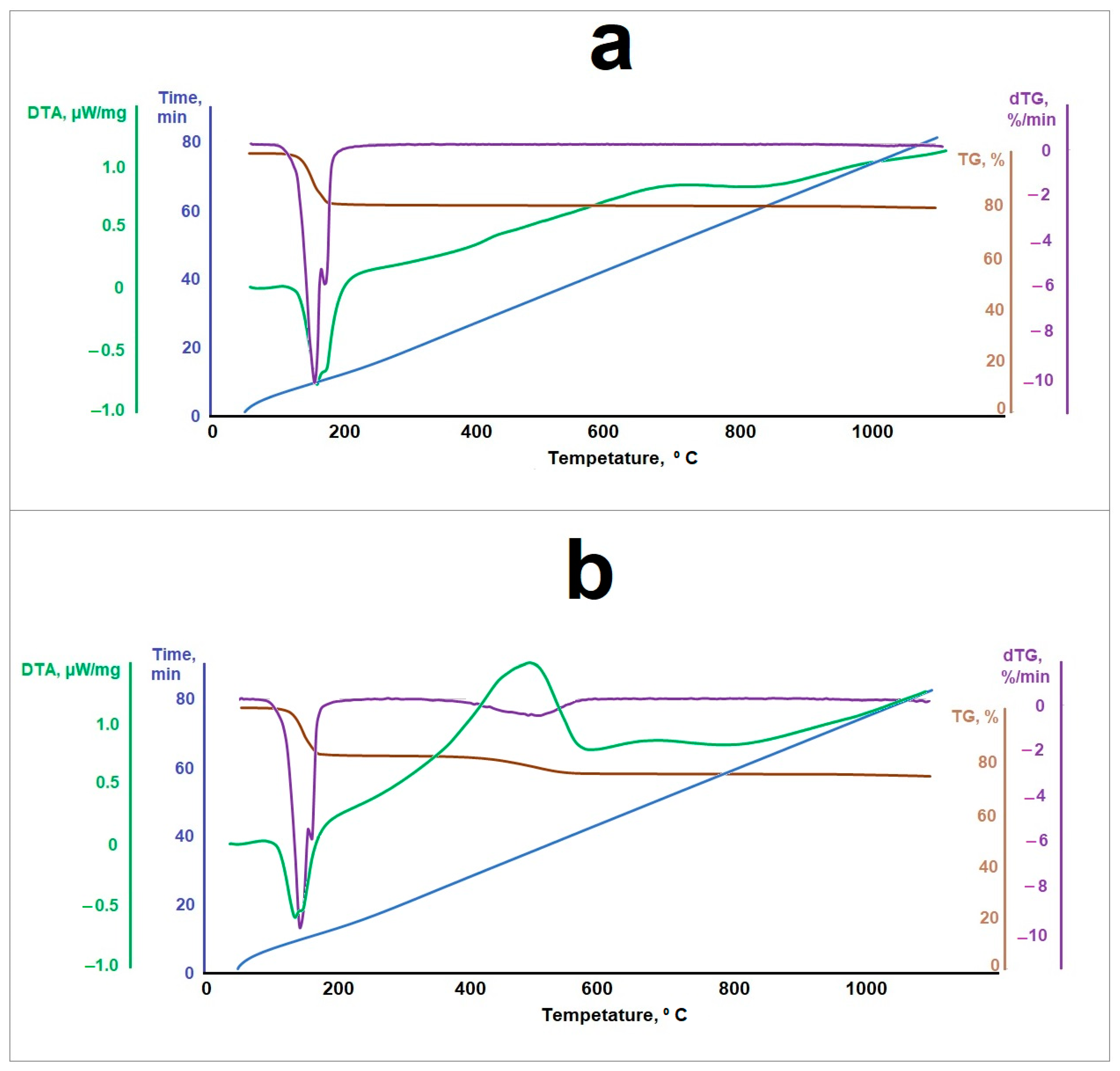
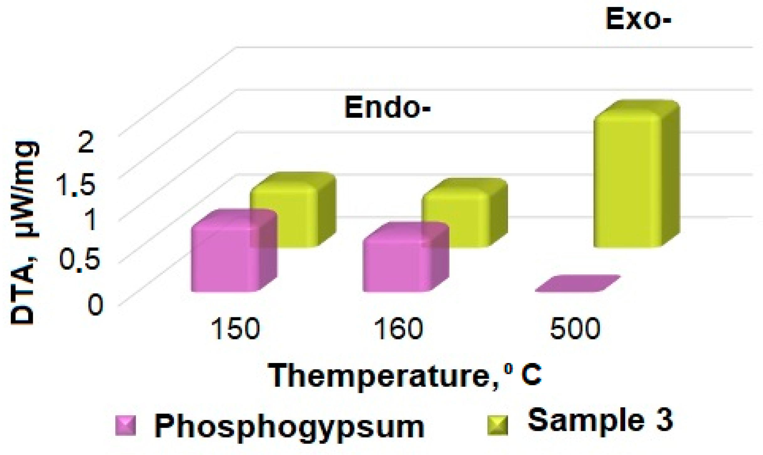
3.1. Samples’ Structures and Morphological Features

Figure 2 shows the X-ray diffraction patterns of the samples: sample 1—phosphogypsum dried at a temperature of 283 K to constant weight; sample 2—phosphogypsum calcined at a temperature of 1073 K for 60 min; and sample 3—phosphogypsum calcined in the presence of a reducing agent—citric acid—at a temperature of 1073 K for 60 min. Figure 2a shows an X-ray image of sample 1. The X-ray image contains reflections belonging to CaSO4·2H2O (PDF #010-70-7008) and CaSO4·0.5H2O (PDF #010-80-1235). Both compounds have a monoclinic system. The grid parameters and the crystallite sizes calculated using the Scherrer formula are presented in Table 1.
The X-ray diffraction pattern of sample 2 is shown in Figure 2b. The X-ray pattern contains reflections belonging to CaSO4 (PDF #010-74-2421) in the orthorhombic modification (see the grid parameters and the crystallite sizes in Table 1). The X-ray diffraction pattern of sample 3 is shown in Figure 2c. The X-ray pattern contains reflections belonging to CaSO4 (PDF #010-70-0909) in the orthorhombic modification and CaS (PDF #000-08-0464) in the cubic modification (see Table 1 for the grid parameters and the crystallite sizes).
As follows from the experimental data obtained, when heat treatment is carried out at a relatively low temperature, water molecules are separated in accordance with Equation (6):
CaSO4·2H2O = CaSO4·0.5H2O + 1.5H2O
In this case, an increase in the volume of the material unit cell is observed (see Table 1). At the same time, the average crystallite size decreases significantly—by 1.9 times. When heat treated at a temperature of 1073 K, phosphogypsum completely loses crystallization water according to Equation (7)
CaSO4·0.5H2O = CaSO4 + 0.5H2O
This leads to a decrease in the crystal grid volume (Table 1) and a more significant decrease (6 times) in the average size of crystallites. Heat treatment in the presence of a reducing agent leads to partial destruction of the structure with the formation of a CaSO4/CaS composite material according to Equations (8) and (9), and the material contains calcium sulfide clusters on the surface of calcium sulfate:
2.25CaSO4 + C6H8O7 = 2.25CaS + 6CO2 + 4H2О
per carbon atom
CaSO4 + 2С = CaS + СО2
In this case, the volume of the unit cell of calcium sulfate and the average crystallite size remain virtually unchanged (Table 1). The formation of CaS crystals on the surface of CaSO4, which have a significantly smaller unit cell volume (Table 1), leads to an increase in the defectiveness of the composite material. Calcium sulfide crystallites are the smallest in size.
Figure 3 shows microphotographs of the samples.
Figure 3a shows a micrograph of the original phosphogypsum. The sample is represented by plate-like crystals characteristic of calcium sulfate dihydrate. After heat treatment at a temperature of 1073 K, cracks appeared on the surface of the crystals (Figure 3b). This may be due to the process of structure destruction when crystallization water is removed. The dehydration process can be represented as shown in Figure 4.
During the heat treatment of phosphogypsum in the presence of a reducing agent, calcium sulfide clusters appeared on the surface of CaSO4 crystals (Figure 3c). Schematically, the composite material formation process can be presented as shown in Figure 5.
The proposed processes for the composite material formation are confirmed by the results of elemental analysis and thermogravimetric analysis. Figure 6 shows the results of phosphogypsum samples’ elemental analyses.
According to the obtained results, the main elements are oxygen (55.5% wt.), calcium (21.4% wt.), sulfur (14.1% wt.), and carbon (6.1% wt.), and impurities of fluorine, silicon, and phosphorus are also present.
Figure 7 shows the results of an elemental analysis of a phosphogypsum sample subjected to heat treatment with a reducing agent. According to the results obtained, the elemental content is as follows: oxygen (48.7% wt.), calcium (31.0% wt.), and sulfur (19.0% wt.).
Compared to the original phosphogypsum sample that was not heat treated with a reducing agent (Figure 6), a decrease in the proportion of oxygen and an increase in the proportion of calcium and sulfur were noted, which means that calcium sulfate was partially reduced to sulfide during the heat treatment.
The IR spectrum of the reduced phosphogypsum sample (sample 3) is shown in Figure 8. The IR spectrum consists of strong absorption bands in the regions of 1100–1000 cm−1 and 700–550 cm−1, which belong to the ν3 and ν4 vibrations of the SO42− ion [31].
The thermogravimetric analysis results for the samples of the original phosphogypsum and the phosphogypsum mixed with a reducing agent in the optimal molar ratio are shown in Figure 9. Analysis of the thermograms is shown in Figure 10 and Table 2.
As follows from the obtained data, a phosphogypsum sample, when heated, had two endothermic effects—at the temperatures of about 424 K and about 440 K—and the sample lost about 20% in mass. Further, the value of the sample mass did not change. These effects can be associated with the separation of crystallization water in accordance with Equations (6) and (7). When phosphogypsum is heated in the presence of a reducing agent, in addition to the indicated two endothermic effects at temperatures of 421 K and 429 K, respectively, a broad exothermic effect is observed at a temperature of 773 K. It may be associated with the thermal decomposition of the reducing agent. The dependence of the measurement of sample 3’s mass on temperature has two inflection points associated with the crystallization water loss (the interval and magnitude of the inflection is similar to the pure phosphogypsum sample) and the reducing agent decomposition processes according to Equations (6) and (7) in the temperature range of 650–770 K.

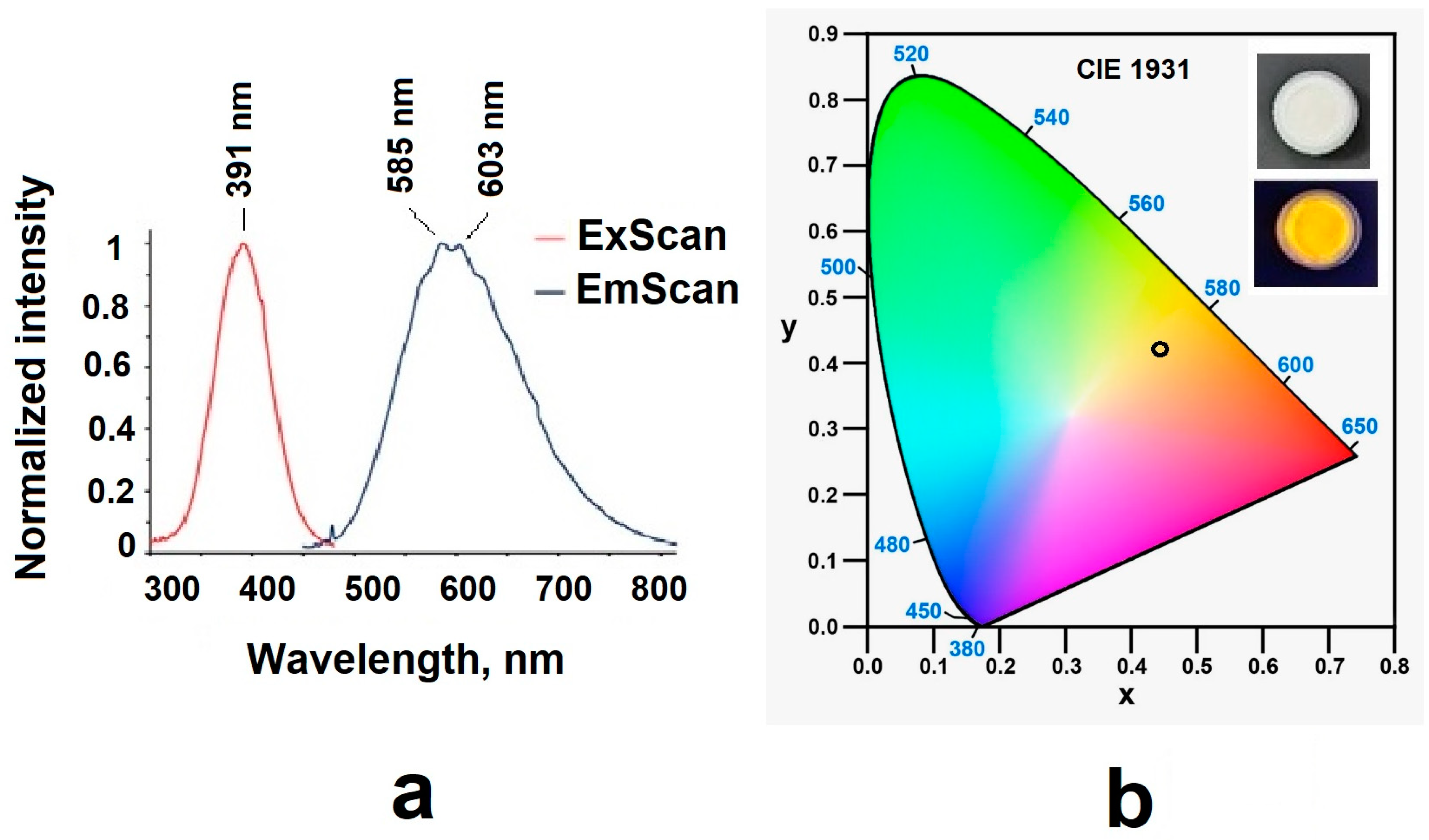
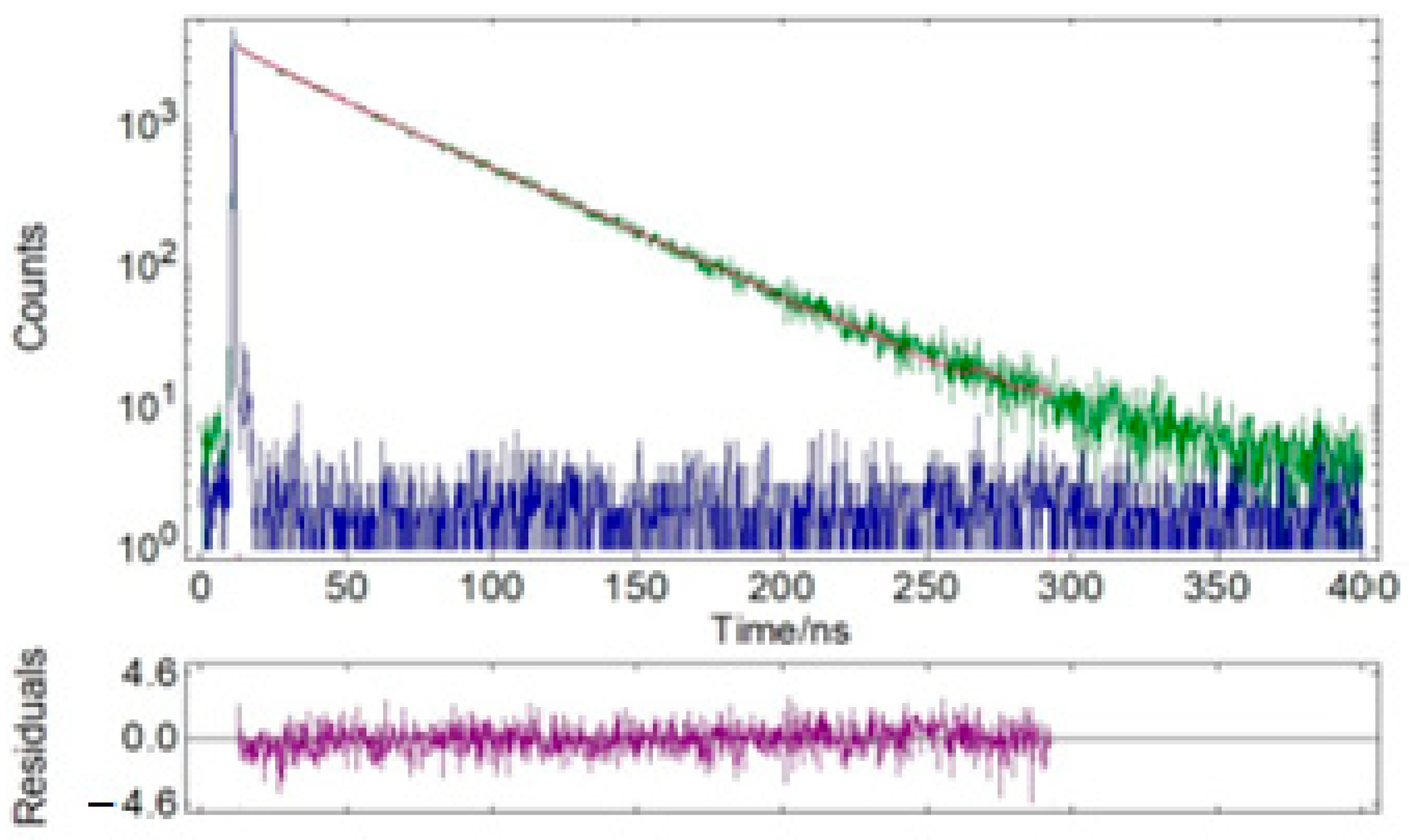
3.2. Characteristics of the Materials’ Luminescences

The emission and excitation spectra of sample 3 were recorded; the data are presented in Table 3 and Figure 11. The sample had orange emission in the range of 500–750 nm with a quantum yield of 0.17. Experimental results showed that the sample’s decomposition process in the solid state consisted of two components with a predominant contribution of the long-lived component (~46 ns) (Table 3 and Table 4; Figure 12 and Figure 13).

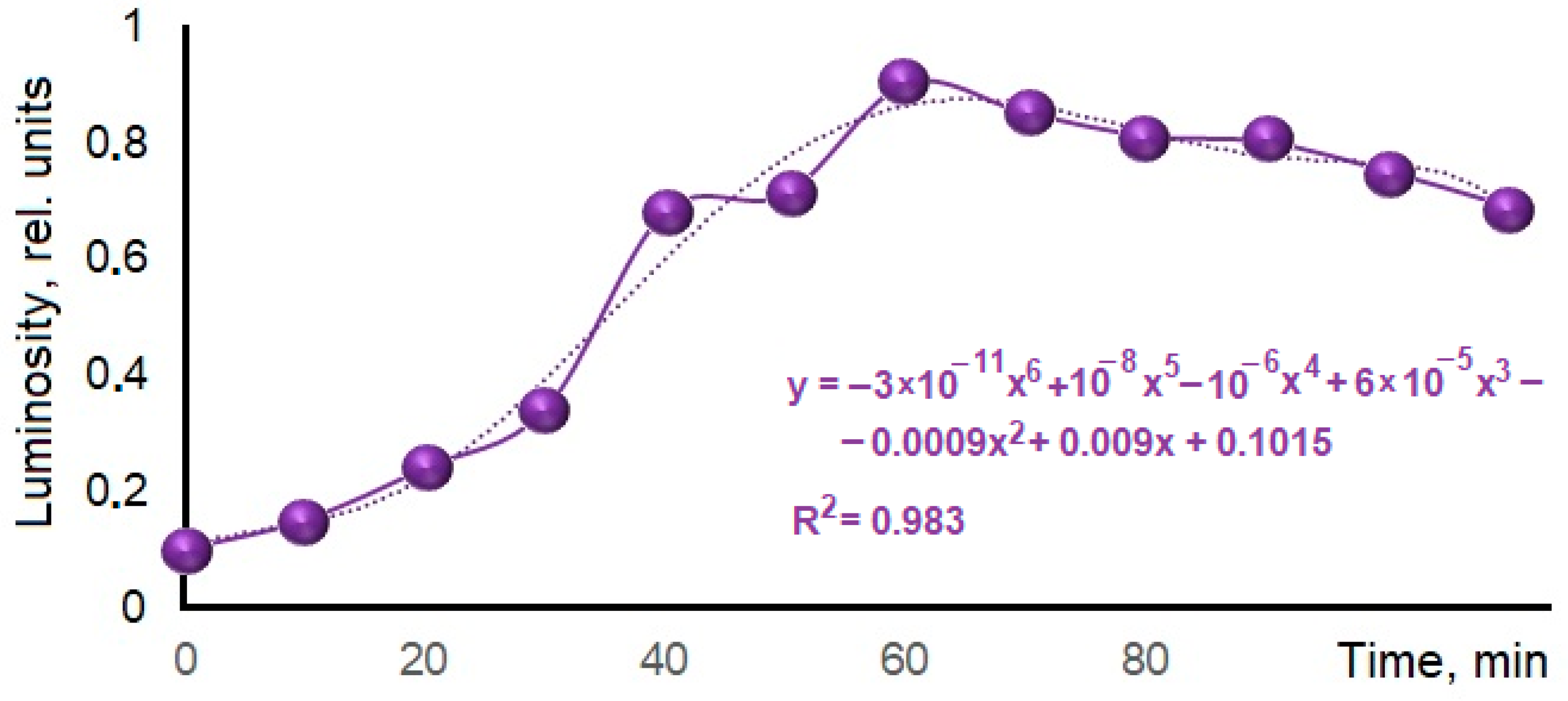
3.3. Selection of the Optimal Heat Treatment Duration for the Luminescent Material Synthesis

To determine the optimal heat treatment time, samples of phosphogypsum weighing 17.2 g and a reducing agent were subjected to heat treatment according to the following regimen: samples were heated at a rate of 13 K/min to a temperature of 1073 K; upon reaching the pre-assigned temperature, some individual samples were moved into a cooling chamber made of thermal insulating material every 10 min, where they slowly cooled to room temperature. After this, the samples were reweighed, crushed in a mortar, and the relative luminous flux emitted by the fixed area of sample surfaces was measured. The results are shown in Figure 14.
The obtained data indicate that at the given temperature, the holding time of 60 min was optimal for obtaining luminescent material. It can be assumed that a shorter holding time is not enough for the reduction process to occur, which is also evidenced by the insufficient mass loss compared to the calculated one and the traces of unreacted coal observed in the calcined samples. A longer calcination time leads to a reverse oxidation reaction. This is evidenced in the samples’ mass increases after calcination at heat treatment temperatures of 1173 K and 1273 K (Table 5). The presence of the first maximum in the calcined sample’s mass increase at the heat treatment temperature of 1173 K may be associated with a denser calcium sulfate modification formation under experimental conditions.

3.4. Selection of the Optimal Heat Treatment Temperature for the Luminescent Material Synthesis

To determine the optimal heat treatment temperature, phosphogypsum weighing 17.2 g and the reducing agent were subjected to heat treatment according to the following modes: The samples were heated at a rate of 13 K/min to the calcination temperature, which was 1073 K, 1173 K, or 1273 K for the three samples, respectively. A holding period of 60 min followed when the calcination temperature was reached. At the end of the heat treatment, the samples were cooled together with the furnace to room temperature. The results of the samples’ relative luminous flux measurements are presented in Table 6.
According to the obtained results, the best luminescent quality was exhibited by phosphogypsum samples which were heat-treated in the presence of citric acid at 1073 K; the maximum value of the relative luminous flux was achieved at the reducing agent mole fraction of 37%. Under experimental conditions, at any heat treatment temperature, a decrease in the sample’s luminescence was observed with the introduction of a significant amount of the reducing agent. This effect may be associated with the formation of carbon-containing compounds that were not detectable by X-ray diffraction but which affect the samples’ luminosity. For the heat treatment temperature of 1173 K, the maximum luminosity of the samples was achieved at a reducing agent mole fraction of 49%. For the temperature of 1273 K, maximum luminosity was achieved at a mole fraction of 74%. This may be due to the formation of a more stable modification of CaSO4 at higher heat treatment temperatures, the reduction of which requires more organic matter.

3.5. Study of the Simultaneous Influence of Temperature and Calcination Time on the Luminescent Quality of the Synthesized Material

To study the simultaneous influence of temperature and calcination time on the luminescent quality of the material synthesized, samples’ heat treatments were carried out with a simultaneous increase in temperature and a decrease in the calcination time, as well as with a simultaneous decrease in temperature and an increase in the calcination time in comparison to the optimal values adopted for the samples. The calcination temperatures and times are given in Table 7.
The obtained data indicate that an increase in temperature with a simultaneous decrease in heat treatment time, as well as a decrease in temperature with a simultaneous increase in heat treatment time, led to a decrease in the luminescent properties of the synthesized material compared to the optimal values, which are a temperature of 1073 K and a duration of 60 min.

4. Conclusions

The process of CaSO4/CaS composite material formation during the reduction of phosphogypsum with citric acid has been studied. The composite structure formation mechanism is proposed.
It is shown that during the process, the oxygen proportion decreases and the calcium and sulfur proportions in the sample increase, which indicate the partial reduction of calcium sulfate to sulfide.
The reduced sample was found to emit orange radiation in the range of 500–750 nm with a quantum yield of 0.17. Experimental results showed that the sample decomposition process in the solid state consisted of two components with a predominant contribution from the long-lived component (~46 ns).
The optimal conditions for producing luminescent materials by reducing phosphogypsum with citric acid were determined to be heat treatment at a temperature of 1073 K, a holding time of 60 min, and a reducing agent mole fraction of 37%.
It was found that an increase in temperature with a simultaneous decrease in heat treatment time, as well as a decrease in temperature with a simultaneous increase in heat treatment time, led to a decrease in the luminescent properties of the synthesized material compared to optimal values.
The results can be used to develop technology for recycling large-tonnage waste from the chemical industry into luminescent materials.

Author Contributions

Conceptualization, O.A.M., M.A.E. and N.P.S.; Methodology, O.A.M., M.A.E. and N.P.S.; Validation, M.A.E. and S.I.S.; Formal analysis, N.P.S., A.R. and E.V.S.; Data curation, S.I.S. and Z.D.K.; Writing—original draft, O.A.M., Z.D.K. and D.I.M.; Writing—review & editing, E.V.S.; Visualization, A.R. and D.I.M.; Funding acquisition, M.A.E. All authors have read and agreed to the published version of the manuscript.

Funding

This research was funded by Ministry of Science and Higher Education of the Russian Federation within the framework of the state assignment of FENN-2024-0006—“Development of inorganic ultraviolet dyes technology” project.

Data Availability Statement

Data are contained within the article.

Acknowledgments

The authors express their gratitude to the staff of the Spectroscopy and Analysis of Organic Compounds Collective Use Center of the Organic Synthesis Institute named after Postovsky, the Ural Branch of the Russian Academy of Sciences, for assistance in luminescence spectra recording for UV-vis and IR samples. The authors express their gratitude to the staff of the Collective Use Center of the Platov South-Russian State Polytechnic University (NPI), and personally to A.N. Yatsenko for assistance in filming and deciphering XRF data.

Conflicts of Interest

The authors declare no conflict of interest.

References

  1. Millán-Becerro, R.; Pérez-López, R.; Cánovas, C.R.; Francisco Macías, F.; León, R. Phosphogypsum weathering and implications for pollutant discharge into an estuary. J. Hydrol. 2023, 617, 128943. [Google Scholar] [CrossRef]
  2. Cao, W.; Yi, W.; Peng, J.; Yin, S. Relationship between the evolution of organic impurities and properties of β-hemihydrate phosphogypsum. Constr. Build. Mater. 2023, 409, 134125. [Google Scholar] [CrossRef]
  3. Bounaga, A.; Danouche, M.; Zeroual, Y.; Boulif, R.; Benhida, R.; Lyamlouli, K. Anaerobic bioremediation of acid phosphogypsum stacks leachates: Assessment of leachate’s biochemical changes and microbial community dynamics. Environ. Technol. Innov. 2024, 33, 103535. [Google Scholar] [CrossRef]
  4. Wu, F.; Chen, B.; Qu, G.; Liu, S.; Zhao, C.; Ren, Y.; Liu, X. Harmless treatment technology of phosphogypsum: Directional stabilization of toxic and harmful substances. J. Environ. Manag. 2022, 311, 114827. [Google Scholar] [CrossRef]
  5. Ennaciri, Y.; Bettach, M. The chemical behavior of the different impurities present in Phosphogypsum: A review. Phosphorus Sulfur Silicon Relat. Elem. 2023, 199, 129–148. [Google Scholar] [CrossRef]
  6. Wang, M.; Yuan, X.; Dong, W.; Fu, Q.; Ao, X.; Chen, Q. Gradient removal of Si and P impurities from phosphogypsum and preparation of anhydrous calcium sulfate. J. Environ. Chem. Eng. 2023, 11, 110312. [Google Scholar] [CrossRef]
  7. Duart, V.M.; Garbuio, F.J.; Caires, E.F. Does direct-seeded rice performance improve upon lime and phosphogypsum use? Soil Tillage Res. 2021, 212, 105055. [Google Scholar] [CrossRef]
  8. Wu, F.; Ren, Y.; Qu, G.; Liu, S.; Chen, B.; Liu, X.; Zhao, C.; Li, J. Utilization path of bulk industrial solid waste: A review on the multi-directional resource utilization path of phosphogypsum. J. Environ. Manag. 2022, 313, 114957. [Google Scholar] [CrossRef] [PubMed]
  9. Murali, G.; Azab, M. Recent research in utilization of phosphogypsum as building materials: Review. J. Mater. Res. Technol. 2023, 25, 960–987. [Google Scholar] [CrossRef]
  10. Shi, Y.; Song, Y.; Min, C.; Du, J.; Wang, X.; Min, J.; Wang, S. Phosphogypsum pretreatment with CaCO3 to improve the backfill performance. Case Stud. Constr. Mater. 2023, 18, e01954. [Google Scholar] [CrossRef]
  11. Chernysh, Y. 10-Recycling of radioactive phosphogypsum wastes. In Woodhead Publishing Series in Civil and Structural Engineering. Advances in the Toxicity of Construction and Building Materials; Pacheco-Torgal, F., Falkinham, J.O., Gałaj, J.A., Eds.; Woodhead Publishing: Sawston, UK, 2022; pp. 225–240. [Google Scholar] [CrossRef]
  12. Meskini, S.; Samdi, A.; Ejjaouani, H.; Remmal, T. Valorization of phosphogypsum as a road material: Stabilizing effect of fly ash and lime additives on strength and durability. J. Clean. Prod. 2021, 323, 129161. [Google Scholar] [CrossRef]
  13. Men, J.; Li, Y.; Cheng, P.; Zhang, Z. Recycling phosphogypsum in road construction materials and associated environmental considerations: A review. Heliyon 2022, 8, e11518. [Google Scholar] [CrossRef] [PubMed]
  14. Ma, J.; Xu, J.; Liu, C.; Yi, Q.; Zheng, M.; Cheng, L.; Song, T. Chemical looping combustion of sulfur paste to SO2 by phosphogypsum oxygen carrier for sulfur acid production. Fuel 2022, 323, 124386. [Google Scholar] [CrossRef]
  15. Pan, Q.; Ma, L.; Du, W.; Yang, J.; Ao, R.; Yin, X.; Qing, S. Hydrogen-enriched syngas production by lignite chemical looping gasification with composite oxygen carriers of phosphogypsum and steel slag. Energy 2022, 241, 122927. [Google Scholar] [CrossRef]
  16. Yang, J.; Ren, Y.; Lu, J.; Liu, H.; Zhang, Z.; Pang, H.; Bounkhong, K. Chemical looping gasification with a CuFe2O4-enhanced phosphogypsum oxygen carrier during reduction in a fluidized bed reactor. Chem. Eng. J. 2021, 426, 131346. [Google Scholar] [CrossRef]
  17. Yang, J.; Ren, Y.; Chen, S.; Lu, J. Study on the mechanism and reaction characteristics of metal-supported phosphogypsum as oxygen carrier in a chemical looping gasification application. J. Environ. Sci. 2024, 138, 428–438. [Google Scholar] [CrossRef]
  18. Hong, C.; Tang, Q.; Liu, S.; Kim, H.; Liu, D. A two-step bioleaching process enhanced the recovery of rare earth elements from phosphogypsum. Hydrometallurgy 2023, 2023, 106140. [Google Scholar] [CrossRef]
  19. Li, L.; Liao, L.; Wang, B.; Li, W.; Liu, T.; Wu, P.; Xu, Q.; Liu, S. Effective Sb(V) removal from aqueous solution using phosphogypsum-modified biochar. Environ. Pollut. 2022, 301, 119032. [Google Scholar] [CrossRef]
  20. Guo, Z.; Zhang, C.; Jiang, H.; Li, L.; Li, Z.; Zhao, L.; Chen, H. Phosphogypsum/titanium gypsum coupling for enhanced biochar immobilization of lead: Mineralization reaction behavior and electron transfer effect. J. Environ. Manag. 2023, 345, 118781. [Google Scholar] [CrossRef] [PubMed]
  21. Alla, M.; Harrou, A.; Elhafiany, M.L.; Azerkane, D.; Ouahabi, M.E.; Gharibi, E.K. Reduction of phosphogypsum to calcium sulfide (CaS) using metallic iron in a hydrochloric acid medium. Phosphorus Sulfur Silicon Relat. Elem. 2022, 197, 1026–1035. [Google Scholar] [CrossRef]
  22. Danouche, M.; Bounaga, A.; Boulif, R.; Zeroual, Y.; Benhida, R.; Lyamlouli, K. Optimization of sulfate leaching from Phosphogypsum for efficient bioreduction in a batch bioreactor using a sulfate-reducing microbial consortium. Chem. Eng. J. 2023, 475, 146072. [Google Scholar] [CrossRef]
  23. Laasri, F.; Garcia, A.C.; Latifi, M.; Chaouki, J. Reaction mechanism of thermal decomposition of Phosphogypsum. Waste Manag. 2023, 171, 482–490. [Google Scholar] [CrossRef] [PubMed]
  24. Bounaga, A.; Alsanea, A.; Lyamlouli, K.; Zhou, C.; Zeroual, Y.; Boulif, R.; Rittmann, B.E. Microbial transformations by sulfur bacteria can recover value from phosphogypsum: A global problem and a possible solution. Biotechnol. Adv. 2022, 57, 107949. [Google Scholar] [CrossRef] [PubMed]
  25. He, H.; Hao, L.; Fan, C.; Li, S.; Lin, W. A two-step approach to phosphogypsum decomposition: Oxidation of CaS with CO2. Thermochim. Acta 2022, 708, 179122. [Google Scholar] [CrossRef]
  26. Zhang, H.; Hu, H.; Di, Y.; Yao, Z.; Yang, F.; Cai, H.; Sun, H.; Liu, Q. Luminescence and stability of CaS: Eu2+, Sm3+ down/up conversion phosphor and film. Mater. Today Commun. 2023, 34, 105457. [Google Scholar] [CrossRef]
  27. Song, Z.; Yan, L.; Peng, M.; Zhou, W.; Zhang, J.; Yu, L.; Qiu, Z.; Lian, S. Inorganic/organic bilayer--modified CaS:Eu2+,Ce3+ phosphor and luminescent film laminated glass for efficient solar energy utilization. Ceram. Int. 2023, 49, 34837–34844. [Google Scholar] [CrossRef]
  28. Yu, F.; Yu, H. Modification of CaS:Eu phosphor under mild conditions toward an extreme stability at harsh environment over 1.5 months. Mater. Lett. X 2023, 18, 100192. [Google Scholar] [CrossRef]
  29. Medennikov, O.A.; Shabelskaya, N.P. Technology for processing phosphogypsum into a fluorescent dye based on calcium sulfide. Tonkie Khimicheskie Tekhnologii 2022, 17, 357–368. [Google Scholar] [CrossRef]
  30. Shabelskaya, N.P.; Medennikov, O.A.; Khliyan, Z.D.; Ulyanova, V.A. Specific features in the processing of phosphogypsum to obtain an inorganic dye. Obogashchenie Rud. 2023, 2, 24–29. [Google Scholar] [CrossRef]
  31. Morozov, A.; Shapovalov, V.; Popov, Y.; Kochur, A.; Yavna, V. Effect of mechanical impact on the microstructure and IR spectra of cohesive soil. Vib. Spectrosc. 2023, 128, 103582. [Google Scholar] [CrossRef]
Figure 1. Setup scheme for measuring the light flux from the surface of a UV-irradiated sample.
Nanomaterials 14 00904 g001
Figure 2. X-ray diffraction patterns of samples: (a) phosphogypsum dried at a temperature of 283 K; (b) phosphogypsum calcined at a temperature of 1073 K; (c) phosphogypsum calcined in the presence of a reducing agent at a temperature of 1073 K.
Nanomaterials 14 00904 g002
Figure 3. Microphotographs of samples: (a) phosphogypsum; (b) phosphogypsum calcined at a temperature of 1073 K; (c) phosphogypsum calcined in the presence of a reducing agent at a temperature of 1073 K.
Nanomaterials 14 00904 g003
Figure 4. Schematic representation of the heat treatment process of phosphogypsum without a reducing agent.
Nanomaterials 14 00904 g004
Figure 5. Schematic representation of the CaSO4/CaS composite material formation process.
Nanomaterials 14 00904 g005
Figure 6. Phosphogypsum elemental analysis.
Nanomaterials 14 00904 g006
Figure 7. Elemental analysis of the obtained material.
Nanomaterials 14 00904 g007
Figure 8. IR spectrum of the reduced phosphogypsum sample.
Nanomaterials 14 00904 g008
Figure 9. Samples’ thermogravimetric analysis data for (a) phosphogypsum and (b) phosphogypsum mixed with a reducing agent.
Nanomaterials 14 00904 g009
Figure 10. Thermogram analysis results.
Nanomaterials 14 00904 g010
Figure 11. (a) Excitation and emission spectra and (b) color diagram for reduced samples.
Nanomaterials 14 00904 g011
Figure 12. Time-resolved fluorescence lifetime decay profiles of solid powder sample 3 (green) and instrumental response function (IRF, blue). λex = 375 nm, λem = 585 nm.
Nanomaterials 14 00904 g012
Figure 13. Time-resolved fluorescence lifetime decay profiles of solid powder sample 3 (green) and instrumental response function (IRF, blue). λex = 375 nm, λem = 603 nm.
Nanomaterials 14 00904 g013
Figure 14. Dependence of samples’ relative luminosities on the treatment duration.
Nanomaterials 14 00904 g014
Table 1. Crystallographic parameters of samples.
SamplePhaseGrid Parameters, nm βVolume, nm3D, nm
abcV
Sample 1CaSO4·2H2O 5.6715.116.49118.5489535
CaSO4·0.5H2O 12.026.9312.6790.21055285
Sample 2CaSO46.236.986.9730347
Sample 3CaSO46.997.006.2430549
CaS5.6918443
Table 2. Thermogravimetric analysis results.
SampleCharacterPhosphogypsumSample 3
Temperature, KValue, µW/mgTemperature, KValue, µW/mg
Peak 1Endo4240.844210.74
Peak 2Endo4400.694290.67
Peak 3Exo--7731.59
Table 3. Photoluminescent data for compound sample in powder at r.t.
Excitation, λem [nm]Emission, λem [nm]τavr, [ns]/χ2ΦF
39158546.02/1.1100.17
60345.81/1.034
Table 4. Detailed data of the fluorescence lifetime measurements of sample 3: τ—lifetime, f—fractional contribution, τavg—average lifetime, χ2—chi-squared distribution.
Solid
λem [nm]τ1, [ns]f1, %τ2 [ns]f2, %τavg, [ns]χ2
58516.225.347.7094.746.021.034
60315.065.247.4994.845.811.110
Table 5. Changes in the samples’ masses depending on the introduced reducing agent amount and heat treatment temperature.
Reducing Agent Fraction, %Heat Treatment Temperature, К
107311731273
6.30.000.070.07
12.60.000.640.07
18.90.141.000.14
25.20.290.070.29
37.80.500.210.50
50.40.860.430.93
63.01.210.713.14
75.61.290.142.29
Table 6. Results of phosphogypsum heat treatment with different reducing agent contents at different temperatures.
Reducing Agent Mass, gReducing Agent Mole Fraction, %Relative Luminous Flux (Rel. Units)
at Treatment Temperature
1073 К1173 К1273 К
1.106.400.240.100.10
2.1012.210.390.120.10
4.3025.000.860.350.15
6.4037.210.930.750.15
8.5049.420.910.900.15
10.7062.210.900.880.10
12.8074.420.840.800.26
17.1099.420.630.600.21
Table 7. Conditions for phosphogypsum samples’ heat treatments with citric acid.
Reducing Agent Mass, g Reducing Agent Mole Fraction, %Relative Luminous Flux (Rel. Units)
973 К,
90 min
1073 К,
60 min
1173 К,
30 min
4.3050.590.380.860.10
6.4075.290.410.930.15
8.50100.000.510.910.72
10.70125.880.460.900.90
12.80150.590.440.840.87
17.10201.180.410.630.74
Disclaimer/Publisher’s Note: The statements, opinions and data contained in all publications are solely those of the individual author(s) and contributor(s) and not of MDPI and/or the editor(s). MDPI and/or the editor(s) disclaim responsibility for any injury to people or property resulting from any ideas, methods, instructions or products referred to in the content.

