
Advances in Phytonanotechnology: A Plant-Mediated Green Synthesis of Metal Nanoparticles Using Phyllanthus Plant Extracts and Their Antimicrobial and Anticancer Applications
 , ,
, ,  and
and
Abstract
:1. Introduction
2. Rationale
3. Review Methods
4. Common Techniques and Characterization of NPs for Surface Chemistry Data Collection
5. Factors That Influence the Synthesis of Nanoparticles from Plants
6. The Role of Plants in the Synthesis of Nanoparticles
6.1. Green Synthesis Methods of MNPs Using Phyllanthus Plant Extracts
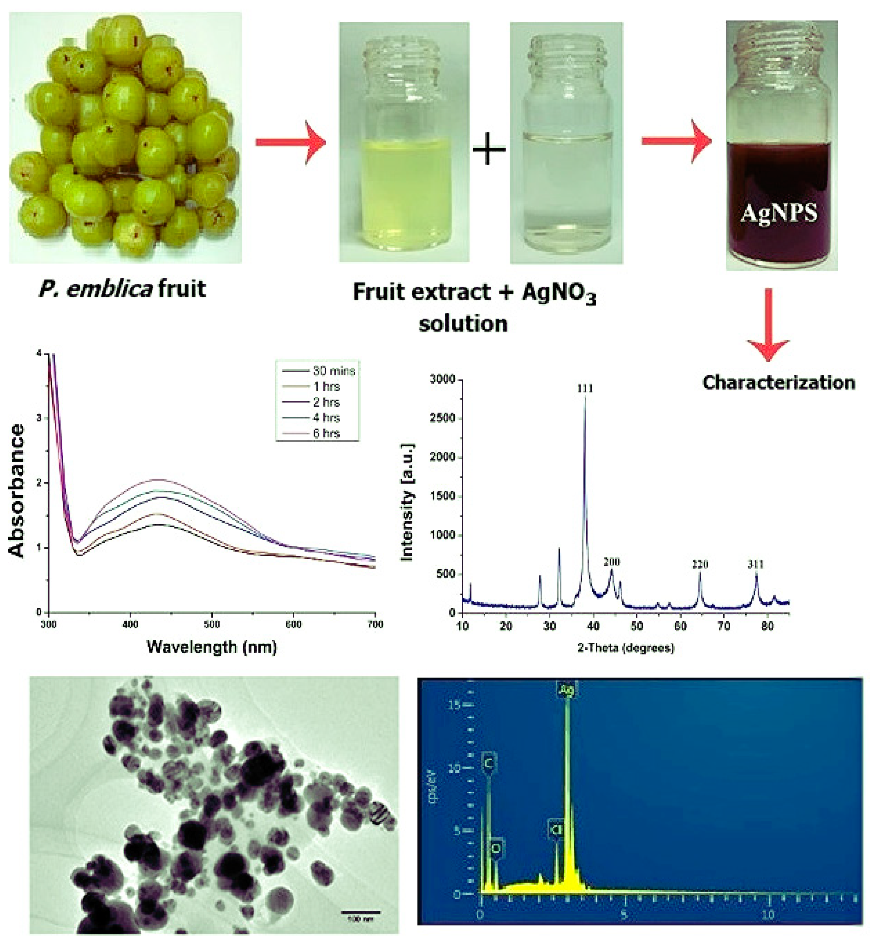
6.1.1. Synthesis of AgNPs Using P. emblica Fruit Extracts by [74]
6.1.2. Synthesis of AgNPs Using P. emblica Methanol Fruit Extracts by [76]
6.1.3. Synthesis of FeNPs Using P. niruri Plant Leaf Extracts by [72]
7. Nanoparticles Stability Test
8. Phytochemical Constituents with Metal Ion Reduction Capacity
8.1. Sugars
8.2. Alkaloids
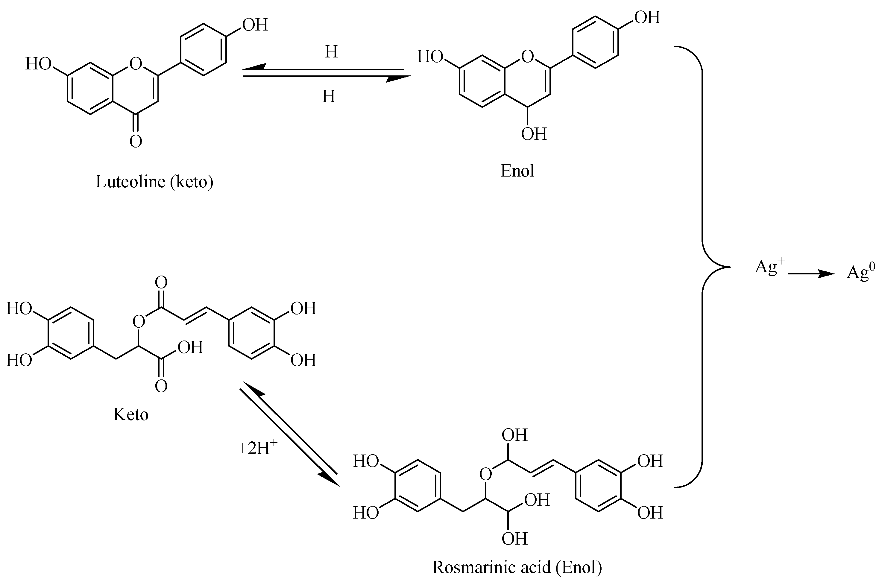
8.3. Flavonoids
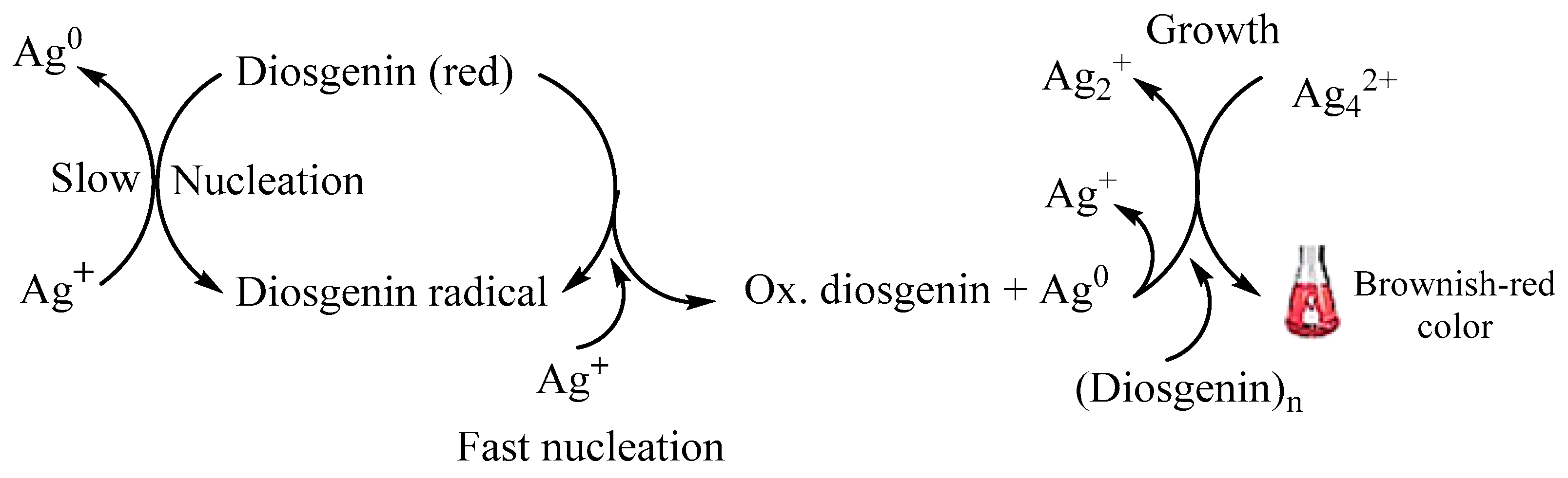
8.4. Terpenoids
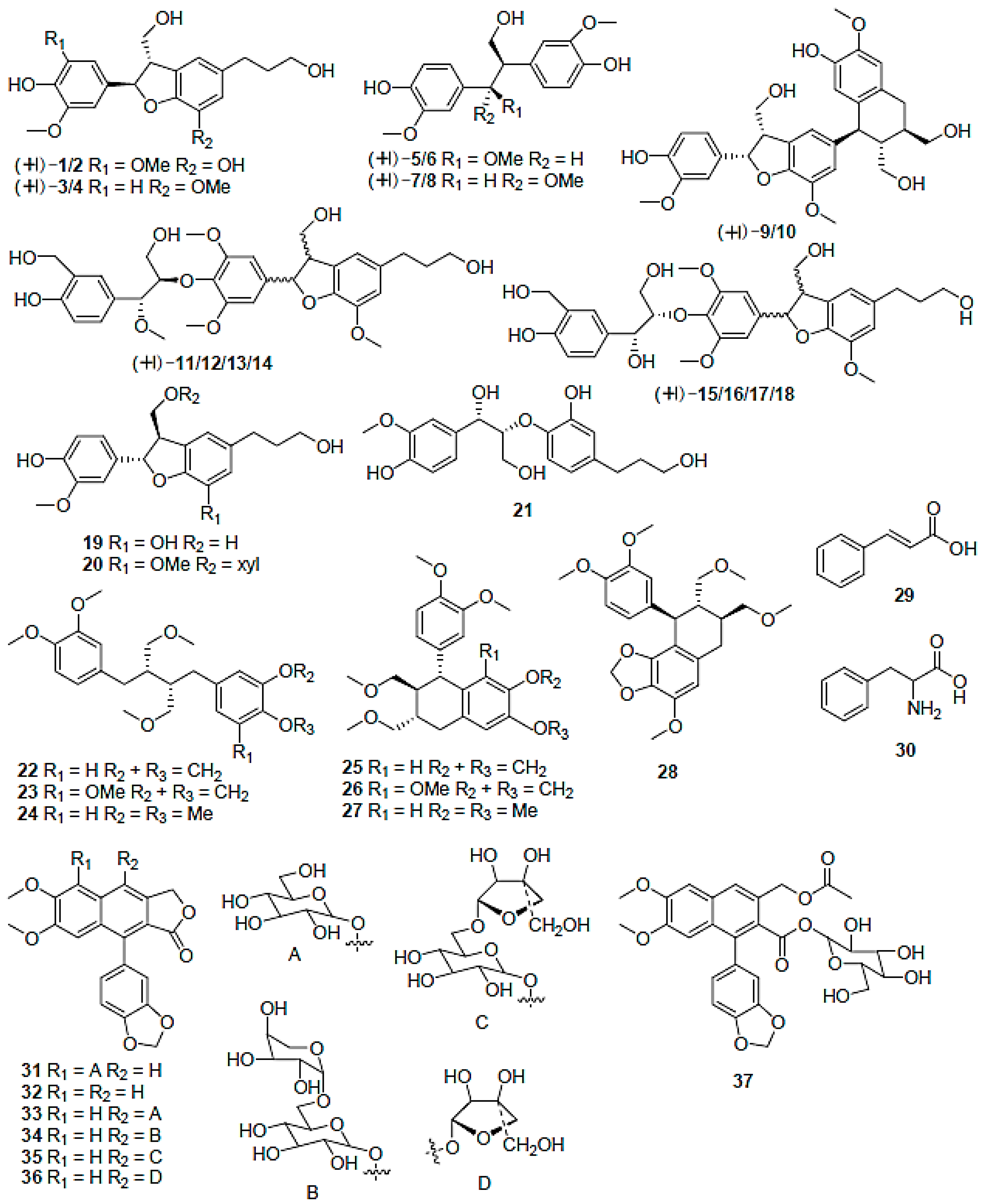
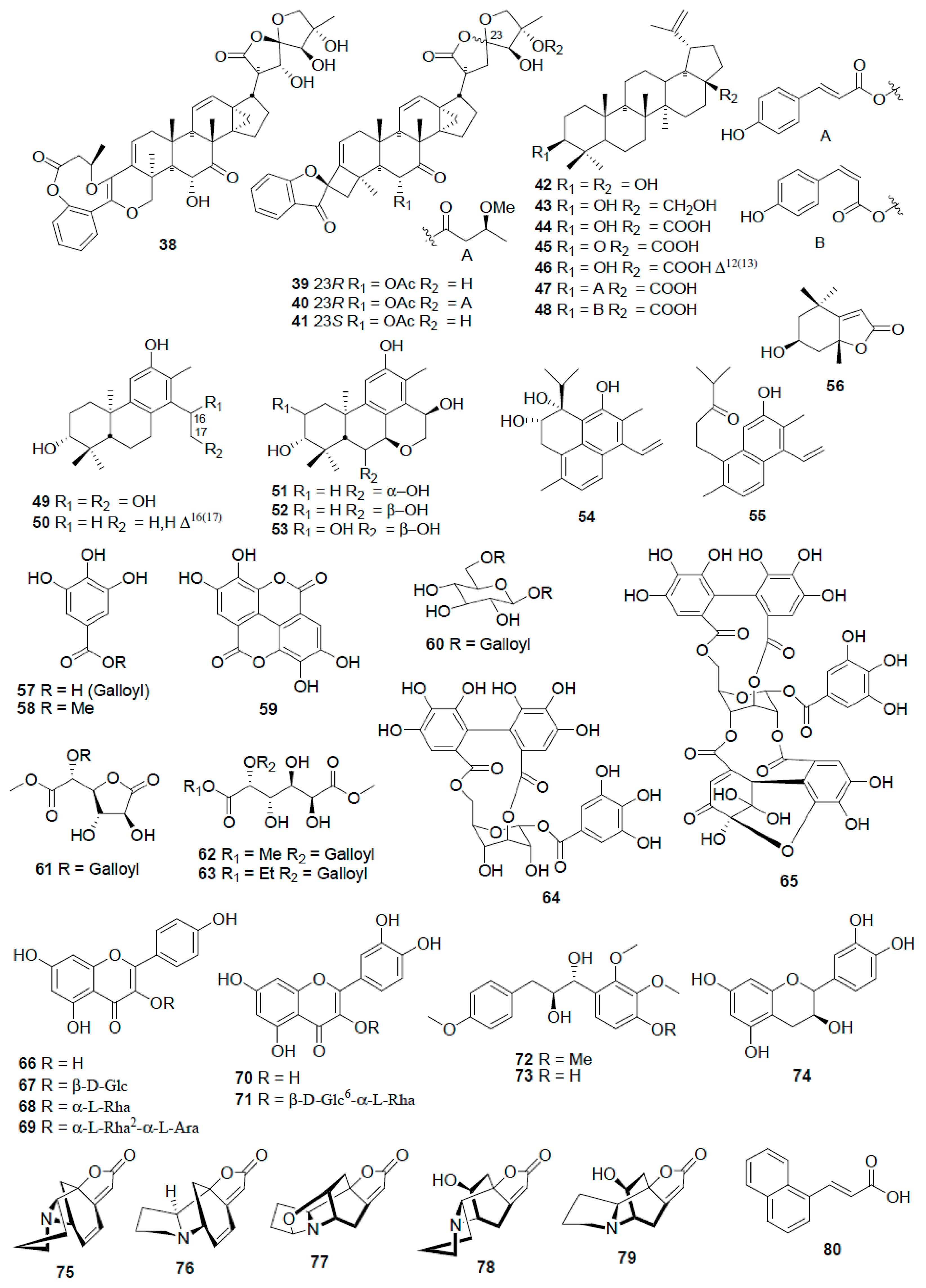
9. The Genus Phyllanthus and Its Phytochemical Constituents
10. Plant-Mediated Synthesis of Nanoparticles Using the Genus Phyllanthus
11. Nanoparticle Uptake and Interaction with Cell Mechanisms
12. Application of Plant-Mediated Synthesized Nanoparticles
Phytonanoparticles for Antimicrobial Activity
13. Phytonanoparticles for Anticancer and Antiviral Activity
14. Other Innovative Applications of Phytonanotechnology
15. Future Innovative Prospects of Phytonanotechnology
16. Conclusions
Author Contributions
Funding
Data Availability Statement
Acknowledgments
Conflicts of Interest
References
- Albanese, A.; Tang, P.S.; Chan, W.C. The effect of nanoparticle size, shape, and surface chemistry on biological systems. Annu. Rev. Biomed. Eng. 2012, 14, 1–16. [Google Scholar] [CrossRef]
- Fakruddin, M.; Hossain, Z.; Afroz, H. Prospects and applications of nanobiotechnology: A medical perspective. J. Nanobiotechnol. 2012, 10, 31. [Google Scholar] [CrossRef]
- Chahardoli, A.; Karimi, N.; Fattahi, A. Biosynthesis, Characterization, Antimicrobial and Cytotoxic Effects of Silver Nanoparticles Using Nigella arvensis Seed Extract. Iran. J. Pharm. Res. 2017, 16, 1167–1175. [Google Scholar] [PubMed]
- Barabadi, H.; Tajani, B.; Moradi, M.; Damavandi Kamali, K.; Meena, R.; Honary, S.; Mahjoub, M.A.; Saravanan, M. Penicillium Family as Emerging Nanofactory for Biosynthesis of Green Nanomaterials: A Journey into the World of Microorganisms. J. Clust. Sci. 2019, 30, 843–856. [Google Scholar] [CrossRef]
- Boomi, D.P.; Poorani, G.; Subramanian, P.; Selvam, D.S.; Ramanathan, G.; Sundaram, R.; Barabadi, H.; Halliah, G.P.; Jeyaraman, J.; Muthupandian, S. Evaluation of Antibacterial and Anticancer Potential of Polyaniline-Bimetal Nanocomposites Synthesized from Chemical Reduction Method. J. Clust. Sci. 2019, 30, 715–726. [Google Scholar] [CrossRef]
- Barabadi, H. Nanobiotechnology: A promising scope of gold biotechnology. Cell. Mol. Biol. 2017, 63, 3–4. [Google Scholar] [CrossRef]
- Salari, S.; Esmaeilzadeh Bahabadi, S.; Samzadeh-Kermani, A.; Yosefzaei, F. In-vitro Evaluation of Antioxidant and Antibacterial Potential of GreenSynthesized Silver Nanoparticles Using Prosopis farcta Fruit Extract. Iran. J. Pharm. Res. 2019, 18, 430–455. [Google Scholar]
- Sarebanhassanabadi, M. Designing a Hydrogen Peroxide Biosensor Using Catalase and Modified Electrode with Magnesium Oxide Nanoparticles. Int. J. Electrochem. Sci. 2014, 9, 257–271. [Google Scholar]
- Ramezani, T.; Nabiuni, M.; Baharara, J.; Parivar, K.; Namvar, F. Sensitization of Resistance Ovarian Cancer Cells to Cisplatin by Biogenic Synthesized Silver Nanoparticles through p53 Activation. Iran. J. Pharm. Res. 2019, 18, 222–231. [Google Scholar]
- Rajan, P.I.; Vijaya, J.J.; Jesudoss, S.K.; Kaviyarasu, K.; Kennedy, L.J.; Jothiramalingam, R.; Al-Lohedan, H.A.; Vaali-Mohammed, M.-A. Green-fuel-mediated synthesis of self-assembled NiO nano-sticks for dual applications—Photocatalytic activity on Rose Bengal dye and antimicrobial action on bacterial strains. Mater. Res. Express 2017, 4, 085030. [Google Scholar] [CrossRef]
- Arasu, M.V.; Arokiyaraj, S.; Viayaraghavan, P.; Kumar, T.S.J.; Duraipandiyan, V.; Al-Dhabi, N.A.; Kaviyarasu, K. One step green synthesis of larvicidal, and azo dye degrading antibacterial nanoparticles by response surface methodology. J. Photochem. Photobiol. B 2019, 190, 154–162. [Google Scholar] [CrossRef]
- Barabadi, H.; Ovais, M.; Shinwari, Z.K.; Saravanan, M. Anti-cancer green bionanomaterials: Present status and future prospects. Green Chem. Lett. Rev. 2017, 10, 285–314. [Google Scholar] [CrossRef]
- Ovais, M.; Khalil, A.T.; Raza, A.; Khan, M.A.; Ahmad, I.; Islam, N.U.; Saravanan, M.; Ubaid, M.F.; Ali, M.; Shinwari, Z.K. Green synthesis of silver nanoparticles via plant extracts: Beginning a new era in cancer theranostics. Nanomedicine 2016, 11, 3157–3177. [Google Scholar] [CrossRef]
- Xu, L.; Wang, Y.Y.; Huang, J.; Chen, C.Y.; Wang, Z.X.; Xie, H. Silver nanoparticles: Synthesis, medical applications and biosafety. Theranostics 2020, 10, 8996–9031. [Google Scholar] [CrossRef] [PubMed]
- Gupta, M.; Seema, K. Living Nano-factories: An Eco-friendly Approach Towards Medicine and Environment. In Bio-Manufactured Nanomaterials: Perspectives and Promotion; Springer International Publishing: Chem, Switzerland, 2021; pp. 95–124. [Google Scholar]
- Tangthong, T.; Piroonpan, T.; Thipe, V.C.; Khoobchandani, M.; Katti, K.; Katti, K.V.; Pasanphan, W. Water-Soluble Chitosan Conjugated DOTA-Bombesin Peptide Capped Gold Nanoparticles as a Targeted Therapeutic Agent for Prostate Cancer. Nanotechnol. Sci. Appl. 2021, 14, 69–89. [Google Scholar] [CrossRef]
- Khoobchandani, M.; Khan, A.; Katti, K.K.; Thipe, V.C.; Al-Yasiri, A.Y.; MohanDoss, D.K.D.; Nicholl, M.B.; Lugão, A.B.; Hans, C.P.; Katti, K.V. Green nanotechnology of MGF-AuNPs for immunomodulatory intervention in prostate cancer therapy. Sci. Rep. 2021, 11, 16797. [Google Scholar] [CrossRef] [PubMed]
- Sibuyi, N.R.S.; Thipe, V.C.; Panjtan-Amiri, K.; Meyer, M.; Katti, K.V. Green synthesis of gold nanoparticles using Acai berry and Elderberry extracts and investigation of their effect on prostate and pancreatic cancer cells. Nanobiomedicine 2021, 8, 1849543521995310. [Google Scholar] [CrossRef] [PubMed]
- Eckelman, M.J.; Zimmerman, J.B.; Anastas, P.T. Toward Green Nano. J. Ind. Ecol. 2008, 12, 316–328. [Google Scholar] [CrossRef]
- Maham, M.; Karami-Osboo, R. Extraction of Sulfathiazole from Urine Samples Using Biosynthesized Magnetic Nanoparticles. Iran. J. Pharm. Res. 2017, 16, 462–470. [Google Scholar]
- Kasithevar, M.; Muthupandian, S.; Periakaruppan, P.; Kumar, H.; Ovais, M.; Barabadi, H.; Shinwari, Z. Green synthesis of silver nanoparticles using Alysicarpus monilifer leaf extract and its antibacterial activity against MRSA and CoNS isolates in HIV patients. J. Interdiscip. Nanomed. 2017, 2, 131–141. [Google Scholar] [CrossRef]
- Rezvani Amin, Z.; Khashyarmanesh, Z.; Fazly Bazzaz, B.S.; Sabeti Noghabi, Z. Does Biosynthetic Silver Nanoparticles Are More Stable with Lower Toxicity than Their Synthetic Counterparts? Iran. J. Pharm. Res. 2019, 18, 210–221. [Google Scholar] [PubMed]
- Saravanan, M.; Barik, S.K.; MubarakAli, D.; Prakash, P.; Pugazhendhi, A. Synthesis of silver nanoparticles from Bacillus brevis (NCIM 2533) and their antibacterial activity against pathogenic bacteria. Microb. Pathog. 2018, 116, 221–226. [Google Scholar] [CrossRef] [PubMed]
- Manimaran, M.; Kannabiran, K. Actinomycetes-mediated biogenic synthesis of metal and metal oxide nanoparticles: Progress and challenges. Lett. Appl. Microbiol. 2017, 64, 401–408. [Google Scholar] [CrossRef]
- Niknejad, F.; Nabili, M.; Daie Ghazvini, R.; Moazeni, M. Green synthesis of silver nanoparticles: Advantages of the yeast Saccharomyces cerevisiae model. Curr. Med. Mycol. 2015, 1, 17–24. [Google Scholar] [CrossRef]
- Barabadi, H.; Honary, S. Biofabrication of gold and silver nanoparticles for pharmaceutical applications. Pharm. Biomed. Res. 2016, 2, 1. [Google Scholar] [CrossRef]
- Dahoumane, S.A.; Mechouet, M.; Wijesekera, K.; Filipe, C.D.M.; Sicard, C.; Bazylinski, D.A.; Jeffryes, C. Algae-mediated biosynthesis of inorganic nanomaterials as a promising route in nanobiotechnology—A review. Green Chem. 2017, 19, 552–587. [Google Scholar] [CrossRef]
- Arya, A.; Gupta, K.; Chundawat, T.S.; Vaya, D. Biogenic Synthesis of Copper and Silver Nanoparticles Using Green Alga Botryococcus braunii and Its Antimicrobial Activity. Bioinorg. Chem. Appl. 2018, 2018, 7879403. [Google Scholar] [CrossRef]
- Kumar, H.; Bhardwaj, K.; Kuča, K.; Kalia, A.; Nepovimova, E.; Verma, R.; Kumar, D. Flower-Based Green Synthesis of Metallic Nanoparticles: Applications beyond Fragrance. Nanomaterials 2020, 10, 766. [Google Scholar] [CrossRef] [PubMed]
- Soni, V.; Raizada, P.; Singh, P.; Cuong, H.N.; S, R.; Saini, A.; Saini, R.V.; Le, Q.V.; Nadda, A.K.; Le, T.T.; et al. Sustainable and green trends in using plant extracts for the synthesis of biogenic metal nanoparticles toward environmental and pharmaceutical advances: A review. Environ. Res. 2021, 202, 111622. [Google Scholar] [CrossRef] [PubMed]
- El-Kassas, H.Y.; Ghobrial, M.G. Biosynthesis of metal nanoparticles using three marine plant species: Anti-algal efficiencies against “Oscillatoria simplicissima”. Environ. Sci. Pollut. Res. Int. 2017, 24, 7837–7849. [Google Scholar] [CrossRef]
- Palomo, J.M.; Filice, M. Biosynthesis of Metal Nanoparticles: Novel Efficient Heterogeneous Nanocatalysts. Nanomaterials 2016, 6, 84. [Google Scholar] [CrossRef] [PubMed]
- Haque, T.; Muhsin, M.; Akhter, T.; Haq, M.; Begum, R.; Chowdhury, S. Antimicrobial and analgesic activity of leaf extracts of Phyllanthus reticulatus Poir. (Family-Euphorbiaceae). Jahangirnagar Univ. J. Biol. Sci. 2016, 5, 81. [Google Scholar] [CrossRef]
- Mao, X.; Wu, L.-F.; Guo, H.-L.; Chen, W.-J.; Cui, Y.-P.; Qi, Q.; Li, S.; Liang, W.-Y.; Yang, G.-H.; Shao, Y.-Y.; et al. The Genus Phyllanthus: An Ethnopharmacological, Phytochemical, and Pharmacological Review. Evid.-Based Complement. Altern. Med. 2016, 2016, 7584952. [Google Scholar] [CrossRef] [PubMed]
- Jiang, J.; Oberdörster, G.; Biswas, P.J. Characterization of Size, Surface Charge, and Agglomeration State of Nanoparticle Dispersions for Toxicological Studies. J. Nanoparticle Res. 2008, 11, 77–89. [Google Scholar] [CrossRef]
- Sepeur, S. Nanotechnology: Technical Basics and Applications; Vincentz Network GmbH & Co KG: Hannover Deutschland, Germany, 2008. [Google Scholar]
- Fedlheim, D.L.; Foss, C.A. Metal Nanoparticles: Synthesis, Characterization, and Applications; CRC Press: Boca Raton, FL, USA, 2001. [Google Scholar]
- Shahverdi, A.R.; Shakibaie, M.; Nazari, P. Basic and Practical Procedures for Microbial Synthesis of Nanoparticles. In Metal Nanoparticles in Microbiology; Springer: Berlin/Heidelberg, Germany, 2011; pp. 177–195. [Google Scholar]
- Pal, S.; Tak, Y.K.; Song, J.M. Does the antibacterial activity of silver nanoparticles depend on the shape of the nanoparticle? A study of the Gram-negative bacterium Escherichia coli. Appl. Environ. Microbiol. 2007, 73, 1712–1720. [Google Scholar] [CrossRef] [PubMed]
- Huang, H.; Yang, X. Synthesis of polysaccharide-stabilized gold and silver nanoparticles: A green method. Carbohydr. Res. 2004, 339, 2627–2631. [Google Scholar] [CrossRef]
- Shankar, S.S.; Rai, A.; Ahmad, A.; Sastry, M. Rapid synthesis of Au, Ag, and bimetallic Au core-Ag shell nanoparticles using Neem (Azadirachta indica) leaf broth. J. Colloid Interface Sci. 2004, 275, 496–502. [Google Scholar] [CrossRef]
- Cao, G. Nanostructures & Nanomaterials: Synthesis, Properties & Applications; Imperial College Press: London, UK, 2004. [Google Scholar]
- Schaffer, B.; Hohenester, U.; Trügler, A.; Hofer, F. High-resolution surface plasmon imaging of gold nanoparticles by energy-filtered transmission electron microscopy. Phys. Rev. B 2009, 79, 041401. [Google Scholar] [CrossRef]
- Eppler, A.; Rupprechter, G.; Anderson, E.; Somorjai, G. Thermal and Chemical Stability and Adhesion Strength of Pt Nanoparticle Arrays Supported on Silica Studied by Transmission Electron Microscopy and Atomic Force Microscopy. J. Phys. Chem. B 2000, 104, 7286–7292. [Google Scholar] [CrossRef]
- Chithrani, B.D.; Ghazani, A.A.; Chan, W.C. Determining the size and shape dependence of gold nanoparticle uptake into mammalian cells. Nano Lett. 2006, 6, 662–668. [Google Scholar] [CrossRef]
- Strasser, P.; Koh, S.; Anniyev, T.; Greeley, J.; More, K.; Yu, C.; Liu, Z.; Kaya, S.; Nordlund, D.; Ogasawara, H.; et al. Lattice-strain control of the activity in dealloyed core-shell fuel cell catalysts. Nat. Chem. 2010, 2, 454–460. [Google Scholar] [CrossRef]
- Sun, S.; Murray, C.B.; Weller, D.; Folks, L.; Moser, A. Monodisperse FePt nanoparticles and ferromagnetic FePt nanocrystal superlattices. Science 2000, 287, 1989–1992. [Google Scholar] [CrossRef]
- Mittal, A.K.; Chisti, Y.; Banerjee, U.C. Synthesis of metallic nanoparticles using plant extracts. Biotechnol. Adv. 2013, 31, 346–356. [Google Scholar] [CrossRef] [PubMed]
- Santhoshkumar, J.; Rajeshkumar, S.; Venkat Kumar, S. Phyto-assisted synthesis, characterization and applications of gold nanoparticles—A review. Biochem. Biophys. Rep. 2017, 11, 46–57. [Google Scholar] [CrossRef] [PubMed]
- Tiwari, M.; Jain, P.; Chandrashekhar Hariharapura, R.; Narayanan, K.; Bhat, K.U.; Udupa, N.; Rao, J.V. Biosynthesis of copper nanoparticles using copper-resistant Bacillus cereus, a soil isolate. Process Biochem. 2016, 51, 1348–1356. [Google Scholar] [CrossRef]
- Agarwal, H.; Kumar, V.; Shanmugam, R. Antidiabetic effect of silver nanoparticles synthesized using lemongrass (Cymbopogon citratus) through conventional heating and microwave irradiation approach. J. Microbiol. Biotechnol. Food Sci. 2018, 7, 371–376. [Google Scholar] [CrossRef]
- Lv, Q.; Zhang, B.; Xing, X.; Zhao, Y.; Cai, R.; Wang, W.; Gu, Q. Biosynthesis of copper nanoparticles using Shewanella loihica PV-4 with antibacterial activity: Novel approach and mechanisms investigation. J. Hazard. Mater. 2018, 347, 141–149. [Google Scholar] [CrossRef]
- Akintelu, S.A.; Folorunso, A.S.; Folorunso, F.A.; Oyebamiji, A.K. Green synthesis of copper oxide nanoparticles for biomedical application and environmental remediation. Heliyon 2020, 6, e04508. [Google Scholar] [CrossRef]
- Maniam, G.P.; Govindan, N.; Rahim, M.H.; Yusoff, M. Plant extracts: Nanoparticle sources. In Phytonanotechnology; Elsevier: Amsterdam, The Netherlands, 2020; pp. 41–49. [Google Scholar]
- Jiang, X.C.; Chen, W.M.; Chen, C.Y.; Xiong, S.X.; Yu, A.B. Role of Temperature in the Growth of Silver Nanoparticles Through a Synergetic Reduction Approach. Nanoscale Res. Lett. 2011, 6, 32. [Google Scholar] [CrossRef]
- Gericke, M.; Pinches, A. Biological synthesis of metal nanoparticles. Hydrometallurgy 2006, 83, 132–140. [Google Scholar] [CrossRef]
- Gamez, G.; Gardea-Torresdey, J.; Tiemann, K.J.; Parsons, J.; Dokken, K.; Yacaman, M. Recovery of gold(III) from multi-elemental solutions by alfafa biomass. Adv. Environ. Res. 2003, 7, 563–571. [Google Scholar] [CrossRef]
- Dubey, S.P.; Lahtinen, M.; Sillanpää, M. Tansy fruit mediated greener synthesis of silver and gold nanoparticles. Process Biochem. 2010, 45, 1065–1071. [Google Scholar] [CrossRef]
- Singh, A.; Talat, M.; Singh, D.; Srivastava, O.N. Biosynthesis of gold and silver nanoparticles by natural precursor clove and their functionalization with amine group. J. Nanoparticle Res. 2010, 12, 1667–1675. [Google Scholar] [CrossRef]
- Dwivedi, A.D.; Gopal, K. Biosynthesis of silver and gold nanoparticles using Chenopodium album leaf extract. Colloids Surf. A Physicochem. Eng. Asp. 2010, 369, 27–33. [Google Scholar] [CrossRef]
- Jha, A.K.; Prasad, K.; Kumar, V.; Prasad, K. Biosynthesis of silver nanoparticles using Eclipta leaf. Biotechnol. Prog. 2009, 25, 1476–1479. [Google Scholar] [CrossRef]
- Shankar, S.S.; Ahmad, A.; Pasricha, R.; Sastry, M. Bioreduction of chloroaurate ions by geranium leaves and its endophytic fungus yields gold nanoparticles of different shapes. J. Mater. Chem. 2003, 13, 1822–1826. [Google Scholar] [CrossRef]
- Malik, P.; Shankar, R.; Malik, V.; Sharma, N.; Mukherjee, T.K. Green Chemistry Based Benign Routes for Nanoparticle Synthesis. J. Nanoparticles 2014, 2014, 302429. [Google Scholar] [CrossRef]
- Li, X.; Xu, H.; Chen, Z.-S.; Chen, G. Biosynthesis of Nanoparticles by Microorganisms and Their Applications. J. Nanomater. 2011, 2011, 270974. [Google Scholar] [CrossRef]
- S, M.; Seetharaman, B. Cashew Apple Juice (Anacardium occidentale L.) Speeds Up the Synthesis of Silver Nanoparticles. Int. J. Green Nanotechnol. 2012, 4, 71–79. [Google Scholar] [CrossRef]
- Tc, P.; Mathew, L.; Chandrasekaran, N.; Raichur, A.; Mukherjee, A. Biomimetic synthesis of nanoparticles: Science, technology & applicability. In Biomimetics Learning from Nature; Intechopen: London, UK, 2010; pp. 1–20. [Google Scholar]
- Makarov, V.V.; Love, A.J.; Sinitsyna, O.V.; Makarova, S.S.; Yaminsky, I.V.; Taliansky, M.E.; Kalinina, N.O. “Green” nanotechnologies: Synthesis of metal nanoparticles using plants. Acta Naturae 2014, 6, 35–44. [Google Scholar] [CrossRef]
- Nahar, D.L.; Sarker, S.; Delazar, A.; Nahar, L.; Sarker, S.D.; Delazar, A. Phytochemistry of the genus Phyllanthus. In Phyllanthus Species: Scientific Evaluation and Medicinal Applications; Harikumar, K.B., Kuttan, R., Eds.; CRC Press: Boca Raton, FL, USA, 2011; pp. 72–91. [Google Scholar]
- Ahmad, N.; Sharma, S.; Alam, M.K.; Singh, V.N.; Shamsi, S.F.; Mehta, B.R.; Fatma, A. Rapid synthesis of silver nanoparticles using dried medicinal plant of basil. Colloids Surf. B Biointerfaces 2010, 81, 81–86. [Google Scholar] [CrossRef]
- Ramesh, P.S.; Kokila, T.; Geetha, D. Plant mediated green synthesis and antibacterial activity of silver nanoparticles using Emblica officinalis fruit extract. Spectrochim. Acta A Mol. Biomol. Spectrosc. 2015, 142, 339–343. [Google Scholar] [CrossRef] [PubMed]
- Jain, S.; Mehata, M.S. Medicinal Plant Leaf Extract and Pure Flavonoid Mediated Green Synthesis of Silver Nanoparticles and their Enhanced Antibacterial Property. Sci. Rep. 2017, 7, 15867. [Google Scholar] [CrossRef] [PubMed]
- V G, V.; Prem, A. Green Synthesis and Characterization of Iron Oxide Nanoparticles Using Phyllanthus Niruri Extract. Orient. J. Chem. 2018, 34, 2583–2589. [Google Scholar] [CrossRef]
- Nguyen, D.H.; Lee, J.S.; Park, K.D.; Ching, Y.C.; Nguyen, X.T.; Phan, V.H.G.; Hoang Thi, T.T. Green Silver Nanoparticles Formed by Phyllanthus urinaria, Pouzolzia zeylanica, and Scoparia dulcis Leaf Extracts and the Antifungal Activity. Nanomaterials 2020, 10, 542. [Google Scholar] [CrossRef] [PubMed]
- Masum, M.M.I.; Siddiqa, M.M.; Ali, K.A.; Zhang, Y.; Abdallah, Y.; Ibrahim, E.; Qiu, W.; Yan, C.; Li, B. Biogenic Synthesis of Silver Nanoparticles Using Phyllanthus emblica Fruit Extract and Its Inhibitory Action Against the Pathogen Acidovorax oryzae Strain RS-2 of Rice Bacterial Brown Stripe. Front. Microbiol. 2019, 10, 820. [Google Scholar] [CrossRef]
- Kattumuri, V.; Katti, K.; Bhaskaran, S.; Boote, E.J.; Casteel, S.W.; Fent, G.M.; Robertson, D.J.; Chandrasekhar, M.; Kannan, R.; Katti, K.V. Gum arabic as a phytochemical construct for the stabilization of gold nanoparticles: In vivo pharmacokinetics and X-ray-contrast-imaging studies. Small 2007, 3, 333–341. [Google Scholar] [CrossRef]
- Dhar, S.; Chowdhury, R.; Das, S.; Nahian, M.K.; Islam, D.; Gafur, M. Plant-mediated green synthesis and characterization of silver nanoparticles using Phyllanthus emblica fruit extract. Mater. Today Proc. 2021, 42, 1867–1871. [Google Scholar] [CrossRef]
- Govindarajan, M.; Rajeswary, M.; Muthukumaran, U.; Hoti, S.L.; Khater, H.F.; Benelli, G. Single-step biosynthesis and characterization of silver nanoparticles using Zornia diphylla leaves: A potent eco-friendly tool against malaria and arbovirus vectors. J. Photochem. Photobiol. B 2016, 161, 482–489. [Google Scholar] [CrossRef]
- Filippo, E.; Serra, A.; Buccolieri, A.; Manno, D. Green synthesis of silver nanoparticles with sucrose and maltose: Morphological and structural characterization. J. Non-Cryst. Solids 2010, 356, 344–350. [Google Scholar] [CrossRef]
- Pattnaik, C.; Mishra, R.; Sahu, A.K.; Sahoo, L.N.; Sahoo, N.K.; Tripathy, S.K.; Sahoo, S. Green synthesis of glucose-capped stable silver nanoparticles: A cost-effective sensor for the selective detection of Hg2+ ions in aqueous solutions. Sens. Diagn. 2023, 2, 647–656. [Google Scholar] [CrossRef]
- Shervani, Z.; Yamamoto, Y. Carbohydrate-directed synthesis of silver and gold nanoparticles: Effect of the structure of carbohydrates and reducing agents on the size and morphology of the composites. Carbohydr. Res. 2011, 346, 651–658. [Google Scholar] [CrossRef]
- Subha, V.; Ramaswami Sachidanandan, E.R.; Sruthi, P. An eco-friendly approach for synthesis of silver nanoparticles using Ipomoea pes-caprae root extract and their antimicrobial properties. Asian J. Pharm. Clin. Res. 2015, 8, 104–107. [Google Scholar]
- Almadiy, A.; Nenaah, G.E. Ecofriendly Synthesis of Silver Nanoparticles Using Potato Steroidal Alkaloids and Their Activity Against Phytopathogenic Fungi. Braz. Arch. Biol. Technol. 2018, 61, e18180013. [Google Scholar] [CrossRef]
- Almadiy, A.; Nenaah, G.E.; Shawer, D. Facile synthesis of silver nanoparticles using harmala alkaloids and their insecticidal and growth inhibitory activities against the khapra beetle. J. Pest Sci. 2018, 91, 727–737. [Google Scholar] [CrossRef]
- El-Seedi, H.R.; El-Shabasy, R.M.; Khalifa, S.A.M.; Saeed, A.; Shah, A.; Shah, R.; Iftikhar, F.J.; Abdel-Daim, M.M.; Omri, A.; Hajrahand, N.H.; et al. Metal nanoparticles fabricated by green chemistry using natural extracts: Biosynthesis, mechanisms, and applications. RSC Adv. 2019, 9, 24539–24559. [Google Scholar] [CrossRef] [PubMed]
- El Shahaby, O. Evaluation of Antimicrobial Activity of Water Infusion Plant-Mediated Silver Nanoparticles. J. Nanomed. Nanotechnol. 2013, 4, 2. [Google Scholar] [CrossRef]
- Hussain, S.; Bashir, O.; Khan, Z.; Al-thabaiti, S. Steroidal saponin based extracellular biosynthesis of AgNPs. J. Mol. Liq. 2014, 199, 489–494. [Google Scholar] [CrossRef]
- Annamalai, J.; Nallamuthu, T. Green synthesis of silver nanoparticles: Characterization and determination of antibacterial potency. Appl. Nanosci. 2016, 6, 259–265. [Google Scholar] [CrossRef]
- Singh, J.; Dutta, T.; Kim, K.H.; Rawat, M.; Samddar, P.; Kumar, P. ‘Green’ synthesis of metals and their oxide nanoparticles: Applications for environmental remediation. J. Nanobiotechnology 2018, 16, 84. [Google Scholar] [CrossRef]
- Nisar, M.F.; He, J.; Ahmed, A.; Yang, Y.; Li, M.; Wan, C. Chemical Components and Biological Activities of the Genus Phyllanthus: A Review of the Recent Literature. Molecules 2018, 23, 2567. [Google Scholar] [CrossRef]
- GBIF Backbone Taxonomy; Phyllanthus L. in GBIF Secretariat. Checklist Dataset; GBIF Secretariat: Copenhagen, Denmark, 2021.
- Kiran, K.R.; Swathy, P.S.; Paul, B.; Shama Prasada, K.; Radhakrishna Rao, M.; Joshi, M.B.; Rai, P.S.; Satyamoorthy, K.; Muthusamy, A. Untargeted metabolomics and DNA barcoding for discrimination of Phyllanthus species. J. Ethnopharmacol. 2021, 273, 113928. [Google Scholar] [CrossRef] [PubMed]
- Husnunnisa, H.; Hartati, R.; Mauludin, R.; Insanu, M. A review of the Phyllanthus genus plants: Their phytochemistry, traditional uses, and potential inhibition of xanthine oxidase. Pharmacia 2022, 69, 681–687. [Google Scholar] [CrossRef]
- Calixto, J.B.; Santos, A.R.; Cechinel Filho, V.; Yunes, R.A. A review of the plants of the genus Phyllanthus: Their chemistry, pharmacology, and therapeutic potential. Med. Res. Re.v 1998, 18, 225–258. [Google Scholar] [CrossRef]
- Bhardwaj, B.; Singh, P.; Kumar, A.; Kumar, S.; Budhwar, V. Eco-Friendly Greener Synthesis of Nanoparticles. Adv. Pharm. Bull. 2020, 10, 566–576. [Google Scholar] [CrossRef] [PubMed]
- Gour, A.; Jain, N.K. Advances in green synthesis of nanoparticles. Artif. Cells Nanomed. Biotechnol. 2019, 47, 844–851. [Google Scholar] [CrossRef] [PubMed]
- Qiao, J.; Qi, L. Recent progress in plant-gold nanoparticles fabrication methods and bio-applications. Talanta 2021, 223, 121396. [Google Scholar] [CrossRef] [PubMed]
- Manikandan, R.; Beulaja, M.; Thiagarajan, R.; Palanisamy, S.; Goutham, G.; Koodalingam, A.; Prabhu, N.M.; Kannapiran, E.; Basu, M.J.; Arulvasu, C.; et al. Biosynthesis of silver nanoparticles using aqueous extract of Phyllanthus acidus L. fruits and characterization of its anti-inflammatory effect against H2O2 exposed rat peritoneal macrophages. Process Biochem. 2017, 55, 172–181. [Google Scholar] [CrossRef]
- Rahimzadeh, C.Y.; Barzinjy, A.A.; Mohammed, A.S.; Hamad, S.M. Green synthesis of SiO2 nanoparticles from Rhus coriaria L. extract: Comparison with chemically synthesized SiO2 nanoparticles. PLoS ONE 2022, 17, e0268184. [Google Scholar] [CrossRef]
- Yadi, M.; Mostafavi, E.; Saleh, B.; Davaran, S.; Aliyeva, I.; Khalilov, R.; Nikzamir, M.; Nikzamir, N.; Akbarzadeh, A.; Panahi, Y.; et al. Current developments in green synthesis of metallic nanoparticles using plant extracts: A review. Artif. Cells Nanomed. Biotechnol. 2018, 46, S336–S343. [Google Scholar] [CrossRef]
- Muñoz, L.E.; Bilyy, R.; Biermann, M.H.C.; Kienhöfer, D.; Maueröder, C.; Hahn, J.; Brauner, J.M.; Weidner, D.; Chen, J.; Scharin-Mehlmann, M.; et al. Nanoparticles size-dependently initiate self-limiting NETosis-driven inflammation. Proc. Natl. Acad. Sci. USA 2016, 113, E5856–E5865. [Google Scholar] [CrossRef]
- Arunachalam, A.; Christinaa, V.L.P.; Christinaa, V.; Ptv, L. Green synthesis and characterisation of Ag NPs using aqueous extract of Phyllanthus maderaspatensis L. J. Exp. Nanosci. 2014, 9, 113–119. [Google Scholar] [CrossRef]
- Balachandar, R.; Gurumoorthy, P.; Karmegam, N.; Barabadi, H.; Subbaiya, R.; Anand, K.; Boomi, P.; Saravanan, M. Plant-Mediated Synthesis, Characterization and Bactericidal Potential of Emerging Silver Nanoparticles Using Stem Extract of Phyllanthus pinnatus: A Recent Advance in Phytonanotechnology. J. Clust. Sci. 2019, 30, 1481–1488. [Google Scholar] [CrossRef]
- Sundarrajan, M.; Jeelani, A.; Santhanam, V.; Durgadevi, S.; Abirami, S. Effect of Concentration, pH and Time on the Morphology of Silver Nanoparticles Synthesized by Green Method using Phyllanthus Niruri and Solanum Nigrum Leaf Extracts. Int. J. Curr. Res. Rev. 2018, 10, 25–29. [Google Scholar] [CrossRef]
- Chellappa, J.; Badhusha, S. Green synthesis of ZnO nanoparticles using Phyllanthus embilica stem extract and their antibacterial activity. Der Pharm. Lett. 2016, 8, 218–223. [Google Scholar]
- Singh, K.; Panghal, M.; Kadyan, S.; Chaudhary, U.; Yadav, J.P. Green silver nanoparticles of Phyllanthus amarus: As an antibacterial agent against multi drug resistant clinical isolates of Pseudomonas aeruginosa. J. Nanobiotechnology 2014, 12, 40. [Google Scholar] [CrossRef]
- Kollur, S.P.; Patra, A. Green synthesis of MnO2 nanorods using Phyllanthus amarus plant extract and their fluorescence studies. Green Process. Synth. 2017, 6, 549–554. [Google Scholar] [CrossRef]
- Acharyulu, N.; Dubey, D.R.S.; Kollu, P.; Swaminadham, V.; Kalyani, R.; Pammi, S.V.N. Green Synthesis of CuO Nanoparticles using Phyllanthus Amarus Leaf Extract and their Antibacterial Activity Against Multidrug Resistance Bacteria Green synthesis of CuO nanoparticles. Int. J. Eng. Res. Technol. 2014, 3, 639. [Google Scholar]
- Ramasamy, S.; Lavanya, R.; Selvapriya, K.; Selvam, M. Green Synthesis of Silver Nanoparticles from Phyllanthus amarus and their Antibacterial and Antioxidant Properties. Int. J. Curr. Microbiol. Appl. Sci. 2014, 3, 600–606. [Google Scholar]
- Sowmya, C.; Lavakumar, V.; Venkateshan, N.; Ravichandiran, V.; Saigopal, D.V.R. Exploration of Phyllanthus acidus mediated silver nanoparticles and its activity against infectious bacterial pathogen. Chem. Cent. J. 2018, 12, 42. [Google Scholar] [CrossRef] [PubMed]
- Renuka, R.; Devi, K.; Sivakami, M.; Thilagavathi, T.; Uthrakumar, R. Biosynthesis of silver nanoparticles using phyllanthus emblica fruit extract for antimicrobial application. Biocatal. Agric. Biotechnol. 2020, 24, 101567. [Google Scholar] [CrossRef]
- Kaur, T.; Putatunda, C.; Vyas, A.; Kumar, G. Zinc oxide nanoparticles inhibit bacterial biofilm formation via altering cell membrane permeability. Prep. Biochem. Biotechnol. 2021, 51, 309–319. [Google Scholar] [CrossRef]
- Ramanujam, K.; Sundrarajan, M. Antibacterial effects of biosynthesized MgO nanoparticles using ethanolic fruit extract of Emblica officinalis. J. Photochem. Photobiol. B 2014, 141, 296–300. [Google Scholar] [CrossRef] [PubMed]
- Vasantharaj, S.; Sripriya, N.; Shanmugavel, M.; Manikandan, E.; Gnanamani, A.; Senthilkumar, P. Surface active gold nanoparticles biosynthesis by new approach for bionanocatalytic activity. J. Photochem. Photobiol. B Biol. 2018, 179, 119–125. [Google Scholar] [CrossRef] [PubMed]
- Dinesh, M.; Roopan, S.M.; Selvaraj, C.I.; Arunachalam, P. Phyllanthus emblica seed extract mediated synthesis of PdNPs against antibacterial, heamolytic and cytotoxic studies. J. Photochem. Photobiol. B 2017, 167, 64–71. [Google Scholar] [CrossRef] [PubMed]
- Ananda, A.; Ramakrishnappa, T.; Archana, S.; Reddy Yadav, L.S.; Shilpa, B.M.; Nagaraju, G.; Jayanna, B.K. Green synthesis of MgO nanoparticles using Phyllanthus emblica for Evans blue degradation and antibacterial activity. Mater. Today: Proc. 2022, 49, 801–810. [Google Scholar] [CrossRef]
- Pathak, M.C. Cytotoxic action of silver nanoparticles synthesized from Phyllanthus fraternus on hepatic and breast cancer cell lines: A green approach. Int. J. Green Pharm. 2019, 13, 229–235. [Google Scholar]
- Thoidingjam, S.; Tiku, A.B. Therapeutic efficacy of Phyllanthus emblica-coated iron oxide nanoparticles in A549 lung cancer cell line. Nanomedicine 2019, 14, 2355–2371. [Google Scholar] [CrossRef]
- Karuppannan, K.; Nagaraj, E.; Sujatha, V. Green Synthesis and Biological Applications of Silver Nanoparticles Using Phyllanthus maderaspatensis L. Root Extract. Smart Sci. 2016, 4, 180–189. [Google Scholar] [CrossRef]
- Wang, R.; Xu, X.; Puja, A.M.; Perumalsamy, H.; Balusamy, S.R.; Kim, H.; Kim, Y.J. Gold Nanoparticles Prepared with Phyllanthus emblica Fruit Extract and Bifidobacterium animalis subsp. lactis Can Induce Apoptosis via Mitochondrial Impairment with Inhibition of Autophagy in the Human Gastric Carcinoma Cell Line AGS. Nanomater. 2021, 11, 1260. [Google Scholar] [CrossRef]
- Thulasidevi, R.; Shah, G.; Snima, K.S.; Kamath, C.R.; Nair, S.; Lakshmanan, V.-K. Enhanced Delivery of Phyllanthus Niruri Nanoparticles for Prostate Cancer Therapy. J. Bionanosciences 2014, 8, 101–107. [Google Scholar] [CrossRef]
- Maheswari, P.; Harish, S.; Ponnusamy, S.; Muthamizhchelvan, C. A novel strategy of nanosized herbal Plectranthus amboinicus, Phyllanthus niruri and Euphorbia hirta treated TiO(2) nanoparticles for antibacterial and anticancer activities. Bioprocess Biosyst. Eng. 2021, 44, 1593–1616. [Google Scholar] [CrossRef] [PubMed]
- Simko, M.; Fiedeler, U.; Gazsó, A.; Nentwich, M. The impact of nanoparticles on cellular functions. Nanotrust Doss. 2011, 7, 1–4. [Google Scholar]
- Kuhn, D.A.; Vanhecke, D.; Michen, B.; Blank, F.; Gehr, P.; Petri-Fink, A.; Rothen-Rutishauser, B. Different endocytotic uptake mechanisms for nanoparticles in epithelial cells and macrophages. Beilstein J. Nanotechnol. 2014, 5, 1625–1636. [Google Scholar] [CrossRef] [PubMed]
- Turrens, J.F. Mitochondrial formation of reactive oxygen species. J. Physiol. 2003, 552, 335–344. [Google Scholar] [CrossRef]
- Binnemars-Postma, K.A.; Ten Hoopen, H.W.; Storm, G.; Prakash, J. Differential uptake of nanoparticles by human M1 and M2 polarized macrophages: Protein corona as a critical determinant. Nanomedicine 2016, 11, 2889–2902. [Google Scholar] [CrossRef]
- Kumar, V.; Yadav, S. Plant-mediated synthesis of silver and gold nanoparticles and their applications. J. Chem. Technol. Biotechnol. 2009, 84, 151–157. [Google Scholar] [CrossRef]
- Sivakumar, S.; Tamizhazhagan, A.; Abdhul, K. Synthesis, Characterization and Anti-Bacterial Activity of Silver Nanoparticles from Leaf Extract of Phyllanthus urinaria L. Eur. J. Biomed. Pharm. Sci. 2017, 4, 544–553. [Google Scholar]
- Dharshini, R.S.; Poonkothai, M.; Srinivasan, P.; Mythili, R.; Syed, A.; Elgorban, A.M.; Selvankumar, T.; Kim, W. Nano-decolorization of methylene blue by Phyllanthus reticulatus iron nanoparticles: An eco-friendly synthesis and its antimicrobial, phytotoxicity study. Appl. Nanosci. 2023, 13, 2527–2537. [Google Scholar] [CrossRef]
- Marslin, G.; Siram, K.; Maqbool, Q.; Selvakesavan, R.K.; Kruszka, D.; Kachlicki, P.; Franklin, G. Secondary Metabolites in the Green Synthesis of Metallic Nanoparticles. Materials 2018, 11, 940. [Google Scholar] [CrossRef]
- Kavitha, S.; Dhamodaran, M.; Prasad, R.; Ganesan, M. Synthesis and characterisation of zinc oxide nanoparticles using terpenoid fractions of Andrographis paniculata leaves. Int. Nano Lett. 2017, 7, 141–147. [Google Scholar] [CrossRef]
- Schröfel, A.; Kratošová, G.; Šafařík, I.; Šafaříková, M.; Raška, I.; Shor, L.M. Applications of biosynthesized metallic nanoparticles—A review. Acta Biomater. 2014, 10, 4023–4042. [Google Scholar] [CrossRef]
- Saratale, R.G.; Karuppusamy, I.; Saratale, G.D.; Pugazhendhi, A.; Kumar, G.; Park, Y.; Ghodake, G.S.; Bharagava, R.N.; Banu, J.R.; Shin, H.S. A comprehensive review on green nanomaterials using biological systems: Recent perception and their future applications. Colloids Surf. B Biointerfaces 2018, 170, 20–35. [Google Scholar] [CrossRef] [PubMed]
- Abdelghany, T.M.; Al-Rajhi, A.M.H.; Al Abboud, M.A.; Alawlaqi, M.M.; Ganash Magdah, A.; Helmy, E.A.M.; Mabrouk, A.S. Recent Advances in Green Synthesis of Silver Nanoparticles and Their Applications: About Future Directions. A Review. BioNanoScience 2018, 8, 5–16. [Google Scholar] [CrossRef]
- Hare, J.I.; Lammers, T.; Ashford, M.B.; Puri, S.; Storm, G.; Barry, S.T. Challenges and strategies in anti-cancer nanomedicine development: An industry perspective. Adv. Drug Deliv. Rev. 2017, 108, 25–38. [Google Scholar] [CrossRef] [PubMed]
- Singh, K. Evaluation of Antimicrobial Activity of Synthesized Silver Nanoparticles using Phyllanthus amarus and Tinospora cordifolia Medicinal Plants. J. Nanomed. Nanotechnol. 2014, 5, 1–5. [Google Scholar] [CrossRef]
- Yang, Z.P.; Sun, L.P. Effects of nanometer ZnO on growth performance of early weaned piglets. J. Shanxi Agric. Sci. 2006, 3, 24. [Google Scholar]
- Fondevilain, M. Silver Nanoparticles. Peres, D.P., Ed.; InTech: Rijeka, Croatia, 2010; pp. 325–334. [Google Scholar]
- Kamalzadeh, A.; Rajabbaigy, M.; Kiasat, A. Livestock production systems and trends in livestock Industry in Iran. J. Agric. Soc. Sci. 2008, 4, 1813–223504. [Google Scholar]
- Maru, G. Isolation and analyses of polymeric polyphenol fractions from Black tea. Food Chem. 2006, 94, 331–340. [Google Scholar] [CrossRef]
- Zuhrotun, A.; Oktaviani, D.J.; Hasanah, A.N. Biosynthesis of Gold and Silver Nanoparticles Using Phytochemical Compounds. Molecules 2023, 28, 3240. [Google Scholar] [CrossRef]
- Ahmed, S.F.; Mofijur, M.; Rafa, N.; Chowdhury, A.T.; Chowdhury, S.; Nahrin, M.; Islam, A.; Ong, H.C. Green approaches in synthesising nanomaterials for environmental nanobioremediation: Technological advancements, applications, benefits and challenges. Environ. Res. 2022, 204, 111967. [Google Scholar] [CrossRef] [PubMed]


| Synthesis Conditions | Characterization | |||||||||
|---|---|---|---|---|---|---|---|---|---|---|
| Plant | NPs | Part of the Plant | Contact Time | Temp. (°C) | pH | Size (nm) | Morphology | Applications | Effective Conc. (MIC) | Ref. |
| P. niruri | Ag | Leaf | 6 and 24 h | 100 | 6 and 9 | 90 to 94 | Spherical, triangular, cuboidal | N.S | N.S | [103] |
| P. maderaspatensis L. | Ag | Leaf | 24 h | 28 | N.S | 59 to 76 | Spherical | N.S | N.S | [101] |
| P. Pinnatus | Ag | Stem | 90 | 28 | N.S | <100 | Spherical | Antimicrobial activity on B. subtilis, P. vulgaris, V. cholera, S. flexneri, M. smegmatis, and P. aeruginosa | 40 µL (N.S) | [102] |
| P. emblica | ZnO | Stem | 2 h | 60 | N.S | 15 to 25 | Spherical | Antimicrobial activity on S. typhi and K. phnemonea | 200 mg/mL (N.S) | [104] |
| P. amarus | Ag | N.S | 15 to 25 min | 60 to 70 | 7.4 | 51 | Distorted spheres | Antibacterial and antifungal against E. faecalis, K. pneumonia, E. coli, S. flexneri, S. typhi, P. mirabilis, P. aeruginosa, S. aureus, S. marcescens, A. niger, and C. albicans | 100 µg/mL (12.5 µg/mL) | [105] |
| P. amarus | MnO | N.S | 1 h | R.T | 6.8 | 40 to 50 | Nanorods | N.S | N.S | [106] |
| P. amarus | CuO | Leaf | 7 h | 130 | N.S | 22 to 50 | Spherical | Antibacteria activity against S. aureus, B. subtilis, P aeruginosa and E. coli | N.S | [107] |
| P. amarus | Ag | Leaf | 3 d | R.T | N.S | N.S | Spherical and cuboidal | Antibacterial activity against E. coli and B. subtilis, and antioxidant properties | N.S | [108] |
| P. acidas | Ag | Leaf | 24 h | 25 | N.S | 200 | Spherical | Antibacterial activity against E. coli | 10 and 20 μg/mL (N.S) | [109] |
| P. amblica | Ag | Fruit | 15 min | R.T | N.S | 19 to 45 | Spherical | Antimicrobial activity against K. pneumoniae and S. aureus | 10 μg/mL (N.S) | [110] |
| P. acidas | Ag | Fruit | 30 min to 9 h | 100 | N.S | 10 to 46 | Cuboidal | Anti-inflammatory effect against H2O2 exposed rat peritoneal macrophages | 25 mg/mL (N.S) | [97] |
| P. amblica | ZnO | N.S | 2 to 3 h | 80 | N.S | 3 to 11 | Quasi spherical | Antibacterial activity against S. pyogenes, B. cereus, E. coli, and P aeruginosa; antibiofilm activity | 5 to 1000 μg/mL (50 μg/mL) | [111] |
| P. amblica | MgO | Fruit | 4 h | R.T | N.S | 27 | Spherical | Antibacterial properties against S. aureus and E. coli | N.S | [112] |
| P. amarus | Ag | Whole plant | 20 | 70 | N.S | 16 to 30 | Spherical | Antimicrobial activity against 15 P. aeruginosa bacteria strains | 13 to 100 μg/mL (6 to 13 μg/mL) | [105] |
| P. acidas | Au | Leaf and twig | N.S | R.T | N.S | 81 to 94 | Spherical and cuboidal | Bionano catalytic activity of α-amylase, cellulose, and xylanase. | Catalytic properties at 0.3 to 0.65 U/μg | [113] |
| P. emblica | Pd | Seed | 3 h | 60 | 6.8 | 30 | Spherical | Antimicrobial activity against B. subtilis, S. aureus, and P. mirabilis. Cytotoxicity effects on HeLa cell lines and hemolytic activity | 75 and 100 mg/mL (29, 27 μg/mL) | [114] |
| P. emblica | MgO | Fruit | 10 min | 120 | N.S | 100 to 200 | Porous and spongy | Antimicrobial activity against P. aeruginosa, S. aureus, A. baumannii, E. coli, and K. pneumoniae. Degradation of Evans blue | 12.5 to 100 mg/mL (50 mg/mL) | [115] |
| P. emblica | Ag | Fruit | 30 min | R.T | 2 to 10 | 10 to 70 | Spherical | Antimicrobial activity against S. aureus, B. subtilis, E. coli and K pneumonia | N.S | [70] |
| Synthesis Conditions | Characterization | |||||||||
|---|---|---|---|---|---|---|---|---|---|---|
| Plant | NPs | Part of the Plant | Contact Time | Temp. (°C) | pH | Size (nm) | Morphology | Applications | Effective Conc. (IC50) | Ref. |
| P. fraternus | Ag | Leaf | 72 | 25 | N.S | 40 to 50 | Spherical and distorted shape | In vitro anticancer activity against HepG-2 and MCF-7 breast cancer cell lines | 0.97–250 µg/mL (62.5, 125 µg/mL) | [116] |
| P. emblica | Fe | Fruit | 12 h | 60 | N.S | 4 to 15 | Spherical | Anticancer activity against human lung cancer cell line A549 | 50–1000 µg/mL (105, 360 µg/mL) | [117] |
| P. maderaspatensis L. | Ag | Root | 24 h | N.S | N.S | 3 to 14 | Spherical | Antioxidant, antibacterial activities against A. aureus, B. subtilis, and E. coli, and anticancer activities against MCF-7 breast cancer cell line | 50–250 µg/mL (67 µg/mL) | [118] |
| P. emblica | Pd | Seed | 3 h | 60 | N.S | 30 | Spherical | Hemolytic activity, antimicrobial activity against P. aeruginosa, B. subtilis, S. aureus, and P. mirabilis, and anticancer activity against HeLa cell lines | 150 µg/mL (15–50 µg/mL) | [114] |
| P. emblica | Au | Fruit | N.S | R.T | N.S | 5 to 60 | Spherical, triangular, and polygonal | Anticancer activity against human gastric carcinoma cell line AGS | 50–100 µg/mL (80 µg/mL) | [119] |
| P. niruri | P.n-NPs | Whole plant | 24 h | R.T | N.S | 150 to 250 | Spherical | Anticancer activity against prostate cancer | 2–5 mg/mL (N.S) (mg/mL) | [120] |
| P. niruri | TiO2 | Leaf | 12 h | 100 | N.S | 8 to 11 | Spherical | Anticancer activity of modified TiO2 nanoparticles against KB oral cancer cell lines | 10–50 µg/mL (N.S) | [121] |
Disclaimer/Publisher’s Note: The statements, opinions and data contained in all publications are solely those of the individual author(s) and contributor(s) and not of MDPI and/or the editor(s). MDPI and/or the editor(s) disclaim responsibility for any injury to people or property resulting from any ideas, methods, instructions or products referred to in the content. |
© 2023 by the authors. Licensee MDPI, Basel, Switzerland. This article is an open access article distributed under the terms and conditions of the Creative Commons Attribution (CC BY) license (https://creativecommons.org/licenses/by/4.0/).
Share and Cite
Thatyana, M.; Dube, N.P.; Kemboi, D.; Manicum, A.-L.E.; Mokgalaka-Fleischmann, N.S.; Tembu, J.V. Advances in Phytonanotechnology: A Plant-Mediated Green Synthesis of Metal Nanoparticles Using Phyllanthus Plant Extracts and Their Antimicrobial and Anticancer Applications. Nanomaterials 2023, 13, 2616. https://doi.org/10.3390/nano13192616
Thatyana M, Dube NP, Kemboi D, Manicum A-LE, Mokgalaka-Fleischmann NS, Tembu JV. Advances in Phytonanotechnology: A Plant-Mediated Green Synthesis of Metal Nanoparticles Using Phyllanthus Plant Extracts and Their Antimicrobial and Anticancer Applications. Nanomaterials. 2023; 13(19):2616. https://doi.org/10.3390/nano13192616
Chicago/Turabian StyleThatyana, Maxwell, Nondumiso P. Dube, Douglas Kemboi, Amanda-Lee E. Manicum, Ntebogeng S. Mokgalaka-Fleischmann, and Jacqueline V. Tembu. 2023. "Advances in Phytonanotechnology: A Plant-Mediated Green Synthesis of Metal Nanoparticles Using Phyllanthus Plant Extracts and Their Antimicrobial and Anticancer Applications" Nanomaterials 13, no. 19: 2616. https://doi.org/10.3390/nano13192616
APA StyleThatyana, M., Dube, N. P., Kemboi, D., Manicum, A.-L. E., Mokgalaka-Fleischmann, N. S., & Tembu, J. V. (2023). Advances in Phytonanotechnology: A Plant-Mediated Green Synthesis of Metal Nanoparticles Using Phyllanthus Plant Extracts and Their Antimicrobial and Anticancer Applications. Nanomaterials, 13(19), 2616. https://doi.org/10.3390/nano13192616