

A Review of the Aquatic Environmental Transformations of Engineered Nanomaterials


 , and
, and 
Abstract
1. Introduction
2. Chemical, Physical and Biological Transformations
2.1. Transformation Overview
2.2. Chemical Transformations
2.2.1. Redox
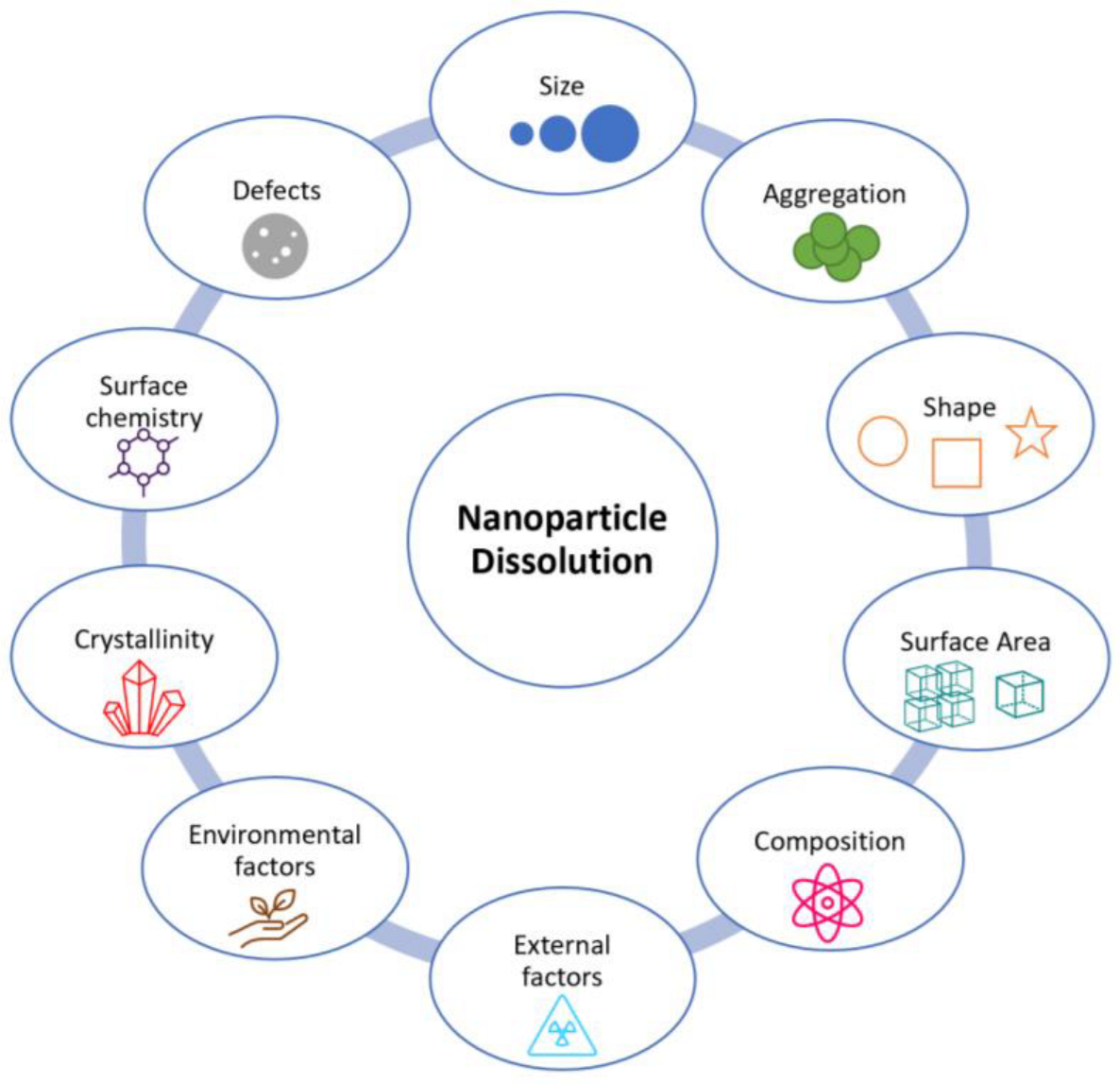
2.2.2. Dissolution
2.2.3. Structural Transformation
Sulfidation
Phosphatisation
Carbonation
2.2.4. Surface Corona Reactions
2.2.5. Photochemical Transformation
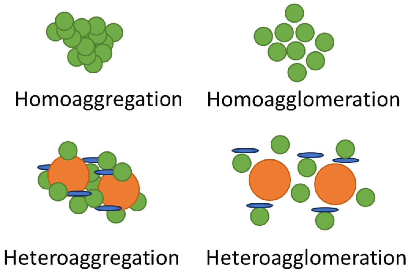
2.3. Physical Transformations
2.4. Biological Transformations
2.4.1. Biodegradation
2.4.2. Biomodification
3. Detection and Quantification of Transformations
4. Knowledge Gaps and Future Challenges
5. Conclusions
Author Contributions
Funding
Conflicts of Interest
References
- Valsami-Jones, E.; Lynch, I. How safe are nanomaterials? Science 2015, 350, 388–389. [Google Scholar] [CrossRef] [PubMed]
- Briffa, S.M.; Lynch, I.; Trouillet, V.; Bruns, M.; Hapiuk, D.; Valsami-Jones, E. Thermal transformations of manufactured nanomaterials as a proposed proxy for ageing. Environ. Sci. Nano 2018, 5, 1618–1627. [Google Scholar] [CrossRef]
- Sengupta, A.; Sarkar, C.K. Introduction to Nano: Basics to Nanoscience and Nanotechnology; Springer: Berlin/Heidelberg, Germany, 2015. [Google Scholar]
- Gottschalk, F.; Sonderer, T.; Scholz, R.W.; Nowack, B. Modeled environmental concentrations of engineered nanomaterials (TiO2, ZnO, Ag, CNT, fullerenes) for different regions. Environ. Sci. Technol. 2009, 43, 9216–9222. [Google Scholar] [CrossRef] [PubMed]
- Lowry, G.V.; Gregory, K.B.; Apte, S.C.; Lead, J.R. Transformations of Nanomaterials in the Environment. Environ. Sci. Technol. 2012, 46, 6893–6899. [Google Scholar] [CrossRef] [PubMed]
- Mitrano, D.M.; Motellier, S.; Clavaguera, S.; Nowack, B. Review of nanomaterial aging and transformations through the life cycle of nano-enhanced products. Environ. Int. 2015, 77, 132–147. [Google Scholar] [CrossRef]
- Miernicki, M.; Hofmann, T.; Eisenberger, I.; von der Kammer, F.; Praetorius, A. Legal and practical challenges in classifying nanomaterials according to regulatory definitions. Nat. Nanotechnol. 2019, 14, 208–216. [Google Scholar] [CrossRef]
- Keller, A.A.; McFerran, S.; Lazareva, A.; Suh, S. Global life cycle releases of engineered nanomaterials. J. Nanoparticle Res. 2013, 15, 1692. [Google Scholar] [CrossRef]
- Maynard, A.D.; Aitken, R.J.; Butz, T.; Colvin, V.; Donaldson, K.; Oberdörster, G.; Philbert, M.A.; Ryan, J.; Seaton, A.; Stone, V.; et al. Safe handling of nanotechnology. Nature 2006, 444, 267–269. [Google Scholar] [CrossRef]
- Hartmann, N.I.B.; Skjolding, L.M.; Hansen, S.F.; Baun, A.; Kjølholt, J.; Gottschalk, F. Environmental Fate and Behaviour of Nanomaterials: New Knowledge on Important Transfomation Processes. 2014. Available online: https://www.semanticscholar.org/paper/Environmental-fate-and-behaviour-of-nanomaterials%3A-Hartmann-Skjolding/e1b467500475ab1255f9ee821d372dba20e35594 (accessed on 23 March 2023).
- Dale, A.L.; Casman, E.A.; Lowry, G.V.; Lead, J.R.; Viparelli, E.; Baalousha, M. Modeling Nanomaterial Environmental Fate in Aquatic Systems. Environ. Sci. Technol. 2015, 49, 2587–2593. [Google Scholar] [CrossRef]
- Bleeker, E.A.J.; Swart, E.; Braakhuis, H.; Fernández Cruz, M.L.; Friedrichs, S.; Gosens, I.; Herzberg, F.; Jensen, K.A.; von der Kammer, F.; Kettelarij, J.A.B.; et al. Towards harmonisation of testing of nanomaterials for EU regulatory requirements on chemical safety—A proposal for further actions. Regul. Toxicol. Pharmacol. 2023, 139, 105360. [Google Scholar] [CrossRef]
- Szpunar, J.; Jiménez-Lamana, J. Environmental Nanopollutants: Sources, Occurrence, Analysis and Fate; Royal Society of Chemistry: London, UK, 2022. [Google Scholar]
- Yang, K.; Lin, D.; Xing, B. Interactions of Humic Acid with Nanosized Inorganic Oxides. Langmuir 2009, 25, 3571–3576. [Google Scholar] [CrossRef]
- Wang, D.; Wang, P.; Wang, C.; Ao, Y. Effects of interactions between humic acid and heavy metal ions on the aggregation of TiO2 nanoparticles in water environment. Environ. Pollut. 2019, 248, 834–844. [Google Scholar] [CrossRef]
- Loosli, F.; Le Coustumer, P.; Stoll, S. TiO2 nanoparticles aggregation and disaggregation in presence of alginate and Suwannee River humic acids. pH and concentration effects on nanoparticle stability. Water Res. 2013, 47, 6052–6063. [Google Scholar] [CrossRef]
- Zhang, P.; Xu, X.Y.; Zhang, X.L.; Zou, K.; Liu, B.Z.; Qing, T.P.; Feng, B. Nanoparticles-EPS corona increases the accumulation of heavy metals and biotoxicity of nanoparticles. J. Hazard. Mater. 2021, 409, 124526. [Google Scholar] [CrossRef]
- Kansara, K.; Kumar, A.; Karakoti, A.S. Combination of humic acid and clay reduce the ecotoxic effect of TiO2 NPs: A combined physico-chemical and genetic study using zebrafish embryo. Sci. Total Environ. 2020, 698, 134133. [Google Scholar] [CrossRef] [PubMed]
- Lee, J.; Kim, J.; Choi, W. TiO2 Photocatalysis for the Redox Conversion of Aquatic Pollutants. In Aquatic Redox Chemistry; American Chemical Society: Washington, DC, USA, 2011; pp. 199–222. [Google Scholar]
- Auffan, M.; Pedeutour, M.; Rose, J.; Masion, A.; Ziarelli, F.; Borschneck, D.; Chaneac, C.; Botta, C.; Chaurand, P.; Labille, J.; et al. Structural degradation at the surface of a TiO2-based nanomaterial used in cosmetics. Environ. Sci. Technol. 2010, 44, 2689–2694. [Google Scholar] [CrossRef]
- Sun, J.; Guo, L.-H.; Zhang, H.; Zhao, L. UV irradiation induced transformation of TiO2 nanoparticles in water: Aggregation and photoreactivity. Environ. Sci. Technol. 2014, 48, 11962–11968. [Google Scholar] [CrossRef]
- Barnard, A.S. One-to-one comparison of sunscreen efficacy, aesthetics and potential nanotoxicity. Nat. Nanotechnol. 2010, 5, 271–274. [Google Scholar] [CrossRef] [PubMed]
- Chekli, L.; Zhao, Y.; Tijing, L.; Phuntsho, S.; Donner, E.; Lombi, E.; Gao, B.; Shon, H. Aggregation behaviour of engineered nanoparticles in natural waters: Characterising aggregate structure using on-line laser light scattering. J. Hazard. Mater. 2015, 284, 190–200. [Google Scholar] [CrossRef] [PubMed]
- Georgantzopoulou, A.; Almeida Carvalho, P.; Vogelsang, C.; Tilahun, M.; Ndungu, K.; Booth, A.M.; Thomas, K.V.; Macken, A. Ecotoxicological Effects of Transformed Silver and Titanium Dioxide Nanoparticles in the Effluent from a Lab-Scale Wastewater Treatment System. Environ. Sci. Technol. 2018, 52, 9431–9441. [Google Scholar] [CrossRef]
- Brunetti, G.; Donner, E.; Laera, G.; Sekine, R.; Scheckel, K.G.; Khaksar, M.; Vasilev, K.; De Mastro, G.; Lombi, E. Fate of zinc and silver engineered nanoparticles in sewerage networks. Water Res. 2015, 77, 72–84. [Google Scholar] [CrossRef]
- Ma, R.; Levard, C.; Michel, F.M.; Brown, G.E.; Lowry, G.V. Sulfidation Mechanism for Zinc Oxide Nanoparticles and the Effect of Sulfidation on Their Solubility. Environ. Sci. Technol. 2013, 47, 2527–2534. [Google Scholar] [CrossRef] [PubMed]
- Poynton, H.C.; Chen, C.; Alexander, S.L.; Major, K.M.; Blalock, B.J.; Unrine, J.M. Enhanced toxicity of environmentally transformed ZnO nanoparticles relative to Zn ions in the epibenthic amphipod Hyalella azteca. Environ. Sci. Nano 2019, 6, 325–340. [Google Scholar] [CrossRef]
- Lee, G.; Lee, B.; Kim, K.-T. Mechanisms and effects of zinc oxide nanoparticle transformations on toxicity to zebrafish embryos. Environ. Sci. Nano 2021, 8, 1690–1700. [Google Scholar] [CrossRef]
- Stetten, L.; Hofmann, T.; Proux, O.; Landrot, G.; Kaegi, R.; von der Kammer, F. Transformation of zinc oxide nanoparticles in freshwater sediments under oxic and anoxic conditions. Environ. Sci. Nano 2022, 9, 4255–4267. [Google Scholar] [CrossRef]
- Zheng, X.; Yang, L.; Shen, Q.; Zhou, C. Evaluation of Zinc Oxide Nanoparticles-Induced Effects on Nitrogen and Phosphorus Removal from Real and Synthetic Municipal Wastewater. Ind. Eng. Chem. Res. 2019, 58, 7929–7936. [Google Scholar] [CrossRef]
- Lv, J.; Zhang, S.; Luo, L.; Han, W.; Zhang, J.; Yang, K.; Christie, P. Dissolution and microstructural transformation of ZnO nanoparticles under the influence of phosphate. Environ. Sci. Technol. 2012, 46, 7215–7221. [Google Scholar] [CrossRef]
- Jones, E.H.; Su, C. Transport and retention of zinc oxide nanoparticles in porous media: Effects of natural organic matter versus natural organic ligands at circumneutral pH. J. Hazard. Mater. 2014, 275, 79–88. [Google Scholar] [CrossRef]
- Khan, R.; Inam, M.A.; Zam, S.Z.; Park, D.R.; Yeom, I.T. Assessment of Key Environmental Factors Influencing the Sedimentation and Aggregation Behavior of Zinc Oxide Nanoparticles in Aquatic Environment. Water 2018, 10, 660. [Google Scholar] [CrossRef]
- Li, M.-R.; Liu, F.-F.; Wang, S.-C.; Cheng, X.; Zhang, H.; Huang, T.-Y.; Liu, G.-Z. Phototransformation of zinc oxide nanoparticles and coexisting pollutant: Role of reactive oxygen species. Sci. Total Environ. 2020, 728, 138335. [Google Scholar] [CrossRef]
- Chunyi, Q.; Qingping, D.; Hongwei, L.; Junxi, L.; Meijun, L.; Yanbin, X. Mitigation Effects of Humic Acid on Toxicity of Zinc Oxide Nanoparticles (ZnO-NPs) on Zebrafish (Danio rerio). Asian J. Ecotoxicol. 2022, 3, 201–209. [Google Scholar]
- Bian, S.-W.; Mudunkotuwa, I.A.; Rupasinghe, T.; Grassian, V.H. Aggregation and Dissolution of 4 nm ZnO Nanoparticles in Aqueous Environments: Influence of pH, Ionic Strength, Size, and Adsorption of Humic Acid. Langmuir 2011, 27, 6059–6068. [Google Scholar] [CrossRef]
- Omar, F.M.; Aziz, H.A.; Stoll, S. Aggregation and disaggregation of ZnO nanoparticles: Influence of pH and adsorption of Suwannee River humic acid. Sci. Total Environ. 2014, 468, 195–201. [Google Scholar] [CrossRef]
- Gogos, A.; Thalmann, B.; Voegelin, A.; Kaegi, R. Sulfidation kinetics of copper oxide nanoparticles. Environ. Sci. Nano 2017, 4, 1733–1741. [Google Scholar] [CrossRef]
- Kansara, K.; Paruthi, A.; Misra, S.K.; Karakoti, A.S.; Kumar, A. Montmorillonite clay and humic acid modulate the behavior of copper oxide nanoparticles in aqueous environment and induces developmental defects in zebrafish embryo. Environ. Pollut. 2019, 255, 113313. [Google Scholar] [CrossRef] [PubMed]
- Wang, Z.; von dem Bussche, A.; Kabadi, P.K.; Kane, A.B.; Hurt, R.H. Biological and environmental transformations of copper-based nanomaterials. ACS Nano 2013, 7, 8715–8727. [Google Scholar] [CrossRef]
- Nanja, A.F.; Focke, W.W.; Musee, N. Aggregation and dissolution of aluminium oxide and copper oxide nanoparticles in natural aqueous matrixes. SN Appl. Sci. 2020, 2, 1164. [Google Scholar] [CrossRef]
- Son, J.; Vavra, J.; Forbes, V.E. Effects of water quality parameters on agglomeration and dissolution of copper oxide nanoparticles (CuO-NPs) using a central composite circumscribed design. Sci. Total Environ. 2015, 521–522, 183–190. [Google Scholar] [CrossRef]
- Yadav, P.K.; Kochar, C.; Taneja, L.; Tripathy, S.S. Study on dissolution behavior of CuO nanoparticles in various synthetic media and natural aqueous medium. J. Nanoparticle Res. 2022, 24, 122. [Google Scholar] [CrossRef]
- Mbanga, O.; Cukrowska, E.; Gulumian, M. Dissolution kinetics of silver nanoparticles: Behaviour in simulated biological fluids and synthetic environmental media. Toxicol. Rep. 2022, 9, 788–796. [Google Scholar] [CrossRef]
- Ross, B.N.; Knightes, C.D. Simulation of the Environmental Fate and Transformation of Nano Copper Oxide in a Freshwater Environment. ACS EST Water 2022, 2, 1532–1543. [Google Scholar] [CrossRef] [PubMed]
- Lin, S.; Taylor, A.A.; Ji, Z.; Chang, C.H.; Kinsinger, N.M.; Ueng, W.; Walker, S.L.; Nel, A.E. Understanding the Transformation, Speciation, and Hazard Potential of Copper Particles in a Model Septic Tank System Using Zebrafish to Monitor the Effluent. ACS Nano 2015, 9, 2038–2048. [Google Scholar] [CrossRef] [PubMed]
- Fathi, P.; Sadeghi, G.; Hosseini, M.-J.; Farahmandkia, Z.; Mehrasbi, M.R. Effects of copper oxide nanoparticles on the Chlorella algae in the presence of humic acid. SN Appl. Sci. 2020, 2, 140. [Google Scholar] [CrossRef]
- Qiu, Y.; Mu, Z.; Wang, N.; Wang, X.; Xu, M.; Li, H. The aggregation and sedimentation of two different sized copper oxide nanoparticles in soil solutions: Dependence on pH and dissolved organic matter. Sci. Total Environ. 2020, 731, 139215. [Google Scholar] [CrossRef]
- Dogra, Y.; Arkill, K.P.; Elgy, C.; Stolpe, B.; Lead, J.; Valsami-Jones, E.; Tyler, C.R.; Galloway, T.S. Cerium oxide nanoparticles induce oxidative stress in the sediment-dwelling amphipod Corophium volutator. Nanotoxicology 2016, 10, 480–487. [Google Scholar] [CrossRef]
- Ray, J.R.; Wu, X.; Neil, C.W.; Jung, H.; Li, Z.; Jun, Y.-S. Redox chemistry of CeO2 nanoparticles in aquatic systems containing Cr(vi)(aq) and Fe2+ ions. Environ. Sci. Nano 2019, 6, 2269–2280. [Google Scholar] [CrossRef]
- Hoecke, K.V.; Quik, J.T.K.; Mankiewicz-Boczek, J.; Schamphelaere, K.A.C.D.; Elsaesser, A.; Meeren, P.V.d.; Barnes, C.; McKerr, G.; Howard, C.V.; Meent, D.V.D.; et al. Fate and Effects of CeO2 Nanoparticles in Aquatic Ecotoxicity Tests. Environ. Sci. Technol. 2009, 43, 4537–4546. [Google Scholar] [CrossRef]
- Rui, Y.; Zhang, P.; Zhang, Y.; Ma, Y.; He, X.; Gui, X.; Li, Y.; Zhang, J.; Zheng, L.; Chu, S.; et al. Transformation of ceria nanoparticles in cucumber plants is influenced by phosphate. Environ. Pollut. 2015, 198, 8–14. [Google Scholar] [CrossRef]
- Römer, I.; Briffa, S.M.; Dasilva, Y.A.R.; Hapiuk, D.; Trouillet, V.; Palmer, R.E.; Valsami-Jones, E. Impact of particle size, oxidation state and capping agent of different cerium dioxide nanoparticles on the phosphate-induced transformations at different pH and concentration. PLoS ONE 2019, 14, e0217483. [Google Scholar] [CrossRef]
- Briffa, S.M.; Lynch, I.; Hapiuk, D.; Valsami-Jones, E. Physical and chemical transformations of zirconium doped ceria nanoparticles in the presence of phosphate: Increasing realism in environmental fate and behaviour experiments. Environ. Pollut. 2019, 252, 974–981. [Google Scholar] [CrossRef]
- Zhang, P.; Ma, Y.; Zhang, Z.; He, X.; Zhang, J.; Guo, Z.; Tai, R.; Zhao, Y.; Chai, Z. Biotransformation of Ceria Nanoparticles in Cucumber Plants. ACS Nano 2012, 6, 9943–9950. [Google Scholar] [CrossRef]
- Quik, J.T.; Stuart, M.C.; Wouterse, M.; Peijnenburg, W.; Hendriks, A.J.; van de Meent, D. Natural colloids are the dominant factor in the sedimentation of nanoparticles. Environ. Toxicol. Chem. 2012, 31, 1019–1022. [Google Scholar] [CrossRef] [PubMed]
- Li, K.; Chen, Y. Effect of natural organic matter on the aggregation kinetics of CeO2 nanoparticles in KCl and CaCl2 solutions: Measurements and modeling. J. Hazard. Mater. 2012, 209–210, 264–270. [Google Scholar] [CrossRef] [PubMed]
- Quik, J.T.K.; Lynch, I.; Hoecke, K.V.; Miermans, C.J.H.; Schamphelaere, K.A.C.D.; Janssen, C.R.; Dawson, K.A.; Stuart, M.A.C.; Meent, D.V.D. Effect of natural organic matter on cerium dioxide nanoparticles settling in model fresh water. Chemosphere 2010, 81, 711–715. [Google Scholar] [CrossRef] [PubMed]
- Elzey, S.; Grassian, V.H. Agglomeration, isolation and dissolution of commercially manufactured silver nanoparticles in aqueous environments. J. Nanoparticle Res. 2010, 12, 1945–1958. [Google Scholar] [CrossRef]
- Levard, C.; Hotze, E.M.; Lowry, G.V.; Brown, G.E., Jr. Environmental transformations of silver nanoparticles: Impact on stability and toxicity. Environ. Sci. Technol. 2012, 46, 6900–6914. [Google Scholar] [CrossRef]
- Liu, J.; Hurt, R.H. Ion release kinetics and particle persistence in aqueous nano-silver colloids. Environ. Sci. Technol. 2010, 44, 2169–2175. [Google Scholar] [CrossRef]
- Römer, I.; Wang, Z.W.; Merrifield, R.C.; Palmer, R.E.; Lead, J. High resolution STEM-EELS study of silver nanoparticles exposed to light and humic substances. Environ. Sci. Technol. 2016, 50, 2183–2190. [Google Scholar] [CrossRef]
- Angel, B.M.; Batley, G.E.; Jarolimek, C.V.; Rogers, N.J. The impact of size on the fate and toxicity of nanoparticulate silver in aquatic systems. Chemosphere 2013, 93, 359–365. [Google Scholar] [CrossRef]
- Zhang, C.; Hu, Z.; Deng, B. Silver nanoparticles in aquatic environments: Physiochemical behavior and antimicrobial mechanisms. Water Res. 2016, 88, 403–427. [Google Scholar] [CrossRef]
- Ma, R.; Levard, C.M.; Judy, J.D.; Unrine, J.M.; Durenkamp, M.; Martin, B.; Jefferson, B.; Lowry, G.V. Fate of zinc oxide and silver nanoparticles in a pilot wastewater treatment plant and in processed biosolids. Environ. Sci. Technol. 2014, 48, 104–112. [Google Scholar] [CrossRef] [PubMed]
- Yu, S.; Yin, Y.; Zhou, X.; Dong, L.; Liu, J. Transformation kinetics of silver nanoparticles and silver ions in aquatic environments revealed by double stable isotope labeling. Environ. Sci. Nano 2016, 3, 883–893. [Google Scholar] [CrossRef]
- Zhao, J.; Wang, X.; Hoang, S.A.; Bolan, N.S.; Kirkham, M.; Liu, J.; Xia, X.; Li, Y. Silver nanoparticles in aquatic sediments: Occurrence, chemical transformations, toxicity, and analytical methods. J. Hazard. Mater. 2021, 418, 126368. [Google Scholar] [CrossRef] [PubMed]
- Zhang, Y.; Xu, J.; Yang, Y.; Sun, B.; Wang, K.; Zhu, L. Impacts of proteins on dissolution and sulfidation of silver nanowires in an aquatic environment: Importance of surface charges. Environ. Sci. Technol. 2020, 54, 5560–5568. [Google Scholar] [CrossRef]
- Stetten, L.; Mackevica, A.; Tepe, N.; Hofmann, T.; von der Kammer, F. Towards Standardization for Determining Dissolution Kinetics of Nanomaterials in Natural Aquatic Environments: Continuous Flow Dissolution of Ag Nanoparticles. Nanomaterials 2022, 12, 519. [Google Scholar] [CrossRef] [PubMed]
- Thalmann, B.; Voegelin, A.; Morgenroth, E.; Kaegi, R. Effect of humic acid on the kinetics of silver nanoparticle sulfidation. Environ. Sci. Nano 2016, 3, 203–212. [Google Scholar] [CrossRef]
- Levard, C.; Hotze, E.M.; Colman, B.P.; Dale, A.L.; Truong, L.; Yang, X.Y.; Bone, A.J.; Brown, G.E.; Tanguay, R.L.; Di Giulio, R.T.; et al. Sulfidation of Silver Nanoparticles: Natural Antidote to Their Toxicity. Environ. Sci. Technol. 2013, 47, 13440–13448. [Google Scholar] [CrossRef]
- Levard, C.; Reinsch, B.C.; Michel, F.M.; Oumahi, C.; Lowry, G.V.; Brown, G.E., Jr. Sulfidation processes of PVP-coated silver nanoparticles in aqueous solution: Impact on dissolution rate. Environ. Sci. Technol. 2011, 45, 5260–5266. [Google Scholar] [CrossRef]
- Kaegi, R.; Voegelin, A.; Ort, C.; Sinnet, B.; Thalmann, B.; Krismer, J.; Hagendorfer, H.; Elumelu, M.; Mueller, E. Fate and transformation of silver nanoparticles in urban wastewater systems. Water Res. 2013, 47, 3866–3877. [Google Scholar] [CrossRef]
- Kampe, S.; Kaegi, R.; Schlich, K.; Wasmuth, C.; Hollert, H.; Schlechtriem, C. Silver nanoparticles in sewage sludge: Bioavailability of sulfidized silver to the terrestrial isopod Porcellio scaber. Environ. Toxicol. Chem. 2018, 37, 1606–1613. [Google Scholar] [CrossRef]
- Prasher, P.; Sharma, M.; Mudila, H.; Verma, A.; Bhatt, P. Silver nanoparticles in natural ecosystems: Fate, transport, and toxicity. In Green Synthesis of Silver Nanomaterials; Elsevier: Amsterdam, The Netherlands, 2022; pp. 649–668. [Google Scholar]
- Cao, C.; Huang, J.; Yan, C.-N.; Zhang, X.-X.; Ma, Y.-X. Impacts of ag and Ag2S nanoparticles on the nitrogen removal within vertical flow constructed wetlands treating secondary effluent. Sci. Total Environ. 2021, 777, 145171. [Google Scholar] [CrossRef] [PubMed]
- Syafiuddin, A.; Salmiati, S.; Hadibarata, T.; Kueh, A.B.H.; Salim, M.R.; Zaini, M.A.A. Silver Nanoparticles in the Water Environment in Malaysia: Inspection, characterization, removal, modeling, and future perspective. Sci. Rep. 2018, 8, 986. [Google Scholar] [CrossRef] [PubMed]
- Piccapietra, F.; Sigg, L.; Behra, R. Colloidal stability of carbonate-coated silver nanoparticles in synthetic and natural freshwater. Environ. Sci. Technol. 2012, 46, 818–825. [Google Scholar] [CrossRef] [PubMed]
- Ellis, L.J.A.; Kissane, S.; Hoffman, E.; Brown, J.B.; Valsami-Jones, E.; Colbourne, J.; Lynch, I. Multigenerational Exposures of Daphnia magna to Pristine and Aged Silver Nanoparticles: Epigenetic Changes and Phenotypical Ageing Related Effects. Small 2020, 16, 2000301. [Google Scholar] [CrossRef]
- Fernando, I.; Zhou, Y. Impact of pH on the stability, dissolution and aggregation kinetics of silver nanoparticles. Chemosphere 2019, 216, 297–305. [Google Scholar] [CrossRef]
- Hou, W.-C.; Stuart, B.; Howes, R.; Zepp, R.G. Sunlight-driven reduction of silver ions by natural organic matter: Formation and transformation of silver nanoparticles. Environ. Sci. Technol. 2013, 47, 7713–7721. [Google Scholar] [CrossRef]
- Zhao, Y.; Liu, Y.; Zhang, X.; Liao, W. Environmental transformation of graphene oxide in the aquatic environment. Chemosphere 2021, 262, 127885. [Google Scholar] [CrossRef]
- Zhao, J.; Wang, Z.; White, J.C.; Xing, B. Graphene in the Aquatic Environment: Adsorption, Dispersion, Toxicity and Transformation. Environ. Sci. Technol. 2014, 48, 9995–10009. [Google Scholar] [CrossRef]
- Hou, W.-C.; Chowdhury, I.; Goodwin, D.G., Jr.; Henderson, W.M.; Fairbrother, D.H.; Bouchard, D.; Zepp, R.G. Photochemical transformation of graphene oxide in sunlight. Environ. Sci. Technol. 2015, 49, 3435–3443. [Google Scholar] [CrossRef]
- Adeleye, A.S.; Ho, K.T.; Zhang, M.; Li, Y.; Burgess, R.M. Fate and transformation of graphene oxide in estuarine and marine waters. Environ. Sci. Technol. 2019, 53, 5858–5867. [Google Scholar] [CrossRef]
- Chen, M.; Qin, X.; Zeng, G. Biodegradation of carbon nanotubes, graphene, and their derivatives. Trends Biotechnol. 2017, 35, 836–846. [Google Scholar] [CrossRef] [PubMed]
- Misra, S.K.; Dybowska, A.; Berhanu, D.; Luoma, S.N.; Valsami-Jones, E. The complexity of nanoparticle dissolution and its importance in nanotoxicological studies. Sci. Total Environ. 2012, 438, 225–232. [Google Scholar] [CrossRef]
- Tantra, R.; Cackett, A.; Peck, R.; Gohil, D.; Snowden, J. Measurement of redox potential in nanoecotoxicological investigations. J. Toxicol. 2012, 2012, 270651. [Google Scholar] [CrossRef] [PubMed]
- Hoffmann, K.; Christl, I.; Kaegi, R.; Kretzschmar, R. Effects of natural organic matter (NOM), metal-to-sulfide ratio and Mn2+ on cadmium sulfide nanoparticle growth and colloidal stability. Environ. Sci. Nano 2020, 7, 3385–3404. [Google Scholar] [CrossRef]
- Lee, W.S.; Kim, E.; Cho, H.-J.; Kang, T.; Kim, B.; Kim, M.Y.; Kim, Y.S.; Song, N.W.; Lee, J.-S.; Jeong, J. The Relationship between Dissolution Behavior and the Toxicity of Silver Nanoparticles on Zebrafish Embryos in Different Ionic Environments. Nanomaterials 2018, 8, 652. [Google Scholar] [CrossRef] [PubMed]
- Zhang, Y.; Xia, J.; Liu, Y.; Qiang, L.; Zhu, L. Impacts of morphology, natural organic matter, cations, and ionic strength on sulfidation of silver nanowires. Environ. Sci. Technol. 2016, 50, 13283–13290. [Google Scholar] [CrossRef]
- Xu, H.; Li, L.; Lv, H.; Liu, X.; Jiang, H. pH-dependent phosphatization of ZnO nanoparticles and its influence on subsequent lead sorption. Environ. Pollut. 2016, 208, 723–731. [Google Scholar] [CrossRef]
- Zhang, P.; Ma, Y.; Xie, C.; Guo, Z.; He, X.; Valsami-Jones, E.; Lynch, I.; Luo, W.; Zheng, L.; Zhang, Z. Plant species-dependent transformation and translocation of ceria nanoparticles. Environ. Sci. Nano 2019, 6, 60–67. [Google Scholar] [CrossRef]
- Diegoli, S.; Manciulea, A.L.; Begum, S.; Jones, I.P.; Lead, J.R.; Preece, J.A. Interaction between manufactured gold nanoparticles and naturally occurring organic macromolecules. Sci. Total Environ. 2008, 402, 51–61. [Google Scholar] [CrossRef]
- Hwang, Y.S.; Li, Q. Characterizing photochemical transformation of aqueous nC60 under environmentally relevant conditions. Environ. Sci. Technol. 2010, 44, 3008–3013. [Google Scholar] [CrossRef]
- Kirschling, T.L.; Golas, P.L.; Unrine, J.M.; Matyjaszewski, K.; Gregory, K.B.; Lowry, G.V.; Tilton, R.D. Microbial Bioavailability of Covalently Bound Polymer Coatings on Model Engineered Nanomaterials. Environ. Sci. Technol. 2011, 45, 5253–5259. [Google Scholar] [CrossRef] [PubMed]
- Allen, B.L.; Kotchey, G.P.; Chen, Y.; Yanamala, N.V.; Klein-Seetharaman, J.; Kagan, V.E.; Star, A. Mechanistic investigations of horseradish peroxidase-catalyzed degradation of single-walled carbon nanotubes. J. Am. Chem. Soc. 2009, 131, 17194–17205. [Google Scholar] [CrossRef] [PubMed]
- Allen, B.L.; Kichambare, P.D.; Gou, P.; Vlasova, I.I.; Kapralov, A.A.; Konduru, N.; Kagan, V.E.; Star, A. Biodegradation of single-walled carbon nanotubes through enzymatic catalysis. Nano Lett. 2008, 8, 3899–3903. [Google Scholar] [CrossRef] [PubMed]
- Nasser, F.; Lynch, I. Secreted protein eco-corona mediates uptake and impacts of polystyrene nanoparticles on Daphnia magna. J. Proteom. 2016, 137, 45–51. [Google Scholar] [CrossRef] [PubMed]
- Mitrano, D.; Nowack, B.; Motellier, S.; Clavaguera, S. Report on Environmental Transformation Reactions. 2014. Available online: https://pubmed.ncbi.nlm.nih.gov/25705000/ (accessed on 24 March 2023).
- Louie, S.M.; Ma, R.; Lowry, G.V. Chapter 2—Transformations of Nanomaterials in the Environment. In Frontiers of Nanoscience; Jamie, R.L., Eugenia, V.-J., Eds.; Elsevier: Amsterdam, The Netherlands, 2014; pp. 55–87. [Google Scholar]
- Geranio, L.; Heuberger, M.; Nowack, B. The Behavior of Silver Nanotextiles during Washing. Environ. Sci. Technol. 2009, 43, 8113–8118. [Google Scholar] [CrossRef]
- Jeyaraj, M.; Sathishkumar, G.; Sivanandhan, G.; MubarakAli, D.; Rajesh, M.; Arun, R.; Kapildev, G.; Manickavasagam, M.; Thajuddin, N.; Premkumar, K. Biogenic silver nanoparticles for cancer treatment: An experimental report. Colloids Surf. B Biointerfaces 2013, 106, 86–92. [Google Scholar] [CrossRef]
- Behzadi, E.; Hosseini, H.M.; Fooladi, A.A.I. The inhibitory impacts of Lactobacillus rhamnosus GG-derived extracellular vesicles on the growth of hepatic cancer cells. Microb. Pathog. 2017, 110, 1–6. [Google Scholar] [CrossRef]
- Dominguez, G.A.; Lohse, S.E.; Torelli, M.D.; Murphy, C.J.; Hamers, R.J.; Orr, G.; Klaper, R.D. Effects of charge and surface ligand properties of nanoparticles on oxidative stress and gene expression within the gut of Daphnia magna. Aquat. Toxicol. 2015, 162, 1–9. [Google Scholar] [CrossRef]
- Avramescu, M.-L.; Rasmussen, P.; Chénier, M.; Gardner, H. Influence of pH, particle size and crystal form on dissolution behaviour of engineered nanomaterials. Environ. Sci. Pollut. Res. 2017, 24, 1553–1564. [Google Scholar] [CrossRef]
- Baralkiewicz, D.; Gramowska, H.; Hanc, A.; Krzyzaniak, I. A comparison of ICP-OES and ICP-MS in the determination of elements in lake water. At. Spectrosc. 2007, 28, 164. [Google Scholar]
- Utembe, W.; Potgieter, K.; Stefaniak, A.B.; Gulumian, M. Dissolution and biodurability: Important parameters needed for risk assessment of nanomaterials. Part. Fibre Toxicol. 2015, 12, 11. [Google Scholar] [CrossRef]
- OECD. Important Issues on Risk Assessment of Manufactured Nanomaterials; OECD: Paris, France, 2022. [Google Scholar]
- OECD. Test No. 105: Water Solubility; OECD: Paris, France, 1995. [Google Scholar]
- OECD. Guidance Document on Transformation/Dissolution of Metals and Metal Compounds in Aqueous Media; OECD: Paris, France, 2002. [Google Scholar]
- Keller, J.G.; Graham, U.M.; Koltermann-Jülly, J.; Gelein, R.; Ma-Hock, L.; Landsiedel, R.; Wiemann, M.; Oberdörster, G.; Elder, A.; Wohlleben, W. Predicting dissolution and transformation of inhaled nanoparticles in the lung using abiotic flow cells: The case of barium sulfate. Sci. Rep. 2020, 10, 458. [Google Scholar] [CrossRef]
- Koltermann-Jülly, J.; Keller, J.G.; Vennemann, A.; Werle, K.; Müller, P.; Ma-Hock, L.; Landsiedel, R.; Wiemann, M.; Wohlleben, W. Abiotic dissolution rates of 24 (nano)forms of 6 substances compared to macrophage-assisted dissolution and in vivo pulmonary clearance: Grouping by biodissolution and transformation. NanoImpact 2018, 12, 29–41. [Google Scholar] [CrossRef]
- Maurer-Jones, M.A.; Gunsolus, I.L.; Murphy, C.J.; Haynes, C.L. Toxicity of Engineered Nanoparticles in the Environment. Anal. Chem. 2013, 85, 3036–3049. [Google Scholar] [CrossRef] [PubMed]
- Borm, P.; Klaessig, F.C.; Landry, T.D.; Moudgil, B.; Pauluhn, J.; Thomas, K.; Trottier, R.; Wood, S. Research strategies for safety evaluation of nanomaterials, part V: Role of dissolution in biological fate and effects of nanoscale particles. Toxicol. Sci. 2006, 90, 23–32. [Google Scholar] [CrossRef] [PubMed]
- Choi, Y.; Choi, Y.-J. The effects of UV disinfection on drinking water quality in distribution systems. Water Res. 2010, 44, 115–122. [Google Scholar] [CrossRef] [PubMed]
- Anderson, J.; Voskerician, G. The challenge of biocompatibility evaluation of biocomposites. In Biomedical Composites; Elsevier: Amsterdam, The Netherlands, 2010; pp. 325–353. [Google Scholar]
- Nowack, B.; Ranville, J.F.; Diamond, S.; Gallego-Urrea, J.A.; Metcalfe, C.; Rose, J.; Horne, N.; Koelmans, A.A.; Klaine, S.J. Potential scenarios for nanomaterial release and subsequent alteration in the environment. Environ. Toxicol. Chem. 2012, 31, 50–59. [Google Scholar] [CrossRef]
- Bae, S.; Hwang, Y.S.; Lee, Y.-J.; Lee, S.-K. Effects of Water Chemistry on Aggregation and Soil Adsorption of Silver Nanoparticles. Environ. Health Toxicol. 2013, 28, e2013006. [Google Scholar] [CrossRef]
- Lin, D.; Xing, B. Root Uptake and Phytotoxicity of ZnO Nanoparticles. Environ. Sci. Technol. 2008, 42, 5580–5585. [Google Scholar] [CrossRef]
- Sharma, V.K. Aggregation and toxicity of titanium dioxide nanoparticles in aquatic environment—A review. J. Environ. Sci. Health Part A 2009, 44, 1485–1495. [Google Scholar] [CrossRef]
- Ward, J.E.; Kach, D.J. Marine aggregates facilitate ingestion of nanoparticles by suspension-feeding bivalves. Mar. Environ. Res. 2009, 68, 137–142. [Google Scholar] [CrossRef] [PubMed]
- Schaumann, G.E.; Philippe, A.; Bundschuh, M.; Metreveli, G.; Klitzke, S.; Rakcheev, D.; Grün, A.; Kumahor, S.K.; Kühn, M.; Baumann, T. Understanding the fate and biological effects of Ag-and TiO2-nanoparticles in the environment: The quest for advanced analytics and interdisciplinary concepts. Sci. Total Environ. 2015, 535, 3–19. [Google Scholar] [CrossRef]
- Mahl, D.; Diendorf, J.; Meyer-Zaika, W.; Epple, M. Possibilities and limitations of different analytical methods for the size determination of a bimodal dispersion of metallic nanoparticles. Colloids Surf. A Physicochem. Eng. Asp. 2011, 377, 386–392. [Google Scholar] [CrossRef]
- Briffa, S.M.; Sullivan, J.; Siupa, A.; Carnell-Morris, P.; Carboni, M.; Jurkschat, K.; Peters, R.J.B.; Schultz, C.; Seol, K.H.; Kwon, S.-J.; et al. Nanoparticle Tracking Analysis of Gold Nanoparticles in Aqueous Media through an Inter-Laboratory Comparison. J. Vis. Exp. 2020, 164, e61741. [Google Scholar]
- Merrifield, R.C.; Stephan, C.; Lead, J.R. Single-particle inductively coupled plasma mass spectroscopy analysis of size and number concentration in mixtures of monometallic and bimetallic (core-shell) nanoparticles. Talanta 2017, 162, 130–134. [Google Scholar] [CrossRef]
- Cascio, C.; Geiss, O.; Franchini, F.; Ojea-Jimenez, I.; Rossi, F.; Gilliland, D.; Calzolai, L. Detection, quantification and derivation of number size distribution of silver nanoparticles in antimicrobial consumer products. J. Anal. At. Spectrom. 2015, 30, 1255–1265. [Google Scholar] [CrossRef]
- Lee, Y.-L.; Shih, Y.-S.; Chen, Z.-Y.; Cheng, F.-Y.; Lu, J.-Y.; Wu, Y.-H.; Wang, Y.-J. Toxic effects and mechanisms of silver and zinc oxide nanoparticles on zebrafish embryos in aquatic ecosystems. Nanomaterials 2022, 12, 717. [Google Scholar] [CrossRef]


| Factors Determining the Outcome of Transformations | Influencing Properties or Variables |
|---|---|
| ENM morphology | Size, shape, available surface area |
| ENM chemistry | Reactivity potential, possible reactions (e.g., oxidation), surface charge, aggregation state |
| Environment | pH, temperature, organic material |
| ENMs | Common Aquatic Environment Transformations | Examples from Literature |
|---|---|---|
| TiO2 | Interaction with organic species and biomodification Photochemical reaction Redox Aggregation | [14,15,16,17,18] [19,20,21] [19,22] [15,16,18,21,23,24] |
| ZnO | Sulfidation Phosphitization Interaction with organic species and biomodification Photochemical Aggregation Dissolution | [25,26,27,28,29] [27,28,30,31] [14,32,33,34,35] [10,34] [32,33,36,37] [29,30,31,36] |
| CuO | Redox Dissolution Sulfidation Interaction with organic species and biomodification Aggregation | [38,39] [40,41,42,43,44,45] [38,40] [39,42,45,46,47,48] [41,42,45,48] |
| CeO2 | Redox Phosphitization Interaction with organic species and biomodification Aggregation | [49,50,51] [51,52,53,54,55] [17,56,57,58] [51,57] |
| Ag | Redox Dissolution Sulfidation Carbonation Interaction with organic species and biomodification Aggregation Photochemical | [59,60,61,62] [59,61,63,64,65,66,67,68,69] [25,68,70,71,72,73,74,75,76] [60,77,78] [60,62,63,70] [63,78,79,80] [66,81] |
| Graphene | Photochemical reactions Aggregation Biodegradation Interaction with organic species and biomodification | [82,83,84,85] [83,85] [83,86] [82,83] |
| Redox | ||
| Nanomaterial | Observations | References |
| Cerium dioxide | Valence state changes upon exposure to elevated temperatures | [2] |
| Redox changes are dependent on exposure medium | [87,88] | |
| Elemental silver (Ag0) | Undergo dissolution, facilitating the release of toxic Ag⁺ ions Significantly enhanced as pH is reduced | [10] |
| An increase in system variables’ concentration, such as chloride and sulfide, will have a proportional effect on the rate of transformation Organic ligands, including NOM, may slow down redox processes. | [60] | |
| Zinc oxide | Redox changes are dependent on exposure medium | [87,88] |
| Cadmium sulfide | Redox changes influenced by the presence of macromolecules and organic ligands from natural organic matter | [87,89] |
| Dissolution | ||
| Nanomaterial | Observations | References |
| Citrate-stabilised silver | Dissolution influenced the toxicity | [90] |
| Silver | Dissolution rate increased at high ionic strength and low pH | [44] |
| Copper oxide | Dissolution is affected by the water characteristics—more soluble in deionised than natural pond water; however, the dissolution rate was faster in pond water compared to deionised water | [43] |
| Zinc oxide | Under oxic conditions, ZnO NPs were dissolved within a few hours. By contrast, ZnO NP dissolution under anoxic conditions was much slower. | [29] |
| Sulfidation | ||
| Nanomaterial | Observations | References |
| Silver | Partly sulfidised Ag ENMs released fewer toxic Ag⁺ ions than pristine Ag ENMs over 48- and 120-h intervals. | [71] |
| Increasing the presence of NOM suppressed the sulfidation of Ag nanowires in the aquatic environment. | [91] | |
| The presence of divalent cations compared to monovalent ions in solution, accelerated sulfidation rates | [91] | |
| Smaller AgNPs could result in an enhanced sulfidation rate owing to the reaction rate’s dependency on the specific surface area of the NP | [75] | |
| The increased HS−/Ag ratio and NOM presence influenced sulfidation. The presence of NOM was also found to influence the sulfidation of AgNPs. | [68,75] | |
| The presence of HA promoted sulfidation by replacing the surface coating, thus increasing the available surface area | [68] | |
| Sulfidation mitigated the toxicity of constructed wetlands | [76] | |
| PVP-coated Ag | Sulfidation was found to reduce dissolution and limit toxicity | [72] |
| Zinc oxide | Stabilisation through sulfidation can reduce toxicity as it reduces dissolution and ion release | [26,28] |
| Posphatization | ||
| Nanomaterial | Observations | References |
| Zinc oxide | pH-dependent and more likely in acidic environments than alkaline environments | [92] |
| A decrease in toxicity in embryonic zebrafish | [28] | |
| Altered morphology | [31] | |
| Transformation products are larger than their pristine counterparts, and thus, surface reactivity is decreased, leading to reduced dissolution and muted toxic potential | [31] | |
| Cerium dioxide | Physical and chemical changes occur | [53,54,55] |
| Increased concentrations of phosphate will encourage desorption, limiting persistence and reducing toxicity risk | [10] | |
| Phosphate was capable of immobilising CeO2 through phosphate complexation in plant roots | [93] | |
| Carbonation | ||
| Nanomaterial | Observations | References |
| Silver | Inorganic silver carbonate (Ag2CO3) coatings have been applied as capping agents to stabilise them against aggregation | [60] |
| At an alkaline pH, negatively charged CO32− surface capping could inhibit aggregation | [78] | |
| Surface Corona Reactions | ||
| Nanomaterial | Observations | References |
| Silver | Positively charged proteins enhanced the dissolution and sulfidation of AgNPs | [68] |
| ZnO, TiO2, SiO2 and Al2O3 | Adsorption of HA was dependent on pH and decreased as the solution became more basic | [14] |
| Titanium dioxide | NPs with clay were toxic to zebrafish embryo development while NPs in the presence of HA displayed a protective effect | [18] |
| Gold | In low ionic-strength solutions, HA provided an additional coating, thereby providing additional resistance from pH induced aggregation | [94] |
| Zinc oxide | Reduction in toxicity in the presence of HA | [35] |
| Photochemical Transformation | ||
| Nanomaterial | Observations | References |
| Titanium dioxide | UV irradiation in the environment significantly increased aggregation | [21] |
| Graphene oxide | Simulated sunlight can rapidly reduce the GO, producing by-products of CO2 and low-molecular weight species | [84] |
| Silver | NOM-facilitated photo-reduction of ionic Ag in river water, could precipitate NPs of different sizes and morphologies | [84] |
| C-60 | Particles underwent surface oxidation and hydroxylation in the presence of dissolved O2 | [95] |
| Physical Transformations | ||
| Nanomaterial | Observations | References |
| Surface coatings | Surface coatings can be lost through biodegradation, which ultimately results in aggregation | [96] |
| Using temperature as a proxy for ageing led to enhanced degradation of the PVP coating | [2] | |
| Silver | Lower aggregation and higher particle stability was reported with increasing pH | [80] |
| Copper oxide | The aggregation and sedimentation of CuO NPs in soil solutions was influenced by the NP size and the soil properties | [48] |
| Biodegradation | ||
| Nanomaterial | Observations | References |
| C-60 (fullerenes) | Not susceptible to biodegradation due to cage-structure | [10] |
| Single walled carbon nanotubes | Biodegradation observed when incubated with horseradish peroxidase and H2O2 via enzyme catalysis | [97] |
| Biomodification | ||
| Nanomaterial | Observations | References |
| Cerium dioxide | Shape changes and presence of aggregation | [93] |
| CNT | Degradation of the lipid-coating which enabled the CNT to aggregate, aiding in their destabilisation | [98] |
| Polystyrene | NPs quickly acquired specific macromolecular coronas on their surfaces, which induced aggregation and increased uptake and gut retention | [99] |
| Titanium dioxide | NPs with EPS coronas adsorbed more heavy metals compared to NPs without EPS coronas | [17] |
| Cerium dioxide | NPs with EPS coronas adsorbed more heavy metals compared to NPs without EPS coronas | [17] |
| Transformation Classification | Type of Transformation | Key Features | Environmental Impact |
|---|---|---|---|
| Chemical | Redox | Influenced by media composition and conditions Dependent on intrinsic physicochemical properties of ENMs Influenced by the presence of macromolecules and organic ligands | Drives reactivity Internalisation may cause ROS production |
| Chemical | Dissolution | Used as a measure of bio-durability and toxicity | Enhanced by environmental parameters Enhances toxicity |
| Chemical | Sulfidation | Influenced by media composition and conditions Usually accompanied by substantial aggregation and sedimentation and a lower dissolution rate | Sulfidation can reduce toxicity in low redox environments, as it reduces the dissolution |
| Chemical | Phosphatization | pH dependent and more likely in acidic environments than alkaline environments Decreases particle surface reactivity | Drives particle size increase and reduces dissolution. Can mitigate toxicity |
| Chemical | Carbonation | Affects particle stability | Inhibits aggregation |
| Chemical | Surface corona reactions | ENM surface properties modified Positively charged proteins enhance dissolution and sulfidation | Biomolecules may result in increased biocompatibility of ENMs, thereby also altering toxicity |
| Chemical | Photochemical | May increase aggregation | Generates ROS and oxidative tissue damage |
| Physical | Aggregation and agglomeration | Potential decrease in reactivity and toxicity, resulting from an increase in size | Increases environmental persistence |
| Biological | Biodegradation | May break down ENMs into less harmful counterparts | Decreases environmental persistence |
| Biological | Biomodification | May convert ENMs to more biocompatible forms | Potentially increases bioavailability and bioaccumulation; may also increase toxicity |
| Characterisation Method | ENM Types Suitable for the Method | Main Information Gained | Advantages | Disadvantages |
|---|---|---|---|---|
| Electron microscopy (SEM, TEM, STEM) | Inorganic ENMs | Morphological and compositional information (quantitative) | Allows direct comparisons between pristine and aged ENMs. Size, morphology, and surface conditions can be studied. Chemical composition provided by EDX. Structural information is available from TEM/STEM. | Ex-situ methods. iSEM may be unsuitable for ultrasmall ENMs. Laborious sample preparation. 2D representation of a 3D material. Operator bias. |
| Dynamic Light Scattering (DLS) | Can be applied to any ENM category but cannot differentiate between different ENMs if present as mixtures | Provides information on the hydrodynamic size (Hd) and zeta potential (ζ) and allows the determination of particle size distribution. | Gives information on hydrodynamic size (Hd) and zeta potential (ζ). | Cannot distinguish between isolated particles, clusters, and mixtures of sizes, or particles with different compositions. Assumes all particles are spherical. |
| Nanoparticle Tracking Analysis (NTA) | Suitable for visualising and analysing inorganic and organic particles in suspension; Cannot differentiate between different ENMs if present as mixtures | Can track different sized particles simultaneously. | Allows visual tracking of particles, allowing observation of aggregates. Can also track different-sized particles simultaneously. | Requires an optimum concentration for analysis. It cannot distinguish between individual particles and agglomerates/aggregates. |
| Inductively coupled plasma mass spectrometry/optical emission spectrometry (ICP-MS, ICP-OES) | Suitable for detecting and quantifying heavy (inorganic) elements. | Directly measure the particle number concentration (in single particle mode). | Allows quantification of dissolved ions in solution and directly measures the particle number concentration. | Signal intensity varies with each isotope. Interferences can occur when plasma-formed species have the same mass as the ionised analyte species. May require prior knowledge of particle composition |
| Ultraviolet-visible spectroscopy (UV-Vis) | Can be used to determine concentrations and provide information about the physical and electronic structures of both organic and inorganic compounds. | Provides information on the optical properties, size, concentration, and agglomeration state of the nanoparticles. | Can provide quantitative and qualitative analysis. Easy to handle and use. Non-destructive. | Unable to analyse compounds that do not interact with light in the UV and visible areas of the spectrum. Can only be used to characterise suspensions and is not suitable to measure solid or gaseous samples. |
| Attenuated Total Reflection—Fourier Transform Infrared Spectroscopy (ATR-FTIR) | Can provide information for both inorganic and organic substances. | Provides information on the surface composition and ligand binding. | Capable of analysing nanomaterial samples in suspension or powder form. Does not require ultra-high vacuum conditions. | Limited spatial resolution. |
| X-ray Photoelectron Spectroscopy (XPS) | Inorganic coatings, but can also be extended to natural organic coatings. | Determination of chemical composition surfaces and oxidation states. | Allows for the determination of electronic structure, elemental composition, and oxidation states. Highly sensitive to surface modifications. | Requires ultra-high vacuum conditions. Overlapping peaks in the spectra can complicate surface analysis. Contamination by adsorbed water or volatile organic compounds makes carbon and oxygen analysis difficult. |
| High-Performance Liquid Chromatography (HPLC) | Quantifies ligands conjugated to soft nanoparticles such as polymers and liposomes. | Suitable for the separation of nanoparticles in mixed samples or for the evaluation of the nanoparticle surface upon interaction with the stationary phase. | Can detect and measure very small amounts of a substance. Can separate and analyse a sample quickly. | Nanoparticles may stick to the column. Limited sample size, which can make it difficult to analyse large samples or many samples at once. |
| Liquid Chromatography-Mass Spectrometry (LCMS) | Used to characterise colloids in natural organic matter but can also be used to study trace metal distributions over colloidal particles. | Quantifies ligand density through cleavage of the surface ligands from the nanoparticles. | Robust analytical technique that provides the high sensitivity and selectivity required to detect the exact molecular weight of a wide range of samples. Can separate and identify solutes in low concentrations (in parts per million) in a complex mixture. Major advantage of LCMS over HPLC is that LCMS can achieve a complete elucidation of the chemical structure of the molecule. | Only works with volatile buffers. Residual impurities being analysed should be ionised. |
Disclaimer/Publisher’s Note: The statements, opinions and data contained in all publications are solely those of the individual author(s) and contributor(s) and not of MDPI and/or the editor(s). MDPI and/or the editor(s) disclaim responsibility for any injury to people or property resulting from any ideas, methods, instructions or products referred to in the content. |
© 2023 by the authors. Licensee MDPI, Basel, Switzerland. This article is an open access article distributed under the terms and conditions of the Creative Commons Attribution (CC BY) license (https://creativecommons.org/licenses/by/4.0/).
Share and Cite
Harrison, D.M.; Briffa, S.M.; Mazzonello, A.; Valsami-Jones, E. A Review of the Aquatic Environmental Transformations of Engineered Nanomaterials. Nanomaterials 2023, 13, 2098. https://doi.org/10.3390/nano13142098
Harrison DM, Briffa SM, Mazzonello A, Valsami-Jones E. A Review of the Aquatic Environmental Transformations of Engineered Nanomaterials. Nanomaterials. 2023; 13(14):2098. https://doi.org/10.3390/nano13142098
Chicago/Turabian StyleHarrison, Daniel Mark, Sophie M. Briffa, Antonino Mazzonello, and Eugenia Valsami-Jones. 2023. "A Review of the Aquatic Environmental Transformations of Engineered Nanomaterials" Nanomaterials 13, no. 14: 2098. https://doi.org/10.3390/nano13142098
APA StyleHarrison, D. M., Briffa, S. M., Mazzonello, A., & Valsami-Jones, E. (2023). A Review of the Aquatic Environmental Transformations of Engineered Nanomaterials. Nanomaterials, 13(14), 2098. https://doi.org/10.3390/nano13142098