
Surface Photovoltage Response of ZnO to Phosphate-Buffered Saline Solution with and without Presence of Staphylococcus aureus

 ,
, 
 ,
,  and
and 
Abstract
1. Introduction
2. Materials and Methods
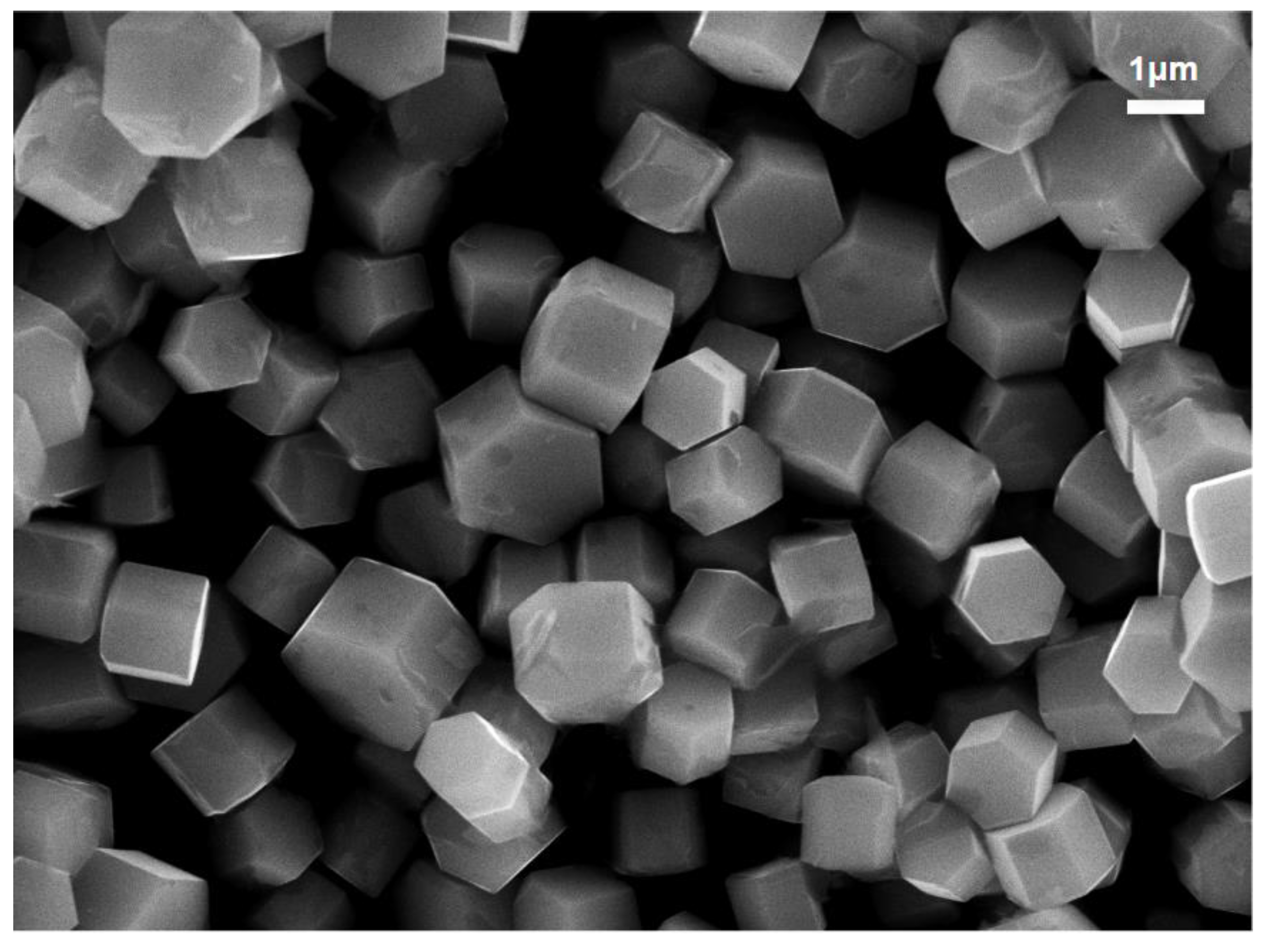
2.1. ZnO MP Synthesis and Morphological Characterization
2.2. Biological Exposure
2.3. Surface Photovoltage Studies
3. Results and Discussion
4. Conclusions
Author Contributions
Funding
Data Availability Statement
Acknowledgments
Conflicts of Interest
References
- Özgür, Ü.; Alivov, Y.I.; Liu, C.; Teke, A.; Reshchikov, M.; Doğan, S.; Avrutin, V.; Cho, S.-J.; Morkoç, A.H. A comprehensive review of ZnO materials and devices. J. Appl. Phys. 2005, 98, 11. [Google Scholar] [CrossRef]
- Puspasari, V.; Ridhova, A.; Hermawan, A.; Amal, M.I.; Khan, M.M. ZnO-based antimicrobial coatings for biomedical applications. Bioprocess Biosyst. Eng. 2022, 45, 1421–1445. [Google Scholar] [CrossRef]
- Kim, I.; Viswanathan, K.; Kasi, G.; Thanakkasaranee, S.; Sadeghi, K.; Seo, J. ZnO Nanostructures in Active Antibacterial Food Packaging: Preparation Methods, Antimicrobial Mechanisms, Safety Issues, Future Prospects, and Challenges. Food Rev. Int. 2022, 38, 537–565. [Google Scholar] [CrossRef]
- Mahlaule-Glory, L.M.; Hintsho-Mbita, N.C. Green Derived Zinc Oxide (ZnO) for the Degradation of Dyes from Wastewater and Their Antimicrobial Activity: A Review. Catalysts 2022, 12, 833. [Google Scholar] [CrossRef]
- Blaskovich, M.A. Antibiotics special issue: Challenges and opportunities in antibiotic discovery and development. ACS Infect. Dis. 2020, 6, 1286–1288. [Google Scholar] [CrossRef] [PubMed]
- Banin, E.; Hughes, D.; Kuipers, O.P. Bacterial pathogens, antibiotics and antibiotic resistance. FEMS Microbiol. Rev. 2017, 41, 450–452. [Google Scholar] [CrossRef]
- Cauda, V.; Gazia, R.; Porro, S.; Stassi, S.; Canavese, G.; Roppolo, I.; Chiolerio, A. Nanostructured ZnO materials: Synthesis, properties and applications. In Handbook of Nanomaterials Properties; Springer: Berlin/Heidelberg, Germany, 2014; pp. 137–177. [Google Scholar]
- Ancona, A.; Dumontel, B.; Garino, N.; Demarco, B.; Chatzitheodoridou, D.; Fazzini, W.; Engelke, H.; Cauda, V. Lipid-coated zinc oxide nanoparticles as innovative ROS-generators for photodynamic therapy in cancer cells. Nanomaterials 2018, 8, 143. [Google Scholar] [CrossRef] [PubMed]
- Premanathan, M.; Karthikeyan, K.; Jeyasubramanian, K.; Manivannan, G. Selective toxicity of ZnO nanoparticles toward Gram-positive bacteria and cancer cells by apoptosis through lipid peroxidation. Nanomed. Nanotechnol. Biol. Med. 2011, 7, 184–192. [Google Scholar] [CrossRef]
- Reddy, K.M.; Feris, K.; Bell, J.; Wingett, D.G.; Hanley, C.; Punnoose, A. Selective toxicity of zinc oxide nanoparticles to prokaryotic and eukaryotic systems. Appl. Phys. Lett. 2007, 90, 213902. [Google Scholar] [CrossRef]
- Baek, Y.-W.; An, Y.-J. Microbial toxicity of metal oxide nanoparticles (CuO, NiO, ZnO, and Sb2O3) to Escherichia coli, Bacillus subtilis, and Streptococcus aureus. Sci. Total Environ. 2011, 409, 1603–1608. [Google Scholar] [CrossRef]
- Kadiyala, U.; Turali-Emre, E.S.; Bahng, J.H.; Kotov, N.A.; VanEpps, J.S. Unexpected insights into antibacterial activity of zinc oxide nanoparticles against methicillin resistant Staphylococcus aureus (MRSA). Nanoscale 2018, 10, 4927–4939. [Google Scholar] [CrossRef] [PubMed]
- Alavi, M.; Nokhodchi, A. An overview on antimicrobial and wound healing properties of ZnO nanobiofilms, hydrogels, and bionanocomposites based on cellulose, chitosan, and alginate polymers. Carbohydr. Polym. 2020, 227, 115349. [Google Scholar] [CrossRef] [PubMed]
- Martinaga Pintarić, L.; Somogi Škoc, M.; Ljoljić Bilić, V.; Pokrovac, I.; Kosalec, I.; Rezić, I. Synthesis, modification and characterization of antimicrobial textile surface containing ZnO nanoparticles. Polymers 2020, 12, 1210. [Google Scholar] [CrossRef] [PubMed]
- Sirelkhatim, A.; Mahmud, S.; Seeni, A.; Kaus, N.H.M.; Ann, L.C.; Bakhori, S.K.M.; Hasan, H.; Mohamad, D. Review on zinc oxide nanoparticles: Antibacterial activity and toxicity mechanism. Nano-Micro Lett. 2015, 7, 219–242. [Google Scholar] [CrossRef] [PubMed]
- da Silva, B.L.; Abuçafy, M.P.; Manaia, E.B.; Junior, J.A.O.; Chiari-Andréo, B.G.; Pietro, R.C.R.; Chiavacci, L.A. Relationship between structure and antimicrobial activity of zinc oxide nanoparticles: An overview. Int. J. Nanomed. 2019, 14, 9395. [Google Scholar] [CrossRef]
- Xu, X.; Chen, D.; Yi, Z.; Jiang, M.; Wang, L.; Zhou, Z.; Fan, X.; Wang, Y.; Hui, D. Antimicrobial mechanism based on H2O2 generation at oxygen vacancies in ZnO crystals. Langmuir 2013, 29, 5573–5580. [Google Scholar] [CrossRef]
- Liu, J.; Wang, Y.; Ma, J.; Peng, Y.; Wang, A. A review on bidirectional analogies between the photocatalysis and antibacterial properties of ZnO. J. Alloys Compd. 2019, 783, 898–918. [Google Scholar] [CrossRef]
- Zakharova, O.; Kolesnikov, E.; Vishnyakova, E.; Strekalova, N.; Gusev, A. Antibacterial activity of ZnO nanoparticles: Dependence on particle size, dispersion media and storage time. Proc. IOP Conf. Ser. Earth Environ. Sci. 2019, 226, 012062. [Google Scholar] [CrossRef]
- Johnson, D.; Reeks, J.M.; Caron, A.; Tzoka, I.; Ali, I.; McGillivray, S.M.; Strzhemechny, Y.M. Influence of Surface Properties and Microbial Growth Media on Antibacterial Action of ZnO. Coatings 2022, 12, 1648. [Google Scholar] [CrossRef]
- Lv, J.; Zhang, S.; Luo, L.; Han, W.; Zhang, J.; Yang, K.; Christie, P. Dissolution and microstructural transformation of ZnO nanoparticles under the influence of phosphate. Environ. Sci. Technol. 2012, 46, 7215–7221. [Google Scholar] [CrossRef]
- Kronik, L.; Shapira, Y. Surface photovoltage phenomena: Theory, experiment, and applications. Surf. Sci. Rep. 1999, 37, 1–206. [Google Scholar] [CrossRef]
- Reeks, J.M.; Ali, I.; Moss, W.J.; Davis, E.; McGillivray, S.M.; Strzhemechny, Y.M. Microscale ZnO with controllable crystal morphology as a platform to study antibacterial action on Staphylococcus aureus. Biointerphases 2021, 16, 031003. [Google Scholar] [CrossRef] [PubMed]
- Meyer, B. First-principles study of the polar O-terminated ZnO surface in thermodynamic equilibrium with oxygen and hydrogen. Phys. Rev. B 2004, 69, 045416. [Google Scholar] [CrossRef]
- Lauritsen, J.V.; Porsgaard, S.; Rasmussen, M.K.; Jensen, M.C.R.; Bechstein, R.; Meinander, K.; Clausen, B.S.; Helveg, S.; Wahl, R.; Kresse, G.; et al. Stabilization Principles for Polar Surfaces of ZnO. ACS Nano 2011, 5, 5987–5994. [Google Scholar] [CrossRef] [PubMed]
- Dulub, O.; Diebold, U.; Kresse, G. Novel stabilization mechanism on polar surfaces: ZnO(0001)-Zn. Phys. Rev. Lett. 2003, 90, 016102. [Google Scholar] [CrossRef]
- Mora-Fonz, D.; Buckeridge, J.; Logsdail, A.J.; Scanlon, D.O.; Sokol, A.A.; Woodley, S.; Catlow, C.R.A. Morphological Features and Band Bending at Nonpolar Surfaces of ZnO. J. Phys. Chem. C 2015, 119, 11598–11611. [Google Scholar] [CrossRef]
- Kronik, L.; Ashkenasy, N.; Leibovitch, M.; Fefer, E.; Shapira, Y.; Gorer, S.; Hodes, G. Surface States and Photovoltaic Effects in CdSe Quantum Dot Films. J. Electrochem. Soc. 1998, 145, 1748. [Google Scholar] [CrossRef]
- Spencer, B.F.; Graham, D.M.; Hardman, S.J.; Seddon, E.A.; Cliffe, M.J.; Syres, K.L.; Thomas, A.G.; Stubbs, S.K.; Sirotti, F.; Silly, M.G. Time-resolved surface photovoltage measurements at n-type photovoltaic surfaces: Si (111) and ZnO (10 1 0). Phys. Rev. B 2013, 88, 195301. [Google Scholar] [CrossRef]
- Lu, Y.; Wang, L.; Wang, D.; Xie, T.; Chen, L.; Lin, Y. A comparative study on plate-like and flower-like ZnO nanocrystals surface photovoltage property and photocatalytic activity. Mater. Chem. Phys. 2011, 129, 281–287. [Google Scholar] [CrossRef]
- Pennelli, G. Transient voltage behavior of free-standing porous silicon layers. J. Appl. Phys. 1996, 80, 5116–5120. [Google Scholar] [CrossRef]
- Weisstein, E.W. Levenberg-Marquardt Method. 2000. Available online: https://mathworld.wolfram.com/ (accessed on 20 February 2023).
- David, C.A.; Galceran, J.; Quattrini, F.; Puy, J.; Rey-Castro, C. Dissolution and Phosphate-Induced Transformation of ZnO Nanoparticles in Synthetic Saliva Probed by AGNES without Previous Solid–Liquid Separation. Comparison with UF-ICP-MS. Environ. Sci. Technol. 2019, 53, 3823–3831. [Google Scholar] [CrossRef] [PubMed]
- Erhart, P.; Albe, K.; Klein, A. First-principles study of intrinsic point defects in ZnO: Role of band structure, volume relaxation, and finite-size effects. Phys. Rev. B 2006, 73, 205203. [Google Scholar] [CrossRef]
- Goldberg, S.; Sposito, G. On the mechanism of specific phosphate adsorption by hydroxylated mineral surfaces: A review. Commun. Soil Sci. Plant Anal. 1985, 16, 801–821. [Google Scholar] [CrossRef]
- Taheri, M.; Ashok, D.; Sen, T.; Enge, T.G.; Verma, N.K.; Tricoli, A.; Lowe, A.; Nisbet, D.R.; Tsuzuki, T. Stability of ZIF-8 nanopowders in bacterial culture media and its implication for antibacterial properties. Chem. Eng. J. 2021, 413, 127511. [Google Scholar] [CrossRef]
- Ma, W.; Li, L.; Lin, X.; Wang, Y.; Ren, X.; Huang, T.-S. Novel ZnO/N-halamine-Mediated Multifunctional Dressings as Quick Antibacterial Agent for Biomedical Applications. ACS Appl. Mater. Interfaces 2019, 11, 31411–31420. [Google Scholar] [CrossRef] [PubMed]
- Calabrese, G.; De Luca, G.; Franco, D.; Morganti, D.; Rizzo, M.G.; Bonavita, A.; Neri, G.; Fazio, E.; Neri, F.; Fazio, B.; et al. Structural and antibacterial studies of novel ZnO and ZnxMn(1−x)O nanostructured titanium scaffolds for biomedical applications. Biomater. Adv. 2023, 145, 213193. [Google Scholar] [CrossRef]
- Flora, R.M.N.; Palani, S.; Kowsalya, P.; Chamundeeswari, M. Sunlight-driven antibacterial activity of a novel zinc oxide quantum dot and its optimization using Box–Behnken design—A medicament for communicable disease protective wearables. Biotechnol. Appl. Biochem. 2023, 70, 221–237. [Google Scholar] [CrossRef]
- Jaberifard, F.; Ghorbani, M.; Arsalani, N.; Mostafavi, H. A novel insoluble film based on crosslinked-starch with gelatin containing ZnO-loaded halloysite nanotube and bacterial nanocellulose for wound healing applications. Appl. Clay Sci. 2022, 230, 106667. [Google Scholar] [CrossRef]


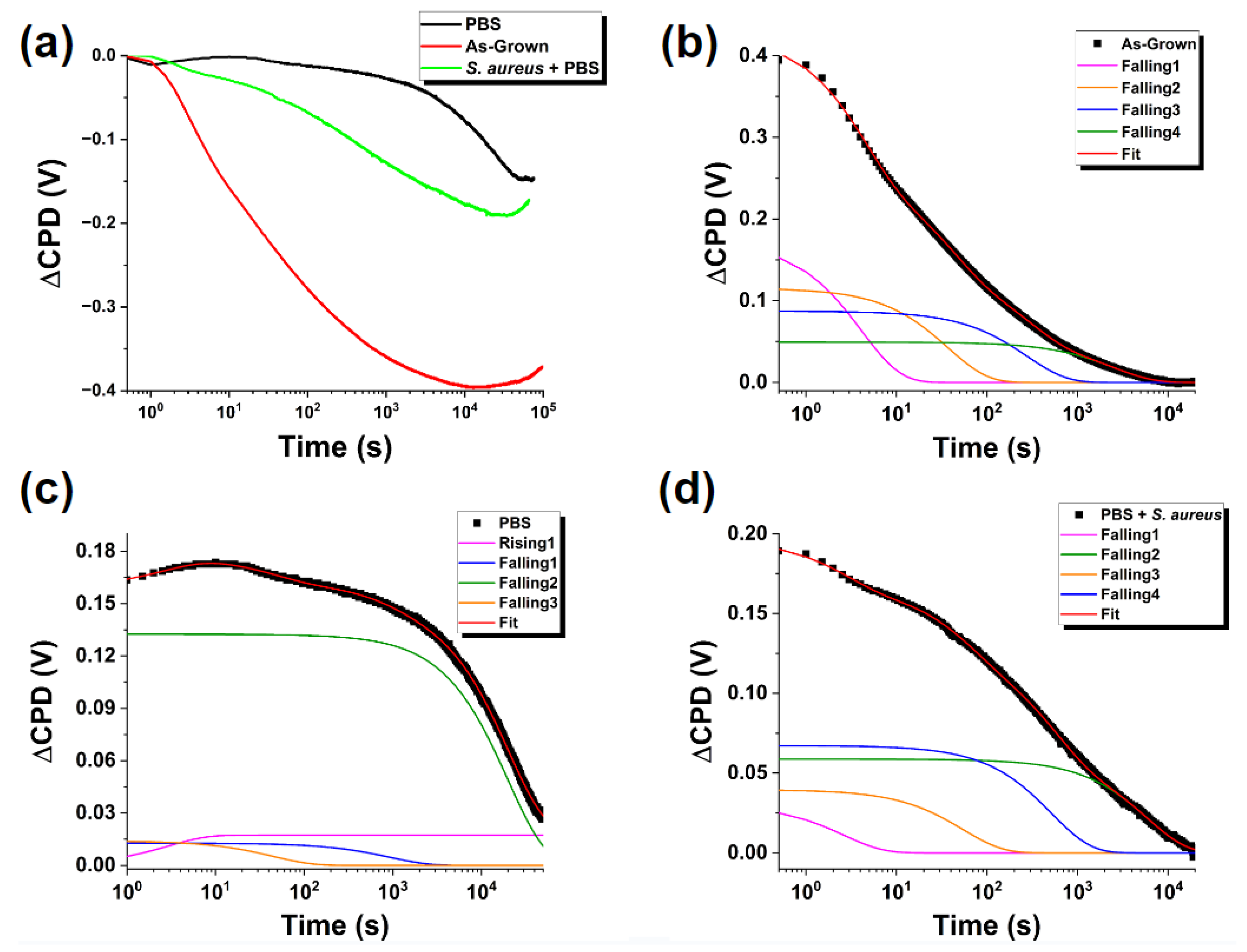
| As-Grown | PBS | PBS + S. aureus | |
|---|---|---|---|
| V0(V) | −3.96 × 10⁻¹ | −1.75 × 10⁻¹ | −1.88 × 10⁻¹ |
| ±3.04 × 10⁻⁴ | ±3.56 × 10⁻⁴ | ±2.42 × 10⁻⁴ | |
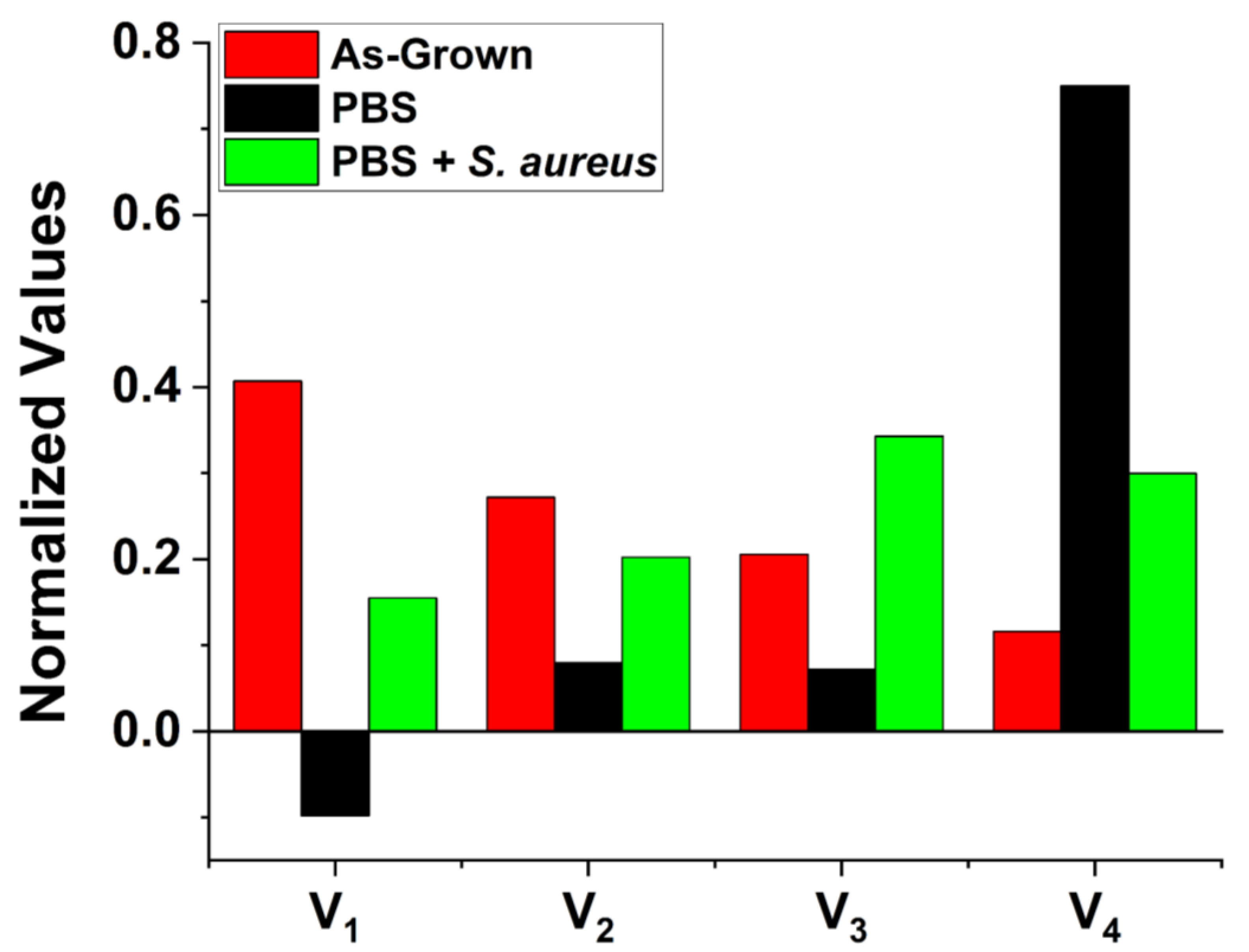
| V1(V) | 1.73 × 10⁻¹ | 1.73 × 10⁻² † | 3.04 × 10⁻² |
| ±1.03 × 10⁻³ | ±2.74 × 10⁻⁴ | ±2.12 × 10⁻⁴ | |
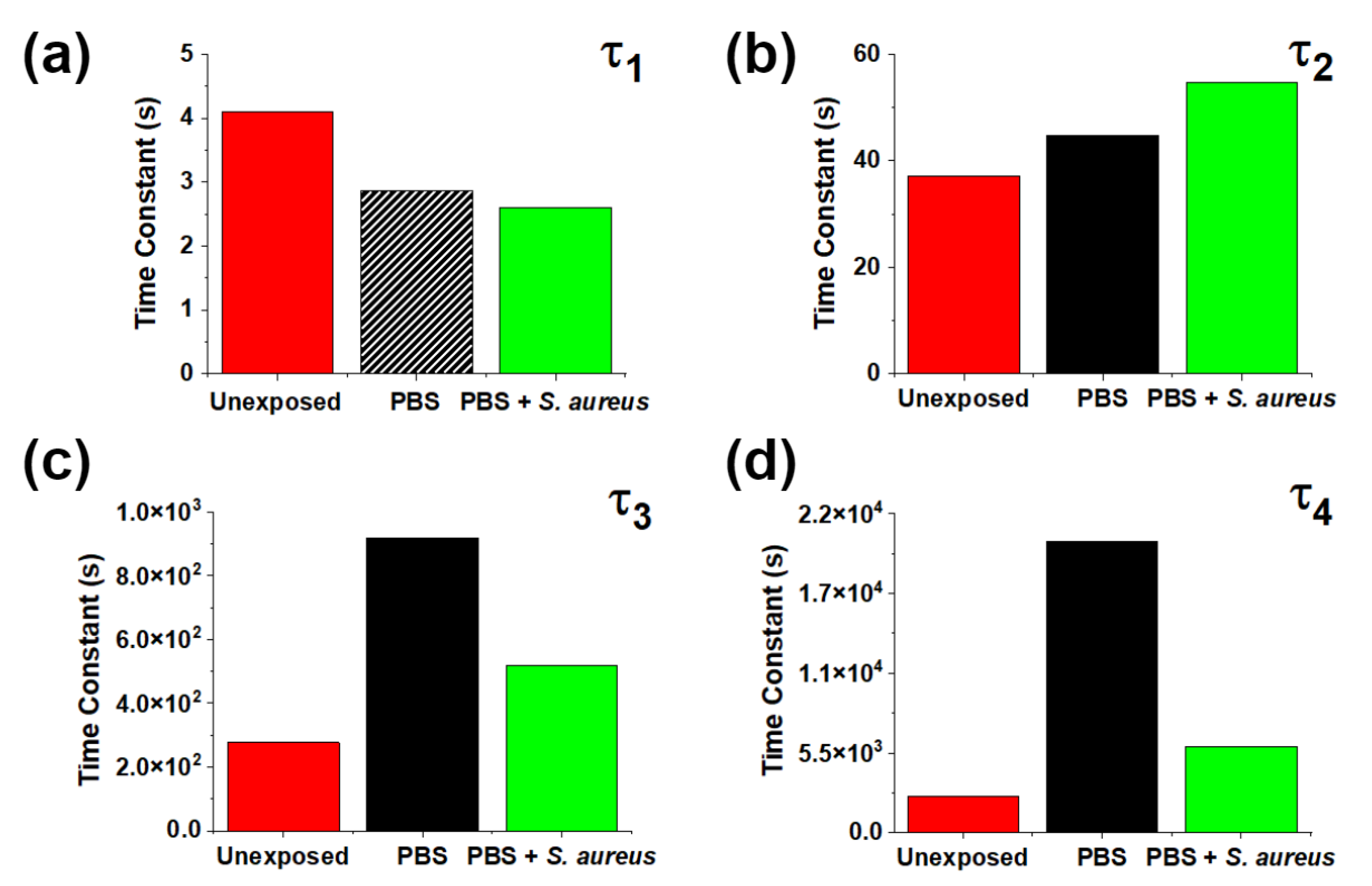
| τ1(s) | 4.09 | 2.87 † | 2.61 |
| ±2.95 × 10⁻¹ | ±1.02 × 10⁻¹ | ±0.04 | |
| V2(V) | 1.15 × 10⁻¹ | 1.40 × 10⁻² | 3.97 × 10⁻² |
| ±1.32 × 10⁻³ | ±2.03 × 10⁻⁴ | ±3.96 × 10⁻⁴ | |
| τ2(s) | 3.71 × 101 | 4.48 × 101 | 5.47 × 101 |
| ±9.16 × 10⁻¹ | ±1.55 | ±1.05 | |
| V3(V) | 8.73 × 10⁻² | 1.27 × 10⁻² | 6.73 × 10⁻² |
| ±1.32 × 10⁻³ | ±1.85 × 10⁻⁴ | ±4.11 × 10⁻⁴ | |
| τ3(s) | 2.77 × 10² | 9.19 × 10² | 5.19 × 10² |
| ±9.13 | ±3.31 × 101 | ±7.51 | |
| V4(V) | 4.93 × 10⁻² | 1.32 × 10⁻¹ | 5.89 × 10⁻² |
| ±1.05 × 10⁻³ | ±1.87 × 10⁻⁴ | ±3.20 × 10⁻⁴ | |
| τ4(s) | 2.53 × 10³ | 2.01 × 10⁴ | 5.97 × 10³ |
| ±9.02 × 101 | ±1.00 × 10² | ±9.50 × 101 |
Disclaimer/Publisher’s Note: The statements, opinions and data contained in all publications are solely those of the individual author(s) and contributor(s) and not of MDPI and/or the editor(s). MDPI and/or the editor(s) disclaim responsibility for any injury to people or property resulting from any ideas, methods, instructions or products referred to in the content. |
© 2023 by the authors. Licensee MDPI, Basel, Switzerland. This article is an open access article distributed under the terms and conditions of the Creative Commons Attribution (CC BY) license (https://creativecommons.org/licenses/by/4.0/).
Share and Cite
Johnson, D.A.; Reeks, J.M.; Caron, A.J.; McGillivray, S.M.; Wiglusz, R.J.; Strzhemechny, Y.M. Surface Photovoltage Response of ZnO to Phosphate-Buffered Saline Solution with and without Presence of Staphylococcus aureus. Nanomaterials 2023, 13, 1652. https://doi.org/10.3390/nano13101652
Johnson DA, Reeks JM, Caron AJ, McGillivray SM, Wiglusz RJ, Strzhemechny YM. Surface Photovoltage Response of ZnO to Phosphate-Buffered Saline Solution with and without Presence of Staphylococcus aureus. Nanomaterials. 2023; 13(10):1652. https://doi.org/10.3390/nano13101652
Chicago/Turabian StyleJohnson, Dustin A., John M. Reeks, Alexander J. Caron, Shauna M. McGillivray, Rafal J. Wiglusz, and Yuri M. Strzhemechny. 2023. "Surface Photovoltage Response of ZnO to Phosphate-Buffered Saline Solution with and without Presence of Staphylococcus aureus" Nanomaterials 13, no. 10: 1652. https://doi.org/10.3390/nano13101652
APA StyleJohnson, D. A., Reeks, J. M., Caron, A. J., McGillivray, S. M., Wiglusz, R. J., & Strzhemechny, Y. M. (2023). Surface Photovoltage Response of ZnO to Phosphate-Buffered Saline Solution with and without Presence of Staphylococcus aureus. Nanomaterials, 13(10), 1652. https://doi.org/10.3390/nano13101652