Article

CdS-Modified TiO2 Nanotubes with Heterojunction Structure: A Photoelectrochemical Sensor for Glutathione

1
College of Chemistry, The Key Laboratory of Advanced Energy Materials Chemistry (Ministry of Education), Tianjin Key Laboratory of Metal and Molecule-Based Material Chemistry, Nankai University, Tianjin 300071, China
2
Chemistry and Chemical Engineering College, Xingtai University, Xingtai 054000, China
3
Department of Microelectronics, Nankai University, Tianjin 300350, China
4
National Demonstration Center for Experimental Chemistry Education (Nankai University), Tianjin 300071, China
*
Author to whom correspondence should be addressed.
Current address: The High School Affiliated to China University of Mining and Technology, Xuzhou 221000, China.
Nanomaterials 2023, 13(1), 13; https://doi.org/10.3390/nano13010013
Submission received: 23 September 2022 / Revised: 20 November 2022 / Accepted: 29 November 2022 / Published: 20 December 2022
(This article belongs to the Section 2D and Carbon Nanomaterials)

Abstract

:
The formation of heterojunction structures can effectively prevent the recombination of photogenerated electron–hole pairs in semiconductors and result in the enhancement of photoelectric properties. Using TiO2 nanotubes (prepared using the hydrothermal-impregnation method) as carriers, CdS-TiO2NTs were fabricated as a photoelectrochemical (PEC) sensor, which can be used under visible light and can exhibit good PEC performance due to the existence of the heterojunction structure. The experimental results show that the prepared CdS-TiO2NTs electrode had a linear response to 2–16 mM glutathione (GSH). The sensor’s sensitivity and detection limit (LOD) were 102.9 µA·mM−1 cm−2 and 27.7 µM, respectively. Moreover, the biosensor had good stability, indicating the potential application of this kind of heterojunction PEC biosensor.

1. Introduction

Glutathione (GSH) is a free-radical scavenger in the body and plays a primary role in functions such as immune regulation, gene expression regulation, enzyme activity, etc. [1]. Many human diseases can influence GSH concentration, and GSH detection is very important due to its significance in physiological circumstances [2]. Among different biomolecule detection methods, photoelectrochemical (PEC) detection is prominent due to its high response speed, high sensitivity, and simple instruments [3].
PEC sensors’ foundations are based on the relation between concentrations of the detected substances and photocurrents. The PEC sensor, which is commonly fabricated by photoactive materials, is firstly photoexcitated to form electron–hole pairs [4,5,6]. During the reaction of photoexcitation species with analytes, the photocurrent is obtained. Thus, a crucial aspect of a prominent PEC sensor is the selecting of an excellent photoelectrode, which will control the sensor’s sensitivity, detection limit (LOD), etc. [7].
Nowadays, the electrode materials with photoelectric responses are focused on inorganic materials, such as TiO2 [8], CdS [9,10], ZnO [11], and CuO [12]. Among them, TiO2 is most prominent due to its stable chemical properties, high PEC performance, and good biocompatibility [13]. Compared with common powder materials, TiO2 nanotubes are more prominent for their unique 1D nanostructure, which can shorten the distance between photogenerated carriers and accelerate electron transport [14]. The modifiers and aimed samples can also be highly dispersed across the nanotubes, and result in a quick response. As is common sense, UV rays are harmful to most biological molecules. Due to its big band gap, the pure TiO2 photoelectrode can only be used under UV light, which limits the utility of TiO2 as a biosensor [15]. The fast recombination of photogenerated charges is another shortcoming of TiO2, which decreases the PEC sensor’s sensitivity.
To improve the PEC performance of TiO2, such as narrowing its energy band and improving its electron-hole separation efficiency, different metals and nonmetals (C [16], S [17], N [18], Fe [19], Ti [20], Bi [21], and so on) were used as dopants, and precious metals (Au [22], Pt [23,24], Ag [25], and so on) were used as modifications. Doping and modification can decrease the recombination ratio of photoelectrons and photoholes and promote their separation in TiO2 [26,27]. However, the impurity level formed by element doping may become the recombination site of the photogenerated electrons and holes, and precious metals are expensive and easily lost in the PEC reaction process.
As has been generally reported, constructing heterojunctions with other semiconductors (CdS [8], CuO [28], MoS2 [29], g-C3N4 [30], SnS2 [31], and so on) is also an effective way to improve TiO2’s PEC performance. Among the above-mentioned semiconductors, CdS’s band gap is 2.3 eV, which can lead to a higher absorption coefficient under visible-light illumination. However, the unstable properties of CdS limits its practical utility. Forming composite materials with stable TiO2 is a good solution to this limitation. In our previous work, we have prepared TiO2 nanotube-supported CdS as a photocatalyst. Compared with pure TiO2 nanotubes or CdS nanoparticles, the fabricated composite exhibited better photoelectric properties [32]. According to the simulation of density functional theory (DFT), the formation of the heterojunction structure can also be considered the existence of an internal electric field [33]. After the heterojunction structure is formed in the CdS-TiO2 system, the charge-transfer barrier is decreased, and the photogenerated electrons and holes can be effectively separated, resulting in a faster charge transport rate and lower recombination efficiency. Therefore, the formed CdS-TiO2 sensors are not only active under visible light but can also lead to better sensing properties being obtained for biomolecules [34,35]. It has been reported that CdS modification can obtain 2.9-fold PEC signal enhancement compared with that of 0.1% for Fe-TiO2 [36].
Based on the improved PEC properties of the heterojunction structure, a TiO2 nanotube was modified with powdery CdS to form a CdS-TiO2NTs electrode, which was further applied for GSH detection in this paper. Through the characterization, investigation of PEC sensitivity, and stability of the CdS-TiO2NTs electrode, the practicality and sensing mechanism of the fabricated CdS-TiO2NTs sensor is also discussed.

2. Experiment

2.1. Synthesis of CdS-TiO2NTs

Hydrogen-titanate nanotubes prepared using the hydrothermal method were soaked in titanium sol for 10 min, naturally dried in the air, and calcined at 400 °C for 2 h to obtain TiO2NTs [37]. A certain amount of sulfur powder was dissolved in tetrahydrofuran solution. After a quantity of Cd(NO3)2·4H2O was slowly added through stirring, the prepared TiO2NTs were added. The calculated CdS/TiO2 molar ratio was 1:1. NaBH4 solution was slowly added under the condition of an ice water bath and N2. After being stirred for 30 min, the solution was ultrasonic treated for 30 min, and then stirred overnight. The samples were successively cleaned by tetrahydrofuran, water, and ethanol, and then treated in a vacuum drying oven to obtain CdS-TiO2NTs.

2.2. Preparation of CdS-TiO2NTs Electrode

20 µL of CdS-TiO2NTs solution was dropped on a glassy-carbon electrode. After the dropped solution was dried, the rest of the solution was added, and 15 µL of 3% chitosan solution was dropped. After naturally air drying overnight, the CdS-TiO2NTs electrode was obtained.

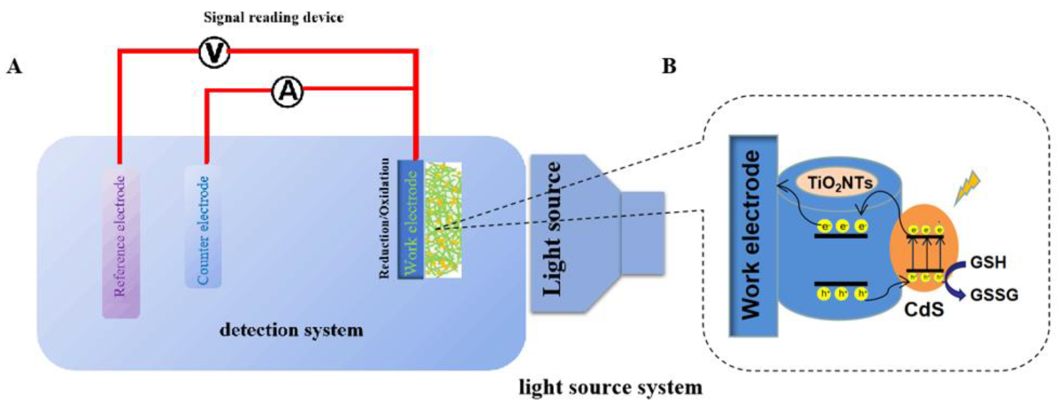
2.3. Photoelectrochemical Testing

The CdS-TiO2NTs electrode’s electrochemical properties were investigated using the three-electrode system from the Zahner electrochemical workstation. The CdS-TiO2NTs electrode, Ag/AgCl electrode, and Pt electrode were used as the working electrode, reference electrode, and auxiliary electrode, respectively. An LED lamp with visible light (429 nm, 35 W/m2) was used as the illumination source, and the pH of electrolytes was maintained at 7.4.

2.4. Instrument Model

XRD patterns were obtained using the Rigaku D/MAX-2500 X-ray diffractometer (Rigaku SmartLab, Rigaku Corporation, Tokyo, Japan). Scanning electron microscope (SEM) and energy dispersive spectrometer (EDS) images were observed on JSM-7800F (Japanese electronics, Beijing, China). To obtain TEM and HR-TEM images, JEM-2800 (Japanese electronics, Shanghai, China) and the Talos F200X G2 transmission electron microscope were used, respectively. X-ray photoelectron spectroscopy (Kratos Axis Ultra DLD (Kratos Analytical Ltd., Manchester, UK), Al target, double anode Al/Mg target) recorded the XPS patterns. All the electrochemical experiments were performed on the Zennium electrochemical workstation (Kronach–Gundelsdorf, Germany, bias voltage: 0.5 V).

3. Results and Discussion

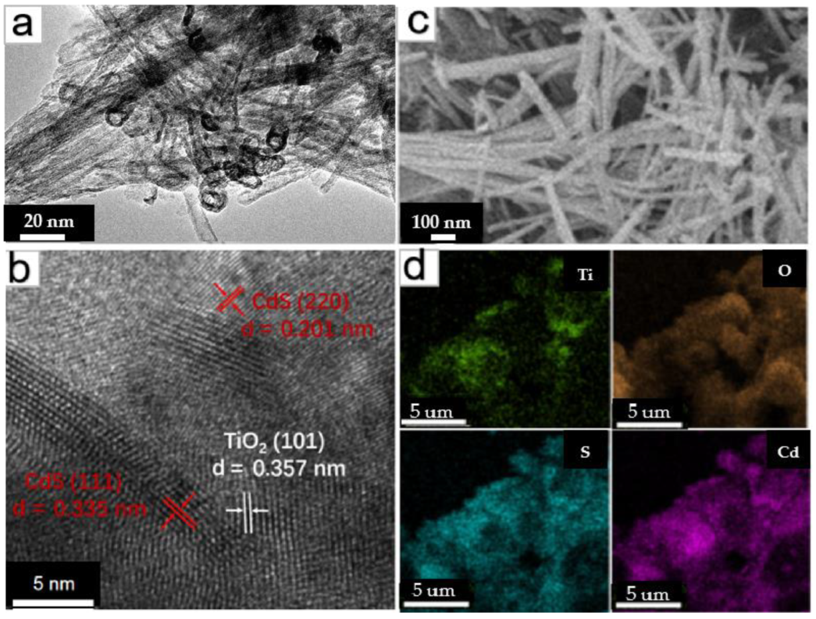
3.1. Surface Morphology Characterization

Figure 1a,b shows the TEM and high-resolution TEM images of CdS-TiO2NTs. From Figure 1a, it can be clearly seen that the open-ended nanotubes have a diameter of about 10 nm. Compared with the pure TiO2NTs, there are many substances with dark color and ring shapes in the tubular materials, which should be the shadow of heavy metal cadmium. In Figure 2b, the measured lattice spacings of 0.335 nm, 0.201 nm, and 0.357 nm, correspond to the (111) and (220) planes of CdS and the (101) plane of anatase TiO2, respectively. It demonstrates the coexistence and association of CdS and TiO2NTs and the formation of the heterojunction structure. To verify the existence of Cd and S more directly, SEM and corresponding mapping images (Figure 1c,d) of the CdS-TiO2NTs were obtained. It is evident that the TiO2NTs are modified by cadmium and sulfide, and the atomic ratio of Cd/S is close to 1:1, corresponding to the composition of CdS. Some regions in Figure 1d exhibit different element contents. This could be attributed to the lack of a uniform distribution in the components of CdS-TiO2NTs.
XPS measurement was used to characterize the chemical composition of the CdS-TiO2NTs. Figure 2 shows the full XPS spectrum (Figure 2a), and Ti 2p, O 1s, Cd 3d, and S 2p high-solution spectra (Figure 2b–e) of CdS-TiO2NTs. As shown in Figure 2a, the CdS-TiO2NTs sample contained chemical elements of O, Ti, Cd, and S. Two Ti 2p peaks emerged at 458.2 eV and 464.3 eV in the CdS-TiO2NTs that corresponded to Ti 2p1/2 and Ti 2p3/2 (Figure 2b), indicating the chemical state Ti4+ of titanium oxide. As displayed in Figure 2c, the O 1s XPS spectrum showed two peaks, with the one at 531.4 eV corresponding to lattice oxygen, and the other at 532.9eV corresponding to adsorbed oxygen. As shown in Figure 2d, the peaks at BE of 404.7 eV and 411.5 eV corresponded to Cd 3d5/2 and Cd 3d3/2, respectively, indicating the existence of Cd2+ in CdS-TiO2NTs. The high-solution S 2p peaked at 161.2 eV and 162.4 eV, which is evidence for S2- in CdS. In summary, the XPS results in Figure 2a–e show the coexistence of CdS and TiO2, consistent with the results of TEM and SEM.
In order to investigate the composition of the prepared materials, XRD patterns were performed. Figure 3 shows the XRD patterns of TiO2NTs and CdS-TiO2NTs. Most of the peaks on pattern a in Figure 3 corresponded to that of anatase TiO2 (JCPDS# 21-1271), indicating that the TiO2NTs are mainly anatase type. The peak at 11° corresponded to the (200) crystal plane of H2Ti3O7 (JCPDS # 47-0561), which is the undecomposed hydrogen titanate. As shown on pattern b in Figure 3, CdS diffraction peaks (JCPDS# 10-0454) emerged in CdS-TiO2NTs. The addition of CdS weakened the diffraction peaks of TiO2, which could be attributed to the high concentration of CdS in TiO2. Moreover, the wide diffraction peaks indicate the small particle sizes of CdS and TiO2. By combining the results of the above TEM, SEM, and XPS analyses, the successful loading of CdS on TiO2 nanotubes can be confirmed.
EIS is commonly used to detect electrode–electrolyte interactions and charge-transfer phenomena [38]. The arc radius in the EIS Nyquist plot reveals the charge-transfer resistance, and charge-transfer ability of the electrode [39]. As shown in Figure 4, the Rs of the samples were almost all the same. However, the Rct for the TiO2NTs and CdS/TiO2NTs were 153 Ω and 102.7 Ω, respectively. Compared with TiO2NTs, the value of the CdS/TiO2NTs was smaller. This could be attributed to the formation of a heterojunction structure in the CdS/TiO2NTs, which resulted in the rapid transmission of charges and reduced charge-transfer resistance. Under light illumination, the effective separation and transmission of photogenerated carriers resulted in the small Rct of CdS/TiO2NTs. In the PEC process, the small Rct can facilitate the transfer of electrons and result in a high photocurrent.

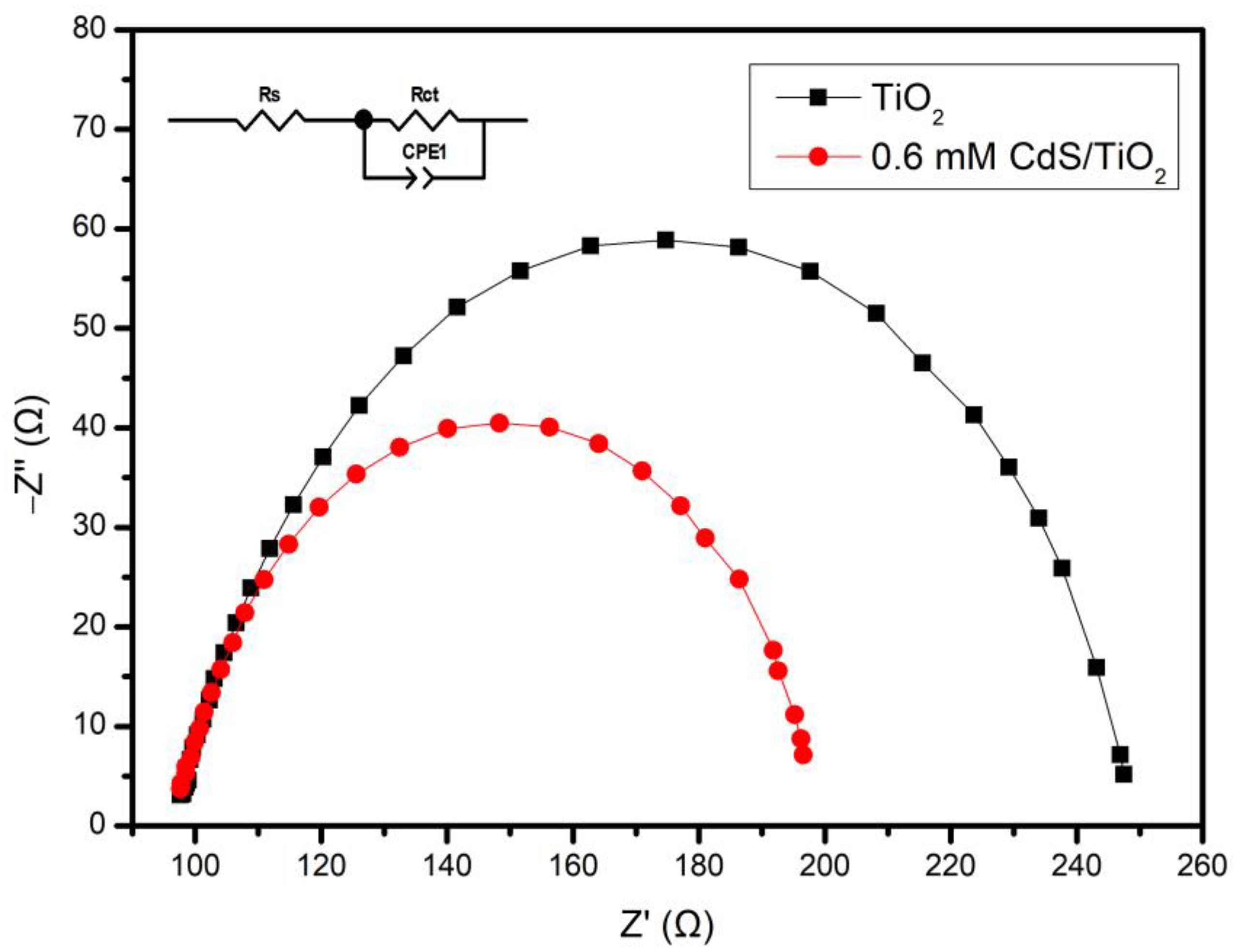
3.2. Electrochemical Characterization

The sensitivity of the fabricated CdS-TiO2NTs for GSH was monitored under 429 nm LED-light irradiation. Figure 5a shows the photocurrent–time curves of the CdS-TiO2NTs in GSH solutions with 18 different concentrations, which ranged from 0.5 mM to 17 mM. Figure 5b displays the relationship between the GSH concentrations and the photocurrents of the CdS-TiO2NTs. The photocurrent-GSH concentration showed a linear relationship in the range of 2–16 mM. The regression equation is Y = −20.2X + 354 (R2 = 0.9953) (Y referring to photocurrent, and X referring to GSH concentration). The detection limit (LOD) of the CdS-TiO2NTs biosensor was calculated based on the equation of LOD = 3 Sb/S (Sb is the standard deviation of the blank signal, and S is the sensitivity of the fabricated sensor), which was equal to 27.7 µM.
The stability of the CdS-TiO2NTs sensor was also investigated. The CdS-TiO2NTs electrode was stored in air at room temperature. Its current response to PBS solution was monitored 9 times under 429 nm LED-light irradiation (bias voltage 0.3 V). As shown in Figure 6, the CdS-TiO2NTs maintained at least 89.3% of the initial value during this test, suggesting the relatively high stability of the fabricated CdS-TiO2NTs electrode [39].
TiO2NTs exhibit no sensitivity to GSH under visible light. However, the CdS-TiO2NTs presented good sensitivity under the same condition. Obviously, the modification of CdS endowed the TiO2 with PEC activity under visible light. Moreover, the heterojunction structure of the CdS-TiO2 composite induced rapid transmission of the charges. In our previous work, a density functional theory (DFT) simulation was conducted for a similar CdS-TiO2 composite [40]. As indicated by the simulation results, tunnels formed at the interlayer section of the CdS-TiO2 composite and facilitated the charge transfer between CdS and TiO2. During the PEC process, CdS was firstly excited under visible light. Because the conduction bands (CBs) of CdS are higher than that of TiO2, the excited electrons in CdS transferred to the CBs of TiO2 and generated a photocurrent.
Based on the experiment results and our former simulation results, the PEC detection process for GSH by CdS-TiO2NTs electrode biosensor in this work was preliminarily put forward to explain the improved performance of CdS-TiO2NTs, as shown in Figure 7. Under the illumination of visible light, electrons are photo-generated in CdS and transferred from the CBs of CdS to the CBs of TiO2, reaching the glassy carbon electrode and eventually the counter electrode. Meanwhile, the holes in CdS react with GSH to form GSSG. During this process, the inner self-combination of electrons and holes in both CdS and TiO2 are decreased, and electrons and holes are accelerated at different parts. Thus, the association of CdS with TiO2 can result in superior PEC performance.

4. Conclusions

In summary, CdS-TiO2NTs with a heterojunction structure were successfully designed as a PEC sensor for GSH detection. The CdS-TiO2NTs exhibited good PEC properties due to the effective charge transfer between CdS and TiO2. The photocurrent signals of the CdS-TiO2NTs biosensor were linear to 2–16 mM GSH solution. In addition to GSH, future applications of this PEC sensor to other substances are anticipated.

Author Contributions

Conceptualization and methodology, G.-N.H. and S.-S.Z.; software, G.-N.H., Y.-L.L. and J.-X.L.; validation, G.-N.H. and S.-S.Z.; formal analysis, G.-N.H., Z.Y. and B.-L.Z.; investigation, G.-N.H. and S.-S.Z.; resources, B.-L.Z. and W.-P.H.; writing—original draft preparation, G.-N.H.; writing—review and editing, B.-L.Z.; visualization, G.-N.H. and B.-L.Z.; supervision, B.-L.Z., S.-M.Z. and W.-P.H.; project administration, B.-L.Z., S.-M.Z. and W.-P.H.; funding acquisition, B.-L.Z., S.-M.Z. and W.-P.H. All authors have read and agreed to the published version of the manuscript.

Funding

This research was supported by the National Natural Science Foundation, China (grant number 61871240); the Natural Science Foundation of Tianjin City, China (grant number 21JCYBJC00330); and the Fundamental Research Funds for Central Universities, Nankai University, China (grant number 63221205).

Institutional Review Board Statement

Not applicable.

Informed Consent Statement

Not applicable.

Data Availability Statement

The data presented in this study are available on request from the corresponding author.

Conflicts of Interest

The authors declare no conflict of interest.

References

  1. Li, Z.; Zhang, J.; Li, Y.; Zhao, S.; Zhang, P.; Zhang, Y.; Bi, J.; Liu, G.; Yue, Z. Carbon dots based photoelectrochemical sensors for ultrasensitive detection of glutathione and its applications in probing of myocardial infarction. Biosens. Bioelectron. 2018, 99, 251–258. [Google Scholar] [CrossRef]
  2. Sui, M.R.; Zhao, Y.; Ni, Z.H.; Gu, X.Q. Photoelectrochemical performance and biosensor application for glutathione (GSH) of W-doped BiVO4 thin films. J. Mater. Sci. Mater. Electron. 2018, 12, 10109–10116. [Google Scholar] [CrossRef]
  3. Shu, J.; Qiu, Z.L.; Lv, S.Z.; Zhang, K.Y.; Tang, D.P. Plasmonic enhancement coupling with defect-engineered TiO2-x: A mode for sensitive photoelectrochemical biosensing. Anal. Chem. 2018, 4, 2425–2429. [Google Scholar] [CrossRef] [PubMed]
  4. Zhang, J.; Tu, L.; Zhao, S.; Liu, G.; Wang, Y.; Wang, Y.; Yue, Z. Fluorescent gold nanoclusters based photoelectrochemical sensors for detection of H2O2 and glucose. Biosens. Bioelectron. 2015, 67, 296–302. [Google Scholar] [CrossRef] [PubMed]
  5. Tu, L.; Liu, G.; Zhang, W.; Qin, J.; Yue, Z. Doped QDs based photoelectrochemical sensors for detection of H2O2 and Glucose. IEEE J. Sel. Top. Quantum. 2014, 20, 175–183. [Google Scholar]
  6. Wu, S.; Song, H.; Song, J.; He, C.; Ni, J.; Zhao, Y.; Wang, X. Development of triphenylamine functional dye for selective photoelectrochemical sensing of cysteine. Anal. Chem. 2014, 86, 5922–5928. [Google Scholar] [CrossRef]
  7. Gao, Y.; Zhang, X.C.; Yan, J.Y.; Guan, Q.L.; Xing, Y.H.; Song, W.B. Split-type cascaded silver etching for “on-super off” PEC-EC dual-mode immunosensing of α-SynO. Sensor. Actuat. B-Chem. 2022, 358, 131449. [Google Scholar] [CrossRef]
  8. Tao, J.; Gong, Z.; Yao, G.; Cheng, Y.; Zhang, M.; Lv, J.; Shi, S.; He, G.; Chen, X.; Sun, Z. Enhanced photocatalytic and photoelectrochemical properties of TiO2 nanorod arrays sensitized with CdS nanoplates. Ceram. Int. 2016, 42, 11716–11723. [Google Scholar] [CrossRef]
  9. Okoth, O.K.; Yan, K.; Feng, J.; Zhang, J. Label-free photoelectrochemical aptasensing of diclofenac based on gold nanoparticles and graphene-doped CdS. Sens. Actuators B 2018, 256, 334–341. [Google Scholar] [CrossRef]
  10. Zhang, J.; Wang, L.; Liu, X.; Li, X.A.; Huang, W. High-performance CdS–ZnS core–shell nanorod array photoelectrode for photoelectrochemical hydrogen generation. J. Mater. Chem. A 2015, 3, 535–541. [Google Scholar] [CrossRef]
  11. Wang, T.; Jin, B.; Jiao, Z.; Lu, G.; Ye, J.; Bi, Y. Electricfield-directed growth and photoelectro chemical properties of cross-linked Au-ZnO hetero-nanowire arrays. Chem. Commun. 2015, 51, 2103–2106. [Google Scholar] [CrossRef] [PubMed]
  12. Lu, N.; Shao, C.L.; Li, X.H.; Shen, T.; Zhang, M.Y.; Miao, F.J.; Zhang, P.; Zhang, X.; Wang, K.X.; Zhang, Y.; et al. CuO/Cu2O nanofibers as electrode materials for non-enzymatic glucose sensors with improved sensitivity. RSC Adv. 2014, 4, 31056–31061. [Google Scholar] [CrossRef]
  13. Tu, W.; Dong, Y.; Lei, J.; Ju, H. Low-potential photoelectrochemical biosensing using porphyrin-functionalized TiO2 nanoparticles. Anal. Chem. 2010, 82, 8711–8716. [Google Scholar] [CrossRef] [PubMed]
  14. Simonetta, P.; Laura, M.; Michele, M.; Annalisa, V. Trend in using TiO2 nanotubes as photoelectrodes in PEC processes for wastewater treatment. Curr. Opin. Electrochem. 2021, 28, 100699. [Google Scholar]
  15. Zhu, J.; Huo, X.; Liu, X.; Ju, H. Gold nanoparticles deposited polyaniline-TiO2 nanotube for surface plasmon resonance enhanced photoelectro chemical biosensing. ACS Appl. Mater. Interfaces 2016, 8, 341–349. [Google Scholar] [CrossRef]
  16. Atchudan, R.; Muthuchamy, N.; Edison, T.N.J.I.; Perumal, S.; Vinodh, R.; Park, K.H.; Lee, Y.R. An ultrasensitive photoelectrochemical biosensor for glucose based on bio-derived nitrogen-doped carbon sheets wrapped titanium dioxide nanoparticles. Biosens. Bioelectron. 2019, 126, 160–169. [Google Scholar] [CrossRef]
  17. Singh, S.C.; Swarnkar, R.K.; Gopal, R. Synthesis of Titanium Dioxide Nanomaterial by Pulsed Laser Ablation in Water. J. Nanosci. Nanotechnol. 2009, 9, 5367–5371. [Google Scholar] [CrossRef]
  18. Huo, G.N.; Ma, L.L.; Liu, X.T.; Zhou, K.H.; Suo, Z.C.; Liu, X.T.; Suo, Z.C.; Zhang, F.F.; Zhu, B.L.; Zhang, S.M.; et al. Fabrication and Photoelectrochemical Sensitivity of N, F-TiO2NTs/Ti with 3D structure. Microchem. J. 2022, 172, 106957. [Google Scholar] [CrossRef]
  19. Tang, J.; Kong, B.; Wang, Y.; Xu, M.; Wang, Y.; Wu, H.; Zheng, G. Photoelectrochemical detection of glutathione by IrO2-hemin-TiO2 nanowire arrays. Nano Lett. 2013, 13, 5350–5354. [Google Scholar] [CrossRef]
  20. Guo, C.Y.; Huo, H.H.; Han, X.; Xu, C.L.; Li, H.L. Ni/CdS Bifunctional Ti@TiO2 Core−Shell Nanowire Electrode for High-Performance Nonenzymatic Glucose Sensing. Anal. Chem. 2014, 86, 876–883. [Google Scholar] [CrossRef]
  21. Ruan, Y.F.; Zhang, N.; Zhu, Y.C.; Zhao, W.W.; Xu, J.J.; Chen, H.Y. Photoelectrochemical bioanalysis platform of gold nanoparticles equipped perovskite Bi4NbO8Cl. Anal. Chem. 2017, 89, 7869–7875. [Google Scholar] [CrossRef] [PubMed]
  22. Zhu, B.; Li, K.; Feng, Y.; Zhang, S.; Wu, S.; Huang, W. Synthesis and catalytic performance of gold-loaded TiO2 nanofibers. Catal. Lett. 2007, 118, 55–58. [Google Scholar] [CrossRef]
  23. Tian, J.; Zhao, P.; Zhang, S.S.; Huo, G.N.; Suo, Z.C.; Yue, Z.; Zhang, S.M.; Huang, W.P.; Zhu, B.L. Platinum and Iridium Oxide Co-modified TiO2 Nanotubes Array Based Photoelectrochemical Sensors for Glutathione. Nanomaterials 2020, 10, 522. [Google Scholar] [CrossRef] [PubMed] [Green Version]
  24. Yang, P.; Jin, S.Y.; Xu, Q.Z.; Yu, S.H. Decorating Pt Co bimetallic alloy nanoparticles on graphene as sensors for glucose detection by catalyzing luminol chemiluminescence. Small 2013, 9, 199–204. [Google Scholar] [CrossRef] [PubMed]
  25. Ding, Y.; Wang, Y.; Su, L.; Zhang, H.; Lei, Y. Preparation and characterization of NiO–Ag nanofibers, NiO nanofibers, and porous Ag: Towards the development of a highly sensitive and selective non-enzymatic glucose sensor. J. Mater. Chem. 2010, 20, 9918–9926. [Google Scholar] [CrossRef]
  26. Bergamonti, L.; Predieri, G.; Paz, Y.; Fornasini, L.; Lottici, P.P.; Bondioli, F. Enhanced self-cleaning properties of N-doped TiO2 coating for Cultural Heritage. Microchem. J. 2017, 133, 1–12. [Google Scholar] [CrossRef]
  27. Shetti, N.P.; Malode, S.J.; Nayak, D.S.; Aminabhavi, T.M.; Reddy, K.R. Nanostructured silver doped TiO2/CNTs hybrid as an efficient electrochemical sensor for detection of anti-inflammatory drug, cetirizine. Microchem. J. 2019, 150, 104–124. [Google Scholar] [CrossRef]
  28. Zhu, Y.H.; Xu, Z.W.; Yan, K.; Zhao, H.B.; Zhang, J.D. One-Step Synthesis of CuO-Cu2O Heterojunction by Flame Spray Pyrolysis for Cathodic Photoelectrochemical Sensing of l-Cysteine. ACS Appl. Mater. Interfaces 2017, 9, 40452–40460. [Google Scholar] [CrossRef]
  29. Li, Y.Y.; Wang, J.H.; Luo, Z.J.; Chen, K.; Cheng, Z.Q.; Ma, L.; Ding, S.J.; Zhou, L.; Wang, Q.Q. Plasmon-Enhanced Photoelectrochemical Current and Hydrogen Production of (MoS2-TiO2)/Au Hybrids. Sci. Rep. 2015, 7, 7178. [Google Scholar] [CrossRef] [Green Version]
  30. Wang, L.B.; Fei, X.G.; Zhang, L.Y.; Yu, J.G.; Cheng, B.; Ma, Y.H. Solar fuel generation over nature-inspired recyclable TiO2/g-C3N4 S-scheme hierarchical thin-film photocatalyst. J. Mater. Sci. Technol. 2022, 112, 1–10. [Google Scholar] [CrossRef]
  31. Yao, P.; Yu, S.; Shen, H.; Yang, J.; Min, L.; Yang, Z.; Zhu, X. A TiO2-SnS2 nanocomposite as a novel matrixfor the development of an enzymatic electrochemical glucose biosensor. New J. Chem. 2019, 43, 16748–16752. [Google Scholar] [CrossRef]
  32. Li, H.; Zhu, B.; Feng, Y.; Wang, S.; Zhang, S.; Huang, W. Synthesis, characterization of TiO2 nanotubes-supported MS (TiO2NTs@MS, M = Cd, Zn) and their photocatalytic activity. J. Solid State Chem. 2007, 180, 2136–2142. [Google Scholar] [CrossRef]
  33. Low, J.X.; Yu, J.G.; Jaroniec, M.; Wageh, S.; Al-Ghamdi, A.A. Heterojunction photocatalysts. Adv. Mater. 2017, 29, 1601694. [Google Scholar] [CrossRef] [PubMed]
  34. Huo, H.H.; Xu, Z.D.; Zhang, T.; Xu, C.L. Ni/CdS/TiO2 nanotube array heterostructures for high performance photoelectrochemical biosensing. J. Mater. Chem. A 2015, 3, 5882–5888. [Google Scholar] [CrossRef]
  35. Cai, J.; Sheng, P.T.; Zhou, L.P.; Shi, L.; Wang, N.Y.; Cai, Q.Y. Label-free photoelectrochemical immunosensor based on CdTe/CdS co-sensitized TiO2 nanotube array structure for octachlorostyrene detection. Biosens. Bioelectron. 2013, 50, 66–71. [Google Scholar] [CrossRef]
  36. Fan, D.W.; Wu, D.; Cui, J.L.; Chen, Y.C.; Ma, H.M.; Liu, Y.X.; Wei, Q.; Du, B. An ultrasensitive label-free immunosensor based on CdS sensitized Fe-TiO2 with high visible-light photoelectrochemical activity. Biosens. Bioelectron. 2015, 74, 843–848. [Google Scholar] [CrossRef]
  37. An, H.Q.; Zhu, B.L.; Li, J.X.; Zhou, J.; Wang, S.R.; Zhang, S.M.; Wu, S.H.; Huang, W.P. Synthesis and Characterization of Thermally Stable Nanotubular TiO2 and Its Photocatalytic Activity. J. Phys. Chem. C 2008, 112, 18772–18775. [Google Scholar] [CrossRef]
  38. Alberto, A.M.; Teresa, M.S.; Liliane, G.D.; Maria, F.M. Application of the Mott-Schottky model to select potentials for EIS studies on electrodes for electrochemical charge storage. Electrochim. Acta 2018, 289, 47–55. [Google Scholar]
  39. Ma, Q.L.; Wang, H.T.; Zhang, H.X.; Cheng, X.W.; Xie, M.Z.; Cheng, Q.F. Fabrication of MnO2/TiO2 nano-tube arrays photoelectrode and its enhanced visible light photoelectrocatalytic performance and mechanism. Sep. Purif. Technol. 2017, 189, 193–203. [Google Scholar] [CrossRef]
  40. Zhang, S.S.; Tian, J.; Yue, Z.; Huo, G.N.; Hu, Z.X.; Zhang, S.M.; Huang, W.P.; Zhu, B.L. Heterostructure-based 3D-CdS/TiO2 nanotubes/Ti: Photoelectrochemical performances and interface simulation investigation. Ceram. Int. 2022, 48, 36731–36738. [Google Scholar] [CrossRef]
Figure 1. TEM (a), HR-TEM (b), and SEM (c) images of the CdS-TiO2NTs and mapping (d) of CdS-TiO2NTs.
Nanomaterials 13 00013 g001
Figure 2. (a) Survey XPS spectrum of the CdS-TiO2NTs and high-resolution spectra for (b) Ti 2p, (c) O 1s, (d) Cd 3d, and (e) S 2p.
Nanomaterials 13 00013 g002aNanomaterials 13 00013 g002b
Figure 3. XRD patterns of TiO2NTs (a) and CdS-TiO2NTs (b).
Nanomaterials 13 00013 g003
Figure 4. EIS of TiO2 and CdS/TiO2NTs.
Nanomaterials 13 00013 g004
Figure 5. (a) Photocurrent response for the detection of different concentrations of GSH (2–16 mM), and (b) corresponding photocurrent-GSH concentration curve of the CdS-TiO2NTs under irradiation of 429 nm.
Nanomaterials 13 00013 g005
Figure 6. Relationship between time and response of the CdS-TiO2NTs electrode.
Nanomaterials 13 00013 g006
Figure 7. Schematic illustration of the PEC process for GSH detection by the CdS-TiO2NTs electrode biosensor ((A) is the detection system and (B) is the work electrode).
Nanomaterials 13 00013 g007
Disclaimer/Publisher’s Note: The statements, opinions and data contained in all publications are solely those of the individual author(s) and contributor(s) and not of MDPI and/or the editor(s). MDPI and/or the editor(s) disclaim responsibility for any injury to people or property resulting from any ideas, methods, instructions or products referred to in the content.

