Article

New Insights into the Magnetic Properties of CoFe2O4@SiO2@Au Magnetoplasmonic Nanoparticles

1
Faculty of Physics, “Babes Bolyai” University, 400084 Cluj-Napoca, Romania
2
Electron Microscopy Center “Prof. C. Crăciun”, Faculty of Biology & Geology, “Babes-Bolyai” University, 400006 Cluj-Napoca, Romania
3
Integrated Electron Microscopy Laboratory, National Institute for Research and Development of Isotopic and Molecular Technologies, 400293 Cluj-Napoca, Romania
4
Department of Bionanoscopy, MedFuture Research Center for Advance Medicine, “Iuliu Hatieganu” University of Medicine and Pharmacy, 400337 Cluj-Napoca, Romania
*
Author to whom correspondence should be addressed.
Nanomaterials 2022, 12(6), 942; https://doi.org/10.3390/nano12060942
Submission received: 20 February 2022 / Revised: 4 March 2022 / Accepted: 10 March 2022 / Published: 12 March 2022

Abstract

:
We report the successful synthesis and a complete magnetic characterization of CoFe2O4@SiO2@Au magnetoplasmonic nanoparticles. The CoFe2O4 magnetic nanoparticles were prepared using the hydrothermal method. A subsequent SiO2 shell followed by a plasmonic Au shell were deposited on the magnetic core creating magnetoplasmonic nanoparticles with a core–shell architecture. A spin-glass-type magnetism was shown at the surface of the CoFe2O4 nanograins. Depending on the external magnetic field, two types of spin-glass were identified and analyzed in correlation with the exchange field acting on octahedral and tetrahedral iron sites. The magnetization per formula unit of the CoFe2O4 core is not changed in the case of CoFe2O4@SiO2@Au nanocomposites. The gold nanoparticles creating the plasmonic shell show a giant diamagnetic susceptibility, dependent on their crystallite sizes.

1. Introduction

The flexibility of spinel ferrite CoFe2O4 or a (Co1−xFex)T(CoxFe2−x)OO4 structure, with a variable occupancy of tetrahedral (T) and octahedral sites (O), provides a wide range of physical properties and applications, particularly in their nanosized form. As a function of the inversion parameter, x, the structure changes from a normal spinel (x = 0) to an inverse spinel type (x = 1). In a bulk state, the CoFe2O4 ferrite has mainly an inverse-type spinel structure and crystallizes in an FCC-type lattice, space group F m 3 ¯ m . The ferrite is ferrimagnetically ordered, and the magnetic moments of the atoms situated in octahedral and tetrahedral sublattices, respectively, being antiparallelly aligned. According to the degree of inversion, a large range of magnetizations can be obtained. The superexchange parameters inside and between magnetic sublattices were determined in the bulk state, starting from a mean field model [1,2].
At the nanometer scale, the magnetic behavior of CoFe2O4 ferrite shows significant differences with respect to that of the bulk state. The crossover to a single domain behavior is 40 nm [3]. The system becomes superparamagnetic between 7 and 10 nm. A core–shell model was proposed for CoFe2O4 nanograins, in which a core of aligned spins is surrounded by a magnetically disordered shell [4]. The low-temperature magnetic behavior of ultra-small CoFe2O4 nanoparticles was also associated with a random freezing of surface spins [5,6]. Thus, associated with size reduction and the formation of single-domain particles, the presence of superparamagnetism, a canted spin structure and surface anisotropy can be present, with different potential applications such as biomedical, electrical, antibacterial, energy storage media, coatings and magnetic refrigerants [7,8,9,10].
The CoFe2O4 nanoparticles are difficult to disperse due to their strong magnetic properties. A method to suppress nanoparticles’ agglomeration consists in surface modification by coating, leading to the creation of so-called core–shell nanocomposites. For medical applications, the coating shell must be non-toxic, biocompatible and stable in physiological environments. A silica shell protects and stabilizes the magnetic core, in addition to having low cytotoxicity, good chemical inertness and high thermal stability [9,10,11,12]. Additionally, it can be functionalized to bind on its surface molecules that possess silanol groups.
The magnetoplasmonic core–shell nanocomposites, having a noble metal shell, are promising materials for biomedical applications. Metallic Au and Ag have a good surface-enhanced Raman scattering effect, whose surface plasma resonance enhances the electromagnetic field near the surface [13,14,15,16,17]. In this way the Raman scattering signals of the adsorbed molecules are greatly enhanced as compared with that of the ordinary Raman molecules. The gold does not interact with biological systems, so the use of Au on the shell surface is useful in supplying biocompatibility characteristics to nanostructures.
The present paper reports the successful synthesis and physical characterization of CoFe2O4@SiO2@Au magnetoplasmonic nanoparticles. The CoFe2O4 magnetic nanoparticles have been synthesized by the hydrothermal method. Subsequently, the SiO2 shell was deposited on CoFe2O4 core nanoparticles. The further deposition of gold was made after the functionalization of CoFe2O4@SiO2 nanocomposites. Once the synthesis process was completed, a complete evaluation of their magnetic properties has been performed. A spin-glass-type magnetism was identified at the surface of CoFe2O4 nanograins. The magnetization per formula unit of CoFe2O4 core was not changed in the CoFe2O4@SiO2@Au nanocomposites, while the coercive fields decreased in the case of magneto-plasmonic nanohybrids. The gold nanoparticles on the shell showed a giant diamagnetic susceptibility, dependent on their crystallite sizes.

2. Materials and Methods

2.1. Samples Preparation

The CoFe2O4 nanoparticles were prepared using a typical hydrothermal method [18,19]. The Fe(III)acetylacetonate (0.4 mmol) and Co(II) acetylacetonate (0.2 mmol) were dissolved in 55 mL ethylene glycol and kept at a temperature of 50 °C. Following this, 0.8 g of polyvinylpyrrolidone (PVP) was added to the solution. After magnetic stirring, the above solution was transferred to an autoclave, and heated to 240 °C at a rate of 0.3 °C/min and maintained at this temperature for 12 h. After cooling, the resulting product was washed several times using a 4/1 volumetric ratio of diethyl ether and ethanol. The CoFe2O4 nanoparticles were obtained by drying the solution at 50 °C.
In order to deposit the SiO2 shell on CoFe2O4 nanoparticles, the Stober method was employed. The CoFe2O4 nanoparticles (0.2 mmol) were dispersed in 80 mL ethanol by ultrasonication and mechanical stirring; the dispersed particles were functionalized using 0.2 mL APTES. After ultrasonication and mechanical stirring, the shell deposition was completed during further sonication by adding TEOS (0.2 mL) to the solution and by increasing the pH upon the addition of the NH4OH solution. The solution was sonicated and stirred for 2 h, keeping the temperature constant at t = 40 °C. The nanocomposites thus obtained were washed several times with ethanol and then dispersed in water.
The CoFe2O4@SiO2 nanostructures were then functionalized with 0.2 mL of 3-aminopropyltriethoxysilane (APTES.) The obtained CoFe2O4@SiO2 particles were dispersed in water. The pH of the solution was lowered to 4 using a 1M HCl solution. Aqueous solutions of chloroauric acid (20 mmol/L), sodium citrate dehydrate(100 mmol/L), mixed trisodium citrate dehydrate (20 mmol/L) and sodium borohydride (50 mmol/L) were added sequentially and dropwise during sonification and mechanical stirring. The temperature was kept at 65 °C. Three series were prepared, denoted CoFe2O4@SiO2@AuN (N = 1–3). Finally, the nanocomposites were then washed.

2.2. Characterization

The morphology of the CoFe2O4 nanoparticles and of the CoFe2O4@SiO2@AuN nanocomposites was investigated by transmission electron microscopy (TEM) and scanning electron microscopy (SEM) using a Hitachi HD2700. The EDS measurements were done in order to analyze the composition of the prepared nanocomposites.
The crystal structure and crystallite sizes of CoFe2O4 nanoparticles and of the CoFe2O4@SiO2@AuN nanocomposites were determined by XRD measurements, performed at ambient temperature, with a Bruker D8 Advance diffractometer. The crystallite sizes were estimated by Rietveld refinement of XRD patterns, using FullProf Suite software.
Magnetic measurements were performed in the 4.2–300 K temperature range, and in external magnetic fields of up to 12 T, using a vibrating sample magnetometer from Cryogenic Limited (London, UK).

3. Results

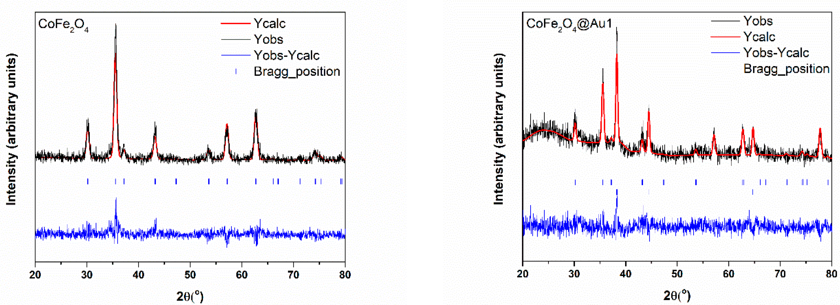
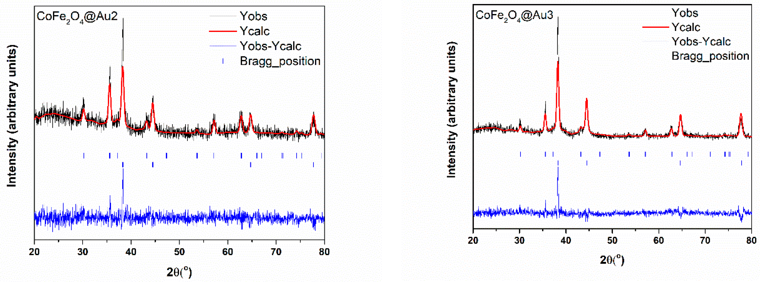
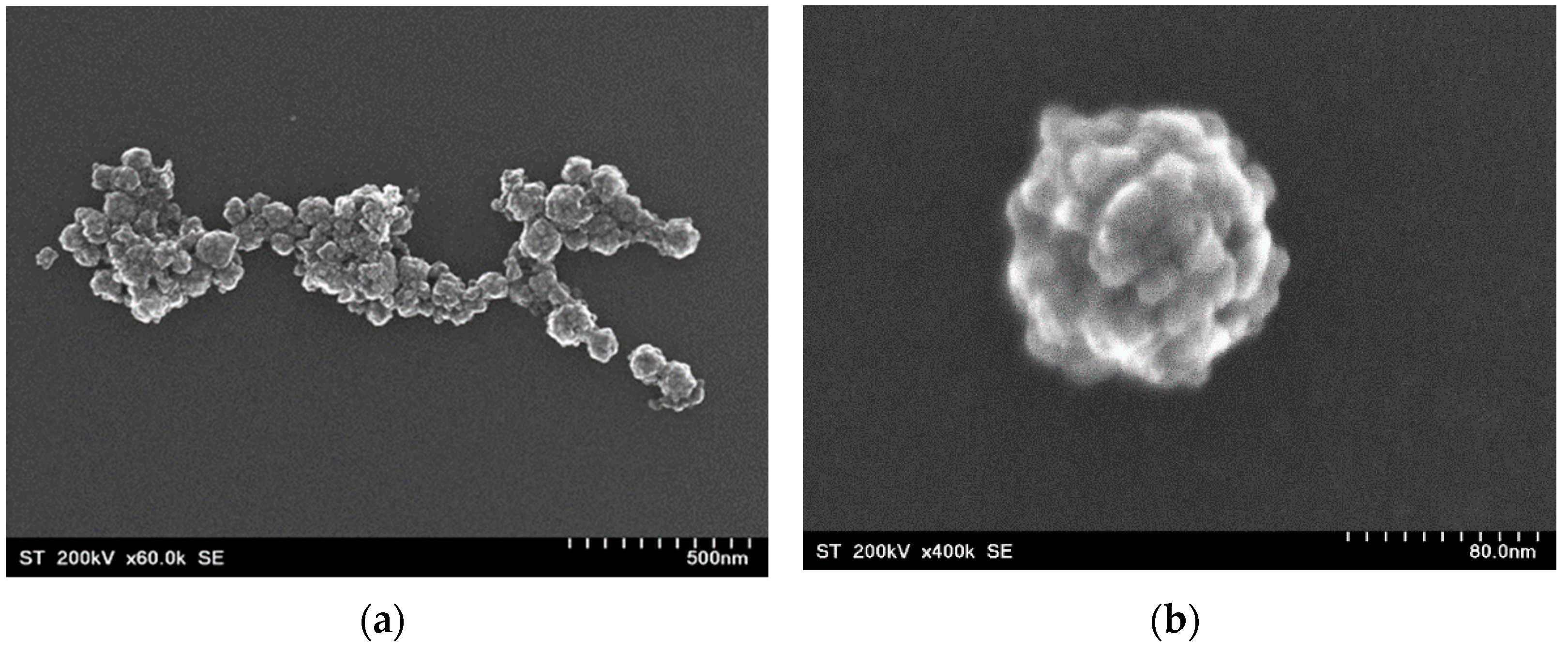
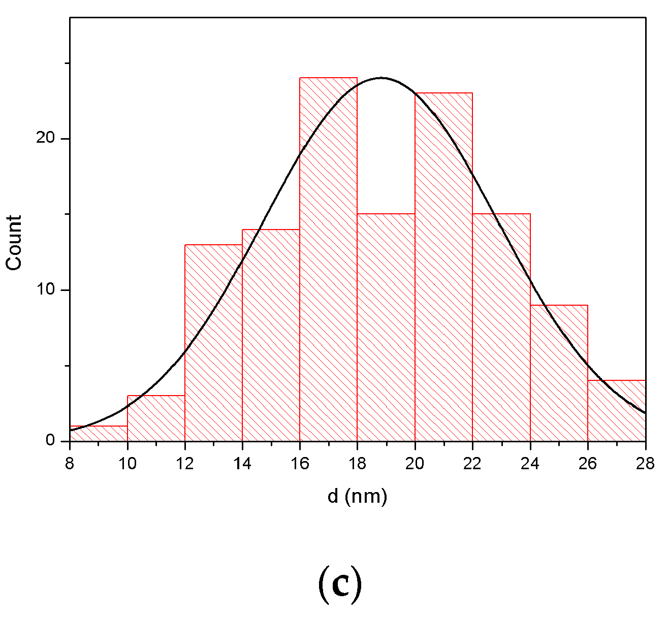
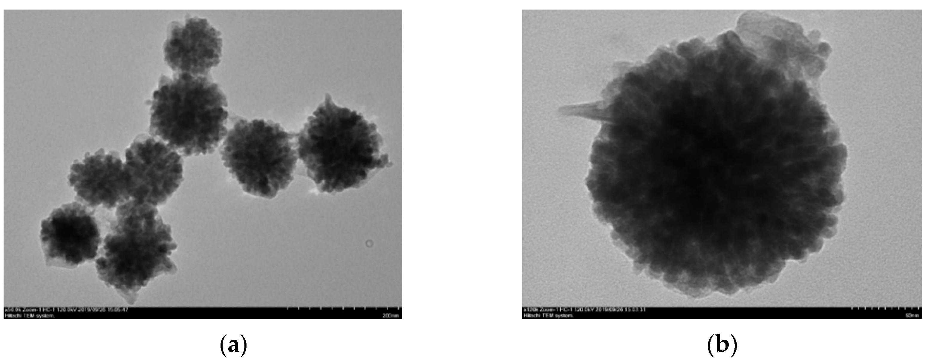
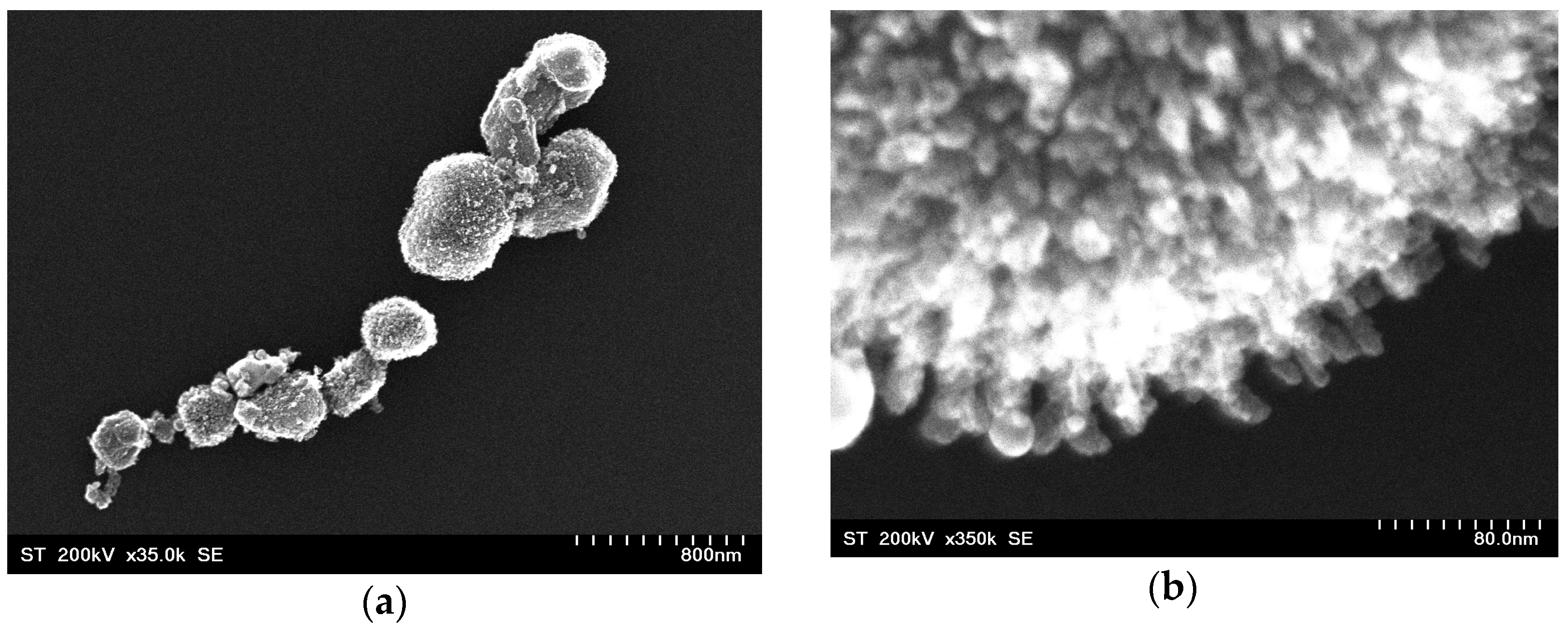
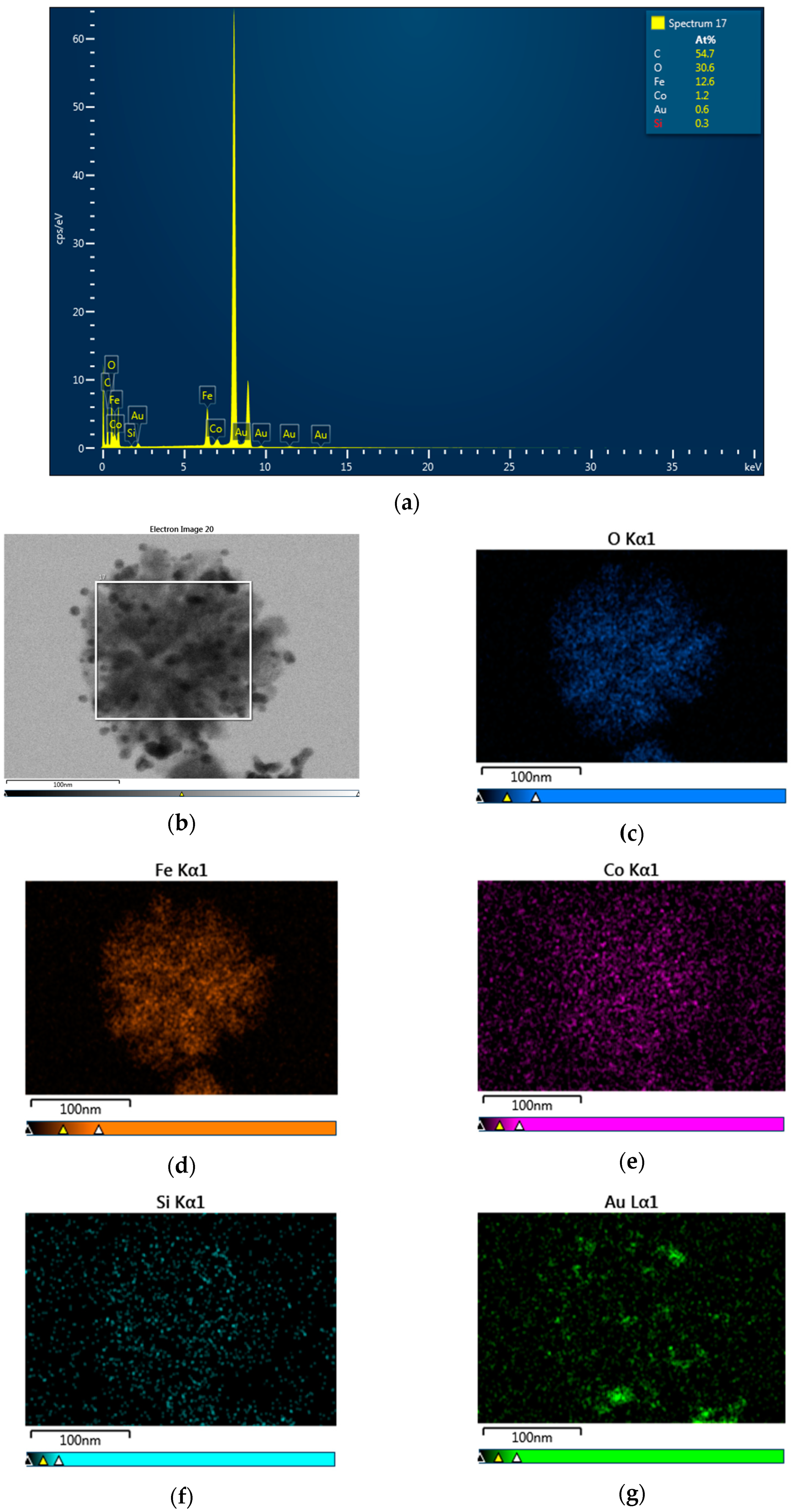
3.1. Morphology and Crystal Structure

The XRD patterns of CoFe2O4 and CoFe2O4@SiO2@AuN nanocomposites are shown in Figure 1. As expected, they correspond to the superposition of the XRD lines of CoFe2O4 and Au (ICDD PDF: 22-1086 for CoFe2O4 and 98-005-0876 for Au). All diffraction peaks including (111), (220), (311), (400), (422), (511) and (440) are fully indexed to the spinel CoFe2O4 phase. The sharp peaks corresponding to the (111), (200), (220) and (311) planes shown in the CoFe2O4@SiO2@AuN nanocomposite patterns confirm the crystallization of cubic FCC Au. The SiO2 was in amorphous state, as evidenced in XRD patterns by a broad feature at low angles. The lattice constants of CoFe2O4 and Au, determined from Rietveld refinements, are not dependent on nanostructures compositions and close to those of bulk samples. The crystallite sizes of CoFe2O4 nanoparticles are in the 14.2 nm and 20.2 nm range, while those of the gold nanoparticles were found to be between 15.9 nm and 23.6 nm (Table 1).
The TEM and SEM images of CoFe2O4 nanoparticles evidenced that these were of nearly spherical form and agglomerated in separate pseudo-spherical “raspberry”-like nanostructures, with an average particle size of approximately 79 nm (Figure 2a,b). The mean size of the crystallites was around 19 nm, as can be seen in Figure 2c.
After the deposition of SiO2, the TEM images show the formation of a shell layer (Figure 3). The nanocomposites thus formed had spherical or pseudo-spherical forms. A similar morphology was shown after gold deposition (Figure 4). The core–shell CoFe2O4@SiO2@AuN nanoparticles were nearly completely covered by gold. High resolution pictures confirmed that the Au nanoparticles had dimensions close to those determined by SEM, being in the 14–21 nm range. The average size of these nanocomposites was 288(57) nm.
The EDS results show that the nanocomposites consisted of Fe, Co, O, Si and Au. As an example, the EDS results for CoFe2O4@SiO2@Au2 are given in Figure 5. The compositions determined for the studied samples are listed in Table 1.

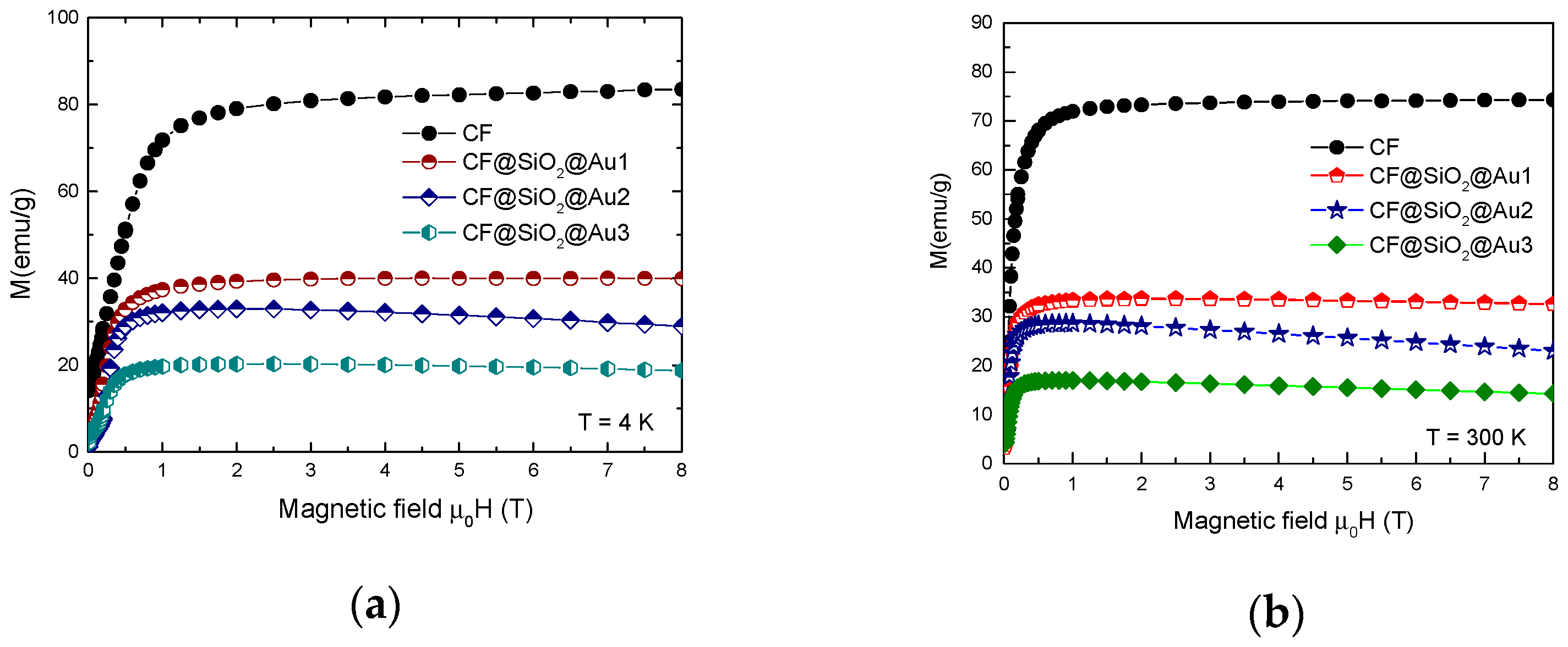
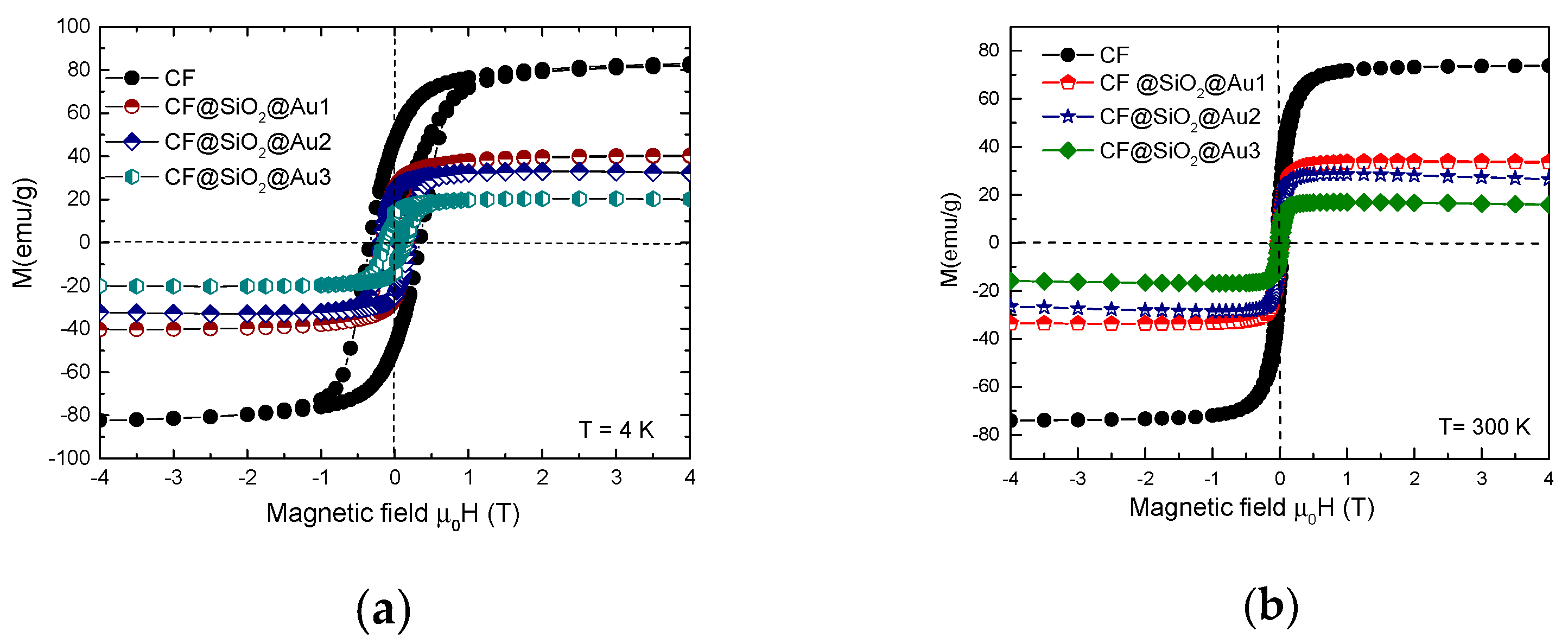
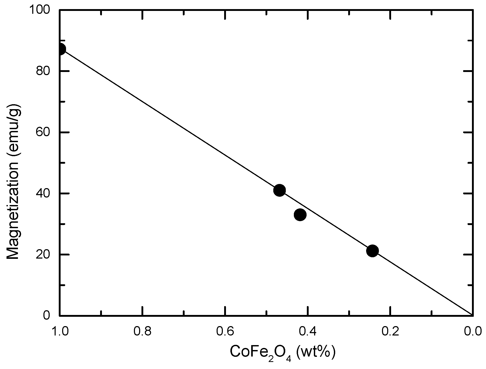
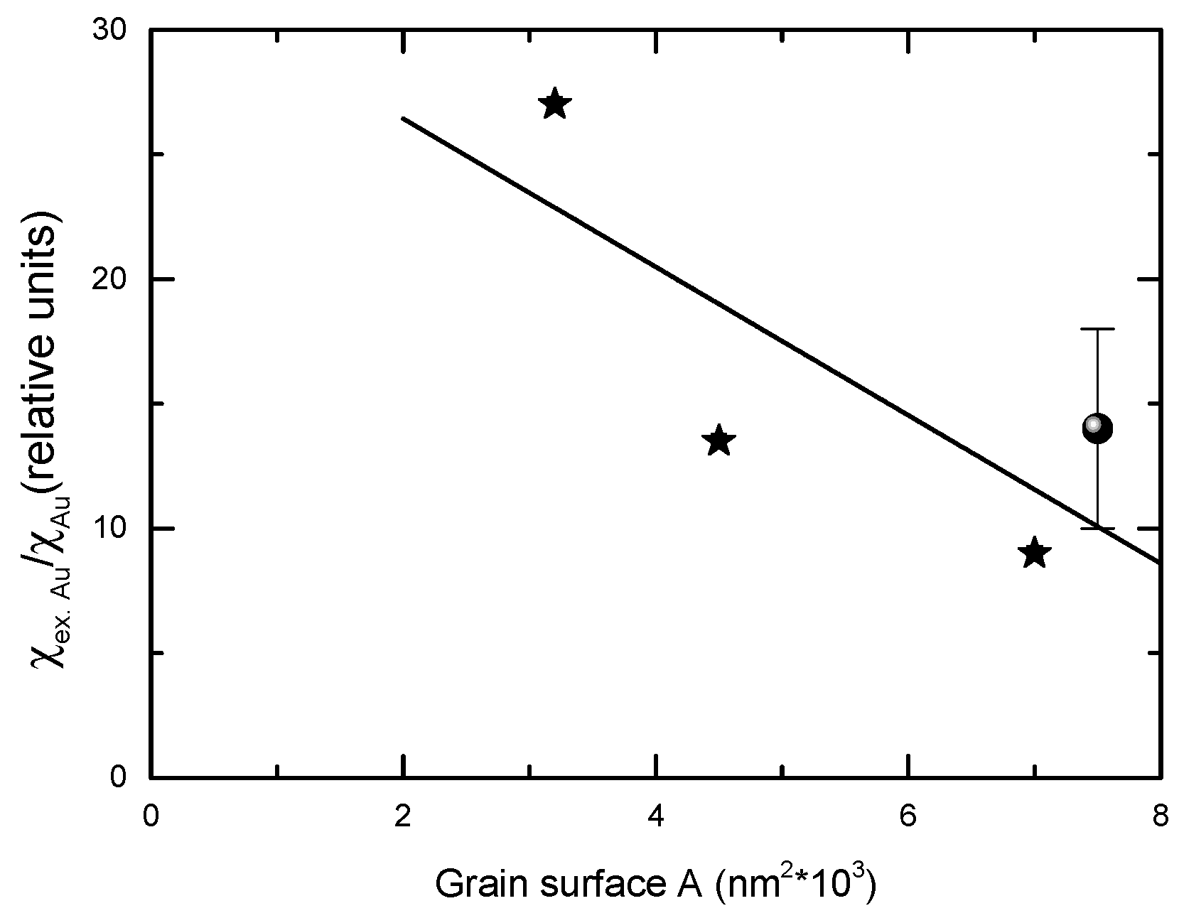
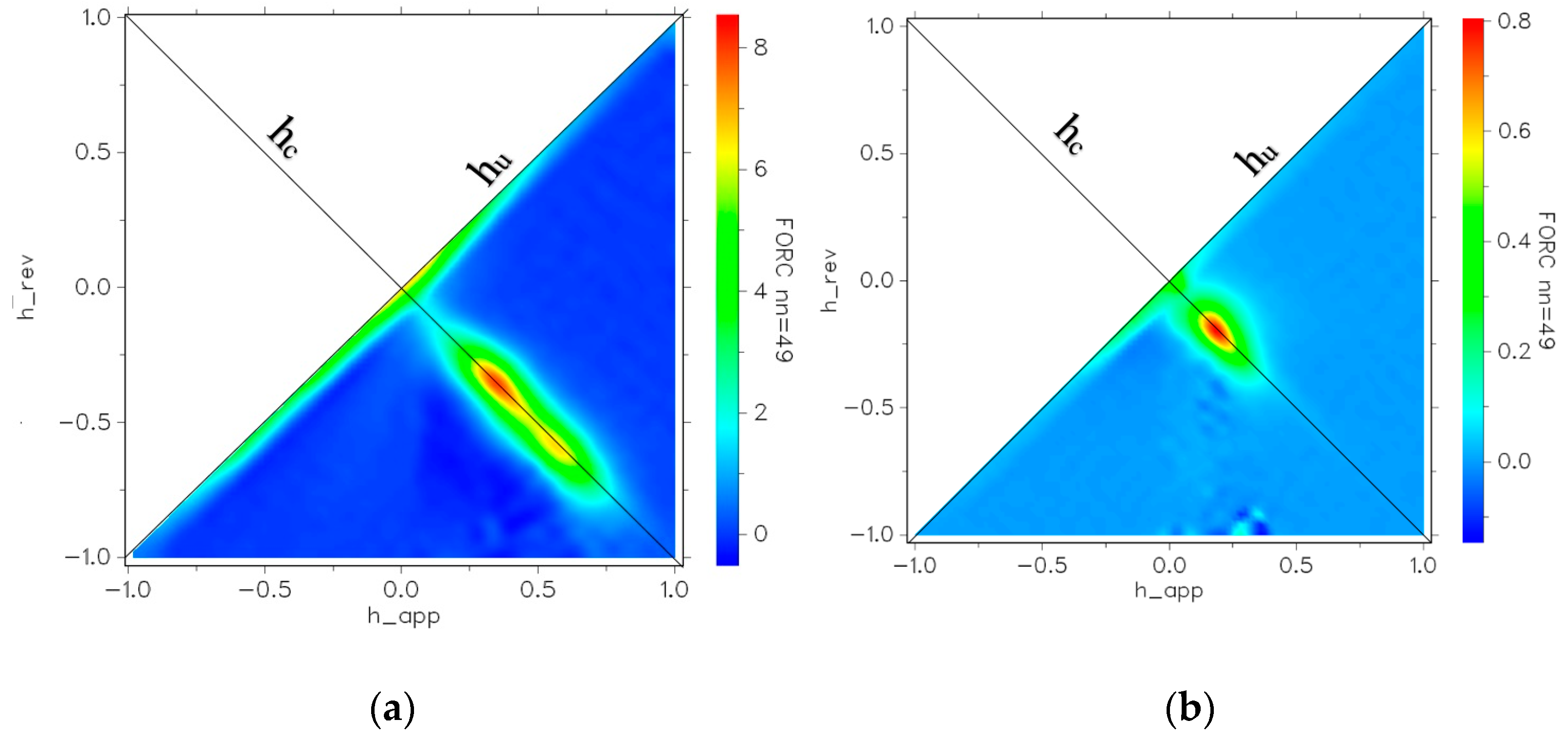
3.2. Magnetic Properties

The magnetization isotherms, recorded at T = 4.2 K and 300 K for nanocrystalline CoFe2O4 and CoFe2O4@SiO2@AuN nanostructures, are shown in Figure 6. The presence of a spin-glass contribution superposed on mainly ferrimagnetic-type ordering is suggested in CoFe2O4 nanocrystalline samples by the present investigations. The spin canting could have been present due to the following: (1) surface effects due to symmetry breaking by the broken exchange bonds at the surface layer, (2) cation distribution in the tetrahedral and octahedral sites and (3) interactions between nanoparticles. In a spin-glass system, instead of having global anisotropy axes, there are easy axes whose directions vary randomly in space. Their direction is determined by the local spin arrangement.
The previous Mössbauer studies [5,6] as well as infrared spectroscopy [4] performed on CoFe2O4 nanocrystalline samples evidenced the presence of a spin-glass state. It can be mentioned that in a large number of studies, ferrimagnetic-type behavior was also reported. The field dependences of the magnetization, in a spin-glass system, have been already analyzed [20]. In such a system, which shows high anisotropy (correlated spin glass), the approach to magnetic saturation follows a 1/H2 law. In systems with weak anisotropy (a ferromagnet with wondering axes) the approach to saturation when the external field, H, is smaller than the exchange field, Hex, can be described by a 1/H1/2 dependence, while for H > Hex it follows the same dependence as for systems having high anisotropy. The spin-glass state of surface magnetization of weak anisotropic perovskites is well described by the 1/H1/2 law, as already reported [21,22,23].
The analysis of magnetization isotherms in CoFe2O4 nanograins suggests the presence of a spin-glass state superposed on mainly ferrimagnetic-type behavior. The dependences of magnetizations, at T = 4.2 K and 300 K on 1/H1/2, are given in Figure 7. Linear variations are present in the field ranges up to 12 T, for data obtained at T = 300 K and up to µ0H = 8 T at T = 4.2 K, described by the relation:
M ( H ) M ( 0 ) = 1 b H 2 1
with a rate b = 0.1275 (T1/2) at T = 4.2 K and 0.0402 (T1/2) at T = 300 K. Nearly the same rate b has been obtained at T = 4.2 K for the field dependence of magnetization at the grain surface of Sr2FeMoO6-based perovskites [22,23].
For fields higher than 8–9 T, the magnetization at 4.2 K follows a 1/H2 law. There is a change in the shell magnetic behavior, suggesting a transition from a spin-glass state with wondering axes to a correlated spin-glass state. The extrapolation of magnetizations at T = 4.2 K, for both 1/H1/2 and 1/H2 trends, to infinite field, as expected, give the same value of saturation magnetization, Ms = 87.0 (3) emu/g. This corresponds to a magnetic moment per formula unit of Ms = 3.65 μB/fu and it is expected to characterize the situation when the moments of both core and shell are oriented along the same axis. Thus, the inversion factor can be determined from saturation magnetization [24,25] as being x = (1/4)(7−Ms) = 0.838. The composition of the sample (Co0.162Fe0.838)T(Co0.838Fe1.162)OO4 is closer to that of an inverse-spinel-type structure.
The analysis of magnetization at T = 4.2 K suggests that the contribution of the spin-glass state to magnetization is around 8% of the total nanoparticle magnetization. This could be due to the cumulative effects of the broken exchange bonds at the surface layer, as well as to the exchange interactions between constituent ions, assuming a spherical grain with d = 20 nm and the shell volume having one CoFe2O4 lattice parameter (0.4 nm) width, corresponding to 11% of that of the nanograin. By Mössbauer spectroscopy, it was shown that the canting angle of iron moments in CoFe2O4 at tetrahedral sites was 410 and at octahedral sites 360 [5]. Thus, the corresponding magnetization of the shell volume, on the field direction, was ~8% of the total magnetization, suggesting that the spin-glass state is mainly due to the surface shell.
The exchange interactions at the level of the unit cell also influences the spin canting. The anisotropy of cobalt is sensitively higher than that of iron and no canting is expected for cobalt moments, unlike for the Fe3+ spins. The exchange fields acting on iron ions in octahedral and tetrahedral sites were estimated starting from the exchange interaction parameters in bulk CoFe2O4, determined in the mean field model, assuming the presence of two [1] or three [2] magnetic sublattices. According to the determined inversion parameter, a tetrahedral Fe3+ ion has as neighbors four Fe and two Co octahedral ions, and an octahedral Fe3+ ion seven Co and five Fe tetrahedral ions, respectively [6]. In the above assumptions, the exchange fields, Hex, acting on iron at octahedral and tetrahedral sites are roughly of 300 T and 160 T, respectively. These values approximate those of the nanoparticle core. The exchange fields at the surface layer are sensitively diminished due to broken bonds and deviations from the parallel alignment of iron moments. Thus, for tetrahedral iron sites, in the one lattice parameter shell, the exchange field can be of a magnitude not highly different from the external field used for measurements. Consequently, there can be different magnetic responses of Fe3+ ions at tetrahedral and octahedral sites in the presence of an external field.
At ambient temperature, only a T−1/2 dependence of surface magnetization is shown, as determined by thermal effects.
The nanoparticle anisotropy seems to influence also the spin-canting-type behavior. The hysteresis curves recorded at 4.2 K and 300 K are given in Figure 8. At T = 4.2 K, the remanent magnetization was Mr/Ms = 0.5, as expected for single domain particles. A smaller Mr/Ms value was obtained at T = 300 K, correlated with the superparamagnetism of some nanoparticles having dimensions d < 10 nm—see Figure 8b. The anisotropy constants of CoFe2O4 nanoparticles were estimated assuming that below the blocking temperature, TB the anisotropy is uniaxial [26]. According to the Stoner–Wohlfarth model for non-interacting single domain particles, the coercive field, Hc, depends both on the anisotropy constant K1 and saturation magnetization [27]:
Hc = 2K10Ms
From experimentally determined Hc values, anisotropy constants K1 = 7 × 10⁵ (T = 4.2 K) and 6.8 × 10⁴ (T = 300 K) J/m3 were obtained.
The effective anisotropy constant, Keff, has been estimated using the blocking temperature TB, according to the relation:
TB = KeffV/25kB
where V is the nanoparticle volume.
For a log normal distribution of particles sizes, the blocking temperature, TB, is related to the temperature corresponding to the maximum in ZFC magnetization, Tmax, by the relation [28]: Tmax = e<TB>, where e is in the 1.5–2.5 range. A careful analysis of the matter in case of CoFe2O4 nanoparticles with dimensions 5–7 nm evidenced a value e = 1.70 [29]. By using this value and taking into account that Tmax = 300 K, an effective anisotropy constant Keff = 4.31 × 10⁴ J/m3 was estimated assuming crystallite sizes of 15 nm. This value is close to that determined at T = 300 K from a coercive field. Somewhat higher Keff values were obtained for nanograins with dimensions of 6.6 nm [16]. Starting from the determined anisotropy constants, the anisotropy field Ha = 2K0M of 9–14 T was estimated. These values are smaller than the exchange field, but close to the field where a change in the spin-glass-type behavior was shown, suggesting possible relations.
The magnetization isotherms for CoFe2O4@SiO2@AuN nanostructures can be described by the contributions of a magnetic ordered phase, attributed to CoFe2O4 and diamagnetic contributions of SiO2 and Au—see Figure 6.
MT = xMCoFe2O4 − (yχSiO2 + zχAu)H
By fitting the experimental data with the above relation, the contributions of CoFe2O4 to the nanostructure magnetizations were obtained. These values are in good agreement with the content of CoFe2O4 in the nanocomposites, as expected in the case of a simple magnetic dilution model—see Figure 9 and Table 1. The covering of the CoFe2O4 core with SiO2 and Au shells do not induce changes in their magnetic properties as compared with that of the single CoFe2O4 phase. Thus, by varying the CoFe2O4 content in nanostructures it is possible to ensure the desired magnetic properties of nanocomposites.
The diamagnetic contributions of the SiO2 and Au content, as determined from VSM studies, are higher than those expected starting from those of bulk SiO2 and Au, χSiO2 = 0.447 × 10⁻⁶ [30] and χAu = 2.74 × 10⁻⁶ [31], respectively. Unlike that of Au, the diamagnetism of SiO2 is not affected by the nanocrystalline sizes. A giant diamagnetism has been observed already in gold nanorods [32] and theoretically investigated [33,34]. After the subtraction of the contribution of SiO2 to nanostructural diamagnetism, the diamagnetic susceptibility of gold nanoparticles was shown to be higher by one order of magnitude than that of the bulk value.
The giant diamagnetic susceptibility of gold nanoparticles is a consequence of field-induced currents in the surface electrons [33]. The diamagnetic susceptibility is originated by steady currents induced by the applied field for quasi-free electrons confined in the surface. The diamagnetic response, induced when the external field is turned on, remains constant during the time the field is acting. As the size of the sample increases, the percentage of surface atoms decreases. Consequently, the magnetism of the surface approached that of the bulk sample. Such a trend is evidenced by the present data. The diamagnetism of gold nanoparticles, in relative units s = χex.AuAu, is plotted in Figure 10, as a function of nanocrystallites surfaces, assuming that these have a spherical form. On the same figure, the s parameter determined for a nanorod with d = 15 nm and l = 80 nm is also given [32]. These data are in agreement with the expected trend between gold diamagnetism and nanoparticle dimensions
The coercivities of CoFe2O4 and CoFe2O4@SiO2@AuN nanostructures determined from hysteresis loops, recorded at 4.2 K and 300 K, are given in Table 1. The coercive fields at T = 4.2 and 300 K increase due to reduction in CoFe2O4 nanosizes. At ambient temperatures, these values are smaller by one order of magnitude than those determined at T = 4.2 K. These magnetic measurements fail to fully describe complex magnetic nanostructures, such as an ensemble of nanoparticles with different magnetic properties, mainly due to the size distribution of the grains. The first-order reversal curve (FORC) diagram offers an image related to the coercivity and interaction fields acting on the different magnetic entities within the sample [35,36,37,38]. After applying a magnetic field to ensure the saturation magnetization, this is reduced to a predefined field Hr, denoted as the reversal field, and the magnetization of the sample is measured while H is returned. The above sequence is repeated with a decreasing Hr, thus obtaining a sequence of magnetization curves M(Hr,H). On this basis, the FORC distribution, P, defined as a second derivative of the magnetization with respect to reversal and applied field, is obtained. The quantitative analysis of the FORC data is performed using the projection of the FORC distribution in the plane of the coercive field, Hc = (1/2)(HHr), and the interaction field, Hu = (1/2)(H + Hr), axes, called the coercivity distribution (PHc) and interaction distribution (PHu), respectively. The FORC distributions for the CoFe2O4 and CoFe2O4@SiO2@Au2 nanostructures are given in Figure 11. In CoFe2O4 nanograins, there is a large distribution of coercive fields and interaction fields.
The latter is due to particle interactions and the former is due to different particles switching at different applied field strengths. A higher maximum in the p value indicates stronger exchange interactions. The maximum of the probability density for CoFe2O4 is one order of magnitude larger than that for CoFe2O4@SiO2@AuN. This means that the FORC distribution of CoFe2O4 is much narrower than that for CoFe2O4@SiO2@AuN nanocomposites. The extended spot shown in the FORC diagram of CoFe2O4 resembles ‘’single phase” behavior, but with large exchange interactions between the nanograins along the hc-axis. In CoFe2O4@SiO2@Au2 nanostructures, the peak distribution is located at an interaction field of 0.19 T. The peak in the FORC distribution is thus shifted towards positive reversal and interaction fields in the core–shell nanostructures, dipolar interactions being increasingly more important due to the presence of a SiO2-Au shell and its screening effect.

4. Conclusions

Magnetoplasmonic CoFe2O4@SiO2@Au nanoparticles were successfully prepared. The SiO2 shell was mainly in an amorphous state, as evidenced in XRD patterns by a broad feature at low angles. The lattice constants of CoFe2O4 and Au were not dependent on nanostructure compositions being close to those of bulk samples. The crystallite sizes of CoFe2O4 nanoparticles were in the 14.2 nm and 20.2 nm range, while those of gold were between 15.9 nm and 23.6 nm. The TEM and SEM images of CoFe2O4 nanoparticles evidenced that these are of nearly spherical form and agglomerate in separate pseudo-spherical “raspberry”-like nanostructures. The presence of a spin-glass contribution superposed on mainly ferrimagnetic-type ordering is suggested. The contribution of the spin-glass state to magnetization was no more than 8% of the total CoFe2O4 nanoparticle magnetization. This could have been due to the cumulative effects of the broken exchange bonds at the surface layer, as well as to the exchange interactions between constituent ions. As a function of the external magnetic field, two types of spin-glasses were observed and correlated with different exchange fields acting on tetrahedral and octahedral iron sites.
The diamagnetic susceptibility of gold nanoparticles was shown to be by one order of magnitude higher than that of the bulk value. The giant diamagnetic susceptibility of gold nanoparticles is a consequence of field induced currents in the surface electrons. The diamagnetic susceptibility originated by the steady currents induced by the applied field for quasi-free electrons was confined in the surface. As the size of the sample increased, the percentage of surface atoms decreased. Consequently, the diamagnetism approached that of the bulk sample.
The FORC diagram of CoFe2O4 highlighted ‘’single phase” behavior, with large exchange interactions between the nanograins along the hc-axis. The peak in the FORC distribution was shifted towards positive reversal and interaction fields in the core–shell nanostructures, dipolar interactions increasing due to the presence of SiO2@Au shells and their screening effects.

Author Contributions

Conceptualization, R.T., R.Ș. and E.B.; investigation, R.B., A.S., G.S., R.D., R.H., L.B.-T. and V.T.; resources, R.B., A.S. and R.T.; formal analysis, R.B., G.S., R.D., R.H., E.B.; writing—original draft preparation, R.B., R.T., E.B.; writing—review and editing, E.B., R.Ș., R.T.; visualization, R.B., R.T.; supervision, R.T., E.B.; project administration, E.B., R.T., R.Ș.; funding acquisition, E.B., R.Ș. and R.T. All authors have read and agreed to the published version of the manuscript.

Funding

This research was funded by the Romanian National Authority for Scientific Research, CNCSIS-UEFISCDI, through the exploratory research project No. PN-III-P4-ID-PCCF-2016-0112.

Institutional Review Board Statement

Not applicable.

Informed Consent Statement

Not applicable.

Data Availability Statement

Not applicable.

Conflicts of Interest

The authors declare no conflict of interest.

References

  1. Sawatzky, G.A.; Van Der Woude, F.; Morrish, A.H. Mössbauer Study of Several Ferrimagnetic Spinels. Phys. Rev. 1969, 187, 747–757. [Google Scholar] [CrossRef]
  2. Srivastava, C.M.; Srinivasan, G.; Nanadikar, N.G. Exchange constants in spinel ferrites. Phys. Rev. B 1979, 19, 499–508. [Google Scholar] [CrossRef] [Green Version]
  3. Chinnasamy, C.N.; Jeyadevan, B.; Shinoda, K.; Tohji, K.; Djayaprawira, D.J.; Takahashi, M.; Joseyphus, J.; Narayanasamy, A. Unusually high coercivity and critical single-domain size of nearly monodispersed CoFe2O4 nanoparticles. Appl. Phys. Lett. 2003, 83, 2862–2864. [Google Scholar] [CrossRef]
  4. Sun, Q.-C.; Birkel, C.S.; Cao, J.; Tremel, W.; Musfeldt, J.L. Spectroscopic signature of the superparamagnetic transition and surface spin disorder in CoFe2O4 nanoparticles. ACS Nano 2012, 6, 4876–4883. [Google Scholar] [CrossRef] [PubMed]
  5. Peddis, D.; Yaacoub, N.; Ferretti, M.; Martinelli, A.; Piccaluga, G.; Musinu, A.; Cannas, C.; Navarra, G.; Greneche, J.-M.; Fiorani, D. Cationic distribution and spin canting in CoFe2O4 nanoparticles. J. Phys. Condens. Matter 2011, 23, 426004. [Google Scholar] [CrossRef]
  6. Peddis, D.; Cannas, C.; Musinu, A.; Piccaluga, G. Coexistence of superparmagnetism and spin-glass like magnetic ordering phenomena in a CoFe2O4−SiO2 nanocomposite. J. Phys. Chem. C 2008, 112, 5141–5147. [Google Scholar] [CrossRef]
  7. Chiu, C.-W.; Lin, P.-H. Core/shell Ag@silicate nanoplatelets and poly(Vinyl Alcohol) spherical nanohybrids fabricated by coaxial electrospraying as highly sensitive SERS substrates. RSC Adv. 2016, 6, 67204–67211. [Google Scholar] [CrossRef]
  8. Prabhakaran, T.; Mangalaraja, R.; Denardin, J.; Jiménez, J.A. The effect of reaction temperature on the structural and magnetic properties of nano CoFe2O4. Ceram. Int. 2017, 43, 5599–5606. [Google Scholar] [CrossRef]
  9. Kiran, B.L.; Rami, R.Y.V. Synthesis and characterization of magnetically core-shell structured CoFe2O4/SiO2 nanoparticles; Their enhanced antibacterial and electrocatalytic properties&rdquo. Coll. Surf. A Physicochem. Eng. Asp. 2020, 598, 124806. [Google Scholar] [CrossRef]
  10. Lin, Y.-S.; Haynes, C.L. Synthesis and characterization of biocompatible and size-tunable multifunctional porous silica nanoparticles. Chem. Mater. 2009, 21, 3979–3986. [Google Scholar] [CrossRef]
  11. Cannas, C.; Musinu, A.; Ardu, A.; Orrù, F.; Peddis, D.; Casu, M.; Sanna, R.; Angius, F.; Diaz, G.; Piccaluga, G. CoFe2O4 and CoFe2O4/SiO2 core/shell nanoparticles: Magnetic and spectroscopic study. Chem. Mater. 2010, 22, 3353–3361. [Google Scholar] [CrossRef]
  12. Parma, A.; Freris, I.; Riello, P.; Cristofori, D.; Fernández, C.D.J.; Amendola, V.; Meneghetti, M.; Benedetti, A. Structural and magnetic properties of mesoporous SiO2 nanoparticles impregnated with iron oxide or cobalt-iron oxide nanocrystals. J. Mater. Chem. 2012, 22, 19276–19288. [Google Scholar] [CrossRef]
  13. Carlà, F.; Campo, G.; Sangregorio, C.; Caneschi, A.; Fernández, C.D.J.; Cabrera, L.I. Electrochemical characterization of core@shell CoFe2O4/Au composite. J. Nanoparticle Res. 2013, 15, 1–16. [Google Scholar] [CrossRef]
  14. Zhang, X.; Niu, Y.; Zhao, J.; Li, Y. Fabrication, structures and molecule detection of gold films coated on γ-Fe2O3@SiO2 ellipsoid ordered arrays. Coll. Surf. A Physicochem. Eng. Asp. 2017, 520, 343–347. [Google Scholar] [CrossRef]
  15. Kassim, S.; Tahrin, R.A.A.; Harun, N.A. Metallic Core-Shell Photonic Crystals for Surface-Enhanced Raman Scattering (SERS). Plasmonics 2020, 15, 1499–1505. [Google Scholar] [CrossRef]
  16. Li, Y.; Duan, W.; Wei, J. CoFe2O4/Ag nanostructures as highly sensitive surface-enhanced raman scattering substrates. ACS Appl. Nano Mater. 2021, 4, 4181–4188. [Google Scholar] [CrossRef]
  17. Huynh, K.-H.; Hahm, E.; Noh, M.; Lee, J.-H.; Pham, X.-H.; Lee, S.; Kim, J.; Rho, W.-Y.; Chang, H.; Kim, D.; et al. Recent advances in surface-enhanced raman scattering magnetic plasmonic particles for bioapplications. Nanomaterials 2021, 11, 1215. [Google Scholar] [CrossRef] [PubMed]
  18. Dudric, R.; Souca, G.; Szatmári, Á.; Szilárd, T.; Nitica, S.; Iacovita, C.; Moldovan, A.I.; Stiufiuc, R.; Tetean, R.; Burzo, E. Magnetite nanoparticles for medical applications. AIP Conf. Proc. 2020, 2218, 030014. [Google Scholar] [CrossRef]
  19. Souca, G.; Dudric, R.; Iacovita, C.; Moldovan, A.; Frentiu, T.; Stiufiuc, R.; Lucaciu, C.M.; Tetean, R.; Burzo, E. Physical properties of zn doped Fe3O4 nanoparticles. J. Optoelectron. Adv. Mater. 2020, 22, 298–302. [Google Scholar]
  20. Tejada, J.; Martinez, B.; Labarta, A.; Chudnovsky, E.M. Correlated spin glass generated by structural disorder in the amorphous Dy6Fe74B20 alloy. Phys. Rev. B 1991, 44, 7698. [Google Scholar] [CrossRef] [Green Version]
  21. Serrate, D.; De Teresa, J.M.; Algarabel, P.A.; Ibarra, M.R.; Galibert, J. Intergrain magnetoresistance up to 50 T in the half-metallic(Ba0.8Sr0.2)2FeMoO6double perovskite: Spin-glass behavior of the grain boundary. Phys. Rev. B 2005, 71, 104409. [Google Scholar] [CrossRef]
  22. Burzo, E.; Balasz, I.; Valeanu, M.; Pop, I. The effects of thermal treatment on the physical properties of Sr2FeMo1−xMxO6 perovskite with M=W, Ta and 0.3. J. Alloy. Compd. 2011, 509, 105–113. [Google Scholar] [CrossRef]
  23. Burzo, E. Magnetic and transport properties of double perovskites. Stud. Univ. Babes-Bolyai. Chem. 2021, 66, 63–72. [Google Scholar] [CrossRef]
  24. Concas, G.; Spano, G.; Cannas, C.; Musinu, A.; Peddis, D.; Piccaluga, G. Inversion degree and saturation magnetization of different nanocrystalline cobalt ferrites. J. Magn. Magn. Mater. 2009, 321, 1893–1897. [Google Scholar] [CrossRef]
  25. Venturini, J.; Tonelli, A.M.; Wermuth, T.B.; Zampiva, R.Y.S.; Arcaro, S.; Viegas, A.D.C.; Bergmann, C.P. Excess of cations in the sol-gel synthesis of cobalt ferrite (CoFe2O4): A pathway to switching the inversion degree of spinels. J. Magn. Magn. Mater. 2019, 482, 1–8. [Google Scholar] [CrossRef]
  26. Caruntu, D.; Caruntu, G.; O’Connor, C.J. Magnetic properties of variable-sized Fe3O4 nanoparticles synthesized from non-aqueous homogeneous solutions of polyols. J. Phys. D Appl. Phys. 2007, 40, 5801–5809. [Google Scholar] [CrossRef] [Green Version]
  27. Stoner, E.C.; Wohlfarth, E.P. A mechanism of magnetic hysteresis in heterogeneous alloys. Philos. Trans. R. Soc. Lond. Ser. A Math. Phys. Sci. 1948, 240, 599–642. [Google Scholar] [CrossRef]
  28. Gittleman, J.I.; Abeles, B.; Bozowski, S. Superparamagnetism and relaxation effects in granular Ni-SiO2 and Ni-Al2O3 films. Phys. Rev. B 1974, 9, 3891. [Google Scholar] [CrossRef]
  29. Omelyanchik, A.; Salvador, M.; D’Orazio, F.; Mameli, V.; Cannas, C.; Fiorani, D.; Musinu, A.; Rivas, M.; Rodionova, V.; Varvaro, G.; et al. Magnetocrystalline and surface anisotropy in CoFe2O4 nanoparticles. Nanomaterials 2020, 10, 1288. [Google Scholar] [CrossRef] [PubMed]
  30. Kondrashova, N.; Valtsifer, V.; Strelnikov, V.; Mitrofanov, V.; Uporov, S. Magnetic Properties of Siilica with Mesoporesd as MCM-48. 2015. Available online: https://www.ariel.ac.il/sites/conf/mmt/ws2015/service%20files/papers/124-130.pdf (accessed on 19 February 2022).
  31. Henry, W.G.; Rogers, J.L. XXI. The magnetic susceptibilities of copper, silver and gold and errors in the Gouy method. Philos. Mag. 1956, 1, 223–236. [Google Scholar] [CrossRef]
  32. Van Rhee, P.G.; Zijlstra, P.; Verhagen, T.G.A.; Aarts, J.; Katsnelson, M.I.; Maan, J.C.; Orrit, M.; Christianen, P.C.M. Giant magnetic susceptibility of gold nanorods detected by magnetic alignment. Phys. Rev. Lett. 2013, 111, 127202. [Google Scholar] [CrossRef] [Green Version]
  33. Hernando, A.; Ayuela, A.; Crespo, P.; Echenique, P.M. Giant diamagnetism of gold nanorods. New J. Phys. 2014, 16, 073043. [Google Scholar] [CrossRef] [Green Version]
  34. Roda-Llordes, M.; Gonzalez-Ballestero, C.; López, A.E.R.; Martínez-Pérez, M.J.; Luis, F.; Romero-Isart, O. Quantum size effects in the magnetic susceptibility of a metallic nanoparticle. Phys. Rev. B 2021, 104, L100407. [Google Scholar] [CrossRef]
  35. Kouhpanji, M.R.Z.; Stadler, B.J.H. Beyond the qualitative description of complex magnetic nanoparticle arrays using FORC measurement. Nano Express 2020, 1, 010017. [Google Scholar] [CrossRef]
  36. Muxworthy, A.; Heslop, D.; Williams, W. Influence of magnetostatic interactions on first-order-reversal-curve (FORC) diagrams: A micromagnetic approach. Geophys. J. Int. 2004, 158, 888–897. [Google Scholar] [CrossRef] [Green Version]
  37. Tanasa, R.; Stancu, A. Statistical Characterization of the FORC Diagram. IEEE Trans. Magn. 2006, 42, 3246–3248. [Google Scholar] [CrossRef]
  38. Dodrill, B.; Ohodnicki, P.; McHenry, M.; Leary, A. High-Temperature First-Order-Reversal-Curve (FORC) Study of Magnetic Nanoparticle Based Nanocomposite Materials. MRS Adv. 2017, 2, 2669–2674. [Google Scholar] [CrossRef]
Figure 1. XRD patterns of CoFe2O4 and CoFe2O4@SiO2@Au nanocomposites and the results of Rietveld refinements.
Nanomaterials 12 00942 g001aNanomaterials 12 00942 g001b
Figure 2. SEM images of CoFe2O4 nanoparticles (a,b) and particle size histogram (c).
Nanomaterials 12 00942 g002aNanomaterials 12 00942 g002b
Figure 3. TEM images of CoFe2O4@SiO2 nanostructures (a) and of one isolated nanoparticle(b).
Nanomaterials 12 00942 g003
Figure 4. SEM images of CoFe2O4@SiO2@Au2 nanocomposite (a) and the edge of a nanoparticle.
Nanomaterials 12 00942 g004
Figure 5. EDS spectrum (a), TEM image (b) EDS mapping (cg) of CoFe2O4@SiO2@Au2 nanocomposite.
Nanomaterials 12 00942 g005
Figure 6. Magnetization isotherms at T = 4.2 K (a) and T = 300 K (b) of CoFe2O4 and CoFe2O4@SiO2@Au nanocomposites.
Nanomaterials 12 00942 g006
Figure 7. Magnetization isotherms of CoFe2O4 at T = 4.2 K and T = 300 K as function of H1/2. In inset, the data at T = 4.2 K for H > 8 T as a function of H−2.
Nanomaterials 12 00942 g007
Figure 8. Hysteresis loops of CoFe2O4@SiO2@Au nanoparticles at T = 4.2 K (a) and T = 300 K (b).
Nanomaterials 12 00942 g008
Figure 9. Magnetizations at T = 4.2 K as a function of CoFe2O4 content in CoFe2O4@SiO2@AuN nanocomposites. The solid line gives the expected trend in a simple dilution magnetic model.
Nanomaterials 12 00942 g009
Figure 10. The gold diamagnetism in CoFe2O4@SiO2@AuN nanostructures as function of the mean surface of Au nanograins, in relative units (referred to bulk gold diamagnetism) (*). The data from [31] are also given (●).
Nanomaterials 12 00942 g010
Figure 11. FORC diagrams for CoFe2O4 (a) and CoFe2O4@SiO2@Au2 (b) nanocomposites. h_rev denotes the applied field for each inversion curve. h_app is the applied field after that inversion is realized. nn is the smoothing factor.
Nanomaterials 12 00942 g011
Table 1. Compositions, lattice parameters, crystallites sizes, magnetizations and coercive fields. AuN is a symbol for CoFe2O4@SiO2@AuN nanostructures with N = 1–3).
Nanostructure CoFe2O4Au1Au2Au3
Composition (weight %)CoFe2O410046.841.824.3
SiO2-11.011.66.3
Au-42.246.669.4
Lattice parameter (nm)CoFe2O4 core0.8379 (9)0.8375 (4)0.8372 (9)0.8376 (9)
Crystallite size (nm14.2 (2)20.2 (3)17.3 (2)20.2 (2)
Lattice parameter (nm)Au shell-0.4073 (2)0.4073 (9)0.4074 (7)
Crystallite size (nm-23.57 (2)15.86 (4)18.88 (6)
Nanocomposite magnetization/CoFe2O4 weight percent (emu/g) 86.986.184.585.5
Coercive field HC(T)T = 4.2 k0.350.200.280.19
T = 300 k0.040.030.030.030
Publisher’s Note: MDPI stays neutral with regard to jurisdictional claims in published maps and institutional affiliations.

