
Exogenous Contrast Agents in Photoacoustic Imaging: An In Vivo Review for Tumor Imaging


Abstract
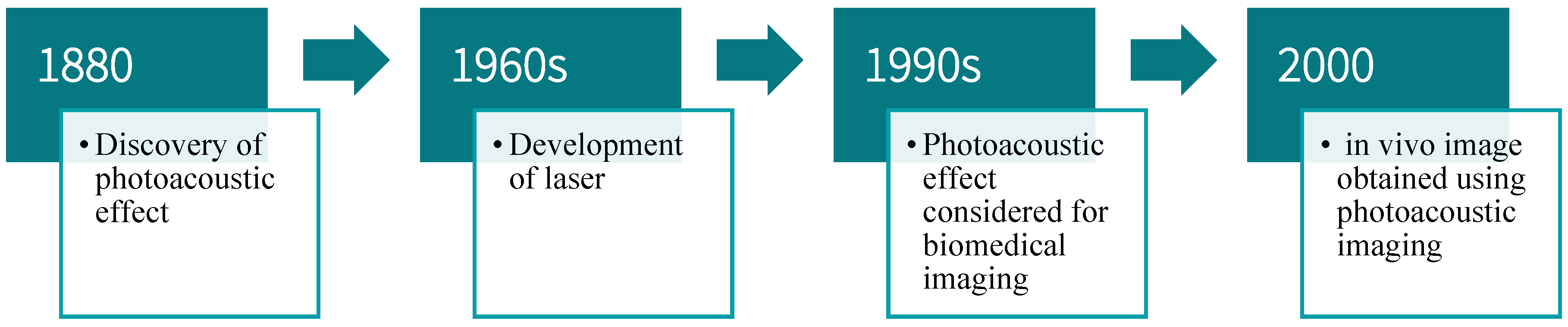
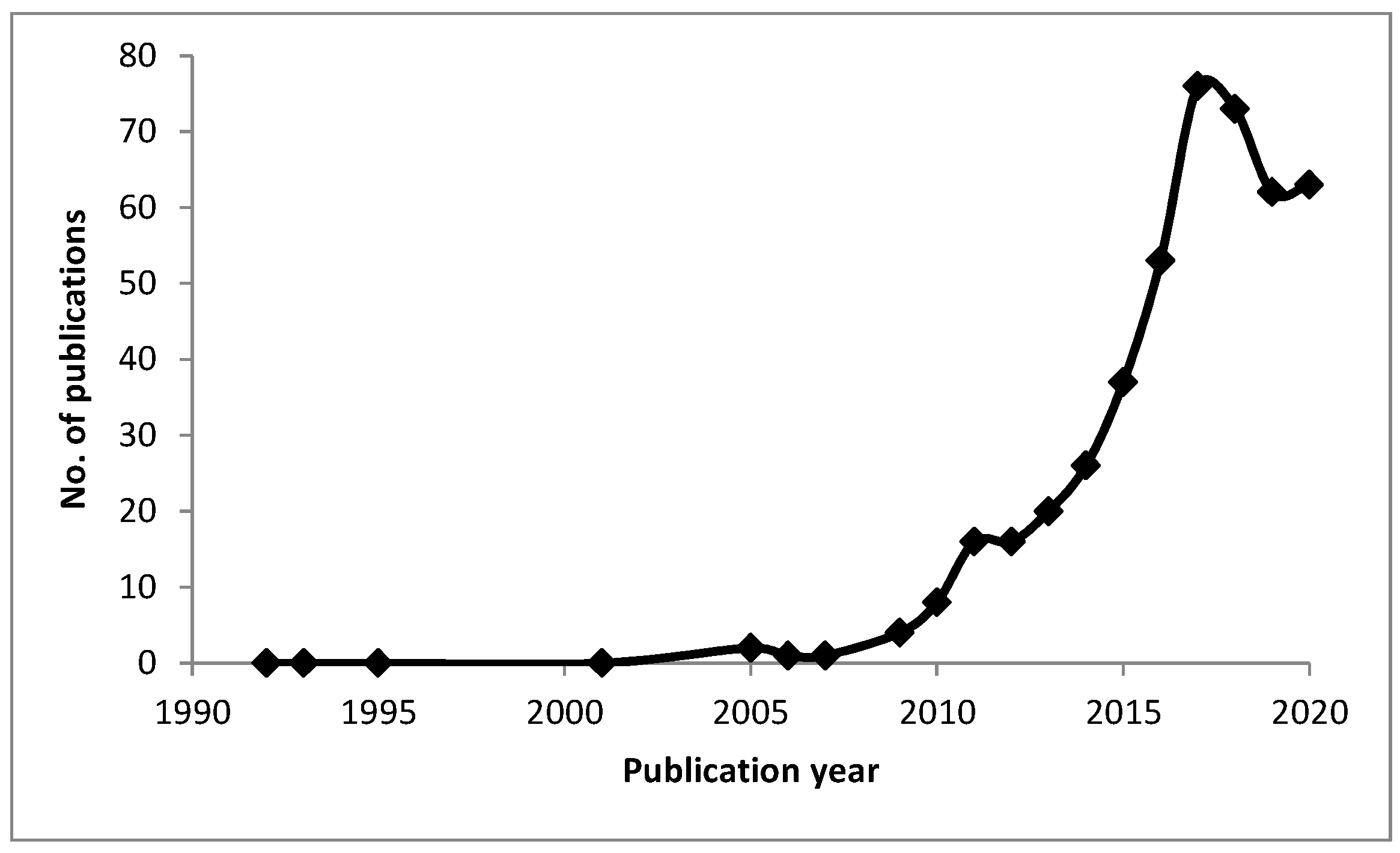
1. Introduction

| Modality | Commonly Used Contrast Agents | References |
|---|---|---|
| MRI | Gadolinium, Super paramagnetic iron oxide nanoparticles (SPIONs), Carbon-13, Nanodiamonds, Carbon nanotubes, Graphene, Manganese, Silicon, Peptides | [35] |
| CT | Gold nanoparticles, Iodine (131I), Bismuth, Lathanide-based (gadolinium, dysprosium, ytterbium) | [35,36] |
| Ultrasound | Nanobubbles, microbubbles (with modifications) | [35,37] |
| PET | Gold nanoparticles, Copper (64Cu), Iodine (124I), Fluorine (18F) | [35,38] |
| SPECT | Gold nanoparticles, Technetium (99mTc) | [35] |
| Optical Imaging | Fluorescence, Quantum dots, Gold nanoparticles, Persistent luminescence nanoparticles | [35] |
| Combinations of these contrast agents can be used to create hybrid contrast agents and optimize imaging | [38,39] | |
2. Contrast Agents for In Vivo Testing
2.1. Basis of PAI and Design Considerations for Contrast Agents
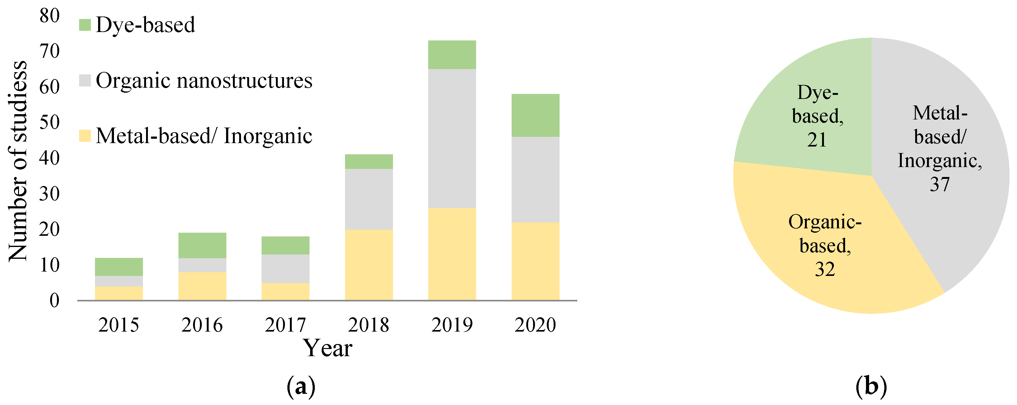
2.2. Organic Contrast Agents
| Classification | Material Used | Imaging Modalities | Application | Studies Conducted | Relevant Measured Parameters | Transducer Used | Computational Techniques | Publication Year/Reference |
|---|---|---|---|---|---|---|---|---|
| Semiconducting Polymer | poly{3-(5-(9-hexyl9-octyl-9H-fluoren-2-yl)thiophen-2-yl)-2,5-bis(2-hexyldecyl)-6- (thiophen-2-yl)pyrrolo [3,4-c]pyrrole-1,4(2H,5H)-dione} (PDPPF, SP0) with SP5 and SP10 (self-quenching SPs) | PAI | Imaging of breast cancer and cervical cancer tumors | HeLa cervical adenocarcinoma epithelial cells for In vitro; In vivo and ex vivo on 4T1 breast cancer tumor in mice/mice organs | Maximum PA signal of SP10 at 4h for both SP10-RGD and SP10 with slower clearance rate for SP10-RGD and 1.78 fold higher PA intensity for SP10-RGD as well | LAZR instrument (Visualsonics, 2100 High-Resolution Imaging System) | - | 2017 [92] |
| Derived from natural resources | DPAHB nanovesicles (hypocrellin B (HB) modified with 1,2-diamino-2-methyl propane encapsulated by PLGA-PEG) | PAI, fluorescence, photodynamic and photothermal therapy | Imaging of 4T1 breast cancer tumors | In vitro and in vivo PAI. | High-intensity signals and enhanced spatial resolution was achieved using DPAHB nanovesicles. PA signal intensity attained maximum peak at 12 h after injection of nanovesicles. | MSOT inVision 128 PAT system | 2018 [93] | |
| Other | 2018 [51] 2017 [52,54] 2015 [53] | |||||||
| Carbon nanodots | Nitrogen-Doped Carbon Nanodots | PAI | Imaging of sentinel lymph node to detect metastatic cancer | In vivo and ex vivo mapping of sentinel lymph node, in vivo PAI of the bladder. | Post injecting N-CNDs PA signal reached a peak at 30 min, and the signal kept decreasing until 180 min. Results show that the contrast agent was circulating in the lymphatic system before being degraded. | Ultrasound transducer with spherical focusing and having a 5-MHz central frequency, Acoustic-resolution reflection-mode PA imaging system | Raster scanning to acquire PA images | 2016 [94] |
| Organic small molecule | Diradicaloid molecular (DRM) structure | PAI and PTT | Imaging of A549 lung cancer | PAI-guided PTT in vitro and in vivo | The average PA signal of tumors excised from the mice injected with DRM NPs is over 4 times higher than that from the control group | Vevo LAZR-X imaging equipment | DFT calculations of optimized geometries of the DRM in the ground and excited states | 2021 [95] |
| Mitochondria-targeted BODIPY NPs | BODIPY NPs with a cationic triphenylphosphine (TPP) group (Mito-BDP1–5 NPs) bearing different lengths of ethylene glycol (0–4 units), along with HO-BDP5 without a cationic TPP group | PAI and PTI | Imaging of mitochondria in HeLa cells | In vitro mitochondrial imaging, and in vivo PTI and PAI | Mito-BDP5 possessed high photothermal conversion efficiency (η) of 76.6%, and was able to accumulate in the tumor sites through the EPR effect, subsequently strong PT and PA signals can be observed in tumor sites. | PAI was conducted on a PA computed-tomography system equipped with a 10 MHz, 10 mJ cm−2, 384-element ring ultrasound array, and a tunable pulsed laser | - | 2021 [96] |
| Carbon nanohorns | carbon nanohorn-polyglycerol-gold (CNH-PG-Au) NPs | PAI and x-ray | Imaging of 4T1 mouse breast cancer cells | In vivo PAI of tumor treatment using DOX@CNH-PG-Au | The photoacoustic intensity of the tumor site increased gradually and reached a maximum 48 h post-injection (735 ± 47), indicating that DOX@CNH-PG-Au NPs steadily accumulated in the tumor during this period | MSOT inVision 256 PAI systems | - | 2021 [97] |
| Laponite (LAP) nanoplatforms | polydopamine (PDA) coated LAP nanoplatforms modified with polyethylene glycol-arginine-glycine-aspartic acid (PEG-RGD) | PAI | Imaging of 4T1 mouse breast cancer cells | In vitro and in vivo PAI-guided chemo-phototherapy of cancer cells | NPs showed an increased PA signal at tumor sites after injection, and the PA signal peaked at 2 h post-injection. | Vevo LAZR PAI system equipped with an 875 nm laser | - | 2021 [98] |
2.3. Metal/Inorganic Contrast Agents
2.4. Dye-Based Contrast Agents
| Classification | Material Used | Imaging Modalities | Application | Studies Conducted | Relevant Measured Parameters | Transducer Used | Computational Techniques | Publication Year/Reference |
|---|---|---|---|---|---|---|---|---|
| Gold nanorods (AuNR)-based | AuNR | PAI | Imaging of lymph vessels/nodes in breast cancer tumors | Phantom using PTFE tubes; in vivo on mice | Attenuation coefficient: −1.90 dB/mm380 times as compared ICG | Concave poly(vinylidene fluoride/trifluoroethylene) (P(VDF-TrFE)) US transducer | Delay-and-sum (DAS) beamforming method | 2018 [154] |
| 89Zr-labeled bGNR@MSN(DOX)-PEG (Zirconium labeled PEGylated gold nanorods, GNR, coated with mesoporous silica nanoshell) | PAI, PET, PTT and chemotherapy | Imaging of 4T1 breast cancer tumors | In vitro and in vivo on mice | NP diameter: 135.9 nm; 4.7 fold stronger signal from PAI 24 h post-injection as compared to pre-injection | VEVO LAZR PA imaging system | - | 2018 [155] | |
| AuNR coated with CTAB. | PAI, US | Imaging of tumor metastases in mice | In vivo EGFR-targeted PAI of lymph node metastases and tumor mass | Enhanced PA signal observed after 24 h in lymph node with metastases post-injection of gold nanorods. | LZ-550 linear array transducer, Vevo 2100 LAZR high-frequency US and PA imaging system. | - | 2016 [127] | |
| Gold nanoparticles | PAI, US | Imaging of micro-metastases in lymph nodes | In vivo imaging of lymph node. | High spatial resolution images of micro-metastases (50 µm) were obtained after 2 h of peritumoral injection. | LZ-550 linear array transducer, Vevo LAZR high-frequency US and PA imaging system. | Spectral unmixing, sPA imaging algorithm to differentiate several optical absorbers. | 2014 [156] | |
| Furin-cleavable RVRR (Arg-Val-Arg-Arg) peptides (Au-RRVR NPs) | PAI, PTT | Imaging HCT 116 colorectal carcinomas | In vitro and in vivo imaging of tumors | The PA signal reached an intensity maximum of approximately 8 h post-injection with a 1.6-fold enhancement compared to the initial background. | A multispectral optoacoustic tomography scanner with excitation light of 680–900 nm | Maynard operation sequence technique (MOST) measurement | 2021 [157] | |
| Gadolinium-/bismuth-based | Gd-PEG-Bi NPs (hydrophobic dodecanethiol-Bi nanoparticles, for CT and PA contrast, coated in gadolinium, for MRI, and PEG) | PAI, CT, MRI and for PTT | Imaging of C6 glial tumors | In vitro and in vivo on mice; hemolysis assay and in vivo blood clearance and bio-distribution | NP diameter: 45 nm; Strong PA signals at low concentrations of 0.625 mg/mL and after 30 min; Strongest PA signal at 3 h and blood half-life at 4.69 h; High biosafety and NIR absorption coefficient | Endra Nexus 128 PA imaging system | - | 2018 [104] |
| Manganese-based | GO/MnWO4/PEG/DOX (Graphene-oxide, GO, grown in situ onto manganese tungsten oxide in the presence of PEG and loaded with doxorubicin) | PAI, MRI, PTT and chemotherapy | Imaging of breast cancer tumors (4T1 mouse mammary carcinoma) | In vitro and in vivo on mice; PTT, chemotherapy and cytotoxicity | Maximum PA signal observed at tumor region 6 h post-injection in vivo; however, the signal was maintained at 1.4 times that of pre-injection at 24 h. Little to no cytotoxicity observed | MOST inVision128, iThera Medical | - | 2018 [105] |
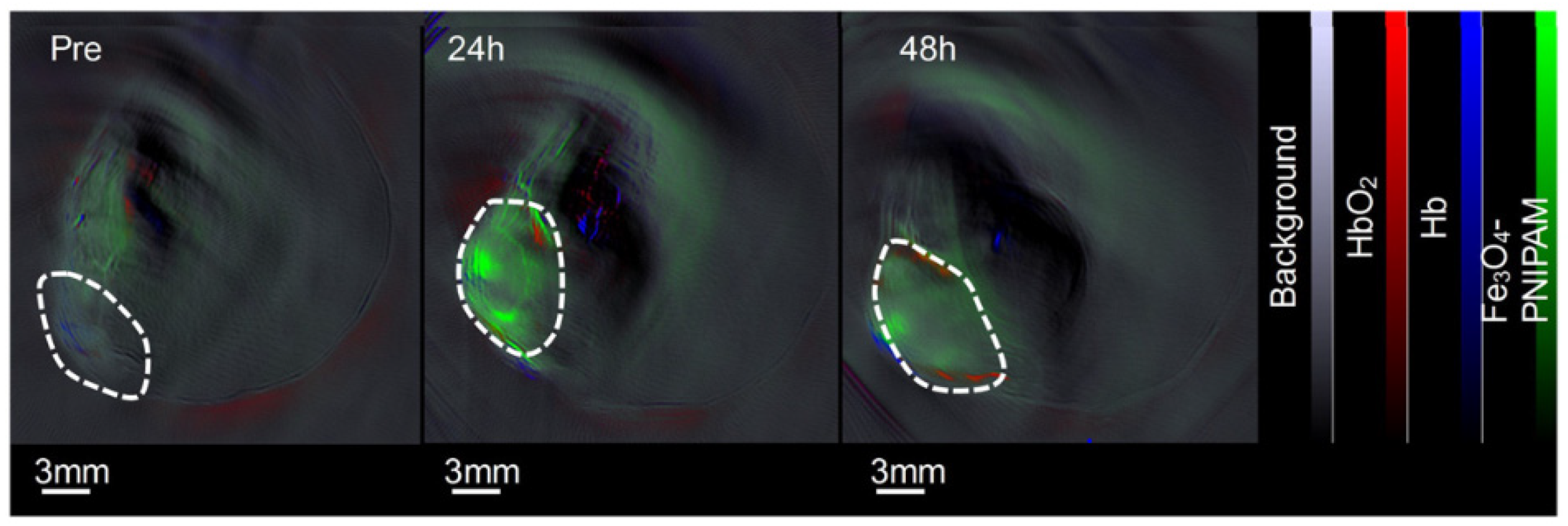
| Iron oxide-based | Magnetic iron oxide nanoparticles | Molecular PAT | Imaging of 4T1 breast cancer tumors | In vivo molecular photoacoustic tomography of breast cancer in mice | Post injection of contrast agents PA signal increased 3 times after 5 min and 10 times after 24 h. | Focused-ultrasound transducer operating at 50 MHz and 3.5 MHz | Raster scanning to acquire PA images, Hilbert transform was used to process acquired signals. | 2014 [158] |
| Copper(II) sulfide nanoparticles (CuS) | Copper(II) chloride, sodium sulfide, methoxy-PEG-thiol to form polyethylene glycol (PEG)-coated copper(II) sulfide nanoparticles | PAT | Imaging of 4T1 breast cancer tumors | In vivo PAT of blood vasculature of 4T1 breast cancer in mouse | After 2 h and 5 min of injecting contrast agent, PA signal had maximum intensity and minute details of blood vessels at tumor site were shown with great clarity. | - | - | 2014 [159] |
| Classification | Material Used | Imaging Modalities | Application | Studies Conducted | Relevant Measured Parameters | Transducer Used | Computational Techniques | Publication Year/Reference |
|---|---|---|---|---|---|---|---|---|
| ICG-based | ICG | PAI | Imaging of lymph vessels/nodes in breast cancer tumors | Phantom using PTFE tubes; in vivo on mice | Attenuation coefficient: −1.90 dB/mm | Concave poly(vinylidenefluoride/trifluoroethylene) (P(VDF-TrFE)) US transducer | Delay-and-sum (DAS) beamforming method | 2018 [154] |
| ICG-cRGD | PAI | Imaging of human glioblastoma (U-87MG, high αvβ3 expression) and epidermoid carcinoma (A431, low αvβ3 expression) | In vitro and in vivo on mice; followed by ex vivo of mice organs | Signal: plateaued after 30–60 min for ICG-RGD in U-87 MG and sustained for 24 h post-injection; 25 times greater for U-97MG than for A431 | Vevo LAZR LZ250 PA imaging system | Spectral unmixing | 2018 [17] | |
| SDF- 1/ICG/PFH/DOX PLGA NPs (PLGA shells encapsulating PFH, Doxorubicin and ICG and conjugated to chemokine SDF-1) | PAI, PTT and chemotherapy | Imaging of metastatic lymph nodes in tongue squamous cell carcinoma | In vitro and in vivo on rabbits | Signal: plateaued at 1 h and was sustained for 24 h post-injection; higher signal intensity for targeted groups than for non-targeted control | VEVO LAZR PA imaging system | - | 2019 [160] | |
| Sodium hyaluronic acid, Ethylenediamine, ICG, single-walled carbon nanotubes | PAI | In vivo Imaging of SCC7 Tumor in mice | In vivo and ex vivo on mice | PA signal was not clear with the injection of free ICG. ICG combined with hyaluronic acid nanoparticles in SWCNT encapsulation provided strong signals. Image contrast decreased after 48 h of injecting IHANPT. | Endra Nexus128 imaging system | - | 2016 [89] | |
| ICG, polyethylene glycol, reduced Nano-graphene oxide composite | PAI, Fluorescence imaging | In vivo imaging of Hela tumor (cervical carcinoma) models in mice | PAI of Phantoms, In Vivo PAI, In Vivo Toxicity Assessment | Nanocomposite produced minimal toxicity. Blood circulation time was 6 h. PAI showed accumulation and distribution of injected contrast agents at the tumor site. | Olympus focused ultrasound transducer with a central frequency of 10 MHz. Acoustic-resolution photoacoustic microscopy system | - | 2016 [149] | |
| Squaraine dye nanoprobe | squaraine dye SQ1 constructed from ethyl-grafted 1,8-naphtholactam and square acid in a donor-acceptor-donor structure | PAI, fluorescence imaging and PTT | PAI of breast cancer cells (MDA-MB-231 and MCF-7) | In vitro and in vivo imaging | SQ1nanoprobe performed well in both PA imaging and PTT of solid tumors. | PA images and corresponding PA intensities at 930 nm were obtained by a PA microscopy system | - | 2020 [161] |
2.5. Biosensors and Nanoprobes for In Vivo Tumor Studies
3. Conclusions, Challenges and Future Directions
Author Contributions
Funding
Acknowledgments
Conflicts of Interest
References
- Deán-Ben, X.L.; Gottschalk, S.; McLarney, B.; Shoham, S.; Razansky, D. Advanced optoacoustic methods for multiscale imaging of in vivo dynamics. Chem. Soc. Rev. 2017, 46, 2158–2198. [Google Scholar] [CrossRef] [PubMed]
- Liu, Y.; Nie, L.; Chen, X. Photoacoustic molecular imaging: From multiscale biomedical applications towards early-stage theranostics. Trends Biotechnol. 2016, 34, 420–433. [Google Scholar] [CrossRef] [PubMed]
- Yao, J.; Wang, L.V. Photoacoustic tomography: Fundamentals, advances and prospects. Contrast Media Mol. Imaging 2011, 6, 332–345. [Google Scholar] [CrossRef]
- Sun, Y.; Jiang, H.; O’Neill, B.E. Photoacoustic imaging: An emerging optical modality in diagnostic and theranostic medicine. J. Biosens. Bioelectron. 2011, 2, 3. [Google Scholar] [CrossRef]
- Bell, A.G. On the production and reproduction of sound by light. Am. J. Sci. 1880, 29, 305–324. [Google Scholar] [CrossRef]
- Esenaliev, R.O.; Karabutov, A.A.; Tittel, F.K.; Fornage, B.D.; Thomsen, S.L.; Stelling, C.; Oraevsky, A.A. Laser optoacoustic imaging for breast cancer diagnostics: Limit of detection and comparison with x-ray and ultrasound imaging. In Optical Tomography and Spectroscopy of Tissue: Theory, Instrumentation, Model, and Human Studies II; International Society for Optics and Photonics: Bellingham, WA, USA, 1997; Volume 2979, pp. 71–83. [Google Scholar]
- Oraevsky, A.A.; Andreev, V.A.; Karabutov, A.A.; Fleming, R.D.; Gatalica, Z.; Singh, H.; Esenaliev, R.O. Laser optoacoustic imaging of the breast: Detection of cancer angiogenesis. In Optical Tomography and Spectroscopy of Tissue III; International Society for Optics and Photonics: Bellingham, WA, USA, 1999; Volume 3597, pp. 352–363. [Google Scholar]
- Oraevsky, A.A.; Jacques, S.L.; Esenaliev, R.O.; Tittel, F.K. Direct measurement of laser fluence distribution and optoacoustic imaging in heterogeneous tissues. In Laser Interaction with Hard and Soft Tissue II; International Society for Optics and Photonics: Bellingham, WA, USA, 1995; Volume 2323, pp. 37–46. [Google Scholar]
- Bertolotti, M.; Voti, R.L. A note on the history of photoacoustic, thermal lensing, and photothermal deflection techniques. J. Appl. Phys. 2020, 128, 230901. [Google Scholar] [CrossRef]
- Oraevsky, A.A.; Karabutov, A.A.; Solomatin, S.V.; Savateeva, E.V.; Andreev, V.A.; Gatalica, Z.; Singh, H.; Fleming, R.D. Laser optoacoustic imaging of breast cancer in vivo. In Biomedical Optoacoustics II; International Society for Optics and Photonics: Bellingham, WA, USA, 2001; Volume 4256, pp. 6–15. [Google Scholar]
- De Mul, F.F.M.; Pilatou, M.C.; Kolkman, R.G.M.; Hondebrink, E.; Steenbergen, W. Photoacoustic imaging of blood vessels in tissues. In Laser Florence 2001: A Window on the Laser Medicine World; International Society for Optics and Photonics: Bellingham, WA, USA, 2002; Volume 4903, pp. 208–213. [Google Scholar]
- Xavierselvan, M.; Singh, M.K.A.; Mallidi, S. In vivo tumor vascular imaging with light emitting diode-based photoacoustic imaging system. Sensors 2020, 20, 4503. [Google Scholar] [CrossRef]
- Hirasawa, T.; Iwatate, R.J.; Kamiya, M.; Okawa, S.; Urano, Y.; Ishihara, M. Multispectral photoacoustic imaging of tumours in mice injected with an enzyme-activatable photoacoustic probe. J. Opt. 2017, 19, 14002. [Google Scholar] [CrossRef]
- Ma, Z.M.; Qin, H.P.; Chen, H.M.; Yang, H.; Xu, J.; Yang, S.P.; Hu, J.P.; Xing, D.P. Phage display-derived oligopeptide-functionalized probes for in vivo specific photoacoustic imaging of osteosarcoma. Nanomed. Nanotechnol. Biol. Med. 2017, 13, 111–121. [Google Scholar] [CrossRef]
- Li, X.; Wang, D.; Ran, H.; Hao, L.; Cao, Y.; Ao, M.; Zhang, N.; Song, J.; Zhang, L.; Yi, H.; et al. A preliminary study of photoacoustic/ultrasound dual-mode imaging in melanoma using MAGE-targeted gold nanoparticles. Biochem. Biophys. Res. Commun. 2018, 502, 255–261. [Google Scholar] [CrossRef]
- Kim, J.-W.; Galanzha, E.I.; Shashkov, E.V.; Moon, H.-M.; Zharov, V.P. Golden carbon nanotubes as multimodal photoacoustic and photothermal high-contrast molecular agents. Nat. Nanotechnol. 2009, 4, 688–694. [Google Scholar] [CrossRef]
- Capozza, M.; Blasi, F.; Valbusa, G.; Oliva, P.; Cabella, C.; Buonsanti, F.; Cordaro, A.; Pizzuto, L.; Maiocchi, A.; Poggi, L. Photoacoustic imaging of integrin-overexpressing tumors using a novel ICG-based contrast agent in mice. Photoacoustics 2018, 11, 36–45. [Google Scholar] [CrossRef] [PubMed]
- de la Zerda, A.; Bodapati, S.; Teed, R.; May, S.Y.; Tabakman, S.M.; Liu, Z.; Khuri-Yakub, B.T.; Chen, X.; Dai, H.; Gambhir, S.S. Family of enhanced photoacoustic imaging agents for high-sensitivity and multiplexing studies in living mice. ACS Nano 2012, 6, 4694–4701. [Google Scholar] [CrossRef] [PubMed]
- Zanganeh, S.; Li, H.; Kumavor, P.D.; Alqasemi, U.S.; Aguirre, A.; Mohammad, I.; Stanford, C.; Smith, M.B.; Zhu, Q. Photoacoustic imaging enhanced by indocyanine green-conjugated single-wall carbon nanotubes. J. Biomed. Opt. 2013, 18, 096006. [Google Scholar] [CrossRef] [PubMed]
- Lavaud, J.; Henry, M.; Coll, J.-L.; Josserand, V. Exploration of melanoma metastases in mice brains using endogenous contrast photoacoustic imaging. Int. J. Pharm. 2017, 532, 704–709. [Google Scholar] [CrossRef]
- Neuwirth, M.; Sinnamon, A.J.; Schultz, S.; Seghal, C.; Xu, G.; Karakousis, G.C. Detection of melanoma metastases in regional lymph nodes using multispectral photoacoustic imaging. J. Am. Coll. Surg. 2017, 225, S189. [Google Scholar] [CrossRef]
- Cheng, K.; Cheng, Z. Diagnostic applications. In Adverse Effects of Engineered Nanomaterials; Fadeel, B., Pietroiusti, A., Shvedova, A.A., Eds.; Elsevier: Amsterdam, The Netherlands, 2012; pp. 265–284. [Google Scholar]
- Johnstonbaugh, K.; Agrawal, S.; Durairaj, D.A.; Homewood, M.; Karri, S.P.K.; Kothapalli, S.-R. Novel deep learning architecture for optical fluence dependent photoacoustic target localization. In Photons Plus Ultrasound: Imaging and Sensing 2019; International Society for Optics and Photonics: Bellingham, WA, USA, 2019; Volume 10878, p. 108781L. [Google Scholar]
- Costa, M.M.; Shah, A.; Rivens, I.; Box, C.; O’Shea, T.; Papaevangelou, E.; Bamber, J.; ter Haar, G. Quantitative photoacoustic imaging study of tumours in vivo: Baseline variations in quantitative measurements. Photoacoustics 2018, 13, 53–65. [Google Scholar] [CrossRef] [PubMed]
- Steinberg, I.; Huland, D.M.; Vermesh, O.; Frostig, H.E.; Tummers, W.S.; Gambhir, S.S. Photoacoustic clinical imaging. Photoacoustics 2019, 14, 77–98. [Google Scholar] [CrossRef]
- Mehrmohammadi, M.; Yoon, S.J.; Yeager, D.; Emelianov, S. Photoacoustic imaging for cancer detection and staging. Curr. Mol. Imaging 2013, 2, 89–105. [Google Scholar] [CrossRef]
- Wilson, K.E.; Wang, T.Y.; Willmann, J.K. Acoustic and photoacoustic molecular imaging of cancer. J. Nucl. Med. 2013, 54, 1851–1854. [Google Scholar] [CrossRef]
- Attia, A.B.E.; Balasundaram, G.; Moothanchery, M.; Dinish, U.; Bi, R.; Ntziachristos, V.; Olivo, M. A review of clinical photoacoustic imaging: Current and future trends. Photoacoustics 2019, 16, 100144. [Google Scholar] [CrossRef] [PubMed]
- Zhao, T.; Desjardins, A.E.; Ourselin, S.; Vercauteren, T.; Xia, W. Minimally invasive photoacoustic imaging: Current status and future perspectives. Photoacoustics 2019, 16, 100146. [Google Scholar] [CrossRef] [PubMed]
- Vu, T.; Razansky, D.; Yao, J. Listening to tissues with new light: Recent technological advances in photoacoustic imaging. J. Opt. 2019, 21, 103001. [Google Scholar] [CrossRef] [PubMed]
- Gargiulo, S.; Albanese, S.; Mancini, M. State-of-the-art preclinical photoacoustic imaging in oncology: Recent advances in cancer theranostics. Contrast Media Mol. Imaging 2019, 2019, 5080267. [Google Scholar] [CrossRef] [PubMed]
- Upputuri, P.K.; Pramanik, M. Recent advances in photoacoustic contrast agents for in vivo imaging. WIREs Nanomed. Nanobiotechnol. 2020, 12, e1618. [Google Scholar] [CrossRef]
- Zhang, J.; Ning, L.; Zeng, Z.; Pu, K. Development of second near-infrared photoacoustic imaging agents. Trends Chem. 2021, 3, 305–317. [Google Scholar] [CrossRef]
- Liu, W.-W.; Li, P.-C. Photoacoustic imaging of cells in a three-dimensional microenvironment. J. Biomed. Sci. 2020, 27, 3. [Google Scholar] [CrossRef]
- Siddique, S.; Chow, J.C.L. Application of nanomaterials in biomedical imaging and cancer therapy. Nanomaterials 2020, 10, 1700. [Google Scholar] [CrossRef]
- Lusic, H.; Grinstaff, M. X-ray-computed tomography contrast agents. Chem. Rev. 2012, 113, 1641–1666. [Google Scholar] [CrossRef]
- Ignee, A.; Atkinson, N.S.; Schuessler, G.; Dietrich, C. Ultrasound contrast agents. Endosc. Ultrasound 2016, 5, 355–362. [Google Scholar] [CrossRef]
- Kiani, A.; Esquevin, A.; Lepareur, N.; Bourguet, P.; Le Jeune, F.; Gauvrit, J. Main applications of hybrid PET-MRI contrast agents: A review. Contrast Media Mol. Imaging 2015, 11, 92–98. [Google Scholar] [CrossRef] [PubMed]
- Criscione, J.M.; Dobrucki, L.W.; Zhuang, Z.W.; Papademetris, X.; Simons, M.; Sinusas, A.J.; Fahmy, T.M. Development and application of a multimodal contrast agent for SPECT/CT hybrid imaging. Bioconjug. Chem. 2011, 22, 1784–1792. [Google Scholar] [CrossRef] [PubMed]
- Xia, J.; Yao, J.; Wang, L. Photoacoustic tomography: Principles and advances. Prog. Electromagn. Res. 2014, 147, 1–22. [Google Scholar] [CrossRef] [PubMed]
- Zhang, Y.; Hong, H.; Cai, W. Photoacoustic imaging. Cold Spring Harb. Protoc. 2011, 2011, 1015–1025. [Google Scholar] [CrossRef] [PubMed]
- Weissleder, R. A clearer vision for in vivo imaging. Nat. Biotechnol. 2001, 19, 316–317. [Google Scholar] [CrossRef] [PubMed]
- Lyu, Y.; Li, J.; Pu, K. Second near-infrared absorbing agents for photoacoustic imaging and photothermal therapy. Small Methods 2019, 3, 1900553. [Google Scholar] [CrossRef]
- Upputuri, P.K.; Pramanik, M. Photoacoustic imaging in the second near-infrared window: A review. J. Biomed. Opt. 2019, 24, 040901. [Google Scholar] [CrossRef]
- Hoshyar, N.; Gray, S.; Han, H.; Bao, G. The effect of nanoparticle size on in vivo pharmacokinetics and cellular interaction. Nanomedicine 2016, 11, 673–692. [Google Scholar] [CrossRef]
- Sharma, A.; Jain, N.; Sareen, R. Nanocarriers for diagnosis and targeting of breast cancer. BioMed. Res. Int. 2013, 2013, 960821. [Google Scholar] [CrossRef]
- Holowka, E.P.; Bhatia, S.K. Drug Delivery: Materials Design and Clinical Perspective; Springer: New York, NY, USA, 2014; ISBN 9781493919987. [Google Scholar]
- Yoo, J.-W.; Doshi, N.; Mitragotri, S. Adaptive micro and nanoparticles: Temporal control over carrier properties to facilitate drug delivery. Adv. Drug Deliv. Rev. 2011, 63, 1247–1256. [Google Scholar] [CrossRef]
- Li, K.; Liu, B. Polymer-encapsulated organic nanoparticles for fluorescence and photoacoustic imaging. Chem. Soc. Rev. 2014, 43, 6570–6597. [Google Scholar] [CrossRef] [PubMed]
- Xiao, W.; Li, Y.; Hu, C.; Huang, Y.; He, Q.; Gao, H. Melanin-originated carbonaceous dots for triple negative breast cancer diagnosis by fluorescence and photoacoustic dual-mode imaging. J. Colloid Interface Sci. 2017, 497, 226–232. [Google Scholar] [CrossRef] [PubMed]
- Jia, Q.; Zheng, X.; Ge, J.; Liu, W.; Ren, H.; Chen, S.; Wen, Y.; Zhang, H.; Wu, J.; Wang, P. Synthesis of carbon dots from Hypocrella bambusae for bimodel fluorescence/photoacoustic imaging-guided synergistic photodynamic/photothermal therapy of cancer. J. Colloid Interface Sci. 2018, 526, 302–311. [Google Scholar] [CrossRef] [PubMed]
- Parvin, N.; Mandal, T.K. Dually emissive P,N-co-doped carbon dots for fluorescent and photoacoustic tissue imaging in living mice. Microchim. Acta 2017, 184, 1117–1125. [Google Scholar] [CrossRef]
- Ge, J.; Jia, Q.; Liu, W.; Guo, L.; Liu, Q.; Lan, M.; Zhang, H.; Meng, X.; Wang, P. Red-emissive carbon dots for fluorescent, photoacoustic, and thermal theranostics in living mice. Adv. Mater. 2015, 27, 4169–4177. [Google Scholar] [CrossRef] [PubMed]
- Chen, Q.; Liang, C.; Sun, X.; Chen, J.; Yang, Z.; Zhao, H.; Feng, L.; Liu, Z. H2O2-responsive liposomal nanoprobe for photoacoustic inflammation imaging and tumor theranostics via in vivo chromogenic assay. Proc. Natl. Acad. Sci. USA 2017, 114, 5343–5348. [Google Scholar] [CrossRef]
- Zhang, R.; Feng, L.; Dong, Z.; Wang, L.; Liang, C.; Chen, J.; Ma, Q.; Chen, Q.; Wang, Y.; Liu, Z. Glucose and oxygen exhausting liposomes for combined cancer starvation and hypoxia-activated therapy. Biomaterials 2018, 162, 123–131. [Google Scholar] [CrossRef]
- Liu, K.; Wang, X.; Ntziachristos, V.; Marsch, S.; Hunziker, P. Polymeric nanosystems for near-infrared multispectral photoacoustic imaging: Synthesis, characterization and in vivo evaluation. Eur. Polym. J. 2017, 88, 713–723. [Google Scholar] [CrossRef]
- AlSawaftah, N.; Pitt, W.G.; Husseini, G.A. Dual-targeting and stimuli-triggered liposomal drug delivery in cancer treatment. ACS Pharmacol. Transl. Sci. 2021, 4, 1028–1049. [Google Scholar] [CrossRef]
- Neumann, P.R.; Erdmann, F.; Holthof, J.; Hädrich, G.; Green, M.; Rao, J.; Dailey, L.A. Different PEG-PLGA matrices influence in vivo optical/photoacoustic imaging performance and biodistribution of NIR-emitting π-conjugated polymer contrast agents. Adv. Health Mater. 2020, 10, 2001089. [Google Scholar] [CrossRef]
- Abelha, T.F.; Dreiss, C.A.; Green, M.A.; Dailey, L.A. Conjugated polymers as nanoparticle probes for fluorescence and photoacoustic imaging. J. Mater. Chem. B 2020, 8, 592–606. [Google Scholar] [CrossRef] [PubMed]
- Kim, H.; Lee, H.; Moon, H.; Kang, J.; Jang, Y.; Kim, D.; Kim, J.; Huynh, E.; Zheng, G.; Kim, H.; et al. Resonance-based frequency-selective amplification for increased photoacoustic imaging sensitivity. ACS Photon. 2019, 6, 2268–2276. [Google Scholar] [CrossRef]
- Li, C.; Liu, C.; Fan, Y.; Ma, X.; Zhan, Y.; Lu, X.; Sun, Y. Recent development of near-infrared photoacoustic probes based on small-molecule organic dye. RSC Chem. Biol. 2021, 2, 743–758. [Google Scholar] [CrossRef] [PubMed]
- Paproski, R.J.; Forbrich, A.; Huynh, E.; Chen, J.; Lewis, J.D.; Zheng, G.; Zemp, R.J. Porphyrin nanodroplets: Sub-micrometer ultrasound and photoacoustic contrast imaging agents. Small 2016, 12, 371–380. [Google Scholar] [CrossRef] [PubMed]
- Gao, D.; Zhang, B.; Liu, Y.; Hu, D.; Sheng, Z.; Zhang, X.; Yuan, Z. Molecular engineering of near-infrared light-responsive BODIPY-based nanoparticles with enhanced photothermal and photoacoustic efficiencies for cancer theranostics. Theranostics 2019, 9, 5315–5331. [Google Scholar] [CrossRef]
- Wang, Z.; Upputuri, P.K.; Zhen, X.; Zhang, R.; Jiang, Y.; Ai, X.; Zhang, Z.; Hu, M.; Meng, Z.; Lu, Y.; et al. pH-sensitive and biodegradable charge-transfer nanocomplex for second near-infrared photoacoustic tumor imaging. Nano Res. 2019, 12, 49–55. [Google Scholar] [CrossRef]
- Miao, Q.; Lyu, Y.; Ding, D.; Pu, K. Semiconducting oligomer nanoparticles as an activatable photoacoustic probe with amplified brightness for in vivo imaging of pH. Adv. Mater. 2016, 28, 3662–3668. [Google Scholar] [CrossRef]
- Dragulescu-Andrasi, A.; Kothapalli, S.-R.; Tikhomirov, G.; Rao, J.; Gambhir, S.S. Activatable oligomerizable imaging agents for photoacoustic imaging of furin-like activity in living subjects. J. Am. Chem. Soc. 2013, 135, 11015–11022. [Google Scholar] [CrossRef]
- Jung, D.; Park, S.; Lee, C.; Kim, H. Recent progress on near-infrared photoacoustic imaging: Imaging modality and organic semiconducting agents. Polymers 2019, 11, 1693. [Google Scholar] [CrossRef]
- Bharathiraja, S.; Manivasagan, P.; Moorthy, M.S.; Bui, N.Q.; Jang, B.; Phan, T.T.V.; Jung, W.-K.; Kim, Y.-M.; Lee, K.D.; Oh, J. Photo-based PDT/PTT dual model killing and imaging of cancer cells using phycocyanin-polypyrrole nanoparticles. Eur. J. Pharm. Biopharm. 2017, 123, 20–30. [Google Scholar] [CrossRef]
- Guo, Y.; Wang, X.-Y.; Chen, Y.-L.; Liu, F.-Q.; Tan, M.-X.; Ao, M.; Yu, J.-H.; Ran, H.-T.; Wang, Z.-X.; Guo, Y.; et al. A light-controllable specific drug delivery nanoplatform for targeted bimodal imaging-guided photothermal/chemo synergistic cancer therapy. Acta Biomater. 2018, 80, 308–326. [Google Scholar] [CrossRef] [PubMed]
- Mattu, C.; Brachi, G.; Menichetti, L.; Flori, A.; Armanetti, P.; Ranzato, E.; Martinotti, S.; Nizzero, S.; Ferrari, M.; Ciardelli, G. Alternating Block Copolymer-Based Nanoparticles as Tools to Modulate the Loading of Multiple Chemotherapeutics and Imaging Probes. Acta Biomater. 2018, 80, 341–351. [Google Scholar] [CrossRef] [PubMed]
- Xiao, W.; Wang, P.; Ou, C.; Huang, X.; Tang, Y.; Wu, M.; Si, W.; Shao, J.; Huang, W.; Dong, X. 2-Pyridone-Functionalized Aza-BODIPY Photosensitizer for Imaging-Guided Sustainable Phototherapy. Biomaterials 2018, 183, 1–9. [Google Scholar] [CrossRef] [PubMed]
- Sheng, D.; Liu, T.; Deng, L.; Zhang, L.; Li, X.; Xu, J.; Hao, L.; Li, P.; Ran, H.; Chen, H.; et al. Perfluorooctyl Bromide & Indocyanine Green Co-Loaded Nanoliposomes for Enhanced Multimodal Imaging-Guided Phototherapy. Biomaterials 2018, 165, 1–13. [Google Scholar] [CrossRef]
- Wang, F.; Huang, Q.; Wang, Y.; Shi, L.; Shen, Y.; Guo, S. NIR-Light and GSH Activated Cytosolic P65-ShRNA Delivery for Precise Treatment of Metastatic Cancer. J. Control. Release 2018, 288, 126–135. [Google Scholar] [CrossRef]
- Deng, L.; Xu, Y.; Sun, C.; Yun, B.; Sun, Q.; Zhao, C.; Li, Z. Functionalization of Small Black Phosphorus Nanoparticles for Targeted Imaging and Photothermal Therapy of Cancer. Sci. Bull. 2018, 63, 917–924. [Google Scholar] [CrossRef]
- Weng, Y.; Guan, S.; Lu, H.; Meng, X.; Kaassis, A.Y.; Ren, X.; Qu, X.; Sun, C.; Xie, Z.; Zhou, S. Confinement of Carbon Dots Localizing to the Ultrathin Layered Double Hydroxides toward Simultaneous Triple-Mode Bioimaging and Photothermal Therapy. Talanta 2018, 184, 50–57. [Google Scholar] [CrossRef] [PubMed]
- Cheng, Y.; Tan, X.; Wang, J.; Wang, Y.; Song, Y.; You, Q.; Sun, Q.; Liu, L.; Wang, S.; Tan, F.; et al. Polymer-Based Gadolinium Oxide Nanocomposites for FL/MR/PA Imaging Guided and Photothermal/Photodynamic Combined Anti-Tumor Therapy. J. Control. Release 2018, 277, 77–88. [Google Scholar] [CrossRef]
- Wang, F.; Huang, Q.; Wang, Y.; Zhang, W.; Lin, R.; Yu, Y.; Shen, Y.; Cui, H.; Guo, S. Rational Design of Multimodal Therapeutic Nanosystems for Effective Inhibition of Tumor Growth and Metastasis. Acta Biomater. 2018, 77, 240–254. [Google Scholar] [CrossRef]
- Wang, S.; Li, Z.; Liu, Y.; Feng, G.; Zheng, J.; Yuan, Z.; Zhang, X. Activatable Photoacoustic and Fluorescent Probe of Nitric Oxide for Cellular and in Vivo Imaging. Sens. Actuators B Chem. 2018, 267, 403–411. [Google Scholar] [CrossRef]
- Lin, G.; Zhang, Y.; Zhu, C.; Chu, C.; Shi, Y.; Pang, X.; Ren, E.; Wu, Y.; Mi, P.; Xia, H.; et al. Photo-excitable hybrid nanocomposites for image-guided photo/TRAIL synergistic cancer therapy. Biomaterials 2018, 176, 60–70. [Google Scholar] [CrossRef] [PubMed]
- Su, Y.Y.; Yao, H.; Zhao, S.; Tian, W.; Liu, W.F.; Wang, S.; Liu, Y.; Tian, Y.; Zhang, X.D.; Teng, Z.G.; et al. Ag-HPBs by a coating-etching strategy and their derived injectable implants for enhanced tumor photothermal treatment. J. Colloid Interface Sci. 2018, 512, 439–445. [Google Scholar] [CrossRef] [PubMed]
- Chen, H.; Zhang, J.; Chang, K.; Men, X.; Fang, X.; Zhou, L.; Li, D.; Gao, D.; Yin, S.; Zhang, X.; et al. Highly absorbing multispectral near-infrared polymer nanoparticles from one conjugated backbone for photoacoustic imaging and photothermal therapy. Biomaterials 2017, 144, 42–52. [Google Scholar] [CrossRef] [PubMed]
- Jiang, Y.; Cui, D.; Fang, Y.; Zhen, X.; Upputuri, P.K.; Pramanik, M.; Ding, D.; Pu, K. Amphiphilic semiconducting polymer as multifunctional nanocarrier for fluorescence/photoacoustic imaging guided chemo-photothermal therapy. Biomaterials 2017, 145, 168–177. [Google Scholar] [CrossRef] [PubMed]
- Li, Y.; Jiang, C.; Zhang, D.; Wang, Y.; Ren, X.; Ai, K.; Chen, X.; Lu, L. Targeted polydopamine nanoparticles enable photoacoustic imaging guided chemo-photothermal synergistic therapy of tumor. Acta Biomater. 2016, 47, 124–134. [Google Scholar] [CrossRef] [PubMed]
- Jin, Y.; Ma, X.; Zhang, S.; Meng, H.; Xu, M.; Yang, X.; Xu, W.; Tian, J. A tantalum oxide-based core/shell nanoparticle for triple-modality image-guided chemo-thermal synergetic therapy of esophageal carcinoma. Cancer Lett. 2017, 397, 61–71. [Google Scholar] [CrossRef]
- Liang, X.; Fang, L.; Li, X.; Zhang, X.; Wang, F. Activatable near infrared dye conjugated hyaluronic acid-based nanoparticles as a targeted theranostic agent for enhanced fluorescence/CT/photoacoustic imaging guided photothermal therapy. Biomaterials 2017, 132, 72–84. [Google Scholar] [CrossRef]
- Hu, D.; Sheng, Z.; Gao, G.; Siu, F.; Liu, C.; Wan, Q.; Gong, P.; Zheng, H.; Ma, Y.; Cai, L. Activatable albumin-photosensitizer nanoassemblies for triple-modal imaging and thermal-modulated photodynamic therapy of cancer. Biomaterials 2016, 93, 10–19. [Google Scholar] [CrossRef]
- Li, W.; Zheng, C.; Pan, Z.; Chen, C.; Hu, D.; Gao, G.; Kang, S.; Cui, H.; Gong, P.; Cai, L. Smart hyaluronidase-actived theranostic micelles for dual-modal imaging guided photodynamic therapy. Biomaterials 2016, 101, 10–19. [Google Scholar] [CrossRef]
- Xie, L.; Wang, G.; Zhou, H.; Zhang, F.; Guo, Z.; Liu, C.; Zhang, X.; Zhu, L. Functional long circulating single walled carbon nanotubes for fluorescent/photoacoustic imaging-guided enhanced phototherapy. Biomaterials 2016, 103, 219–228. [Google Scholar] [CrossRef]
- Attia, A.B.E.; Ho, C.J.H.; Chandrasekharan, P.; Balasundaram, G.; Tay, H.C.; Burton, N.C.; Chuang, K.-H.; Ntziachristos, V.; Olivo, M. Multispectral optoacoustic and MRI coregistration for molecular imaging of orthotopic model of human glioblastoma. J. Biophoton. 2016, 9, 701–708. [Google Scholar] [CrossRef] [PubMed]
- Moon, H.; Kang, J.; Sim, C.; Kim, J.; Lee, H.; Chang, J.H.; Kim, H. Multifunctional theranostic contrast agent for photoacoustics- and ultrasound-based tumor diagnosis and ultrasound-stimulated local tumor therapy. J. Control. Release 2015, 218, 63–71. [Google Scholar] [CrossRef] [PubMed]
- Sim, C.; Kim, H.; Moon, H.; Lee, H.; Chang, J.H.; Kim, H. Photoacoustic-based nanomedicine for cancer diagnosis and therapy. J. Control. Release 2015, 203, 118–125. [Google Scholar] [CrossRef] [PubMed]
- Xie, C.; Upputuri, P.K.; Zhen, X.; Pramanik, M.; Pu, K. Self-Quenched Semiconducting Polymer Nanoparticles for Amplified in Vivo Photoacoustic Imaging. Biomaterials 2017, 119, 1–8. [Google Scholar] [CrossRef]
- Zheng, X.; Ge, J.; Wu, J.; Liu, W.; Guo, L.; Jia, Q.; Ding, Y.; Zhang, H.; Wang, P. Biodegradable Hypocrellin Derivative Nanovesicle as a Near-Infrared Light-Driven Theranostic for Dually Photoactive Cancer Imaging and Therapy. Biomaterials 2018, 185, 133–141. [Google Scholar] [CrossRef]
- Lee, C.; Kwon, W.; Beack, S.; Lee, D.; Park, Y.; Kim, H.; Hahn, S.K.; Rhee, S.-W.; Kim, C. Biodegradable Nitrogen-Doped Carbon Nanodots for Non-Invasive Photoacoustic Imaging and Photothermal Therapy. Theranostics 2016, 6, 2196–2208. [Google Scholar] [CrossRef]
- Li, X.; Zhang, D.; Yin, C.; Lu, G.; Wan, Y.; Huang, Z.; Tan, J.; Li, S.; Luo, J.; Lee, C.-S. A Diradicaloid Small Molecular Nanotheranostic with Strong Near-Infrared Absorbance for Effective Cancer Photoacoustic Imaging and Photothermal Therapy. ACS Appl. Mater. Interfaces 2021, 13, 15983–15991. [Google Scholar] [CrossRef]
- Wang, J.-L.; Zhang, L.; Zhao, M.-J.; Zhang, T.; Liu, Y.; Jiang, F.-L. Mitochondria-Targeted BODIPY Nanoparticles for Enhanced Photothermal and Photoacoustic Imaging In Vivo. ACS Appl. Bio Mater. 2021, 4, 1760–1770. [Google Scholar] [CrossRef]
- Li, D.; Zhang, Y.; Xu, J.; Yoshino, F.; Xu, H.; Chen, X.; Zhao, L. Surface-Engineered Carbon Nanohorns as a Theranostic Nanodevice for Photoacoustic Imaging and Effective Radiochemotherapy of Cancer. Carbon 2021, 180, 185–196. [Google Scholar] [CrossRef]
- Liu, R.; Xu, F.; Wang, L.; Liu, M.; Cao, X.; Shi, X.; Guo, R. Polydopamine-Coated Laponite Nanoplatforms for Photoacoustic Imaging-Guided Chemo-Phototherapy of Breast Cancer. Nanomater. 2021, 11, 394. [Google Scholar] [CrossRef]
- Cho, E.C.; Glaus, C.; Chen, J.; Welch, M.J.; Xia, Y. Inorganic Nanoparticle-Based Contrast Agents for Molecular Imaging. Trends Mol. Med. 2010, 16, 561–573. [Google Scholar] [CrossRef] [PubMed]
- Li, J.; Arnal, B.; Wei, C.-W.; Shang, J.; Nguyen, T.-M.; O’Donnell, M.; Gao, X. Magneto-Optical Nanoparticles for Cyclic Magnetomotive Photoacoustic Imaging. ACS Nano 2015, 9, 1964–1976. [Google Scholar] [CrossRef] [PubMed]
- O’Donnell, M. Magnetic Nanoparticles as Contrast Agents for Molecular Imaging in Medicine. Phys. C Supercond. Appl. 2018, 548, 103–106. [Google Scholar] [CrossRef]
- Quyen Chau, N.D.; Ménard-Moyon, C.; Kostarelos, K.; Bianco, A. Multifunctional Carbon Nanomaterial Hybrids for Magnetic Manipulation and Targeting. Biochem. Biophys. Res. Commun. 2015, 468, 454–462. [Google Scholar] [CrossRef] [PubMed]
- Yang, M.; Fan, Q.; Zhang, R.; Cheng, K.; Yan, J.; Pan, D.; Ma, X.; Lu, A.; Cheng, Z. Dragon Fruit-like Biocage as an Iron Trapping Nanoplatform for High Efficiency Targeted Cancer Multimodality Imaging. Biomaterials 2015, 69, 30–37. [Google Scholar] [CrossRef] [PubMed]
- Wu, B.; Lu, S.-T.; Yu, H.; Liao, R.-F.; Li, H.; Lucie Zafitatsimo, B.V.; Li, Y.-S.; Zhang, Y.; Zhu, X.-L.; Liu, H.-G.; et al. Gadolinium-Chelate Functionalized Bismuth Nanotheranostic Agent for in Vivo MRI/CT/PAI Imaging-Guided Photothermal Cancer Therapy. Biomaterials 2018, 159, 37–47. [Google Scholar] [CrossRef]
- Chang, X.; Zhang, Y.; Xu, P.; Zhang, M.; Wu, H.; Yang, S. Graphene Oxide/MnWO4 Nanocomposite for Magnetic Resonance/Photoacoustic Dual-Model Imaging and Tumor Photothermo-Chemotherapy. Carbon 2018, 138, 397–409. [Google Scholar] [CrossRef]
- Voti, R.L.; Leahu, G.; Sibilia, C.; Matassa, R.; Familiari, G.; Cerra, S.; Salamone, T.A.; Fratoddi, I. Photoacoustics for Listening to Metal Nanoparticle Super-Aggregates. Nanoscale Adv. 2021, 3, 4692–4701. [Google Scholar] [CrossRef]
- Lamastra, F.R.; Grilli, M.L.; Leahu, G.; Belardini, A.; Li Voti, R.; Sibilia, C.; Salvatori, D.; Cacciotti, I.; Nanni, F. Diatom Frustules Decorated with Zinc Oxide Nanoparticles for Enhanced Optical Properties. Nanotechnology 2017, 28, 375704. [Google Scholar] [CrossRef]
- Tan, L.; Wan, J.; Guo, W.; Ou, C.; Liu, T.; Fu, C.; Zhang, Q.; Ren, X.; Liang, X.-J.; Ren, J.; et al. Renal-Clearable Quaternary Chalcogenide Nanocrystal for Photoacoustic/Magnetic Resonance Imaging Guided Tumor Photothermal Therapy. Biomaterials 2018, 159, 108–118. [Google Scholar] [CrossRef]
- Li, W.; Chen, X. Gold Nanoparticles for Photoacoustic Imaging. Nanomedicine 2015, 10, 299–320. [Google Scholar] [CrossRef] [PubMed]
- Lee, S.; Lee, C.; Park, S.; Lim, K.; Kim, S.S.; Kim, J.O.; Lee, E.S.; Oh, K.T.; Choi, H.-G.; Youn, Y.S. Facile Fabrication of Highly Photothermal-Effective Albumin-Assisted Gold Nanoclusters for Treating Breast Cancer. Int. J. Pharm. 2018, 553, 363–374. [Google Scholar] [CrossRef] [PubMed]
- Li, W.; Rong, P.; Yang, K.; Huang, P.; Sun, K.; Chen, X. Semimetal Nanomaterials of Antimony as Highly Efficient Agent for Photoacoustic Imaging and Photothermal Therapy. Biomaterials 2015, 45, 18–26. [Google Scholar] [CrossRef] [PubMed]
- Hou, M.; Yan, C.; Chen, Z.; Zhao, Q.; Yuan, M.; Xu, Y.; Zhao, B. Multifunctional NIR-Responsive Poly(Vinylpyrrolidone)-Cu-Sb-S Nanotheranostic Agent for Photoacoustic Imaging and Photothermal/Photodynamic Therapy. Acta Biomater. 2018, 74, 334–343. [Google Scholar] [CrossRef] [PubMed]
- Xu, L.; Hong, S.H.; Sun, Y.; Sun, Z.; Shou, K.; Cheng, K.; Chen, H.; Huang, D.; Xu, H.; Cheng, Z. Dual T1 and T2 Weighted Magnetic Resonance Imaging Based on Gd3+ Loaded Bioinspired Melanin Dots. Nanomed. Nanotechnol. Biol. Med. 2018, 14, 1743–1752. [Google Scholar] [CrossRef] [PubMed]
- Hong, S.H.; Sun, Y.; Tang, C.; Cheng, K.; Zhang, R.; Fan, Q.; Xu, L.; Huang, D.; Zhao, A.; Cheng, Z. Chelator-Free and Biocompatible Melanin Nanoplatform with Facile-Loading Gadolinium and Copper-64 for Bioimaging. Bioconjug. Chem. 2017, 28, 1925–1930. [Google Scholar] [CrossRef]
- Liu, Q.; Qian, Y.; Li, P.; Zhang, S.; Wang, Z.; Liu, J.; Sun, X.; Fulham, M.; Feng, D.; Chen, Z.; et al. The Combined Therapeutic Effects of 131iodine-Labeled Multifunctional Copper Sulfide-Loaded Microspheres in Treating Breast Cancer. Acta Pharm. Sin. B 2018, 8, 371–380. [Google Scholar] [CrossRef]
- Shen, S.; Ding, B.; Zhang, S.; Qi, X.; Wang, K.; Tian, J.; Yan, Y.; Ge, Y.; Wu, L. Near-Infrared Light-Responsive Nanoparticles with Thermosensitive Yolk-Shell Structure for Multimodal Imaging and Chemo-Photothermal Therapy of Tumor. Nanomed. Nanotechnol. Biol. Med. 2017, 13, 1607–1616. [Google Scholar] [CrossRef]
- Feng, L.; Dong, Z.; Liang, C.; Chen, M.; Tao, D.; Cheng, L.; Yang, K.; Liu, Z. Iridium Nanocrystals Encapsulated Liposomes as Near-Infrared Light Controllable Nanozymes for Enhanced Cancer Radiotherapy. Biomaterials 2018, 181, 81–91. [Google Scholar] [CrossRef]
- Tang, Y.; Yang, T.; Wang, Q.; Lv, X.; Song, X.; Ke, H.; Guo, Z.; Huang, X.; Hu, J.; Li, Z.; et al. Albumin-Coordinated Assembly of Clearable Platinum Nanodots for Photo-Induced Cancer Theranostics. Biomaterials 2018, 154, 248–260. [Google Scholar] [CrossRef]
- Zhou, B.; Zhao, J.; Qiao, Y.; Wei, Q.; He, J.; Li, W.; Zhong, D.; Ma, F.; Li, Y.; Zhou, M. Simultaneous Multimodal Imaging and Photothermal Therapy via Renal-Clearable Manganese-Doped Copper Sulfide Nanodots. Appl. Mater. Today 2018, 13, 285–297. [Google Scholar] [CrossRef]
- Tang, X.; Tan, L.; Shi, K.; Peng, J.; Xiao, Y.; Li, W.; Chen, L.; Yang, Q.; Qian, Z. Gold Nanorods Together with HSP Inhibitor-VER-155008 Micelles for Colon Cancer Mild-Temperature Photothermal Therapy. Acta Pharm. Sin. B 2018, 8, 587–601. [Google Scholar] [CrossRef] [PubMed]
- Guo, Z.; Chen, M.; Peng, C.; Mo, S.; Shi, C.; Fu, G.; Wen, X.; Zhuang, R.; Su, X.; Liu, T.; et al. PH-Sensitive Radiolabeled and Superfluorinated Ultra-Small Palladium Nanosheet as a High-Performance Multimodal Platform for Tumor Theranostics. Biomaterials 2018, 179, 134–143. [Google Scholar] [CrossRef]
- Liang, R.; Liu, L.; He, H.; Chen, Z.; Han, Z.; Luo, Z.; Wu, Z.; Zheng, M.; Ma, Y.; Cai, L. Oxygen-Boosted Immunogenic Photodynamic Therapy with Gold Nanocages@manganese Dioxide to Inhibit Tumor Growth and Metastases. Biomaterials 2018, 177, 149–160. [Google Scholar] [CrossRef] [PubMed]
- He, W.; Ai, K.; Jiang, C.; Li, Y.; Song, X.; Lu, L. Plasmonic Titanium Nitride Nanoparticles for in Vivo Photoacoustic Tomography Imaging and Photothermal Cancer Therapy. Biomaterials 2017, 132, 37–47. [Google Scholar] [CrossRef]
- Li, Z.; Liu, J.; Hu, Y.; Li, Z.; Fan, X.; Sun, Y.; Besenbacher, F.; Chen, C.; Yu, M. Biocompatible PEGylated Bismuth Nanocrystals: “All-in-One” Theranostic Agent with Triple-Modal Imaging and Efficient in Vivo Photothermal Ablation of Tumors. Biomaterials 2017, 141, 284–295. [Google Scholar] [CrossRef]
- Yu, X.; Yang, K.; Chen, X.; Li, W. Black Hollow Silicon Oxide Nanoparticles as Highly Efficient Photothermal Agents in the Second Near-Infrared Window for in Vivo Cancer Therapy. Biomaterials 2017, 143, 120–129. [Google Scholar] [CrossRef]
- Zhou, G.; Xiao, H.; Li, X.; Huang, Y.; Song, W.; Song, L.; Chen, M.; Cheng, D.; Shuai, X. Gold Nanocage Decorated PH-Sensitive Micelle for Highly Effective Photothermo-Chemotherapy and Photoacoustic Imaging. Acta Biomater. 2017, 64, 223–236. [Google Scholar] [CrossRef]
- Zhang, M.; Kim, H.S.; Jin, T.; Yi, A.; Moon, W.K. Ultrasound-Guided Photoacoustic Imaging for the Selective Detection of EGFR-Expressing Breast Cancer and Lymph Node Metastases. Biomed. Opt. Express 2016, 7, 1920–1931. [Google Scholar] [CrossRef]
- Zhang, R.; Cheng, K.; Antaris, A.L.; Ma, X.; Yang, M.; Ramakrishnan, S.; Liu, G.; Lu, A.; Dai, H.; Tian, M.; et al. Hybrid Anisotropic Nanostructures for Dual-Modal Cancer Imaging and Image-Guided Chemo-Thermo Therapies. Biomaterials 2016, 103, 265–277. [Google Scholar] [CrossRef]
- Zhong, J.; Yang, S.; Wen, L.; Xing, D. Imaging-Guided Photoacoustic Drug Release and Synergistic Chemo-Photoacoustic Therapy with Paclitaxel-Containing Nanoparticles. J. Control. Release 2016, 226, 77–87. [Google Scholar] [CrossRef] [PubMed]
- Chou, S.-W.; Liu, C.-L.; Liu, T.-M.; Shen, Y.-F.; Kuo, L.-C.; Wu, C.-H.; Hsieh, T.-Y.; Wu, P.-C.; Tsai, M.-R.; Yang, C.-C.; et al. Infrared-Active Quadruple Contrast FePt Nanoparticles for Multiple Scale Molecular Imaging. Biomaterials 2016, 85, 54–64. [Google Scholar] [CrossRef] [PubMed]
- Bao, T.; Yin, W.; Zheng, X.; Zhang, X.; Yu, J.; Dong, X.; Yong, Y.; Gao, F.; Yan, L.; Gu, Z.; et al. One-Pot Synthesis of PEGylated Plasmonic MoO3–x Hollow Nanospheres for Photoacoustic Imaging Guided Chemo-Photothermal Combinational Therapy of Cancer. Biomaterials 2016, 76, 11–24. [Google Scholar] [CrossRef] [PubMed]
- Feng, Q.; Zhang, Y.; Zhang, W.; Shan, X.; Yuan, Y.; Zhang, H.; Hou, L.; Zhang, Z. Tumor-Targeted and Multi-Stimuli Responsive Drug Delivery System for near-Infrared Light Induced Chemo-Phototherapy and Photoacoustic Tomography. Acta Biomater. 2016, 38, 129–142. [Google Scholar] [CrossRef] [PubMed]
- Anamaria, O.; Yang, Y.; Feng, T.; Wang, X.; Wu, H.; Li, Y.; Yang, L.; Tang, X.; Mao, H. A Nanocomposite of Au-AgI Core/Shell Dimer as a Dual-Modality Contrast Agent for x-Ray Computed Tomography and Photoacoustic Imaging. Med. Phys. 2016, 43, 589–599. [Google Scholar]
- Mallidi, S.; Kim, S.; Karpiouk, A.; Joshi, P.P.; Sokolov, K.; Emelianov, S. Visualization of Molecular Composition and Functionality of Cancer Cells Using Nanoparticle-Augmented Ultrasound-Guided Photoacoustics. Photoacoustics 2015, 3, 26–34. [Google Scholar] [CrossRef] [PubMed]
- Kanazaki, K.; Sano, K.; Makino, A.; Shimizu, Y.; Yamauchi, F.; Ogawa, S.; Ding, N.; Yano, T.; Temma, T.; Ono, M.; et al. Development of Anti-HER2 Fragment Antibody Conjugated to Iron Oxide Nanoparticles for in Vivo HER2-Targeted Photoacoustic Tumor Imaging. Nanomed. Nanotechnol. Biol. Med. 2015, 11, 2051–2060. [Google Scholar] [CrossRef]
- Mou, J.; Liu, C.; Li, P.; Chen, Y.; Xu, H.; Wei, C.; Song, L.; Shi, J.; Chen, H. A Facile Synthesis of Versatile Cu2-XS Nanoprobe for Enhanced MRI and Infrared Thermal/Photoacoustic Multimodal Imaging. Biomaterials 2015, 57, 12–21. [Google Scholar] [CrossRef]
- Kishimoto, S.; Oshima, N.; Yamamoto, K.; Munasinghe, J.; Ardenkjaer-Larsen, J.H.; Mitchell, J.B.; Choyke, P.L.; Krishna, M.C. Molecular Imaging of Tumor Photoimmunotherapy: Evidence of Photosensitized Tumor Necrosis and Hemodynamic Changes. Free Radic. Biol. Med. 2018, 116, 1–10. [Google Scholar] [CrossRef]
- Samykutty, A.; Grizzle, W.E.; Fouts, B.L.; McNally, M.W.; Chuong, P.; Thomas, A.; Chiba, A.; Otali, D.; Woloszynska, A.; Said, N.; et al. Optoacoustic Imaging Identifies Ovarian Cancer Using a Microenvironment Targeted Theranostic Wormhole Mesoporous Silica Nanoparticle. Biomaterials 2018, 182, 114–126. [Google Scholar] [CrossRef]
- Haedicke, K.; Brand, C.; Omar, M.; Ntziachristos, V.; Reiner, T.; Grimm, J. Sonophore Labeled RGD: A Targeted Contrast Agent for Optoacoustic Imaging. Photoacoustics 2017, 6, 1–8. [Google Scholar] [CrossRef] [PubMed]
- Lin, S.; Shah, A.; Hernández-Gil, J.; Stanziola, A.; Harriss, B.I.; Matsunaga, T.O.; Long, N.; Bamber, J.; Tang, M.-X. Optically and Acoustically Triggerable Sub-Micron Phase-Change Contrast Agents for Enhanced Photoacoustic and Ultrasound Imaging. Photoacoustics 2017, 6, 26–36. [Google Scholar] [CrossRef] [PubMed]
- Li, Y.; Liu, G.; Ma, J.; Lin, J.; Lin, H.; Su, G.; Chen, D.; Ye, S.; Chen, X.; Zhu, X.; et al. Chemotherapeutic Drug-Photothermal Agent Co-Self-Assembling Nanoparticles for near-Infrared Fluorescence and Photoacoustic Dual-Modal Imaging-Guided Chemo-Photothermal Synergistic Therapy. J. Control. Release 2017, 258, 95–107. [Google Scholar] [CrossRef] [PubMed]
- Wang, G.; Zhang, F.; Tian, R.; Zhang, L.; Fu, G.; Yang, L.; Zhu, L. Nanotubes-Embedded Indocyanine Green-Hyaluronic Acid Nanoparticles for Photoacoustic-Imaging-Guided Phototherapy. ACS Appl. Mater. Interfaces 2016, 8, 5608–5617. [Google Scholar] [CrossRef]
- Chen, J.; Liu, C.; Zeng, G.; You, Y.; Wang, H.; Gong, X.; Zheng, R.; Kim, J.; Kim, C.; Song, L. Indocyanine Green Loaded Reduced Graphene Oxide for In Vivo Photoacoustic/Fluorescence Dual-Modality Tumor Imaging. Nanoscale Res. Lett. 2016, 11, 85. [Google Scholar] [CrossRef]
- Gao, C.; Deng, Z.-J.; Peng, D.; Jin, Y.-S.; Ma, Y.; Li, Y.-Y.; Zhu, Y.-K.; Xi, J.-Z.; Tian, J.; Dai, Z.-F.; et al. Near-Infrared Dye-Loaded Magnetic Nanoparticles as Photoacoustic Contrast Agent for Enhanced Tumor Imaging. Cancer Biol. Med. 2016, 13, 349–359. [Google Scholar] [CrossRef]
- Wang, J.; Guo, F.; Yu, M.; Liu, L.; Tan, F.; Yan, R.; Li, N. Rapamycin/DiR Loaded Lipid-Polyaniline Nanoparticles for Dual-Modal Imaging Guided Enhanced Photothermal and Antiangiogenic Combination Therapy. J. Control. Release 2016, 237, 23–34. [Google Scholar] [CrossRef]
- Chen, Q.; Liu, X.; Zeng, J.; Cheng, Z.; Liu, Z. Albumin-NIR Dye Self-Assembled Nanoparticles for Photoacoustic PH Imaging and PH-Responsive Photothermal Therapy Effective for Large Tumors. Biomaterials 2016, 98, 23–30. [Google Scholar] [CrossRef]
- Zhang, Y.; Chen, W.; Yang, C.; Fan, Q.; Wu, W.; Jiang, X. Enhancing Tumor Penetration and Targeting Using Size-Minimized and Zwitterionic Nanomedicines. J. Control. Release 2016, 237, 115–124. [Google Scholar] [CrossRef]
- Song, X.; Zhang, R.; Liang, C.; Chen, Q.; Gong, H.; Liu, Z. Nano-Assemblies of J-Aggregates Based on a NIR Dye as a Multifunctional Drug Carrier for Combination Cancer Therapy. Biomaterials 2015, 57, 84–92. [Google Scholar] [CrossRef]
- Beziere, N.; Lozano, N.; Nunes, A.; Salichs, J.; Queiros, D.; Kostarelos, K.; Ntziachristos, V. Dynamic Imaging of PEGylated Indocyanine Green (ICG) Liposomes within the Tumor Microenvironment Using Multi-Spectral Optoacoustic Tomography (MSOT). Biomaterials 2015, 37, 415–424. [Google Scholar] [CrossRef] [PubMed]
- Yin, W.; Kimbrough, C.W.; Gomez-Gutierrez, J.G.; Burns, C.T.; Chuong, P.; Grizzle, W.E.; McNally, L.R. Tumor Specific Liposomes Improve Detection of Pancreatic Adenocarcinoma in Vivo Using Optoacoustic Tomography. J. Nanobiotechnol. 2015, 13, 90. [Google Scholar] [CrossRef] [PubMed]
- Kimbrough, C.W.; Hudson, S.; Khanal, A.; Egger, M.E.; McNally, L.R. Orthotopic Pancreatic Tumors Detected by Optoacoustic Tomography Using Syndecan-1. J. Surg. Res. 2015, 193, 246–254. [Google Scholar] [CrossRef] [PubMed][Green Version]
- Yang, B.; Lin, H.; Dai, C.; Chen, Y.; Shi, J. “Stepwise Extraction” Strategy-Based Injectable Bioresponsive Composite Implant for Cancer Theranostics. Biomaterials 2018, 166, 38–51. [Google Scholar] [CrossRef]
- Zhang, W.; Deng, G.; Li, B.; Zhao, X.; Ji, T.; Song, G.; Xiao, Z.; Cao, Q.; Xiao, J.; Huang, X.; et al. Degradable Rhenium Trioxide Nanocubes with High Localized Surface Plasmon Resonance Absorbance like Gold for Photothermal Theranostics. Biomaterials 2018, 159, 68–81. [Google Scholar] [CrossRef]
- Nagaoka, R.; Tabata, T.; Yoshizawa, S.; Umemura, S.; Saijo, Y. Visualization of Murine Lymph Vessels Using Photoacoustic Imaging with Contrast Agents. Photoacoustics 2018, 9, 39–48. [Google Scholar] [CrossRef]
- Xu, C.; Chen, F.; Valdovinos, H.F.; Jiang, D.; Goel, S.; Yu, B.; Sun, H.; Barnhart, T.E.; Moon, J.J.; Cai, W. Bacteria-like Mesoporous Silica-Coated Gold Nanorods for Positron Emission Tomography and Photoacoustic Imaging-Guided Chemo-Photothermal Combined Therapy. Biomaterials 2018, 165, 56–65. [Google Scholar] [CrossRef]
- Luke, G.P.; Myers, J.N.; Emelianov, S.Y.; Sokolov, K. V Sentinel Lymph Node Biopsy Revisited: Ultrasound-Guided Photoacoustic Detection of Micrometastases Using Molecularly Targeted Plasmonic Nanosensors. Cancer Res. 2014, 74, 5397–5408. [Google Scholar] [CrossRef]
- Cheng, X.; Zhou, X.; Xu, J.; Sun, R.; Xia, H.; Ding, J.; Chin, Y.E.; Chai, Z.; Shi, H.; Gao, M. Furin Enzyme and PH Synergistically Triggered Aggregation of Gold Nanoparticles for Activated Photoacoustic Imaging and Photothermal Therapy of Tumors. Anal. Chem. 2021, 93, 9277–9285. [Google Scholar] [CrossRef]
- Xi, L.; Grobmyer, S.R.; Zhou, G.; Qian, W.; Yang, L.; Jiang, H. Molecular Photoacoustic Tomography of Breast Cancer Using Receptor Targeted Magnetic Iron Oxide Nanoparticles as Contrast Agents. J. Biophotonics 2014, 7, 401–409. [Google Scholar] [CrossRef]
- Zhou, M.; Ku, G.; Pageon, L.; Li, C. Theranostic Probe for Simultaneous in Vivo Photoacoustic Imaging and Confined Photothermolysis by Pulsed Laser at 1064 Nm in 4T1 Breast Cancer Model. Nanoscale 2014, 6, 15228–15235. [Google Scholar] [CrossRef] [PubMed]
- Xiong, J.; Feng, J.; Qiu, L.; Gao, Z.; Li, P.; Pang, L.; Zhang, Z. SDF-1-Loaded PLGA Nanoparticles for the Targeted Photoacoustic Imaging and Photothermal Therapy of Metastatic Lymph Nodes in Tongue Squamous Cell Carcinoma. Int. J. Pharm. 2019, 554, 93–104. [Google Scholar] [CrossRef] [PubMed]
- Yao, D.; Wang, Y.; Zou, R.; Bian, K.; Liu, P.; Shen, S.; Yang, W.; Zhang, B.; Wang, D. Molecular Engineered Squaraine Nanoprobe for NIR-II/Photoacoustic Imaging and Photothermal Therapy of Metastatic Breast Cancer. ACS Appl. Mater. Interfaces 2020, 12, 4276–4284. [Google Scholar] [CrossRef] [PubMed]
- Zhao, J.; Jin, G.; Weng, G.; Li, J.; Zhu, J.; Zhao, J. Recent Advances in Activatable Fluorescence Imaging Probes for Tumor Imaging. Drug Discov. Today 2017, 22, 1367–1374. [Google Scholar] [CrossRef] [PubMed]
- Bouvier-Müller, A.; Ducongé, F. Application of Aptamers for in Vivo Molecular Imaging and Theranostics. Adv. Drug Deliv. Rev. 2018, 134, 94–106. [Google Scholar] [CrossRef] [PubMed]
- Yang, Y.; Xu, L.; Zhu, W.; Feng, L.; Liu, J.; Chen, Q.; Dong, Z.; Zhao, J.; Liu, Z.; Chen, M. One-Pot Synthesis of PH-Responsive Charge-Switchable PEGylated Nanoscale Coordination Polymers for Improved Cancer Therapy. Biomaterials 2018, 156, 121–133. [Google Scholar] [CrossRef] [PubMed]
- Silva, M.L.S. Lectin-Based Biosensors as Analytical Tools for Clinical Oncology. Cancer Lett. 2018, 436, 63–74. [Google Scholar] [CrossRef]
- Zhen, X.; Pu, K.; Jiang, X. Photoacoustic Imaging and Photothermal Therapy of Semiconducting Polymer Nanoparticles: Signal Amplification and Second Near-Infrared Construction. Small 2021, 17, 2004723. [Google Scholar] [CrossRef]
- Goh, Y.; Balasundaram, G.; Moothanchery, M.; Attia, A.; Li, X.; Lim, H.Q.; Burton, N.; Qiu, Y.; Putti, T.C.; Chan, C.W.; et al. Multispectral Optoacoustic Tomography in Assessment of Breast Tumor Margins During Breast-Conserving Surgery: A First-in-Human Case Study. Clin. Breast Cancer 2018, 18, e1247–e1250. [Google Scholar] [CrossRef]
- Vedantham, S.; Karellas, A. Emerging Breast Imaging Technologies on the Horizon. Semin. Ultrasound, CT MRI 2018, 39, 114–121. [Google Scholar] [CrossRef]


| Material | MCDs [50] | P-NP and N-NPs [56] | PGNR-PT6 and PGNR-PT7 [14] | MAGE-Au-PFH-NP [15] |
|---|---|---|---|---|
| Purpose | In vivo breast cancer imaging in mice | In vivo organ imaging in mice | In vivo osteosarcoma cancer imaging in mice | In vivo melanoma-tumor imaging in mice |
| No. of array elements | 128 with a circular arc of 270° from 680 to 900 nm | 256 with a circular arc of 270° | - | |
| Pulse duration (ns) | 10 | 10 | - | |
| Repetition rate (Hz) | 10 | 10 | - | |
| Central frequency (MHz) | 5 | 5 | - | 21 |
| Average size (nm) | 40 | 20 for P-NP, and 100 for N-NP | 81.7 for PGNR-PT6, and 82.7 for PGNR-PT7 | 354.27 |
| Contrast | High | High | PGNR-PT6 and PGNR-PT7 enhanced contrast by 170% and 230%, respectively | High |
| Bio distribution (hours) | 24 | 1 (more work needed) | 24 | 24 |
| Peak time (@Concentrationmax) (hours) | 2 | 0.2 | 4 | 2 |
| Biosafety | Low | Not measured | High | High |
| Physical efficacy | High | High | Very High | High |
Publisher’s Note: MDPI stays neutral with regard to jurisdictional claims in published maps and institutional affiliations. |
© 2022 by the authors. Licensee MDPI, Basel, Switzerland. This article is an open access article distributed under the terms and conditions of the Creative Commons Attribution (CC BY) license (https://creativecommons.org/licenses/by/4.0/).
Share and Cite
Farooq, A.; Sabah, S.; Dhou, S.; Alsawaftah, N.; Husseini, G. Exogenous Contrast Agents in Photoacoustic Imaging: An In Vivo Review for Tumor Imaging. Nanomaterials 2022, 12, 393. https://doi.org/10.3390/nano12030393
Farooq A, Sabah S, Dhou S, Alsawaftah N, Husseini G. Exogenous Contrast Agents in Photoacoustic Imaging: An In Vivo Review for Tumor Imaging. Nanomaterials. 2022; 12(3):393. https://doi.org/10.3390/nano12030393
Chicago/Turabian StyleFarooq, Afifa, Shafiya Sabah, Salam Dhou, Nour Alsawaftah, and Ghaleb Husseini. 2022. "Exogenous Contrast Agents in Photoacoustic Imaging: An In Vivo Review for Tumor Imaging" Nanomaterials 12, no. 3: 393. https://doi.org/10.3390/nano12030393
APA StyleFarooq, A., Sabah, S., Dhou, S., Alsawaftah, N., & Husseini, G. (2022). Exogenous Contrast Agents in Photoacoustic Imaging: An In Vivo Review for Tumor Imaging. Nanomaterials, 12(3), 393. https://doi.org/10.3390/nano12030393