Review

Nanoparticle-Induced m6A RNA Modification: Detection Methods, Mechanisms and Applications

State Key Laboratory of Cellular Stress Biology, School of Life Sciences, Shenzhen Research Institute of Xiamen University, Xiamen University, Xiamen 361005, China
*
Author to whom correspondence should be addressed.
Nanomaterials 2022, 12(3), 389; https://doi.org/10.3390/nano12030389
Submission received: 24 December 2021 / Revised: 19 January 2022 / Accepted: 21 January 2022 / Published: 25 January 2022
(This article belongs to the Special Issue Nanobiotechnology for Drug Delivery System)

Abstract

:
With the increasing application of nanoparticles (NPs) in medical and consumer applications, it is necessary to ensure their safety. As m6A (N6-methyladenosine) RNA modification is one of the most prevalent RNA modifications involved in many diseases and essential biological processes, the relationship between nanoparticles and m6A RNA modification for the modulation of these events has attracted substantial research interest. However, there is limited knowledge regarding the relationship between nanoparticles and m6A RNA modification, but evidence is beginning to emerge. Therefore, a summary of these aspects from current research on nanoparticle-induced m6A RNA modification is timely and significant. In this review, we highlight the roles of m6A RNA modification in the bioimpacts of nanoparticles and thus elaborate on the mechanisms of nanoparticle-induced m6A RNA modification. We also summarize the dynamic regulation and biofunctions of m6A RNA modification. Moreover, we emphasize recent advances in the application perspective of nanoparticle-induced m6A RNA modification in medication and toxicity of nanoparticles to provide a potential method to facilitate the design of nanoparticles by deliberately tuning m6A RNA modification.

Graphical Abstract

1. Introduction

As innovative nanotechnology has developed, nanoparticles (NPs), sized between 1 and 110 nm, have been created to improve the quality of life and are widely applied, including in household items, building materials, food products, cosmetic products, biomedical imaging, biomedical diagnostics, drug delivery and anticancer therapy [1,2,3]. For example, silica dioxide nanoparticles (SiO2-NPs) [4] and titanium dioxide nanoparticles (TiO2-NPs) [5] have the ability to self-clean, repel water and resist heat, and they can be used as coatings. Nanoparticles coated with modified polyethylene glycol (PEG) through chemical bonds can function as drug carriers for oral peptide drugs, as they are more stable in gastrointestinal fluid and improve peptide absorption [6,7]. Quantum dots (QDs) are specific fluorescent nanomaterials that can be added to proteins to serve as molecular biomarkers of diseases and as tools for living imaging [8]. Nanoparticles have been used in diagnosis, imaging and drug delivery regarding inflammatory bowel disease (IBD) [9]. Additionally, nanoparticles were modified for possible anticancer application [10].
Because of the widespread use of NPs, their biological effects on organisms and the mechanisms involved have been extensively studied over the past few decades using many models, both in vitro and in vivo [11,12,13,14,15]. On the one hand, NPs have potential toxicity, and constant exposure to NPs has increased risks of neurodegeneration, immunological diseases and even cancers [16,17,18]. It has also been shown through the interactions between NPs and biomacromolecules, such as proteins, lipids and DNA, that NPs can cause inflammation, lipid peroxidation and oxidative stress [19,20,21,22]. Increasing evidence suggests that DNA methylation, miRNA regulation, histone modification, RNA modification and other epigenetic modulations play a significant role in the biological activity of NPs [23,24,25]. An increasing amount of research suggests that environmentally induced epigenetic modifications are involved in the initiation and processing of various diseases [26], such as obesity, dysplasia and even cancer, implying that NPs may have long-term negative health consequences. Many studies have been performed, but unfortunately, there is limited knowledge regarding the relationship between nanoparticles and m6A RNA modification, which is one of the most abundant mRNA modifications.
In this review, we provide a comprehensive overview of recent research progress on the dynamic regulation and bioimpacts of m6A RNA modification as well as its relationship to NP-induced bioimpacts, and we discuss future research directions with the goal of providing a theoretical foundation based on currently available literature for further investigating the relationship between m6A RNA modification and nanoparticle bioimpacts.

2. m6A RNA Modification

There have been more than 150 various RNA modifications reported in different organisms; since the first modification was found in 1957, the most abundant modification is the m6A RNA modification [27]. As the first RNA modification to have its “writers”, “erasers” and “readers” characterized, knowledge of its regulation and biofunctions is of great importance to better understand RNA modification. This modification is associated with the fate of mRNA, indicating that it may have an essential role in exact posttranscriptional regulation during gene expression. The correlation between m6A RNA modification and diseases indicates its potential application in toxicity- and medication-involved NPs.

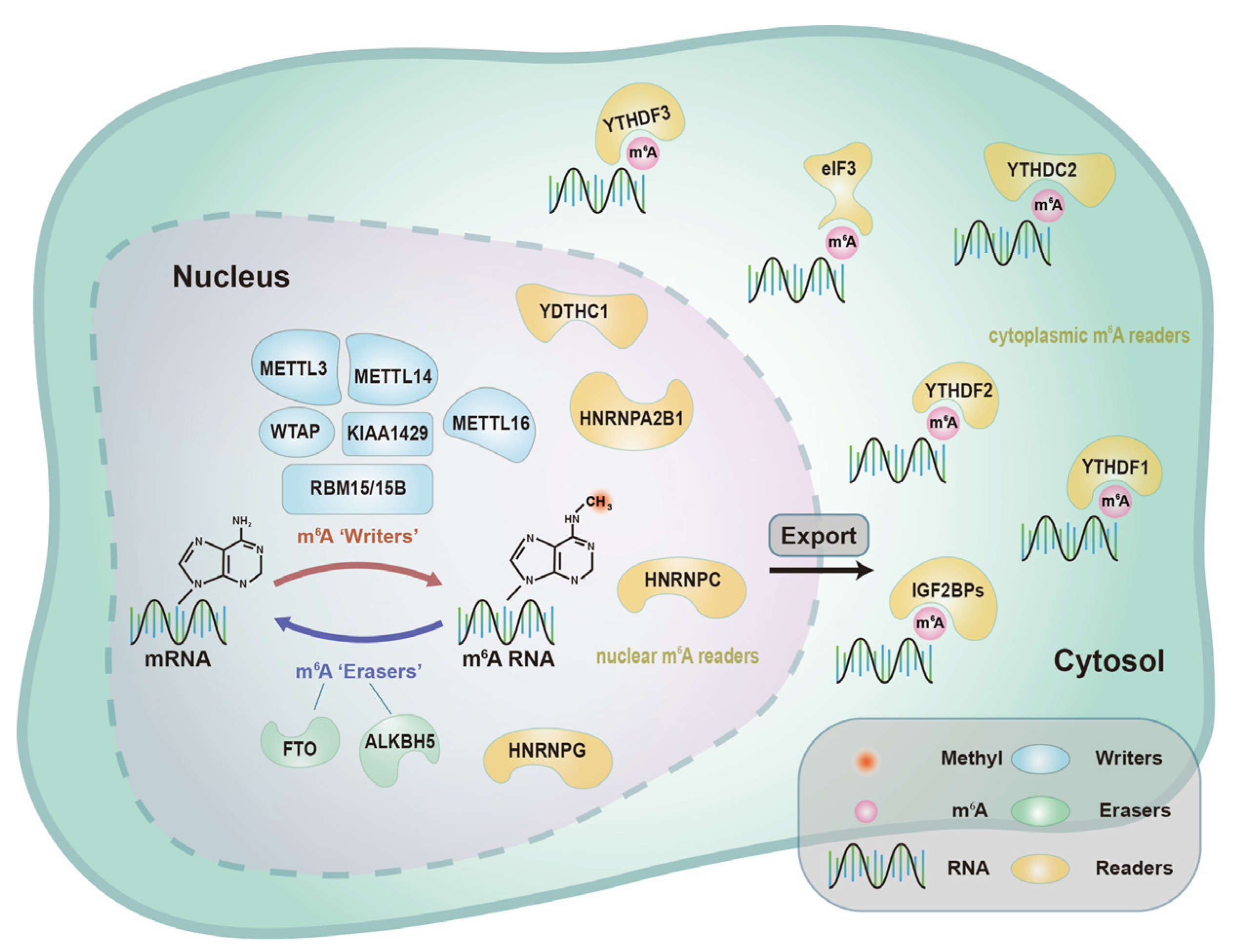
2.1. Dynamic Regulation of m6A RNA Modification

Due to the shortage of proteins related to m6A and the short half-life of most types of RNA, m6A RNA modifications have been considered to be stable and unchanged for a long time. The discovery of the first authentic m6A demethylase, namely, fat mass and obesity associated protein (FTO) [28], resolved this conundrum, demonstrating that m6A RNA modification is a dynamic process that is tightly regulated. As more studies have been performed, it is known that m6A RNA modification is involved in “writers”, “erasers” and “readers”, which is a dynamic process to add or remove a methyl group to adenosine and usually occurs in the RRACH sequence of adenines. To visualize the location and distribution of these proteins involved in m6A, we outlined the types of proteins involved in m6A RNA modification (Figure 1).

2.1.1. m6A “Writers”—Adenosine Methyltransferases

m6A RNA modification is modified by “writers”, including adenosine methyltransferases, methyltransferase-like 3 (METTL3), methyltransferase-like 14 (METTL14), Wilms tumour 1-associated protein (WTAP), KIAA1429 and RNA-binding motif protein 15/15B (RBM15/RBM15B). Well-known methyltransferases can add a methyl group to adenosine. The METTL3-METTL14-WTAP methyltransferase complex can add m6A, and METTL3 has been previously discovered to be an S-adenosyl-L-methionine (SAM)-binding site in this multiprotein complex, which is widely expressed and highly conserved. Through knockdown of Mettl3 in blastocysts achieved by mating Mettl3+/− mice, it has been shown that METTL3 is a regulator for terminating murine naive pluripotency [29], underlining the crucial role of METTL3 in m6A RNA modification. METTL14 and METTL3 are close homologues, and they always work together as METTL3-14 complexes. This complex subsequently interacts with WTAP to regulate the activity and precise localization of m6A methyltransferase [30]. Additionally, other unknown proteins may be involved in this complex. By proteomics, a recent study uncovered several candidate proteins, including KIAA1429 [31], required for m6A RNA modification in mammals. During mammalian female development, loss of function of RBM15/RBM15B results in defective XIST-mediated gene silencing on the X chromosome [32].

2.1.2. m6A “Erasers”—Demethylases

FTO was the first m6A mRNA demethylase to be discovered, confirming that m6A RNA modification is not unalterable but can be removed. Later investigations revealed that the dysregulation of FTO may be associated with obesity, brain malformations and growth retardation [33,34,35,36], suggesting that m6A RNA modification may have essential regulatory functions in these diseases. Moreover, in human cell lines, knockdown of ALKB homologue 5 (ALKBH5), which belongs to the same ALKB family of FTO, causes higher m6A levels [37]. However, unlike FTO, ALKBH5 catalyses direct the removal of the methyl group from m6A-methylated adenosine instead of indirect oxidative demethylation [38].

2.1.3. m6A “Readers”—Binding Proteins

m6A RNA modification works in one of two ways [25], as follows: modifying the structure of target transcripts to restrain or trigger interactions between RNAs and proteins or being directly recognized by “readers” to initiate downstream processes. According to recent studies, proteins in the YT521-B homology (YTH) family are correlated with the modulation of mRNA fate. In the cytoplasm, YTH domain-containing 1 (YTHDF1) facilitates m6A mRNA translation [39], and YTH domain-containing 2 (YTHDF2) aids in the degradation of mRNAs [40]. In addition, YTH domain-containing 3 (YTHDF3) collaborates with YTHDF1 to promote mRNA translation, and it enhances YTHDF2-mediated mRNA decay [41]. YTH domain-containing 1 (YTHDC1) is engaged in alternative splicing and plays an important role in nuclear export. YTH domain-containing 2 (YTHDC2) prefers to bind to transcripts that contain m6A RNA to accelerate RNA translation and decay [42] as well as to decrease the amount of mRNA and improve the efficiency of translation. Eukaryotic initiation factor 3 (eIF3) has been reported to directly bind to 5′UTR m6A mRNA [43] independent of YTHDF1. Moreover, other m6A “readers” have been reported, such as insulin-like growth factor 2 mRNA-binding proteins (IGF2BPs), heterogeneous nuclear ribonucleoprotein A2B1 (HNRNPA2B1), heterogeneous nuclear ribonucleoprotein C (HNRNPC) and heterogeneous nuclear ribonucleoprotein G (HNRNPG). As more m6A “readers” are discovered, the knowledge of m6A RNA modification will be more comprehensive.

2.2. Biofunctions of m6A RNA Modification

Due to breakthroughs in transcriptome sequencing of m6A over the past few decades, the vital roles of m6A RNA modification have been verified in a number of essential cellular events, including the fate of mRNA, the maintenance of stemness and even tumorigenesis [44]. The “writers”, “erasers” and “readers” involved in m6A RNA modification have also been linked to certain diseases [45,46,47], such as infertility, obesity, dysplasia and even cancer. Given its ubiquity in mRNA and lncRNA, further regulatory roles for m6A RNA modification as well as the discovery of additional m6A-related proteins are expected.

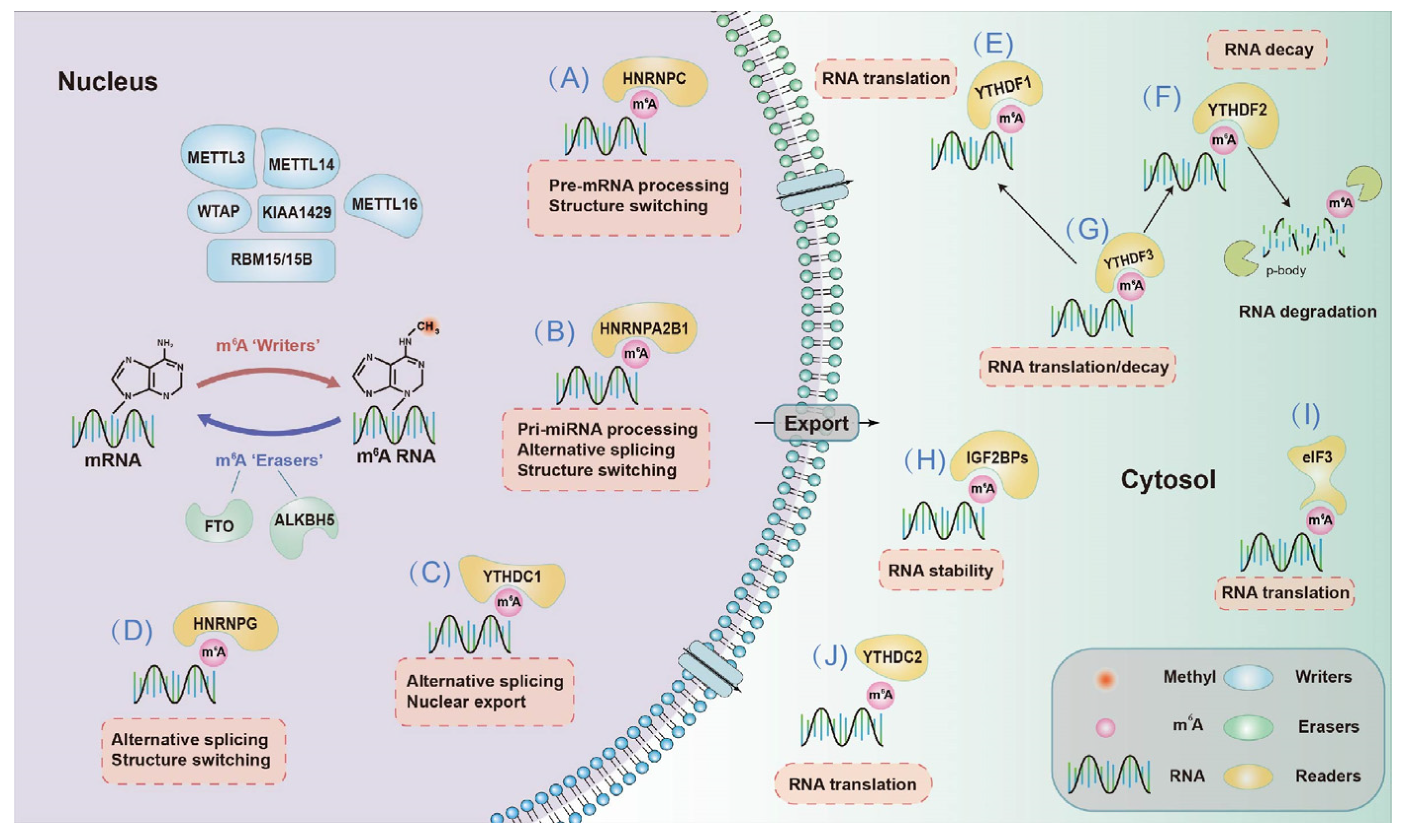
2.2.1. Effects on mRNA Fate—Splicing, Processing, Translation and Degradation

For mRNA, m6A regulates the processing and alternative splicing of precursor mRNA (pre-mRNA) in the nucleus as well as the translation, degradation and decay of mRNA in the cytosol (Figure 2). Processing alternative splicing and pre-mRNA to form accurate RNA in posttranscriptional regulation allows eukaryotic organisms with limited gene counts to perform a variety of functions. METTL3, METTL14, WTAP and ALKBH5 [48] are mainly located in nuclear speckles where pre-mRNA processing occurs, implying that m6A RNA modification is involved in pre-mRNA processing. HNRNPC, HNRNPG and HNRNPA2B1 are associated with mRNA structure switching, thus regulating gene expression. A recent study has suggested that m6A RNA modification affects the binding between HNRNPC and mRNA [49], thus regulating the processing and alternative splicing of pre-mRNA. Further research has indicated that in HEK-293T cells, knockdown of Mettl3/14 comodulates the expression of more than 5000 genes with knockdown of Hnrnpc, and nearly one in five of these genes may contain an m6A-mediated structural remodelling switch. In addition, researchers have found that the depletion of FTO enhances the m6A level and thus promotes the affinity of serine- and arginine-rich splicing factor 2 (SRSF2) binding with RNA, thereby increasing the number of target exons included. These data provide strong evidence of the essential role of m6A RNA modification during mRNA splicing.
As proteins are executors of biological functions, their synthesis and translation processes are vital to life and are regulated by various factors [37], and m6A is one of the factors because the presence of m6A in exons and surrounding stop codon regions may have an impact on protein production. Mettl3 ablation significantly enhances translation efficiency in mouse embryonic stem cells (mESCs) and embryoid bodies (EBs) [29], indicating that m6A may play a translational regulatory role. Additionally, another m6A “reader”, YTHDF1, has been reported to increase translational output by interacting with initiation factors and ribosomes [38], providing direct evidence for the roles of m6A in RNA translation. Identifying more m6A “readers” will aid in better understanding the regulatory role of m6A in the translation process [50].
The flexible and rapid degradation of mRNA is critical for the response of living organisms to changeable environments. Knockdown of Mettl3/14 modestly increases the stability of target mRNAs in mESCs [51], demonstrating that mRNA instability is associated with m6A RNA modification. RNA degradation has been verified to be enhanced by YTHDF2 through the transportation of mRNA for degradation [52], revealing the role of m6A RNA modification in mRNA degradation. The same conclusions have been reached in Hnrnpc [47] and Alkbh5 [36] knockdown studies.

2.2.2. Biological Consequences of m6A—Dysregulation in Cellular Processes and Diseases

Recent studies have revealed that m6A RNA is important in both physiological and pathological circumstances, including cellular stress, differentiation, development, immune response and tumorigenesis (Table 1).

Cellular Stress

Cellular stress includes oxidative stress, hypoxia, metabolic stress, heat shock, DNA damage ER stress and autophagy [53]. Overexpression of Mettl3 leads to more reactive oxygen species (ROS) production through the Keap1Nrf2 antioxidant pathway in mouse renal tubular epithelial cells (mRTECs) [54]. A recent study has identified that in normal human hepatocytes, FTO triggers oxidative stress through m6A demethylation at the 3′UTR of peroxisome proliferator-activated receptor-γ coactivator-1α (PGC1α), an important modulator of mitochondrial metabolism, which increases the stability and enhances translation of PGC1α mRNA, thereby causing increased production of ROS [55]. YTHDF1 has been discovered to be a negative regulator in the Keap1Nrf2 pathway, as its knockdown induces the overexpression of NRF2 and increases the production of antioxidants in human bronchial epithelium (Beas-2B) cells [56]. In hepatocellular carcinoma cells (HCCs), downregulation of METTL3 inhibits glycolytic capacity through the mTOR signalling pathway [57]. Depletion of Ythdf2 causes heat shock stress by increasing HSP90, HSP60 and HSPB1 mRNA expression [58,59]. As an m6A “writer”, METTL16 has been confirmed to be recruited to DNA damage sites after 20–30 min of UV exposure and to methylate RNAs transported to DNA damage sites, which may be related to the nucleotide excision repair (NER) mechanism [60,61]. Additionally, FTO is correlated with the stability of HSP70 and other genes related to ER stress through the NF-κB pathway in osteoblasts [62]. Moreover, FTO was reported to be related to autophagy by modulating the m6A level of Atg5 and Atg7, which was recognized by YTHDF2 [63]. FTO also participates in the demethylation of the transcripts of ULK1, thus promoting autophagy by the upregulated protein abundance of ULK1 [64].

Hematopoietic Development

m6A RNA modification plays an important role in haematopoietic and progenitor stem cell (HSPC) differentiation as well as in haematopoietic development in vertebrates. Depletion of Mettl3 prevents double-stranded RNAs (dsRNAs) from being synthesized and activates MDA5 and RIG-I [65], ultimately causing haematopoietic stem cell (HSC) development to fail. Moreover, by elevating m6A levels, METTL3 stimulates c-MYC, PTEN and BCL2 mRNA translation, which inhibits the proliferation of HSPCs [66]. By degrading mRNAs involved in the Wnt signalling pathway, such as Ccnd1, c-Myc and Axin2, YTHDF2 suppresses this signalling pathway, thereby inhibiting the proliferation and differentiation of HSCs [67]. In HSPCs, METTL3 and YTHDF2 work together to suppress Notch1a [68], providing compelling evidence that m6A RNA modification governs haematopoietic stem cell-directed differentiation.

Neurogenesis

m6A RNA modification regulates the development of the central nervous and reproductive systems. First, through the JAK/STAT and PI3K/AKT signalling pathways, YTHDF2 [69] and FTO [70] increase the self-renewal and proliferation of neural stem/progenitor cells (NSCs). By speeding up the cell cycle of radial glial cells (RGCs), METTL3 and METTL14 promote cortical biogenesis [71]. YTHDF1 promotes synaptic transmission and transcription of LTP-related target genes in neurons, which regulates learning and memory [72].

Fertilization

In addition, METTL3 [73] and ALKBH5 [37,74] are involved in the modulation of motility and proliferation of sperm cells by regulating m6A levels. YTHDC1 [75] and YTHDF2 [76] promote oocyte proliferation and maturation by regulating the maturation and translation of CPSF6 and Trcp5 transcripts, respectively. Abnormal embryo development thus may result in spontaneous abortion (SA), which is related to the imbalance of redox reaction. Research about SA patients showed that FTO is downregulated, causing immune tolerance and angiogenesis at the placenta, thus leading to spontaneous abortion [77]. Knockdown of ALKBH5 can promote trophoblast invasion by regulating the mRNA stability of CYR61, thus resulting in spontaneous abortion [78].

Immune Response

In the immune response, research has shown that m6A protects RNA from being recognized by TLR3 [79] and TLR7 [80] as antigens for degradation. Additional evidence suggests that m6A modification plays an indispensable role in the immune response by influencing related factors, such as methylating a specific mRNA. A recent study has found an m6A score is useful to measure immune features of tumorigenesis in clinicopathology, such as inflammatory stages [81]. METTL3 has been shown to be responsible for the stability of Socs mRNA and thus regulate the TLR4/NF-κB pathway, which is related to inflammation [82]. By inactivating the TLR4/NF-κB pathway, deletion of Mettl14 in dendritic cells (DCs) severely impairs B cell development, which affects the processes of IL-7-induced pro-B cell proliferation and causes abnormalities in gene expression critical for B cell development [83], confirming its important role in the immune response. In addition, ALKBH5 has been found to be highly expressed in spleens and lungs [70], which contain a large number of immune cells and are frequently involved in immunological reactions. Additionally, a recent study has shown that HNRNPA2B1 and HNRNPC may be associated with the abnormal immune response in endometriosis [84].

Cancer

Recent studies have shown a strong correlation of m6A RNA modification and cancer [85], including glioblastoma (GBM) and acute myeloid leukaemia (AML). In GBM, m6A regulates the proliferation, invasion and migration of tumours as well as the maintenance of the stemness of cancer stem cells by targeting several oncogenes [86]. By decreasing m6A levels of ADAM19 and increasing its expression in glioblastoma stem cells (GSCs), downregulation of METTL3/14 stimulates the proliferation and self-renewal of GSCs, ultimately resulting in GBM [87]. During the development of AML, abnormal m6A RNA modification of oncogenes and tumour suppressor genes plays an important role in many stages of tumorigenesis, such as proliferation and differentiation of cancer cells as well as self-renewal and cellular death of cancer stem cells [88,89,90]. Further research has revealed that AML cells have higher levels of METTL3 expression than those of normal haematological cells [91]. CEBPZ recruits METTL3 to the SP1 promoter area to increase m6A levels and induce translation, and SP1 subsequently activates c-MYC, a notorious oncogene, resulting in the formation of AML. Moreover, another study has found that a decreased copy number of Alkbh5 is common in AML and has been linked to TP53 mutations, which predict a poor prognosis for AML patients [92].

Diabetes

Lately, the significant role of m6A modification in diabetes has been confirmed. In 2019, β-cell-specific Mettl14 knockout mice were used to mimic the features of human type 2 diabetes (T2D). Some transcripts involved in the regulation of cell cycle progression and the secretion of insulin are hypomethylated and were detected by m6A sequencing. This study highlights the importance of RNA methylation in regulating metabolism of glucose and provides a potential therapeutic targeting of m6A modulators in type 2 diabetes [93]. In HepG2 cells, the expression of FTO is upregulated by high glucose, and with its upregulation, mRNA expression levels of FOXO1, G6PC and DGAT2 were significantly increased [94].
Table 1. Roles of m6A RNA methylation and dysregulation in cellular processes and diseases.
Processes
/Diseases
m6A RegulatorCells/
Organisms
Effect of Gene Loss/Gain of FunctionMechanismRef.
METTL3mRTECs↑ROS↑METTL3/Keap1/Nrf2[54]
FTOL02 cells↓ROS↓FTO/↑PGC-1α[55]
YTHDF1Beas-2B cells↓Hypoxia adaptation↓YTHDF1/Keap1/Nrf2-AKR1C1[56]
Cell stressMETTL3HCCs↓Glycolytic capacity↓METTL3/mTORC[57]
YTHDF2MEFs↑Heat shock stress↓YTHDF2/↑HSP90, HSP60, HSPB1[58,59]
METTL16MEFs↑DNA damage↑METTL16/γH2AX[60,61]
FTOMice↑ER stress↓FTO/↓HSP70/↑NF-κB[62]
FTOMice↑autophagy↓FTO/↓Atg5, Atg7[63]
FTO293T cells↑autophagy↓FTO/↑ULK1[64]
HaematopoieticMETTL3HSCs↓Proliferation,
↓differentiation
↓METTL3/↑MDA5/RIG-I[65]
developmentMETTL3HSPCs↑Differentiation,
↓cell proliferation
↑METTL3/c-MYC/BCL2/PTEN[66]
YTHDF2HSCs↑Regeneration↓YTHDF2/↑Wnt target genes[67]
METTL3HSPCs↑Endothelial to haematopoietic transition↓METTL3/↑YTHDF2/↓Notch1a[68]
NeurogenesisYTHDF2NSPCs↓Self-renewal↓YTHDF1/JAK–STAT[69]
FTONSCs↓Proliferation,
↓differentiation
↓FTO/BDNF/PI3K/Akt2/Akt3[70]
METTL3/14RGCs↑Neurogenesis,
↑cell cycle
↓METTL3/14/↑Neurog2/Neurod1[71]
YTHDF1Mice↓Learning, memory defects↓YTHDF1/Camk2a[72]
FertilizationMETTL3Zebrafish↓Sperm motility↓METTL14/11-KT/17β-E2[73]
ALKBH5Mice↓Fertility↓ALKBH5/↑Dnmt1[37,74]
YTHDC1Germ cells↓Oocyte growth, maturation↓YTHDC1/CPSF6/SRSF3[75]
YTHDF2Mice↓Oocyte maturation↓YTHDF2/Trpc5[76]
FTOSA patients↑Spontaneous abortion↓FTO/VEGFA, VEGFR[77]
ALKBH5SA patients↑Spontaneous abortion↓FTO/↑CYR61[78]
ImmuneMETTL3Mice↓T cell proliferation↓METTL3/IL-7/STAT5/SOCS[82]
responseMETTL14DCs↓B cell development↓METTL14/TLR4/NF-κB[83]
CancerMETTL3/14GBM↑Proliferation and self-renewal of GSCs↓METTL3/14/↑ADAM19[87]
METTL3AML↓Cell cycle and differentiation of leukaemic cells↓METTL3/↑c-MYC[91]
ALKBH5AML↓Prognosis of AML patients↓ALKBH5/↑TP53[92]
DiabetesMETTL14Mice↓Insulin secretion↓METTL14/IGF1–AKT–PDX1[93]
FTOHepG2 cells↓Glucose metabolism↓FTO/FOXO1/G6PC/DGAT2[94]

2.3. Detection

The direct detection of m6A bases is difficult because the base pairing properties remain unchanged and cannot be distinguished from regular bases by reverse transcription. Methylated RNA immunoprecipitation followed by high-throughput sequencing (MeRIP-seq), also known as m6A RNA immunoprecipitation sequencing (m6A-seq), has been widely used to detect m6A at a resolution of 110–200 nt since it was developed in 2012 [95].
In the past decade, several approaches with the resolution of a single base, including photo crosslinking-assisted m6A sequencing (PA-m6A-seq) [96], m6A individual nucleotide resolution cross-linking and immunoprecipitation sequencing (miCLIP-seq) [97], site-specific cleavage, and radioactive labelling, followed by ligation-assisted extraction and thin-layer chromatography (SCARLET) [98], have been developed (Table 2). A recent study has introduced an efficient method to directly detect m6A bases by m6A-sensitive RNA endoribonuclease-facilitated sequencing (m6A-REF-seq) [99] with higher sensitivity and a lower false-positive rate, eliminating the dependence of traditional identification methods on antibodies.
Moreover, several methods have been developed to detect the total amount of m6A in RNA [100], including colorimetry, m6A dot blot analysis and high-performance liquid chromatography–tandem mass spectrometry (HPLC–MS/MS). Furthermore, directly detecting the effect of changing any site and in any organism is promising with the advancement of genome engineering.

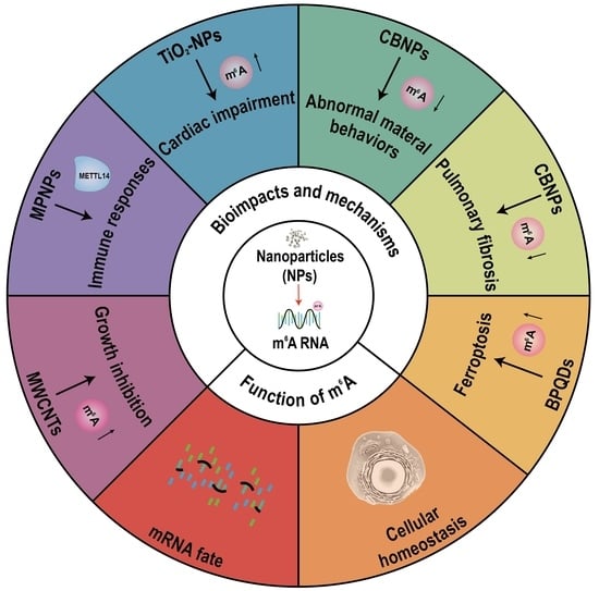
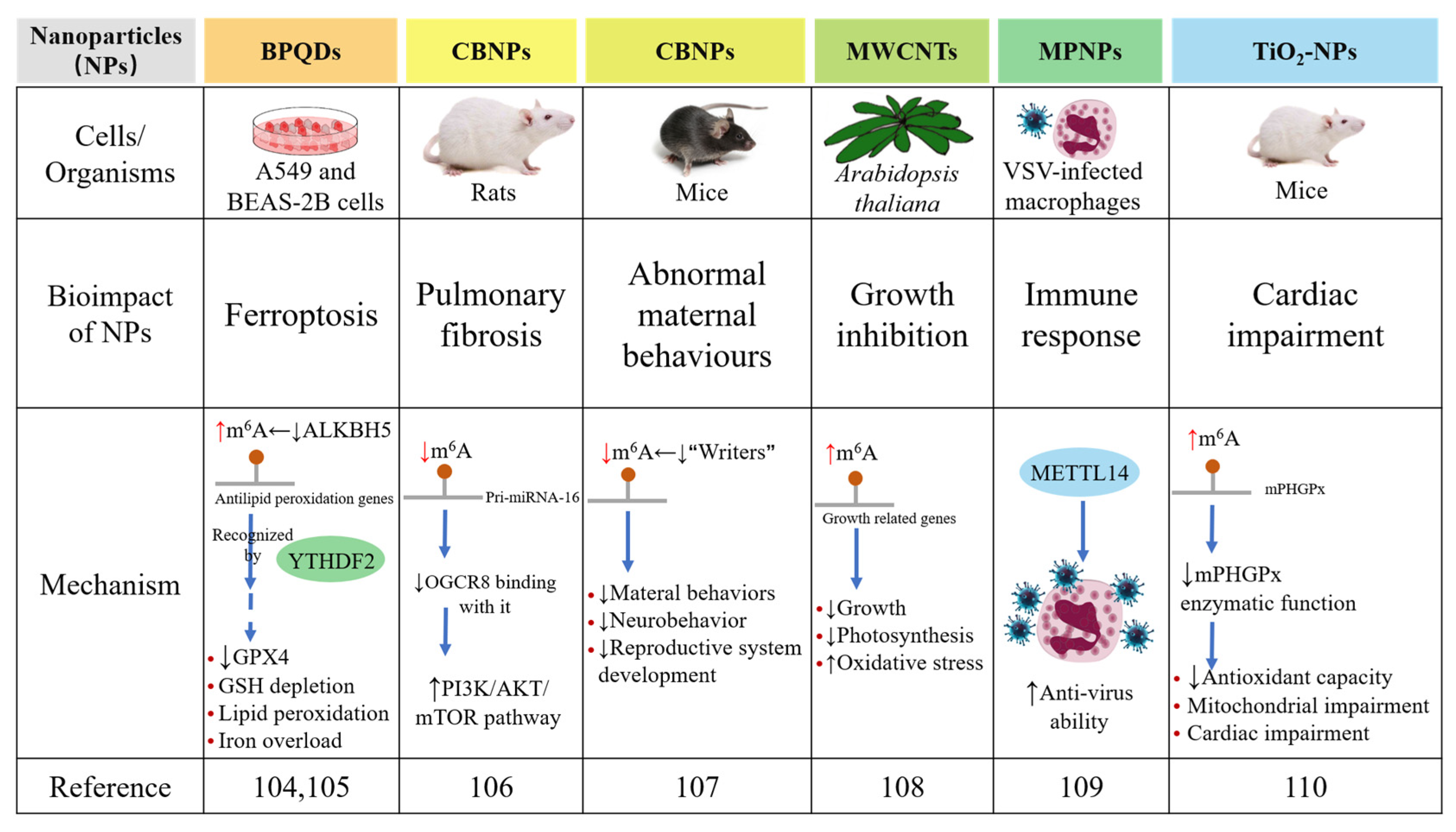
3. m6A RNA Modification Modulates the Bioimpacts of Nanoparticles

NPs have been widely used in production and life due to their distinct physicochemical features, which is accompanied with concerns over their bioimpacts and mechanisms involved. Therefore, there is a need to study and comprehend the impacts in terms of the cellular and molecular mechanisms of the bioimpacts of NPs. The mechanisms involved are partly clarified, including oxidative stress, cytotoxicity, neurotoxicity and genotoxicity [101], which depend on physiochemical features, such as the size and crystallinity of nanoparticles.
However, there is more evidence suggesting the possibility of NP-induced genetic toxicity. DNA methylation, histone modifications and RNA modifications as well as many other epigenetic mechanisms have been proposed and widely studied in recent years. As more research has been performed and more knowledge is available, the roles of epigenetics in the biological function of NPs are gaining increasing attention. Most notably, epigenetic dysregulation has been linked to clinical disorders, including cancer and neurological diseases. Thus, it is possible that NP-mediated epigenetic modulations can also contribute to pathogenesis to a certain level. One of the characteristics of cancer is global DNA hypomethylation, which can be passed down from generation to generation [102]. For instance, the levels of pri-miRNA-1275 are increased after Ag-NP exposure in neural stem/progenitor cells derived from human embryonic stem cells (hESCs) with obvious downregulation of their target genes, ADAMTS9 and SHANK2, which encode proteins involved in axonal guidance signalling and are linked to brain damage and neurodegenerative diseases [103]. The link between nanoparticles and m6A RNA modification is less well understood, but evidence is beginning to emerge in the past two years. Therefore, we will introduce these studies and summarize the role of m6A RNA modification in the bioimpacts induced by NPs.
Recently, we established a 3D high-throughput screening system using constructed kidney organoids to study the nephrotoxicity of black phosphorus quantum dots (BPQDs) [104]. Interestingly, we found significant endoplasmic reticulum (ER) stress and insulin sensitivity induced by BPQDs in the kidney. Furthermore, considering their easy inhalation ability, we used lung cells to further evaluate the bioimpacts of BPQDs and their correlation with m6A RNA modification. Surprisingly, the global m6A level was increased after BPQD exposure in lung cells, and after the protein expression of m6A-related proteins was examined, we further clarified the significant role of ALKBH5 in this event; the expression of “writers” and other “erasers” did not change, but ALKBH5 decreased with BPQD exposure [105]. Transcriptome and epitranscriptome data have provided evidence of the correlation between the abnormal elevation of total m6A levels and the aberrant expression of genes related to ferroptosis involving ER stress, iron homeostasis and iron homeostasis, which has also been verified by evidence of iron overload, lipid peroxidation, glutathione peroxidase 4 (GPX4) downregulation and glutathione (GSH) depletion in vitro. Moreover, the mechanism of the downregulation of antiferroptosis-related genes is that YTHDF2, a m6A “reader”, recognizes and promotes the decay of mitochondrial homeostasis-containing and antilipid peroxidation genes modulated by m6A, which has been verified by m6A RNA immunoprecipitation (RIP)-qPCR in lung cells. Our study reported the roles of m6A RNA modification in BPQD-induced ferroptosis and for the first time revealed the mechanism involved, thereby providing a promising biomarker and a drug target for BPQD-induced ferroptosis (Figure 3).
Carbon black nanoparticles (CBNPs) have been reported to have a close correlation with many diseases, some of which are involved in reproductive systems and abnormal behaviours. A recent study has shown the relationship between CBNP-induced pulmonary fibrosis and m6A RNA modification [106]. The processing of pri-miRNA-126 is a DiGeorge syndrome critical region gene 8 (DGCR8)-dependent process that regulates the downstream PI3K/AKT/mTOR pathway. This study showed that CB exposure causes pulmonary fibrosis and activates the PI3K–AKT–mTOR pathway by decreasing the m6A level of pri-miRNA-126 and its binding with DGCR8. Additionally, another study has reported that CBNP exposure during pregnancy influences maternal behaviours and partially causes abnormal neurobehaviors and the development of the reproductive system in offspring, all of which are linked to m6A RNA modification [107]. This study revealed the potential correlation between the abnormal maternal behaviours induced by CBNPs and the decreased m6A level. Further studies need to be performed to provide more information about this intriguing phenomenon.
Moreover, a previous study on the bioimpacts of high dots of multiwalled carbon nanotubes (MWCNTs) on Arabidopsis thaliana [108] has suggested the potential correlation of m6A RNA modification and the bioimpacts of MECNTs (Figure 4). In detail, the growth of Arabidopsis thaliana is severely inhibited after MWCNT exposure, presenting abnormal behaviours of roots and leaves as well as suppression of auxin and photosynthesis signalling. MWCNTs also cause oxidative stress, thus activating the antioxidant mechanism. Combined data from m6A-seq and RNA-seq have shown that m6A RNA modification may negatively regulate the transcription of related genes after MWCNT exposure, including plant hormone transduction signalling pathways and protein phosphorylation. For the first time, these findings offer insight into the molecular pathways underlying MWCNT phytotoxicity and plant defensive responses to MWCNTs. In addition, many studies have shown that cellular metabolism can be reprogrammed through m6A RNA modification during viral infection. Recently, a study uncovered that metal–protein nanoparticles (MPNPs) in macrophages infected by vesicular stomatitis virus (VSVs) polarize macrophages and stimulate immunological responses related to METTL14 [109]. Further research has revealed that a high dose of interferon-beta (IFN-β) increases the expression of METTL14, an anti-VSV protein. The same conclusions have been reached for influenza viruses (H1N1(WSN)), which are also negative-sense single-stranded RNA viruses. Overall, these findings shed light on the antiviral role of METTL14 and suggest that manipulating METTL14 may be a viable technique for combating additional negative-sense single-stranded RNA virus infections.
In 2021, research related to TiO2-NPs and m6A RNA modification was reported [110]. In this study, researchers aimed to determine whether mitochondrial phospholipid hydroperoxide glutathione peroxidase (mPHGPx) can sustain cardiovascular function and bioenergetics in offspring after direct exposure to TiO2-NPs by inhalation during pregnancy in female mice and to reveal an epitranscriptomic mechanism that contributes to this phenomenon. The results showed that in offspring, the enzymatic function of mPHGPx is considerably decreased, and the m6A levels are increased. These findings attribute the abnormally high m6A level of mPHGPx to reduced antioxidant capacity and the resulting mitochondrial and cardiac impairments that last into adulthood after gestational nTiO2 exposure through inhalation.
In addition to those studies related to bioimpacts of NPs induced by m6A RNA modification, there is some research about electrochemical immunosensors for m6A detection based on nanotechnology. Silver nanoparticles and SiO2 nanospheres are used in the electrochemical immunosensor, providing a promising detection platform for m6A [111]. Moreover, nanoparticles have emerged as promising carriers in cancer therapy by delivering drugs to target. A recent study showed that N6-methyladenosine (m6A) mediated by METTL3 upregulated long noncoding RNA LINC00958, which subsequently promoted HCC progression through the miR-3619-5p/HDGF axis [112]. Therefore, it is promising that RNA m6A modification functions as a robust tool to modify RNA used in cancer therapy, as the veil of RNA m6A modification in the bioimpacts of NPs is lifted completely.

4. Conclusions and Outlook

Despite a large number of findings connected to the functional roles of epigenetics in the bioimpacts of NPs [23], many vital gaps still have to be filled, particularly in the area of RNA m6A modification. Epigenetic modifications can be utilized to study nanoparticle toxicity and, more crucially, to predict their toxicity in preclinical research, according to recent evidence from both experimental and epidemiological studies. A link between nanotoxicity and epigenetic changes following exposure to specific nanoparticles has been demonstrated in many experimental studies in vivo and in vitro [113,114,115]. YTHDC1 and METTL14, in particular, have been identified as possible endometrial carcinoma diagnostic and prognostic indicators [116]. Therefore, RNA m6A modifications may function as biomarkers not only to detect and predict the toxicity of NPs but also to evaluate the process of diseases triggered by NPs as the role of RNA m6A modification in the bioimpacts of NPs is further understood.
The roles of DNA methylation and histone acetylation in the biological functions induced by NPs have been reported in great detail [23,25,117]. However, information on the roles of RNA m6A modification is lacking. Moreover, most studies focus on the bioeffects of one type of NP on RNA m6A modification, whereas clarifying the process involved in RNA m6A modification and NPs in greater depth could provide broader insights into the understanding of nanomaterials. The precise impacts of the physicochemical features of NPs, including their size and surface charge, in modulating RNA m6A modification are also unknown to some extent.
The genetic effects of NP-induced bioimpacts related to RNA m6A modification are another gap that must be filled. Studies involving longer research periods are needed to illustrate the consequences of RNA m6A modification correlated with NPs and to determine whether these biological effects can be passed from generation to generation [118]. Several model organisms, such as Danio rerio, can serve as efficient in vivo models for determining the potential epigenetic mechanisms in the toxicity triggered by NPs and the genetic effects of the bioimpacts of NPs in this field [119,120]. Zebrafish are low-cost and simple to feed in the lab, and they can be used to efficiently test agents through multiple routes of exposure, including direct exposure to water, which is especially significant for the toxicology of environmental pollution. Because of the short life cycle and distinct developmental stages, transgenerational studies can be completed in a relatively short period of time. Furthermore, specific physiological effects can be studied at diverse stages of the whole development of zebrafish.
Finally, dysregulation of RNA m6A modification has been linked to some pathological diseases, such as obesity, as well as several mental disorders, such as neurodegenerative diseases, and even cancer [16,42]. Recent studies have found that NPs are closely related to dysregulation in some cellular processes and diseases when exposed to the body through the digestive system, respiratory system and skin tissue [121]. Thus, it is possible that NP-induced RNA m6A modification can similarly lead to some diseases to a certain extent. Therefore, addressing the roles of RNA m6A modification in the bioimpacts of NPs and the involved mechanisms as well as using various practical in vivo and in vitro models are important challenges for the future.
Last but not least, with the remarkable development of RNA nanotechnology, RNA nanoparticles can be used as carriers in drug delivery systems [122]. In order to improve targeting efficiency and achieve optimal therapeutic efficacy, physiochemical features can be modulated on purpose. Disease diagnosis and healthcare based on RNA nanotechnology can achieve early diagnosis, early detection and early treatment, lowering disease preventive costs and improving human health protection. Furthermore, RNA nanotechnology has a wide range of potential applications in disease prevention based on vaccine creation. With more investigation, it is promising to target m6A-methylation-based epitranscriptomics using the nanoparticle as an “epigenetic drug” for cancer therapy.

Author Contributions

Y.W. wrote the manuscript. C.H. and F.R. guided the work and revised the manuscript. Z.Z. provided suggestions. All authors have read and agreed to the published version of the manuscript.

Funding

This research was funded by grants from the National Natural Science Foundation of China (32071301, 31971234 and 42177411) and the Natural Science Foundation of Fujian Province, China (2020J01027).

Institutional Review Board Statement

Not applicable.

Informed Consent Statement

Not applicable.

Data Availability Statement

Not applicable.

Conflicts of Interest

The authors declare no conflict of interest.

Abbreviations

NPsnanoparticles
m6AN6-methyladenosine
SiO2-NPssilica dioxide nanoparticles
TiO2-NPstitanium dioxide nanoparticles
PEGpolyethylene glycol
QDsquantum dots
FTOfat mass and obesity-associated protein
METTL3methyltransferase-like 3
METTL14methyltransferase-like 14
WTAPWilms tumour 1-associated protein
RBM15/RBM15BRNA-binding motif protein 15/15B
ALKBH5ALKB homologue 5
YTHYT521-B homology
YTHDF1YTH domain-containing 1
YTHDF2YTH domain-containing 2
YTHDF3YTH domain-containing 3
YTHDC1YTH domain-containing 1
YTHDC2YTH domain-containing 2
eIF3eukaryotic initiation factor 3
IGF2BPsinsulin-like growth factor 2 mRNA-binding proteins
HNRNPA2B1heterogeneous nuclear ribonucleoprotein A2B1
HNRNPCheterogeneous nuclear ribonucleoprotein C
HNRNPGheterogeneous nuclear ribonucleoprotein G
pre-mRNAprecursor mRNA
SRSF2serine and arginine rich splicing factor 2
mESCsmouse embryonic stem cells
EBsembryoid bodies
mRTECsmouse renal tubular epithelial cells
ROSreactive oxygen species
mRTECsmouse renal tubular epithelial cells
PGC1αperoxisome proliferator-activated receptor-γ coactivator-1α
Beas-2B cellshuman bronchial epithelium cells
HCCshepatocellular carcinoma cells
NERnucleotide excision repair
dsRNAsdouble-stranded RNAs
HSCshaematopoietic stem cells
NSCsneural stem/progenitor cells
RGCsradial glial cells
DCsdendritic cells
GBMglioblastoma
AMLacute myeloid leukaemia
GSCsglioblastoma stem cells
MeRIP-seqmethylated RNA immunoprecipitation followed by high-throughput sequencing
m6A-seqm6A RNA immunoprecipitation sequencing
PA-m6A-seqphotocrosslinking-assisted m6A sequencing
miCLIP-seqm6A individual nucleotide resolution cross-linking and immunoprecipitation sequencing
SCARLETsite-specific cleavage and radioactive labelling followed by ligation-assisted extraction and thin-layer chromatography
m6A-REF-seqm6A-sensitive RNA endoribonuclease-facilitated sequencing
HPLC–MShigh-performance liquid chromatography-tandem mass spectrometry
hESCshuman embryonic stem cells
BPQDsblack phosphorus quantum dots
ERendoplasmic reticulum
GPX4glutathione peroxidase 4
GSHglutathione
RIPRNA immunoprecipitation
CBNPscarbon black nanoparticles
MWCNTsmultiwalled carbon nanotubes
VSVvesicular stomatitis virus
MPNPsmetal–protein nanoparticles
IFN-βinterferon-beta
mPHGPxmitochondrial phospholipid hydroperoxide glutathione peroxidase

References

  1. Gupta, R.; Xie, H. Nanoparticles in daily life: Applications, toxicity and regulations. J. Environ. Pathol. Toxicol. Oncol. 2018, 37, 209–230. [Google Scholar] [CrossRef]
  2. Hoseinnejad, M.; Jafari, S.M.; Katouzian, I. Inorganic and metal nanoparticles and their antimicrobial activity in food packaging applications. Crit. Rev. Microbiol. 2018, 44, 161–181. [Google Scholar] [CrossRef]
  3. Lu, P.J.; Cheng, W.L.; Huang, S.C.; Chen, Y.P.; Chou, H.K.; Cheng, H.F. Characterizing titanium dioxide and zinc oxide nanoparticles in sunscreen spray. Int. J. Cosmet. Sci. 2015, 37, 620–626. [Google Scholar] [CrossRef]
  4. Knowles, B.; Wagner, P.; MacLaughlin, S.; Higgins, M.; Molino, P.J. Silica nanoparticles functionalized with zwitterionic sulfobetaine siloxane for application as a versatile antifouling coating System. ACS Appl. Mater. Interfaces 2017, 9, 18584–18594. [Google Scholar] [CrossRef]
  5. Bogdan, J.; Jackowska-Tracz, A.; Zarzynska, J.; Pławińska-Czarnak, J. Chances and limitations of nanosized titanium dioxide practical application in view of its physicochemical properties. Nanoscale Res. Lett. 2016, 10, 57. [Google Scholar] [CrossRef] [Green Version]
  6. Ashfaq, U.A.; Riaz, M.; Yasmeen, E.; Yousaf, M.Z. Recent advances in nanoparticle-based targeted drug-delivery systems against cancer and role of tumor microenvironment. Crit. Rev. Ther. Drug Carr. Syst. 2017, 34, 317–353. [Google Scholar] [CrossRef]
  7. Gill, K.K.; Kaddoumi, A.; Nazzal, S. PEG–lipid micelles as drug carriers: Physiochemical attributes, formulation principles and biological implication. J. Drug Target. 2015, 23, 222–231. [Google Scholar] [CrossRef]
  8. Mansuriya, B.D.; Altintas, Z. Applications of graphene quantum dots in biomedical sensors. Sensors 2020, 20, 1072. [Google Scholar] [CrossRef] [Green Version]
  9. Barani, M.; Reza Hajinezhad, M.; Sargazi, S.; Zeeshan, M.; Rahdar, A.; Pandey, S.; Khatami, M.; Zargari, F. Simulation, in vitro, and in vivo cytotoxicity assessments of methotrexate-loaded pH-responsive nanocarriers. Polymers 2021, 13, 3153. [Google Scholar] [CrossRef]
  10. Barani, M.; Rahdar, A.; Sargazi, S.; Amiri, M.S.; Sharma, P.K.; Bhalla, N. Nanotechnology for inflammatory bowel disease management: Detection, imaging and treatment. Sens. Bio-Sens. Res. 2021, 32, 100417. [Google Scholar] [CrossRef]
  11. Arora, S.; Rajwade, J.M.; Paknikar, K. Nanotoxicology and in vitro studies: The need of the hour. Toxicol. Appl. Pharmacol. 2012, 258, 151–165. [Google Scholar] [CrossRef]
  12. Cao, Y.; Li, S.; Chen, J. Modeling better in vitro models for the prediction of nanoparticle toxicity: A review. Toxicol. Mech. Methods 2020, 31, 1–17. [Google Scholar] [CrossRef]
  13. Lewinski, N.; Colvin, V.; Drezek, R. Cytotoxicity of nanoparticles. Small 2008, 4, 26–49. [Google Scholar] [CrossRef]
  14. Li, B.; Tang, M. Research progress of nanoparticle toxicity signaling pathway. Life Sci. 2020, 263, 118542. [Google Scholar] [CrossRef]
  15. Manohar, A.; Vijayakanth, V.; Kim, K.H. Influence of Ca doping on ZnFe2O4 nanoparticles magnetic hyperthermia and cytotoxicity study. J. Alloy. Compd. 2021, 886, 161276. [Google Scholar] [CrossRef]
  16. Kononenko, V.; Narat, M.; Drobne, D. Nanoparticle interaction with the immune system. Arch. Ind. Hyg. Toxicol. 2015, 66, 97–108. [Google Scholar] [CrossRef] [Green Version]
  17. Sun, T.; Wu, R.; Ming, L. The role of m6A RNA methylation in cancer. Biomed. Pharmacother. 2019, 112, 108613. [Google Scholar] [CrossRef]
  18. Wang, Z.; Wang, Z. Nanoparticles induced embryo–fetal toxicity. Toxicol. Ind. Health 2020, 36, 181–213. [Google Scholar] [CrossRef]
  19. Ahsan, S.M.; Rao, C.M.; Ahmad, M.F. Nanoparticle-protein interaction: The significance and role of protein corona. Adv. Exp. Med. Biol. 2018, 1048, 175–198. [Google Scholar] [CrossRef]
  20. Dinischiotu, A.; Stanca, L.; Gradinaru, D.; Petrache, S.N.; Radu, M.; Serban, A.I. Lipid peroxidation due to in vitro and in vivo exposure of biological samples to nanoparticles. Methods Mol. Biol. 2013, 1028, 155–164. [Google Scholar] [CrossRef]
  21. Fu, P.P.; Xia, Q.; Hwang, H.-M.; Ray, P.C.; Yu, H. Mechanisms of nanotoxicity: Generation of reactive oxygen species. J. Food Drug Anal. 2014, 22, 64–75. [Google Scholar] [CrossRef] [Green Version]
  22. Manshian, B.B.; Poelmans, J.; Saini, S.; Pokhrel, S.; Grez, J.J.; Himmelreich, U.; Mädler, L.; Soenen, S.J. Nanoparticle-induced inflammation can increase tumor malignancy. Acta Biomater. 2018, 68, 99–112. [Google Scholar] [CrossRef]
  23. Pogribna, M.; Hammons, G. Epigenetic effects of nanomaterials and nanoparticles. J. Nanobiotechnol. 2021, 19, 2. [Google Scholar] [CrossRef]
  24. Sierra, M.I.; Valdés, A.; Fernández, A.; Torrecillas, R.; Fraga, M.F. The effect of exposure to nanoparticles and nanomaterials on the mammalian epigenome. Int. J. Nanomed. 2016, 11, 6297–6306. [Google Scholar] [CrossRef] [Green Version]
  25. Wong, B.S.E.; Hu, Q.; Baeg, G.H. Epigenetic modulations in nanoparticle-mediated toxicity. Food Chem. Toxicol. 2017, 109, 746–752. [Google Scholar] [CrossRef]
  26. Cayir, A.; Byun, H.-M.; Barrow, T.M. Environmental epitranscriptomics. Environ. Res. 2020, 189, 109885. [Google Scholar] [CrossRef] [PubMed]
  27. Cao, G.; Li, H.-B.; Yin, Z.; Flavell, R.A. Recent advances in dynamic m6A RNA modification. Open Biol. 2016, 6, 160003. [Google Scholar] [CrossRef] [Green Version]
  28. Jia, G.; Fu, Y.; Zhao, X.; Dai, Q.; Zheng, G.; Yang, Y.; Yi, C.; Lindahl, T.; Pan, T.; Yang, Y.-G.; et al. N6-Methyladenosine in nuclear RNA is a major substrate of the obesity-associated FTO. Nat. Chem. Biol. 2011, 7, 885–887. [Google Scholar] [CrossRef]
  29. Geula, S.; Moshitch-Moshkovitz, S.; Dominissini, D.; Mansour, A.A.; Kol, N.; Salmon-Divon, M.; Hershkovitz, V.; Peer, E.; Mor, N.; Manor, Y.S.; et al. m6A mRNA methylation facilitates resolution of naive pluripotency toward differentiation. Science 2015, 347, 1002–1006. [Google Scholar] [CrossRef]
  30. Liu, J.; Yue, Y.; Han, D.; Wang, X.; Fu, Y.; Zhang, L.; Jia, G.; Yu, M.; Lu, Z.; Deng, X.; et al. A METTL3–METTL14 complex mediates mammalian nuclear RNA N6-adenosine methylation. Nat. Chem. Biol. 2013, 10, 93–95. [Google Scholar] [CrossRef] [Green Version]
  31. Schwartz, S.; Mumbach, M.; Jovanovic, M.; Wang, T.; Maciag, K.; Bushkin, G.G.; Mertins, P.; Ter-Ovanesyan, D.; Habib, N.; Cacchiarelli, D.; et al. Perturbation of m6A writers reveals two distinct classes of mRNA methylation at internal and 5′ sites. Cell Rep. 2014, 8, 284–296. [Google Scholar] [CrossRef] [PubMed] [Green Version]
  32. Patil, D.P.; Chen, C.-K.; Pickering, B.F.; Chow, A.; Jackson, C.; Guttman, M.; Jaffrey, S.R. m6A RNA methylation promotes XIST-mediated transcriptional repression. Nature 2016, 537, 369–373. [Google Scholar] [CrossRef] [PubMed]
  33. Boissel, S.; Reish, O.; Proulx, K.; Kawagoe-Takaki, H.; Sedgwick, B.; Yeo, G.S.H.; Meyre, D.; Golzio, C.; Molinari, F.; Kadhom, N.; et al. Loss-of-function mutation in the dioxygenase-encoding FTO gene causes severe growth retardation and multiple malformations. Am. J. Hum. Genet. 2009, 85, 106–111. [Google Scholar] [CrossRef] [Green Version]
  34. Dina, C.; Meyre, D.; Gallina, S.; Durand, E.; Körner, A.; Jacobson, P.; Carlsson, L.M.S.; Kiess, W.; Vatin, V.; Lecoeur, C.; et al. Variation in FTO contributes to childhood obesity and severe adult obesity. Nat. Genet. 2007, 39, 724–726. [Google Scholar] [CrossRef] [PubMed]
  35. Frayling, T.M.; Timpson, N.J.; Weedon, M.N.; Zeggini, E.; Freathy, R.M.; Lindgren, C.M.; Perry, J.R.B.; Elliott, K.S.; Lango, H.; Rayner, N.W.; et al. A common variant in the FTO gene is associated with body mass index and predisposes to childhood and adult obesity. Science 2007, 316, 889–894. [Google Scholar] [CrossRef] [Green Version]
  36. Scuteri, A.; Sanna, S.; Chen, W.-M.; Uda, M.; Albai, G.; Strait, J.; Najjar, S.; Nagaraja, R.; Orrú, M.; Usala, G.; et al. Genome-wide association scan shows genetic variants in the FTO gene are associated with obesity-related traits. PLoS Genet. 2007, 3, e115. [Google Scholar] [CrossRef]
  37. Zheng, G.; Dahl, J.A.; Niu, Y.; Fedorcsak, P.; Huang, C.-M.; Li, C.J.; Vågbø, C.B.; Shi, Y.; Wang, W.-L.; Song, S.-H.; et al. ALKBH5 is a mammalian RNA demethylase that impacts RNA metabolism and mouse fertility. Mol. Cell 2013, 49, 18–29. [Google Scholar] [CrossRef] [Green Version]
  38. Zheng, G.; Dahl, J.A.; Niu, Y.; Fu, Y.; Klungland, A.; Yang, Y.-G.; He, C. Sprouts of RNA epigenetics: The discovery of mammalian RNA demethylases. RNA Biol. 2013, 10, 915–918. [Google Scholar] [CrossRef] [Green Version]
  39. Wang, X.; Zhao, B.; Roundtree, I.A.; Lu, Z.; Han, D.; Ma, H.; Weng, X.; Chen, K.; Shi, H.; He, C. N6-methyladenosine modulates messenger RNA translation efficiency. Cell 2015, 161, 1388–1399. [Google Scholar] [CrossRef] [Green Version]
  40. Wang, X.; Lu, Z.; Gomez, A.; Hon, G.C.; Yue, Y.; Han, D.; Fu, Y.; Parisien, M.; Dai, Q.; Jia, G.; et al. N6-methyladenosine-dependent regulation of messenger RNA stability. Nature 2013, 505, 117–120. [Google Scholar] [CrossRef]
  41. Shi, H.; Wang, X.; Lu, Z.; Zhao, B.S.; Ma, H.; Hsu, P.J.; Liu, C.; He, C. YTHDF3 facilitates translation and decay of N6-methyladenosine-modified RNA. Cell Res. 2017, 27, 315–328. [Google Scholar] [CrossRef] [PubMed] [Green Version]
  42. Mao, Y.; Dong, L.; Liu, X.-M.; Guo, J.; Ma, H.; Shen, B.; Qian, S.-B. M6A in mRNA coding regions promotes translation via the RNA helicase-containing YTHDC2. Nat. Commun. 2019, 10, 5332. [Google Scholar] [CrossRef] [PubMed]
  43. Meyer, K.; Patil, D.; Zhou, J.; Zinoviev, A.; Skabkin, M.A.; Elemento, O.; Pestova, T.V.; Qian, S.-B.; Jaffrey, S.R. 5′ UTR m6A promotes cap-independent translation. Cell 2015, 163, 999–1010. [Google Scholar] [CrossRef] [PubMed] [Green Version]
  44. Jiang, X.; Liu, B.; Nie, Z.; Duan, L.; Xiong, Q.; Jin, Z.; Yang, C.; Chen, Y. The role of m6A modification in the biological functions and diseases. Signal Transduct. Target. Ther. 2021, 6, 74. [Google Scholar] [CrossRef] [PubMed]
  45. Ding, C.; Zou, Q.; Ding, J.; Ling, M.; Wang, W.; Li, H.; Huang, B. Increased N6-methyladenosine causes infertility is associated with FTO expression. J. Cell. Physiol. 2018, 233, 7055–7066. [Google Scholar] [CrossRef] [PubMed]
  46. Lan, N.; Lu, Y.; Zhang, Y.; Pu, S.; Xi, H.; Nie, X.; Liu, J.; Yuan, W. FTO—A common genetic basis for obesity and cancer. Front. Genet. 2020, 11, 559138. [Google Scholar] [CrossRef]
  47. Meng, T.-G.; Lu, X.; Guo, L.; Hou, G.-M.; Ma, X.-S.; Li, Q.-N.; Huang, L.; Fan, L.-H.; Zhao, Z.-H.; Ou, X.-H.; et al. Mettl14 is required for mouse postimplantation development by facilitating epiblast maturation. FASEB J. 2018, 33, 1179–1187. [Google Scholar] [CrossRef]
  48. Schöller, E.; Weichmann, F.; Treiber, T.; Ringle, S.; Treiber, N.; Flatley, A.; Feederle, R.; Bruckmann, A.; Meister, G. Interactions, localization, and phosphorylation of the m6A generating METTL3-METTL14-WTAP complex. RNA 2018, 24, 499–512. [Google Scholar] [CrossRef] [PubMed] [Green Version]
  49. Liu, N.; Dai, Q.; Zheng, G.; He, C.; Parisien, M.; Pan, T. N6-methyladenosine-dependent RNA structural switches regulate RNA–protein interactions. Nature 2015, 518, 560–564. [Google Scholar] [CrossRef] [Green Version]
  50. Coker, H.; Wei, G.; Brockdorff, N. m6A modification of non-coding RNA and the control of mammalian gene expression. Biochim. Biophys. Acta 2018, 1862, 310–318. [Google Scholar] [CrossRef]
  51. Wang, Y.; Li, Y.; Toth, J.I.; Petroski, M.D.; Zhang, Z.; Zhao, J.C. N6-methyladenosine modification destabilizes developmental regulators in embryonic stem cells. Nat. Cell Biol. 2014, 16, 191–198. [Google Scholar] [CrossRef] [PubMed]
  52. Lee, Y.; Choe, J.; Park, O.H.; Kim, Y.K. Molecular mechanisms driving mRNA degradation by m6A modification. Trends Genet. 2020, 36, 177–188. [Google Scholar] [CrossRef] [PubMed] [Green Version]
  53. Wilkinson, E.; Cui, Y.-H.; He, Y.-Y. Context-dependent roles of RNA modifications in stress responses and diseases. Int. J. Mol. Sci. 2021, 22, 1949. [Google Scholar] [CrossRef] [PubMed]
  54. Wang, J.; Ishfaq, M.; Xu, L.; Xia, C.; Chen, C.; Li, J. METTL3/m6A/miRNA-873-5p attenuated oxidative stress and apoptosis in colistin-induced kidney injury by modulating Keap1/Nrf2 pathway. Front. Pharmacol. 2019, 10, 517. [Google Scholar] [CrossRef] [Green Version]
  55. Zhuang, C.; Zhuang, C.; Luo, X.; Huang, X.; Yao, L.; Li, J.; Li, Y.; Xiong, T.; Ye, J.; Zhang, F.; et al. N6-methyladenosine demethylase FTO suppresses clear cell renal cell carcinoma through a novel FTO-PGC-1α signalling axis. J. Cell. Mol. Med. 2019, 23, 2163–2173. [Google Scholar] [CrossRef]
  56. Shi, Y.; Fan, S.; Wu, M.; Zuo, Z.; Li, X.; Jiang, L.; Shen, Q.; Xu, P.; Zeng, L.; Zhou, Y.; et al. YTHDF1 links hypoxia adaptation and non-small cell lung cancer progression. Nat. Commun. 2019, 10, 4892. [Google Scholar] [CrossRef] [Green Version]
  57. Lin, Y.; Wei, X.; Jian, Z.; Zhang, X. METTL3 expression is associated with glycolysis metabolism and sensitivity to glycolytic stress in hepatocellular carcinoma. Cancer Med. 2020, 9, 2859–2867. [Google Scholar] [CrossRef] [Green Version]
  58. Yu, J.; Li, Y.; Wang, T.; Zhong, X. Modification of N6-methyladenosine RNA methylation on heat shock protein expression. PLoS ONE 2018, 13, e0198604. [Google Scholar] [CrossRef]
  59. Zhou, J.; Wan, J.; Gao, X.; Zhang, X.; Jaffrey, S.; Qian, S.-B. Dynamic m6A mRNA methylation directs translational control of heat shock response. Nature 2015, 526, 591–594. [Google Scholar] [CrossRef] [Green Version]
  60. Kovaříková, A.S.; Stixová, L.; Kovařík, A.; Komůrková, D.; Legartová, S.; Fagherazzi, P.; Bártová, E. N6-adenosine methylation in RNA and a reduced m3G/TMG level in non-coding RNAs appear at microirradiation-induced DNA lesions. Cells 2020, 9, 360. [Google Scholar] [CrossRef] [Green Version]
  61. Xiang, Y.; Laurent, B.; Hsu, C.-H.; Nachtergaele, S.; Lu, Z.; Sheng, W.; Xu, C.; Chen, H.; Ouyang, J.; Wang, S.; et al. RNA m6A methylation regulates the ultraviolet-induced DNA damage response. Nature 2017, 543, 573–576. [Google Scholar] [CrossRef] [PubMed]
  62. Zhang, Q.; Riddle, R.C.; Yang, Q.; Rosen, C.R.; Guttridge, D.C.; Dirckx, N.; Faugere, M.-C.; Farber, C.R.; Clemens, T.L. The RNA demethylase FTO is required for maintenance of bone mass and functions to protect osteoblasts from genotoxic damage. Proc. Natl. Acad. Sci. USA 2019, 116, 17980–17989. [Google Scholar] [CrossRef] [PubMed] [Green Version]
  63. Wang, X.; Wu, R.; Liu, Y.; Zhao, Y.; Bi, Z.; Yao, Y.; Liu, Q.; Shi, H.; Wang, F.; Wang, Y. M6A mRNA methylation controls autophagy and adipogenesis by targeting Atg5 and Atg7. Autophagy 2019, 16, 1221–1235. [Google Scholar] [CrossRef] [PubMed]
  64. Jin, S.; Zhang, X.; Miao, Y.; Liang, P.; Zhu, K.; She, Y.; Wu, Y.; Liu, D.-A.; Huang, J.; Ren, J.; et al. M6A RNA modification controls autophagy through upregulating ULK1 protein abundance. Cell Res. 2018, 28, 955–957. [Google Scholar] [CrossRef]
  65. Gao, Y.; Vasic, R.; Song, Y.; Teng, R.; Liu, C.; Gbyli, R.; Biancon, G.; Nelakanti, R.; Lobben, K.; Kudo, E.; et al. M6A modification prevents formation of endogenous double-stranded RNAs and deleterious innate immune responses during hematopoietic development. Immunity 2020, 52, 1007.e8–1021.e8. [Google Scholar] [CrossRef]
  66. Vu, L.; Pickering, B.F.; Cheng, Y.; Zaccara, S.; Nguyen, D.; Minuesa, G.; Chou, T.; Chow, A.; Saletore, Y.; Mackay, M.; et al. The N6-methyladenosine (m6A)-forming enzyme METTL3 controls myeloid differentiation of normal hematopoietic and leukemia cells. Nat. Med. 2017, 23, 1369–1376. [Google Scholar] [CrossRef]
  67. Wang, H.; Zuo, H.; Liu, J.; Wen, F.; Gao, Y.; Zhu, X.; Liu, B.; Xiao, F.; Wang, W.; Huang, G.; et al. Loss of YTHDF2-mediated m6A-dependent mRNA clearance facilitates hematopoietic stem cell regeneration. Cell Res. 2018, 28, 1035–1038. [Google Scholar] [CrossRef] [Green Version]
  68. Zhang, C.; Chen, Y.; Sun, B.; Wang, L.; Yang, Y.; Ma, D.; Lv, J.; Yusheng, C.; Ding, Y.; Xue, Y.; et al. M6A modulates haematopoietic stem and progenitor cell specification. Nature 2017, 549, 273–276. [Google Scholar] [CrossRef]
  69. Li, M.; Zhao, X.; Wang, W.; Shi, H.; Pan, Q.; Lu, Z.; Perez, S.P.; Suganthan, R.; He, C.; Bjørås, M.; et al. Ythdf2-mediated m6A mRNA clearance modulates neural development in mice. Genome Biol. 2018, 19, 69. [Google Scholar] [CrossRef] [Green Version]
  70. Li, L.; Zang, L.; Zhang, F.; Chen, J.; Shen, H.; Shu, L.; Liang, F.; Feng, C.; Chen, D.; Tao, H.; et al. Fat mass and obesity-associated (FTO) protein regulates adult neurogenesis. Hum. Mol. Genet. 2017, 26, 2398–2411. [Google Scholar] [CrossRef]
  71. Yoon, K.-J.; Ringeling, F.R.; Vissers, C.; Jacob, F.; Pokrass, M.; Jimenez-Cyrus, D.; Su, Y.; Kim, N.-S.; Zhu, Y.; Zheng, L.; et al. Temporal control of mammalian cortical neurogenesis by m6A methylation. Cell 2017, 171, 877–889. [Google Scholar] [CrossRef] [PubMed] [Green Version]
  72. Shi, H.; Zhang, X.; Weng, Y.-L.; Lu, Z.; Liu, Y.; Lu, Z.; Li, J.; Hao, P.; Zhang, Y.; Zhang, F.; et al. M6A facilitates hippocampus-dependent learning and memory through YTHDF1. Nature 2018, 563, 249–253. [Google Scholar] [CrossRef] [PubMed]
  73. Xia, H.; Zhong, C.; Wu, X.; Chen, J.; Tao, B.; Xia, X.; Shi, M.; Zhu, Z.; Trudeau, V.L.; Hu, W. Mettl3 mutation disrupts gamete maturation and reduces fertility in zebrafish. Genetics 2018, 208, 729–743. [Google Scholar] [CrossRef] [PubMed] [Green Version]
  74. Landfors, M.; Nakken, S.; Fusser, M.; Dahl, J.-A.; Klungland, A.; Fedorcsak, P. Sequencing of FTO and ALKBH5 in men undergoing infertility work-up identifies an infertility-associated variant and two missense mutations. Fertil. Steril. 2016, 105, 1170–1179.e5. [Google Scholar] [CrossRef] [PubMed] [Green Version]
  75. Kasowitz, S.; Ma, J.; Anderson, S.J.; Leu, N.A.; Xu, Y.; Gregory, B.D.; Schultz, R.M.; Wang, P.J. Nuclear m6A reader YTHDC1 regulates alternative polyadenylation and splicing during mouse oocyte development. PLoS Genet. 2018, 14, e1007412. [Google Scholar] [CrossRef]
  76. Qi, S.; Ma, J.-Y.; Wang, Z.-B.; Guo, L.; Hou, Y.; Sun, Q.-Y. N6-methyladenosine sequencing highlights the involvement of mRNA methylation in oocyte meiotic maturation and embryo development by regulating translation in Xenopus laevis. J. Biol. Chem. 2016, 291, 23020–23026. [Google Scholar] [CrossRef] [Green Version]
  77. Qiu, W.; Zhou, Y.; Wu, H.; Lv, X.; Yang, L.; Ren, Z.; Tian, H.; Yu, Q.; Li, J.; Lin, W.; et al. RNA demethylase FTO mediated RNA m6A modification is involved in maintaining maternal-fetal interface in spontaneous abortion. Front. Cell Dev. Biol. 2021, 9, 617172. [Google Scholar] [CrossRef]
  78. Li, X.-C.; Jin, F.; Wang, B.-Y.; Yin, X.-J.; Hong, W.; Tian, F.-J. The m6A demethylase ALKBH5 controls trophoblast invasion at the maternal-fetal interface by regulating the stability of CYR61 mRNA. Theranostics 2019, 9, 3853–3865. [Google Scholar] [CrossRef]
  79. Karikó, K.; Buckstein, M.; Ni, H.; Weissman, D. Suppression of RNA recognition by Toll-like receptors: The impact of nucleoside modification and the evolutionary origin of RNA. Immunity 2005, 23, 165–175. [Google Scholar] [CrossRef] [Green Version]
  80. Kawai, T.; Akira, S. Toll-like receptor and RIG-1-like receptor signaling. Ann. N. Y. Acad. Sci. 2008, 1143, 1–20. [Google Scholar] [CrossRef]
  81. Zhang, B.; Wu, Q.; Li, B.; Wang, D.; Wang, L.; Zhou, Y.L. m6A regulator-mediated methylation modification patterns and tumor microenvironment infiltration characterization in gastric cancer. Mol. Cancer 2020, 19, 53. [Google Scholar] [CrossRef] [PubMed]
  82. Lina, K.; Tong, J.; Zhu, S.; Batista, P.J.; Duffy, E.E.; Zhao, J.; Bailis, W.; Cao, G.; Kroehling, L.; Chen, Y.; et al. m6A mRNA methylation controls T cell homeostasis by targeting the IL-7/STAT5/SOCS pathways. Nature 2017, 548, 338–342. [Google Scholar] [CrossRef] [Green Version]
  83. Wang, H.; Hu, X.; Huang, M.; Liu, J.; Gu, Y.; Ma, L.; Zhou, Q.; Cao, X. Mettl3-mediated mRNA m6A methylation promotes dendritic cell activation. Nat. Commun. 2019, 10, 1898. [Google Scholar] [CrossRef] [PubMed] [Green Version]
  84. Jiang, L.; Zhang, M.; Wu, J.; Wang, S.; Yang, X.; Yi, M.; Zhang, X.; Fang, X. Exploring diagnostic m6A regulators in endometriosis. Aging 2020, 12, 25916–25938. [Google Scholar] [CrossRef] [PubMed]
  85. Lan, Q.; Liu, P.Y.; Haase, J.; Bell, J.L.; Hüttelmaier, S.; Liu, T. The critical role of RNA m6A methylation in cancer. Cancer Res. 2019, 79, 1285–1292. [Google Scholar] [CrossRef] [Green Version]
  86. Li, F.; Zhang, C.; Zhang, G. M6A RNA methylation controls proliferation of human glioma cells by influencing cell apoptosis. Cytogenet. Genome Res. 2019, 159, 119–125. [Google Scholar] [CrossRef]
  87. Cui, Q.; Shi, H.; Ye, P.; Li, L.; Qu, Q.; Sun, G.; Sun, G.; Lu, Z.; Huang, Y.; Yang, C.-G.; et al. M6A RNA methylation regulates the self-renewal and tumorigenesis of glioblastoma stem cells. Cell Rep. 2017, 18, 2622–2634. [Google Scholar] [CrossRef]
  88. Paris, J.; Morgan, M.; Campos, J.; Spencer, G.J.; Shmakova, A.; Ivanova, I.; Mapperley, C.; Lawson, H.; Wotherspoon, D.A.; Sepulveda, C.; et al. Targeting the RNA m6A reader YTHDF2 selectively compromises cancer stem cells in acute myeloid leukemia. Cell Stem Cell 2019, 25, 137.e6–148.e6. [Google Scholar] [CrossRef] [Green Version]
  89. Shen, C.; Sheng, Y.; Zhu, A.C.; Robinson, S.; Jiang, X.; Dong, L.; Chen, H.; Su, R.; Yin, Z.; Li, W.; et al. RNA demethylase ALKBH5 selectively promotes tumorigenesis and cancer stem cell self-renewal in acute myeloid leukemia. Cell Stem Cell 2020, 27, 64.e9–80.e9. [Google Scholar] [CrossRef]
  90. Wang, J.; Li, Y.; Wang, P.; Han, G.; Zhang, T.; Chang, J.; Yin, R.; Shan, Y.; Wen, J.; Xie, X.; et al. Leukemogenic chromatin alterations promote AML leukemia stem cells via a KDM4C-ALKBH5-AXL Signaling Axis. Cell Stem Cell 2020, 27, 81.e8–97.e8. [Google Scholar] [CrossRef]
  91. Barbieri, I.; Tzelepis, K.; Pandolfini, L.; Namshik, H.; Millan-Zambrano, G.; Robson, S.C.; Aspris, D.; Migliori, V.; Bannister, A.J.; Hannes, P.; et al. Promoter-bound METTL3 maintains myeloid leukaemia by m6A-dependent translation control. Nature 2017, 552, 126–131. [Google Scholar] [CrossRef]
  92. Kwok, C.-T.; Marshall, A.; Rasko, J.E.J.; Wong, J.J.L. Genetic alterations of m6A regulators predict poorer survival in acute myeloid leukemia. J. Hematol. Oncol. 2017, 10, 39. [Google Scholar] [CrossRef] [Green Version]
  93. De Jesus, D.F.; Zhang, Z.; Brown, N.K.; Hu, J.; Ahriman, S.; Mathews, C.E.; Powers, A.C.; Atkinson, M.A.; Eizirik, D.L.; He, C.; et al. 287-OR: M6A mRNA methylation regulates the innate immune response in human β-Cells. Diabetes 2021, 70. [Google Scholar] [CrossRef]
  94. Yang, Y.; Shen, F.; Huang, W.; Qin, S.; Huang, J.-T.; Sergi, C.; Yuan, B.-F.; Liu, S.-M. Glucose is involved in the dynamic regulation of m6A in patients with type 2 diabetes. J. Clin. Endocrinol. Metab. 2018, 104, 665–673. [Google Scholar] [CrossRef] [Green Version]
  95. Dominissini, D.; Moshitch-Moshkovitz, S.; Schwartz, S.; Salmon-Divon, M.; Ungar, L.; Osenberg, S.; Cesarkas, K.; Jacob-Hirsch, J.; Amariglio, N.; Kupiec, M.; et al. Topology of the human and mouse m6A RNA methylomes revealed by m6A-seq. Nature 2012, 485, 201–206. [Google Scholar] [CrossRef]
  96. Chen, K.; Lu, Z.; Wang, X.; Fu, Y.; Luo, G.-Z.; Liu, N.; Han, D.; Dominissini, D.; Dai, Q.; Pan, T.; et al. High-resolution N6-methyladenosine (m6A) map using photo-crosslinking-assisted m6A sequencing. Angew. Chem. Int. Ed. 2014, 54, 1587–1590. [Google Scholar] [CrossRef] [Green Version]
  97. Linder, B.; Grozhik, A.V.; Olarerin-George, A.O.; Meydan, C.; Mason, C.E.; Jaffrey, S.R. Single-nucleotide-resolution mapping of m6A and m6Am throughout the transcriptome. Nat. Methods 2015, 12, 767–772. [Google Scholar] [CrossRef]
  98. Liu, N.; Pan, T. Probing RNA modification status at single-nucleotide resolution in total RNA. Methods Enzymol. 2015, 560, 149–159. [Google Scholar] [CrossRef]
  99. Shu, X.; Cao, J.; Cheng, M.; Xiang, S.; Gao, M.; Li, T.; Ying, X.; Wang, F.; Yue, Y.; Lu, Z.; et al. A metabolic labeling method detects m6A transcriptome-wide at single base resolution. Nat. Chem. Biol. 2020, 16, 887–895. [Google Scholar] [CrossRef]
  100. Zhu, W.; Wang, J.; Xu, Z.; Cao, M.; Hu, Q.; Pan, C.; Guo, M.; Wei, J.; Yang, H. Detection of N6-methyladenosine modification residues (Review). Int. J. Mol. Med. 2019, 43, 2267–2278. [Google Scholar] [CrossRef] [Green Version]
  101. Yang, W.; Wang, L.; Mettenbrink, E.M.; DeAngelis, P.L.; Wilhelm, S. Nanoparticle toxicology. Annu. Rev. Pharmacol. Toxicol. 2021, 61, 269–289. [Google Scholar] [CrossRef] [PubMed]
  102. Choudhury, S.R.; Ordaz, J.; Lo, C.-L.; Damayanti, N.P.; Zhou, F.; Irudayaraj, J. ZnO nanoparticles induced reactive oxygen species promotes multimodal cyto- and epigenetic toxicity. Toxicol. Sci. 2017, 156, 261–274. [Google Scholar] [CrossRef] [PubMed] [Green Version]
  103. Ong, C.; Lee, Q.Y.; Cai, Y.; Liu, X.; Ding, J.; Yung, L.-Y.L.; Bay, B.-H.; Baeg, G.-H. Silver nanoparticles disrupt germline stem cell maintenance in the Drosophila testis. Sci. Rep. 2016, 6, 20632. [Google Scholar] [CrossRef] [PubMed] [Green Version]
  104. He, C.; Ruan, F.; Jiang, S.; Zeng, J.; Yin, H.; Liu, R.; Zhang, Y.; Huang, L.; Wang, C.; Ma, S.; et al. Black phosphorus quantum dots cause nephrotoxicity in organoids, mice, and human cells. Small 2020, 16, e2001371. [Google Scholar] [CrossRef]
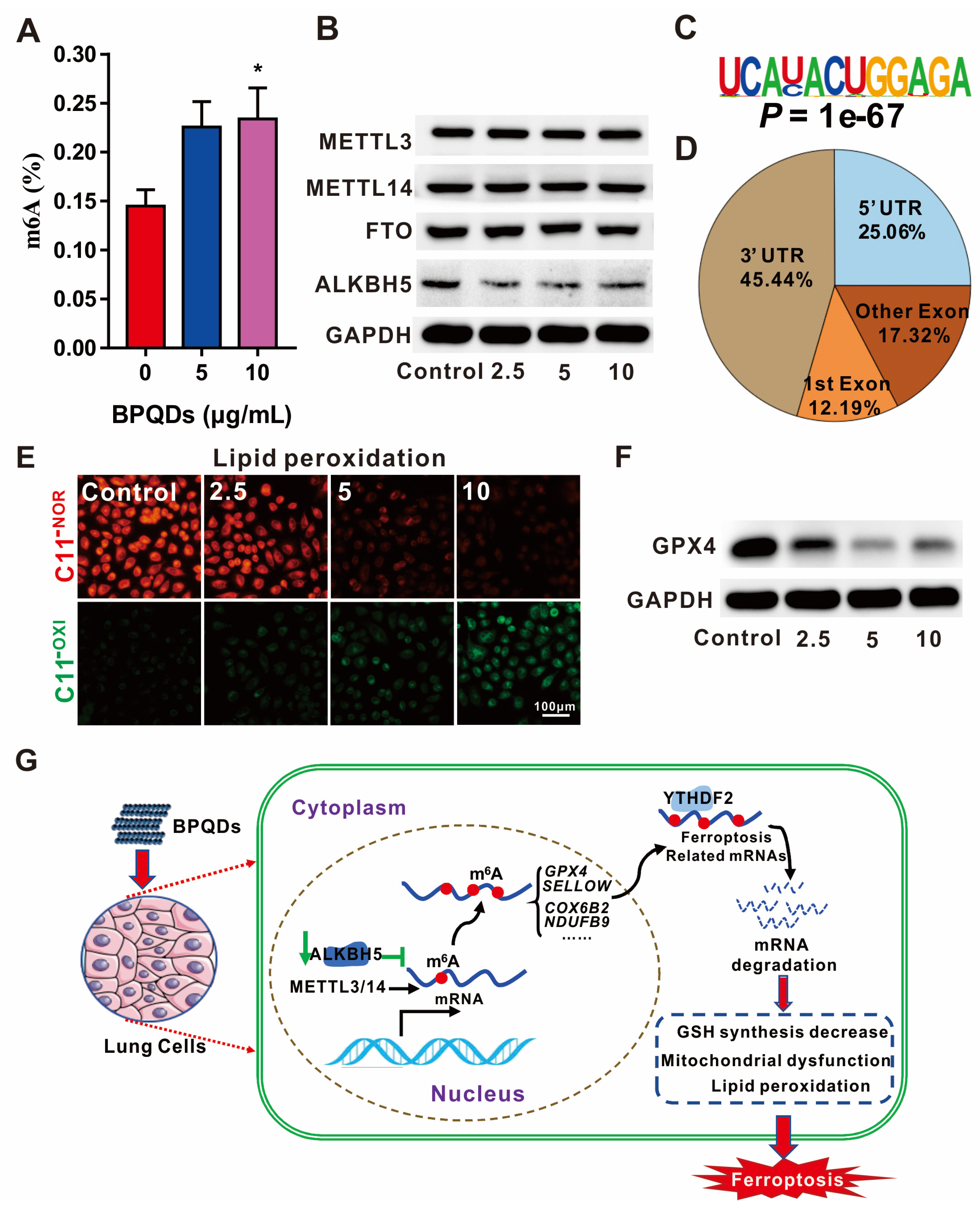
  105. Ruan, F.; Zeng, J.; Yin, H.; Jiang, S.; Cao, X.; Zheng, N.; Han, C.; Zhang, C.; Zuo, Z.; He, C. RNA m6A modification alteration by black phosphorus quantum dots regulates cell ferroptosis: Implications for nanotoxicological assessment. Small Methods 2021, 5, 2001045. [Google Scholar] [CrossRef]
  106. Han, B.; Chu, C.; Su, X.; Zhang, N.; Zhou, L.; Zhang, M.; Yang, S.; Shi, L.; Zhao, B.; Niu, Y.; et al. N6-methyladenosine-dependent primary microRNA-126 processing activated PI3K-AKT-mTOR pathway drove the development of pulmonary fibrosis induced by nanoscale carbon black particles in rats. Nanotoxicology 2019, 14, 1–20. [Google Scholar] [CrossRef]
  107. Zhang, S.; Meng, P.; Cheng, S.; Jiang, X.; Zhang, J.; Qin, X.; Tang, Q.; Bai, L.; Zou, Z.; Chen, C. Pregnancy exposure to carbon black nanoparticles induced neurobehavioral deficits that are associated with altered m6A modification in offspring. NeuroToxicology 2020, 81, 40–50. [Google Scholar] [CrossRef]
  108. Yang, Z.; Deng, C.; Wu, Y.; Dai, Z.; Tang, Q.; Cheng, C.; Xu, Y.; Hu, R.; Liu, C.; Chen, X.; et al. Insights into the mechanism of multi-walled carbon nanotubes phytotoxicity in Arabidopsis through transcriptome and m6A methylome analysis. Sci. Total Environ. 2021, 787, 147510. [Google Scholar] [CrossRef]
  109. Zhu, X.; Feng, J.; Zheng, M.; Yang, Z.; Zhao, L.; Zhang, W.; Zhong, W.; Chen, Y.; Lin, J. Metal–protein nanoparticles facilitate anti-VSV and H1N1 viruses through the coordinative actions on innate immune responses and METTL14. Macromol. Biosci. 2021, 21, 2000382. [Google Scholar] [CrossRef]
  110. Kunovac, A.; Hathaway, Q.A.; Pinti, M.V.; Durr, A.J.; Taylor, A.D.; Goldsmith, W.T.; Garner, K.L.; Nurkiewicz, T.R.; Hollander, J.M. Enhanced antioxidant capacity prevents epitranscriptomic and cardiac alterations in adult offspring gestationally-exposed to ENM. Nanotoxicology 2021, 15, 812–831. [Google Scholar] [CrossRef]
  111. Yin, H.; Wang, H.; Jiang, W.; Zhou, Y.; Ai, S. Electrochemical immunosensor for N6-methyladenosine detection in human cell lines based on biotin-streptavidin system and silver-SiO2 signal amplification. Biosens. Bioelectron. 2017, 90, 494–500. [Google Scholar] [CrossRef] [PubMed]
  112. Zuo, X.; Chen, Z.; Gao, W.; Zhang, Y.; Wang, J.; Wang, J.; Cao, M.; Cai, J.; Wu, J.; Wang, X. M6A-mediated upregulation of LINC00958 increases lipogenesis and acts as a nanotherapeutic target in hepatocellular carcinoma. J. Hematol. Oncol. 2020, 13, 5. [Google Scholar] [CrossRef] [Green Version]
  113. Balasubramanian, S.; Gunasekaran, K.; Sasidharan, S.; Mathan, V.J.; Perumal, E. MicroRNAs and xenobiotic toxicity: An overview. Toxicol. Rep. 2020, 7, 583–595. [Google Scholar] [CrossRef] [PubMed]
  114. Pogribna, M.; Koonce, N.A.; Mathew, A.; Word, B.; Patri, A.K.; Lyn-Cook, B.; Hammons, G. Effect of titanium dioxide nanoparticles on DNA methylation in multiple human cell lines. Nanotoxicology 2020, 14, 534–553. [Google Scholar] [CrossRef] [PubMed]
  115. Rossner, J.P.; Vrbova, K.; Rossnerova, A.; Zavodna, T.; Milcova, A.; Klema, J.; Vecera, Z.; Mikuska, P.; Coufalik, P.; Capka, L.; et al. Gene expression and epigenetic changes in mice following inhalation of copper (II) oxide nanoparticles. Nanomaterials 2020, 10, 550. [Google Scholar] [CrossRef] [PubMed] [Green Version]
  116. Ma, J.; Yang, D.; Ma, X.-X. Immune infiltration-related N6-methyladenosine RNA methylation regulators influence the malignancy and prognosis of endometrial cancer. Aging 2021, 13, 16287–16315. [Google Scholar] [CrossRef]
  117. Rossnerova, A.; Honkova, K.; Pelclova, D.; Zdimal, V.; Hubacek, J.A.; Chvojkova, I.; Vrbova, K.; Rossner, J.P.; Topinka, J.; Vlckova, S.; et al. DNA methylation profiles in a group of workers occupationally exposed to nanoparticles. Int. J. Mol. Sci. 2020, 21, 2420. [Google Scholar] [CrossRef] [Green Version]
  118. Poma, A.; Colafarina, S.; Fontecchio, G.; Chichiriccò, G. Transgenerational Effects of NMs; Springer: Berlin/Heidelberg, Germany, 2014; Volume 811, pp. 235–254. [Google Scholar] [CrossRef]
  119. Bhagat, J.; Zang, L.; Nishimura, N.; Shimada, Y. Zebrafish: An emerging model to study microplastic and nanoplastic toxicity. Sci. Total Environ. 2020, 728, 138707. [Google Scholar] [CrossRef]
  120. Hu, J.; Lin, W.; Lin, B.; Wu, K.; Fan, H.; Yu, Y. Persistent DNA methylation changes in zebrafish following graphene quantum dots exposure in surface chemistry-dependent manner. Ecotoxicol. Environ. Saf. 2018, 169, 370–375. [Google Scholar] [CrossRef]
  121. Yee, M.; Hii, L.-W.; Looi, C.; Lim, W.-M.; Wong, S.-F.; Kok, Y.-Y.; Tan, B.-K.; Wong, C.-Y.; Leong, C.-O. Impact of microplastics and nanoplastics on human health. Nanomaterials 2021, 11, 496. [Google Scholar] [CrossRef]
  122. Guo, P.X. The emerging field of RNA nanotechnology. Nat. Nanotechnol. 2010, 5, 833–842. [Google Scholar] [CrossRef]
Figure 1. The molecular composition of m6A RNA methylation. “Writers” of m6A are methyltransferases that add methyl groups to mRNA, including METTL3, METTL14, WTAP, KIAA1429 METTL16 and RBM15/15B. “Erasers” of m6A are demethylases that remove methyl groups from m6A RNA, including FTO and ALKBH5. “Readers” of m6A recognize m6A RNA and thus trigger downstream events, including nuclear m6A “readers” (such as YTHDC1, HNRNPA2B1, HNRNPC and HNRNPG) and cytoplasmic m6A “readers” (such as YTHDF1, YTHDF2, YTHDF3, YTHDC2, IGF2BPs and eIF3).
Nanomaterials 12 00389 g001
Figure 2. m6A RNA modification influences mRNA fate. (A) HNRNPC is involved in the processing of pre-mRNA and structural switching. (B) HNRNPA2B1 plays an important role in pri-miRNA processing, alternative splicing and structural switching of mRNA. (C) YTHDC1 is engaged in alternative splicing and nuclear export. (D) HNRNPG participates in alternative splicing and mRNA structure switching. (E) YTHDF1 facilitates mRNA translation. (F) YTHDF2 aids in the decay of mRNAs. (G) YTHDF3 collaborates with YTHDF1 to promote mRNA translation, and it enhances YTHDF2-mediated mRNA decay. (H) IGF2BPs containing IGF2BP1/2/3 enhance RNA stability. (I) eIF3 is engaged in RNA translation. (J) YTHDC2 accelerates RNA translation.
Nanomaterials 12 00389 g002
Figure 3. The roles of m6A RNA modification in ferroptosis induced by BPQDs. (A) The global m6A level is increased after BPQD exposure in a concentration-dependent manner. (B) The expression of m6A-related proteins, as detected by Western blotting, suggests that BPQDs decrease the expression of the demethylase ALKBH5 rather than FTO or the METTL3/14 methylases. (C) Most significant m6A-binding motif. (D) m6A peaks are mainly located at 3′ and 5′ UTR of mRNA. (E) Fluorescent images showing the lipid peroxidation level as measured with C11 BODIPY staining. Scale bar = 100 µm. (F) The expression of GPX4 protein is decreased after BPQD exposure with GAPDH as a loading control. (G) BPQDs decrease ALKBH5 expression and thus lower the m6A level of ferroptosis-related mRNAs recognized by the YTHDF2 m6A “reader”, promoting mRNA degradation. * p < 0.05. * compared to no treatment. As a result, GSH synthesis decreases and mitochondrial dysfunction, lipoperoxidation and ferroptosis occur. Reproduced from [97] with permission from Small Methods.
Nanomaterials 12 00389 g003
Figure 4. Roles of m6A RNA modification in the bioimpacts of nanoparticles and the mechanisms involved. BPQDs induce ferroptosis by upregulating the total m6A level in A549 and BEAS-2B cells. CBNPs induce pulmonary fibrosis by downregulating the total m6A level in rats and also cause abnormal maternal behaviours by downregulating the total m6A level in mice. MWCNTs cause growth inhibition by upregulating the total m6A level in Arabidopsis thaliana. MPNPs improve anti-virus ability in VSV-infected macrophages. TiO2-NPs cause cardiac impairment by upregulating the total m6A level [104,105,106,107,108,109,110].
Nanomaterials 12 00389 g004
Table 2. Methods for detecting m6A residues. (Extremely large: ≥300 μg; Large: 300 ng < n < 300 μg; Little: ≤300 ng).
MethodsResolutionSample RNA Demand (n)Need for AntibodiesReferences
MeRIP-seq100–200 ntExtremely largeYes[95]
PA-m6A-seq20–30 ntLargeYes[96]
miCLIP-seqSingle baseLargeYes[97]
SCARLETSingle baseLargeNo[98]
m6A-REF-seqSingle baseLittleNo[99]
ColorimetryTotal
amount
of m6A
LittleYes[100]
m6A dot-blotLargeYes
HPLC–MS/MSLargeNo
Publisher’s Note: MDPI stays neutral with regard to jurisdictional claims in published maps and institutional affiliations.

