Review

Compositionally Disordered Crystalline Compounds for Next Generation of Radiation Detectors

1
National Research Center “Kurchatov Institute”, Moscow 123098, Russia
2
National Research Center “Kurchatov Institute”—Institute of Reactives, IREA, Moscow 107076, Russia
3
Institute for Nuclear Problems, Belarus State University, 220030 Minsk, Belarus
*
Author to whom correspondence should be addressed.
Nanomaterials 2022, 12(23), 4295; https://doi.org/10.3390/nano12234295
Submission received: 31 October 2022 / Revised: 23 November 2022 / Accepted: 29 November 2022 / Published: 3 December 2022
(This article belongs to the Special Issue State-of-the-Art Inorganic Materials and Metal-Organic Frameworks)

Abstract

:
The review is devoted to the analysis of the compositional disordering potential of the crystal matrix of a scintillator to improve its scintillation parameters. Technological capabilities to complicate crystal matrices both in anionic and cationic sublattices of a variety of compounds are examined. The effects of the disorder at nano-level on the landscape at the bottom of the conduction band, which is adjacent to the band gap, have been discussed. The ways to control the composition of polycationic compounds when creating precursors, the role of disorder in the anionic sublattice in alkali halide compounds, a positive role of Gd based matrices on scintillation properties, and the control of the heterovalent state of the activator by creation of disorder in silicates have been considered as well. The benefits of introducing a 3D printing method, which is prospective for the engineering and production of scintillators at the nanoscale level, have been manifested.

1. Introduction

Scintillation materials play a notable role in ensuring further progress in the techniques and methods of detecting various types of ionizing radiation. Historically, their development has involved several phases driven by application. The nuclear projects in the 1940s of the 20th century led to the discovery of alkali halide scintillation single crystals. Later on, the drivers of the development were both scientific research in the field of nuclear and high energy physics and medical imaging [1]. Nowadays, security systems, environmental monitoring, and non-proliferation of fissile materials are the most significant industries that consume novelties in the field of scintillation materials. Unlike liquid, gaseous, and organic media, which are also used to detect ionizing radiation by the scintillation method, inorganic materials make it possible to combine high density, i.e., stopping power for ionizing radiation, with a wide variation in spectral and luminescent parameters due to the choice of material composition and activating ions. The widespread application of modern analytical methods for studying materials at the submicron level [2,3,4] and measuring electronic excitation transfer processes in the subpicosecond range [5,6] made possible the development of materials at a new level. It is worth noting the progress in the development of the theory of scintillations [7,8], and the use of big data analysis of spectroscopic data [9,10,11,12,13,14,15,16] to identify patterns in scintillation properties when varying the composition of relatively simple compounds. The accumulated experience and knowledge in this area, as well as the available tools, have made it possible to proceed with the targeted engineering of more complex compounds at the submicron level.
Since the publication of the second edition on Inorganic Scintillators for Detectors Systems by P. Lecoq et al. [17], quite popular among specialists, five years have passed. During this time, new directions in the creation of scintillation materials and detector elements have been formed. Particularly noteworthy are structured materials and detector elements [18,19,20,21,22], as well as the incorporation or creation of nanoparticles of alkali halide materials with a high scintillation yield in various media, in order to obtain new properties and improve the manufacturability of these materials [23]. These developments put forward the work with nanoscale objects for luminescence generation high on the list of priorities. Nonetheless, the investigation of inorganic scintillation materials in a variety of forms, from nanoparticles to bulk single and polycrystalline ceramic blocks, as well as films [24], remains at the heart of this field’s innovations. Therefore, the progress in the luminescence properties of the inorganic materials remains an important task for the creation of detector elements that meet the ever-increasing requirements for operational characteristics: energy and time resolution and tolerance to various types of ionizing radiation.
Analyzing the properties of the most commonly used scintillators, it can be stated that the greatest progress in improving their properties was achieved through the use of codoping: LaBr3: Ce [25], NaI:Tl [26,27], CsI:Tl [28], PbWO4 [29]. Another way to improve the consumer performance is the use of the sampling of detector elements with the best parameters among those produced, which significantly increases the cost of final products.
The main limiting factor for alkali halide compounds is the isolation of all technological stages of production from an oxygen-containing atmosphere. The incorporation of oxygen ions into the crystal lattice, in particular, based on Br- and I-anions, leads to the formation of point defects competing for trapping nonequilibrium carriers while the material interacting with ionizing radiation. These issues are the limiting factors for the application of the new generation halides LaCl3:Ce and LaBr3:Ce [30,31] in widely used pixelated detectors in medical scanners.
A limiting factor for self-activated scintillators of the bi-cation tungstate family and materials of the olivine structural type is the temperature quenching of luminescence and, as a result, the low scintillation yield, which cannot be increased by technological methods. The only example is a twofold increase in the scintillation yield in a PWO-II crystal. However, it was achieved by reducing the concentration of the doping of La3+ and Y3+ ions. Such combining doping is used to stabilize the radiation hardness of the material.
In Ce-activated oxide compounds, investigation has focused on the search for scintillators with a combination of fast kinetics, high density, and high scintillation yield [32]. Besides orthosilicates [33], binary compounds with garnet Lu3Al5O12: Ce(Pr) (LuAG) [34,35,36,37] and oxyorthoaluminates YAlO3 (YAP) and Lu(Y)AlO3 (LuAP) [38,39,40] structures activated by Ce3+ and Pr3+ ions were discovered.
Among the listed oxide scintillators, materials with a garnet structure have the greatest potential for improving scintillation properties. We note three important features of these compounds:
First, compounds of the garnet structural type have a cubic space group Oh10-Ia3d [41]. The isotropy of the matrix properties allows the use of many technological methods for shaping, annealing, and growth in order to obtain a high-quality crystalline mass. They have a good optical transparency, which can be varied over a wide range by the composition of matrix-forming and doping elements [42,43,44,45].
Second, the isomorphism of garnets is a unique opportunity to obtain new compounds of this class. Their general formula can be represented as C3B2A3O12, where the C, B, and A positions correspond to cations in different crystal positions: dodecahedral, octahedral, and tetrahedral, respectively [46]. Ions of Ce3+ and other rare earth (RE) ions are predominantly localized in dodecahedral positions.
Thirdly, the isovalent localization of RE activators in the above positions in the structure is accompanied by a change in the corresponding metal–oxygen bond length. This affects their luminescent properties. This mechanism is an additional tool for adjusting the spectral-luminescent properties of the garnet type compounds. It should be noted that the garnet lattice can be formed both by pairs of di-and tetravalent ions and by pairs of trivalent ions. A heterovalent substitution in garnets is also possible, but it is limited by the necessity for charge compensation [47,48].
In the family of the artificial garnets, compounds based on trivalent ions are the most widely used. Compounds with a garnet structure can be expressed as C3+3[B3+]2(A3+)3O12, where C = Y, Gd, Tb, Er, Tm, Ho, Yb, Lu; B, C = Al, Ga, Sc, etc.
One of the most promising compositions for combining high scintillation parameters and high stopping power was lutetium garnet [49]. However, from the experience of the technological development of the binary Lu garnet crystal, a few problems have been disclosed, which have become a significant limiting factor. Note the low, no more than 1 mm/h, rate of pulling out the commercial crystalline mass on the seed, which leads to a gradient in the properties of single crystals along the crystal axis. Moreover, due to a difference in segregation coefficients of various ions from the melt at a low pulling rate, an axial gradient of defects arises, which significantly limits the volume of the crystalline mass suitable for manufacturing the high-quality detector elements. Finally, in the single crystal with a garnet structure, when a small-radius Lu3+ ion is included in the composition, antisite defects occur. The lutetium ion is localized in dodecahedrons and, in a small quantity, in octahedrons as well [50,51].
Technologies to produce ceramic materials are devoid of these disadvantages. Moreover, the capability of strict control at the production of precursors (powders for further densification) provides better scintillation characteristics of YAG:Ce [52], LuAG:Ce [53], LuAG:Pr [54] ceramic scintillators than their single-crystal counterparts due to the provision of homogeneous distribution and increased dopant concentration, as well as the use of technological methods to reduce the concentration of antisite defects.
Notable progress in improving the scintillation yield of LuAG:Ce ceramics has been made in just a decade. LuAG:Ce,Mg ceramics showed a high yield of 21,900 ph/MeV with a signal integration time in a time gate of 1 µs [55], and a yield of 25,000 ph/MeV has been obtained so far. An even more significant effect of improving the scintillation parameters was observed when the LuAG:Ce ceramic was codoped with Li+ ions. LuAG:Ce,Li ceramics demonstrated a scintillation yield of 29,200 ph/MeV [56].
Additional improvements in the properties of LuAG:Ce ceramics are achieved by adjusting the composition of the precursor, which provides a variation in the composition near the stoichiometric ratio. The Lu3+ deficient nonstoichiometric LuAG:Ce ceramics showed poor optical transmission due to the presence of a secondary phase but improved scintillation performance [57,58].
Thus, it can be stated that intensive research on improving the scintillation properties of binary crystalline garnets, although it resulted in some progress, still did not allow reaching the set of scintillation parameters better than the lutetium oxyorthosilicate Lu2SiO5:Ce family owes. Similarly, one can also be declared that there is a stagnation in the breakthrough increase in scintillation yield in simple (two-component) alkali halide materials with various activators.
This review is devoted to the analysis of the compositional disordering potential of the crystal matrix of a scintillator to improve its scintillation parameters. The effects of the disorder on the landscape at the bottom of the conduction band, which is adjacent to the band gap, have been discussed. The ways to control the composition of polycationic compounds when creating precursors, the role of disorder in the anionic sublattice in alkali halide compounds, and a new kind of disordered material on a base of silicates have been considered as well. The benefits of introducing a 3D printing method, which is prospective for the engineering and production of scintillators at the nanoscale level have been argued.

2. Next Step: Compositional Disordering of the Materials

Let us turn to the analysis of a simplified expression describing the scintillation yield (LY) [7]. It usually includes three factors: LY = ηSQ, where η describes the efficiency of ionizing radiation energy conversion; S is the efficiency of nonequilibrium carrier energy transfer to an ensemble of luminescent centers; and Q is the quantum yield of intracenter luminescence of these centers. The first is exclusively due to the properties of the matrix, in particular, the size of the band gap which determines the energy of thermalized carriers and, accordingly, excitons. The latter is defined by the properties of the luminescent center. For intracenter spontaneous transitions in a distinct luminescent center, Q may exceed unity. For example, relaxation from the radiative state of Ce3+ ions can be accompanied by the emission of an optical photon upon transition to the 2F7/2 level and then by the emission of a photon with an energy of ~2000 cm−1 in the far IR region when thermodynamic equilibrium is established between the 2F7/2 and 2F5/2 states. However, photodetectors used in ionizing radiation measurement are typically not sensitive in the far infrared spectrum. The value of Q can exceed unity upon excitation in polyatomic systems when the excitation energy of one of the participants in the cooperative process is divided between neighboring centers emitting the same spectrum [59,60,61,62,63].
The efficiency of energy transfer S is determined by the conditions for the transfer of both bound nonequilibrium carriers (excitons) and the migration rates of electrons and holes in the conduction and valence bands, respectively. The effective mass of holes in the valence band in dielectrics is, as a rule, an order of magnitude or even greater than the mass of electrons in the conduction band. Therefore, the migration of electrons along the band, as well as the mobility of excitons, determine the efficiency of excitation of luminescent centers. In binary systems, which typically have the regular shape of the crystal potential forming the bottom of the conduction band, there is a significant expansion of nonequilibrium electrons from holes. This prevents the creation of excitons and, consequently, the low efficiency of the excitation transfer through the excitons [8]. This lack causes the low contribution of this mechanism to scintillation, for example, in binary oxide compounds. The reduction in the expansion of nonequilibrium carriers can be facilitated by modulation of the crystal potential at the atomic level, which may be created by introducing a compositional disorder into the matrix.
Compositional disorder in a crystalline system does not mean amorphization of a compound. A polycationic ensemble of the cationic sublattice with identical crystallographic sites creates a disordered distribution of cations in the matrix. However, the conservation of the space symmetry group occurs due to the ordered distribution of anions. Thus, the long-range order in the ion stabilization positions, i.e., crystallinity, is preserved. This approach is typical for oxygen systems. For alkali-halide systems, where anions may also be mixed, the long-range order is preserved due to the ordering of the cationic subsystem. Highly disordered crystalline systems consisting of many cations occupying the same positions in the crystal lattice are often referred to as high-entropy materials [64,65]. They also find application in photocatalysis [66] and the creation of more complex systems [67] than those that have been discussed in this review. In [68], promising high-entropy ceramics have been considered specifically for photonics applications as well.
Apparently, research on oxyhalides with RE activators [69] and Gd2O2S:Tb [70] may be considered as a pioneer in the creation of compositionally disordered systems in the field of scintillators. Later on, an improvement of the technological capabilities for single phase stabilization and better scintillation properties by growing a mixed perovskite-type LuxY1-xAlO3:Ce instead of LuAlO3:Ce and oxyorthosilicate type (LuxY1-x)2SiO5:Ce (LYSO) instead of Lu2SiO5:Ce (LSO) demonstrated [71,72,73].
The most technologically advanced combinations for introducing compositional disorder into a garnet-type crystal system are either the partial isomorphic substitution of aluminum ions by gallium ions or the mixing of isovalent cations localized in the dodecahedral position. There are other types of disorder in crystalline systems [74], but for the development of scintillators, compositional disorder is of particular interest.

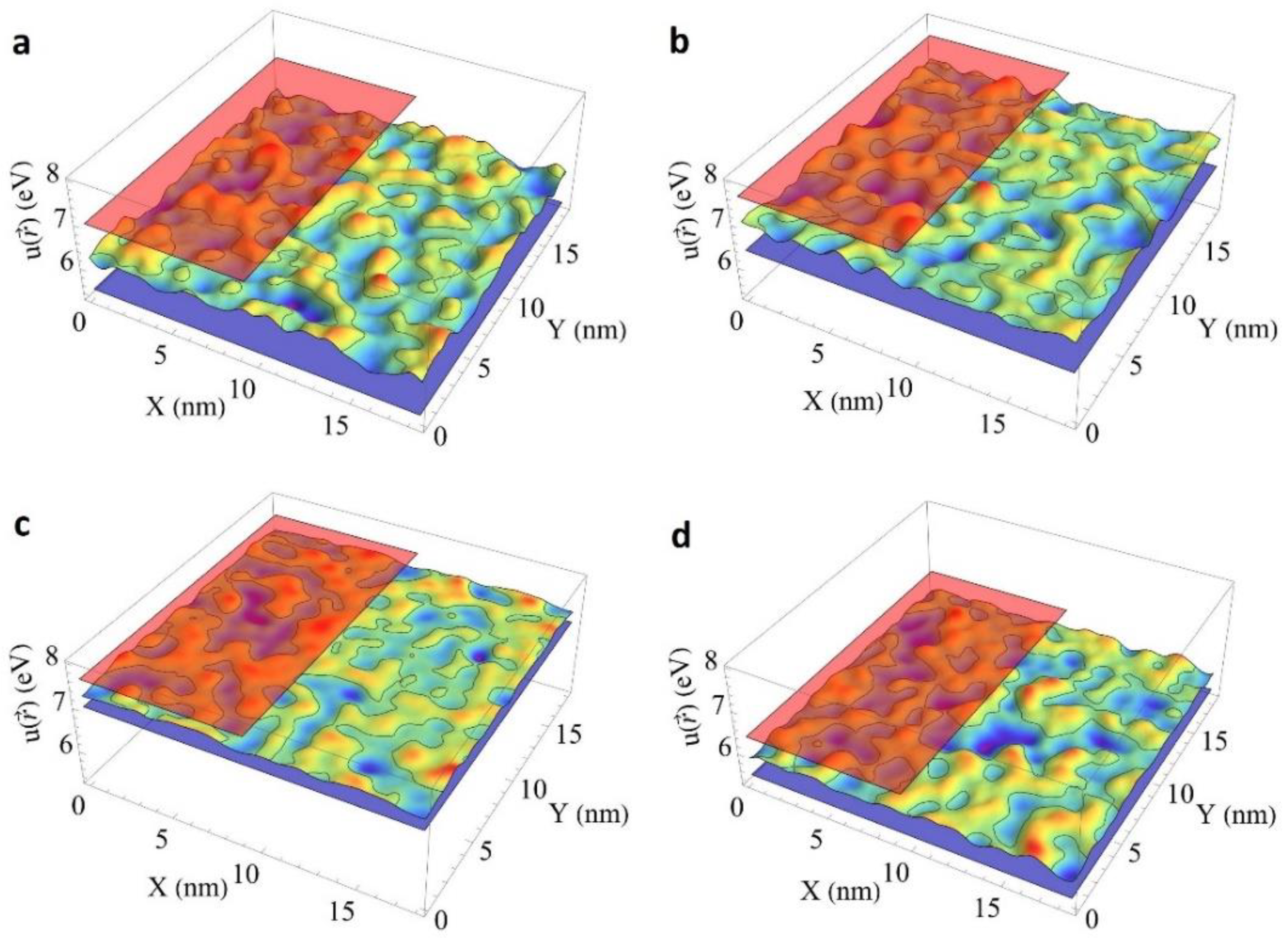
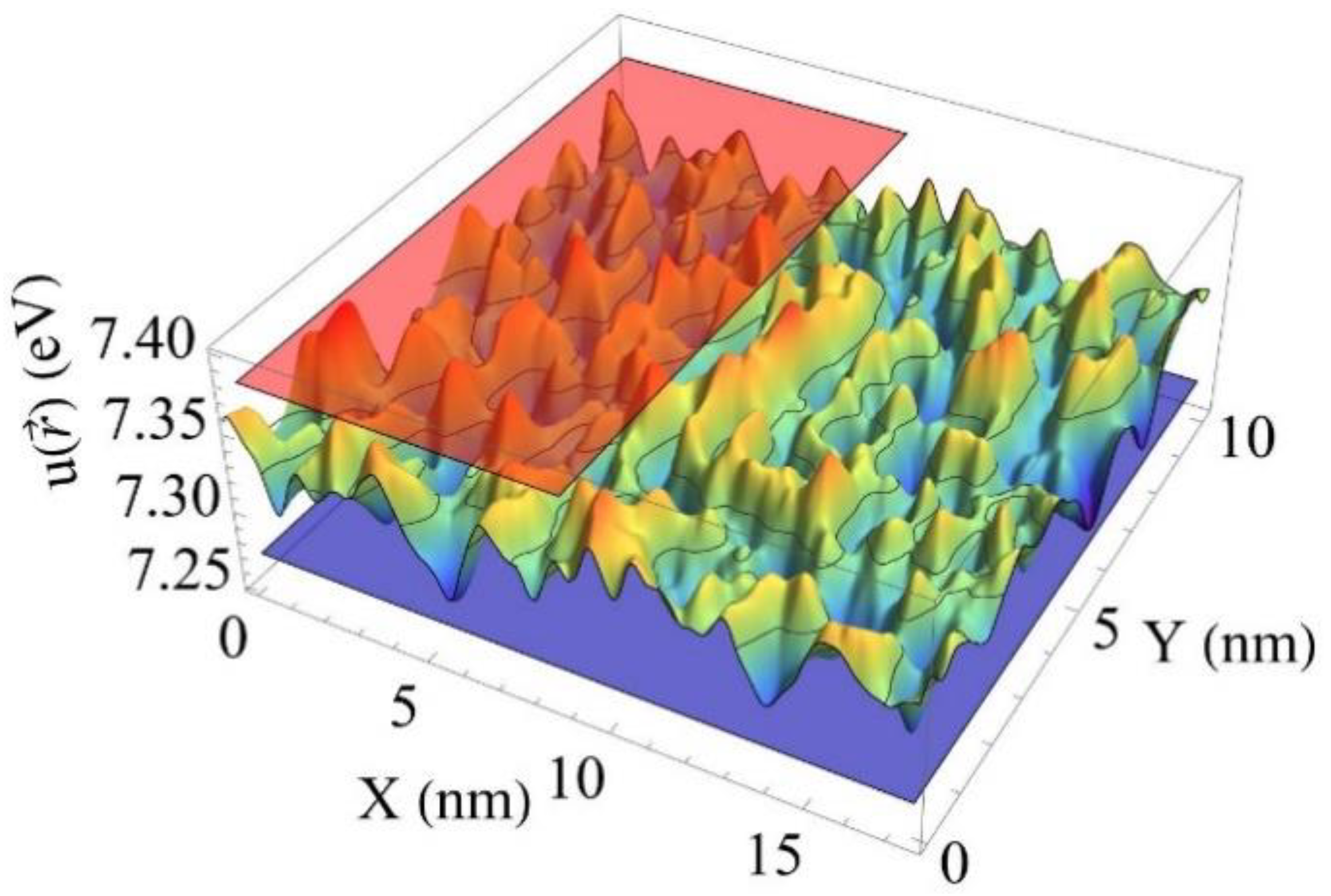
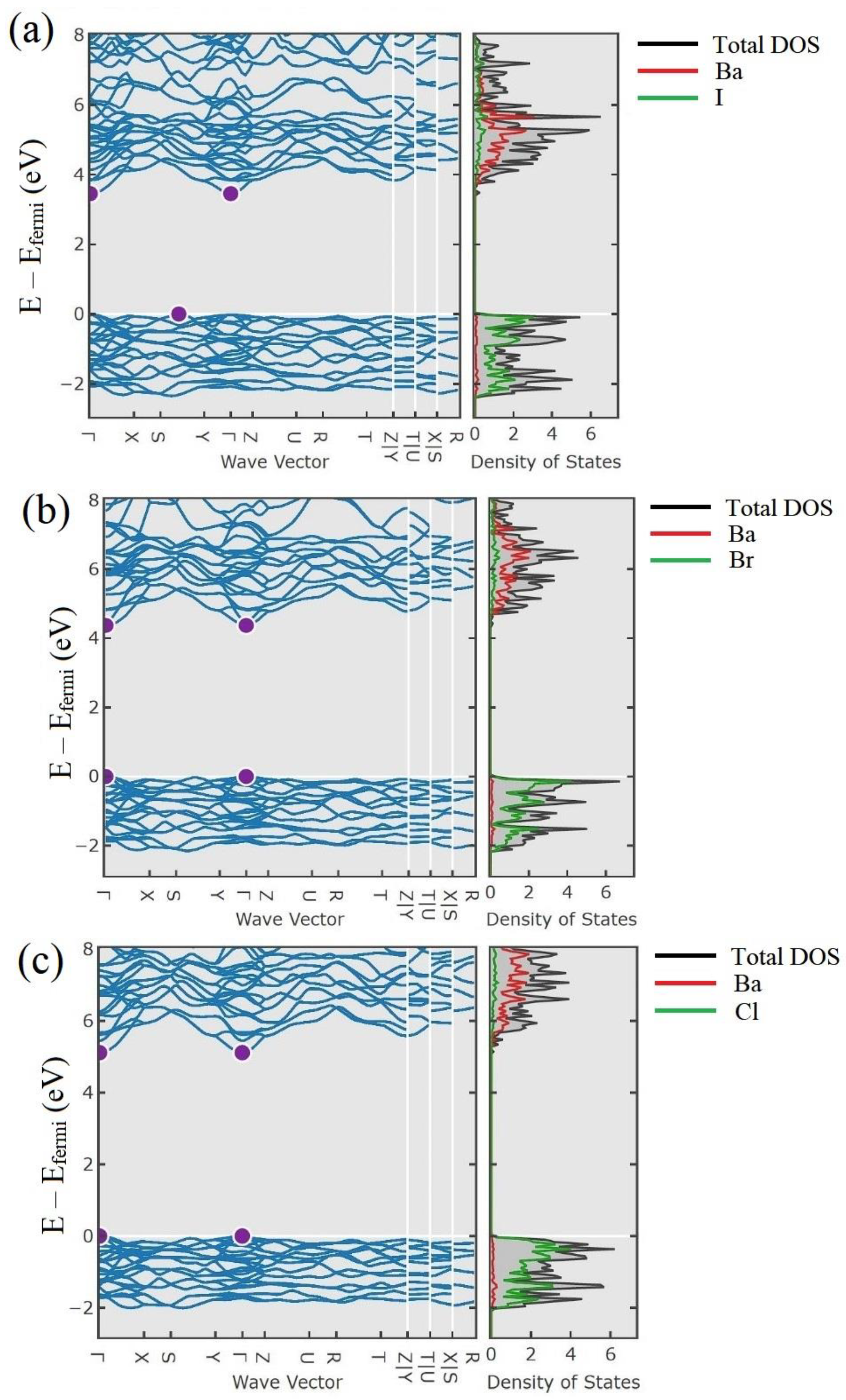
2.1. Conduction Band Bottom Landscape Modulation

The difference in the formation of the spatial distribution at the bottom of the conduction band might be revealed by considering fluctuations in the effective potential in the supercell of a mixed crystal with a random cation distribution. Fluctuations of the effective potential in the single-electron approximation, causing the scattering of electron states of the first branch in the conduction band of the crystal, can be built using the pseudopotential method according to the model proposed in [75]. The pseudopotential landscape can be expressed as:
u ( r ) = R p R Δ E c b 4 π V 0 ( κ a ) 3 s ( | r | κ a ) ,
s ( x ) = { 0 ,   | x | > 1   2 ( 1 + x ) 2 , 1 x < 0.5 1 2 x 2 , 0.5 x < 0.5 2 ( 1 x ) 2 ,   0.5 x 1
where x is independent variable of the function s(x), p R is the matrix of nodes occupied by Al (Y) ions in a supercell, Δ E c b is the difference between bottoms of the conduction bands of two components of a solid solution, V 0 is the volume of the unit cell, a is the lattice constant, κ is the scale factor. The scale parameter means the distance (in units of a lattice constant) at which the pseudopotential of a certain substituting ion equals zero.
Figure 1 presents a 2D cross-section of the spatial distribution of the pseudopotential u ( r ) in binary crystals: Y3Al5O12 and Y3Ga5O12, Lu3Al5O12 and Lu3Ga5O12, Lu3Al5O12, Y3Al5O12, Lu3Gd5O12 and Y3Ga5O12, as well as ternary crystals having the same content of Al3+ and Ga3+ or Lu3+ and Y3+ ions, respectively. The spatial distribution of the substituting ions was chosen to be homogeneous, without clustering. The 2D cross sections are built in 15 × 15 × 15 supercells for garnets and in 20 × 20 × 20 supercells for oxyorthosilicates at z = 15 2 ( a + b + c ) z , i.e., at the half height of the supercells. The parameters of the unit cells of binary components, the positions of ions in the unit cells and the band gaps are obtained from [76,77,78,79,80,81].
The amplitude of the fluctuations of the pseudopotential reaches the largest values in a ternary system composing Al3+ and Ga3+ ions. It is caused by the largest difference in the band gaps of the binary compounds Lu3Ga5O12 and Y3Ga5O12 or Lu3Al5O12 and Y3Al5O12. At the same time, the substitution of yttrium by lutetium ions leads to a less modulation of the effective potential. A smaller effect is observed when yttrium is substituted by lutetium in the Lu2SiO5-Y2SiO5 system as well. Figure 2 presents a minor modulation of the effective potential in (L0.5-Y0.5)2SiO5 solid solution.
The effects of the modulation of the effective potential in a disordered crystal on the mobility of nonequilibrium carriers can be classified into two groups: elastic scattering of free carriers with the kinetic energy above the localization threshold on potential fluctuations, and localization of thermalized carriers in potential wells with the possibility of subsequent activation due to phonon absorption or tunneling into a neighboring potential well [82]. The latter effect has a lower probability in comparison with the thermal activation of carriers at room temperature. In both cases, the modulation of the effective potential leads to a decrease in the diffusion length of free carriers, increasing the concentration of geminate electron-hole pairs, and, consequently, increasing the transfer efficiency to the luminescent centers.
Eventually, the modulation of the effective potential changes the energy structure of single-electron states in the crystal. The interruption of the periodicity of the crystal field leads to a spatial compression of states. In such systems, the wave vector is already a “bad” quantum number. Unfortunately, such quantum states cannot be described by any set of quantum numbers. However, such mixed systems can be considered in the context of a virtual (average and regular) crystal with a finite lifetime of single-electron states due to scattering on fluctuations of the crystal field, as described in [75]. Such an approach uses the coherent potential (CPA) method [82] to calculate the band structure in a mixed solid solution. The branches in the conduction band of such mixed crystals are broadening, which width corresponds to the uncertainty of the energy of the states.
The branch broadening changes the shape of the density of states, which acquires a tail in the energy region below the edge of the fundamental absorption in the virtual crystal E g v c x E g A + ( 1 x ) E g B , where E g A and E g B are the band gaps of binary components. The broadening of the lower branch in the conduction band at the neighborhood of the Γ point can be considered as the blurring of the bottom of the conduction band. The blurring of the bottom of the conduction band is very often interpreted as a change in the width of the band gap. This approach is based on linear absorption spectroscopy results when the dynamic range of the setup does not exceed three decades, and blurring causes the observed shift of the cutoff absorption.
The significant predominance of the concentration of one of the cationic components, which generates the disorder, might lead to clustering in the compound, i.e., the appearance of regions of individual compounds with the same structural type [83], and the scintillation parameters of such materials are quite close to the parameters of initial components in a series of solid solutions.

2.2. Changing the Parameters of the Local Crystal Field

A change in the relief of zones also affects the local crystal field, causing a strength variation in the Stark splitting of activator levels [84,85]. Disordering in the Al-Ga sublattice to a greater extent, and in the Y(Lu)-RE sublattice to a lesser extent, leads to fluctuations of the crystal field in the localization position of the Ce3+ activator ions. In the crystal lattice of garnet, dodecahedrons containing Ce3+ ions are surrounded by tetrahedra and octahedra occupied by Ga3+ and Al3+ ions. The strongest effect on the 5d states of the Ce3+ ion can be expected from two tetrahedra sharing d48 edges with the dodecahedron containing Ce.
In a garnet-type lattice, Ga3+ ions tend to occupy tetrahedral positions [86,87]. The population of the tetrahedra with Ga3+ or Al3+ ions lead to substantially nonequivalent positions for Ce3+ ions. It has been experimentally established that the length of common edges between the dodecahedron and the tetrahedron d48 plays a decisive role in the splitting Δ1,2 between the states 5d1 and 5d2 [88]. The length d48 is longer in the tetrahedra occupied by Ga3+ than in the tetrahedra occupied by Al3+ ions. The greater the d48, the smaller the Δ1,2. This contributes to a decrease in the energy gap between the 5d1(Ce3+) radiative state and the modulated bottom of the conduction band, while the opposite trend is observed at a predominant concentration of Al3+ ions in the compound composition. LuGAG:Ce single crystals exhibit a lower slow scintillation component and a higher LY value compared to LuAG:Ce [89,90]. However, with an increase in the Ga content due to a decrease in the strength of the crystal field in the dodecahedral position, the energy level 5d1 of the Ce3+ activator shifts towards higher energies, while the average crystal potential shifts towards lower energies, approaching each other. This reduces the energy of thermal ionization of Ce3+ ions into the conduction band, makes the process of thermal ionization more probable at room temperature, and, as a result, leads to a slowing down of the scintillation kinetics. A similar change in parameters was also found in LuGAG doped with Pr3+ ions [91].
The effect of shallow electron traps caused by compositional disordering can be diminished by codoping with a divalent ion. The effect of codoping in Y(Lu)-aluminum-gallium garnets turned out to be similar to the effects in binary compounds with a garnet structure. Furthermore, codoping with Ca2+ and Mg2+ ions affects the concentration of vacancies and retrapping processes in disordered systems, which influences the scintillation characteristics [92,93]. At a high concentration of codoping, they can contribute to the localization of tetravalent cerium ions in the lattice. Ce4+ can act as a fast nonradiative relaxation channel, and thus suppress the scintillations caused by Ce3+. However, at a low concentration of Mg2+ ion codoping in disordered compounds of both oxyorthosilicates and garnets, a broad structureless absorption band with a maximum near 260 nm appears in the optical absorption spectrum [94]. The intensity of this band does not show a change with time. Therefore, it is not associated with a metastable color center. It is most likely due to a charge–transfer transition from the top of the valence band to the unfilled level of the Mg2+-based defect. No luminescence bands associated with excitation into this band were found, which indicates a very fast nonradiative relaxation. This means that, during the formation of scintillation, carriers trapped in shallow traps due to matrix disorder having a thermal activation energy of the order of fluctuations of the crystal potential (>0.5 eV) will be predominantly recaptured by the magnesium impurity-based defects. For this reason, their delocalization into the conduction band will be minimized, resulting in faster scintillation kinetics by Ce3+ ions. An inhomogeneous broadening of the luminescence of Cr3+ ions and a change in the local symmetry of the activator site were noted in LuYAG [95]. Compositional disordering leads to inhomogeneous broadening of the luminescence band of Ce3+ ions and, consequently, to a gradient in the luminescence kinetics along the band contour [96]. The transition from the ternary compound Gd3Ga3Al2O12:Ce to the quaternary (Gd,Lu)3Ga3Al2O12 leads to even greater inhomogeneity of the luminescence kinetics, as can be seen from Figure 3.
Thus, the creation of compositional disorder in the Al-Ga and Y-RE systems in the garnet structure leads to the possibility of controlling the spectral-luminescent properties of the scintillator and the position of the emitting level of Ce3+ ions relative to the modulated bottom of the conduction band, which makes it possible to purposefully adjust the luminous properties of the material.

2.3. Modulation of Local Charge Distribution

Compositional disorder, in addition to a modulation of the crystal potential, can provide a local fluctuation of the charge. Recently, new results on phosphors [97,98,99] were announced. Attempts have been made to create materials with a garnet structure that combine di-, tri-, and tetravalent cations in a matrix. Such an approach promises certain benefits in further reducing the scattering length of nonequilibrium carriers formed by ionizing radiation. However, the development in this direction is hindered by technological factors associated with the difficulties in controlling the charge state of activators in such systems.

2.4. Variation in Stopping Power and Sensitivity to Various Types of Ionizing Radiation

The compositional disorder in the sublattice of a heavy cation makes it possible to solve the problem of adjusting the stopping power to various types of ionizing radiation. The compound stopping power for hard electromagnetic radiation depends on the density and the effective charge Zeff of the compound as ~Z4eff. It is enough to have a light Y cation in the dodecahedral position in the matrix to detect soft X-rays. To detect higher-energy gamma quanta, especially annihilation quanta with an energy of 511 keV, most typical in PET scanners, this position should be filled with heavier cations like lutetium and gadolinium. By controlling the compositional disorder in the dodecahedral position, the density of the compound can vary from 4.55 (YAG) to 7 (LuAGG) g/cm3. To detect hard gamma quanta, when the formation of e–e+ pairs become dominant, the radiation length X0 of the garnet material can be brought to values of less than 2 cm by filling dodecahedral positions with heavy cations. This makes it possible to use garnet-type materials to create compact calorimetric detectors in high-energy physics. The variability of the stopping power of the garnet compound by controlling the composition in dodecahedral positions can bring the partial ionization loss for high-energy charged minimally ionizing particles (MIP) to a level of 0.75 MeV/mm, which is five times higher than for scintillators based on organic plastics. This makes it possible to create detectors with increased time resolution for collider experiments.
The use of Gd to fill the dodecahedral positions allows one to solve another important detection problem—the detection of neutrons by the scintillation method. Natural gadolinium is in the form of a mixture of six isotopes, two of which, 155Gd and 157Gd, have the highest cross section of interaction with thermal neutrons of all known isotopes, 61 and 255 kbarn, respectively.
Figure 4 shows the part of absorbed neutrons (in percent) in gadolinium layer thicknesses of 0.2–10 mm of natural isotopic composition for incident neutron energies in the range from thermal 0.0253 eV to fast 15 MeV. Since crystalline compounds with a garnet structure have a density of gadolinium nuclei not much less than that of a metal, it is obvious that relatively thin layers of a crystalline compound, less than 5 cm, can provide effective absorption of neutrons in a wide energy range. Gadolinium nuclei possess a wide range of resonances for neutron absorption in the energy range from 1 eV to 10 keV, which makes them effective for detecting epithermal neutrons.
The products of the neutron capture reaction are γ-quanta and conversion electrons with a total energy of ~8 MeV. At the radiative capture of neutrons by gadolinium nuclei, compound nuclei are formed in an excited state, which excitation relaxation is accompanied by the emission of a cascade of γ-quanta, the average number of emitted gamma-quanta is four at an energy of detected neutrons of 0.0253 eV [100,101]. For detection, the most interesting are the intense and highest-yielding soft lines of gamma radiation with energies in the range of 80–90, 180–200 and 511 keV. Control of sensitivity to various types of ionizing radiation can also be achieved in alkali halide compounds, where Li and Na ions are isovalently mixed in the (Na,Li)I:Tl compound [102]. It could also be achieved by eutectic compound [103]. Eutectic Ce:6LiBr/LaBr3 with a high Li concentration was produced via the vertical Bridgman method and exhibited a lamellar eutectic structure and optical transparency. The light yield under neutrons excitation was estimated to be 74,000 photons/neutron. The scintillation decay time was defined to be 18.7 ns.

3. Producing and Utilizing of the Precursors

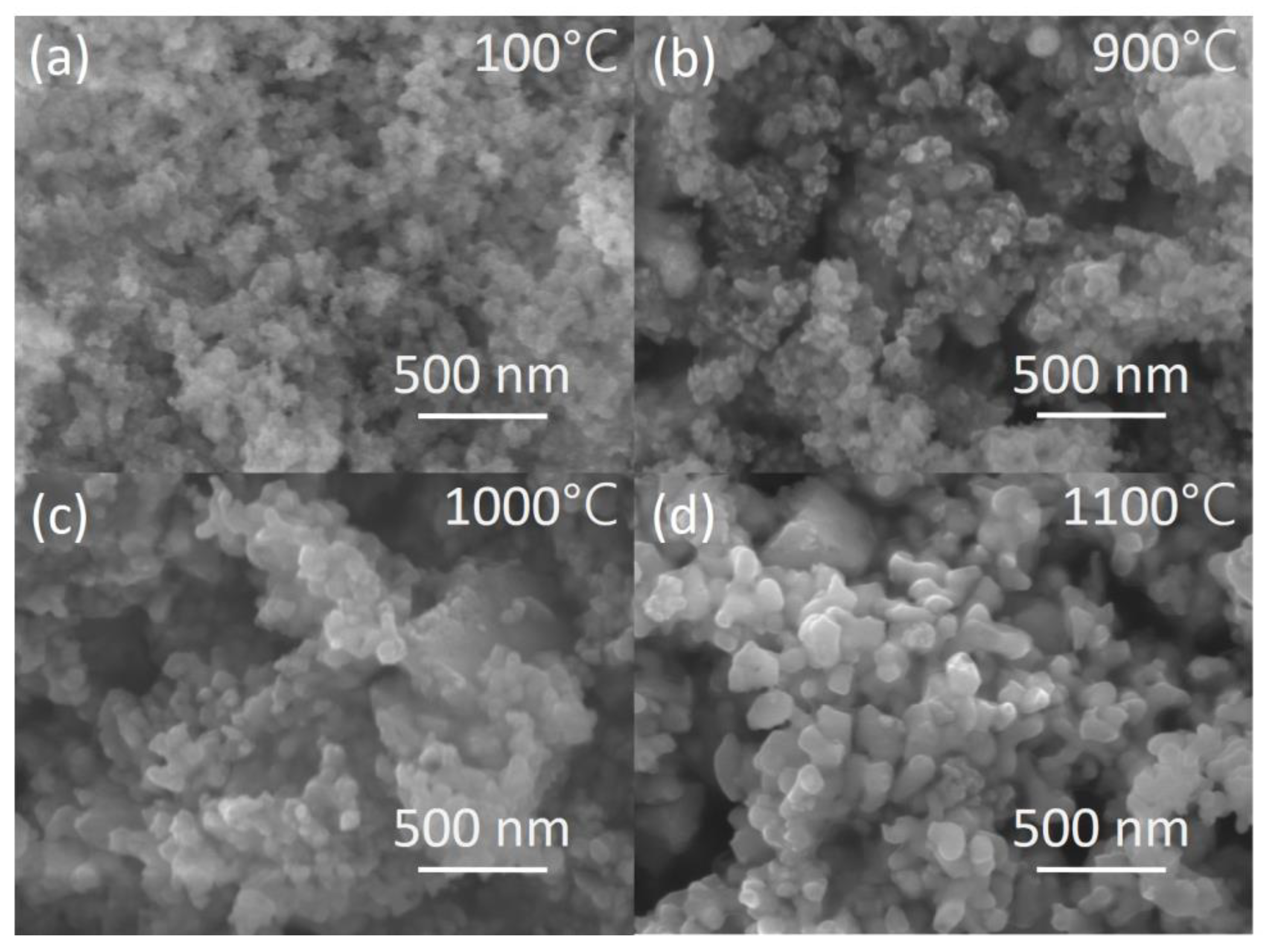
3.1. Development of Nanosized Powders

With the increasing complexity of the elemental composition of the material, the quality of precursors for crystal growth and for ceramics production comes to the fore. A precursor is a mixture of compounds suitable for further thermal treatment or compacting, with a minimal deviation from a given composition, usually stoichiometric, during further treatment (melting, sintering). Obviously, the precursor that provides the highest rate of transformation into a commercial crystalline mass is of the greatest interest. The precursor can be obtained in a relatively simple way by mechanical mixing and milling of powders for further solid-phase synthesis [104,105]. Furthermore, more complex methods such as co-precipitation [106,107], sol-gel [108,109], and spray-pyrolysis [110,111] are used.
For gallium-containing mixtures, a characteristic process is the evaporation of gallium oxide [112] during heat treatment. During single crystal growth, Ga2O3 evaporation is minimized by using a slightly oxidizing atmosphere (e.g., Ar/O2) [113]. In the case of ceramics, a known way to solve this problem is sintering in an oxygen atmosphere [114]. The listed methods for the synthesis of precursor powders of a given stoichiometry were used to obtain single crystals and ceramics. Authors [115] demonstrated that nanostructuring plays a positive role in the production of complex compounds. Precursors for transparent ceramics of binary and ternary garnets were obtained by solid phase synthesis as well as by co-precipitation and spray pyrolysis [116,117,118,119,120,121]. Among these methods, co-precipitation has an advantage of the fact that the necessary crystalline phase has already formed during the preparation of the precursor at the stage of calcination of the precipitate. This feature makes it possible to significantly reduce the evaporation of gallium oxide due to gallium bounding in a more complex compound. Figure 5 shows SEM images of the precipitate dried at 100 °C and then calcined at temperatures of 900, 1000, and 1100 °C for 2 h. SEM images show that at 900 °C agglomerates are formed from particles of the garnet habitus. Grain growth is observed with an increase in the annealing temperature of the precursors above 1000 °C. The choice heat treatment temperature of powder depends on the composition of the compound and should be experimentally selected. Definitely, it has to ensure the release of pores at the minimum acceptable sintering temperature of the compacted powder.
Worth noting, the formation of grains of garnet habitus in precursor obtained by spray pyrolysis is not observed even at a temperature of 1050 °C [110,111,122]. Spherical shape of the particles is retained by surface tension at high temperatures but does not prevent the evaporation of gallium. At the same time, the spray pyrolysis method is a well-established technology for the synthesis of nanopowders with a high specific surface area and low agglomeration. Precursors obtained by this method do not require additional milling and can be used for compaction without additional processing.
Several modifications of the co-precipitation method have been developed for disordered compounds with a garnet structure [107,123,124,125]. Among them, the method of reverse coprecipitation, which can significantly reduce the duration of the process of ceramic production is notable. The hydroxocarbonate precursor obtaining process includes four stages: (1) preparation of two solutions: a precipitant—ammonium bicarbonate and mixed metals solution; (2) precipitation of precursor by adding a mixture solution to the precipitant; (3), (4) washing and drying of the precipitate precursor. At the first stage solutions of metals nitrates are mixed in the required ratio and diluted to a concentration 0.5 M. Wherein the accuracy of concentration measurement should be ±0.1 g Me/kg of solution. The mixture solution was added in a thin stream with a flow rate of 40–60 mL/min to a precipitant with a concentration of about 1.5 M with continuous stirring, formed slurry was separated from mother liquor under vacuum. Then the precipitate was thoroughly washed with a water–isopropanol mixture. The product was dried to constant weight in a forced convection oven at 80–100 °C and then subjected to heat treatment at 800–1000 °C for 2 h to decompose the carbonates and form the garnet phase powder. Depending on the further compacting technology, the powder after low-temperature treatment is either used for milling and pressing, or it is subjected to a secondary heat treatment at a higher temperature to reduce its specific surface area and prepare it for compaction by colloidal methods.

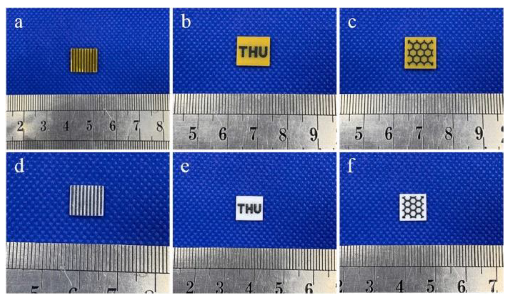
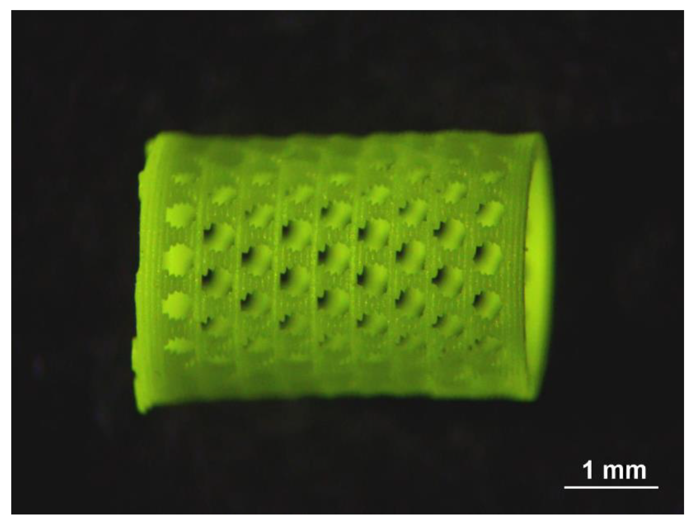
3.2. 3D Printing Advances in Precursors Compacting

Consolidation of precursors under pressure remains the main method in the production of dense scintillation ceramics. At the same time, 3D printing of crystalline compounds with cubic space group with further annealing is becoming increasingly popular for the production of luminescent, laser, and scintillation materials. Among the promising methods of 3D printing, direct ink writing (DIW) might provide promising results [126,127,128,129,130,131]. DIW is an extrusion-based additive manufacturing method widely used to fabricate solid and/or mesh-like periodic structures. The DIW method has several advantages, namely its versatility, relative simplicity, and low cost of equipment. In the DIW method, “ink” is dispensed from small nozzles with variable-speed and deposited along digitally defined paths to produce planar or relatively simple 3D structures layer-by-layer. The rheological characteristics of such suspensions do not depend on the chemical nature of filler powder, its optical or other functional properties, but on its dispersion, the nature and concentration of a dispersant used, and the type of organic binder. The main limitation of the DIW method is the size of nozzles. Small nozzles provide relatively good resolution but result in relatively slow print speeds. The typical nozzle size used is 0.50–0.61 mm. The formulations described include sintering additives such as TEOS (tetraethyl orthosilicate) and MgO. To obtain dense ceramics and composites from nominally pure or doped YAG, mixtures of commercial simple oxides or ready-made commercial YAG nanopowders are used. Sintering is carried out, as a rule, at high temperatures of 1750–1850 °C under high vacuum conditions. Additionally, cold isostatic pressing (CIP) and hot-isostatic pressing (HIP) can be utilized.
In addition, we note another promising method—Stereolithography (or Vat photopolymerization) [132,133,134,135,136,137,138,139,140,141]. The method of stereolithography is based on the layer-by-layer controlled polymerization of photosensitive monomers (or suspensions with ceramic powders) under light irradiation. In 2002, the production of a regular columnar structure (a 9-rod array) from low-viscosity suspensions by stereolithography was demonstrated from composite acrylate polymers-Pb(Zr,Ti)O3 [138] with a typical size of a hundred microns. Recently, in 2020, the fabrication of a similar structure was demonstrated from another related dielectric ceramic high-Z material, BaTiO3 [139].
The first 3D printed complex inorganic polycrystalline YAG:Ce scintillator was successfully obtained by stereolithography in 2017 [132], and afterwards, the possibility of manufacturing transparent YAG:Yb ceramic was demonstrated [133]. Figure 6 shows an example of successful stereolithographic 3D printing of plate-shaped samples. The picture on the left panel is as-printed composites consisting of a mixture of Al2O3, Y2O3, Yb2O3, TEOS ceramic powders and an acrylate polymer(s). The picture on the right shows a translucent YAG:Yb ceramic plate after solid-state reaction and sintering in a high vacuum furnace at 1750 °C for 16 h. High temperatures up to 1800 °C, vacuum conditions, and sintering aids (TEOS, MgO, etc.) allow to produce transparent ceramics using the stereolithography method as well.
Stereolithography produces surfaces with high accuracy, good quality and low roughness. Its disadvantages include obvious restrictions on the available wall thickness and the size of holes in product prototypes (both top and bottom). A bottleneck in stereolithography could be the light source in the 3D printer, whose irradiation may be absorbed by the doping ions in the ceramic powders. Therefore, direct stereolithographic 3D printing with powders of the garnets activated by RE ions or transition metals requires a careful selection of the light emitter.
The possibility of printing several materials in one product looks promising, as recently demonstrated [139,140]. Figure 7 shows an example of successful stereolithographic 3D printing from several ceramic materials at the same time in one sample. In the upper row, as-printed composites are present; in the lower row, ceramic samples after sintering are shown.
Figure 8 shows a mesh scintillator in the form of a cylinder, which can be used for measurements in a liquid or gas flow [142]. It was obtained by the 3D stereolithography method from the GAGG:Ce powder with further annealing at a temperature of 1600 °C for 2 h.
Such an approach will make it possible in the near future to manufacture high-precision structured scintillator detecting elements consisting entirely of ceramics. For example, translucent GYAGG:Ce or other suitable garnet-type material will be used to print discrete pixels interacting with radiation, whereas undoped white GYAGG fill interpixel space, as a reflector. It should be noted that two-photon 3D printing has spatial resolution at the level of a few microns [136], which will certainly require the use of exclusively nanosized initial powders for ceramic production.

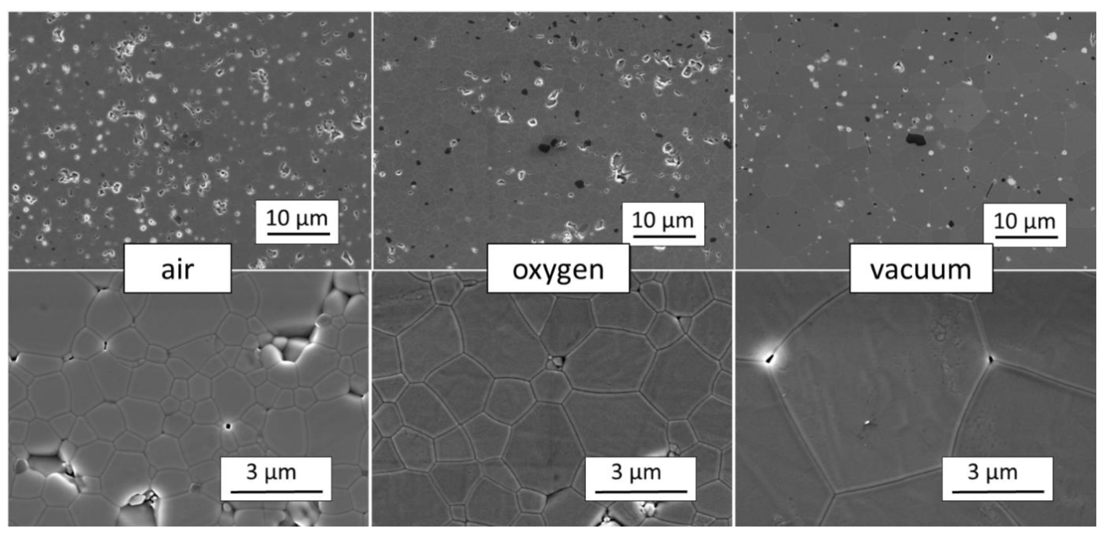
3.3. Sintering Process and Polycrystalline Structure Formation

The final step of producing a ceramic material is the sintering process, which is the formation of a dense polycrystalline mass under high temperature. When carrying out the sintering process under atmospheric conditions (in the air) it is difficult to achieve a complete exit of pores from the volume of ceramics. The residual porosity remains at a level of 0.5–1.0%. Transparent ceramics are obtained by sintering under vacuum conditions [105,111,143,144,145]. An alternative approach, which is suitable for transparent ceramics production, is sintering in an oxygen atmosphere, which prevents the evaporation of gallium [107,146,147,148] or hot isostatic pressing [122]. Figure 9 compares the microstructure of GYAGG:Ce ceramics obtained in different sintering atmospheres.
To obtain the ceramics with a minimum number of residual pores, sintering additives are used, which, when combined with a precursor, can affect the sintering process [149] and references there.
As an example, Figure 10 shows samples of GYAGG:Ce (a, c) and GYAGG:Tb (b, d) ceramics obtained by sintering in an oxygen atmosphere. The duration of sintering in this case was less than 4 h at a temperature of 1650 °C.

4. Disordered Doped Scintillation Materials

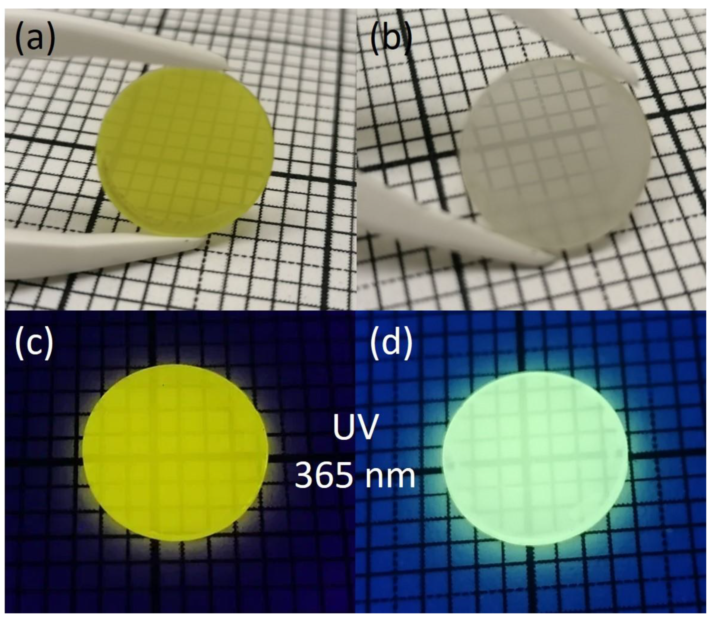
4.1. Heavy and Light Eu-Doped Scintillation Materials

Historically, the first scintillator doped with RE, the divalent europium ions, that found application was strontium iodide SrI2:Eu [150]. Material has been revisited in the last decade, and new halide compositions have been invited [151,152]. Several new alkali halide crystals have demonstrated impressive energy resolution parameters. Such materials, like other alkali halide compounds, were obtained in a single crystalline form by various pulling methods. The creation of a series of solid solutions [153] was a further development of this work. It was discovered that simultaneous incorporation of the heavy halide anions (I, Br) into the composition provides the best scintillation yield. We also note the outstanding characteristics of a mixture of bromide and barium iodide [154,155,156,157], as well as the preparation of BaBrCl compound by the Czochralski method [157], which indicates the prospect of producing a large ingot. An important improvement from mixing Cl and Br in an alkali halide composition is the reduction in the hygroscopicity of the compound. However, when mixing light and heavy halides in the lattice, no significant change in the scintillation yield was found [158]. The positive role of codoping with Au ions has been established [159,160] as well.
The influence of anion substitution in the lattice of an alkali-halide compound might be considered by analyzing the density of states in single-anionic compounds (Figure 11). Since the valence bands in BaI2, BaBr2, and BaCl2 crystals are formed dominantly by the p-states of I, Br and Cl-ions, the substitution disorder in mixed BaI2(1-x)Br2x and BaBr2(1-x)Cl2x crystals affects the states of the valence band, i.e., it leads to the scattering and localization of nonequilibrium holes. However, the effective mass of holes in the valence band of the investigated ionic crystals is quite large. Therefore, nonequilibrium holes have a small diffusion length and scattering due to the compositional disorder further limiting it. At the same time, nonequilibrium electrons are still more mobile than holes, which leads to the strong spatial dispersion of carriers. This situation is typical for BaBr2(1-x)Cl2x, in which the binary components forming the solid solution are direct bandgap crystals. The light yield of such an anionic mixture is not expected to be changed.
On the contrary, the mixed BaI2(1-x)Br2x crystal has a peculiarity in the valence band when considering this system in the virtual crystal (VC) approximation, described in [75]. BaI2 is an indirect bandgap crystal. That leads to the degeneration of the highest energy state in the valence band in the mixed system BaI2(1-x)Br2x. Although fluctuations of the effective potential due to the compositional disorder destroy this degeneracy, there are two states of similar energy relating to different regions in the cell. However, the probability of hole tunneling between these states may be significant due to the strong uncertainty of the quasi-momentum in a mixed system. This effect might lead to an increase in the diffusion length of holes. Therefore, more efficient formation of excitons and, consequently, increased light yield are expected.
In addition to heavy Eu2+-doped scintillators, lithium-containing compounds containing atoms of alkaline earth elements (Ca, Sr), which are isovalently replaced by divalent europium, are of particular interest. Such compounds are light and have a practical demand in the field of thermal neutron detection when used in the form of scintillation coatings for neutron radiography setups. The “gold standards” scintillators of neutron sensitive screens are commercially available terbium-doped gadolinium oxysulfide Gd2O2S:Tb (GOS, Gadox) and 6LiF/ZnS:Ag mixtures. The advantages of these compounds include their high scintillation light yield and, in the case of GOS, also the possibility to get a high spatial resolution of screens, which is associated both with the short free path of conversion electrons in the material (~6 μm) and a high neutron cross-section making efficient thin scintillator layers [164,165]. However, the bottleneck of these scintillators is the long scintillation decay kinetics, which limits their use under high flux neutron beams. Furthermore, both have a large Zeff, which causes sensitivity to background gamma-quanta. This is more suitable for GOS, but for 6LiF/ZnS:Ag, the opacity of the ZnS to their scintillation light deteriorates spatial resolution of the composites as well as efficiency of light collection from a layer.
An innovative option to combine high light output, fast scintillation kinetics, and neutron absorption efficiency is a composite coating based on (Gd,Y)3(Al,Ga)5O12:Ce and 6LiF, which demonstrates a high response to neutrons at the level of commercial 6LiF/ZnS:Ag screens [166]. The light oxide lithium-containing compounds having a few cations seem to be prospective alternatives for GOS and 6LiF/ZnS:Ag.
The lithium aluminate LiAlO2 is well known among a variety of light lithium-containing binary compounds. However, the light yield of scintillations is low when doped with rare-earth and transition metal ions and does not exceed 9000 ph/neutron [167,168]. Light ternary compounds containing lithium and alkaline earth metals have also been studied quite well as phosphors of different luminescence colors. For scintillators, over the past year attempts have been made to grow crystals of relatively heavy lithium-containing eutectics: the lithium-strontium and lithium-barium chlorides [169,170]. An LiCaAlF6 scintillator doped with cerium [171] and divalent europium [172] is also known from non-oxide systems, but it has low scintillation yield: 9000–12,000 ph/neutron when doped with Eu2+. Among the three-component oxide systems containing Li2O and AE (where AE = Ca, Sr, Ba), one can mark those which contain boron oxide as the third component: LiCaBO3 [173] and a silicon oxide. Li2CaSiO4 (LCS) is one of the compounds with the least amount of silicon tetrahedra. The scintillation properties of cerium doped LCS were first described in [174], where a light yield of 7780 ph/MeV was reported upon excitation with gamma-quanta. In [175], the cathodoluminescence of Li2CaSiO4 doped with Eu2+ was studied. However, as applied to the detection of neutrons, this compound was first proposed by the authors of [176,177]. A thin (180 µm) two-component composite LCS layer demonstrated the same response to neutrons as a commercial 470 µm 6LiF/ZnS:Ag screen and a 14% lower response to gamma-quanta. The achieved light yield under 5.5 MeV alpha-particles was measured to be 21,600 photons/MeV, which corresponds to the yield under thermal neutrons of ~100,000 ph/neutron, which is associated with a fast scintillation kinetics equal to 500 ns. The measured properties are similar to those of known lithium-containing scintillators CLYC and CLLB [178] and, at the same time, they have the potential to further increase the scintillation light yield. One of the important factors affecting the luminescence intensity of Li2CaSiO4:Eu2+ is the localization of Eu3+ in the compound, which is a quenching agent of the Eu2+ luminescence. Oxidation of Eu2+ to Eu3+ occurs during synthesis due to the evaporation of lithium and charge compensation of the defects by oxygen vacancies. To stabilize europium in the divalent state, the authors of [179,180] proposed introducing the RE metal ions into the matrix. The occupation by trivalent RE ions of the positions of the divalent Ca2+ may compensate for the loss of Li+ by the following Equation (3):
V L i + 1 2 V Ö + E u 2 O 3 V L i + 1 2 V Ö + E u C a × V L i + E u C a . + R E 2 O 3 V L i + R E C a . + E u C a ×    
However, the incorporation of additional RE ions into Ca sites in the matrix increases the effective charge of the compound and thereby increases the sensitivity of the material to background gamma radiation. Another method of stabilizing Eu2+: in the compound was found to be the substitution of Si ions by Al ions in tetrahedra. Both ions are light. The nonisovalent substitution of Si4+ by Al3+ ions act as a nonisovalent substitution in Ca sites in the compound. As a result, europium is dominantly stabilized in the divalent state according to the following Equation (4):
A l S i = A l S i × + e
e + E u C a . E u C a ×
This technological approach to dilute Si cationic sublattice made it possible to increase the intensity of photoluminescence by a factor of two, which correlates with the proportions of Eu2+ and Eu3+ in the compound (Figure 12).
The relative amount of Eu2+ and Eu3+ in the compounds was estimated from the photoluminescence spectra at 395 nm excitation, which ensures the excitation of both Eu3+ and Eu2+ ions. It was estimated as the ratio of the integrated photoluminescence intensity of the characteristic Eu2+ peak at 480 nm and integrated photoluminescence intensity of the peaks at 593, 617, and 700 nm corresponding to Eu3+ ions to total integrated intensity in 430–730 nm spectral range (Table 1).
In the context of compositional disorder, it is also worth mentioning the partial substitution of O2− ions by N3− ions, as is realized in oxynitride phosphors [181]. The most studies aimed at improving the luminescence properties. However, the door is open for researchers to use the same techniques as a tool to improve the scintillation properties of desired groups of compounds.

4.2. Ce-Doped Materials

Introducing disorder into both the cationic and anionic subsystems in Ce-activated crystals also gave a new impetus to research on improving the scintillation properties of alkali halide and oxide scintillators. Some results, after technological experiments, could be explained in the way described above for Eu2+ doped halides. A solid solution of LaI3 and LaBr3 having a mixture of anions was found to have a smaller LY in a comparison with pure LaBr3. However, it demonstrated faster scintillation kinetics [182]. The significant shortening of the scintillation kinetics can be explained by the possible contribution of tunneling to hole mobility, as in disordered BaI2(1-x)Br2x. The decrease in the scintillation yield can be explained by scattering in a solid solution due to the difficulty of stabilizing one phase when mixing hexagonal and orthorhombic phases [183]. The creation of disorder due to the substitution of cations in the compounds LuI3–YI3 or LuI3–GdI3 also does not lead to an increase in the yield of scintillations [184]. This is also due to technological difficulties in obtaining a single-phase solid solution of these compounds; YI3 has a trigonal symmetry group, while GdI3 has a hexagonal one. All three iodides are indirect-gap compounds, which makes it difficult to describe the dynamics of nonequilibrium carriers in such mixed systems yet.
Studies of the scintillation properties of oxyhalides have been carried out by the authors [185]. The creation of disorder due to the inclusion of oxygen and halide ions as ligands in the composition led to a significant deterioration in the parameters. The samples were obtained in the form of powders by solid-phase synthesis. Apparently, the deterioration of the parameters is associated with the difficulty of obtaining a homogeneous single crystalline phase.
Doping the well-known BaF2 scintillation material with Ce ions results in a slowing of the kinetics but a significant increase in scintillation yield [186,187,188]. However, in the LaF3-CeF3 system, a cation mixture does not significantly improve the parameters [189]. Cs2HfCl6:Ce [190] is another recently discovered material worth noting. The material is not hygroscopic and has a cubic spatial symmetry. The introduction of disorder into the anionic subsystem does not increase light yield but does reduce the slow component of scintillation [191].
Summarizing the available experimental data, it can be concluded that the introduction of disorder by mixing direct-gap and indirect-gap alkali halide compounds can lead to improvements in some scintillation parameters, as is observed in the listed examples of Eu2+ and Ce3+ doped materials.
In contrast to alkali-halide compounds, the binary and ternary oxide compounds, which have the structure of garnet, perovskite, pyrosilicate or oxyorthosilicate, provide the formation of disordered compounds when mixed with compounds with the same space symmetry group. All of these compounds are also direct-gap. Therefore, the change in the scintillation parameters in such compounds exclusively occurs due to a change in the dynamics of nonequilibrium carriers in the conduction band and excitons as well. As noted above (Section 2.1), creating disorder in the Gd/Al subsystem creates the potential to increase the scintillation yield. The scintillation yield in GAGG:Ce ternary garnet can reach 51,000 ph/MeV [192,193,194], which is nearly 70% higher than in binary YAG:Ce or LuAG:Ce compositions [195]. On the contrary, when the modulation of the crystal potential is not large, as in the case of a combination of Y and Lu in the composition [196], Y and Gd in the composition (Y,Gd)3Al5O12 [197] and Lu and Gd [198], the increase in the scintillation yield is no more than twenty percent.
Here we underline the role of trivalent gadolinium ions in the crystal lattice [199]. Contrary to the crystals on a base of Y, La, Lu, which have no peculiarities of the electron density states into the forbidden zone, the Gd3+ based crystal host has numerous f-levels (P, I, D) creating sub-zones in the bandgap. Moreover, following to extended Dieke diagram [200], one can suppose that numerous Gd3+ levels in the energy ranger above 40,000 cm−1 will provide an effective capture of nonequilibrium carriers by Gd3+ ions and further intracenter relaxation to the lowest 6P state. The intracenter relaxation process is quite short and carries on within a few picoseconds [6]. Consequently, the significant part of nonequilibrium carriers create Frenkel type excitons, which have a capability for migration along with P-, S-states of Gd sublattice. Moreover, holes can be quickly delocalized to the top of a valence band from 8S state [94]. Spatially, the hole can be localized in the same polyhedron with the mother Gd3+ ion, but predominantly located at the coordinating oxygen, and the Frenkel exciton might be considered as localized on the Gd-based oxyanionic complex. Migration of excitons between oxyanionic complexes provides delivery of excitation to cerium ions populating the Ce3+ radiating level 5d1.
The transition from ternary to quaternary garnets should take into account the breaking of the gadolinium sublattice and the features introduced in this way into the process of relaxation of nonequilibrium carriers. The authors of [192] found that in single crystals, such a transition, as a rule, leads to a lower scintillation yield. At the same time, in ceramics, where it becomes possible to use higher concentrations of the Ce3+ activator, it is possible to maintain or even exceed the scintillation yield in comparison with GAGG ceramics [201], while the scintillation kinetics can be significantly accelerated. An increase in the concentration of the activator, although it leads to the segregation of the activator near the grain boundaries [202], does not lead to a significant change in the scintillation parameters. There is a small fraction of such ions compared to those localized in the bulk of the grains. In a composition where some fraction of the Gd3+ ions are substituted by yttrium ions, it is possible to combine a high yield of 51,000 photons/MeV and fast scintillation kinetics (decay constant 50 ns) [203]. Of particular interest is the combination of Lu3+ and Gd3+ ions in a quaternary garnet to increase the stopping power of the material. At the same time, it was found that the partial substitution of gadolinium by lutetium ions leads to a significant decrease in the scintillation yield in both single crystal and polycrystalline samples compared to ternary GAGG [204,205,206]. Moreover, in such quaternary systems, a slow component appears in the scintillation. Breaking the integrity of the gadolinium sublattice by substitution with heavy lutetium ions increases the role of self-trapped states in the excitation of Ce3+ ions, which ensures both an increase in the fraction of short ~20 ns and very long ~600 ns components in the scintillation kinetics [207]. It is possible to preserve the fast kinetics, significantly reduce the slow kinetics, and increase the scintillation yield by changing from a quadruple to a quintuple composition by diluting the lutetium sublattice with yttrium ions. The ceramics sample with the composition Gd2Y0.5Lu0.5Al2Ga3O12 doped with Ce3+ and codoped with a small Mg2+ concentration (20 ppm) demonstrates fast scintillation kinetics having components of 14 ns (84%); 78 ns (16%) and the light yield at the level of 41,000 ph/MeV [208]. Scintillation materials based on disordered compounds with a garnet structure have come close in their parameters to advanced alkali-halide materials, surpassing them in stopping power as well as in manufacturability.
In oxyorthosilicates, when turning from Lu2SiO5 to (Lu,Y)2SiO5, it is hard to improve the scintillation yield due to the small depth of modulation at the bottom of the conduction band (Section 2.1). However, the introduction of yttrium into the composition allows solving the problem of phase homogeneity as well as minimizing the role of deep traps in capturing nonequilibrium electrons, that is, reducing the level of phosphorescence.
In pyrosilicates, mixing lanthanum and gadolinium ions (La,Gd)2Si2O7:Ce [209,210] in the composition makes it possible to maintain high scintillation yields at high temperatures and increase the yield by 20–25% compared to Gd2Si2O7:Ce.
In halide perovskites [211], it is not yet possible to obtain better results than for end compounds, while in (Lu, Gd)AlO3:Ce [212] the light yield approaches 21,000 ph/MeV, which is close to that of but much less dense YAlO3:Ce and 30% higher than the best (Lu,Y)AlO3:Ce. The compound may reach a Zeff~65, which is comparable to commercial LSO:Ce. Nonetheless, its scintillation kinetics are found to be three times slower when compared to YAlO3: Ce.

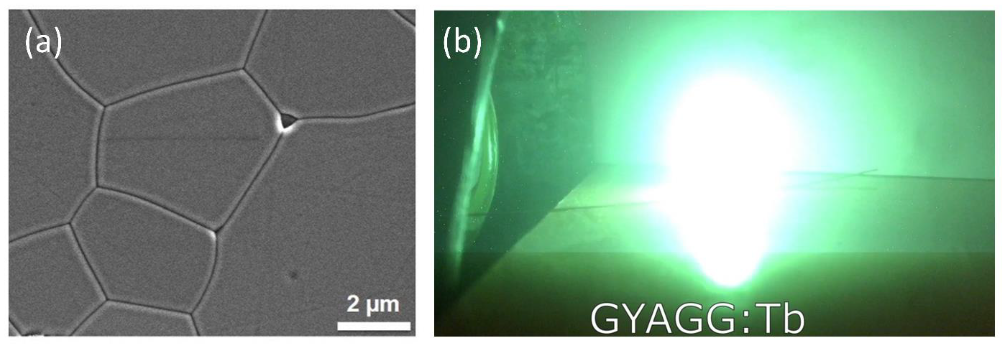
4.3. Tb Doped Materials

Terbium Tb3+ ion is another activator for creating bright scintillators. It can be hardly used for pulse height spectra acquisition due to its long scintillation kinetics (~2 ms), but can be utilized in a current mote with sensitive photo-diodes. Widely used in X-ray scanners is Gd2O2S translucent ceramics, activated with Tb3+ ions [213]. The host compounds for activation with terbium ions, which can be created in a polycrystalline form, are of particular interest [214]. A high concentration of the Tb activator is achievable compared to Ce activator in garnet type crystals [215]. This can significantly expand the field of utilization of such scintillation materials, for example, in alpha-, beta-voltaics [216], where the efficiency of energy conversion of ionizing radiation can reach or even exceed the efficiency of thermoelectric generators or cathodoluminescent light sources [217]. Moreover, due to high neutron cross-section, cerium doped Gd-based ternary garnets were shown to be promising scintillation materials to detect neutrons in a wide energy range [218,219]. Terbium provides slower but brighter scintillation than cerium, and this makes terbium-activated gadolinium-containing materials promising for application in scintillation threshold neutron counters or neutron sensitive screens, where sensitivity is the most important property. Figure 13a shows SEM image Tb-doped (Gd,Y)3Al2Ga3O12 ceramics; its microstructure is identical to those doped with Ce. Figure 13b demonstrates shining of Tb-doped ceramics under electron beam.
Worth noting, Tb-doped sample shows a small temperature coefficient for the light yield LY(T) in the temperature range 300–540 K. Quaternary Gd-Y compounds with a garnet structure doped with Tb were found to be a good platform to create high-bright scintillators to be exploited in indirect converters with both β- and α- isotopes. Distinctive features of the developed scintillators are their tolerance to irradiation and temperature stability, which are of especial importance for application in converters. The materials tested were fabricated as ceramics to utilize a flexibility with concentration of doping ions and to achieve high doping levels, which are hardly attained in the single crystalline matrix.

5. Conclusions

An extensive overview of the research and creation of scintillation crystal materials with compositionally disordered structures is provided. Both single- and poly-crystalline RE doped materials show an improvement in the scintillation properties. Amorphization of a chemical does not necessarily result from compositional disorder in a crystalline structure. A disordered distribution of ions occurs in the matrix when a polycationic ensemble with identical crystallographic sites forms in the cationic sublattice. The ordered distribution of anions, however, causes the space symmetry group to be conserved. As a result, crystallinity—or long-range order—in the ion stabilization locations is maintained. This approach is typical for oxide systems. For alkali-halides, where there is more latitude in the admixing, anions may be mixed as well. In this case, the long-range order is preserved due to the ordering of the cationic subsystem. The compositional disorder causes a modulation of the effective crystalline potential. Apparently, most of the positive effects on scintillation properties are due to the effects of the modulation of the effective potential in a disordered crystal on the mobility of nonequilibrium carriers. The modulation of the effective potential leads to a decrease in the diffusion length of free carriers, increasing the concentration of geminate electron-hole pairs and, consequently, increasing the transfer efficiency to the luminescent centers. A production cycle for multiunit oxide compositions with a garnet structure is considered in detail. Inverse coprecipitation of semiproducts, combined with a short annealing of the green bodies in the oxygen atmosphere at a temperature significantly lower than the melting point, could result in transparent materials doped with different RE ions. Future optimization of the production process chain, which includes engineering at the nano-scale level, might be based on significant progress in the last decade on the topic. In our opinion, more of the materials discussed will emerge as candidates for commercialization. Crystalline scintillators with disordered composition may become more prevalent in future ionizing radiation detectors due to the demand for new applications requiring purposeful tuning of the scintillation parameters.

Author Contributions

Conceptualization, V.R. and M.K.; methodology, D.K. and I.K.; validation, V.D. and P.K.; investigation, P.S. and Y.T.; writing—original draft preparation, V.R., M.K., D.K., I.K., V.D., P.K., P.S. and Y.T; writing—review and editing, V.R., M.K. and P.S. All authors have read and agreed to the published version of the manuscript.

Funding

This research received no external funding.

Data Availability Statement

Not applicable.

Acknowledgments

Authors are grateful to G. Dosovitskiy and V. Mechinsky for polemic involvement. Support from the Russian Ministry of Science and Education through grant No. 075-15-2021-1353 dated 12 October 2021 is acknowledged. The fabrication of mesh-like scintillation ceramic elements was supported by the Russian Science Foundation under grant No. 22-13-00172, https://rscf.ru/project/22-13-00172/ (accessed on 30 October 2022) Analytical studies have been carried out using the scientific equipment of CKP NRC “Kurchatov Institute–IREA”, with the financial support of the project by the Russian Federation represented by the Ministry of Education and Science of Russia, Agreement No. 075-15-2022-1157 dated 16 August 2022. Corresponding author thanks to Y. Wu, A. Yoshikawa and E. Bourret-Courchesne for fruitful discussion of the references on topic.

Conflicts of Interest

The authors declare no conflict of interest.

References

  1. Weber, M.J. Inorganic scintillators: Today and tomorrow. J. Lumin. 2002, 100, 35–45. [Google Scholar] [CrossRef]
  2. Yao, N.; Wang, Z.L. Handbook of Microscopy for Nanotechnology; Kluwer Academic Publishers: Boston, MA, USA, 2005; p. 745. [Google Scholar]
  3. Courtland, R. The microscope revolution that’s sweeping through materials science. Nature 2018, 563, 462–464. [Google Scholar] [CrossRef] [PubMed] [Green Version]
  4. Ali, A.; Chiang, Y.W.; Santos, R. X-Ray Diffraction Techniques for Mineral Characterization: A Review for Engineers of the Fundamentals, Applications, and Research Directions. Minerals 2021, 12, 205. [Google Scholar] [CrossRef]
  5. Tamulaitis, G.; Nargelas, S.; Korjik, M.; Mechinsky, V.; Talochka, Y.; Vaitkevičius, A.; Vasil’ev, A. Transient optical absorption as a powerful tool for engineering of lead tungstate scintillators towards faster response. J. Mater. Chem. C 2022, 10, 9521–9529. [Google Scholar] [CrossRef]
  6. Nargelas, S.; Korjik, M.; Vengris, M.; Tamulaitis, G. Study of electronic excitation relaxation at cerium ions in Gd3Al2Ga3O12 matrix using multipulse transient absorption technique. J. Appl. Phys. 2020, 128, 103104. [Google Scholar] [CrossRef]
  7. Gektin, A.V.; Belsky, A.N.; Vasil’ev, A.N. Scintillation Efficiency Improvement by Mixed Crystal Use. IEEE Trans. Nucl. Sci. 2014, 61, 262–270. [Google Scholar] [CrossRef]
  8. Korzhik, M.; Tamulaitis, G.; Vasil’ev, A.N. Physics of Fast Processes in Scintillators; Particle Acceleration and Detection; Springer International Publishing: Cham, Switzerland, 2020; ISBN 978-3-030-21965-9. [Google Scholar]
  9. Dorenbos, P. Fundamental Limitations in the Performance of Ce3+, Pr3+, and Eu2+ Activated Scintillators. IEEE Trans. Nucl. Sci. 2010, 57, 1162–1167. [Google Scholar] [CrossRef]
  10. Dorenbos, P. Energy of the First 4f7→4f65d Transition of Eu2+ in Inorganic Compounds. J. Lumin. 2003, 104, 239–260. [Google Scholar] [CrossRef]
  11. Dorenbos, P. The 5d Level Positions of the Trivalent Lanthanides in Inorganic Compounds. J. Lumin. 2000, 91, 155–176. [Google Scholar] [CrossRef]
  12. Dorenbos, P. The 4fn↔4fn−15d Transitions of the Trivalent Lanthanides in Halogenides and Chalcogenides. J. Lumin. 2000, 91, 91–106. [Google Scholar] [CrossRef]
  13. Dorenbos, P. 5d-Level Energies of Ce3+ and the Crystalline Environment. I. Fluoride Compounds. Phys. Rev. B 2000, 62, 15640–15649. [Google Scholar] [CrossRef] [Green Version]
  14. Dorenbos, P. 5d-level Energies of Ce3+ and the Crystalline Environment. II. Chloride, Bromide, and Iodide Compounds. Phys. Rev. B 2000, 62, 15650–15659. [Google Scholar] [CrossRef] [Green Version]
  15. Dorenbos, P. 5d-Level Energies of Ce3+ and the Crystalline Environment. III. Oxides Containing Ionic Complexes. Phys. Rev. B 2001, 64, 125117. [Google Scholar] [CrossRef] [Green Version]
  16. Zhuo, Y.; Hariyani, S.; You, S.; Dorenbos, P.; Brgoch, J. Machine Learning 5d-Level Centroid Shift of Ce3+ Inorganic Phosphors. J. Appl. Phys. 2020, 128, 013104. [Google Scholar] [CrossRef]
  17. Lecoq, P.; Gektin, A.; Korzhik, M. Inorganic Scintillators for Detector Systems; Particle Acceleration and Detection; Springer International Publishing: Cham, Switzerland, 2017; ISBN 978-3-319-45521-1. [Google Scholar]
  18. Yoshikawa, A.; Kamada, K.; Kurosawa, S.; Yokota, Y.; Yamaji, A.; Chani, V.I.; Ohashi, Y.; Yoshino, M. Growth and Characterization of Directionally Solidified Eutectic Systems for Scintillator Applications. J. Cryst. Growth 2018, 498, 170–178. [Google Scholar] [CrossRef]
  19. Yamamoto, S.; Kamada, K.; Yoshikawa, A. Ultrahigh Resolution Radiation Imaging System Using an Optical Fiber Structure Scintillator Plate. Sci. Rep. 2018, 8, 3194. [Google Scholar] [CrossRef] [Green Version]
  20. Kim, K.J.; Kamada, K.; Murakami, R.; Yoshino, M.; Kurosawa, S.; Yamaji, A.; Shoji, Y.; Kochurikhin, V.V.; Sato, H.; Toyoda, S.; et al. Growth and Scintillation Properties of Directionally Solidified Ce:LaCl3/AECl2 (AE = Mg, Ca, Sr) Eutectic Scintillators. J. Cryst. Growth 2022, 584, 126549. [Google Scholar] [CrossRef]
  21. Takizawa, Y.; Kamada, K.; Kim, K.J.; Yoshino, M.; Yamaji, A.; Kurosawa, S.; Yokota, Y.; Sato, H.; Toyoda, S.; Ohashi, Y.; et al. Large Size Growth of Terbium Doped BaCl2/NaCl/KCl Eutectic for Radiation Imaging. Jpn. J. Appl. Phys. 2022, 61, SC1009. [Google Scholar] [CrossRef]
  22. Lin, Z.; Lv, S.; Yang, Z.; Qiu, J.; Zhou, S. Structured Scintillators for Efficient Radiation Detection. Adv. Sci. 2022, 9, 2102439. [Google Scholar] [CrossRef]
  23. Vaněček, V.; Děcká, K.; Mihóková, E.; Čuba, V.; Král, R.; Nikl, M. Advanced Halide Scintillators: From the Bulk to Nano. Adv. Photonics Res. 2022, 3, 2200011. [Google Scholar] [CrossRef]
  24. Sidletskiy, O.; Gorbenko, V.; Zorenko, T.; Syrotych, Y.; Witkiwicz-Łukaszek, S.; Mares, J.A.; Kucerkova, R.; Nikl, M.; Gerasymov, I.; Kurtsev, D.; et al. Composition Engineering of (Lu,Gd,Tb)3(Al,Ga)5O12:Ce Film/Gd3(Al,Ga)5O12:Ce Substrate Scintillators. Crystals 2022, 12, 1366. [Google Scholar] [CrossRef]
  25. Alekhin, M.S.; de Haas, J.T.M.; Khodyuk, I.V.; Krämer, K.W.; Menge, P.R.; Ouspenski, V.; Dorenbos, P. Improvement of γ-Ray Energy Resolution of LaBr3:Ce3+ Scintillation Detectors by Sr2+ and Ca2+ Co-Doping. Appl. Phys. Lett. 2013, 102, 161915. [Google Scholar] [CrossRef] [Green Version]
  26. Yang, K.; Menge, P.R. Improving γ-Ray Energy Resolution, Non-Proportionality, and Decay Time of NaI:Tl+ with Sr2+ and Ca2+ Co-Doping. J. Appl. Phys. 2015, 118, 213106. [Google Scholar] [CrossRef]
  27. Khodyuk, I.V.; Messina, S.A.; Hayden, T.J.; Bourret, E.D.; Bizarri, G.A. Optimization of Scintillation Performance via a Combinatorial Multi-Element Co-Doping Strategy: Application to NaI:Tl. J. Appl. Phys. 2015, 118, 084901. [Google Scholar] [CrossRef] [Green Version]
  28. Brecher, C.; Lempicki, A.; Miller, S.R.; Glodo, J.; Ovechkina, E.E.; Gaysinskiy, V.; Nagarkar, V.V.; Bartram, R.H. Suppression of Afterglow in CsI:Tl by Codoping with Eu2+—I: Experimental. Nucl. Instrum. Methods A 2006, 558, 450–457. [Google Scholar] [CrossRef]
  29. Novotny, R.W.; Bremer, D.; Dormenev, V. The PANDA electro- magnetic calorimeter–a high-resolution detector based on PWO-II. In Proceedings of the IEEE 10th International Conference Inorganic Scintillators and Their Applications, Jeju, Republic of Korea, 14 June 2009; pp. 7–12. [Google Scholar]
  30. van Loef, E.V.D.; Dorenbos, P.; van Eijk, C.W.E.; Krämer, K.; Güdel, H.U. High-Energy-Resolution Scintillator: Ce3+ Activated LaCl3. Appl. Phys. Lett. 2000, 77, 1467–1468. [Google Scholar] [CrossRef]
  31. van Loef, E.V.D.; Dorenbos, P.; van Eijk, C.W.E.; Krämer, K.; Güdel, H.U. High-Energy-Resolution Scintillator: Ce3+ Activated LaBr3. Appl. Phys. Lett. 2001, 79, 1573–1575. [Google Scholar] [CrossRef]
  32. Derenzo, S.E.; Moses, W.W. Experimental efforts and results in finding new heavy scintillators. In Proceedings of the Crystal 2000 International Workshop on Heavy Scintillators for Scientific and Industrial Applications, Chamonix, France, 22–26 September 1992. [Google Scholar]
  33. Melcher, C.L.; Schweitzer, J.S. Cerium-doped lutetium oxyorthosilicate: A fast, efficient new scintillator. IEEE Trans. Nucl. Sci. 1992, 39, 502–505. [Google Scholar] [CrossRef]
  34. Nikl, M.; Mihoková, E.; Mareš, J.A.; Vedda, A.; Martini, M.; Nejezchleb, K.; Blažek, K. Traps and Timing Characteristics of LuAG:Ce3+ Scintillator. Phys. Status Solidi A 2000, 181, R10–R12. [Google Scholar] [CrossRef]
  35. Nikl, M.; Ogino, H.; Krasnikov, A.; Beitlerova, A.; Yoshikawa, A.; Fukuda, T. Photo- and Radioluminescence of Pr-Doped Lu3Al5O12 Single Crystal. Phys. Status Solidi A 2005, 202, R4–R6. [Google Scholar] [CrossRef]
  36. Ogino, H.; Yoshikawa, A.; Nikl, M.; Kamada, K.; Fukuda, T. Scintillation Characteristics of Pr-Doped Lu3Al5O12 Single Crystals. J. Cryst. Growth 2006, 292, 239–242. [Google Scholar] [CrossRef]
  37. Shi, Y.; Nikl, M.; Feng, X.; Mares, J.A.; Shen, Y.; Beitlerova, A.; Kucerkova, R.; Pan, Y.; Liu, Q. Microstructure, Optical, and Scintillation Characteristics of Pr3+ Doped Lu3Al5O12 Optical Ceramics. J. Appl. Phys. 2011, 109, 013522. [Google Scholar] [CrossRef]
  38. Baryshevskij, V.G.; Zuevskij, R.F.; Korzhik, M.V.; Lobko, A.S.; Moroz, V.I.; Smirnova, S.A.; Pavlenko, V.B.; Fedorov, A.A. YAlO3: Pr fast-response scintillation crystals. Pis’ma V Zhurnal Tekhnicheskoj Fiz. 1991, 17, 82–85. [Google Scholar]
  39. Moses, W.W.; Fyodorov, A.A.; Korzhik, M.V.; Aslanov, V.; Minkov, B.; Derenzo, S.E.; Gektin, A.V. LuAlO3:Ce-A high density, high speed scintillator for gamma detection. IEEE Nucl. Sci. Symp. 1995, 42, 597–600. [Google Scholar] [CrossRef] [Green Version]
  40. Moszyński, M.; Wolski, D.; Ludziejewski, T.; Kapusta, M.; Lempicki, A.; Brecher, C.; Wiśniewski, D.; Wojtowicz, A.J. Properties of the New LuAP:Ce Scintillator. Nucl. Instrum. Methods A 1997, 385, 123–131. [Google Scholar] [CrossRef]
  41. Euler, F.; Bruce, J.A. Oxygen Coordinates of Compounds with Garnet Structure. Acta Crystallogr. 1965, 19, 971–978. [Google Scholar] [CrossRef]
  42. Autrata, R.; Schauer, P.; Kuapil, J.; Kuapil, J. A Single Crystal of YAG-New Fast Scintillator in SEM. J. Phys. E 1978, 11, 707. [Google Scholar] [CrossRef] [Green Version]
  43. Yanagida, T.; Takahashi, H.; Ito, T.; Kasama, D.; Enoto, T.; Sato, M.; Hirakuri, S.; Kokubun, M.; Makishima, K.; Yanagitani, T.; et al. Evaluation of Properties of YAG (Ce) Ceramic Scintillators. IEEE Trans. Nucl. Sci. 2005, 52, 1836–1841. [Google Scholar] [CrossRef]
  44. Ludziejewski, T.; Moszyński, M.; Kapusta, M.; Wolski, D.; Klamra, W.; Moszyńska, K. Investigation of Some Scintillation Properties of YAG:Ce Crystals. Nucl. Instrum. Methods Phys. Res. Sect. Accel. Spectrometers Detect. Assoc. Equip. 1997, 398, 287–294. [Google Scholar] [CrossRef]
  45. Wang, X.; Dai, Y.; Zhang, Z.; Su, L.; Kou, H.; Wang, Y.; Wu, A. Optical and Scintillation Properties of Ce:Y3Al5O12 Single Crystal Fibers Grown by Laser Heated Pedestal Growth Method. J. Rare Earths 2021, 39, 1533–1539. [Google Scholar] [CrossRef]
  46. Hawthorne, F.C. Some Systematics of the Garnet Structure. J. Solid State Chem. 1981, 37, 157–164. [Google Scholar] [CrossRef]
  47. Zhang, Y.; Li, L.; Zhang, X.; Xi, Q. Temperature Effects on Photoluminescence of YAG:Ce3+ Phosphor and Performance in White Light-Emitting Diodes. J. Rare Earths 2008, 26, 446–449. [Google Scholar] [CrossRef]
  48. Zhong, J.; Zhuang, W.; Xing, X.; Liu, R.; Li, Y.; Liu, Y.; Hu, Y. Synthesis, Crystal Structures, and Photoluminescence Properties of Ce3+-Doped Ca2LaZr2Ga3O12: New Garnet Green-Emitting Phosphors for White LEDs. J. Phys. Chem. C 2015, 119, 5562–5569. [Google Scholar] [CrossRef]
  49. Swiderski, L.; Moszynski, M.; Nassalski, A.; Syntfeld-Kazuch, A.; Szczesniak, T.; Kamada, K.; Tsutsumi, K.; Usuki, Y.; Yanagida, T.; Yoshikawa, A.; et al. Scintillation Properties of Praseodymium Doped LuAG Scintillator Compared to Cerium Doped LuAG, LSO and LaBr3. IEEE Trans. Nucl. Sci. 2009, 56, 2499–2505. [Google Scholar] [CrossRef]
  50. Nikl, M.; Mares, J.A.; Solovieva, N.; Li, H.-L.; Liu, X.-J.; Huang, L.-P.; Fontana, I.; Fasoli, M.; Vedda, A.; D’Ambrosio, C. Scintillation Characteristics of Lu3Al5O12:Ce Optical Ceramics. J. Appl. Phys. 2007, 101, 033515. [Google Scholar] [CrossRef]
  51. Hu, C.; Liu, S.; Shi, Y.; Kou, H.; Li, J.; Pan, Y.; Feng, X.; Liu, Q. Antisite Defects in Nonstoichiometric Lu3Al5O12:Ce Ceramic Scintillators. Phys. Status Solidi B 2015, 252, 1993–1999. [Google Scholar] [CrossRef]
  52. Takahashi, H.; Yanagida, T.; Kasama, D.; Ito, T.; Kokubun, M.; Makishima, K.; Yanagitani, T.; Yagi, H.; Shigeta, T.; Ito, T. The Temperature Dependence of Gamma-Ray Responses of YAG:Ce Ceramic Scintillators. IEEE Trans. Nucl. Sci. 2006, 53, 2404–2408. [Google Scholar] [CrossRef] [Green Version]
  53. Yanagida, T.; Fujimoto, Y.; Yokota, Y.; Kamada, K.; Yanagida, S.; Yoshikawa, A.; Yagi, H.; Yanagitani, T. Comparative Study of Transparent Ceramic and Single Crystal Ce Doped LuAG Scintillators. Radiat. Meas. 2011, 46, 1503–1505. [Google Scholar] [CrossRef]
  54. Yanagida, T.; Fujimoto, Y.; Kamada, K.; Totsuka, D.; Yagi, H.; Yanagitani, T.; Futami, Y.; Yanagida, S.; Kurosawa, S.; Yokota, Y.; et al. Scintillation Properties of Transparent Ceramic Pr:LuAG for Different Pr Concentration. IEEE Trans. Nucl. Sci. 2012, 59, 2146–2151. [Google Scholar] [CrossRef]
  55. Liu, S.; Feng, X.; Zhou, Z.; Nikl, M.; Shi, Y.; Pan, Y. Effect of Mg2+ Co-Doping on the Scintillation Performance of LuAG:Ce Ceramics. Phys. Status Solidi RRL–Rapid Res. Lett. 2014, 8, 105–109. [Google Scholar] [CrossRef]
  56. Liu, S.; Feng, X.; Mares, J.A.; Babin, V.; Nikl, M.; Beitlerova, A.; Shi, Y.; Zeng, Y.; Pan, Y.; D’Ambrosio, C.; et al. Effect of Li+ ions co-doping on luminescence, scintillation properties and defects characteristics of LuAG:Ce ceramics. Opt. Mater. 2017, 64, 245–249. [Google Scholar] [CrossRef]
  57. Liu, S.; Feng, X.; Mares, J.A.; Babin, V.; Nikl, M.; Beitlerova, A.; Shi, Y.; Zeng, Y.; Pan, Y.; D’Ambrosio, C.; et al. Optical, Luminescence and Scintillation Characteristics of Non-Stoichiometric LuAG:Ce Ceramics. J. Lumin. 2016, 169, 72–77. [Google Scholar] [CrossRef]
  58. Liu, S.; Mares, J.A.; Babin, V.; Hu, C.; Kou, H.; D’Ambrosio, C.; Pan, Y.; Nikl, M. Effect of Reducing Lu3+ Content on the Fabrication and Scintillation Properties of Non-Stoichiometric Lu3−xAl5O12:Ce Ceramics. Opt. Mater. 2017, 63, 179–184. [Google Scholar] [CrossRef] [Green Version]
  59. Dexter, D.L. Possibility of Luminescent Quantum Yields Greater than Unity. Phys. Rev. 1957, 108, 630–633. [Google Scholar] [CrossRef]
  60. van der Kolk, E.; Dorenbos, P.; Vink, A.P.; Perego, R.C.; van Eijk, C.W.E.; Lakshmanan, A.R. Vacuum Ultraviolet Excitation and Emission Properties of Pr3+ and Ce3+ in MSO4 (MA=Ba, Sr, and Ca) and Predicting Quantum Splitting by Pr3+ in Oxides and Fluorides. Phys. Rev. B 2001, 64, 195129. [Google Scholar] [CrossRef] [Green Version]
  61. Lakshmanan, A.R.; Kim, S.-B.; Jang, H.M.; Kum, B.G.; Kang, B.K.; Heo, S.; Seo, D. A Quantum-Splitting Phosphor Exploiting the Energy Transfer from Anion Excitons to Tb3+ in CaSO4:Tb,Na. Adv. Funct. Mater. 2007, 17, 212–218. [Google Scholar] [CrossRef]
  62. Kudryavtseva, I.; Lushchik, A.; Lushchik, C.; Maaroos, A.; Nagirnyi, V.; Pazylbek, S.; Tussupbekova, A.; Vasil’chenko, E. Complex Terbium Luminescence Centers in Spectral Transformers Based on CaSO4. Phys. Solid State 2015, 57, 2191–2201. [Google Scholar] [CrossRef]
  63. Lushchik, A.; Lushchik, C.; Kudryavtseva, I.; Maaroos, A.; Nagirnyi, V.; Savikhin, F. Resonant Processes Causing Photon Multiplication in CaSO4:Tb3+. Radiat. Meas. 2013, 56, 139–142. [Google Scholar] [CrossRef] [Green Version]
  64. Oses, C.; Toher, C.; Curtarolo, S. High-Entropy Ceramics. Nat. Rev. Mater. 2020, 5, 295–309. [Google Scholar] [CrossRef]
  65. Zhang, R.-Z.; Reece, M.J. Review of High Entropy Ceramics: Design, Synthesis, Structure and Properties. J. Mater. Chem. A 2019, 7, 22148–22162. [Google Scholar] [CrossRef] [Green Version]
  66. Nundy, S.; Tatar, D.; Kojčinović, J.; Ullah, H.; Ghosh, A.; Mallick, T.K.; Meinusch, R.; Smarsly, B.M.; Tahir, A.A.; Djerdj, I. Bandgap Engineering in Novel Fluorite-Type Rare Earth High-Entropy Oxides (RE-HEOs) with Computational and Experimental Validation for Photocatalytic Water Splitting Applications. Adv. Sustain. Syst. 2022, 6, 2200067. [Google Scholar] [CrossRef]
  67. Corey, Z.J.; Lu, P.; Zhang, G.; Sharma, Y.; Rutherford, B.X.; Dhole, S.; Roy, P.; Wang, Z.; Wu, Y.; Wang, H.; et al. Structural and Optical Properties of High Entropy (La,Lu,Y,Gd,Ce)AlO3 Perovskite Thin Films. Adv. Sci. 2022, 9, 2202671. [Google Scholar] [CrossRef] [PubMed]
  68. Zhang, G.; Wu, Y. High-Entropy Transparent Ceramics: Review of Potential Candidates and Recently Studied Cases. Int. J. Appl. Ceram. Technol. 2022, 19, 644–672. [Google Scholar] [CrossRef]
  69. Rabatin, J.G. Luminescence of Rare Earth Activated Lutetium Oxyhalide Phosphors. J. Electrochem. Soc. 1982, 129, 1552–1555. [Google Scholar] [CrossRef]
  70. Yoshida, M.; Nakagawa, M.; Fujii, H.; Kawaguchi, F.; Yamada, H.; Ito, Y.; Takeuchi, H.; Hayakawa, T.; Tsukuda, Y. Application of Gd2O2S Ceramic Scintillator for X-Ray Solid State Detector in X-Ray CT. Jpn. J. Appl. Phys. 1988, 27, L1572. [Google Scholar] [CrossRef]
  71. Korzhik, M.V. Physics of Scintillators on a Base of Oxide Single Crystals; BSU: Minsk, Belarus, 2003; p. 263. ISBN 985-485-061-7. [Google Scholar]
  72. Chai, B.H.T.; Chai, D.Y.; Randall, A. Method of Enhancing Performance of Doped Scintillation Crystals. U.S. Patent 7,397,034 B2, 8 July 2008. [Google Scholar]
  73. Belsky, A.N.; Auffray, E.; Lecoq, P.; Dujardin, C.; Garnier, N.; Canibano, H.; Pedrini, C.; Petrosyan, A.G. Progress in the Development of LuAlO3-Based Scintillators. IEEE Trans. Nucl. Sci. 2001, 48, 1095–1100. [Google Scholar] [CrossRef]
  74. Simonov, A.; Goodwin, A.L. Designing Disorder into Crystalline Materials. Nat. Rev. Chem. 2020, 4, 657–673. [Google Scholar] [CrossRef]
  75. Talochka, Y.; Vasil’ev, A.; Korzhik, M.; Tamulaitis, G. Impact of Compositional Disorder on Electron Migration in Lutetium–Yttrium Oxyorthosilicate Scintillator. J. Appl. Phys. 2022, 132, 053101. [Google Scholar] [CrossRef]
  76. Mp-3050: Y3Al5O12 (Cubic, Ia-3d, 230). Available online: https://materialsproject.org/materials/mp-3050/ (accessed on 24 October 2022).
  77. Mp-5444: Y3Ga5O12 (Cubic, Ia-3d, 230). Available online: https://materialsproject.org/materials/mp-5444#electronic_structure (accessed on 24 October 2022).
  78. Mp-14132: Lu3Al5O12 (Cubic, Ia-3d, 230). Available online: https://materialsproject.org/materials/mp-14132/ (accessed on 24 October 2022).
  79. Mp-14134: Lu3Ga5O12 (Cubic, Ia-3d, 230). Available online: https://materialsproject.org/materials/mp-14134/ (accessed on 24 October 2022).
  80. Mp-16969: Lu2SiO5 (Monoclinic, C2/c, 15). Available online: https://materialsproject.org/materials/mp-16969/ (accessed on 24 October 2022).
  81. Mp-3520: Y2SiO5 (Monoclinic, C2/c, 15). Available online: https://materialsproject.org/materials/mp-3520/#electronic_structure (accessed on 24 October 2022).
  82. Yonezawa, F.; Morigaki, K. Coherent Potential Approximation. Basic Concepts and Applications. Prog. Theor. Phys. Suppl. 1973, 53, 1–76. [Google Scholar] [CrossRef] [Green Version]
  83. Sidletskiy, O. Trends in Search for Bright Mixed Scintillators. Phys. Status Solidi A 2018, 215, 1701034. [Google Scholar] [CrossRef]
  84. Dorenbos, P. Electronic Structure and Optical Properties of the Lanthanide Activated RE3(Al1−xGax)5O12 (RE=Gd, Y, Lu) Garnet Compounds. J. Lumin. 2013, 134, 310–318. [Google Scholar] [CrossRef]
  85. Yadav, S.K.; Uberuaga, B.P.; Nikl, M.; Jiang, C.; Stanek, C.R. Band-Gap and Band-Edge Engineering of Multicomponent Garnet Scintillators from First Principles. Phys. Rev. Appl. 2015, 4, 054012. [Google Scholar] [CrossRef] [Green Version]
  86. Kanai, T.; Satoh, M.; Miura, I. Characteristics of a Nonstoichiometric Gd3+δ(Al,Ga)5−δO12:Ce Garnet Scintillator. J. Am. Ceram. Soc. 2008, 91, 456–462. [Google Scholar] [CrossRef]
  87. Sifat, R.; Grosvenor, A.P. Examination of the Site Preference in Garnet Type (X3A2B3O12; X = Y, A/B = Al, Ga, Fe) Materials. Solid State Sci. 2018, 83, 56–64. [Google Scholar] [CrossRef]
  88. Ueda, J.; Tanabe, S. (INVITED) Review of Luminescent Properties of Ce3+-Doped Garnet Phosphors: New Insight into the Effect of Crystal and Electronic Structure. Opt. Mater. X 2019, 1, 100018. [Google Scholar] [CrossRef]
  89. Ogino, H.; Yoshikawa, A.; Nikl, M.; Kucerkova, R.; Shimoyama, J.; Kishio, K. Suppression of Defect Related Host Luminescence in LuAG Single Crystals. Phys. Procedia 2009, 2, 191–205. [Google Scholar] [CrossRef] [Green Version]
  90. Fasoli, M.; Vedda, A.; Nikl, M.; Jiang, C.; Uberuaga, B.P.; Andersson, D.A.; McClellan, K.J.; Stanek, C.R. Band-Gap Engineering for Removing Shallow Traps in Rare-Earth Lu3Al5O12 Garnet Scintillators Using Ga3+ Doping. Phys. Rev. B 2011, 84, 081102. [Google Scholar] [CrossRef] [Green Version]
  91. Ogino, H.; Yoshikawa, A.; Nikl, M.; Pejchal, J.; Fukuda, T. Growth and Luminescence Properties of Pr-Doped Lu3(Ga,Al)5O12 Single Crystals. Jpn. J. Appl. Phys. 2007, 46, 3514–3517. [Google Scholar] [CrossRef]
  92. Hu, C.; Liu, S.; Fasoli, M.; Vedda, A.; Nikl, M.; Feng, X.; Pan, Y. O– Centers in LuAG:Ce,Mg Ceramics. Phys. Status Solidi RRL–Rapid Res. Lett. 2015, 9, 245–249. [Google Scholar] [CrossRef]
  93. Nikl, M.; Kamada, K.; Babin, V.; Pejchal, J.; Pilarova, K.; Mihokova, E.; Beitlerova, A.; Bartosiewicz, K.; Kurosawa, S.; Yoshikawa, A. Defect Engineering in Ce-Doped Aluminum Garnet Single Crystal Scintillators. Cryst. Growth Des. 2014, 14, 4827–4833. [Google Scholar] [CrossRef]
  94. Auffray, E.; Augulis, R.; Fedorov, A.; Dosovitskiy, G.; Grigorjeva, L.; Gulbinas, V.; Koschan, M.; Lucchini, M.; Melcher, C.; Nargelas, S.; et al. Excitation Transfer Engineering in Ce-Doped Oxide Crystalline Scintillators by Codoping with Alkali-Earth Ions. Phys. Status Solidi A 2018, 215, 1700798. [Google Scholar] [CrossRef]
  95. Feofilov, S.P.; Kaplyanskii, A.A.; Kulinkin, A.B.; Zakharchenya, R.I.; Ovanesyan, K.; Petrosyan, A.; Dujardin, C. Inhomogeneous Broadening: Symmetry of Centers and Disorder in Solid Solutions and Nanocrystals. EPJ Web Conf. 2015, 103, 01003. [Google Scholar] [CrossRef] [Green Version]
  96. Nargelas, S.; Talochka, Y.; Vaitkevičius, A.; Dosovitskiy, G.; Buzanov, O.; Vasil’ev, A.; Malinauskas, T.; Korzhik, M.; Tamulaitis, G. Influence of Matrix Composition and Its Fluctuations on Excitation Relaxation and Emission Spectrum of Ce Ions in (GdxY1-x)3Al2Ga3O12:Ce Scintillators. J. Lumin. 2022, 242, 118590. [Google Scholar] [CrossRef]
  97. Shakhno, A.; Markovskyi, A.; Zorenko, T.; Witkiewicz-Łukaszek, S.; Vlasyuk, Y.; Osvet, A.; Elia, J.; Brabec, C.J.; Batentschuk, M.; Zorenko, Y. Micropowder Ca2YMgScSi3O12:Ce Silicate Garnet as an Efficient Light Converter for White LEDs. Materials 2022, 15, 3942. [Google Scholar] [CrossRef] [PubMed]
  98. Meng, Q.; Li, J.-G.; Zhu, Q.; Li, X.; Sun, X. The Effects of Mg2+/Si4+ Substitution on Crystal Structure, Local Coordination and Photoluminescence of (Gd,Lu)3Al5O12:Ce Garnet Phosphor. J. Alloys Compd. 2019, 797, 477–485. [Google Scholar] [CrossRef]
  99. Tratsiak, Y.; Bokshits, Y.; Borisevich, A.; Korjik, M.; Vaitkevičius, A.; Tamulaitis, G. Y2CaAlGe(AlO4)3:Ce and Y2MgAlGe(AlO4)3:Ce Garnet Phosphors for White LEDs. Opt. Mater. 2017, 67, 108–112. [Google Scholar] [CrossRef]
  100. Chyzh, A.; Baramsai, B.; Becker, J.A.; Bečvář, F.; Bredeweg, T.A.; Couture, A.; Dashdorj, D.; Haight, R.C.; Jandel, M.; Kroll, J.; et al. Measurement of the 157Gd(n,γ) Reaction with the DANCE γ Calorimeter Array. Phys. Rev. C 2011, 84, 014306. [Google Scholar] [CrossRef] [Green Version]
  101. Hagiwara, K.; Yano, T.; Tanaka, T.; Reen, M.S.; Das, P.K.; Lorenz, S.; Ou, I.; Sudo, T.; Yamada, Y.; Mori, T.; et al. Gamma-Ray Spectrum from Thermal Neutron Capture on Gadolinium-157. Prog. Theor. Exp. Phys. 2019, 2019, 023D01. [Google Scholar] [CrossRef] [Green Version]
  102. Menge, P.R.; Yang, K.; Ouspenski, V. Large Format Li Co-Doped Nai:Tl (NailTM) Scintillation Detector for Gamma-Ray and Neutron Dual Detection. In Proceedings of the 12th Pacific Rim Conference on Ceramic and Glass Technology; John Wiley & Sons, Ltd.: Hoboken, NJ, USA, 2018; pp. 201–208, ISBN 978-1-119-49409-6. [Google Scholar]
  103. Takizawa, Y.; Kamada, K.; Yoshino, M.; Kim, K.J.; Yamaji, A.; Kurosawa, S.; Yokota, Y.; Sato, H.; Toyoda, S.; Ohashi, Y.; et al. Growth and Scintillation Properties of Ce Doped 6LiBr/LaBr3 Eutectic Scintillator for Neutron Detection. Nucl. Instrum. Methods A 2022, 1028, 166384. [Google Scholar] [CrossRef]
  104. Ye, S.; Xiao, F.; Pan, Y.X.; Ma, Y.Y.; Zhang, Q.Y. Phosphors in phosphor-converted white lightemitting diodes: Recent advances in materials, techniques and properties. Mater. Sci. Eng. R. 2010, 71, 1–34. [Google Scholar] [CrossRef]
  105. Kanai, T.; Satoh, M.; Miura, I. Hot-Pressing Method to Consolidate Gd3(Al,Ga)5O12:Ce Garnet Scintillator Powder for use in an X-ray CT Detector. Int. J. Appl. Ceram. Technol. 2013, 10, E1–E10. [Google Scholar] [CrossRef]
  106. Li, J.G.; Ikegami, T.; Lee, J.H.; Mori, T.; Yajima, Y. Reactive yttrium aluminate garnet powder via coprecipitation using ammonium hydrogen carbonate as the precipitant. J. Mater. Res. 2000, 15, 1864–1867. [Google Scholar] [CrossRef]
  107. Luo, Z.; Jiang, H.; Jiang, J. Synthesis of Cerium-Doped Gd3(Al,Ga)5O12 Powder for Ceramic Scintillators with Ultrasonic-Assisted Chemical Coprecipitation Method. J. Am. Ceram. Soc. 2013, 96, 3038–3041. [Google Scholar] [CrossRef]
  108. Hassanzadeh-Tabrizi, S.A.; Taheri-Nassaj, E.; Sarpoolaky, H. Synthesis of an alumina–YAG nanopowder via sol–gel method. J. Alloys Compd. 2008, 456, 282–285. [Google Scholar] [CrossRef]
  109. Drozdowski, W.; Witkowski, M.E.; Solarz, P.; Głuchowski, P.; Głowacki, M.; Brylew, K. Scintillation properties of Gd3Al2Ga3O12:Ce (GAGG:Ce): A comparison between monocrystalline and nanoceramic samples. Opt. Mater. 2018, 79, 227–231. [Google Scholar] [CrossRef]
  110. Marchal, J.; John, T.; Baranwal, R.; Hinklin, T.; Laine, R.M. Yttrium aluminum garnet nanopowders produced by liquid-feed flame spray pyrolysis (LF-FSP) of metalloorganic precursors. Chem. Mater. 2004, 16, 822–831. [Google Scholar] [CrossRef]
  111. Seeley, Z.M.; Cherepy, N.J.; Payne, S.A. Expanded phase stability of Gd-based garnet transparent ceramic scintillators. J. Mater. Res. 2014, 29, 2332–2337. [Google Scholar] [CrossRef]
  112. Lamoreaux, R.H.; Hildenbrand, D.L.; Brewer, L. High-Temperature Vaporization Behavior of Oxides II. Oxides of Be, Mg, Ca, Sr, Ba, B, Al, Ga, In, Tl, Si, Ge, Sn, Pb, Zn, Cd, and Hg. J. Phys. Chem. Ref. Data 1987, 16, 419–443. [Google Scholar] [CrossRef]
  113. Kurosawa, S.; Shoji, Y.; Yokota, Y.; Kamada, K.; Chani, V.I.; Yoshikawa, A. Czochralski growth of Gd3(Al5−xGax)O12 (GAGG) single crystals and their scintillation properties. J. Cryst. Growth 2014, 393, 134–137. [Google Scholar] [CrossRef] [Green Version]
  114. Zhang, J.Y.; Luo, Z.H.; Jiang, H.C.; Jiang, J.; Gui, Z.Z.; Chen, C.H.; Liu, Y.F.; Liu, F.; Ci, M.M. Sintering of GGAG: Ce3+, xY3+ transparent ceramics in oxygen atmosphere. Ceram. Int. 2017, 43, 16036–16041. [Google Scholar] [CrossRef]
  115. Korzhik, M.; Alenkov, V.; Buzanov, O.; Fedorov, A.; Dosovitskiy, G.; Grigorjeva, L.; Mechinsky, V.; Sokolov, P.; Tratsiak, Y.; Zolotarjovs, A.; et al. Nanoengineered Gd3Al2Ga3O12 Scintillation Materials with Disordered Garnet Structure for Novel Detectors of Ionizing Radiation. Cryst. Res. Technol. 2019, 54, 1800172. [Google Scholar] [CrossRef]
  116. Li, H.L.; Liu, X.J.; Huang, L.P. Fabrication of transparent cerium-doped lutetium aluminum garnet (LuAG:Ce) ceramics by a solid-state reaction method. J. Am. Ceram. Soc. 2005, 88, 3226–3228. [Google Scholar] [CrossRef]
  117. Li, H.L.; Liu, X.J.; Huang, L.P. Fabrication of transparent Ce:LuAG ceramics by a solid-state reaction method. J. Inorg. Mater. 2006, 21, 1161–1166. [Google Scholar] [CrossRef]
  118. Li, H.L.; Liu, X.J.; Xie, R.J.; Zeng, Y.; Huang, L.P. Fabrication of transparent cerium-doped lutetium aluminum garnet ceramics by co-precipitation routes. J. Am. Ceram. Soc. 2006, 89, 2356–2358. [Google Scholar] [CrossRef]
  119. Li, H.L.; Liu, X.J.; Xie, R.J.; Zhou, G.H.; Hirosaki, N.; Pu, X.P.; Huang, L.P. Cerium-doped lutetium aluminum garnet phosphors and optically transparent ceramics prepared from powder precursors by a urea homogeneous precipitation method. Jpn. J. Appl. Phys. 2008, 47, 1657–1661. [Google Scholar] [CrossRef]
  120. Cherepy, N.J.; Payne, S.A.; Asztalos, S.J.; Hull, G.; Kuntz, J.D.; Niedermayr, T.; Pimputkar, S.; Roberts, J.J.; Sanner, R.D.; Tillotson, T.M.; et al. Scintillators with potential to supersede lanthanum bromide. IEEE Trans. Nucl. Sci. 2009, 56, 873–880. [Google Scholar] [CrossRef] [Green Version]
  121. Cherepy, N.J.; Kuntz, J.D.; Roberts, J.J.; Hurst, T.A.; Drury, O.B.; Sanner, R.D.; Tillotson, T.M.; Payne, S.A. Transparent ceramic scintillator fabrication, properties, and applications. In Proceedings of the SPIE 7079, Hard X-ray, Gamma-Ray, and Neutron Detector Physics X, San Diego, CA, USA, 4 September 2008. [Google Scholar] [CrossRef]
  122. Seely, Z.M.; Cherepy, N.J.; Payne, S.A. Homogenity of Gd-based garnet transparent ceramic scintillators for gamma spectroscopy. J. Cryst. Growth 2013, 379, 79–83. [Google Scholar] [CrossRef]
  123. Yang, S.H.; Sun, Y.S.; Chen, X.Q.; Zhang, Y.; Luo, Z.; Jiang, J.; Jiang, H. The effects of cation concentration in the salt solution on the cerium doped gadolinium gallium aluminum oxide nanopowders prepared by co-precipitation method. IEEE Trans. Nucl. Sci. 2014, 61, 301–305. [Google Scholar] [CrossRef]
  124. Sun, Y.; Yang, S.; Zhang, Y.; Jiang, J.; Jiang, H. Coprecipitation synthesis of gadolinium aluminum gallium oxide (GAGG) via different precipitants. IEEE Trans. Nucl. Sci. 2014, 61, 306–311. [Google Scholar] [CrossRef]
  125. Luo, Z.H.; Liu, Y.F.; Zhang, C.H.; Zhang, J.X.; Qin, H.M.; Jiang, H.C.; Jiang, J. Effect of Yb3+ on the Crystal Structural Modification and Photoluminescence Properties of GGAG:Ce3+. Inorg. Chem. 2016, 55, 3040–3046. [Google Scholar] [CrossRef]
  126. Chen, J.; Ji, H.; Wang, S.; Liu, Y. Fabrication of YAG ceramic tube by UV-assisted direct ink writing. Ceram. Int. 2022, 48, 19703–19708. [Google Scholar] [CrossRef]
  127. Ji, H.; Zhao, J.; Chen, J.; Shimai, S.; Chen, H.; Zhou, G.; Liu, Y.; Zhang, J.; Wang, S.; Yang, D. Direct ink writing of cellulose-plasticized aqueous ceramic slurry for YAG transparent ceramics. MRS Commun. 2022, 12, 206–212. [Google Scholar] [CrossRef]
  128. Zhang, G.; Carloni, D.; Wu, Y. 3D printing of transparent YAG ceramics using copolymer-assisted slurry. Ceram. Int. 2020, 46, 17130–17134. [Google Scholar] [CrossRef]
  129. Seeley, Z.; Yee, T.; Cherepy, N.; Drobshoff, A.; Herrara, O.; Ryerson, R.; Payne, S.A. 3D printed transparent ceramic YAG laser rods: Matching the core-clad refractive index. Opt. Mater. 2020, 107, 110121. [Google Scholar] [CrossRef]
  130. Jones, I.K.; Seeley, Z.M.; Cherepy, N.J.; Duoss, E.B.; Payne, S.A. Direct ink write fabrication of transparent ceramic gain media. Opt.Mater. 2018, 75, 19. [Google Scholar] [CrossRef]
  131. Carloni, D.; Zhang, G.; Wu, Y. Transparent alumina ceramics fabricated by 3D printing and vacuum sintering. J. Eur. Ceram. Soc. 2021, 41, 781–791. [Google Scholar] [CrossRef]
  132. Dosovitskiy, G.A.; Karpyuk, P.V.; Evdokimov, P.V.; Kuznetsova, D.E.; Mechinsky, V.A.; Borisevich, A.E.; Fedorov, A.A.; Putlayev, V.I.; Dosovitskiy, A.E.; Korjik, M.V. First 3D-printed complex inorganic polycrystalline scintillator. CrystEngComm 2017, 19, 4260–4264. [Google Scholar] [CrossRef]
  133. Hostasa, J.; Schwentenwein, M.; Toci, G.; Esposito, L.; Brouczek, D.; Piancastelli, A.; Pirri, A.; Patrizi, B.; Vannini, M.; Biasini, V. Transparent laser ceramics by stereolithography. Scr. Mater. 2020, 187, 194–196. [Google Scholar] [CrossRef]
  134. Shen, Y.; Sun, Y.; Jin, B.; Li, M.; Xing, B.; Zhao, Z. Effect of debinding and sintering profile on the optical properties of DLP-3D printed YAG transparent ceramic. Ceram. Int. 2022, 48, 21134–21140. [Google Scholar] [CrossRef]
  135. Hu, S.; Liu, Y.; Zhang, Y.; Xue, Z.; Wang, Z.; Zhou, G.; Lu, C.; Li, H.; Wang, S. 3D printed ceramic phosphor and the photoluminescence property under blue laser excitation. J. Eur. Ceram. Soc. 2019, 39, 2731–2738. [Google Scholar] [CrossRef]
  136. Cooperstein, I.; Indukuri, S.R.K.C.; Bouketov, A.; Levy, U.; Magdassi, S. 3D printing of micrometer-sized transparent ceramics with on-demand optical-gain properties. Adv. Mater. 2020, 32, 2001675. [Google Scholar] [CrossRef] [PubMed]
  137. Sun, Y.; Li, M.; Jiang, Y.; Xing, B.; Shen, M.; Cao, C.; Wang, C.; Zhao, Z. High-quality translucent alumina ceramic through digital light processing stereolithography method. Adv. Eng. Mater. 2021, 23, 2001475. [Google Scholar] [CrossRef]
  138. Duaud, O.; Marchal, P.; Corbel, S. Rheological properties of PZT suspensions for stereolithography. J. Eur. Ceram. Soc. 2002, 22, 2081–2092. [Google Scholar] [CrossRef]
  139. Wang, W.; Sun, J.; Guo, B.; Chen, X.; Ananth, P.; Bai, J. Fabrication of piezoelectric nano-ceramics via stereolithography of low viscous and non-aqueous suspensions. J. Eur. Ceram. Soc. 2020, 40, 682–688. [Google Scholar] [CrossRef]
  140. Hu, K.; Zhao, P.; Li, J.; Lu, Z. High-resolution multiceramic additive manufacturing based on digital light processing. Addit. Manuf. 2022, 54, 102732. [Google Scholar] [CrossRef]
  141. Schlacher, J.; Hofer, A.-K.; Geier, S.; Kraleva, I.; Papsik, R.; Schwentenwein, M.; Bermejo, R. Additive manufacturing of high-strength alumina through a multi-material approach. Open Ceram. 2021, 5, 100082. [Google Scholar] [CrossRef]
  142. Fedorov, A.A.; Dubov, V.V.; Ermakova, L.V.; Bondarev, A.G.; Karpyuk, P.V.; Korzhik, M.V.; Kuznetsova, D.E.; Mechinsky, V.A.; Smyslova, V.G.; Dosovitskiy, G.A.; et al. Gd3Al2Ga3O12:Ce: Scintillation ceramic elements for measuring ionising radiation in gases and liquids. Instrum. Exp. Tech. 2023, 2. in press. [Google Scholar]
  143. Parker, G. Encyclopedia of materials: Science and technology. In Guide-Wave Optical Communications: Materials; Elsevier: Amsterdam, The Netherlands, 2001. [Google Scholar]
  144. Li, J.; Wu, Y.S.; Pan, Y.B.; Liu, W.B.; Huang, L.P.; Guo, J.K. Fabrication, microstructure and properties of highly transparent Nd:YAG laser ceramics. Opt. Mater. 2008, 31, 6–17. [Google Scholar] [CrossRef]
  145. Nishiura, S.; Tanabe, S.; Fujioka, K.; Fujimoto, Y. Properties of transparent Ce: YAG ceramic phosphors for white LED. Opt. Mater. 2011, 33, 688–691. [Google Scholar] [CrossRef]
  146. Luo, Z.; Jiang, H.; Jiang, J.; Mao, R. Microstructure and optical characteristics of Ce:Gd3(Ga,Al)5O12 ceramic for scintillator application. Ceram. Int. 2015, 41, 873–876. [Google Scholar] [CrossRef]
  147. Chen, X.; Qin, H.; Zhang, Y.; Jiang, J.; Jiang, H. Highly transparent ZrO2-doped (Ce,Gd)3Al3Ga2O12 ceramics prepared via oxygen sintering. J. Eur. Ceram. Soc. 2015, 35, 3879–3883. [Google Scholar] [CrossRef]
  148. Liu, Y.; Liu, S.; Sun, P.; Du, Y.; Lin, S.; Xie, R.J.; Jiang, H. Transparent Ceramics Enabling High Luminous Flux and Efficacy for the Next-Generation High-Power LED Light. ACS Appl. Mater. Interfaces 2019, 24, 21697–21701. [Google Scholar] [CrossRef] [PubMed]
  149. Karpyuk, P.; Shurkina, A.; Kuznetsova, D.; Smyslova, V.; Dubov, V.; Dosovitskiy, G.; Korzhik, M.; Retiviv, V.; Bondarev, A. Effect of Sintering Additives on the Sintering and Spectral-Luminescent Characteristics of Quaternary GYAGG:Ce Scintillation Ceramics. J. Electron. Mater. 2022, 51, 6481–6491. [Google Scholar] [CrossRef]
  150. Hofstadter, R. Europium Activated Strontium Iodide Scintillators. U.S. Patent 3,373,279, 2 March 1968. [Google Scholar]
  151. Wilson, C.M.; van Loef, E.V.; Glodo, J.; Cherepy, N.; Hull, G.; Payne, S.; Choong, W.-S.; Moses, W.; Shah, K.S. Strontium Iodide Scintillators for High Energy Resolution Gamma Ray Spectroscopy. In Proceedings of the Hard X-Ray, Gamma-Ray, and Neutron Detector Physics X, San Diego, CA, USA, 5 September 2008; pp. 334–340. [Google Scholar] [CrossRef]
  152. Cherepy, N.J.; Hull, G.; Drobshoff, A.D.; Payne, S.A.; van Loef, E.; Wilson, C.M.; Shah, K.S.; Roy, U.N.; Burger, A.; Boatner, L.A.; et al. Strontium and Barium Iodide High Light Yield Scintillators. Appl. Phys. Lett. 2008, 92, 083508. [Google Scholar] [CrossRef] [Green Version]
  153. Bourret-Courchesne, E.D.; Bizarri, G.A.; Borade, R.; Gundiah, G.; Samulon, E.C.; Yan, Z.; Derenzo, S.E. Crystal Growth and Characterization of Alkali-Earth Halide Scintillators. J. Cryst. Growth 2012, 352, 78–83. [Google Scholar] [CrossRef]
  154. Bourret-Courchesne, E.D.; Bizarri, G.; Hanrahan, S.M.; Gundiah, G.; Yan, Z.; Derenzo, S.E. BaBrI:Eu2+, a New Bright Scintillator. Nucl. Instrum. Methods A 2010, 613, 95–97. [Google Scholar] [CrossRef] [Green Version]
  155. Gundiah, G.; Bizarri, G.; Hanrahan, S.M.; Weber, M.J.; Bourret-Courchesne, E.D.; Derenzo, S.E. Structure and Scintillation of Eu2+-Activated Solid Solutions in the BaBr2–BaI2 System. Nucl. Instrum. Methods A 2011, 652, 234–237. [Google Scholar] [CrossRef]
  156. Bizarri, G.; Bourret-Courchesne, E.D.; Yan, Z.; Derenzo, S.E. Scintillation and Optical Properties of BaBrI:Eu2+ and CsBa2I5Eu2+. IEEE Trans. Nucl. Sci. 2011, 58, 3403–3410. [Google Scholar] [CrossRef]
  157. Yan, Z.; Shalapska, T.; Bourret, E.D. Czochralski Growth of the Mixed Halides BaBrCl and BaBrCl:Eu. J. Cryst. Growth 2016, 435, 42–45. [Google Scholar] [CrossRef]
  158. Gundiah, G.; Yan, Z.; Bizarri, G.; Derenzo, S.E.; Bourret-Courchesne, E.D. Structure and Scintillation of Eu2+-Activated BaBrCl and Solid Solutions in the BaCl2–BaBr2 System. J. Lumin. 2013, 138, 143–149. [Google Scholar] [CrossRef]
  159. Shalapska, T.; Moretti, F.; Bourret, E.; Bizarri, G. Effect of Au Codoping on the Scintillation Properties of BaBrCl:Eu Single Crystals. J. Lumin. 2018, 202, 497–501. [Google Scholar] [CrossRef]
  160. Li, P.; Gridin, S.; Ucer, K.B.; Williams, R.T.; Del Ben, M.; Canning, A.; Moretti, F.; Bourret, E. Picosecond Absorption Spectroscopy of Excited States in BaBrCl with and without Eu Dopant and Au Codopant. Phys. Rev. Appl. 2019, 12, 014035. [Google Scholar] [CrossRef]
  161. Mp-23260: BaI2 (Orthorhombic, Pnma, 62). Available online: https://materialsproject.org/materials/mp-23260/ (accessed on 21 October 2022).
  162. Mp-27456: BaBr2 (Orthorhombic, Pnma, 62). Available online: https://materialsproject.org/materials/mp-27456/ (accessed on 21 October 2022).
  163. Mp-23199: BaCl2 (Orthorhombic, Pnma, 62). Available online: https://materialsproject.org/materials/mp-23199/ (accessed on 21 October 2022).
  164. Hussey, D.S.; LaManna, J.M.; Baltic, E.; Jacobson, D.L. Neutron Imaging Detector with 2 µm Spatial Resolution Based on Event Reconstruction of Neutron Capture in Gadolinium Oxysulfide Scintillators. Nucl. Instrum. Methods A 2017, 866, 9–12. [Google Scholar] [CrossRef] [PubMed]
  165. Jiang, X.; Xiu, Q.; Zhou, J.; Yang, J.; Tan, J.; Yang, W.; Zhang, L.; Xia, Y.; Zhou, X.; Zhou, J.; et al. Study on the Neutron Imaging Detector with High Spatial Resolution at China Spallation Neutron Source. Nucl. Eng. Technol. 2021, 53, 1942–1946. [Google Scholar] [CrossRef]
  166. Fedorov, A.; Komendo, I.; Amelina, A.; Gordienko, E.; Gurinovich, V.; Guzov, V.; Dosovitskiy, G.; Kozhemyakin, V.; Kozlov, D.; Lopatik, A.; et al. GYAGG/6LiF Composite Scintillation Screen for Neutron Detection. Nucl. Eng. Technol. 2022, 54, 1024–1029. [Google Scholar] [CrossRef]
  167. Fujimoto, Y.; Kamada, K.; Yanagida, T.; Kawaguchi, N.; Kurosawa, S.; Totsuka, D.; Fukuda, K.; Watanabe, K.; Yamazaki, A.; Yokota, Y.; et al. Lithium Aluminate Crystals as Scintillator for Thermal Neutron Detection. IEEE Trans. Nucl. Sci. 2012, 59, 2252–2255. [Google Scholar] [CrossRef]
  168. Yanagida, T.; Fujimoto, Y.; Koshimizu, M.; Kawano, N.; Okada, G.; Kawaguchi, N. Comparative Studies of Optical and Scintillation Properties between LiGaO2 and LiAlO2 Crystals. J. Phys. Soc. Jpn. 2017, 86, 094201. [Google Scholar] [CrossRef]
  169. Kamada, K.; Takizawa, Y.; Yoshino, M.; Kutsuzawa, N.; Kim, K.J.; Murakami, R.; Kochurikhin, V.; Yoshikawa, A. Growth and Scintillation Properties of Eu Doped Li2SrCl4/LiSr2Cl5 Eutectic. J. Cryst. Growth 2022, 581, 126500. [Google Scholar] [CrossRef]
  170. Takizawa, Y.; Kamada, K.; Yoshino, M.; Yajima, R.; Kim, K.J.; Kochurikhin, V.V.; Yoshikawa, A. Growth of 6Li-Enriched LiCl/BaCl2 Eutectic as a Novel Neutron Scintillator. Jpn. J. Appl. Phys. 2022, 61, SC1038. [Google Scholar] [CrossRef]
  171. Gektin, A.; Shiran, N.; Neicheva, S.; Gavrilyuk, V.; Bensalah, A.; Fukuda, T.; Shimamura, K. LiCaAlF6:Ce Crystal: A New Scintillator. Nucl. Instrum. Methods A 2002, 486, 274–277. [Google Scholar] [CrossRef]
  172. Kawaguchi, N.; Yanagida, T.; Novoselov, A.; Kim, K.J.; Fukuda, K.; Yoshikawa, A.; Miyake, M.; Baba, M. Neutron Responses of Eu2+ Activated LiCaAlF6 Scintillator. In Proceedings of the 2008 IEEE Nuclear Science Symposium Conference Record, Dresden, Germany, 19–25 October 2008; pp. 1174–1176. [Google Scholar]
  173. Zhou, W.; Hou, D.; Pan, F.; Zhang, B.; Dorenbos, P.; Huang, Y.; Tao, Y.; Liang, H. VUV-Vis Photoluminescence, X-Ray Radioluminescence and Energy Transfer Dynamics of Ce3+ and Pr3+ Doped LiCaBO3. J. Mater. Chem. C 2015, 3, 9161–9169. [Google Scholar] [CrossRef]
  174. Pierron, L.; Kahn-Harari, A.; Viana, B.; Dorenbos, P.; van Eijk, C.W.E. X-Ray Excited Luminescence of Ce:Li2CaSiO4, Ce:CaBPO5 and Ce:LiCaPO4. J. Phys. Chem. Solids 2003, 64, 1743–1747. [Google Scholar] [CrossRef]
  175. Xie, M.; Liang, H.; Su, Q.; Huang, Y.; Gao, Z.; Tao, Y. Intense Cyan-Emitting of Li2CaSiO4:Eu2+ Under Low-Voltage Cathode Ray Excitation. Electrochem. Solid-State Lett. 2011, 14, J69. [Google Scholar] [CrossRef]
  176. Komendo, I.; Bondarev, A.; Fedorov, A.; Dosovitskiy, G.; Gurinovich, V.; Kazlou, D.; Kozhemyakin, V.; Mechinsky, V.; Mikhlin, A.; Retivov, V.; et al. New scintillator 6Li2CaSiO4:Eu2+for neutron sensitive screens. Nucl. Instrum. Methods A 2022, 1045, 167637. [Google Scholar] [CrossRef]
  177. Komendo, I.; Mechinsky, V.; Fedorov, A.; Dosovitskiy, G.; Schukin, V.; Kuznetsova, D.; Zykova, M.; Velikodny, Y.; Korzhik, M. Effect of the Synthesis Conditions on the Morphology, Luminescence and Scintillation Properties of a New Light Scintillation Material Li2CaSiO4:Eu2+ for Neutron and Charged Particle Detection. Inorganics 2022, 10, 127. [Google Scholar] [CrossRef]
  178. Bessiere, A.; Dorenbos, P.; van Eijk, C.; Kramer, K.; Gudel, H.U. New Thermal Neutron Scintillators: Cs2LiYCl6: Ce3+ and Cs2LiYBr6:Ce3+. IEEE Trans. Nucl. Sci. 2004, 51, 2970–2972. [Google Scholar] [CrossRef] [Green Version]
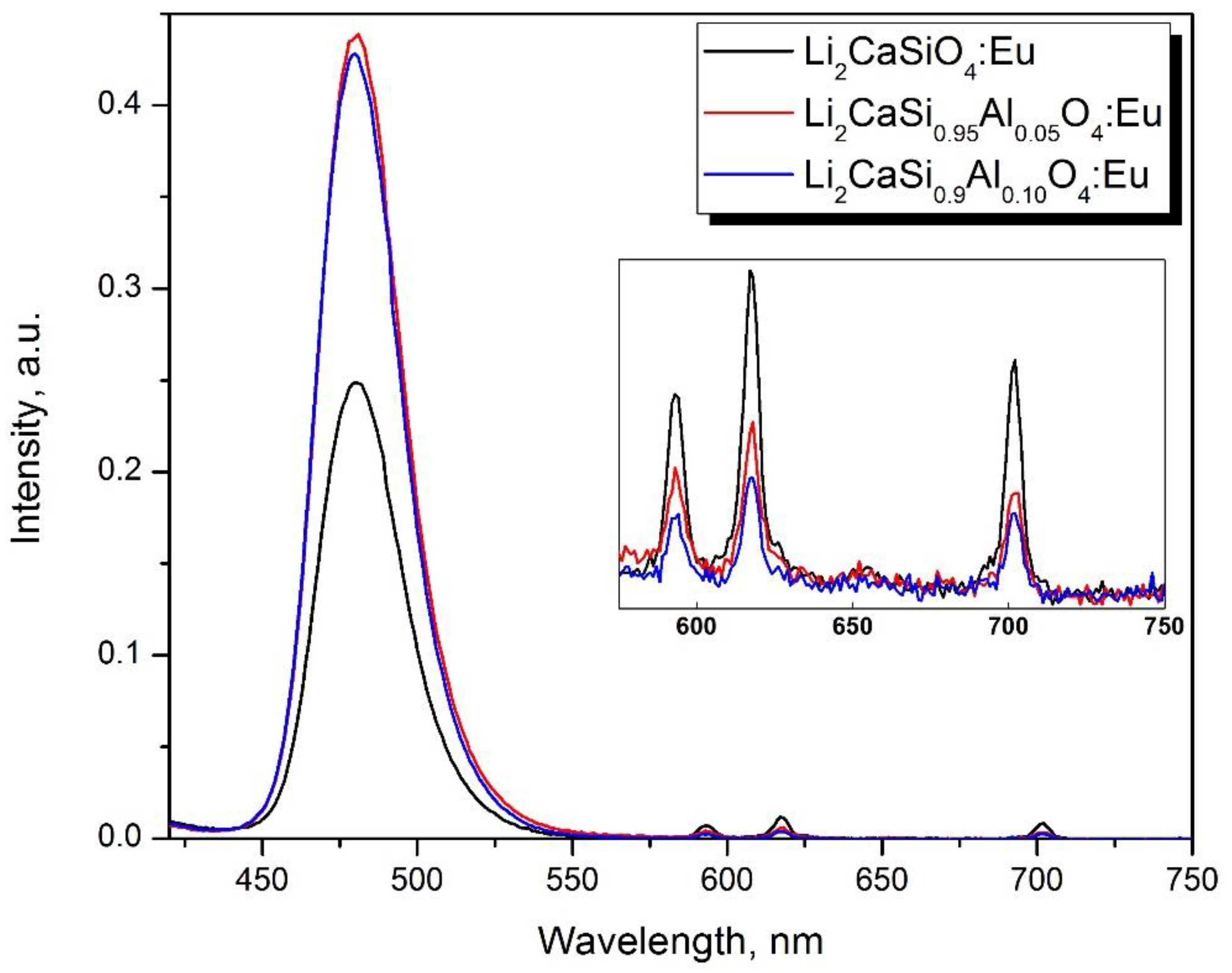
  179. Zhong, J.; Zhao, W.; Lan, L.; Wang, J.; Chen, J.; Wang, N. Enhanced Emission from Li2CaSiO4:Eu2+ Phosphors by Doping with Y3+. J. Alloys Compd. 2014, 592, 213–219. [Google Scholar] [CrossRef]
  180. Zhong, J.; Zhao, W.; Lan, L.; Wang, J. Strong Luminescence Enhancement of Li2CaSiO4:Eu2+ Phosphors by Codoping with La3+. J. Mater. Sci. Mater. Electron. 2014, 25, 736–741. [Google Scholar] [CrossRef]
  181. Yin, L.-J.; Ji, W.-W.; Liu, S.-Y.; He, W.-D.; Zhao, L.; Xu, X.; Fabre, A.; Dierre, B.; Lee, M.-H.; van Ommen, J.R.; et al. Intriguing Luminescence Properties of (Ba, Sr)3Si6O9N4:Eu2+ Phosphors via Modifying Synthesis Method and Cation Substitution. J. Alloys Compd. 2016, 682, 481–488. [Google Scholar] [CrossRef] [Green Version]
  182. Birowosuto, M.D.; Dorenbos, P.; Krämer, K.W.; Güdel, H.U. Ce3+ Activated LaBr3−xIx: High-Light-Yield and Fast-Response Mixed Halide Scintillators. J. Appl. Phys. 2008, 103, 103517. [Google Scholar] [CrossRef]
  183. Dorenbos, P.; Birowosuto, M.D.; Kraemer, K.W.; Guedel, H.-U. Scintillator Based on Lanthanum Iodide and Lanthanum Bromide. U.S. Patent 20100224798A1, 09 September 2010. [Google Scholar]
  184. Glodo, J.; van Loef, E.V.D.; Higgins, W.M.; Shah, K.S. Mixed Lutetium Iodide Compounds. IEEE Trans. Nucl. Sci. 2008, 55, 1496–1500. [Google Scholar] [CrossRef]
  185. Eagleman, Y.D.; Bourret-Courchesne, E.; Derenzo, S.E. Room-Temperature Scintillation Properties of Cerium-Doped REOX (RE=Y, La, Gd, and Lu; X = F, Cl, Br, and I). J. Lumin. 2011, 131, 669–675. [Google Scholar] [CrossRef] [Green Version]
  186. Wojtowicz, A.; Szupryczynski, P.; Glodo, J.; Drozdowski, W.; Wisniewski, D. Radioluminescence and recombination process in BaF2:Ce. J. Phys. Condens. Matter 2000, 12, 4097. [Google Scholar] [CrossRef]
  187. Luo, J.; Sahi, S.; Groza, M.; Wang, Z.; Ma, L.; Chen, W.; Burger, A.; Kenarangui, R.; Sham, T.-K.; Selim, F.A. Luminescence and scintillation properties of BaF2Ce transparent ceramics. Opt. Mater. 2016, 58, 353–356. [Google Scholar] [CrossRef] [Green Version]
  188. Rodnyĭ, P.A.; Gain, S.D.; Mironov, I.A.; Garibin, E.A.; Demidenko, A.A.; Seliverstov, D.M.; Kuznetsov, S.V. Spectral-kinetic characteristics of crystals and nanoceramics based on BaF2 and BaF2: Ce. Phys. Solid State 2010, 52, 1910–1914. [Google Scholar] [CrossRef]
  189. Auffray, E.; Baccaro, S.; Beckers, T.; Benhammou, Y.; Belsky, A.; Borgia, B.; Boutet, D.; Chipaux, R.; Dafinei, I.; de Notaristefani, F.; et al. Extensive studies on CeF3 crystals, a good candidate for electromagnetic calorimetry at future accelerators. Nucl. Instrum. Methods Phys. Res. Sect. A 1996, 388, 367–390. [Google Scholar] [CrossRef] [Green Version]
  190. Burger, A.; Rowe, E.; Groza, M.; Figueroa, K.M.; Cherepy, N.J.; Beck, P.R.; Hunter, S.; Payne, S.A. Cesium hafnium chloride: A high light yield, non-hygroscopic cubic crystal scintillator for gamma spectroscopy. Appl. Phys. Lett. 2015, 107, 143505. [Google Scholar] [CrossRef]
  191. Hawrami, R.; Ariesanti, E.; Buliga, V.; Matei, L.; Motakef, S.; Burger, A. Advanced high-performance large diameter Cs2HfCl6 (CHC) and mixed halides scintillator. J. Cryst. Growth 2020, 533, 125473. [Google Scholar] [CrossRef] [Green Version]
  192. Nikl, M.; Yoshikawa, A.; Kamada, K.; Nejezchleb, K.; Stanek, C.R.; Mares, J.A.; Blazek, K. Development of LuAG-Based Scintillator Crystals–A Review. Prog. Cryst. Growth Charact. Mater. 2013, 59, 47–72. [Google Scholar] [CrossRef]
  193. Kamada, K.; Endo, T.; Tsutumi, K.; Yanagida, T.; Fujimoto, Y.; Fukabori, A.; Yoshikawa, A.; Pejchal, J.; Nikl, M. Composition Engineering in Cerium-Doped (Lu,Gd)3(Ga,Al)5O12 Single-Crystal Scintillators. Cryst. Growth Des. 2011, 11, 4484–4490. [Google Scholar] [CrossRef]
  194. Kamada, K.; Yanagida, T.; Endo, T.; Tsutumi, K.; Usuki, Y.; Nikl, M.; Fujimoto, Y.; Fukabori, A.; Yoshikawa, A. 2inch Diameter Single Crystal Growth and Scintillation Properties of Ce:Gd3Al2Ga3O12. J. Cryst. Growth 2012, 352, 88–90. [Google Scholar] [CrossRef]
  195. Nikl, M.; Yoshikawa, A. Recent R&D Trends in Inorganic Single-Crystal Scintillator Materials for Radiation Detection. Adv. Opt. Mater. 2015, 3, 463–481. [Google Scholar] [CrossRef]
  196. Kuwano, Y.; Suda, K.; Ishizawa, N.; Yamada, T. Crystal Growth and Properties of (Lu,Y)3Al5O12. J. Cryst. Growth 2004, 260, 159–165. [Google Scholar] [CrossRef]
  197. Yanagida, T.; Itoh, T.; Takahashi, H.; Hirakuri, S.; Kokubun, M.; Makishima, K.; Sato, M.; Enoto, T.; Yanagitani, T.; Yagi, H.; et al. Improvement of Ceramic YAG(Ce) Scintillators to (YGd)3Al5O12(Ce) for Gamma-Ray Detectors. Nucl. Instrum. Methods A 2007, 579, 23–26. [Google Scholar] [CrossRef]
  198. Chewpraditkul, W.; Wantong, K.; Chewpraditkul, W.; Yawai, N.; Kamada, K.; Yoshikawa, A.; Witkowski, M.E.; Makowski, M.; Drozdowski, W.; Nikl, M. Scintillation Yield and Temperature Dependence of Radioluminescence of (Lu,Gd)3Al5O12:Ce Garnet Crystals. Opt. Mater. 2021, 120, 111471. [Google Scholar] [CrossRef]
  199. Korzhik, M.; Borisevich, A.; Fedorov, A.; Gordienko, E.; Karpyuk, P.; Dubov, V.; Sokolov, P.; Mikhlin, A.; Dosovitskiy, G.; Mechninsky, V.; et al. The Scintillation Mechanisms in Ce and Tb Doped (GdxY1-x)Al2Ga3O12 Quaternary Garnet Structure Crystalline Ceramics. J. Lumin. 2021, 234, 117933. [Google Scholar] [CrossRef]
  200. Wegh, R.T.; Meijerink, A.; Lamminmäki, R.-J. Jorma Hölsä Extending Dieke’s Diagram. J. Lumin. 2000, 87–89, 1002–1004. [Google Scholar] [CrossRef]
  201. Retivov, V.; Dubov, V.; Kuznetsova, D.; Ismagulov, A.; Korzhik, M. Gd3+ Content Optimization for Mastering High Light Yield and Fast GdxAl2Ga3O12:Ce3+ Scintillation Ceramics. J. Rare Earths 2022, in press. [Google Scholar] [CrossRef]
  202. Dosovitskiy, G.; Dubov, V.; Karpyuk, P.; Volkov, P.; Tamulaitis, G.; Borisevich, A.; Vaitkevičius, A.; Prikhodko, K.; Kutuzov, L.; Svetogorov, R.; et al. Activator Segregation and Micro-Luminescence Properties in GAGG:Ce Ceramics. J. Lumin. 2021, 236, 118140. [Google Scholar] [CrossRef]
  203. Korzhik, M.; Alenkov, V.; Buzanov, O.; Dosovitskiy, G.; Fedorov, A.; Kozlov, D.; Mechinsky, V.; Nargelas, S.; Tamulaitis, G.; Vaitkevičius, A. Engineering of a New Single-Crystal Multi-Ionic Fast and High-Light-Yield Scintillation Material (Gd0.5–Y0.5)3Al2Ga3O12:Ce,Mg. CrystEngComm 2020, 22, 2502–2506. [Google Scholar] [CrossRef]
  204. Kamada, K.; Yoshikawa, A.; Endo, T.; Tsutsumi, K.; Shoji, Y.; Kurosawa, S.; Yokota, Y.; Prusa, P.; Nikl, M. Growth of 2-Inch Size Ce:Doped Lu2Gd1Al2Ga3O12 Single Crystal by the Czochralski Method and Their Scintillation Properties. J. Cryst. Growth 2015, 410, 14–17. [Google Scholar] [CrossRef] [Green Version]
  205. Witkiewicz-Lukaszek, S.; Gorbenko, V.; Zorenko, T.; Sidletskiy, O.; Gerasymov, I.; Fedorov, A.; Yoshikawa, A.; Mares, J.A.; Nikl, M.; Zorenko, Y. Development of Composite Scintillators Based on Single Crystalline Films and Crystals of Ce3+-Doped (Lu,Gd)3(Al,Ga)5O12 Mixed Garnet Compounds. Cryst. Growth Des. 2018, 18, 1834–1842. [Google Scholar] [CrossRef]
  206. Chewpraditkul, W.; Pattanaboonmee, N.; Chewpraditkul, W.; Szczesniak, T.; Moszynski, M.; Kamada, K.; Yoshikawa, A.; Kucerkova, R.; Nikl, M. Optical and Scintillation Properties of LuGd2Al2Ga3O12:Ce, Lu2GdAl2Ga3O12:Ce, and Lu2YAl2Ga3O12:Ce Single Crystals: A Comparative Study. Nucl. Instrum. Methods A 2021, 1004, 165381. [Google Scholar] [CrossRef]
  207. Korzhik, M.; Retivov, V.; Bondarau, A.; Dosovitskiy, G.; Dubov, V.; Kamenskikh, I.; Karpuk, P.; Kuznetsova, D.; Smyslova, V.; Mechinsky, V.; et al. Role of the Dilution of the Gd Sublattice in Forming the Scintillation Properties of Quaternary (Gd,Lu)3Al2Ga3O12: Ce Ceramics. Crystals 2022, 12, 1196. [Google Scholar] [CrossRef]
  208. Kuznetsova, D.; Dubov, V.; Bondarev, A.; Dosovitskiy, G.; Mechinsky, V.; Retivov, V.; Kucherov, O.; Saifutyarov, R.; Korzhik, M. Tailoring of the Gd-Y-Lu ratio in quintuple (Gd, Lu, Y)3Al2Ga3O12:Ce ceramics for better scintillation properties. J. Appl. Phys. 2022, 132, 203104. [Google Scholar] [CrossRef]
  209. Tsubota, Y.; Kaneko, J.H.; Higuchi, M.; Minagawa, M.; Ishibashi, H. Crystal Growth and Scintillation Properties of La0.2Ce0.05Gd1.75Si2O7 (La-GPS) Single Crystal with Triclinic Structure. Opt. Mater. 2014, 36, 665–669. [Google Scholar] [CrossRef]
  210. Murakami, R.; Kurosawa, S.; Yamane, H.; Horiai, T.; Shoji, Y.; Yokota, Y.; Yamaji, A.; Ohashi, Y.; Kamada, K.; Yoshikawa, A. Crystal Structure of Ce-Doped (La,Gd)2Si2O7 Grown by the Czochralski Process. J. Alloys Compd. 2018, 748, 404–410. [Google Scholar] [CrossRef]
  211. Lu, L.; Sun, M.; Wu, T.; Lu, Q.; Chen, B.; Huang, B. All-Inorganic Perovskite Nanocrystals: Next-Generation Scintillation Materials for High-Resolution X-Ray Imaging. Nanoscale Adv. 2022, 4, 680–696. [Google Scholar] [CrossRef]
  212. Pokorný, M.; Babin, V.; Beitlerová, A.; Jurek, K.; Polák, J.; Houžvička, J.; Pánek, D.; Parkman, T.; Vaněček, V.; Nikl, M. Gd-Admixed (Lu,Gd)AlO3 Single Crystals: Breakthrough in Heavy Perovskite Scintillators. NPG Asia Mater. 2021, 13, 1–9. [Google Scholar] [CrossRef]
  213. Brixner, L.H. New X-Ray Phosphors. Mater. Chem. Phys. 1987, 16, 253–281. [Google Scholar] [CrossRef]
  214. Naik, R.; Prashantha, S.; Nagabhushana, H.; Naik, Y.; Girish, K. Green Light Emitting Tb3+ Doped Phosphors-A Review. Mat. Sci. Res. India 2018, 15, 252–255. [Google Scholar] [CrossRef]
  215. Zorenko, Y.; Gorbenko, V.; Savchyn, V.; Zorenko, T.; Martin, T.; Douissard, P.-A.; Nikl, M.; Mares, J.A. Luminescent Properties and Energy Transfer Processes in Ce–Tb Doped Single Crystalline Film Screens of Lu-Based Silicate, Perovskite and Garnet Compounds. Radiat. Meas. 2013, 56, 415–419. [Google Scholar] [CrossRef]
  216. Langley, J.; Litz, M.; Russo, J.; Ray, W. Design of Alpha-Voltaic Power Source Using Americium-241 (241Am) and Diamond with a Power Density of 10 mW/cm3; US Army Research Laboratory Adelphi United States: Adelphi, MD, USA, 2017. [Google Scholar]
  217. Sheshin, E.P.; Kolodyazhnyj, A.Y.; Chadaev, N.N.; Getman, A.O.; Danilkin, M.I.; Ozol, D.I. Prototype of Cathodoluminescent Lamp for General Lighting Using Carbon Fiber Field Emission Cathode. J. Vac. Sci. Technol. B 2019, 37, 031213. [Google Scholar] [CrossRef]
  218. Korjik, M.; Brinkmann, K.-T.; Dosovitskiy, G.; Dormenev, V.; Fedorov, A.; Kozlov, D.; Mechinsky, V.; Zaunick, H.-G. Compact and Effective Detector of the Fast Neutrons on a Base of Ce-Doped Gd3Al2Ga3O12 Scintillation Crystal. IEEE Trans. Nucl. Sci. 2019, 66, 536–540. [Google Scholar] [CrossRef]
  219. Dosovitskiy, G.; Karpyuk, P.; Gordienko, E.; Kuznetsova, D.; Vashchenkova, E.; Volkov, P.; Retivov, V.; Dormenev, V.; Brinkmann, K.-T.; Zaunick, H.-G.; et al. Neutron Detection by Gd-Loaded Garnet Ceramic Scintillators. Radiat. Meas. 2019, 126, 106133–106137. [Google Scholar] [CrossRef]
  220. Korzhik, M.; Abashev, R.; Fedorov, A.; Dosovitskiy, G.; Gordienko, E.; Kamenskikh, I.; Kazlou, D.; Kuznecova, D.; Mechinsky, V.; Pustovarov, V.; et al. Towards Effective Indirect Radioisotope Energy Converters with Bright and Radiation Hard Scintillators of (Gd,Y)3Al2Ga3O12 Family. Nucl. Eng. Technol. 2022, 54, 2579–2585. [Google Scholar] [CrossRef]
Figure 1. 2D cross-sections of the spatial distribution of the pseudopotential u ( r ) : (a) Y3Al5O12 (red plane, E g = 7.01   eV ), Y3Ga5O12 (blue plane, E g = 5.46   eV ) and Y3(Al,Ga)5O12; (b) Lu3Al5O12 (red plane, E g = 7.61   eV ), Lu3Ga5O12 (blue plane, E g = 6.38   eV ) and Lu3(Al,Ga)5O12; (c) Lu3Al5O12 (red plane), Y3Al5O12 (blue plane) and (Lu,Y)3Al5O12 and (d) Lu3Ga5O12 (red plane), Y3Ga5O12 (blue plane) and (Lu,Y)3Ga5O12.
Nanomaterials 12 04295 g001
Figure 2. 2D cross-sections of the spatial distribution of the pseudopotential u ( r ) in Lu2SiO5 (blue plane, E g = 7.27   eV ), Y2SiO5 (red plane, E g = 7.38   eV ) and (L0.5-Y0.5)2SiO5.
Nanomaterials 12 04295 g002
Figure 3. A change in the decay constant of the initial stage of the kinetics (a decrease by a factor e) of the photoluminescence of the Gd1.495Lu1.495Ce0.015 Ga3Al2O12 upon excitation 2.78 eV (1) and 3.67 eV (2) in comparison to Gd2.985Ce0.015Ga3Al2O12 upon excitation 2.78 eV (3).
Nanomaterials 12 04295 g003
Figure 4. Percentage of absorbed neutrons in gadolinium metal of natural isotopic composition depending on the particle energy. The region of resonances is highlighted in color.
Nanomaterials 12 04295 g004
Figure 5. SEM images of GYAGG:Ce nanopowders calcined at 100 °C (a), 900 °C (b), 1000 °C (c), 1100 °C (d).
Nanomaterials 12 04295 g005
Figure 6. Photograph of as-printed samples (left) and of a polished sintered Yb:YAG ceramic sample after annealing (right). After [133].
Nanomaterials 12 04295 g006
Figure 7. Photograph of as-printed green bodies (ac) and sintered parts (df). After [140].
Nanomaterials 12 04295 g007
Figure 8. The mesh scintillator in the form of a cylinder obtained by the 3D stereolithography method from the GAGG:Ce powder with further annealing at a temperature of 1600 °C for 2 h. After [142].
Nanomaterials 12 04295 g008
Figure 9. SEM images of ceramic samples obtained in different sintering atmospheres and vacuum.
Nanomaterials 12 04295 g009
Figure 10. Images of ceramic samples GYAGG:Ce (a,c) and GYAGG:Tb (b,d) under natural light and under UV-light excitation (λ = 365 nm). The ceramic samples were double side polished.
Nanomaterials 12 04295 g010
Figure 11. Band structure and the density of states: (a) BaI2 [161]; (b) BaBr2 [162]; (c) BaCl2 [163] crystals.
Nanomaterials 12 04295 g011
Figure 12. Photoluminescence spectra of Li2CaSiO4 samples and samples with compositional disordering in Si-sublattice obtained by partial Al substitution registered at excitation wavelength of 395 nm.
Nanomaterials 12 04295 g012
Figure 13. Scanning electron microscopy image of Gd1.13Y1.72Tb0.15Al2Ga3O12 sample (a), this sample under excitation with 150 keV electron beam, current density 1A/ cm2 (b). After [220].
Nanomaterials 12 04295 g013
Table 1. The fraction of the luminescence of Eu2+ and Eu3+.
Composition.Eu2+%Eu3+%
Li2CaSiO4: Eu98.7 ± 0.11.3 ± 0.1
Li2CaSi0.95Al0.05O4: Eu99.5 ± 0.10.5 ± 0.1
Li2CaSi0.90Al0.10O4: Eu99.6 ± 0.10.4 ± 0.1
Publisher’s Note: MDPI stays neutral with regard to jurisdictional claims in published maps and institutional affiliations.

