
Graphene-Based Electrochemical Sensors for Psychoactive Drugs


Abstract
1. Introduction
2. Graphene-Based Electrochemical Drug Sensors and Biosensors
2.1. Depressants
2.2. Traditional and Semisynthetic Opioids
2.3. Tramadol
2.4. Methadone and Buprenorphine
2.5. Ketamine
2.6. Stimulants
2.7. Cocaine
2.8. Methamphetamine
3. Other 2D Materials as Electrochemical Drug Sensors
Characteristics and Applications of Other 2D Materials for Sensing Purposes

| Analyte | 2D Base Material | Sensing Materials | Limit of Detection | Ref. |
|---|---|---|---|---|
| Dopamine | Graphene | G-C3N4/CuO | 0.1 nM | [100] |
| rGO-poly(Cu-AMT) | 3.48 nM | [125] | ||
| PEDOT/GO/in vivo carbon fiber | 85 nM | [126] | ||
| TMDs | MoSe2/Graphene | 1 nM | [127] | |
| MoS2 | 1 µM | [128] | ||
| MXenes | Ti3C2Tx/Pt-NPs | 6 nM | [129] | |
| Ti3C2Tx | 100 nM | [130] | ||
| h-BN | MIP/G-QDs/h-BN | 20 pM | [131] | |
| BP | BP | NA | [132] |
4. Comparison of Electrochemical Sensing and Other Conventional Drug-Detection Techniques
5. Conclusions and Outlook
Author Contributions
Funding
Conflicts of Interest
References
- Agius, R.; Kintz, P. Guidelines for European workplace drug and alcohol testing in hair. Drug Test. Anal. 2010, 2, 367–376. [Google Scholar] [CrossRef] [PubMed]
- Plumb, R.S.; Stumpf, C.L.; Granger, J.H.; Castro-Perez, J.; Haselden, J.N.; Dear, G.J. Use of liquid chromatography/time-of-flight mass spectrometry and multivariate statistical analysis shows promise for the detection of drug metabolites in biological fluids. Rapid Commun. Mass Spectrom. 2003, 17, 2632–2638. [Google Scholar] [CrossRef] [PubMed]
- Li, W.; Li, X.; Yang, T.; Guo, X.; Song, Y. Detection of saliva morphine using surface-enhanced Raman spectroscopy combined with immunochromatographic assay. J. Raman Spectrosc. 2020, 51, 642–648. [Google Scholar] [CrossRef]
- Paul, R.; Tsanaclis, L.; Murray, C.; Boroujerdi, R.; Facer, L.; Corbin, A. Ethyl Glucuronide as a Long-term Alcohol Biomarker in Fingernail and Hair. Matrix Comparison and Evaluation of Gender Bias. Alcohol Alcohol. 2019, 54, 402–407. [Google Scholar] [CrossRef]
- Peng, S.X. Hyphenated HPLC-NMR and its applications in drug discovery. Alcohol Alcohol. 2000, 14, 430–441. [Google Scholar] [CrossRef]
- Ryu, J.; Manivannan, R.; Son, Y.-A. Azo dye-based optical probe for the detection toward mimic molecule of date rape drug. Microchem. J. 2021, 166, 106237. [Google Scholar] [CrossRef]
- McCrae, K.; Tobias, S.; Grant, C.; Lysyshyn, M.; Laing, R.; Wood, E.; Ti, L. Assessing the limit of detection of Fourier-transform infrared spectroscopy and immunoassay strips for fentanyl in a real-world setting. Drug Alcohol Rev. 2020, 39, 98–102. [Google Scholar] [CrossRef]
- Wang, H.; Xue, Z.; Wu, Y.; Gilmore, J.; Wang, L.; Fabris, L. Rapid SERS quantification of trace fentanyl laced in recreational drugs with a portable Raman module. Anal. Chem. 2021, 93, 9373–9382. [Google Scholar] [CrossRef]
- Uchiyama, N.; Kikura-Hanajiri, R.; Shoda, T.; Fukuhara, K.; Goda, Y. Isomeric analysis of synthetic cannabinoids detected as designer drugs. J. Pharm. Soc. Jpn. 2011, 131, 1141–1147. [Google Scholar] [CrossRef]
- Ding, Y.; Li, X.; Guo, Y.; Yan, J.; Ling, J.; Li, W.; Lan, L.; Chang, Y.; Cai, J.; Zha, L.J.A.; et al. Rapid and sensitive detection of ketamine in blood using novel fluorescence genosensor. Anal. Bioanal. Chem. 2017, 409, 7027–7034. [Google Scholar] [CrossRef]
- Cinti, S.; Basso, M.; Moscone, D.; Arduini, F. A paper-based nanomodified electrochemical biosensor for ethanol detection in beers. Anal. Chim. Acta 2017, 960, 123–130. [Google Scholar] [CrossRef] [PubMed]
- Zheng, D.; Hu, H.; Liu, X.; Hu, S. Application of graphene in elctrochemical sensing. Curr. Opin. Colloid Interface Sci. 2015, 20, 383–405. [Google Scholar] [CrossRef]
- Nodehi, M.; Baghayeri, M.; Veisi, H.J.T. Preparation of GO/Fe3O4@PMDA/AuNPs nanocomposite for simultaneous determination of As3+ and Cu2+ by stripping voltammetry. Talanta 2021, 230, 122288. [Google Scholar] [CrossRef] [PubMed]
- Boroujerdi, R.; Abdelkader, A.; Paul, R. State of the Art in Alcohol Sensing with 2D Materials. Nano-Micro Lett. 2020, 12, 33. [Google Scholar] [CrossRef] [PubMed]
- Akça, A.; Karaman, O.; Karaman, C. Mechanistic insights into catalytic reduction of N2O by CO over Cu-embedded graphene: A density functional theory perspective. ECS J. Solid State Sci. Technol. 2021, 10, 041003. [Google Scholar] [CrossRef]
- Karaman, C.; Karaman, O.; Atar, N.; Yola, M. Tailoring of cobalt phosphide anchored nitrogen and sulfur co-doped three dimensional graphene hybrid: Boosted electrocatalytic performance towards hydrogen evolution reaction. Electrochim. Acta 2021, 380, 138262. [Google Scholar] [CrossRef]
- Karaman, C.; Karaman, O.; Atar, N.; Yola, M.L. Electrochemical immunosensor development based on core-shell high-crystalline graphitic carbon nitride@ carbon dots and Cd0.5Zn0.5S/d-Ti3C2TxMXene composite for heart-type fatty acid–binding protein detection. Microchim. Acta 2021, 188, 182. [Google Scholar] [CrossRef]
- Boroujerdi, R.; Paul, R. Introducing Graphene–Indium Oxide Electrochemical Sensor for Detecting Ethanol in Aqueous Samples with CCD-RSM Optimization. Chemosensors 2022, 10, 42. [Google Scholar] [CrossRef]
- Boroujerdi, R.; Abdelkader, A.; Paul, R. Highly selective detection of ethanol in biological fluids and alcoholic drinks using indium ethylenediamine functionalized graphene. Sens. Diagn. 2022, 1, 566–578. [Google Scholar] [CrossRef]
- Shao, Y.; Wang, J.; Wu, H.; Liu, J.; Aksay, I.A.; Lin, Y. Graphene based electrochemical sensors and biosensors: A review. Electroanalysis 2010, 22, 1027–1036. [Google Scholar] [CrossRef]
- Sun, D.; Luo, Y.; Debliquy, M.; Zhang, C. Graphene-enhanced metal oxide gas sensors at room temperature: A review. Beilstein J. Nanotechnol. 2018, 9, 2832–2844. [Google Scholar] [CrossRef] [PubMed]
- Jha, S.K.; Hayashi, K.; Yadava, R.D. Drugs of abuse and their detection methodologies: Contribution of chemical sensor. Curr. Org. Chem. 2015, 19, 1191–1201. [Google Scholar] [CrossRef]
- Majeed, M.I.; Nawaz, H.; Arshad, F.N. Detecting the Presence of Illicit Drugs Using Biosensors. Nanobiosensors 2020, 223–253. [Google Scholar]
- Stein, C. Opioid Receptors. Annu. Rev. Med. 2016, 67, 433–451. [Google Scholar] [CrossRef] [PubMed]
- Gomes, I.; Jordan, B.; Gupta, A.; Trapaidze, N.; Nagy, V.; Devi, L.A. Heterodimerization of μ and δ opioid receptors: A role in opiate synergy. J. Neurosci. 2000, 20, RC110. [Google Scholar] [PubMed]
- Armentia, A.; Ruiz-Munoz, P.; Quesada, J.M.; Postigo, I.; Herrero, M.; Martín-Gil, F.; Gonzalez-Sagrado, M.; Martín, B.; Castrodeza, J. Clinical value of morphine, pholcodine and poppy seed IgE assays in drug-abusers and allergic people. Allergol. Et Immunopathol. 2013, 41, 37–44. [Google Scholar] [CrossRef]
- Atta, N.F.; Galal, A.; Abdel-Gawad, F.M.; Mohamed, E.F. Electrochemical morphine sensor based on gold nanoparticles metalphthalocyanine modified carbon paste electrode. Electroanalysis 2015, 27, 415–428. [Google Scholar] [CrossRef]
- Abd-Rabboh, H.S.; Amr, A.E.-G.E.; Elsayed, E.A.; Sayed, A.Y.; Kamel, A.H. Paper-based potentiometric sensing devices modified with chemically reduced graphene oxide (CRGO) for trace level determination of pholcodine (opiate derivative drug). RSC Adv. 2021, 11, 12227–12234. [Google Scholar] [CrossRef]
- Santos, V.; López, K.J.V.; Santos, L.M.; Yonamine, M.; Carmona, M.J.C.; Santos, S.R. Determining plasma morphine levels using GC-MS after solid phase extraction to monitor drug levels in the postoperative period. Clinics 2008, 63, 307–314. [Google Scholar] [CrossRef]
- Norouzi, P.; Ganjali, M.R.; Moosavi-Movahedi, A.A.; Larijani, B. Fast Fourier transformation with continuous cyclic voltammetry at an Au microelectrode for the determination of morphine in a flow injection system. Talanta 2007, 73, 54–61. [Google Scholar] [CrossRef][Green Version]
- Pejić, N.D.; Blagojević, S.M.; Anić, S.R.; Vukojević, V.B.; Mijatović, M.D.; Ćirić, J.S.; Marković, Z.S.; Marković, S.D.; Kolar-Anić, L.Z. Kinetic determination of morphine by means of Bray–Liebhafsky oscillatory reaction system using analyte pulse perturbation technique. Anal. Chim. Acta 2007, 582, 367–374. [Google Scholar] [CrossRef] [PubMed]
- Abraham, P.; Renjini, S.; Vijayan, P.; Nisha, V.; Sreevalsan, K.; Anithakumary, V. Review on the progress in electrochemical detection of morphine based on different modified electrodes. J. Electrochem. Soc. 2020, 167, 037559. [Google Scholar] [CrossRef]
- Navaee, A.; Salimi, A.; Teymourian, H. Graphene nanosheets modified glassy carbon electrode for simultaneous detection of heroine, morphine and noscapine. Biosens. Bioelectron. 2012, 31, 205–211. [Google Scholar] [CrossRef]
- Mohamed, M.A.; El-Gendy, D.M.; Ahmed, N.; Banks, C.E.; Allam, N.K. 3D spongy graphene-modified screen-printed sensors for the voltammetric determination of the narcotic drug codeine. Biosens. Bioelectron. 2018, 101, 90–95. [Google Scholar] [CrossRef] [PubMed]
- Li, Y.; Li, K.; Song, G.; Liu, J.; Zhang, K.; Ye, B.J.S.; Chemical, A.B. Electrochemical behavior of codeine and its sensitive determination on graphene-based modified electrode. Sens. Actuators B Chem. 2013, 182, 401–407. [Google Scholar] [CrossRef]
- Atta, N.F.; Hassan, H.K.; Galal, A. Rapid and simple electrochemical detection of morphine on graphene–palladium-hybrid-modified glassy carbon electrode. Anal. Bioanal. Chem. 2014, 406, 6933–6942. [Google Scholar] [CrossRef]
- Li, Y.; Zou, L.; Li, Y.; Li, K.; Ye, B. A new voltammetric sensor for morphine detection based on electrochemically reduced MWNTs-doped graphene oxide composite film. Sens. Actuators B Chem. 2014, 201, 511–519. [Google Scholar] [CrossRef]
- Bagheri, H.; Khoshsafar, H.; Afkhami, A.; Amidi, S. Sensitive and simple simultaneous determination of morphine and codeine using a Zn2SnO4 nanoparticle/graphene composite modified electrochemical sensor. New J. Chem. 2016, 40, 7102–7112. [Google Scholar] [CrossRef]
- Beitollahi, H.; Nejad, F.G. Magnetic Core–shell Graphene Oxide/Fe3O4@SiO2 Nanocomposite for Sensitive and Selective Electrochemical Detection of Morphine using Modified Graphite Screen Printed Electrode. J. Anal. Chem. 2020, 75, 127–134. [Google Scholar] [CrossRef]
- Afkhami, A.; Khoshsafar, H.; Bagheri, H.; Madrakian, T. Facile simultaneous electrochemical determination of codeine and acetaminophen in pharmaceutical samples and biological fluids by graphene-CoFe2O4 nancomposite modified carbon paste electrode. Sens. Actuators B Chem. 2014, 203, 909–918. [Google Scholar] [CrossRef]
- Atta, N.F.; Galal, A.; El-Ads, E.H.; Hassan, S.H. Cobalt oxide nanoparticles/graphene/ionic liquid crystal modified carbon paste electrochemical sensor for ultra-sensitive determination of a narcotic drug. Adv. Pharm. Bull. 2019, 9, 110. [Google Scholar] [CrossRef] [PubMed]
- Abraham, P.; Renjini, S.; Nancy, T.M.; Kumary, V.A. Electrochemical synthesis of thin-layered graphene oxide-poly (CTAB) composite for detection of morphine. J. Appl. Electrochem. 2020, 50, 41–50. [Google Scholar] [CrossRef]
- Eissa, S.; Zourob, M. Competitive voltammetric morphine immunosensor using a gold nanoparticle decorated graphene electrode. Microchim. Acta 2017, 184, 2281–2289. [Google Scholar] [CrossRef]
- Kumary, V.A.; Abraham, P.; Renjini, S.; Swamy, B.K.; Nancy, T.M.; Sreevalsan, A. A novel heterogeneous catalyst based on reduced graphene oxide supported copper coordinated amino acid—A platform for morphine sensing. J. Electroanal. Chem. 2019, 850, 113367. [Google Scholar] [CrossRef]
- Prakash, V.; Garima; Prabhakar, N.; Kaur, G.; Diwan, A.; Mehta, S.K.; Sharma, S. Structural insight on thiourea doped graphene: An efficient electrochemical sensor for voltammetric detection of morphine in alcoholic and non-alcoholic beverages. Curr. Res. Green Sustain. Chem. 2022, 5, 100267. [Google Scholar] [CrossRef]
- Lucena, F.; de Oliveira, D.M.C.; Noroes, M.M.; Mujica, E.M.; Melleu, F.F.; Benedet, P.d.O.; Stein, T.; Ribeiro, L.F.; Tonussi, C.R. Intrathecally injected tramadol reduces articular incapacitation and edema in a rat model of lipopolysaccharide (LPS)-induced reactive arthritis. Life Sci. 2019, 236, 116860. [Google Scholar] [CrossRef]
- Jahromi, Z.; Mirzaei, E.; Savardashtaki, A.; Afzali, M.; Afzali, Z. A rapid and selective electrochemical sensor based on electrospun carbon nanofibers for tramadol detection. Microchem. J. 2020, 157, 104942. [Google Scholar] [CrossRef]
- Hassamal, S.; Miotto, K.; Dale, W.; Danovitch, I. Tramadol: Understanding the risk of serotonin syndrome and seizures. Am. J. Med. 2018, 131, 1382.e1–1382.e6. [Google Scholar] [CrossRef]
- Diouf, A.; Aghoutane, Y.; Burhan, H.; Sen, F.; Bouchikhi, B.; El Bari, N. Tramadol sensing in non-invasive biological fluids using a voltammetric electronic tongue and an electrochemical sensor based on biomimetic recognition. Int. J. Pharm. 2021, 593, 120114. [Google Scholar] [CrossRef]
- Soleimani, M.; Afshar, M.G.; Shafaat, A.; Crespo, G.A. High-Selective Tramadol Sensor Based on Modified Molecularly Imprinted Polymer Carbon Paste Electrode with Multiwalled Carbon Nanotubes. Electroanalysis 2013, 25, 1159–1168. [Google Scholar] [CrossRef]
- Bloor, M.; Paech, M.; Kaye, R. Tramadol in pregnancy and lactation. Int. J. Obstet. Anesth. 2012, 21, 163–167. [Google Scholar] [CrossRef] [PubMed]
- Cobianchi, M.R. Study of codeine-paracetamol combination treatment compared with tramadol-paracetamol in the control of moderate-to-severe low back pain. Minerva Med. 2012, 103, 177–182. [Google Scholar]
- Hallinan, R.; Ray, J.; Byrne, A.; Agho, K.; Attia, J. Therapeutic thresholds in methadone maintenance treatment: A receiver operating characteristic analysis. Drug Alcohol. Depend. 2006, 81, 129–136. [Google Scholar] [CrossRef] [PubMed]
- Xia, W.; Liu, G.; Shao, Z.; Xu, E.; Yuan, H.; Liu, J.; Gao, L. Toxicology of tramadol following chronic exposure based on metabolomics of the cerebrum in mice. Sci. Rep. 2020, 10, 11130. [Google Scholar] [CrossRef]
- Al Samarrai, S.Y.; Abdoon, F.M.; Hashim, K.K. A simple method to determine tramadol using a coated-wire electrode as a detector in the flow injection analysis. Microchem. J. 2019, 146, 588–591. [Google Scholar] [CrossRef]
- Mohamed, M.A.; Atty, S.A.; Salama, N.N.; Banks, C.E. Highly selective sensing platform utilizing graphene oxide and multiwalled carbon nanotubes for the sensitive determination of tramadol in the presence of co-formulated drugs. Electroanalysis 2017, 29, 1038–1048. [Google Scholar] [CrossRef]
- Afkhami, A.; Khoshsafar, H.; Bagheri, H.; Madrakian, T. Preparation of NiFe2O4/graphene nanocomposite and its application as a modifier for the fabrication of an electrochemical sensor for the simultaneous determination of tramadol and acetaminophen. Anal. Chim. Acta 2014, 831, 50–59. [Google Scholar] [CrossRef]
- Rokhsefid, N.; Shishehbore, M.R. Synthesis and characterization of an Au nanoparticles/graphene nanosheet nanocomposite and its application for the simultaneous determination of tramadol and acetaminophen. Anal. Methods 2019, 11, 5150–5159. [Google Scholar] [CrossRef]
- Aflatoonian, M.R.; Tajik, S.; Aflatoonian, B.; Beitollahi, H.; Zhang, K.; Le, Q.V.; Cha, J.H.; Jang, H.W.; Shokouhimehr, M.; Peng, W. A Screen-Printed Electrode Modified With Graphene/Co3O4 Nanocomposite for Electrochemical Detection of Tramadol. Front. Chem. 2020, 8, 1065. [Google Scholar] [CrossRef]
- Bagheri, H.; Shirzadmehr, A.; Rezaei, M.; Khoshsafar, H. Determination of tramadol in pharmaceutical products and biological samples using a new nanocomposite carbon paste sensor based on decorated nanographene/tramadol-imprinted polymer nanoparticles/ionic liquid. Ionics 2018, 24, 833–843. [Google Scholar] [CrossRef]
- Sanati, A.L.; Faridbod, F.J.A.; Electrochemistry, B. A New Nanostructure Approach based on Pr (OH)3/GQD and Imidazolium Ionic Liquid for Voltammetric Analysis of Tramadol. Anal. Bioanal. Electrochem. 2022, 14, 510–522. [Google Scholar]
- Amiri-Aref, M.; Raoof, J.B.; Ojani, R.J.C.; Biointerfaces, S.B. Electrocatalytic oxidation and selective determination of an opioid analgesic methadone in the presence of acetaminophen at a glassy carbon electrode modified with functionalized multi-walled carbon nanotubes: Application for human urine, saliva and pharmaceutical samples analysis. Colloids Surf. B Biointerfaces 2013, 109, 287–293. [Google Scholar] [PubMed]
- Ottomanelli, G. Methadone patients and alcohol abuse. J. Subst. Abus. Treat. 1999, 16, 113–121. [Google Scholar] [CrossRef]
- Alipour, E.; Majidi, M.R.; Hoseindokht, O. Development of Simple Electrochemical Sensor for Selective Determination of Methadone in Biological Samples Using Multi-walled Carbon Nanotubes Modified Pencil Graphite Electrode. J. Chin. Chem. Soc. 2015, 62, 461–468. [Google Scholar] [CrossRef]
- Baghayeri, M.; Nabavi, S.; Hasheminejad, E.; Ebrahimi, V. Introducing an electrochemical sensor based on two layers of Ag nanoparticles decorated graphene for rapid determination of methadone in human blood serum. Top. Catal. 2022, 65, 623–632. [Google Scholar] [CrossRef]
- Nazari, Z.; Es’haghi, Z. A New Electrochemical Sensor for the Simultaneous Detection of Morphine and Methadone based on Thioglycolic Acid Decorated CdSe Doped Graphene Oxide Multilayers. Anal. Bioanal. Electrochem. 2022, 14, 228–245. [Google Scholar]
- Fakhari, A.R.; Sahragard, A.; Ahmar, H. Development of an Electrochemical Sensor Based on Reduced Graphene Oxide Modified Screen-Printed Carbon Electrode for the Determination of Buprenorphine. Electroanalysis 2014, 26, 2474–2483. [Google Scholar] [CrossRef]
- Alizadeh, T.; Atashi, F.; Akhoundian, M.; Ganjali, M.R. Highly selective extraction and voltammetric determination of the opioid drug buprenorphine via a carbon paste electrode impregnated with nano-sized molecularly imprinted polymer. Microchim. Acta 2019, 186, 654. [Google Scholar] [CrossRef]
- White, P.F.; Way, W.L.; Trevor, A.J. Ketamine—Its pharmacology and therapeutic uses. Anesthesiology 1982, 56, 119–136. [Google Scholar] [CrossRef]
- Bolze, S.; Boulieu, R. HPLC determination of ketamine, norketamine, and dehydronorketamine in plasma with a high-purity reversed-phase sorbent. Clin. Chem. 1998, 44, 560–564. [Google Scholar] [CrossRef]
- Giné, C.V.; Vilamala, M.V.; Espinosa, I.F.; Lladanosa, C.G.; Álvarez, N.C.; Fruitós, A.F.; Rodríguez, J.R.; Salvany, A.D.; de la Torre Fornell, R. Crystals and tablets in the Spanish ecstasy market 2000–2014: Are they the same or different in terms of purity and adulteration? Forensic Sci. Int. 2016, 263, 164–168. [Google Scholar] [CrossRef] [PubMed]
- Gjerde, H.; Nordfjærn, T.; Bretteville-Jensen, A.L.; Edland-Gryt, M.; Furuhaugen, H.; Karinen, R.; Øiestad, E.L. Comparison of drugs used by nightclub patrons and criminal offenders in Oslo, Norway. Forensic. Sci. Int. 2016, 265, 1–5. [Google Scholar] [CrossRef] [PubMed]
- Demoranville, L.T.; Verkouteren, J.R. Measurement of drug facilitated sexual assault agents in simulated sweat by ion mobility spectrometry. Talanta 2013, 106, 375–380. [Google Scholar] [CrossRef] [PubMed]
- Yang, Y.; Zhai, S.; Liu, C.; Wang, X.; Tu, Y. Disposable immunosensor based on electrochemiluminescence for ultrasensitive detection of ketamine in human hair. ACS Omega 2019, 4, 801–809. [Google Scholar] [CrossRef]
- Narang, J.; Malhotra, N.; Singhal, C.; Mathur, A.; Chakraborty, D.; Anil, A.; Ingle, A.; Pundir, C.S. Point of care with micro fluidic paper based device integrated with nano zeolite–graphene oxide nanoflakes for electrochemical sensing of ketamine. Biosens. Bioelectron. 2017, 88, 249–257. [Google Scholar] [CrossRef] [PubMed]
- Fu, K.; Zhang, R.; He, J.; Bai, H.; Zhang, G. Sensitive detection of ketamine with an electrochemical sensor based on UV-induced polymerized molecularly imprinted membranes at graphene and MOFs modified electrode. Biosens. Bioelectron. 2019, 143, 111636. [Google Scholar] [CrossRef]
- Breiter, H.C.; Gollub, R.L.; Weisskoff, R.M.; Kennedy, D.N.; Makris, N.; Berke, J.D.; Goodman, J.M.; Kantor, H.L.; Gastfriend, D.R.; Riorden, J.P. Acute effects of cocaine on human brain activity and emotion. Neuron 1997, 19, 591–611. [Google Scholar] [CrossRef]
- Kroener, S.; Lavin, A.J.N. Altered dopamine modulation of inhibition in the prefrontal cortex of cocaine-sensitized rats. Neuropsychopharmacology 2010, 35, 2292–2304. [Google Scholar] [CrossRef]
- Ersche, K.D.; Jones, P.S.; Williams, G.B.; Turton, A.J.; Robbins, T.W.; Bullmore, E.T. Abnormal brain structure implicated in stimulant drug addiction. Science 2012, 335, 601–604. [Google Scholar] [CrossRef]
- Shi, Y.; Dai, H.; Sun, Y.; Hu, J.; Ni, P.; Li, Z. Fluorescent sensing of cocaine based on a structure switching aptamer, gold nanoparticles and graphene oxide. Analyst 2013, 138, 7152–7156. [Google Scholar] [CrossRef]
- Mendelson, J.H.; Mello, N.K. Management of cocaine abuse and dependence. N. Engl. J. Med. 1996, 334, 965–972. [Google Scholar] [CrossRef] [PubMed]
- Moore, C.; Negrusz, A.; Lewis, D. Determination of drugs of abuse in meconium. J. Chromatogr. B Biomed. Sci. Appl. 1998, 713, 137–146. [Google Scholar] [CrossRef]
- Dragan, A.-M.; Truta, F.M.; Tertis, M.; Florea, A.; Schram, J.; Cernat, A.; Feier, B.; De Wael, K.; Cristea, C.; Oprean, R. Electrochemical fingerprints of illicit drugs on graphene and multi-walled carbon nanotubes. Front. Chem. 2021, 9, 641147. [Google Scholar] [CrossRef]
- Qiu, L.; Zhou, H.; Zhu, W.; Qiu, L.; Jiang, J.; Shen, G.; Yu, R. A novel label-free fluorescence aptamer-based sensor method for cocaine detection based on isothermal circular strand-displacement amplification and graphene oxide absorption. New J. Chem. 2013, 37, 3998–4003. [Google Scholar] [CrossRef]
- Karlsson, M.; Strandqvist, C.; Jussi, J.; Öberg, O.; Petermann, I.; Elmlund, L.; Dunne, S.; Fu, Y.; Wang, Q. Chemical sensors generated on wafer-scale epitaxial graphene for application to front-line drug detection. Sensors 2019, 19, 2214. [Google Scholar] [CrossRef] [PubMed]
- Florea, A.; Cowen, T.; Piletsky, S.; De Wael, K. Electrochemical sensing of cocaine in real samples based on electrodeposited biomimetic affinity ligands. Analyst 2019, 144, 4639–4646. [Google Scholar] [CrossRef]
- Rocha, R.G.; Ribeiro, J.S.; Santana, M.H.; Richter, E.M.; Muñoz, R.A. 3D-printing for forensic chemistry: Voltammetric determination of cocaine on additively manufactured graphene–polylactic acid electrodes. Anal. Methods 2021, 13, 1788–1794. [Google Scholar] [CrossRef]
- Cardoso, R.M.; Mendonça, D.M.; Silva, W.P.; Silva, M.N.; Nossol, E.; da Silva, R.A.; Richter, E.M.; Muñoz, R.A. 3D printing for electroanalysis: From multiuse electrochemical cells to sensors. Anal. Chim. Acta 2018, 1033, 49–57. [Google Scholar] [CrossRef]
- De La Torre, R.; Yubero-Lahoz, S.; Pardo-Lozano, R.; Farré, M. MDMA, methamphetamine, and CYP2D6 pharmacogenetics: What is clinically relevant? Front. Genet. 2012, 3, 235. [Google Scholar] [CrossRef]
- Inoue, H.; Kuwayama, K.; Iwata, Y.T.; Kanamori, T.; Tsujikawa, K.; Miyaguchi, H. Simple and simultaneous detection of methamphetamine and dimethyl sulfone in crystalline methamphetamine seizures by fast gas chromatography. Forensic Toxicol. 2008, 26, 19–22. [Google Scholar] [CrossRef]
- Mandani, S.; Rezaei, B.; Ensafi, A.A. Sensitive imprinted optical sensor based on mesoporous structure and green nanoparticles for the detection of methamphetamine in plasma and urine. Spectrochim. Acta Part A: Mol. Biomol. Spectrosc. 2020, 231, 118077. [Google Scholar] [CrossRef] [PubMed]
- Khorablou, Z.; Shahdost-Fard, F.; Razmi, H.; Yola, M.L.; Karimi-Maleh, H. Recent advances in developing optical and electrochemical sensors for analysis of methamphetamine: A review. Chemosphere 2021, 278, 130393. [Google Scholar] [CrossRef] [PubMed]
- Saisahas, K.; Soleh, A.; Somsiri, S.; Senglan, P.; Promsuwan, K.; Saichanapan, J.; Kanatharana, P.; Thavarungkul, P.; Lee, K.; Chang, K.H. Electrochemical Sensor for Methamphetamine Detection Using Laser-Induced Porous Graphene Electrode. Nanomaterials 2022, 12, 73. [Google Scholar] [CrossRef] [PubMed]
- Riahifar, V.; Haghnazari, N.; Keshavarzi, F.; Ahmadi, E. A sensitive voltammetric sensor for methamphetamine determination based on modified glassy carbon electrode using Fe3O4@ poly pyrrole core-shell and graphene oxide. Microchem. J. 2021, 170, 106748. [Google Scholar] [CrossRef]
- Anvari, L.; Ghoreishi, S.M.; Faridbod, F.; Ganjali, M.R. Electrochemical Determination of Methamphetamine in Human Plasma on a Nanoceria Nanoparticle Decorated Reduced Graphene Oxide (rGO) Glassy Carbon Electrode (GCE). Anal. Lett. 2021, 54, 1–14. [Google Scholar] [CrossRef]
- Hashemi, P.; Bagheri, H.; Afkhami, A.; Ardakani, Y.H.; Madrakian, T. Fabrication of a novel aptasensor based on three-dimensional reduced graphene oxide/polyaniline/gold nanoparticle composite as a novel platform for high sensitive and specific cocaine detection. Anal. Chim. Acta 2017, 996, 10–19. [Google Scholar] [CrossRef]
- Jiang, B.; Wang, M.; Chen, Y.; Xie, J.; Xiang, Y. Highly sensitive electrochemical detection of cocaine on graphene/AuNP modified electrode via catalytic redox-recycling amplification. Biosens. Bioelectron. 2012, 32, 305–308. [Google Scholar] [CrossRef]
- Willuhn, I.; Burgeno, L.M.; Everitt, B.J.; Phillips, P.E.M. Hierarchical recruitment of phasic dopamine signaling in the striatum during the progression of cocaine use. Proc. Natl. Acad. Sci. USA 2012, 109, 20703–20708. [Google Scholar] [CrossRef]
- Reyes, S.; Fu, Y.; Double, K.L.; Cottam, V.; Thompson, L.H.; Kirik, D.; Paxinos, G.; Watson, C.; Cooper, H.M.; Halliday, G.M. Trophic factors differentiate dopamine neurons vulnerable to Parkinson’s disease. Neurobiol. Aging 2013, 34, 873–886. [Google Scholar] [CrossRef]
- Zou, J.; Wu, S.; Liu, Y.; Sun, Y.; Cao, Y.; Hsu, J.-P.; Shen Wee, A.T.; Jiang, J. An ultra-sensitive electrochemical sensor based on 2D g-C3N4/CuO nanocomposites for dopamine detection. Carbon 2018, 130, 652–663. [Google Scholar] [CrossRef]
- Jeong, S.; Yoo, D.; Ahn, M.; Miró, P.; Heine, T.; Cheon, J. Tandem intercalation strategy for single-layer nanosheets as an effective alternative to conventional exfoliation processes. Nat. Commun. 2015, 6, 5763. [Google Scholar] [CrossRef] [PubMed]
- Wang, Y.; Zhuang, Q.; Ni, Y. Fabrication of riboflavin electrochemical sensor based on homoadenine single-stranded DNA/molybdenum disulfide-graphene nanocomposite modified gold electrode. J. Electroanal. Chem. 2015, 736, 47–54. [Google Scholar] [CrossRef]
- Heine, T. Transition metal chalcogenides: Ultrathin inorganic materials with tunable electronic properties. Acc. Chem. Res. 2015, 48, 65–72. [Google Scholar] [CrossRef] [PubMed]
- Grayfer, E.D.; Kozlova, M.N.; Fedorov, V.E. Colloidal 2D nanosheets of MoS2 and other transition metal dichalcogenides through liquid-phase exfoliation. Adv. Colloid Interface Sci. 2017, 245, 40–61. [Google Scholar] [CrossRef]
- Hillebrecht, H.; Schmidt, P.; Rotter, H.; Thiele, G.; Zönnchen, P.; Bengel, H.; Cantow, H.-J.; Magonov, S.; Whangbo, M.-H. Structural and scanning microscopy studies of layered compounds mcl3 (m = mo, ru, cr) and mocl2 (m = v, nb, mo, ru, os). J. Alloys Compd. 1997, 246, 70–79. [Google Scholar] [CrossRef]
- Miró, P.; Audiffred, M.; Heine, T. An atlas of two-dimensional materials. Chem. Soc. Rev. 2014, 43, 6537–6554. [Google Scholar] [CrossRef]
- Tanwar, S.; Arya, A.; Gaur, A.; Sharma, A.L. Transition metal dichalcogenide (tmds) electrodes for supercapacitors: A comprehensive review. J. Phys. Condens. Matter. 2021, 33, 303002. [Google Scholar] [CrossRef]
- Naguib, M.; Mochalin, V.N.; Barsoum, M.W.; Gogotsi, Y. 25th anniversary article: MXenes: A new family of two-dimensional materials. Adv. Mater. 2014, 26, 992–1005. [Google Scholar] [CrossRef]
- Angizi, S.; Khalaj, M.; Alem, S.A.A.; Pakdel, A.; Willander, M.; Hatamie, A.; Simchi, A. Towards the two-dimensional hexagonal boron nitride (2D h-BN) electrochemical sensing platforms. J. Electrochem. Soc. 2020, 167, 126513. [Google Scholar] [CrossRef]
- Pumera, M. Phosphorene and black phosphorus for sensing and biosensing. TrAC Trends Anal. Chem. 2017, 93, 1–6. [Google Scholar] [CrossRef]
- Liu, H.; Neal, A.T.; Zhu, Z.; Luo, Z.; Xu, X.; Tománek, D.; Ye, P.D. Phosphorene: An unexplored 2D semiconductor with a high hole mobility. ACS Nano 2014, 8, 4033–4041. [Google Scholar] [CrossRef] [PubMed]
- Eswaraiah, V.; Zeng, Q.; Long, Y.; Liu, Z. Black phosphorus nanosheets: Synthesis, characterization and applications. Small 2016, 12, 3480–3502. [Google Scholar] [CrossRef] [PubMed]
- Akhtar, M.; Anderson, G.; Zhao, R.; Alruqi, A.; Mroczkowska, J.E.; Sumanasekera, G.; Jasinski, J.B. Recent Advances in Synthesis, Properties, and Applications of Phosphorene. npj 2D Mater. Appl. 2017, 1, 5. [Google Scholar] [CrossRef]
- Tran, V.; Soklaski, R.; Liang, Y.; Yang, L. Layer-controlled band gap and anisotropic excitons in few-layer black phosphorus. Phys. Rev. B Condens. Matter Mater. Phys. 2014, 89, 235319. [Google Scholar] [CrossRef]
- Takao, Y.; Morita, A. Electronic structure of black phosphorus: Tight binding approach. Physica B+C 1981, 105, 93–98. [Google Scholar] [CrossRef]
- Li, L.; Yu, Y.; Ye, G.J.; Ge, Q.; Ou, X.; Wu, H.; Feng, D.; Chen, X.H.; Zhang, Y. Black phosphorus field-effect transistors. Nat. Nanotechnol. 2014, 9, 372–377. [Google Scholar] [CrossRef] [PubMed]
- Koenig, S.P.; Doganov, R.A.; Schmidt, H.; Castro Neto, A.H.; Özyilmaz, B. Electric field effect in ultrathin black phosphorus. Appl. Phys. Lett. 2014, 104, 103106. [Google Scholar] [CrossRef]
- Li, L.; Ye, G.J.; Tran, V.; Fei, R.; Chen, G.; Wang, H.; Wang, J.; Watanabe, K.; Taniguchi, T.; Yang, L.; et al. Quantum oscillations in a two-dimensional electron gas in black phosphorus thin films. Nat. Nanotechnol. 2015, 10, 608–613. [Google Scholar] [CrossRef]
- Gillgren, N.; Wickramaratne, D.; Shi, Y.; Espiritu, T.; Yang, J.; Hu, J.; Wei, J.; Liu, X.; Mao, Z.; Watanabe, K.; et al. Gate tunable quantum oscillations in air-stable and high mobility few-layer phosphorene heterostructures. 2D Mater. 2015, 2, 011001. [Google Scholar] [CrossRef]
- Wang, H.; Yang, X.; Shao, W.; Chen, S.; Xie, J.; Zhang, X.; Wang, J.; Xie, Y. Ultrathin black phosphorus nanosheets for efficient singlet oxygen generation. J. Am. Chem. Soc. 2015, 137, 11376–11382. [Google Scholar] [CrossRef]
- Sun, Z.; Xie, H.; Tang, S.; Yu, X.F.; Guo, Z.; Shao, J.; Zhang, H.; Huang, H.; Wang, H.; Chu, P.K. Ultrasmall black phosphorus quantum dots: Synthesis and use as photothermal agents. Angew. Chem. Int. Ed. 2015, 127, 11688–11692. [Google Scholar] [CrossRef]
- Sun, C.; Wen, L.; Zeng, J.; Wang, Y.; Sun, Q.; Deng, L.; Zhao, C.; Li, Z. One-pot solventless preparation of PEGylated black phosphorus nanoparticles for photoacoustic imaging and photothermal therapy of cancer. Biomaterials 2016, 91, 81–89. [Google Scholar] [CrossRef] [PubMed]
- Latiff, N.M.; Teo, W.Z.; Sofer, Z.; Fisher, A.C.; Pumera, M. The cytotoxicity of layered black phosphorus. Chem.–A Eur. J. 2015, 21, 13991–13995. [Google Scholar] [CrossRef] [PubMed]
- Chen, Y.; Ren, R.; Pu, H.; Chang, J.; Mao, S.; Chen, J. Field-effect transistor biosensors with two-dimensional black phosphorus nanosheets. Biosens. Bioelectron. 2017, 89, 505–510. [Google Scholar] [CrossRef]
- Li, Y.; Gu, Y.; Zheng, B.; Luo, L.; Li, C.; Yan, X.; Zhang, T.; Lu, N.; Zhang, Z. A novel electrochemical biomimetic sensor based on poly(Cu-AMT) with reduced graphene oxide for ultrasensitive detection of dopamine. Talanta 2017, 162, 80–89. [Google Scholar] [CrossRef] [PubMed]
- Taylor, I.M.; Robbins, E.M.; Catt, K.A.; Cody, P.A.; Happe, C.L.; Cui, X.T. Enhanced dopamine detection sensitivity by PEDOT/graphene oxide coating on in vivo carbon fiber electrodes. Biosens. Bioelectron. 2017, 89, 400–410. [Google Scholar] [CrossRef]
- Huang, K.J.; Zhang, J.Z.; Cai, J.L. Preparation of porous layered molybdenum selenide-graphene composites on Ni foam for high-performance supercapacitor and electrochemical sensing. Electrochim. Acta 2015, 180, 770–777. [Google Scholar] [CrossRef]
- Zou, H.L.; Li, B.L.; Luo, H.Q.; Li, N.B. 0D-2D heterostructures of Au nanoparticles and layered MoS2 for simultaneous detections of dopamine, ascorbic acid, uric acid, and nitrite. Sens. Actuators B Chem. 2017, 253, 352–360. [Google Scholar] [CrossRef]
- Lorencová, L.; Bertok, T.; Filip, J.; Jerigová, M.; Velic, D.; Kasák, P.; Mahmoud, K.A.; Tkac, J.J.S.; Chemical, A.B. Highly stable Ti3C2Tx (MXene)/Pt nanoparticles-modified glassy carbon electrode for H2O2 and small molecules sensing applications. Sens. Actuators B Chem. 2018, 263, 360–368. [Google Scholar] [CrossRef]
- Xu, B.; Zhu, M.; Zhang, W.; Zhen, X.; Pei, Z.; Xue, Q.; Zhi, C.; Shi, P. Ultrathin MXene-Micropattern-Based Field-Effect Transistor for Probing Neural Activity. Adv. Mater. 2016, 28, 3333–3339. [Google Scholar] [CrossRef]
- Yola, M.L.; Atar, N. A novel detection approach for serotonin by graphene quantum dots/two-dimensional (2D) hexagonal boron nitride nanosheets with molecularly imprinted polymer. Appl. Surf. Sci. 2018, 458, 648–655. [Google Scholar] [CrossRef]
- Sofer, Z.; Sedmidubský, D.; Huber, Š.; Luxa, J.; Bouša, D.; Boothroyd, C.; Pumera, M. Layered Black Phosphorus: Strongly Anisotropic Magnetic, Electronic, and Electron-Transfer Properties. Angew. Chem. Int. Ed. 2016, 55, 3382–3386. [Google Scholar] [CrossRef] [PubMed]
- Al-Sanea, M.M.; Gamal, M. Critical analytical review: Rare and recent applications of refractive index detector in HPLC chromatographic drug analysis. Microchem. J. 2022, 178, 107339. [Google Scholar] [CrossRef]
- Nahar, L.; Onder, A.; Sarker, S.D. A review on the recent advances in HPLC, UHPLC and UPLC analyses of naturally occurring cannabinoids (2010–2019). Phytochem. Anal. 2020, 31, 413–457. [Google Scholar] [CrossRef] [PubMed]
- Tuzimski, T.; Petruczynik, A. Review of chromatographic methods coupled with modern detection techniques applied in the therapeutic drugs monitoring (TDM). Molecules 2020, 25, 4026. [Google Scholar] [CrossRef]
- Lindon, J.C.; Nicholson, J.K.; Wilson, I.D. The development and application of coupled HPLC-NMR spectroscopy. Adv. Chromatogr. 2021, 315–382. [Google Scholar] [CrossRef]
- Soni, S.; Jain, U.; Burke, D.H.; Chauhan, N. Recent trends and emerging strategies for aptasensing technologies for illicit drugs detection. J. Electroanal. Chem. 2022, 910, 116128. [Google Scholar] [CrossRef]
- Vaccaro, G.; Massariol, A.; Guirguis, A.; Kirton, S.B.; Stair, J.L. NPS detection in prison: A systematic literature review of use, drug form, and analytical approaches. Drug Test. Anal. 2022, 1–18. [Google Scholar] [CrossRef]
- Aslani, S.; Armstrong, D.W. High Information Spectroscopic Detection Techniques for Gas Chromatography. J. Chromatogr. A 2022, 463255. [Google Scholar] [CrossRef]
- Roberson, Z.R.; Goodpaster, J.V. Optimization of the qualitative and quantitative analysis of cocaine and other drugs of abuse via gas chromatography—Vacuum ultraviolet spectrophotometry (GC–VUV). Talanta 2021, 222, 121461. [Google Scholar] [CrossRef]
- Tamama, K. Advances in drugs of abuse testing. Clin. Chim. Acta 2021, 514, 40–47. [Google Scholar] [CrossRef] [PubMed]
- Ghorani-Azam, A.; Balali-Mood, M.; Khatami, S.-M.; Asoodeh, A.; Es’ haghi, Z.; Riahi-Zanjani, B. Plant extract and herbal products as potential source of sorbent for analytical purpose: An experimental study of morphine and codeine determination using HPLC and LC–MSMS. J. Chromatogr. Sci. 2021, 59, 482–489. [Google Scholar] [CrossRef] [PubMed]
- Özbunar, E.; Aydoğdu, M.; Döğer, R.; Bostancı, H.İ.; Koruyucu, M.; Akgür, S.A. Morphine Concentrations in Human Urine Following Poppy Seed Paste Consumption. Forensic Sci. Int. 2019, 295, 121–127. [Google Scholar] [CrossRef]
- Yoo, O.; Tang, E.K.Y.; Nguyen, M.N.; Salman, S.; Hua, A.J.; von Ungern Sternberg, B.S.; Lim, L.Y. HPLC-UV assay of tramadol and O-desmethyltramadol in human plasma containing other drugs potentially co-administered to participants in a paediatric population pharmacokinetic study. J. Chromatogr. B 2021, 1184, 122971. [Google Scholar] [CrossRef]
- Xu, F.; Liu, L. Simultaneous determination of free methamphetamine, pethidine, ketamine and tramadol in urine by dispersive liquid–liquid microextraction combined with GC–MS. Forensic Sci. Res. 2019, 4, 188–194. [Google Scholar] [CrossRef] [PubMed]
- Mercolini, L.; Mandrioli, R.; Saladini, B.; Conti, M.; Baccini, C.; Raggi, M.A. Quantitative analysis of cocaine in human hair by HPLC with fluorescence detection. J. Pharm. Biomed. Anal. 2008, 48, 456–461. [Google Scholar] [CrossRef]
- Romolo, F.S.; Rotolo, M.C.; Palmi, I.; Pacifici, R.; Lopez, A. Optimized conditions for simultaneous determination of opiates, cocaine and benzoylecgonine in hair samples by GC–MS. Forensic Sci. Int. 2003, 138, 17–26. [Google Scholar] [CrossRef]
- Loganathan, D.; Yi, R.; Patel, B.; Zhang, J.; Kong, N.J.A.; Chemistry, B. A sensitive HPLC-MS/MS method for the detection, resolution and quantitation of cathinone enantiomers in horse blood plasma and urine. Anal. Bioanal. Chem. 2021, 413, 2147–2161. [Google Scholar] [CrossRef]
- Alawi, A.I.; Dhabbah, A.M.; Morrison, C.; Ben-Jaber, S.; AlAngari, W.A.; Bin Jassas, M.; Badjah-Hadj-Ahmed, Y. Indirect chiral separation of crystal methamphetamine seized in Saudi Arabia using GC-MS. Aust. J. Forensic Sci. 2022, 1–14. [Google Scholar] [CrossRef]


| NO | Drug(s) Detected | Sensing Material | Main Method(s) | Detection Limit(s) | Sample(s) | Ref. |
|---|---|---|---|---|---|---|
| 1 | Morphine | Graphene oxide–Co3O4 | DPV/CV | 0.54 nM | Urine, Medical tablets | [41] |
| 2 | Morphine | Graphene–Pd NPs | DPV | 12.95 nM | Urine | [36] |
| 3 | Morphine | Graphene–Cu-poly(Ala) | DPV | 47 nM | Blood serum | [44] |
| 4 | Morphine | Graphene-MWCNTs | CV | 50 nM | Urine | [37] |
| 5 | Morphine | Graphene oxide–poly(CTAB) | DPV/CV | 360 nM | Blood serum, Urine | [42] |
| 6 | Morphine | Graphene oxide–Fe3O4@SiO2 | DPV/CV | 750 nM | Urine, Medical ampoule | [39] |
| 7 | Morphine | Graphene–Au NPs-MO antibody | SWV | 90 pg/mL | Saliva | [43] |
| 8 | Morphine | N-S-doped graphene | CV | 0.26 μg/mL | Soft drinks, Beer, Vodka | [45] |
| 9 | Heroin, Morphine, Noscapine | Graphene | DPV | 500, 400 and 200 nM | None | [33] |
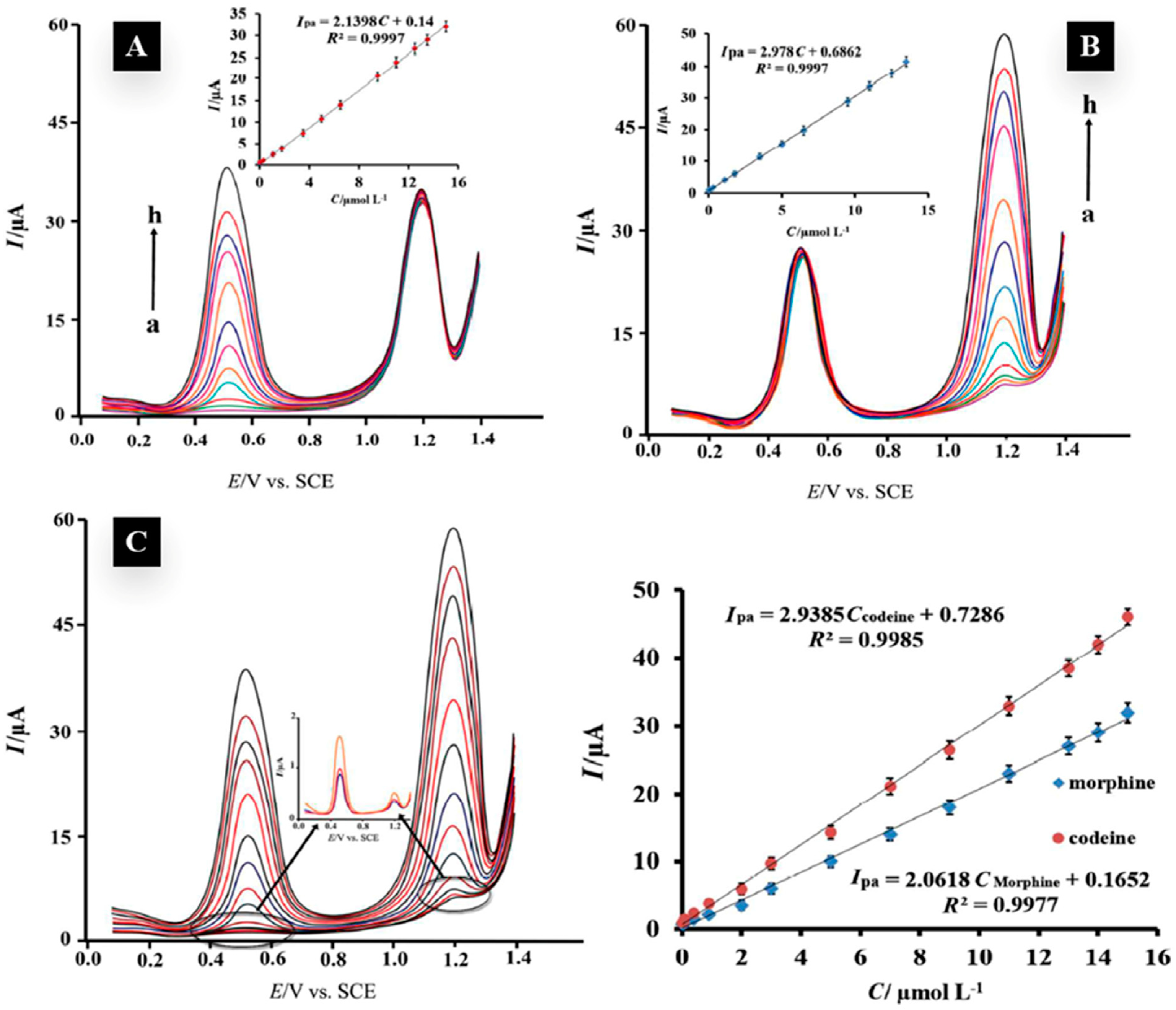
| 10 | Morphine and Codeine | Graphene–Zn2SO4 | DPV/CV | 11 and 9 nM | Urine, Plasma, Medical tablets | [38] |
| 11 | Codeine | 3D spongy graphene–adenine | SWV | 5.8 nM | Plasma, Solpadeine tablets | [34] |
| 12 | Codeine | Graphene–Nafion | SWV | 15 nM | Urine, Cough syrup | [35] |
| 13 | Codeine and Acetaminophen | Graphene–CoFe2O4 | SWV | 25 and 11 nM | Plasma, Urine, Medical tablets, Expectorant Cod syrup | [40] |
| 14 | pholcodine | Graphene–(PHL)2+-(NB)− | POT | 0.04 μg/mL | Medical suspensions, Medical syrups | [28] |
| 15 | Ketamine | Graphene oxide–Zeo | CV | 0.001 nM | Whisky, Juice | [75] |
| 16 | Ketamine and Norketamine | Graphene–MOF-MIM | DPV | 0.04 nM | Urine, Saliva | [76] |
| 17 | Tramadol | Graphene–Ag NPs-MIP | CV | 2.04 nM | Urine, Medical tablets | [60] |
| 18 | Tramadol | 1-M-3-BBr-Pr(OH)3-GQD | CV | 3 nM | Medical injection, Blood serum | [61] |
| 19 | Tramadol | Graphene oxide–MWCNTs | CV | 15 nM | Plasma, Medical tablets | [56] |
| 20 | Tramadol | Graphene–Co3O4 | LSV/DPV | 30 nM | Urine, Medical tablets | [59] |
| 21 | Tramadol and Acetaminophen | Graphene–NiFe2O4 | SWV | 3.6 and 3 nM | Blood serum, Urine, Medical tablets, Ultracet tablet | [57] |
| 22 | Tramadol and Acetaminophen | Graphene–Au NPs-HTP | DPV | 820 nM | Urine, Medical tablets | [58] |
| 23 | Methadone | Graphene–Ag NPs | DPV | 120 nM | Blood serum | [65] |
| 24 | Methadone and Morphine | Graphene–TGA@CdSe | DPV | 40 nM and 30 nM | Blood serum | [66] |
| 25 | Buprenorphine | Graphene | DPV/CV | 160 nM | Urine | [67] |
| 26 | Cocaine | 3D printed Graphene–PLA | SWV/CV | 6 μM | None | [87] |
| 27 | Cocaine | Graphene–Pd NPs-MIP | SWV | 50 μM | Saliva, River water, street sample | [86] |
| 28 | Cocaine and Amphetamine | Graphene–(pyrene-NHS)-antibody | TLM | NA | None | [85] |
| 29 | Methamphetamine | Graphene–Fe3O4@PPy | CV/SWV | 1 nM | Blood serum, Urine | [94] |
| 30 | Methamphetamine | Graphene–Ce2O3 | SWV | 8.75 μM | Plasma | [95] |
| 31 | Methamphetamine | 3D Porous Graphene | DPV/CV | 0.31 μg/mL | Saliva, Household surfaces | [93] |
| Class of 2D Material | Mono-Layer Molecular Structure | Possible “M/M’”s | Possible “X/Y”s |
|---|---|---|---|
| Semimetal Chalcogenides (MCs) | M’X | Ga and In | S, Se and Te |
| Transition Metal Chalcogenides (TMCs) | MX | Transition metals between group 4 and group 10 | |
| M3X4 | |||
| Transition Metal Dichalcogenides (TMDs) | MX2 | ||
| Transition Metal Halides (TMHs) | MY2 | Cl, Br and I | |
| MY3 | |||
| Transition Metal Carbides, Nitrides and Carbonitrides (MXenes) | M2X | C and N | |
| M3X2 | |||
| M4X3 |
| Analyte | Experimental Technique | Duration of Analysis a | Detection Limit | Ref. |
|---|---|---|---|---|
| Morphine | HPLC-UV | 10 min | 1 ng/mL | [142] |
| GC-MS | 11 min | 3 ng/mL | [143] | |
| CV | 24 s b | 0.54 nM | [41] | |
| Tramadol | HPLC-UV | 40 min | 6.7 ng/mL | [144] |
| GC-MS | 9 min | 0.77 ng/mL | [145] | |
| CV | 3.4 s b | 2.04 nM | [60] | |
| Cocaine | HPLC-Fluorescence | 8 min | 1 ng/mL | [146] |
| GC-MS | 16 min | 0.05 ng/mL | [147] | |
| CV | 60 s b | 6 μM | [87] | |
| Methamphetamine | HPLC-MS | 12 min | 0.05 ng/mL | [148] |
| GC-MS | 18 min | 0.06 μg/mL | [149] | |
| CV | 40 s b | 1 nM | [94] |
Publisher’s Note: MDPI stays neutral with regard to jurisdictional claims in published maps and institutional affiliations. |
© 2022 by the authors. Licensee MDPI, Basel, Switzerland. This article is an open access article distributed under the terms and conditions of the Creative Commons Attribution (CC BY) license (https://creativecommons.org/licenses/by/4.0/).
Share and Cite
Boroujerdi, R.; Paul, R. Graphene-Based Electrochemical Sensors for Psychoactive Drugs. Nanomaterials 2022, 12, 2250. https://doi.org/10.3390/nano12132250
Boroujerdi R, Paul R. Graphene-Based Electrochemical Sensors for Psychoactive Drugs. Nanomaterials. 2022; 12(13):2250. https://doi.org/10.3390/nano12132250
Chicago/Turabian StyleBoroujerdi, Ramin, and Richard Paul. 2022. "Graphene-Based Electrochemical Sensors for Psychoactive Drugs" Nanomaterials 12, no. 13: 2250. https://doi.org/10.3390/nano12132250
APA StyleBoroujerdi, R., & Paul, R. (2022). Graphene-Based Electrochemical Sensors for Psychoactive Drugs. Nanomaterials, 12(13), 2250. https://doi.org/10.3390/nano12132250