Abstract
Metal halide perovskite nanocrystals (NCs) attract much attention for light-emitting applications due to their exceptional optical properties. More recently, perovskite NCs have begun to be considered a promising material for nonlinear optical applications. Numerous strategies have recently been developed to improve the properties of metal halide perovskite NCs. Among them, B-site doping is one of the most promising ways to enhance their brightness and stability. However, there is a lack of study of the influence of B-site doping on the nonlinear optical properties of inorganic perovskite NCs. Here, we demonstrate that Cd2+ doping simultaneously improves both the linear (higher photoluminescence quantum yield, larger exciton binding energy, reduced trap states density, and faster radiative recombination) and nonlinear (higher two- and three-photon absorption cross-sections) optical properties of CsPbBr3 NCs. Cd2+ doping results in a two-photon absorption cross-section, reaching 2.6 × 10⁶ Goeppert-Mayer (GM), which is among the highest reported for CsPbBr3 NCs.
1. Introduction
Doping of cesium lead halide perovskites has become a novel approach to tuning their optoelectronic properties and enhancing their stability [1,2,3,4]. Doping may enhance their luminosity and promote charge carrier transport and crystal phase stabilization [2]. Different divalent metal ions including Mn2+, Sn2+, Ca2+, Zn2+, Cu2+, and Ni2+ have been used for isovalent B-site doping (where ABX3 is a general structural formula) of cesium lead halide perovskite nanocrystals (NCs) [5,6,7,8,9,10,11,12,13,14,15,16,17]. B-site doping is also considered a new approach to achieving better characteristics of devices based on perovskite NCs, and that was demonstrated for CsPbBr3 NCs, which are important nanomaterials to produce green-emitting light-emitting diodes (LEDs) [12,13,14,15,16,17]. Recently, the doping of CsPbBr3 NCs with Mn2+ [18], Sn2+ [19], Ce3+ [20], Rb+ [21], and Co2+ [22] has been used to enhance the external quantum efficiency (EQE) and maximum luminance of the LEDs. Moreover, the doping may facilitate better environmental stability of the device [18] and optimization of the energy band diagram [23].
Metal halide perovskites can now be considered a new platform for nonlinear optics [24]. For instance, they demonstrate enormous values of nonlinear absorption cross-sections. The study of the processes involving simultaneous absorption of multiple photons is of great importance for numerous applications, including high-resolution microscopy [25,26] and biomedical imaging [27,28]. Despite the great practical interest in these processes, their utilization is limited due to the lack of highly efficient nonlinear materials, and the employment of perovskites in this field is of tremendous importance. B-cite doping is considered to be an effective tool for the further optimization of nonlinear optical responses from metal halide perovskite NCs [29,30]; however, very few reports have been published on this topic. Ketavath et al., recently showed that 0.08–0.1% Ni2+ doping of 2D CsPbBr3 NCs induced a ~2.5-fold increase in the two-photon absorption (2PA) cross-section [31]. He et al., studied the influence of Mn2+ doping on the nonlinear optical properties of CsPbCl3 NCs [32] and nanoplatelets (NPLs) [33]. They reported the wavelength-dependent 2PA cross-section of Mn2+-doped CsPbCl3 NCs with a maximum value up to ~3.2 × 10⁵ GM (1 GM = 10−50 cm−4 s photon−1) [32].
Cd2+ doping is an important example of engineering of perovskite NC properties. Cd2+ doping of CsPbBr3 NCs was first reported by van der Stam and co-workers, who proposed a post-synthetic cation exchange reaction to replace some of the Pb2+ cations [11]. They showed that the doping caused a blue shift of optical transitions, induced by lattice contraction, while preserving a high photoluminescence quantum yield (PLQY) and narrow full width at half maximum (FWHM) of the PL band. A similar observation was recently made by Zhao et al., who synthesized Cd2+ doped CsPbBr3 NCs inside a borosilicate glass matrix [34]. As an alternative approach, Mondal and co-workers demonstrated the post-synthetic treatment of CsPbCl3 NCs with CdCl2 that resulted in the tremendous growth of PLQY [35]. Very recently, Xie et al., demonstrated that post-synthetic surface treatment of CsPbBr3 NCs with CdX2 precursors enlarged their PLQY from 85% to 92% [36].
Although these results point to the undeniable effect of Cd2+ doping on all-inorganic NC optical properties, there is no information regarding its influence on their nonlinear optical properties. Here, we developed methods for obtaining Cd2+-doped perovskite NCs by direct synthesis using a mixture of PbBr2 and CdBr2 precursors. The investigation of the temperature dependencies of the PL responses allowed us to reveal additional features induced by Cd2+ doping. For the first time, we demonstrate that Cd2+ doping modifies nonlinear optical responses of CsPbBr3 NCs: Cd2+ doped CsPbBr3 NCs demonstrated bright one- and multi-photon excited emission, while a two-photon absorption cross-section reached 2.6 × 10⁶ GM. First, we describe the influence of doping on the structural and linear optical properties of CsPbBr3 NCs that allow the fabrication of an LED with a maximum luminance of 24,221 cd·m−2, and then we demonstrate the improvement of nonlinear optical responses studied by means of two- and three-photon excited photoluminescent spectroscopy.
2. Materials and Methods
Materials. Lead bromide (PbBr2, 99%), cesium carbonate (Cs2CO3, 99.95%), octadecene (ODE, 90%), oleic acid (OA, 90%), cadmium bromide tetrahydrate (CdBr2∙4H2O, 98%), and oleylamine (OlAm, 80–90%) were purchased from Aladdin (Shanghai, China). Hexane, ethanol, and acetone were purchased from Sinopharm Chemical Reagent Co., Ltd. (Shanghai, China). Poly(3,4-ethylenedioxythiophene): polystyrene sulphonate (PEDOT:PSS), Poly[bis(4-phenyl) (4-butylphenyl) amine] (Poly-TPD) and 2,2′,2″-(1,3,5-Benzinetriyl)-tris(1-phenyl-1-Hbenzimidazole) (TPBi) were purchased from Xi’an Polymer (Xi’an, Shannxi, China). Lithium fluoride (LiF) was purchased from Lumtec (New Taipei City, Taiwan).
Preparation of cesium oleate (Cs-OA) solution. In brief, Cs2CO3 (0.814 g), OA (2.5 mL), and ODE (30 mL) were loaded into a 100 mL three-neck flask. The flask was degassed at 120 °C for 60 min and then switched to N2 flow. Finally, the flask was heated up to 150 °C until the Cs2CO3 completely dissolved.
Preparation of CsPbBr3 and CsPb0.78Cd0.22Br3 NCs. Briefly, PbBr2 (0.376 mmol) and ODE (10 mL) were added to a 50 mL three-neck round bottom flask. The solution was degassed at 120 °C for 60 min under vacuum and then switched to N2 flow. After injection of OA (1 mL) and OlAm (1 mL) the temperature was increased to 180 °C. Subsequently, as-synthesized Cs-OA (1 mL) was promptly injected to the flask. After 5 s, an ice bath was used to stop the reaction. CsPb0.78Cd0.22Br3 NCs were synthesized by adding 0.188 mmol of CdBr2·4H2O at the beginning of reaction while keeping the rest of the protocol intact.
Purification. First, the crude solution was purified by centrifugation at 5000 RPM for 10 min; the supernatant was discarded, and the precipitate was re-dispersed in hexane to form stable colloidal solutions. Second, ethyl acetate was introduced into the colloidal solutions at a volume ratio of 1:1, and then the mixture was centrifuged for 10 min at 10,000 RPM. Lastly, the final precipitate was dispersed in 2 mL hexane for LED fabrication.
LED fabrication. Glass substrates coated with indium tin oxide (ITO) were first sonicated consecutively with ethanol and acetone for 20 min each. To increase the work function, ITO substrates were treated with UV-ozone for 15 min. Then, PEDOT: PSS (25 nm thickness) was spin-coated on the ITO substrates at a speed of 4000 RPM for 40 s and then annealed at 150 °C for 15 min in ambient air. Subsequently, these substrates were transferred into a glovebox. The Poly-TPD solution (dissolved in chlorobenzene, 8 mg/mL, 30 nm) was spin coated onto the PEDOT: PSS film at a speed of 4000 RPM for 40 s and annealed at 150 °C for 15 min. The washed CsPb0.78Cd0.22Br3 NCs (~35 nm thickness) were spin coated onto the Poly-TPD film at 1500 RPM for 40 s. Finally, TPBi (18 nm), LiF, and Al (LiF/Al, 55 nm) electrodes were deposited by thermal evaporation in a vacuum deposition chamber of ~3 × 10⁻⁶ Torr.
Characterization. UV-VIS absorption was measured using a Shimadzu UV-1800 (Shimadzu corporation, Kyoto, Japan) spectrophotometer. A JEM-2100F (JEOL Ltd., Tokyo, Japan) transmission electron microscope (TEM) was used to analyze the morphology of the NCs. X-ray diffraction (XRD) patterns were acquired using a Bruker D8 Advance X diffractometer (Cu Kα, λ = 1.5406 Å). X-ray Photoelectron Spectroscopy (XPS) was performed on an ESCALAB 250 (Thermo Fisher Scientific Inc., Waltham, MA, USA) spectrometer with a mono X-Ray source Al Kα excitation (1486.6 eV). Ultraviolet photoelectron spectra (UPS) were collected using a PREVAC (PREVAC, Rogów, Poland) system. A Keithley 2400 (Keysight technologies, Santa Rosa, CA, USA) source meter and a PR-750 Spectroradiometer (Photo Research Inc., Chatsworth, CA, USA) were used to acquire the current density–luminance–voltage (J-V-L) characteristics. The molar concentration of the nanocrystals in the solution was obtained from the absorption spectra; see Appendix A for details.
PL spectroscopy. PL spectra were measured using a custom-built setup based on an Acton-2500 monochromator equipped with an Andor iDus 401A CCD camera (Andor Technology Ltd., Belfast, Northern Ireland) [37,38,39]. To avoid any thermal effects, the samples were excited with unfocused 80 µW 405 nm laser (Lasever Inc., Ningbo, China) excitation passed through a 0.8 mm diaphragm. The obtained spectra were corrected using the blackbody radiation source (Thorlabs SLS201/M, Thorlabs, Newton, NJ, USA). For the measurements at decreased temperatures, NCs were added to 8 wt% PMMA solution in toluene under an inert atmosphere and were left stirring for 1 h at 1000 RPM. The solution was dropped onto a precleaned glass substrate and spin-coated at 2000 RPM. The optical density of the films did not exceed 0.1 at 400 nm. The samples were transferred to a Linkam THMS600 Microscope Stage cryostat (Linkam scientific instruments, Tadworth, UK). For the transient measurements, a MicroTime 100 microscope (PicoQuant, Berlin, Germany) employing time-correlated single-photon counting was used. The microscope was equipped with 3× objective lens and a 50 ps pulse diode laser head operated at 405 nm. PL lifetimes were found to be dependent on the excitation power density, as was previously reported [40]. The excitation power density was maintained below the value at which the dependence of PL lifetimes on excitation intensity occurred. The IRF was measured and deconvoluted from the decay curves. After the deconvolution, the PL decays were fitted by the 3-exponential decay law.
2-photon excited PL measurements. Nanocrystals were dispersed in toluene in 1-mm-thick optical glass cuvettes. Two-photon PL was excited using an Avesta femtosecond laser system (Avesta project Ltd., Troitsk, Moscow, Russia) with a peak center at 800 nm, pulse width of ~30 fs, 1 kHz repetition rate, and power density ranging from 0.035 W/cm2 to 1.150 W/cm2. PL was detected using a USB4000-UV-VIS-ES (Ocean Insight, Largo, FL, USA) spectrometer [41]. The obtained spectra were corrected using the blackbody radiation source (Thorlabs SLS201/M, Thorlabs, Newton, NJ, USA). Rhodamine 6G in ethanol was used as a standard for the 2PA cross-section calculation. Rhodamine 6G cross-section was taken to be 128 ± 84 GM (10−50 cm−4·s·photon−1) [42,43,44]. A detailed description of the two-photon excited PL analysis can be found in Appendix B.
3-photon excited PL measurements. An optical parametric amplifier (OPA-TOPAS, Light Conversion, Vilnius, Lithuania) was used as an excitation source for the generation of 3-photon excited PL from perovskite NC solutions in toluene. The femtosecond pump radiation from a regenerative Ti-sapphire amplifier (pulse duration 35 fs, energy 2.2 mJ, 1 kHz repetition rate, 800 nm central wavelength) was used as a pump radiation for OPA. Optical radiation from OPA in the range 1300–1560 nm and under a pulse duration of 60 fs passed through quartz cuvette with sample solutions. The diameter of the laser beam was 5 mm, and the excitation power density varied from 0.35 W/cm2 to 1.52 W/cm2. To measure the dependence of the integrated PL intensity vs. power density, the excitation wavelength was fixed at 1300 nm, and an optical attenuator with neutral filters was used to change the excitation power. For the wavelength-dependent measurements, the pulse energy was fixed at 250 ± 20 μJ. The PL signal was detected using a USB4000-UV-VIS-ES (Ocean Insight, Largo, FL, USA) spectrometer. 3PA absorption cross-section was calculated analogous to the two-photon excitation (see Appendix B for the details) with Rhodamine 6G in ethanol as a reference (3PA cross section of 3 × 10−80 cm6 s2 [45].
3. Results
3.1. Synthesis and Characterization of NCs
The morphology and crystal structure of the as-synthesized CsPbBr3 NCs and Cd2+-doped CsPbBr3 (CsPb0.78Cd0.22Br3, the element content was confirmed by X-ray photoelectron spectroscopy) NCs were investigated by transmission electron microscopy (TEM). As shown in Figure 1a,d, the TEM image of CsPb0.78Cd0.22Br3 NCs exhibited a more uniform morphology and narrower size distribution than CsPbBr3 NCs, as clearly seen from the particle size distribution histograms (Figure 1c,f). Both samples were highly crystalline, which can be confirmed by the ordered crystal lattices in high-resolution TEM (HRTEM) images of Figure 1e. The obvious lattice fringes were identified to be 5.8 and 5.7 Å, which is in good agreement with the (100) crystal facets of the CsPbBr3 cubic phase. As for the reduction in lattice spacing in CsPb0.78Cd0.22Br3 NCs, it can be attributed to the lattice contraction derived from the Cd2+ doping. To further characterize the crystal structure, we measured X-ray diffraction patterns (XRD); the results are presented in Figure 1g. The two main peaks located at ~14.8° and 30.2° were assigned to the (100) and (200) planes of CsPbBr3 cubic phase, respectively. In contrast to CsPbBr3 NCs, the characteristic peaks of CsPb0.78Cd0.22Br3 NCs showed an obvious shift to higher angles without introducing any extra peaks, which indicates that the Cd2+ incorporation did not change the crystal structure but only contracted the lattice spacing, corresponding to the HRTEM results. To verify the effect on optical properties, the absorption spectra of these samples were measured and are summarized in Figure 1h. The characteristic absorption peak shifted from 501 nm to 495 nm after Cd2+ doping, and the absorption largely decreased in the 520–600 nm range, indicating the reduction of the trap state density in CsPb0.78Cd0.22Br3 NCs.
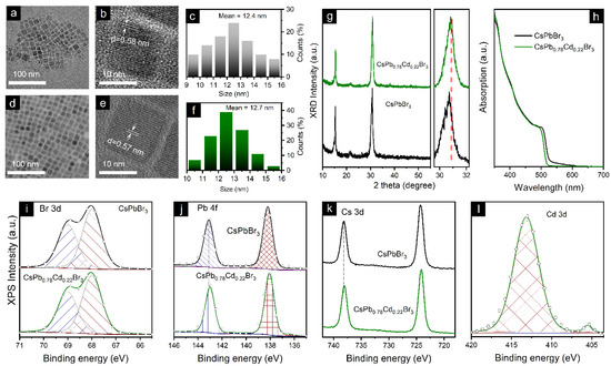
Figure 1.
TEM images of (a) CsPbBr3 and (d) CsPb0.78Cd0.22Br3 NCs; their corresponding HR-TEM images are exhibited in (b,e). (c,f) are particle size distribution histograms of CsPbBr3 and CsPb0.78Cd0.22Br3 NCs; their corresponding XRD patterns and UV−vis absorption spectra are shown in (g,h), respectively. (i–l) XPS spectra for the (a) Br, (b) Pb, (c) Cs, and (d) Cd elements collected for the CsPbBr3 and CsPb0.78Cd0.22Br3 NCs.
To identify the influence of Cd2+ doping on the chemical states of constituting elements in the perovskite NC film, we performed X-ray photoelectron spectroscopy (XPS) measurements of CsPbBr3 and CsPb0.78Cd0.22Br3 NC films. Figure 1i–l shows XPS spectra for the Br, Pb, Cs, and Cd elements, respectively, all calibrated with C 1 s. No peak shift was observed for Br 3d, while the Pb 4f and Cs 3d peaks shifted to higher binding energy in the CsPb0.78Cd0.22Br3 NC film compared to the CsPbBr3 NC film. The result is consistent with the crystal lattice contraction observed in the HRTEM images and XRD patterns. As expected, CsPb0.78Cd0.22Br3 NC films demonstrated a noticeable Cd 3d peak, and the elemental ratio of Cd:Pb was around 0.29:1.
3.2. One-Photon Excited PL
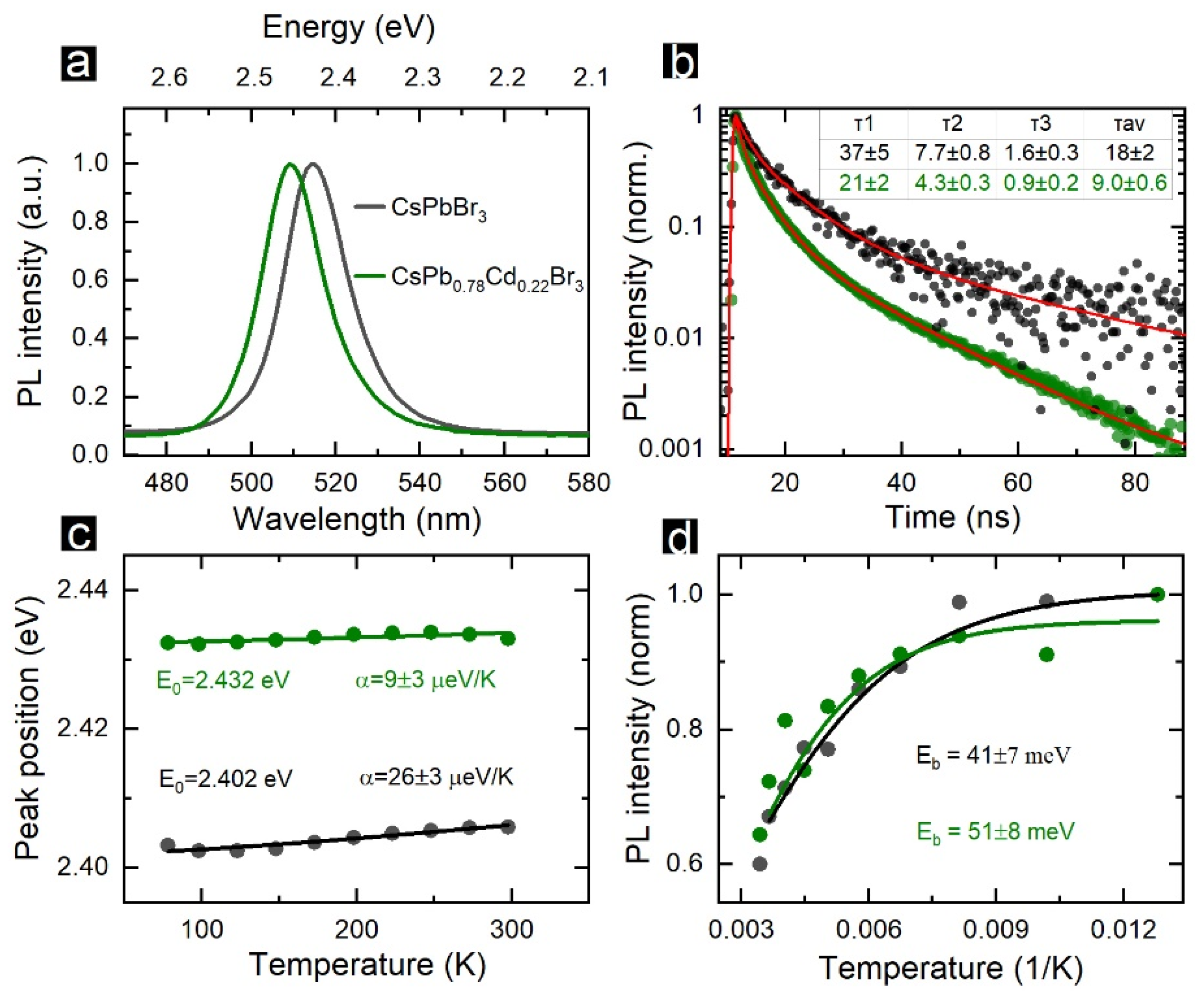
The PL spectra for CsPbBr3 (black) and CsPb0.78Cd0.22Br3 (green) NCs in toluene are shown in Figure 2a. The Cd2+ doping induced a 25-meV blue shift of the PL peak position. The blue shift of PL peak position and absorption onset (Figure 1h) are typical for the doping of perovskite NCs by guest cations with smaller ionic radii [3]. The measured change of the bandgap fitted well with the theoretical predictions reported by Navas et al., for Cd2+-doped MAPbI3 NCs [46]. Van der Stam et al. [11] recently demonstrated that introducing smaller cations, including Cd2+, induced a parent NC lattice contraction. A small shift of the band-edge emission was recently observed for Cd2+-doped CsPbCl3 NCs [47], which indicates that Cd2+ ions diffuse into the NC volume rather than passivate their surface. Both spectra shown in Figure 2a had similar FWHMs, i.e., 90 and 91 meV. After the Cd2+ doping, the PLQY increased from 75% to 90%. The PL decay for pristine and doped NCs is shown in Figure 2b. The decay curves were fitted by a 3-exponential function. The averaged PL lifetime decreased from 18 ± 2 to 9.0 ± 0.5 ns after the doping, which corresponds well with the data obtained by Van der Stam et al., for Cd2+-doped CsPbBr3 NCs [11]. Since we observed both a decrease in the PL decay time and an increase in the PLQY, this points to an increase in the radiative recombination rate and not the appearance of additional fast nonradiative recombination channels.

Figure 2.
One-photon excited PL from CsPbBr3 (black) and CsPb0.78Cd0.22Br3 (green) NCs. (a) PL spectra recorded under 405 nm laser excitation. (b) PL decay curves recorded under pulsed 405 nm laser excitation; red lines show 3-exponential fitting, and the estimated decay times constants are listed in the table along with the average PL decay times. (c) Temperature dependencies of the PL peak position; solid lines show Varshni fitting. (d) Temperature dependencies of integrated PL intensity; solid lines show Arrhenius-type fitting.
The analysis of the temperature dependencies of PL spectra revealed more differences between the pristine and doped NCs. Figure 2 shows the temperature dependencies of the PL peak positions and their approximation with a Varshnii function (Equation (1)) [48].
where EG0 is the bandgap (PL peak position) at 0 K, α is the coefficient of the temperature shift, and β is the Debye temperature, i.e., 224.8 K [49]. The peak position temperature shift coefficient α was 26 ± 3 µeV/K for CsPbCl3 NCs, which is almost an order smaller than that reported in Refs. [40,50,51]. Since many physical mechanisms contribute to a temperature shift of the bandgap for nanosized semiconductors, many factors may influence the determined α value, and the reports for different synthetic procedures may vary in a wide range [52]. The CsPb0.78Cd0.22Br3 NCs demonstrated an even smaller α value, 9 ± 3 µeV/K. Such a negligible temperature shift of the PL band is beneficial for the development of LEDs with stable color purity [53]. Both types of NCs demonstrated an increase in PL intensity with a temperature decrease (Figure 2d). Fitting the integrated PL intensity vs. temperature with Arrhenius-type equation (Equation (2)) allowed us to determine the exciton binding energy (Eb) in perovskite NCs [54].
where I0 is the integrated PL intensity at 0 K, kB is the Boltzmann constant, and Eb is the binding energy. Eb was 41 ± 7 meV for the pristine CsPbBr3 NCs. This value is very close to those recently determined by Yuan et al., using the same experimental approach [51] and estimated by Protesescu et al., within the effective mass approximation [55]. The CsPb0.78Cd0.22Br3 NCs had a higher Eb value, 51 ± 8 meV, which makes them attractive candidates to be used in perovskite-based LEDs [56,57].
3.3. LEDs
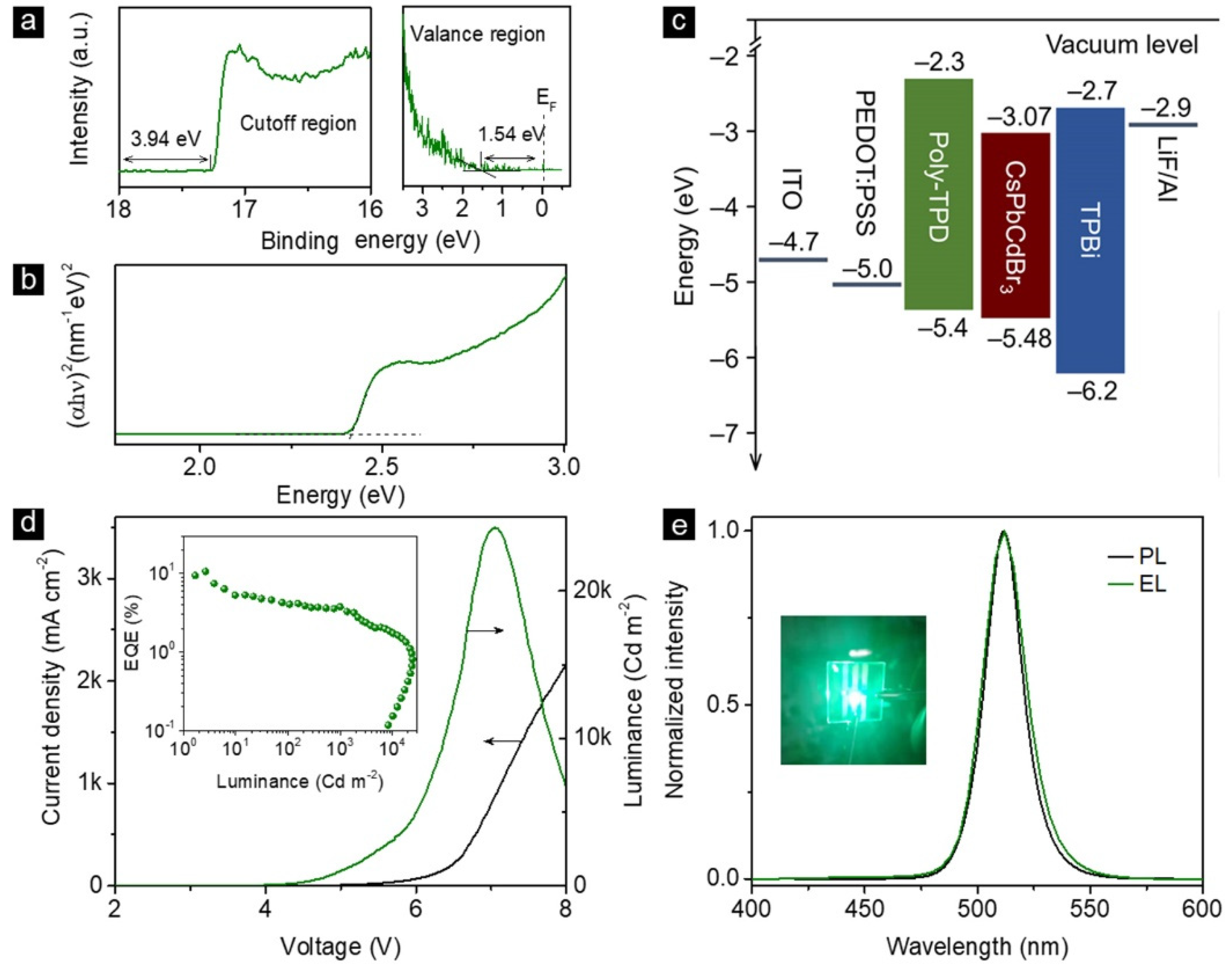
To demonstrate their practical applicability, the CsPb0.78Cd0.22Br3 NCs were adopted as emitters to fabricate LEDs. The lowest unoccupied molecular orbital (LUMO) and the highest occupied molecular orbital (HOMO) were determined by the ultraviolet photoelectron spectroscopy (UPS, Figure 3a) of CsPb0.78Cd0.22Br3 NC film [58]. The Tauc plot of the CsPb0.78Cd0.22Br3 NC film (Figure 3b) revealed a bandgap of 2.41 eV [59]. Thus, the LUMO and HOMO values of CsPb0.78Cd0.22Br3 NCs were confirmed to be −3.07 and −5.48 eV, respectively. The conventional multilayered LED was fabricated using patterned indium−tin oxide (ITO) as the anode, poly(ethylenedioxythiophene):polystyrenesulfonate (PEDOT:PSS, 25 nm) film and poly(N,N′-bis(4-butylphenyl)-N,N′-bis(phenyl)-benzidine) (poly-TPD, 40 nm) film as the hole transporting layer (HTL), CsPb0.78Cd0.22Br3 NC film (40 nm) as the emitting layer, 1,3,5-tris(N-phenylbenzimidazol-2-yl) benzene (TPBi, 40 nm) film as the electron-transporting layer (ETL), and LiF/Al as the cathode. The energy band diagram for all functional layers of LED is given in Figure 3c. Figure 3d demonstrates the current density–voltage (J–V) and luminance–voltage (L–V) curves of the LEDs, which showed a peak luminance of 24,221 Cd·m−2 under the working bias of 7.0 V. The peak EQE of 10.6% was achieved at 0.3 mA cm−2 (inset of Figure 3d). Normalized PL and electroluminescence (EL) spectra of a typical LED are given in Figure 3e. Apparently, the EL originated from CsPb0.78Cd0.22Br3 NCs without noticeable contribution from any charge transport layers, indicating that the perovskite NCs served as the exciton recombination centers for the device, and a balanced charge transport was achieved. The device PL and EL both exhibited emission peaks at 511 nm. The inset of Figure 3e is the image of a working LED.

Figure 3.
(a) UPS spectra for the cutoff (left) and valence (right) regions and (b) Tauc plot of the CsPb0.78Cd0.22Br3 NCs film; (c) overall energy band diagram of the CsPb0.78Cd0.22Br3 NC-based LED, (d) current density and brightness vs. driving voltage of the CsPb0.78Cd0.22Br3 NC-based LED, (e) normalized PL and EL spectra for CsPb0.78Cd0.22Br3 NCs in the film and LEDs; the inset is a photograph of the working LED.
3.4. Multiphoton-Induced PL
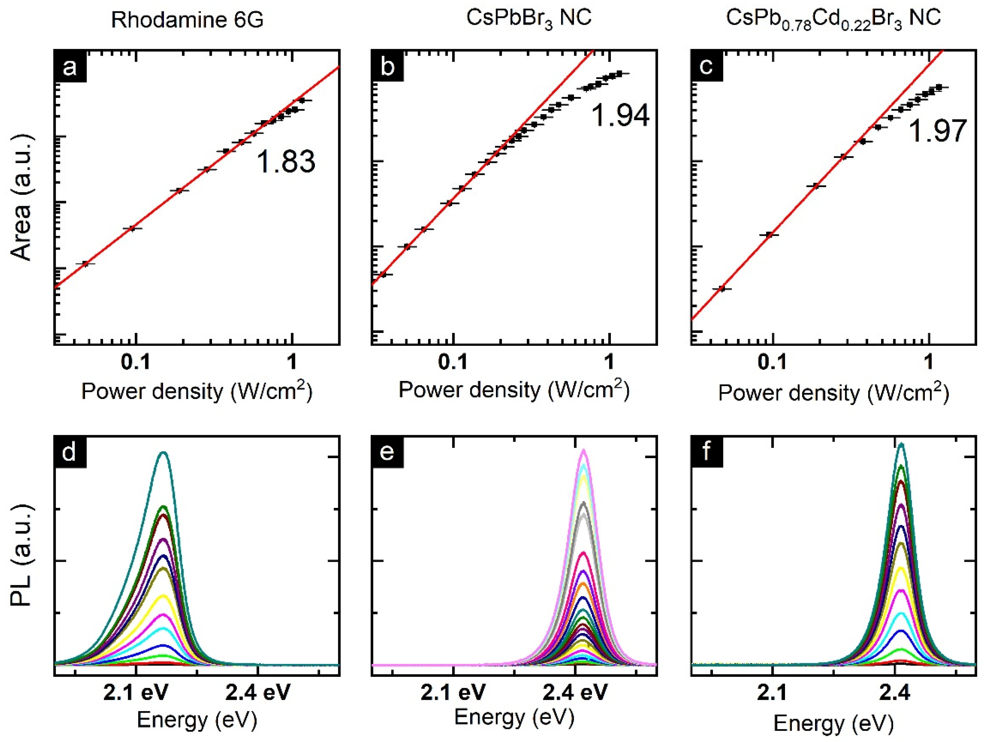
Two-photon excited PL spectra of perovskite NCs were recorded using non-resonant 800 nm laser excitation. To estimate the type of multi-photon process, we measured NC PL spectra with different excitation power densities. Figure 4a–c demonstrates the log–log plots of integrated PL intensity vs. excitation power density for the Rhodamine 6G (used as a reference), CsPbBr3 NCs, and CsPb0.78Cd0.22Br3 NCs, respectively. Nearly quadratic dependencies of integrated PL intensity vs. power density clearly indicate the two-photon absorption (2PA) process [60]. At higher excitation power density, the integrated 2PA-excited PL intensity demonstrated a saturation-like behavior as previously reported for the MAPbBr3 films [61] and 2D (PEA)2PbI4 perovskites [62]. We can attribute this observation to the influence of strong laser radiation exposure, which can lead to perovskite NС partial destruction caused by heating. It should also be noted that CsPb0.78Cd0.22Br3 NCs showed a smaller deviation from the theoretical power law, which may indicate their improved stability under laser radiation.

Figure 4.
The log–log plots of integrated PL area vs. power density for Rhodamine 6G (a), CsPbBr3 NCs (b), and CsPb0.78Cd0.22Br3 NCs (c). (d–f) corresponding 2PA-excited PL spectra measured at different excitation power densities.
The 2PA absorption cross-sections were calculated using Rhodamine 6G as a standard (see experimental section and Appendix B). The reported NC values of third-order nonlinear optical parameters strongly depend on their stoichiometry [63], size [64], and shape [65]. The value obtained for the pristine CsPbBr3 NCs (3.2 ± 1.9) × 105 GM was very close to those recently reported (see Table 1) [64,65,66,67,68]. CsPb0.78Cd0.22Br3 NCs demonstrated an almost one-order enhanced value of the 2PA cross-section, and the obtained value is among the highest reported for CsPbBr3 NCs [69,70].

Table 1.
Pristine and volume-normalized 2PA and 3PA cross-sections for the CsPbBr3 NCs, nanoplatelets (NP), nanorods (NR), and nanowires (NW).
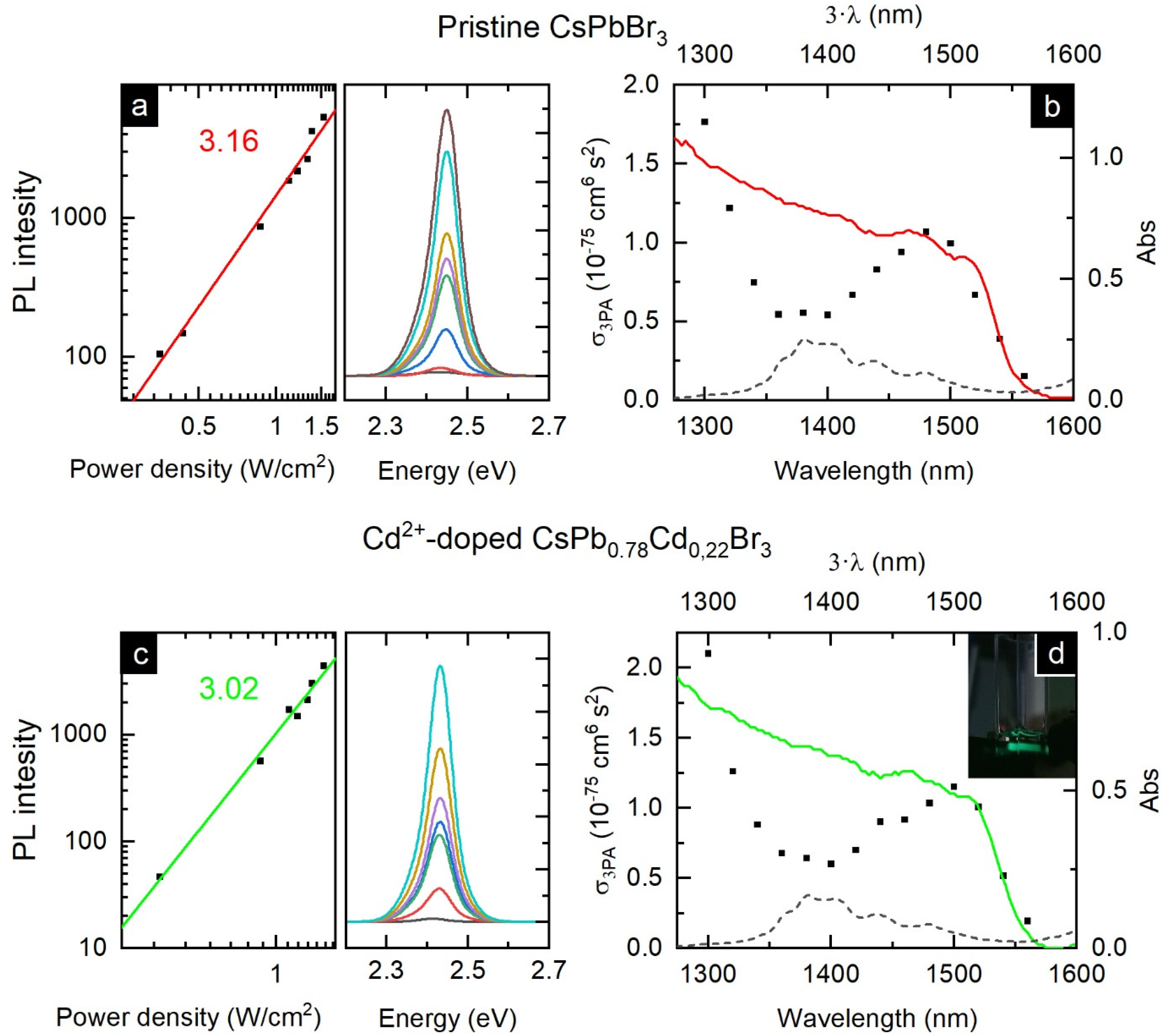
To study the 5th-order nonlinear optical response, three-photon absorption (3PA)-induced PL was investigated using the wavelength-tunable excitation in the 1300–1560 nm spectral range. First, the excitation wavelength was fixed at 1300 nm, and the integrated PL intensity vs. excitation power dependencies for the pristine and doped perovskite NCs were measured (Figure 5a,c). For both samples, the dependencies were similar to a cubic function, indicating the 3-photon excited process solely [74]. To calculate the 3PA cross-sections, Rhodamine 6G was used as a standard. Similar to the results obtained for the 2PA excited PL, the 3PA cross-section was higher for CsPb0.78Cd0.22Br3 NCs (2.1 ± 0.7 vs. 1.7 ± 1.0 ×10−75 cm6 s2 photon−2). Spectral tuning of the used excitation allowed us to obtain the wavelength-dependencies of the 3PA cross-section for the pristine CsPbBr3 and doped CsPb0.78Cd0.22Br3 NC (Figure 5b,d). For both samples, the wavelength dependencies of the 3PA cross-sections well matched the linear absorption spectra of the NC solutions, except the region of intense absorption of toluene (1340–1440 nm). This observation is in line with previously reported wavelength dependencies obtained for both 2PA and 3PA in CsPbBr3 NCs [64,68].
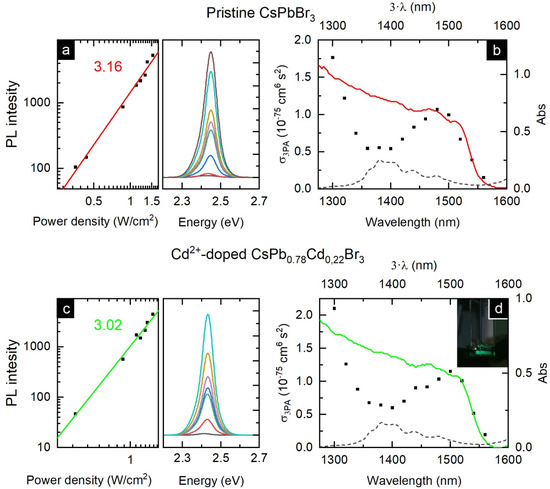
Figure 5.
Three-photon excited photoluminescence of the studied NC. The log–log plots of the integrated PL area vs. power density for CsPbBr3 NCs (a) and CsPb0.78Cd0.22Br3 NCs (c). On the side are corresponding 3PA-excited PL spectra measured at different excitation power densities. 3PA absorption cross-section spectral dependencies for (b) CsPbBr3 NCs and (d) CsPb0.78Cd0.22Br3 NCs (on the inset is the photo of the PL from the CsPb0.78Cd0.22Br3 NC solution when excited at 1560 nm). Dashed lines in (b,d) are the absorption of toluene at 1300–1600 nm, while colored lines show corresponding linear absorption drawn as a function of Abs(3·λ).
4. Conclusions
To conclude, we demonstrate that the engineering of the B-site in metal halide perovskite NCs has great potential for the improvement of their optical properties. Importantly, we revealed that this strategy may induce a huge change in their nonlinear optical responses. The insertion of Cd2+ into CsPbBr3 NCs resulted in an improvement of their morphological and optical properties, including greater exciton binding energy of 51 meV, reduced trap state density, faster radiative recombination, and high photoluminescence quantum yield reaching 90%. An even more significant improvement was associated with a change in the nonlinear optical response of the Cd2+-doped CsPbBr3 NCs. The TPA cross-section of 2.6 × 10⁶ GM was evaluated, which is among the highest reported for CsPbBr3 perovskite NCs. The findings demonstrate that the perovskite NCs with a rationally engineered B-site have great potential for light-emission and nonlinear optical applications.
Author Contributions
I.D.S. and W.Y. contributed equally. Conceptualization: A.P.L.; Investigation: I.D.S., W.Y., A.O.I., A.N.T., H.H., H.W., X.Z., A.P.L.; Project administration: A.P.L. and W.Z.; Visualization: I.D.S. and W.Y.; writing original draft: I.D.S. and W.Y.; writing review and editing: X.Z. and A.P.L.; Validation: W.Z. All authors have read and agreed to the published version of the manuscript.
Funding
The work was supported by the Russian Science Foundation (21-73-10131), the National Natural Science Foundation of China (52072141), the Natural Science Foundation of Jilin Province (Grant No. 20180101294JC), and the Fundamental Research Funds for the Central Universities (JLU, 1018320194002, 2019JCXK-31).
Data Availability Statement
All required data is provided within the manuscript.
Acknowledgments
A.P.L. thanks the Ministry of Education of the Russian Federation for the financial support (Scholarship of the President of the Russian Federation for young scientists and graduate students, SP-149.2021.1).
Conflicts of Interest
The authors declare no conflict of interest.
Appendix A. Calculation of Perovskite NC Molar Concentration
The perovskite NC extinction coefficient (ε) at 400 nm was calculated from the following equation:
where D is the absorbance of the sample at 400 nm; CM—NC molar concentration; l—cuvette length; mNC—molar mass of the single NC; Cmass—mass concentration; NA—the Avogadro number; VNC—NC volume (determined from the known NC size); ρ—the NC density. NC density was calculated by normalizing the crystalline cell mass to the cell volume:
where mcell and Vcell are the respective cell mass and cell volume, r is the edge length, determined from TEM (see Figure 1). Cell mass was calculated from known NC stoichiometry. The resulting densities were 4.935 g/cm3 for CsPbBr3 and 5.012 g/cm3 for CsPb0.78Cd0.22Br3. Increased NC density in doped NCs arises from the lattice contraction observed from TEM (see Figure 1b,e). Mass concentration of the solution was determined from the weight of thoroughly washed and dried NCs.
The ε values for the CsPbBr3 and CsPb0.78Cd0.22Br3 NCs were (3.63 ± 0.18) × 107 mol−1cm−1 and (3.57 ± 0.09) × 107 mol−1cm−1, respectively. To verify the calculated extinction coefficients, we compared the values with the ones that were obtained for the pristine CsPbBr3 using Equation (A3) [75].
where d is the edge length of the NCs in nanometers. The calculated value was (4.46 ± 0.22) × 107 mol−1cm−1, which is fairly close to the values obtained from the aforementioned determination of extinction coefficients. Some difference may be caused by the overestimation of the NCs mass because some ligands still may remain at the NC surface. Using the determined mass absorption coefficient and NC molar weight, linear absorption cross-sections may be easily calculated. The linear absorption cross-sections were 4.94 × 10⁻¹⁴ cm2 and 4.86 × 10⁻¹⁴ cm2 for CsPbBr3 and CsPb0.78Cd0.22Br3 NCs, respectively. Recently, absorption cross-sections at 400 nm in the range of 1.3–3.4 × 10⁻¹⁴ cm2 were reported for CsPbBr3 NCs with sizes of 6.2–9.3 nm, [76,77,78,79]; our values are similar and expectedly slightly higher due to a larger NC size. This supports the applicability of the estimated concentration for the correct measurements of multiphoton absorption cross-sections.
Concentrations of NCs and Rhodamine 6G were determined from Beer’s law. For Rhodamine 6G, the known extinction coefficient (~40,000 mol−1cm−1) at 500 nm was used. [80,81].
Appendix B. Multiphoton Absorption Cross-Section Calculation
Rhodamine 6G in ethanol was used as a standard for the 2PA cross-section calculation. The Rhodamine 6G cross-section was taken to be 128 ± 84 GM (10−50 cm−4·s·photon−1), as averaged from the literature data (taken at 800 nm with 100 fs pulse duration) [42,43,44]. To estimate the 2PA cross-section, one-photon excited PLQY was measured first. PLQY was measured against a standard, Rhodamine 6G in ethanol (PLQY ~0.9). PL spectra were measured using a Cary Eclipse spectrofluorometer (Varian Inc., Palo Alto, CA, USA). Absorption spectra were taken using a Shimadzu UV3600 spectrophotometer (Shimadzu corporation, Kyoto, Japan). PLQY was calculated using Equation (A4):
where φ—one-photon excited PL quantum yield; I—PL spectra area, D—the optical density at the excitation wavelength, n—solution refractive index; indices r and s represent values for the reference and the sample, respectively.
Then, the 2PA cross-section was calculated using the Equation (A5):
where σ2PA—2PA cross-section (in GM); φ—one-photon excited PLQY; η—two-photon excited PLQY; I2P—two-photon excited PL spectra area, C—concentration (in M), n—solution refractive index; indices r and s represent values for the reference and the sample, respectively. Assuming that φ = η, the equation can be re-written as follows:
The 3PA absorption cross-section was calculated analogous to the 2PA cross-section with Rhodamine 6G in ethanol as a reference using the following equation:
where σ3PA—3PA cross-section (in GM); φ—one-photon excited PLQY; I3P—three-photon excited PL spectra area, C—concentration (in mol), n—solution refractive index; indices r and s represent values for the reference and the sample, respectively. The 3PA cross-section of Rhodamine 6G was taken as 3 × 10−80 cm6 s2 [45].
References
- Kumawat, N.K.; Yuan, Z.; Bai, S.; Gao, F. Metal Doping/Alloying of Cesium Lead Halide Perovskite Nanocrystals and their Applications in Light-Emitting Diodes with Enhanced Efficiency and Stability. Isr. J. Chem. 2019, 59, 695–707. [Google Scholar] [CrossRef]
- Xu, L.; Yuan, S.; Zeng, H.; Song, J. A comprehensive review of doping in perovskite nanocrystals/quantum dots: Evolution of structure, electronics, optics, and light-emitting diodes. Mater. Today Nano 2019, 6, 100036. [Google Scholar] [CrossRef]
- Lu, C.-H.; Biesold-McGee, G.V.; Liu, Y.; Kang, Z.; Lin, Z. Doping and ion substitution in colloidal metal halide perovskite nanocrystals. Chem. Soc. Rev. 2020, 49, 4953–5007. [Google Scholar] [CrossRef]
- Chen, Y.; Liu, Y.; Hong, M. Cation-doping matters in caesium lead halide perovskite nanocrystals: From physicochemical fundamentals to optoelectronic applications. Nanoscale 2020, 12, 12228–12248. [Google Scholar] [CrossRef]
- Shen, X.; Zhang, Y.; Kershaw, S.V.; Li, T.; Wang, C.; Zhang, X.; Wang, W.; Li, D.; Wang, Y.; Lu, M.; et al. Zn-Alloyed CsPbI3 Nanocrystals for Highly Efficient Perovskite Light-Emitting Devices. Nano Lett. 2019, 19, 1552–1559. [Google Scholar] [CrossRef]
- Zhang, R.; Yuan, Y.; Li, J.; Qin, Z.; Zhang, Q.; Xiong, B.; Wang, Z.; Chen, F.; Du, X.; Yang, W. Ni and K ion doped CsPbX3 NCs for the improvement of luminescence properties by a facile synthesis method in ambient air. J. Lumin. 2020, 221, 117044. [Google Scholar] [CrossRef]
- Chen, J.-K.; Ma, J.-P.; Guo, S.-Q.; Chen, Y.-M.; Zhao, Q.; Zhang, B.-B.; Li, Z.-Y.; Zhou, Y.; Hou, J.; Kuroiwa, Y.; et al. High-Efficiency Violet-Emitting All-Inorganic Perovskite Nanocrystals Enabled by Alkaline-Earth Metal Passivation. Chem. Mater. 2019, 31, 3974–3983. [Google Scholar] [CrossRef]
- Yong, Z.-J.; Guo, S.-Q.; Ma, J.-P.; Zhang, J.-Y.; Li, Z.-Y.; Chen, Y.-M.; Zhang, B.-B.; Zhou, Y.; Shu, J.; Gu, J.-L.; et al. Doping-Enhanced Short-Range Order of Perovskite Nanocrystals for Near-Unity Violet Luminescence Quantum Yield. J. Am. Chem. Soc. 2018, 140, 9942–9951. [Google Scholar] [CrossRef] [Green Version]
- Pan, G.C.; Bai, X.; Xu, W.; Chen, X.; Zhai, Y.; Zhu, J.; Shao, H.; Ding, N.; Xu, L.; Dong, B.; et al. Bright Blue Light Emission of Ni2+ Ion-Doped CsPbClxBr3−x Perovskite Quantum Dots Enabling Efficient Light-Emitting Devices. ACS Appl. Mater. Interfaces 2020, 12, 14195–14202. [Google Scholar] [CrossRef]
- Chen, Y.-C.; Chou, H.-L.; Lin, J.-C.; Lee, Y.-C.; Pao, C.-W.; Chen, J.-L.; Chang, C.-C.; Chi, R.-Y.; Kuo, T.-R.; Lu, C.-W.; et al. Enhanced Luminescence and Stability of Cesium Lead Halide Perovskite CsPbX3 Nanocrystals by Cu2+-Assisted Anion Exchange Reactions. J. Phys. Chem. C 2019, 123, 2353–2360. [Google Scholar] [CrossRef]
- Van der Stam, W.; Geuchies, J.J.; Altantzis, T.; van den Bos, K.H.W.; Meeldijk, J.D.; Van Aert, S.; Bals, S.; Vanmaekelbergh, D.; De Mello Donega, C.D.M. Highly Emissive Divalent-Ion-Doped Colloidal CsPb1−xMxBr3 Perovskite Nanocrystals through Cation Exchange. J. Am. Chem. Soc. 2017, 139, 4087–4097. [Google Scholar] [CrossRef]
- Li, Y.; Zhang, X.; Huang, H.; Kershaw, S.V.; Rogach, A.L. Advances in metal halide perovskite nanocrystals: Synthetic strategies, growth mechanisms, and optoelectronic applications. Mater. Today 2020, 32, 204–221. [Google Scholar] [CrossRef]
- Shamsi, J.; Urban, A.S.; Imran, M.; De Trizio, L.; Manna, L. Metal Halide Perovskite Nanocrystals: Synthesis, Post-Synthesis Modifications, and Their Optical Properties. Chem. Rev. 2019, 119, 3296–3348. [Google Scholar] [CrossRef]
- Liu, M.; Grandhi, G.K.; Matta, S.; Mokurala, K.; Litvin, A.; Russo, S.; Vivo, P. Halide Perovskite Nanocrystal Emitters. Adv. Photonics Res. 2021, 2, 2000118. [Google Scholar] [CrossRef]
- Yan, F.; Tan, S.T.; Li, X.; Demir, H.V. Light Generation in Lead Halide Perovskite Nanocrystals: LEDs, Color Converters, Lasers, and Other Applications. Small 2019, 15, e1902079. [Google Scholar] [CrossRef]
- Gualdrón-Reyes, A.F.; Masi, S.; Mora-Seró, I. Progress in halide-perovskite nanocrystals with near-unity photoluminescence quantum yield. Trends Chem. 2021, 3, 499–511. [Google Scholar] [CrossRef]
- Dey, A.; Ye, J.; De, A.; Debroye, E.; Ha, S.K.; Bladt, E.; Kshirsagar, A.S.; Wang, Z.; Yin, J.; Wang, Y.; et al. State of the Art and Prospects for Halide Perovskite Nanocrystals. ACS Nano 2021, 15, 10775–10981. [Google Scholar] [CrossRef]
- Zou, S.; Liu, Y.; Li, J.; Liu, C.; Feng, R.; Jiang, F.; Li, Y.; Song, J.; Zeng, H.; Hong, M.; et al. Stabilizing Cesium Lead Halide Perovskite Lattice through Mn(II) Substitution for Air-Stable Light-Emitting Diodes. J. Am. Chem. Soc. 2017, 139, 11443–11450. [Google Scholar] [CrossRef]
- Wang, H.-C.; Wang, W.; Tang, A.-C.; Tsai, H.-Y.; Bao, Z.; Ihara, T.; Yarita, N.; Tahara, H.; Kanemitsu, Y.; Chen, S.; et al. High-Performance CsPb1−xSnxBr3 Perovskite Quantum Dots for Light-Emitting Diodes. Angew. Chem. 2017, 129, 13838–13842. [Google Scholar] [CrossRef]
- Yao, J.-S.; Ge, J.; Han, B.-N.; Wang, K.-H.; Yao, H.-B.; Yu, H.-L.; Li, J.-H.; Zhu, B.-S.; Song, J.-Z.; Chen, C.; et al. Ce3+-Doping to Modulate Photoluminescence Kinetics for Efficient CsPbBr3 Nanocrystals Based Light-Emitting Diodes. J. Am. Chem. Soc. 2018, 140, 3626–3634. [Google Scholar] [CrossRef]
- Todorović, P.; Ma, D.; Chen, B.; Quintero-Bermudez, R.; Saidaminov, M.I.; Dong, Y.; Lu, Z.; Sargent, E.H. Spectrally Tunable and Stable Electroluminescence Enabled by Rubidium Doping of CsPbBr3 Nanocrystals. Adv. Opt. Mater. 2019, 7, 1470–1474. [Google Scholar] [CrossRef]
- Yang, H.; Yin, W.; Dong, W.; Gao, L.; Tan, C.-H.; Li, W.; Zhang, X.; Zhang, J. Enhancing the light-emitting performance and stability in CsPbBr3 perovskite quantum dots via simultaneous doping and surface passivation. J. Mater. Chem. C 2020, 8, 14439–14445. [Google Scholar] [CrossRef]
- Hoang, M.T.; Pannu, A.S.; Tang, C.; Yang, Y.; Pham, N.D.; Gui, K.; Wang, X.; Yambem, S.; Sonar, P.; Du, A.; et al. Potassium Doping to Enhance Green Photoemission of Light-Emitting Diodes Based on CsPbBr3 Perovskite Nanocrystals. Adv. Opt. Mater. 2020, 8, 2000742. [Google Scholar] [CrossRef]
- Xu, J.; Li, X.; Xiong, J.; Yuan, C.; Semin, S.; Rasing, T.; Bu, X. Halide Perovskites for Nonlinear Optics. Adv. Mater. 2020, 32, e1806736. [Google Scholar] [CrossRef] [PubMed]
- Denk, W.; Strickler, J.H.; Webb, W.W. Two-photon laser scanning fluorescence microscopy. Science 1990, 248, 73–76. [Google Scholar] [CrossRef] [Green Version]
- Hell, S.W.; Bahlmann, K.; Schrader, M.; Soini, A.; Malak, H.M.; Gryczynski, I.; Lakowicz, J.R. Three-photon excitation in fluorescence microscopy. J. Biomed. Opt. 1996, 1, 71–74. [Google Scholar] [CrossRef]
- Yu, J.H.; Kwon, S.-H.; Petrasek, Z.; Park, O.K.; Jun, S.W.; Shin, K.; Choi, M.; Park, Y.I.; Park, K.; Bin Na, H.; et al. High-resolution three-photon biomedical imaging using doped ZnS nanocrystals. Nat. Mater. 2013, 12, 359–366. [Google Scholar] [CrossRef]
- Tong, L.; Cobley, C.M.; Chen, J.; Xia, Y.; Cheng, J.-X. Bright Three-Photon Luminescence from Gold/Silver Alloyed Nanostructures for Bioimaging with Negligible Photothermal Toxicity. Angew. Chem. Int. Ed. 2010, 49, 3485–3488. [Google Scholar] [CrossRef] [Green Version]
- Chen, W.; Zhang, F.; Wang, C.; Jia, M.; Zhao, X.; Liu, Z.; Ge, Y.; Zhang, Y.; Zhang, H. Nonlinear Photonics Using Low-Dimensional Metal-Halide Perovskites: Recent Advances and Future Challenges. Adv. Mater. 2021, 33, 2004446. [Google Scholar] [CrossRef]
- Shen, W.; Chen, J.; Wu, J.; Li, X.; Zeng, H. Nonlinear Optics in Lead Halide Perovskites: Mechanisms and Applications. ACS Photonics 2021, 8, 113–124. [Google Scholar] [CrossRef]
- Ketavath, R.; Katturi, N.K.; Ghugal, S.G.; Kolli, H.K.; Swetha, T.; Soma, V.R.; Murali, B. Deciphering the Ultrafast Nonlinear Optical Properties and Dynamics of Pristine and Ni-Doped CsPbBr3 Colloidal Two-Dimensional Nanocrystals. J. Phys. Chem. Lett. 2019, 10, 5577–5584. [Google Scholar] [CrossRef]
- He, T.; Li, J.; Ren, C.; Xiao, S.; Li, Y.; Chen, R.; Lin, X. Strong two-photon absorption of Mn-doped CsPbCl3 perovskite nanocrystals. Appl. Phys. Lett. 2017, 111, 211105. [Google Scholar] [CrossRef] [Green Version]
- He, T.; Li, J.; Qiu, X.; Xiao, S.; Lin, X. Superior multiphoton absorption properties in colloidal Mn-doped CsPbCl3 two-dimensional nanoplatelets. Photonics Res. 2018, 6, 1021–1027. [Google Scholar] [CrossRef]
- Zhao, Y.; Shen, C.; Ding, L.; Liu, J.; Xiang, W.; Liang, X. Novel B-site Cd2+ doped CsPbBr3 quantum dot glass toward strong fluorescence and high stability for wLED. Opt. Mater. 2020, 107, 110046. [Google Scholar] [CrossRef]
- Mondal, N.; De, A.; Samanta, A. Achieving Near-Unity Photoluminescence Efficiency for Blue-Violet-Emitting Perovskite Nanocrystals. ACS Energy Lett. 2019, 4, 32–39. [Google Scholar] [CrossRef]
- Xie, C.; Zhao, Y.; Shi, W.; Yang, P. Postsynthetic Surface-Treatment of CsPbX3 (X = Cl, Br, or I) Nanocrystals via CdX2 Precursor Solution toward High Photoluminescence Quantum Yield. Langmuir 2021, 37, 1183–1193. [Google Scholar] [CrossRef] [PubMed]
- Parfenov, P.S.; Litvin, A.P.; Baranov, A.V.; Ushakova, E.V.; Fedorov, A.V.; Prudnikov, A.V.; Artemyev, M.V. Measurement of the luminescence decay times of PbS quantum dots in the near-IR spectral range. Opt. Spectrosc. 2012, 112, 868–873. [Google Scholar] [CrossRef]
- Parfenov, P.S.; Litvin, A.P.; Ushakova, E.V.; Fedorov, A.V.; Baranov, A.V.; Berwick, K. Note: Near infrared spectral and transient measurements of PbS quantum dots luminescence. Rev. Sci. Instrum. 2013, 84, 116104. [Google Scholar] [CrossRef]
- Skurlov, I.D.; Onishchuk, D.A.; Parfenov, P.S.; Litvin, A.P. An Experimental Setup for Analysis of Weak Photoluminescence in the Near-Infrared Spectral Region. Opt. Spectrosc. 2018, 125, 756–759. [Google Scholar] [CrossRef]
- Han, Q.; Wu, W.; Liu, W.; Yang, Q.; Yang, Y. Temperature-dependent photoluminescence of CsPbX3 nanocrystal films. J. Lumin. 2018, 198, 350–356. [Google Scholar] [CrossRef]
- Das, A.; Arefina, I.A.; Danilov, D.V.; Koroleva, A.V.; Zhizhin, E.V.; Parfenov, P.S.; Kuznetsova, V.A.; Ismagilov, A.O.; Litvin, A.P.; Fedorov, A.V.; et al. Chiral carbon dots based on l/d-cysteine produced via room temperature surface modification and one-pot carbonization. Nanoscale 2021, 13, 8058–8066. [Google Scholar] [CrossRef]
- Albota, M.A.; Xu, C.; Webb, W.W. Two-photon fluorescence excitation cross sections of biomolecular probes from 690 to 960 nm. Appl. Opt. 1998, 37, 7352–7356. [Google Scholar] [CrossRef] [Green Version]
- Kapoor, R.; Friend, C.S.; Patra, A. Two-photon-excited absolute emission cross-sectional measurements calibrated with a luminance meter. J. Opt. Soc. Am. B 2003, 20, 1550–1554. [Google Scholar] [CrossRef]
- Fischer, A.; Cremer, C.; Stelzer, E.H.K. Fluorescence of coumarins and xanthenes after two-photon absorption with a pulsed titanium–sapphire laser. Appl. Opt. 1995, 34, 1989–2003. [Google Scholar] [CrossRef]
- Lanin, A.A.; Chebotarev, A.S.; Pochechuev, M.S.; Kelmanson, I.V.; Kotova, D.A.; Bilan, D.S.; Ermakova, Y.G.; Fedotov, A.B.; Ivanov, A.A.; Belousov, V.V.; et al. Two- and three-photon absorption cross-section characterization for high-brightness, cell-specific multiphoton fluorescence brain imaging. J. Biophotonics 2020, 13, e201900243. [Google Scholar] [CrossRef]
- Navas, J.; Sánchez-Coronilla, A.; Gallardo, J.J.; Cruz Hernández, N.; Piñero, J.C.; Alcántara, R.; Fernández-Lorenzo, C.; De los Santos, D.M.; Aguilar, T.; Martín-Calleja, J. New insights into organic–inorganic hybrid perovskite CH3NH3PbI3 nanoparticles. An experimental and theoretical study of doping in Pb2+ sites with Sn2+, Sr2+, Cd2+ and Ca2+. Nanoscale 2015, 7, 6216–6229. [Google Scholar] [CrossRef]
- Cai, T.; Yang, H.; Hills-Kimball, K.; Song, J.-P.; Zhu, H.; Hofman, E.; Zheng, W.; Rubenstein, B.M.; Chen, O. Synthesis of All-Inorganic Cd-Doped CsPbCl3 Perovskite Nanocrystals with Dual-Wavelength Emission. J. Phys. Chem. Lett. 2018, 9, 7079–7084. [Google Scholar] [CrossRef]
- Varshni, Y.P. Temperature dependence of the energy gap in semiconductors. Physica 1967, 34, 149–154. [Google Scholar] [CrossRef]
- Lee, W.; Li, H.; Wong, A.; Zhang, D.; Lai, M.; Yu, Y.; Kong, Q.; Lin, E.; Urban, J.J.; Grossman, J.C.; et al. Ultralow thermal conductivity in all-inorganic halide perovskites. Proc. Natl. Acad. Sci. USA 2017, 114, 8693–8697. [Google Scholar] [CrossRef] [Green Version]
- Saran, R.; Heuer-Jungemann, A.; Kanaras, A.G.; Curry, R.J. Giant Bandgap Renormalization and Exciton-Phonon Scattering in Perovskite Nanocrystals. Adv. Opt. Mater. 2017, 5, 1700231. [Google Scholar] [CrossRef]
- Li, J.; Yuan, X.; Jing, P.; Li, J.; Wei, M.; Hua, J.; Zhao, J.; Tian, L. Temperature-dependent photoluminescence of inorganic perovskite nanocrystal films. RSC Adv. 2016, 6, 78311–78316. [Google Scholar] [CrossRef]
- Litvin, A.P.; Parfenov, P.S.; Ushakova, E.V.; Simões Gamboa, A.L.; Fedorov, A.V.; Baranov, A.V. Size and Temperature Dependencies of the Low-Energy Electronic Structure of PbS Quantum Dots. J. Phys. Chem. C 2014, 118, 20721–20726. [Google Scholar] [CrossRef]
- Diroll, B.T.; Zhou, H.; Schaller, R.D. Low-Temperature Absorption, Photoluminescence, and Lifetime of CsPbX3 (X = Cl, Br, I) Nanocrystals. Adv. Funct. Mater. 2018, 28, 1800945. [Google Scholar] [CrossRef]
- Baranowski, M.; Plochocka, P. Excitons in Metal-Halide Perovskites. Adv. Energy Mater. 2020, 10, 1903659. [Google Scholar] [CrossRef]
- Protesescu, L.; Yakunin, S.; Bodnarchuk, M.I.; Krieg, F.; Caputo, R.; Hendon, C.H.; Yang, R.X.; Walsh, A.; Kovalenko, M.V. Nanocrystals of Cesium Lead Halide Perovskites (CsPbX3, X = Cl, Br, and I): Novel Optoelectronic Materials Showing Bright Emission with Wide Color Gamut. Nano Lett. 2015, 15, 3692–3696. [Google Scholar] [CrossRef] [Green Version]
- Lu, M.; Zhang, Y.; Wang, S.; Guo, J.; Yu, W.W.; Rogach, A.L. Metal Halide Perovskite Light-Emitting Devices: Promising Technology for Next-Generation Displays. Adv. Funct. Mater. 2019, 29, 1902008. [Google Scholar] [CrossRef]
- Van Le, Q.; Jang, H.W.; Kim, S.Y. Recent Advances toward High-Efficiency Halide Perovskite Light-Emitting Diodes: Review and Perspective. Small Methods 2018, 2, 1700419. [Google Scholar] [CrossRef]
- Zhang, X.; Lin, H.; Huang, H.; Reckmeier, C.; Zhang, Y.; Choy, W.C.H.; Rogach, A.L. Enhancing the Brightness of Cesium Lead Halide Perovskite Nanocrystal Based Green Light-Emitting Devices through the Interface Engineering with Perfluorinated Ionomer. Nano Lett. 2016, 16, 1415–1420. [Google Scholar] [CrossRef]
- Zhang, X.; Sun, C.; Zhang, Y.; Wu, H.; Ji, C.; Chuai, Y.; Wang, P.; Wen, S.; Zhang, C.; Yu, W.W. Bright Perovskite Nanocrystal Films for Efficient Light-Emitting Devices. J. Phys. Chem. Lett. 2016, 7, 4602–4610. [Google Scholar] [CrossRef]
- He, G.S.; Tan, L.-S.; Zheng, A.Q.; Prasad, P.N. Multiphoton Absorbing Materials: Molecular Designs, Characterizations, and Applications. Chem. Rev. 2008, 108, 1245–1330. [Google Scholar] [CrossRef]
- Ganeev, R.A.; Rao, K.S.; Yu, Z.; Yu, W.; Yao, C.; Fu, Y.; Zhang, K.; Guo, C. Strong nonlinear absorption in perovskite films. Opt. Mater. Express 2018, 8, 1472–1483. [Google Scholar] [CrossRef]
- Liu, W.; Xing, J.; Zhao, J.; Wen, X.; Wang, K.; Lu, P.; Xiong, Q. Giant Two-Photon Absorption and Its Saturation in 2D Organic-Inorganic Perovskite. Adv. Opt. Mater. 2017, 5, 1601045. [Google Scholar] [CrossRef]
- Pramanik, A.; Gates, K.; Gao, Y.; Begum, S.; Ray, P.C. Several Orders-of-Magnitude Enhancement of Multiphoton Absorption Property for CsPbX3 Perovskite Quantum Dots by Manipulating Halide Stoichiometry. J. Phys. Chem. C 2019, 123, 5150–5156. [Google Scholar] [CrossRef]
- Chen, J.; Žídek, K.; Chábera, P.; Liu, D.; Cheng, P.; Nuuttila, L.; Al-Marri, M.J.; Lehtivuori, H.; Messing, M.E.; Han, K.; et al. Size- and Wavelength-Dependent Two-Photon Absorption Cross-Section of CsPbBr3 Perovskite Quantum Dots. J. Phys. Chem. Lett. 2017, 8, 2316–2321. [Google Scholar] [CrossRef]
- Krishnakanth, K.N.; Seth, S.; Samanta, A.; Rao, S.V. Broadband femtosecond nonlinear optical properties of CsPbBr_3 perovskite nanocrystals. Opt. Lett. 2018, 43, 603–606. [Google Scholar] [CrossRef] [PubMed]
- Wang, Y.; Li, X.; Zhao, X.; Xiaoming, L.; Zeng, H.; Sun, H. Nonlinear Absorption and Low-Threshold Multiphoton Pumped Stimulated Emission from All-Inorganic Perovskite Nanocrystals. Nano Lett. 2015, 16, 448–453. [Google Scholar] [CrossRef]
- Han, Q.; Wu, W.; Liu, W.; Yang, Q.; Yang, Y. Two-photon absorption and upconversion luminescence of colloidal CsPbX3 quantum dots. Opt. Mater. 2018, 75, 880–886. [Google Scholar] [CrossRef]
- He, T.; Li, J.; Qiu, X.; Xiao, S.; Yin, C.; Lin, X. Highly Enhanced Normalized-Volume Multiphoton Absorption in CsPbBr3 2D Nanoplates. Adv. Opt. Mater. 2018, 6, 1800843. [Google Scholar] [CrossRef]
- Xu, Y.; Chen, Q.; Zhang, C.; Wang, R.; Wu, H.; Zhang, X.; Xing, G.; Yu, W.W.; Wang, X.; Zhang, Y.; et al. Two-Photon-Pumped Perovskite Semiconductor Nanocrystal Lasers. J. Am. Chem. Soc. 2016, 138, 3761–3768. [Google Scholar] [CrossRef]
- Chen, W.; Bhaumik, S.; Veldhuis, S.A.; Xing, G.; Xu, Q.; Grätzel, M.; Mhaisalkar, S.; Mathews, N.; Sum, T.C. Giant five-photon absorption from multidimensional core-shell halide perovskite colloidal nanocrystals. Nat. Commun. 2017, 8, 15198. [Google Scholar] [CrossRef] [PubMed]
- Li, M.; Bhaumik, S.; Goh, T.W.; Kumar, M.S.; Yantara, N.; Grätzel, M.; Mhaisalkar, S.; Mathews, N.; Sum, T.C. Slow cooling and highly efficient extraction of hot carriers in colloidal perovskite nanocrystals. Nat. Commun. 2017, 8, 14350. [Google Scholar] [CrossRef] [PubMed]
- Li, J.; Jing, Q.; Xiao, S.; Gao, Y.; Wang, Y.; Zhang, W.; Sun, X.W.; Wang, K.; He, T. Spectral Dynamics and Multiphoton Absorption Properties of All-Inorganic Perovskite Nanorods. J. Phys. Chem. Lett. 2020, 11, 4817–4825. [Google Scholar] [CrossRef] [PubMed]
- Pramanik, A.; Patibandla, S.; Gao, Y.; Gates, K.; Ray, P.C. Water Triggered Synthesis of Highly Stable and Biocompatible 1D Nanowire, 2D Nanoplatelet, and 3D Nanocube CsPbBr3 Perovskites for Multicolor Two-Photon Cell Imaging. JACS Au 2021, 1, 53–65. [Google Scholar] [CrossRef]
- Catalano, I.M.; Cingolani, A. Three-photon absorption coefficient determination by means of nonlinear luminescence experiments. J. Appl. Phys. 1979, 50, 5638–5641. [Google Scholar] [CrossRef]
- Maes, J.; Balcaen, L.; Drijvers, E.; Zhao, Q.; De Roo, J.; Vantomme, A.; Vanhaecke, F.; Geiregat, P.; Hens, Z. Light Absorption Coefficient of CsPbBr3 Perovskite Nanocrystals. J. Phys. Chem. Lett. 2018, 9, 3093–3097. [Google Scholar] [CrossRef]
- Wang, Y.; Li, X.; Song, J.; Xiaoming, L.; Zeng, H.; Sun, H. All-Inorganic Colloidal Perovskite Quantum Dots: A New Class of Lasing Materials with Favorable Characteristics. Adv. Mater. 2015, 27, 7101–7108. [Google Scholar] [CrossRef]
- Makarov, N.S.; Guo, S.; Isaienko, O.; Liu, W.; Robel, I.; Klimov, V.I. Spectral and Dynamical Properties of Single Excitons, Biexcitons, and Trions in Cesium–Lead-Halide Perovskite Quantum Dots. Nano Lett. 2016, 16, 2349–2362. [Google Scholar] [CrossRef] [PubMed]
- Zhang, F.; Liu, Y.; Wei, S.; Chen, J.; Zhou, Y.; He, R.; Pullerits, T.; Zheng, K. Microscopic morphology independence in linear absorption cross-section of CsPbBr3 nanocrystals. Sci. China Mater. 2021, 64, 1418–1426. [Google Scholar] [CrossRef]
- Puthenpurayil, J.; Cheng, O.H.-C.; Qiao, T.; Rossi, D.; Son, D.H. On the determination of absorption cross section of colloidal lead halide perovskite quantum dots. J. Chem. Phys. 2019, 151, 154706. [Google Scholar] [CrossRef] [Green Version]
- Beaumont, P.C.; Johnson, D.G.; Parsons, B.J. Photophysical properties of laser dyes: Picosecond laser flash photolysis studies of Rhodamine 6G, Rhodamine B and Rhodamine 101. J. Chem. Soc. Faraday Trans. 1993, 89, 4185–4191. [Google Scholar] [CrossRef]
- Dempster, D.N.; Morrow, T.; Quinn, M.F. The photochemical characteristics of rhodamine 6G-ethanol solutions. J. Photochem. 1973, 2, 343–359. [Google Scholar] [CrossRef]
Publisher’s Note: MDPI stays neutral with regard to jurisdictional claims in published maps and institutional affiliations. |
© 2022 by the authors. Licensee MDPI, Basel, Switzerland. This article is an open access article distributed under the terms and conditions of the Creative Commons Attribution (CC BY) license (https://creativecommons.org/licenses/by/4.0/).