Article

Superparamagnetic α-Fe2O3/Fe3O4 Heterogeneous Nanoparticles with Enhanced Biocompatibility

1
School of Pharmacy, Jiangsu University, Zhenjiang 212013, China
2
College of Vanadium and Titanium, Panzhihua University, Panzhihua 617000, China
*
Author to whom correspondence should be addressed.
Nanomaterials 2021, 11(4), 834; https://doi.org/10.3390/nano11040834
Submission received: 18 February 2021 / Revised: 16 March 2021 / Accepted: 22 March 2021 / Published: 24 March 2021

Abstract

:
A novel type of magnetic α-Fe2O3/Fe3O4 heterogeneous nanoparticles was prepared via a facile solution combustion process with ferric nitrate and urea as raw materials, and they were characterized by XRD, SEM, TEM, and VSM techniques. The effects of the calcination temperature, the calcination time, the ratio of ferric nitrate and urea, and the heating rate on the relative content of Fe3O4 in the heterogeneous nanoparticles were investigated. The toxicity of α-Fe2O3/Fe3O4 heterogeneous nanoparticles to human hepatocytes L-02, the blood routine, and the histopathological section observation of mice were explored. The results showed that the ratio of ferric nitrate and urea was a key factor to affect the relative content of Fe3O4 in the heterogeneous nanoparticles. The calcination temperature and the calcination time had similar influences, and the corresponding calcination temperature and the calcination time were selected according to their own needs. The CCK8 results initially revealed that α-Fe2O3/Fe3O4 heterogeneous nanoparticles had no effect on cell viability when the concentration of the heterogeneous nanoparticles was less than 100 ng/mL, which suggested their excellent biocompatibility. At the same time, the tail vein administration concentration of 0.9 mg/kg had good biological safety.

1. Introduction

Magnetic nanoparticles are of great interest to scientists and technologists due to their favorable magnetic and electrical properties, especially iron-oxide-based nanoparticles (MFe2O4, M = Fe, Mn, Co, Zn, or Ni, etc.). Although MFe2O4 nanoparticles have exhibited excellent characteristics in the biomedical field, their safe application in the human body is a major problem owing to the heavy metal elements presented in MFe2O4 [1], which also limits their applications in the biomedical field. So, as one of the typical nanomaterials, spinel iron oxide nanoparticles without heavy metal elements have been widely applied in many aspects of the biomedicine field due to their good biocompatibility and no toxicity or low toxicity in organisms [2,3], such as magnetic resonance imaging (MRI) [4], clinical diagnosis and the treatment of diseases [5,6], magnetic carriers for drug targeting [7], catalysis [8,9,10,11,12], immune assays [13], targeting photodynamic therapy [14,15,16], cell separation [17], DNA extraction [18], and so on. Therefore, the preparations and applications of iron oxide nanoparticles have attracted more and more researchers [19,20].
Iron oxide nanomaterials were mainly divided into three types, i.e., α-Fe2O3, γ-Fe2O3, and Fe3O4 [21]. In biomedical research and application processes, the saturation magnetization of magnetic α-Fe2O3 nanoparticles was small, so their magnetic targeting was low, which limited the applications of iron oxide nanoparticles in various fields [22,23,24]. Although the saturation magnetization of Fe3O4 nanoparticles was the largest in these three types, their magnetic targeting was enhanced; however, the agglomeration of magnetic Fe3O4 nanoparticles was also enhanced, which was not conducive to regulating the dispersion degree of magnetic Fe3O4 nanoparticles, thereby limiting their wide applications [25]. Therefore, the combined application of α-Fe2O3/Fe3O4 heterogeneous materials might provide a feasible solution for this problem, and it has been documented that FDA-approved iron oxide nanoparticles (Fe2O3/Fe3O4) could be redeployed for the treatment and control of COVID-19 infection using molecular docking technology [26]. In order to meet the application requirements of iron oxide nanoparticles in living organisms, the preparation of magnetic iron oxide heterogeneous nanoparticles with controllable size, shape, composition, and structure, as well as suitable saturation magnetization for better in vivo applications, has become a major trend of research.
In this project, we successfully prepared novel magnetic α-Fe2O3/Fe3O4 heterogeneous nanoparticles via a facile solution combustion process. The innovation point was that we explored the process conditions of each stage that affect its performance, and under the appropriate raw material ratio and calcination conditions, we prepared the ideal nanomaterials. At the same time, as the most important detoxification organ of the human body, the liver played a significant role in the recognition and transport of nanoparticles into the human body [27,28], so we selected the human normal hepatocytes L-02 as the research object to investigate the cytotoxicity of magnetic α-Fe2O3/Fe3O4 heterogeneous nanoparticles. The blood routine and histopathological section observation of mice injected magnetic α-Fe2O3/Fe3O4 heterogeneous nanoparticles through the tail vein were detected.

2. Materials and Methods

2.1. Preparation and Characteristics of Magnetic α-Fe2O3/Fe3O4 Heterogeneous Nanoparticles

Magnetic α-Fe2O3/Fe3O4 heterogeneous nanoparticles were prepared via a facile solution combustion process with ferric nitrate and urea as raw materials. Typically, 15.13 g Fe(NO3)3·9H2O and the urea were dissolved in 25 mL of distilled water. The quantity of urea was determined according to the ratio of ferric nitrate and urea (1:1, 1:2, 1:3, 1:4, 1:5, 1:6), and the mixture was magnetically stirred at room temperature until a homogeneous solution was formed. The solution was poured into a crucible and calcined in the programmed temperature control furnace at various temperatures (250, 300, 350, 400, 450, 500, 600 °C) for different times (0.5, 1, 1.5, 2, 2.5, 3, 4 h) with various heating rates (1, 3, 5, 7 °C·min−1). When the calcination was out, the crucible in the programmed temperature control furnace was cooled naturally, and the product was ground and collected to obtain magnetic α-Fe2O3/Fe3O4 heterogeneous nanoparticles.
The phase identification and composition analyses of magnetic α-Fe2O3/Fe3O4 heterogeneous nanoparticles were characterized by D/max 2500 PC X-ray diffraction (XRD, Rigaku, Tokyo, Japan) with Cu-Kα radiation. The morphologies were investigated with scanning electron microscopy (SEM, JSM-7800F, JEOL, Tokyo, Japan) and transmission electron microscopy (TEM, JEM-2100, JEOL, Tokyo, Japan). The magnetic properties of the nanomaterials were obtained by an ADE DMS-HF-4 vibrating sample magnetometer (VSM). The saturation magnetizations could be obtained from the measured hysteresis loops.

2.2. In Vitro Cytotoxicity Assay

Human L-02 hepatocytes (Shanghai Institute of Cell Science, Chinese Academy of Sciences, Shanghai, China) were cultured in RPMI 1640 medium (Gibco) supplemented with 10% fetal bovine serum (Gibco) at 37 °C in a humidified and 5% CO2 incubator. Cell lines were maintained with regular passaging, and the medium was replaced every 2 days. L-02 cells in 24-well plates were treated with different concentrations of α-Fe2O3/Fe3O4 heterogeneous nanoparticles for 24 h. Next, cells were fixed with 4% paraformaldehyde for 30 min and then stained with Perls stain staining solution at 37 °C for 30 min. After washing twice with deionized water, cells were counterstained with Perls for 1 min and then prepared for observation under a microscope (Olympus, Tokyo, Japan).
The Cell Counting Kit-8 (CCK-8) assay was performed according to the manufacturer’s instructions (Beyotime, Nantong, China). Cells were inoculated in a 96-well culture plate at a density of 4.0 × 10³/100 μL per well. The following day, the cells were grown in medium containing different concentrations of α-Fe2O3/Fe3O4 for 24 h. Before detection, 10 μL of CCK-8 was added to each well, and the plate was further incubated at 37 °C for 1 h. The absorbance of each well at 450 nm was read with a fully automatic microplate reader (SpectraMAX 190, Molecular Devices, California, USA).
After treating with different concentrations of α-Fe2O3/Fe3O4 nanomaterials for 24 h in L-02 cells, cell apoptosis was quantitatively determined with double stain with Annexin-V-FITC and Propidium Iodide (PI) following the manufacturer’s protocol (KeyGEN Biotech, Nanjing, China). Briefly, cells were digested with 0.25% trypsin without EDTA, washed by PBS, stained with Annexin-V-FITC and PI for 15 min in the dark, and then analyzed by flow cytometry (Calibur, BD Biosciences, New Jersey, USA). The number of apoptotic cells was analyzed using Flowjo V10 software and expressed as a percentage of the number of total cells. After the intervention of different concentrations of α-Fe2O3/Fe3O4 heterogeneous nanoparticles for 24 h, the supernatants were collected for detecting total antioxidant capacity (T-AOC) and malondialdehyde (MDA) concentration using ELISA kits (Beyotime, Nantong, China) according to the manufacturer’s instructions.

2.3. In Vivo Toxicity Assay

The local animal ethics committee approved all animal experiments (SYXK 2018-0053). To detect the effect of Fe2O3/Fe3O4 heterogeneous nanoparticles on the adult C57 mice, blood routine and blood biochemistry tests were performed. The 200 μL Fe2O3/Fe3O4 heterogeneous nanoparticle solutions with various concentrations of 0.9, 4.5, 45, and 450 mg/kg were separately injected via the tail vein, and the control group was injected with the same volume of normal saline. On Days 0, 14, and 28 after injection, the mice were fasted the night before each blood sampling time point to prevent some components in the food from interfering with the blood routine and blood biochemical detection. Blood samples from the orbital artery plexus were collected and divided into two parts. One part was dripped into the whole blood of the test tube added with anticoagulant heparin sodium for routine blood analysis. In another test tube with separation gel, serum was separated for blood biochemical detection. To ensure the quality of collection blood, the mice were prevented from excessive struggling and shocking during the process of blood collection.
Liver and kidney tissues were dissected and collected from mice that had been sacrificed by cervical dislocation under anesthesia. The tissues were fixed in 4% paraformaldehyde (PFA) for 12 h at 4 °C. Subsequently, they were dehydrated with ethanol and xylene, embedded in paraffin, and sliced continuously (5 μm in thickness). The tissue sections were incubated in a hematoxylin staining solution for 5 min and washed with flowing water for 20 min. After rinsing with water for 15–20 min and PBS for 30 s (nuclei turn blue), tissue sections were placed in eosin staining solution for 30 s; afterward, they were dehydrated in ethanol and xylene and then sealed with neutral resin.
All experiments were performed at least in triplicate. A comparison of different groups was determined using Student’s t-test. A value of p < 0.05 was considered statistically significant. The statistical data were analyzed by SPSS software.

3. Results and Discussion

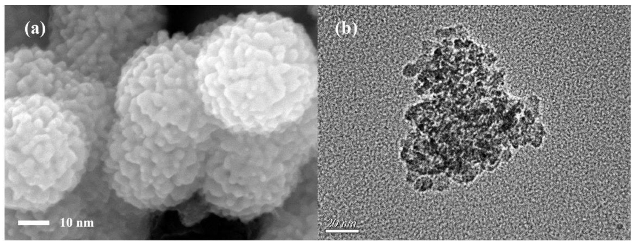
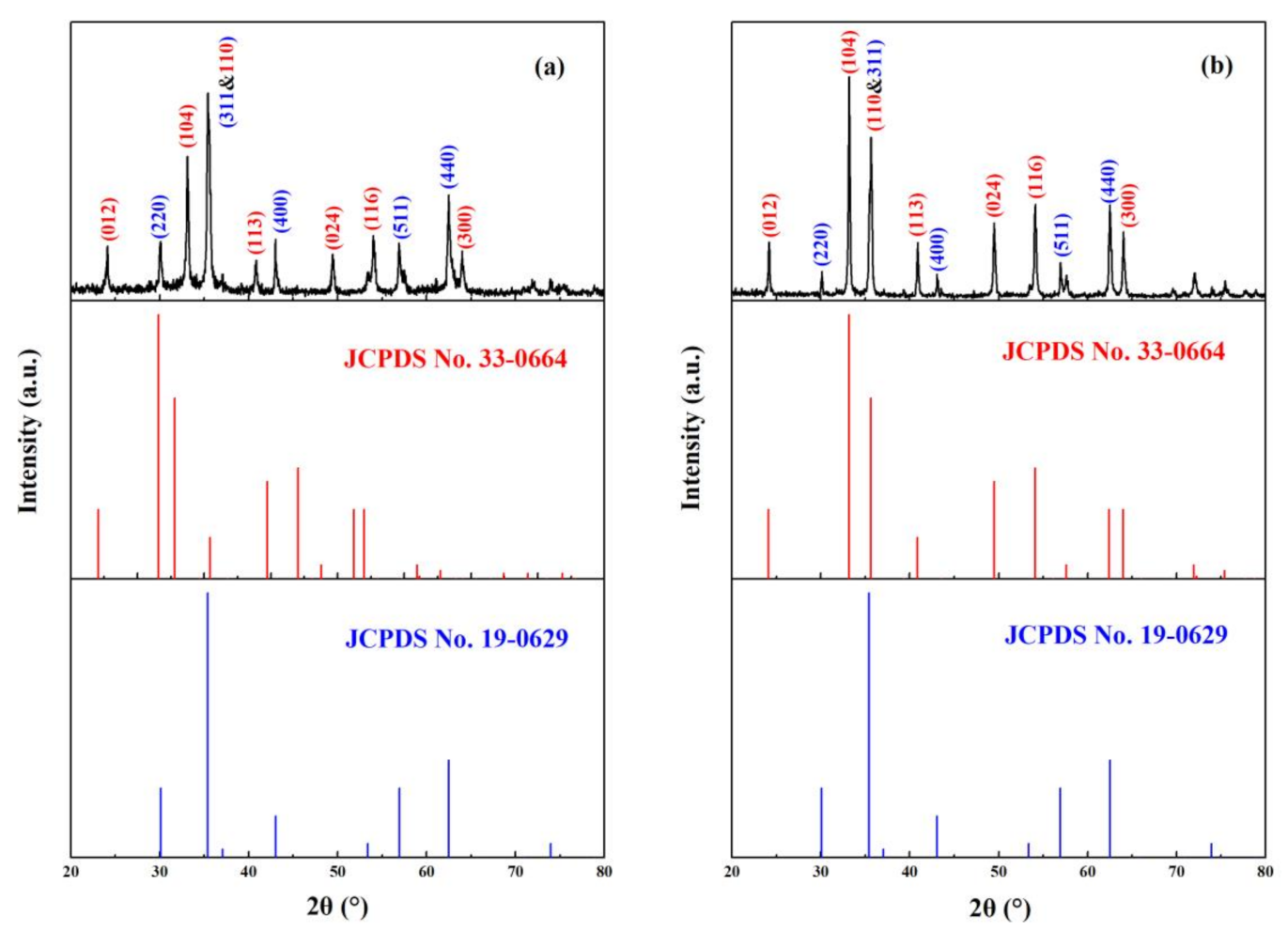
3.1. Characteristics of Magnetic α-Fe2O3/Fe3O4 Heterogeneous Nanoparticles

The SEM morphology and TEM image of α-Fe2O3/Fe3O4 heterogeneous nanoparticles calcined at 350 °C for 2 h with the heating rate of 3 °C/min and the molar ratio of ferric nitrate and urea of 1:4 are revealed in Figure 1. From the SEM morphology (shown in Figure 1a), it can be seen that the size distribution of magnetic α-Fe2O3/Fe3O4 heterogeneous nanoparticles was uniform. The nanomaterials were composed of several nanograins, which resulted in irregular sphere particles. The roughness of the particle surface increased the specific surface area of the nanoparticles, and the average particle size of them was around 42 nm. The TEM image of magnetic α-Fe2O3/Fe3O4 heterogeneous nanoparticles is displayed in Figure 1b; obviously, the magnetic α-Fe2O3/Fe3O4 heterogeneous nanoparticles had a polycrystalline structure, and the average particle size was approximately 42 nm, which agreed with the result of SEM morphology.
Figure 2a,b displays the X-ray diffraction patterns of magnetic α-Fe2O3/Fe3O4 heterogeneous nanoparticles calcined at 400 and 500 °C for 2 h with the heating rate of 3 °C/min and the molar ratio of ferric nitrate and urea of 1:4, respectively. Among them, the diffraction peaks of Figure 2a,b basically corresponded to the standard PDF cards of α-Fe2O3 and Fe3O4. Compared with the α-Fe2O3 standard PDF card (JCPDS No. 33-0664) and the Fe3O4 standard PDF card (JCPDS No. 19-0629), the ratio of diffraction peaks at 33° and 35.6° in Figure 2a were significantly smaller than those in the α-Fe2O3 standard PDF card, indicating that there was Fe3O4 diffraction peak at 35.6°. The results proved the existence of Fe3O4. Although the peak of α-Fe2O3 also existed at 35.6°, the d-spacing values of Sample A at the diffraction peak at 35.6° (shown in Table 1) belonged to Fe3O4 instead of α-Fe2O3 because the d-spacing values (2.5322) matched Fe3O4 (2.5320) more. In iron oxides, both Fe3O4 and γ-Fe2O3 had the same cubic spinel crystalline structure, and it was very difficult to differentiate them with XRD patterns. However, they could be distinguished using d-spacing values. The d-spacing values of Samples A and B, together with the JCPDS files of Fe3O4 and γ-Fe2O3, are listed in Table 1. It can be seen that experimental data matched with the d-spacing values of Fe3O4 better, indicating the formation of Fe3O4. There are no γ-Fe2O3-specific diffraction peaks at 26° in the two figures, implying that the other substance was Fe3O4 instead of γ-Fe2O3. The difference between Samples A and B was that as the calcination temperature increased, the intensity of the diffraction peak at 35.6° significantly decreased, indicating that the relative content of Fe3O4 was lowered at 500 °C, but it could not be said that Fe3O4 disappeared completely after the calcination temperature of 500 °C. The presence of a weak Fe3O4 diffraction peak can still be seen in Figure 2b. These characterizations confirmed that magnetic α-Fe2O3/Fe3O4 heterogeneous nanoparticles were successfully obtained.

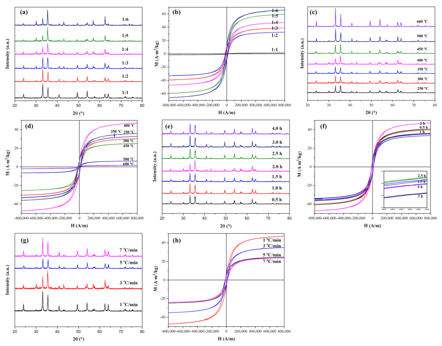
3.2. Effects of Experimental Conditions on the Ratio of α-Fe2O3 and Fe3O4 in Heterogeneous Nanoparticles

The effects of the calcination temperature, the calcination time, the ratio of ferric nitrate and urea, and the heating rate on the relative content of Fe3O4 in the heterogeneous nanoparticles were investigated by changing the preparation conditions. As mentioned above, the phase composition and grain characteristics of the sample could be analyzed from XRD. However, for this nanomaterial, the magnetic properties of nanoparticles were also needed to be measured in order to better apply them. VSM was a magnetic parameter measurement system that made use of the small sample to make tiny wobbles in the magnetic field and induced the electromotive force from the adjacent coil. The magnetic properties of the heterogeneous nanoparticles could be obtained by comparing the magnetic hysteresis loops measured by VSM. Figure 3a,b shows the XRD and VSM patterns of the heterogeneous nanoparticles obtained at a calcination temperature of 400 °C for 2 h with the heating rate of 3 °C·min−1 with various molar ratios of ferric nitrate and urea. As can be seen from Figure 3b, the saturation magnetization of the heterogeneous nanoparticles became larger with the increase of urea amount, and the saturation magnetizations could reach 66.6 A·m2/kg when the molar ratio of ferric nitrate and urea was 1:6. Because α-Fe2O3 showed weak magnetic properties [29], the enhancement of the magnetic properties proved the increase of the Fe3O4 content in the nanomaterial.
The formation mechanism of Fe2O3/Fe3O4 heterogeneous nanoparticles could be understood as similar to the thermal decomposition reaction of [Fe(CON2H4)6](NO3)3 [30]. As the temperature rose, the water in the mixed solution evaporated quickly, which caused Fe3+ to react with CON2H4 and finally formed [Fe(CON2H4)6](NO3)3. The thermal decomposition of [Fe(CON2H4)6](NO3)3 involved a two-stage decomposition process. Fe(NO3)3 and CON2H4 were the main products in the first step. The products of the second stage were likely to depend on the heat treatment atmosphere. As the temperature increased, the evaporation of water in the crucible temporarily created a vacuum environment around the crucible. At the same time, the ammonia gas generated by the decomposition of urea would act to isolate oxygen around the crucible. The released CON2H4 in the first stage could not escape completely from the crucible by evaporation; hence, it could partially reduce Fe3+ to Fe2+, leading to the formation of Fe3O4. Fe2O3 was generated by redundant Fe(NO3)3 decomposition and also formed by oxidation of Fe3O4 during calcination.
Fe [ CON 2 H 4 ) 6 ( NO 3 ) 3 3 Fe ( NO 3 ) 3 + CON 2 H 4
3 Fe ( NO 3 ) 3 +   CON 2 H 4   Fe 3 O 4 + CO 2 + 4 NO + 7 NO 2 + O 2 +   2 H 2
When the ratio of ferric nitrate and urea was 1:1, the product was completely Fe2O3. It could be considered that the mixed reaction system formed due to the lack of air blocked by the solution, resulting in the inability to form Fe3O4, or the amount of Fe3O4 formed by the reaction was so little that Fe3O4 was completely oxidized to Fe2O3. When the ratio reached 1:2, Fe3O4 started to be generated, which was inferred from the sudden increase in magnetism. According to the reaction mechanism, it could be foreseen that the amount of Fe3O4 was gradually increased with the increase of the amount of urea, but the complete generation of Fe3O4 did not occur. This was due to the decomposition of urea itself and the oxidation of Fe3O4 during calcination.
Figure 3c,d shows XRD and VSM patterns of Fe2O3/Fe3O4 heterogeneous nanoparticles obtained at different calcination temperatures under preparation conditions. The molar ratio of ferric nitrate and urea was 1:4, the eating rate was 3 °C·min−1, and the calcination time was set as 2 h. From the XRD patterns shown in Figure 3c, it can be observed that the diffraction peak became higher and narrower as the temperature increased, indicating that the crystallinity became higher, and the particle size became larger. The ratio of diffraction peaks at 33° and 35.6° became smaller and smaller until the temperature reached 400 °C and rose again after 400 °C. As displayed in Figure 3d, it is apparent that Fe3O4 began to form at 250 °C due to the higher magnetic properties of Fe3O4. The content of Fe3O4 reached the maximum at 400 °C, which illustrated that the reduction reaction completed at 400 °C and partial Fe3O4 was gradually transformed to α-Fe2O3 after 400 °C. Because the amount of urea volatilized in the reaction temperature was difficult to control (urea involved in the reaction may be less), it showed an increase in the reaction temperature but a decrease in magnetic properties, such as magnetic properties of the heterogeneous nanoparticles at 250, 300, and 350 °C.
The effect of the calcination time on Fe2O3/Fe3O4 heterogeneous nanoparticles was investigated and is shown in Figure 3e,f. The molar ratio of ferric nitrate and urea was 1:4, the heating rate was 3 °C·min−1, and the calcination temperature was 400 °C. The saturation magnetization of Fe2O3/Fe3O4 heterogeneous nanoparticles reached maximum after 2 h of calcination. Combined with the effect of the calcination temperature on Fe2O3/Fe3O4 heterogeneous nanoparticles, the reaction was completed at 400 °C for 2 h. Figure 3g,h shows XRD and VSM patterns of Fe2O3/Fe3O4 heterogeneous nanoparticles obtained with different heating rates, the molar ratio of ferric nitrate and urea of 1:4 at the calcination temperature of 400 °C, for a calcination time of 2 h. The difference in magnetic properties caused by various heating rates might be related to the total reaction time (including heating, calcination, and cooling time). In short, 3 °C·min−1 became the condition that Fe3O4 was most generated when it was calcined at 400 °C for 2 h.

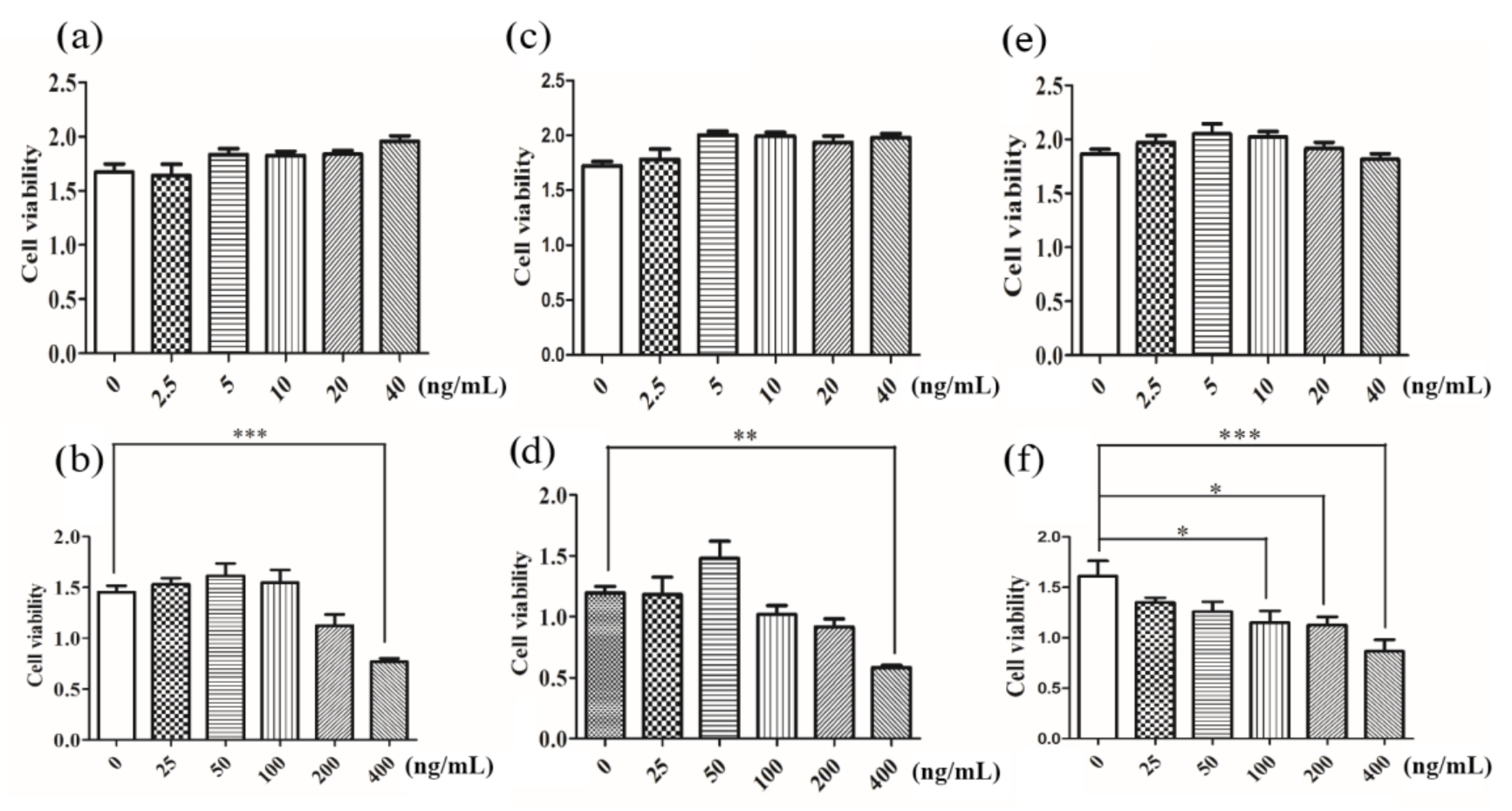
3.3. In Vitro Cytotoxicity Assay

Increasing the use of metal oxide nanoparticles requires a better understanding of their potential impact on human health. Whether the Fe2O3/Fe3O4 heterogeneous nanoparticles can enter the cell also depended on the particle size of the heterogeneous nanoparticles [31,32,33]. In the experiments, Prussian blue staining was first applied to determine whether the nanoparticles entered the cells, and most of the nanoparticles entered the cells through the endocytic pathway. The cytotoxicity of Fe2O3/Fe3O4 heterogeneous nanoparticles to human hepatocytes L-02 was further evaluated by the different cytotoxic effects through CCK 8 assay, reactive oxygen species (ROS), lactate dehydrogenase (LDH), MDA, and T-AOC to assess the end points of toxicity.

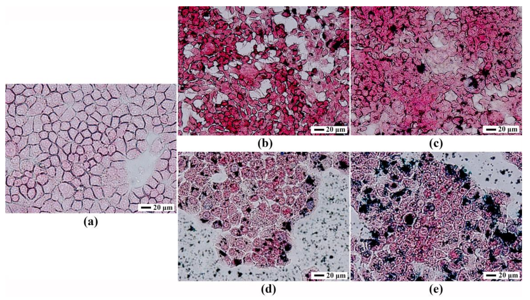
3.4. Prussian Blue Staining

Prussian blue was a common and sensitive stain to detect the presence of iron in cells or biopsy specimens by pathologists [34]. Ferric iron deposited in tissues and cells reacted with soluble ferrocyanide in the stain and transformed into insoluble Prussian blue dye, a complex hydrated ferric ferrocyanide substance. This allowed Prussian blue to be used to detect whether the material entered into the cells. Figure 4 shows the results of Prussian blue staining. After the Fe2O3/Fe3O4 heterogeneous nanoparticles were cocultured with the hepatocytes for 24 h, the Prussian blue staining results visually showed the distribution of the heterogeneous nanoparticles in the cells. Under the inverted microscope, blue iron particles were observed in the cells, indicating that the Fe2O3/Fe3O4 heterogeneous entered into the cells. However, the localization to the cytoplasm or nucleus required subsequent electron microscopy.

3.5. Cell Counting Kit-8 and LDH Detection of Cell Viability

Assessment of cellular toxicity was an essential prerequisite to the as-prepared magnetic Fe2O3/Fe3O4 heterogeneous nanoparticles in biomedical applications. CCK8 (Cell Counting Kit-8) cell viability assay was a common method to measure the cytotoxicity of drugs [35,36]. Its advantages were accurate, rapid, and stable, and the reagent itself was less cytotoxic. The amount of formazan was proportional to the number of living cells. Therefore, this property could be used to directly perform cell proliferation and toxicity analysis. As evidenced in Figure 5, the results of cell viability demonstrated that the effect of the low dose (0–40 ng/mL) of Fe2O3/Fe3O4 heterogeneous nanoparticles on cell viability was not statistically significant, which identified that the Fe2O3/Fe3O4 heterogeneous nanoparticles had excellent biocompatibility in the in vitro test. At a high dose (40–400 ng/mL), when the concentration was greater than 100 ng/mL, the cell activities were significantly decreased, which was attributed to the cell biological effects induced by nanoparticles due to the size, morphology, surface charge, composition of the different nanomaterial particles, and the selected cell model. The CCK8 results initially showed that the concentration of Fe2O3/Fe3O4 heterogeneous nanoparticles had less effect on cell viability when the concentration was less than 100 ng/mL.

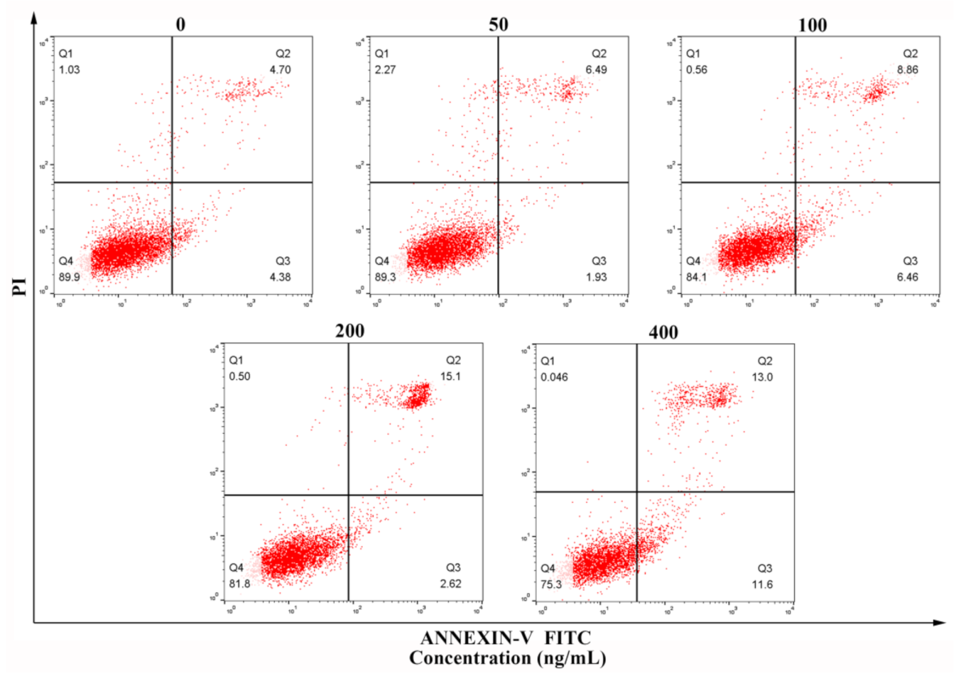
3.6. Flow Cytometry Detection of Apoptosis

Apoptosis might be one of the mechanisms of Fe2O3/Fe3O4 heterogeneous nanoparticles cytotoxicity [37]. Apoptosis was the autonomous and orderly death of cells under gene control, which was a process of nuclear condensation, chromosomal DNA degradation, cell shrinkage, and finally the formation of apoptotic bodies [38]. Figure 6 shows that when the concentration of Fe2O3/Fe3O4 heterogeneous nanoparticles was more than 100 ng/mL, the apoptosis increased significantly. When the concentration was less than 100 ng/mL, there was no effect on apoptosis.

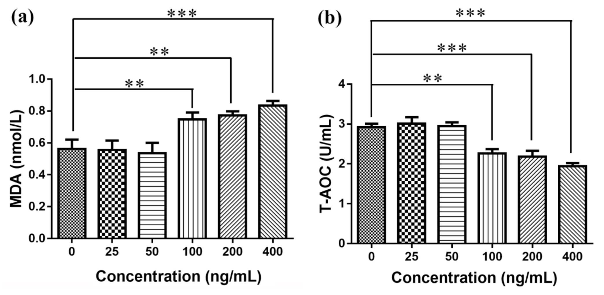
3.7. Elisa Detects MDA Production and T-AOC

At present, the toxicity mechanism of nanomaterials was mainly oxidative stress, and oxidative stress was one of the basic ways of cell damage in organisms. Oxidative stress referred to the process of oxidative damage caused by the increase or elimination of oxygen free radicals in the body tissues or cells, resulting in the accumulation of reactive oxygen species in the body or cells [39]. ROS could attack biofilm unsaturated fatty acids to form lipid peroxides, and its product MDA was a harmful substance, which could be often used as an indicator to evaluate the strength of lipid peroxidation [40]. T-AOC was the reaction tissue cell enzymatic and an important indicator of the total antioxidant capacity of nonenzymatic systems [41]. This study found that when the concentration was greater than 100 ng/mL, compared with the normal control group, the amount of MDA production in each group was significantly increased, and the T-AOC ability was significantly decreased, as shown in Figure 7. When the concentration was less than 100 ng/mL, compared with the normal control group, the amount of MDA produced and T-AOC ability were not statistically significant.

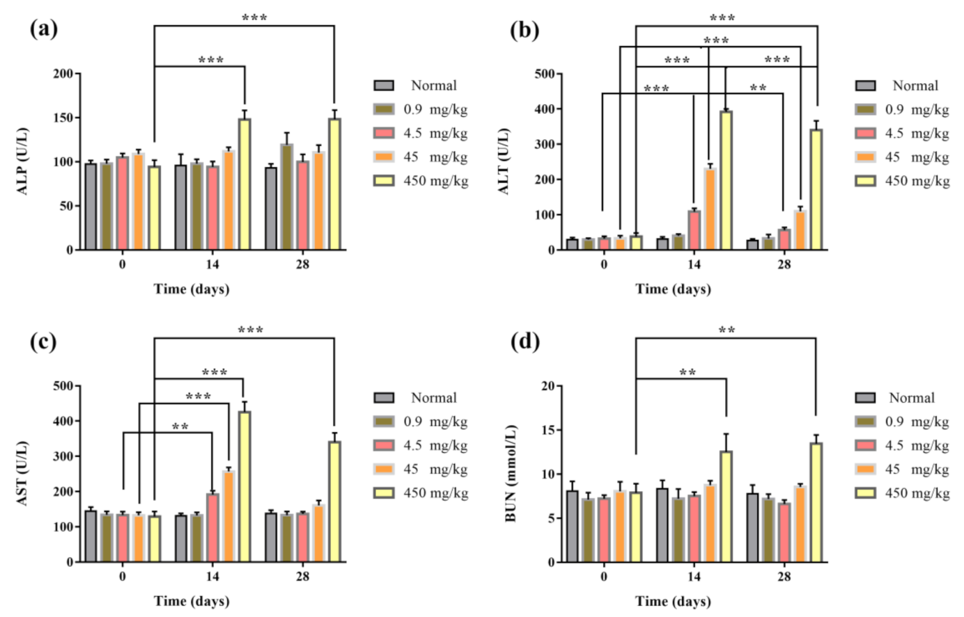
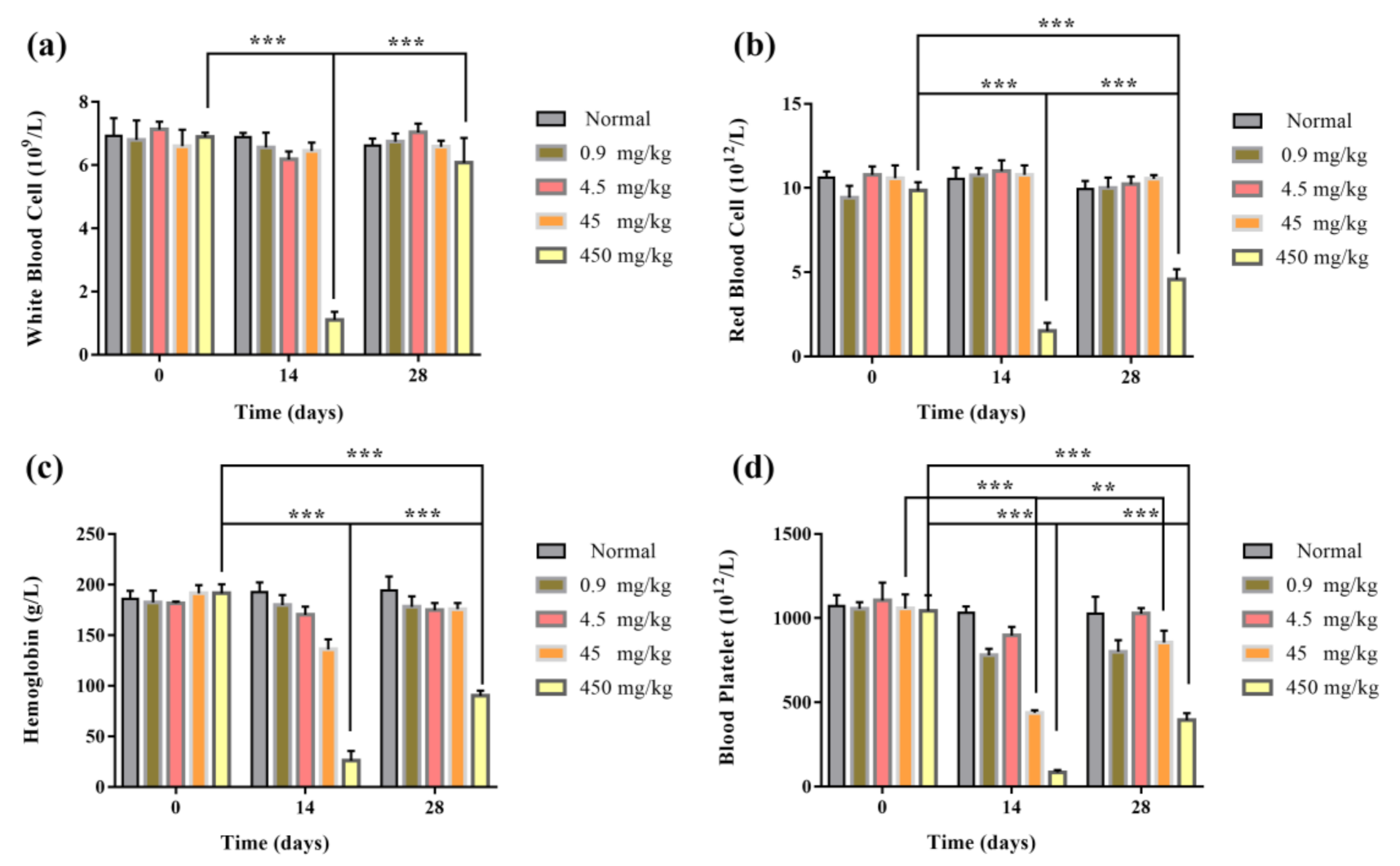
3.8. Blood Routine and Biochemical Assay of Mice

After the tail vein injection of Fe2O3/Fe3O4 heterogeneous nanoparticles, the blood routine and blood biochemical results of mice in each group were detected at Days 0, 14, and 28. The blood routine indexes included white blood cell (WBC), red blood cell (RBC), hemoglobin (HB), and blood platelet (blood). Blood biochemical indexes included alanine aminotransferase (ALT), aspartate aminotransferase (AST), alkaline phosphatase (ALP), and blood urea nitrogen (BUN). The results of the blood routine test showed that the blood routine values of 0.9 and 4.5 mg/kg groups were in the normal range. After 14 days of administration, compared with the control group, the platelet content of the 45 mg/kg group was significantly decreased, and the number of white blood cells in the 450 mg/kg group decreased significantly after 14 days and returned to normal value at 28 days. It might be that the nanomaterials had a certain damage effect on white blood cells (Figure 8). Blood biochemical results showed that the values of ALT and AST in the 4.5, 45, and 450 mg/kg groups were significantly lower than those in the control group after 14 days compared with the control group, indicating that liver injury might have been caused. In the 450 mg/kg group, after 14 days of administration, the renal function index blood urea nitrogen increased significantly, indicating that renal injury might have been caused (Figure 9). In conclusion, the results of the blood routine test and blood biochemistry in the 0.9 mg/kg group were within the normal range within 0–28 days after administration, and the tail vein administration concentration of 0.9 mg/kg had good biological safety.

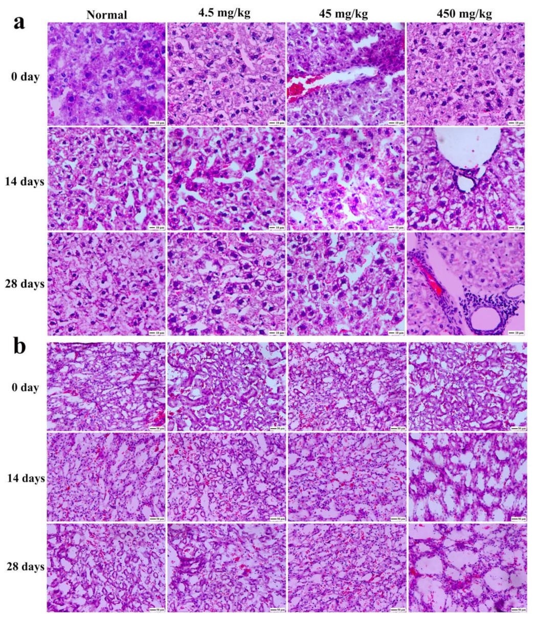
3.9. Histopathological Section Observation of Mice

Figure 10 showed the hematoxylin and eosin (H&E) staining of tumors on various days with different doses of Fe2O3/Fe3O4 heterogeneous nanoparticles. It could be observed that the tissue section had no obvious pathological changes within the injection range of 45 mg/kg, and there were no significant differences between a and b, which indicated that the Fe2O3/Fe3O4 heterogeneous nanoparticles had high biocompatibility in vivo. Additionally, H&E staining with the injection dose of 45 mg/kg revealed higher apoptosis compared with the control group, confirming that the Fe2O3/Fe3O4 heterogeneous nanoparticles induced cell apoptosis at a late stage at a high dose.

4. Conclusions

A novel type of magnetic Fe2O3/Fe3O4 heterogeneous nanoparticles, which were spherical nanomaterials with a uniform size distribution, was successfully prepared via a facile solution combustion process, using ferric nitrate and urea as raw materials. The average particle size of the heterogeneous nanoparticles was 42 nm, and the content of Fe3O4 in Fe2O3/Fe3O4 heterogeneous nanoparticles was mainly affected by the ratio of ferric nitrate and urea, the reaction temperature, the reaction time, and the heating rate. Under the condition that the ratio of ferric nitrate and urea was 1:4, the Fe2O3/Fe3O4 heterogeneous nanoparticles with the highest Fe3O4 content could be obtained at 400 °C for 2 h with the heating rate of 3 °C·min−1. The saturation magnetization strength of the heterogeneous nanoparticles was 47.1 A·m2/kg. As the amount of urea increased, the amount of Fe3O4 naturally increased, and the selectivity was high. The Fe2O3/Fe3O4 heterogeneous nanoparticles could enter cells by endocytosis. The CCK8 results initially showed that Fe2O3/Fe3O4 heterogeneous nanoparticles had no effect on cell viability when the concentration of nanoparticles was less than 100 ng·mL−1. When the concentration was greater than 100 ng·mL−1, oxidative stress and apoptosis were observed in L-02 cells, and the heterogeneous nanoparticles displayed cytotoxicity. MDA production increased and the total antioxidant capacity declined. The nanoparticles were cytotoxic, the toxicity was concentration dependent, and they had good biological safety within the concentration of 0.9 mg/kg.

Author Contributions

Conceptualization, Z.W.; Formal analysis, Y.L.; Funding acquisition, R.L.; Investigation, Y.L.; Methodology, Z.W. and R.L.; Project administration, R.L.; Resources, R.L.; Software, Y.L.; Supervision, R.L.; Validation, R.L.; Visualization, Z.W.; Writing—original draft, Y.L.; Writing—review & editing, Z.W. and R.L. All authors have read and agreed to the published version of the manuscript.

Funding

The work was supported by the Jiangsu Provincial Postgraduate Scientific Practice and Innovation Project (Grant No. SJCX20_1432) and the Science and Technology Innovation Project of CHN Energy (Grant No. GJNY-20-109).

Institutional Review Board Statement

The study was conducted according to the guidelines of the Declaration of Helsinki, and approved by the Laboratory Animal Center of Jiangsu University (protocol code SYXK 2018-0053 and date of approval is 26 October 2018 to 25 October 2023).

Data Availability Statement

The data presented in this study are openly available in ICDD-PDF.

Conflicts of Interest

The authors declare that they have no known competing financial interests or personal relationships that could have appeared to influence the work reported in this paper.

References

  1. Gujre, N.; Mitra, S.; Soni, A.; Agnihotri, R.; Rangan, L.; Rene, E.R.; Sharma, M.P. Speciation, contamination, ecological and human health risks assessment of heavy metals in soils dumped with municipal solid wastes. Chemosphere 2021, 262, 128013. [Google Scholar] [CrossRef]
  2. Arsalani, S.; Oliveira, J.; Guidelli, E.J.; Araujo, J.F.D.F.; Wiekhorst, F.; Baffa, O. Synthesis of radioluminescent iron oxide nanoparticles functionalized by anthracene for biomedical applications. Colloids Surf. A 2020, 602, 125105. [Google Scholar] [CrossRef]
  3. Duan, M.; Shapter, J.G.; Qi, W.; Yang, S.; Gao, G. Recent progress in magnetic nanoparticles: Synthesis, properties, and applications. Nanotechnology 2018, 29, 452001. [Google Scholar] [CrossRef]
  4. Mu, L.; Zhou, Q.; Sun, D.W.; Wang, M.; Chai, X.; Wang, M. The application of resting magnetic resonance imaging in the cognitive judgment of Parkinson. World Neurosurg. 2020, 138, 672–679. [Google Scholar] [CrossRef] [PubMed]
  5. Rost, N.C.V.; Sen, K.; Savliwala, S.; Singh, I.; Liu, S.; Unni, M.; Raniero, L. Magnetic particle imaging performance of liposomes encapsulating iron oxide nanoparticles. J. Magn. Magn. Mater. 2020, 504, 166675. [Google Scholar] [CrossRef]
  6. Zhi, D.F.; Yang, T.; Yang, J.; Fu, S.; Zhang, S.B. Targeting strategies for superparamagnetic iron oxide nanoparticles in cancer therapy. Acta Biomater. 2020, 102, 13–34. [Google Scholar] [CrossRef] [PubMed]
  7. Teng, Y.; Du, Y.M.; Shi, J.; Pong, W.T. Magnetic iron oxide nanoparticle-hollow mesoporous silica Spheres: Fabrication and potential application in drug delivery. Curr. Appl. Phys. 2020, 20, 320–325. [Google Scholar] [CrossRef]
  8. Elrahman, A.A.A.; Mansour, F.R. Targeted magnetic iron oxide nanoparticles: Preparation, functionalization and biomedical application. J. Drug Deliv. Sci. Tec. 2019, 52, 702–712. [Google Scholar] [CrossRef]
  9. Huang, C.Z.; Wu, J.L.; Jiang, W.; Liu, R.L.; Li, Z.H.; Luan, Y.X. Amphiphilic prodrug-decorated graphene oxide as a multi-functional drug delivery system for efficient cancer therapy. Mat. Sci. Eng. C 2018, 89, 15–24. [Google Scholar] [CrossRef]
  10. Kashkouli, K.I.; Torkzadeh-Mahani, M.; Mosaddegh, E. Synthesis and characterization of aminotetrazole-functionalized magnetic chitosan nanocomposite as a novel nanocarrier for targeted gene delivery. Mat. Sci. Eng. C 2018, 89, 166–174. [Google Scholar] [CrossRef]
  11. Mebrahtu, C.; Abate, S.; Chen, S.M.; Salazar, A.F.S.; Perathoner, S.; Krebs, F.; Palkovits, R. Enhanced catalytic activity of iron-promoted nickel on γ-Al2O3 nanosheets for carbon dioxide methanation. Energy Technol. 2018, 6, 1196–1207. [Google Scholar] [CrossRef]
  12. Nagai, S.; Ueda, K.; Ohyama, J.; Satsuma, A. Enhanced three way catalytic activity of NiFe2O4 by physically mixed metal oxides. Cataly. Today 2018, 303, 40–45. [Google Scholar] [CrossRef]
  13. Taebi, S.; Keyhanfar, M.; Noorbakhsh, A. A novel method for sensitive, low-cost and portable detection of hepatitis B surface antigen using a personal glucose meter. J. Immunol. Methods 2018, 458, 26–32. [Google Scholar] [CrossRef]
  14. Santana, W.M.O.S.D.; Caetano, B.L.; Annunzio, S.R.D.; Pulcinelli, S.H.; Ménager, C.; Fontana, C.R.; Santilli, C.V. Conjugation of superparamagnetic iron oxide nanoparticles and curcumin photosensitizer to assist in photodynamic therapy. Colloids Surf. B 2020, 196, 111297. [Google Scholar] [CrossRef] [PubMed]
  15. Zheng, Y.L.; Li, Z.Y.; Chen, H.J.; Gao, Y. Nanoparticle-based drug delivery systems for controllable photodynamic cancer therapy. Eur. J. Pharm. Sci. 2020, 144, 105213. [Google Scholar] [CrossRef] [PubMed]
  16. Stanković, A.; Mihailović, J.; Mirković, M.; Radović, M.; Milanović, Z.; Ognjanović, M.; Janković, D.; Antić, B.; Mijović, M.; Vranješ-Đurić, S.; et al. Aminosilanized flower-structured superparamagnetic iron oxide nanoparticles coupled to 131I-labeled CC49 antibody for combined radionuclide and hyperthermia therapy of cancer. Int. J. Pharm. 2020, 587, 119628. [Google Scholar] [CrossRef]
  17. Mühlberger, M.; Janko, C.; Unterweger, H.; Band, J.; Schreiber, E.; Lehmann, C.; Dudziak, D.; Lee, G.; Alexiou, C.; Tietze, R. Non-magnetic chromatographic separation of colloidally metastable superparamagnetic iron oxide nanoparticles and suspension cells. J. Chromatogr. B 2019, 1122–1123, 83–89. [Google Scholar] [CrossRef]
  18. Pérez, A.G.; González-Martínez, E.; Águila, C.R.D.; González-Martínez, D.A.; Ruiz, G.G.; Artalejo, A.G.; Yee-Madeira, H. Chitosan-coated magnetic iron oxide nanoparticles for DNA and rhEGF separation. Colloids Surf. A 2020, 591, 124500. [Google Scholar] [CrossRef]
  19. Leal, E.; Dantas, J.; Santos, S.P.T.A.D.; Bicalho, M.D.C.M.; Kiminami, R.H.G.A.; Silva, M.R.D.; Costa, A.C.F.D.M. Effect of the surface treatment on the structural, morphological, magnetic and biological properties of MFe2O4 iron spinels (M=Cu, Ni, Co, Mn and Fe). Appl. Surf. Sci. 2018, 455, 635–645. [Google Scholar] [CrossRef]
  20. Srinivasan, S.Y.; Paknikar, K.M.; Bodas, D.; Gajbhiye, V. Applications of cobalt ferrite nanoparticles in biomedical nanotechnology. Nanomedicine 2018, 13, 1221–1328. [Google Scholar] [CrossRef]
  21. Suturin, S.; Kaveev, A.; Korovin, A.; Fedorov, V.; Sawada, M.; Sokolov, N. Structural transformations and interfacial iron reduction in heterostructures with epitaxial layers of 3d metals and ferrimagnetic oxides. J. Appl. Crystallogr. 2018, 51, 1069–1081. [Google Scholar] [CrossRef]
  22. Lassoued, A.; Lassoued, M.S.; Dkhil, B.; Ammar, S.; Gadri, A. Synthesis, structural, morphological, optical and magnetic characterization of iron oxide (α-Fe2O3) nanoparticles by precipitation method: Effect of varying the nature of precursor. Phys. E 2018, 97, 328–334. [Google Scholar] [CrossRef]
  23. Ghimire, S.; Dho, J. Anisotropic lattice disorder and enhanced magnetic anisotropy in Fe3O4 films on (110) SrTiO3. J. Magn. Magn. Mater. 2018, 468, 209–214. [Google Scholar] [CrossRef]
  24. Amiri, M.; Salavati-Niasari, M.; Akbari, A. Magnetic nanocarriers: Evolution of spinel ferrites for medical applications. Adv. Colloids Interface 2019, 265, 29–44. [Google Scholar] [CrossRef]
  25. Sharafi, Z.; Bakhshi, B.; Javidi, J.; Adrangi, S. Synthesis of Silica-coated Iron Oxide Nanoparticles: Preventing Aggregation Without Using Additives or Seed Pretreatment. Iran. J. Pharm. Res. 2018, 17, 386–395. [Google Scholar]
  26. Abo-zeid, Y.; Ismail, N.S.M.; McLean, G.R.; Hamdy, N.M. A molecular docking study repurposes FDA approved iron oxide nanoparticles to treat and control COVID-19 infection. Eur. J. Pharm. Sci. 2020, 153, 105465. [Google Scholar] [CrossRef]
  27. Han, Y.N.; Hu, B.; Wang, M.Y.; Yang, Y.; Zhang, L.; Zhou, J.; Chen, J.H. pH-Sensitive tumor-targeted hyperbranched system based on glycogen nanoparticles for liver cancer therapy. Appl. Mater. Today 2020, 18, 100521. [Google Scholar]
  28. Qiao, J.B.; Fan, Q.Q.; Zhang, C.L.; Lee, J.; Byun, J.; Xing, L.; Gao, X.D.; Oh, Y.K.; Jiang, H.L. Hyperbranched lipoid-based lipid nanoparticles for bidirectional regulation of collagen accumulation in liver fibrosis. J. Control. Releas. 2020, 321, 629–640. [Google Scholar] [CrossRef] [PubMed]
  29. Huang, W.; Pan, S.; Li, Y.; Yu, L.L.; Liu, R.J. Immobilization and characterization of cellulase on hydroxy and aldehyde functionalized magnetic Fe2O3/Fe3O4 nanocomposites prepared via a novel rapid combustion process. Int. J. Biol. Macromol. 2020, 162, 845–852. [Google Scholar] [CrossRef] [PubMed]
  30. Asuha, S.; Suyala, B.; Siqintana, X.; Zhao, S. Direct synthesis of Fe3O4 nanopowder by thermal decomposition of Fe–urea complex and its properties. J. Alloy. Compd. 2011, 509, 2870–2873. [Google Scholar] [CrossRef]
  31. Chakraborty, A.; Das, A.; Raha, S.; Barui, A. Size-dependent apoptotic activity of gold nanoparticles on osteosarcoma cells correlated with SERS signal. J. Photoch. Photobio. B 2020, 203, 111778. [Google Scholar] [CrossRef] [PubMed]
  32. Nwoko, K.C.; Raab, A.; Cheyne, L.; Dawson, D.; Krupp, E.; Feldmann, J. Matrix-dependent size modifications of iron oxide nanoparticles (Ferumoxytol) spiked into rat blood cells and plasma: Characterisation with TEM, AF4-UV-MALS-ICP-MS/MS and spICP-MS. J. Chromatogr. B 2019, 1124, 356–365. [Google Scholar] [CrossRef]
  33. Ha, S.W.; Viggeswarapu, M.; Habib, M.M.; Beck, G.R. Bioactive effects of silica nanoparticles on bone cells are size, surface, and composition dependent. Acta Biomater. 2018, 82, 184–196. [Google Scholar] [CrossRef] [PubMed]
  34. Catala, L.; Mallah, T. Nanoparticles of Prussian blue analogs and related coordination polymers: From information storage to biomedical applications. Coordin. Chem. Rev. 2017, 346, 32–61. [Google Scholar] [CrossRef]
  35. Zhao, X.Y.; Xing, F.J.; Cong, Y.W.; Zhuang, Y.; Han, M.X.; Wu, Z.Q.; Yu, S.L.; Wei, H.Y.; Wang, X.K.; Chen, G. Antimony trichloride induces a loss of cell viability via reactive oxygen species-dependent autophagy in A549 cells. Int. J. Biochem. Cell B. 2017, 93, 32–40. [Google Scholar] [CrossRef] [PubMed]
  36. Ooi, L.; Okazaki, K.; Arias-Barreiro, C.R.; Heng, L.Y.; Mori, I.C. Application of the cellular oxidation biosensor to Toxicity Identification Evaluations for high-throughput toxicity assessment of river water. Chemosphere 2020, 247, 125933. [Google Scholar] [CrossRef]
  37. Ghasemi, A.; Jafari, S.; Saeidi, J.; Mohtashami, M.; Salehi, I. Synthesis and characterization of polyglycerol coated superparamagnetic iron oxide nanoparticles and cytotoxicity evaluation on normal human cell lines. Colloids Surf. A 2018, 551, 128–136. [Google Scholar] [CrossRef]
  38. Won, Y.S.; Seo, K.I. Sanggenol L promotes apoptotic cell death in melanoma skin cancer cells through activation of caspase cascades and apoptosis-inducing factor. Food Chem. Toxicol. 2020, 138, 111221. [Google Scholar] [CrossRef]
  39. McGarry, T.; Biniecka, M.; Veale, D.J.; Fearon, U. Hypoxia, oxidative stress and inflammation. Free Radic. Biol. Med. 2018, 125, 15–24. [Google Scholar] [CrossRef]
  40. Sunilkumar, D.; Drishya, G.; Chandrasekharan, A.; Shaji, S.K.; Nair, B.G. Oxyresveratrol drives caspase-independent apoptosis-like cell death in MDA-MB-231 breast cancer cells through the induction of ROS. Biochem. Pharmacol. 2020, 173, 113724. [Google Scholar] [CrossRef] [PubMed]
  41. Li, W.; Cai, Z.N.; Mehmood, S.; Wang, Y.; Pan, W.J.; Zhang, W.N.; Lu, Y.M.; Chen, Y. Polysaccharide FMP-1 from Morchella esculenta attenuates cellular oxidative damage in human alveolar epithelial A549 cells through PI3K/AKT/Nrf2/HO-1 pathway. Int. J. Biol. Macromol. 2018, 120, 865–875. [Google Scholar] [CrossRef] [PubMed]
Figure 1. SEM morphology (a) and TEM image (b) of Fe2O3/Fe3O4 heterogeneous nanoparticles calcined at 350 °C for 2 h with the heating rate of 3 °C/min and the molar ratio of ferric nitrate and urea of 1:4.
Nanomaterials 11 00834 g001
Figure 2. Comparisons of the XRD patterns of Fe2O3/Fe3O4 heterogeneous nanoparticles calcined at 400 (a) and 500 °C (b) for 2 h with the heating rate of 3 °C/min and the molar ratio of ferric nitrate and urea of 1:4 with the standard PDF cards (Fe2O3: JCPDS No. 33-0664; Fe3O4: JCPDS No. 19-0629).
Nanomaterials 11 00834 g002
Figure 3. XRD patterns and hysteresis loops of Fe2O3/Fe3O4 heterogeneous nanoparticles with various molar ratios of ferric nitrate and urea (a,b) calcined at various temperatures (c,d) for different times (e,f) with different heating rates (g,h).
Nanomaterials 11 00834 g003
Figure 4. Prussian blue staining of human hepatocytes L-02 incubated by Fe2O3/Fe3O4 heterogeneous nanoparticles at 0 (a), 50 (b), 100 (c), 200 (d), and 400 ng/mL (e).
Nanomaterials 11 00834 g004
Figure 5. Cell viability assay of Fe2O3/Fe3O4 heterogeneous nanoparticles calcined at various temperatures: (a,b) 250, (c,d) 300, and (e,f) 350 °C. * p ≤ 0.05, ** p ≤ 0.01, *** p ≤ 0.001 versus control group, n = 6.
Nanomaterials 11 00834 g005
Figure 6. Flow cytometry detection of apoptosis.
Nanomaterials 11 00834 g006
Figure 7. Elisa detection of malondialdehyde (MDA) production and total antioxidant capacity (T-AOC): (a) MDA generation and (b) T-AOC detection. ** p ≤ 0.01, *** p ≤ 0.001 versus control group, n = 6.
Nanomaterials 11 00834 g007
Figure 8. Blood biochemical indexes of mice injected via tail vein with 200 μL of Fe2O3/Fe3O4 heterogeneous nanoparticles solutions of various concentrations: (a) alkaline phosphatase (ALP), (b) alanine aminotransferase (ALT), (c) aspartate aminotransferase (AST), and (d) blood urea nitrogen (BUN) (the control groups were injected with the same volumes of normal saline). ** p ≤ 0.01, *** p ≤ 0.001 versus control group, n = 6.
Nanomaterials 11 00834 g008
Figure 9. The blood routine indexes of mice injected via tail vein with 200 μL of Fe2O3/Fe3O4 heterogeneous nanoparticles solutions of various concentrations: (a) white blood cell (WBC), (b) red blood cell (RBC), (c) hemoglobin (HB), and (d) blood platelet (blood) (the control groups were injected with the same volumes of normal saline). ** p ≤ 0.01, *** p ≤ 0.001 versus control group, n = 6.
Nanomaterials 11 00834 g009
Figure 10. Physiological section of lung from mice of the Fe2O3/Fe3O4 heterogeneous nanoparticles (a) and control groups (b) with different doses (hematoxylin staining).
Nanomaterials 11 00834 g010
Table 1. D-spacings of Samples A and B and those of Fe3O4, γ-Fe2O3, and α-Fe2O3 from the JCPDS files: 1 JCPDS file No. 19-0629; 2 JCPDS file No. 39-1346; 3 JCPDS file No. 33-0664.
Sample A (Å)Sample B (Å)Fe3O4 (Å) 1γ-Fe2O3 (Å) 2hklα-Fe2O3(Å) 3hkl
2.96662.96272.96702.9530220--
2.53222.51562.53202.51773112.5190110
2.10082.09802.09932.0886400--
1.61591.61441.61581.6073511--
1.48441.48441.48451.4758440--
Publisher’s Note: MDPI stays neutral with regard to jurisdictional claims in published maps and institutional affiliations.

