Nanocellulose/PEGDA Aerogels with Tunable Poisson’s Ratio Fabricated by Stereolithography for Mouse Bone Marrow Mesenchymal Stem Cell Culture
Abstract
1. Introduction
2. Materials and Methods
2.1. Materials and Reagents
2.2. Preparation of CNF Suspensions
2.3. Structural Design of Tissue Engineering Scaffolds
2.4. Preparation of CNF/PEGDA Photocurable Resin Mixtures
2.5. Fabrication of CNF/PEGDA Scaffolds
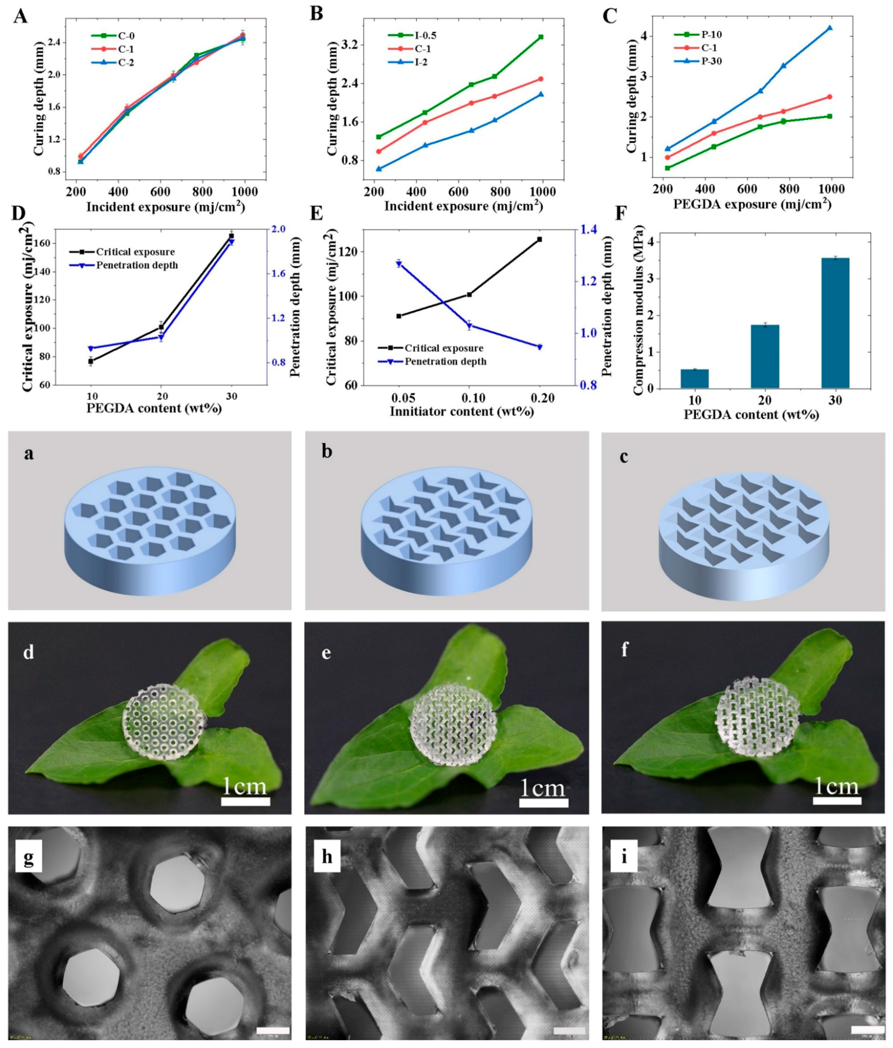
2.6. Analysis of the Clarity of CNF/PEGDA Hydrogel Structure Formation
2.7. Macromorphology and Micromorphology of CNF/PEGDA Scaffolds
2.8. Compressive Test of CNF/PEGDA Scaffolds
2.9. Stem Cell Culture and Differentiation on CNF/PEGDA Scaffolds
2.10. SEM Analysis of Cell–Scaffold Constructs
2.11. CLSM Analysis of Cell–Scaffold Constructs
2.12. MTT Analysis of Cell–Scaffold Constructs
2.13. Staining Analysis of Cartilage Matrix Secretion
2.14. HE Staining of Cell–Scaffold Constructs
3. Results and Discussion
3.1. Fabrication of CNF/PEGDA Hydrogels
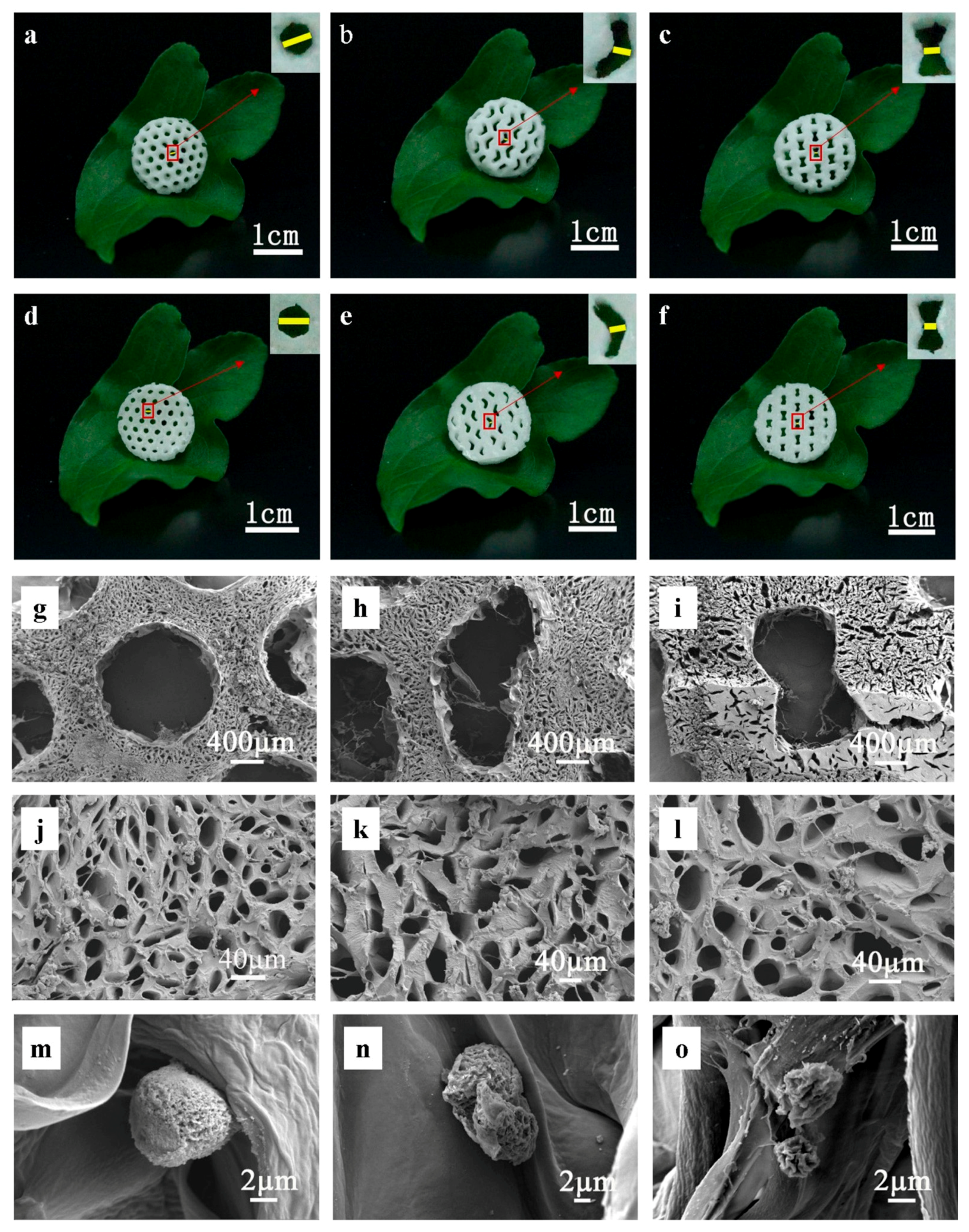
3.2. Fabrication of Variable Poisson’s Ratio CNF/PEGDA Aerogel Structures
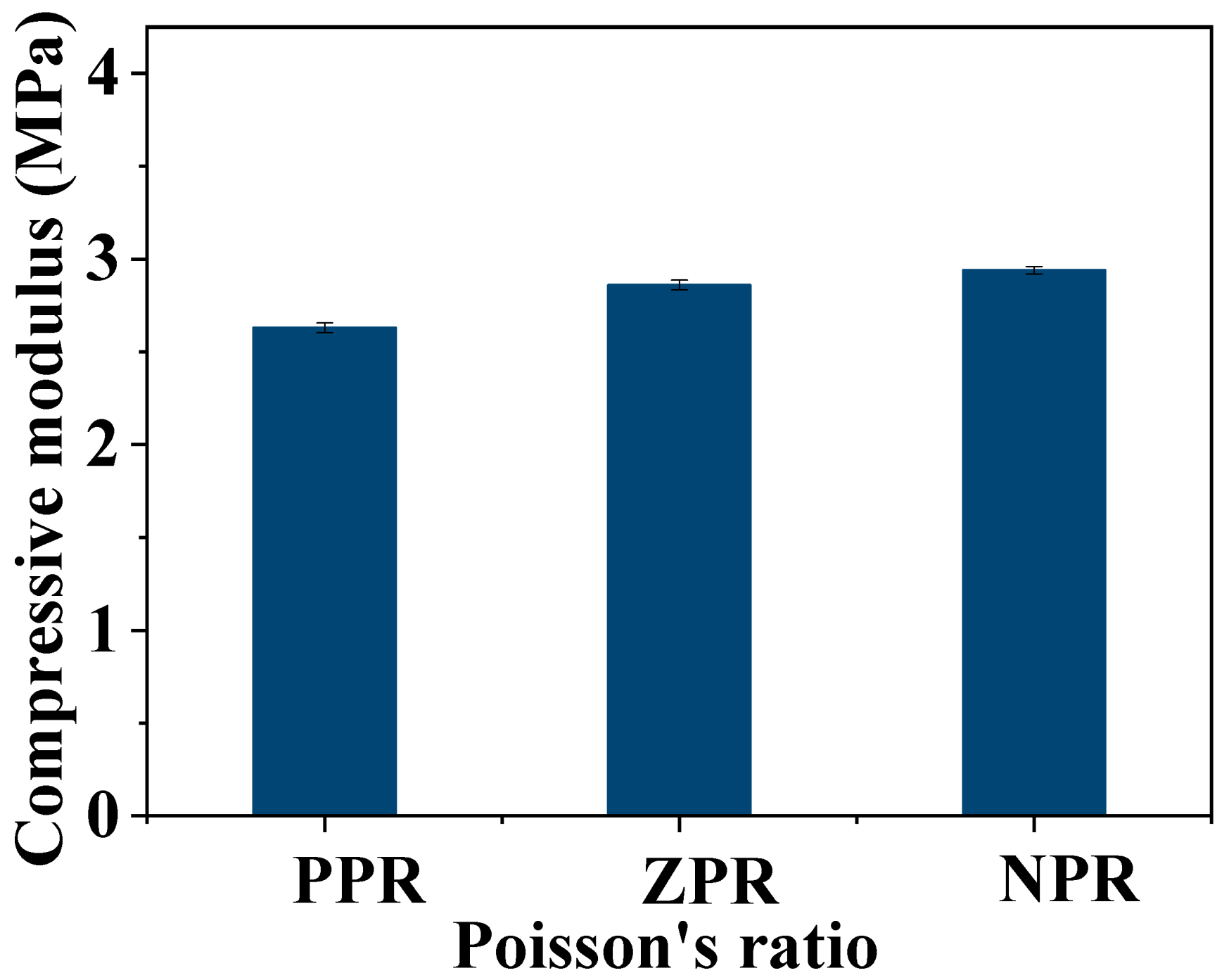
3.3. Compression Modulus of CNF/PEGDA Scaffolds
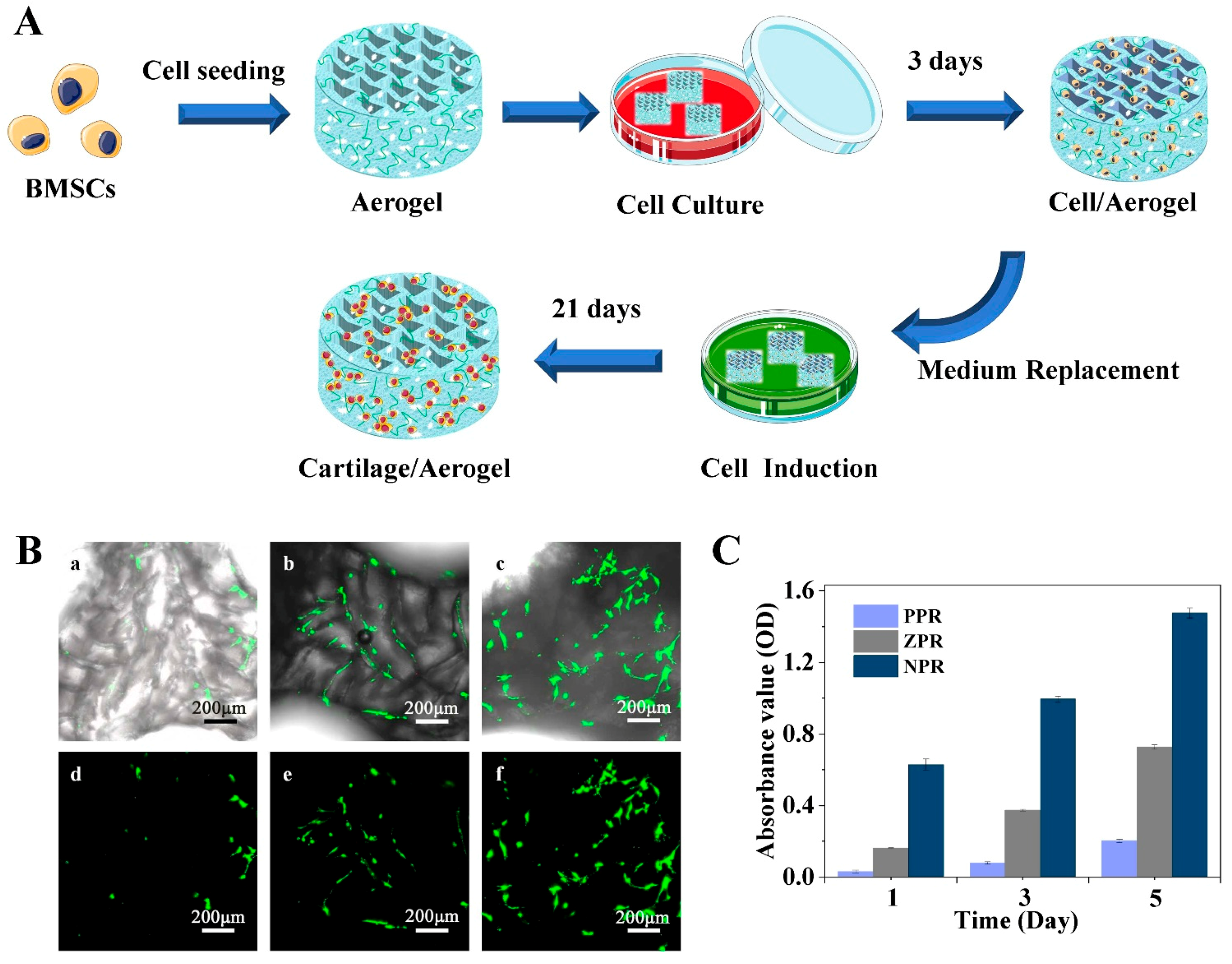
3.4. Survival Rate Analysis of Stem Cells on CNF/PEGDA Scaffolds
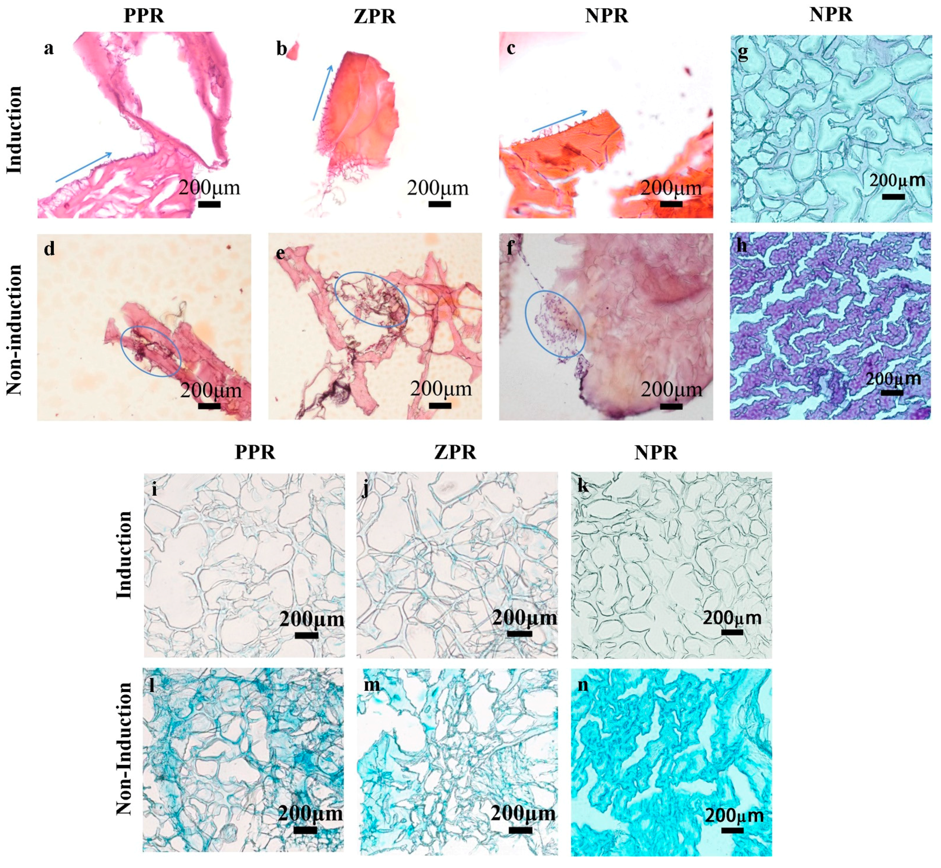
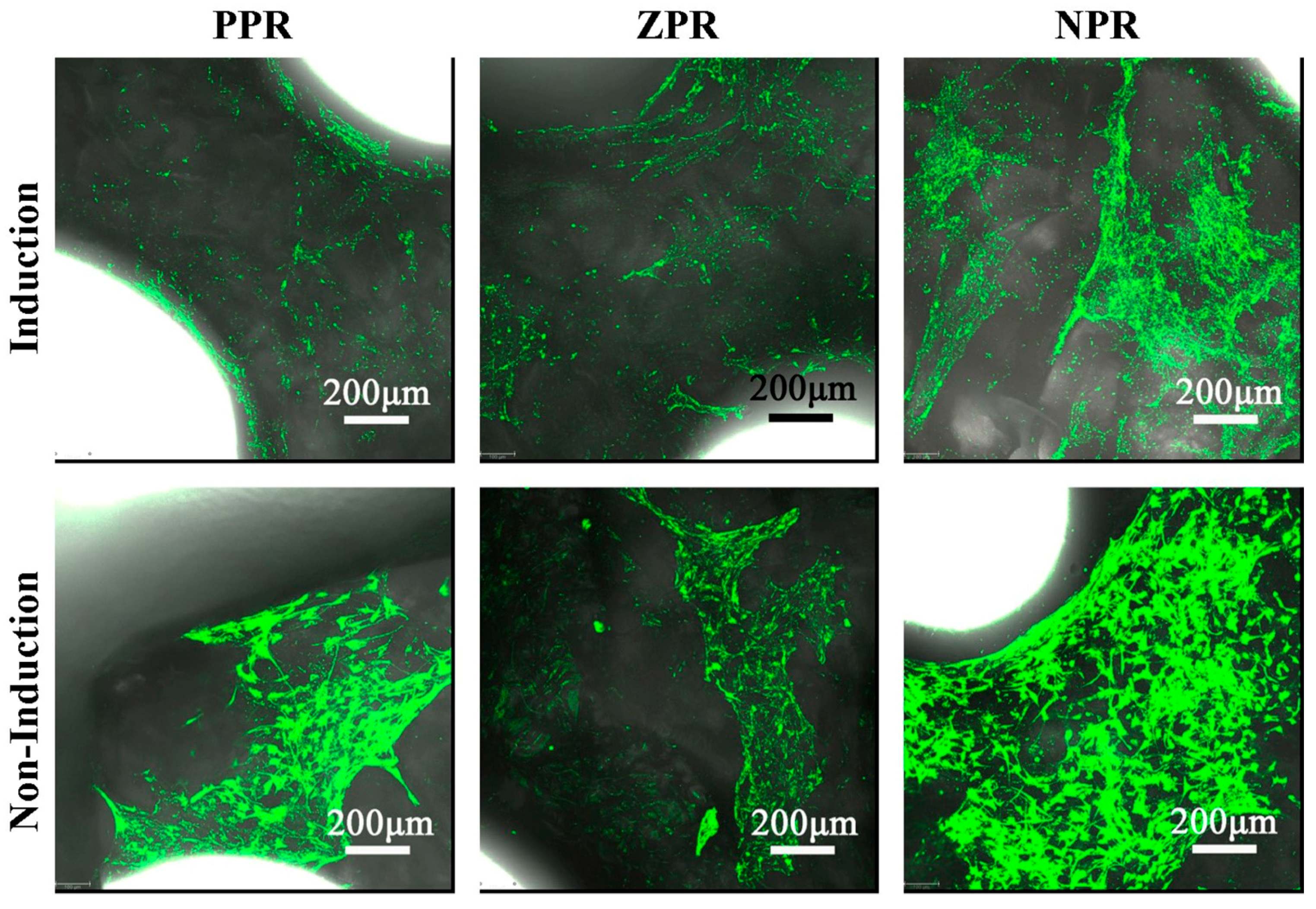
3.5. Analysis of Stem Cell Induction on CNF/PEGDA Scaffolds
4. Conclusions
Supplementary Materials
Author Contributions
Funding
Acknowledgments
Conflicts of Interest
References
- Loh, Q.L.; Choong, C. Three-Dimensional Scaffolds for Tissue Engineering Applications: Role of Porosity and Pore Size. Tissue Eng. Part B Rev. 2013, 19, 485–502. [Google Scholar] [CrossRef]
- Mirtaghavi, A.; Luo, J.; Muthuraj, R. Recent Advances in Porous 3D Cellulose Aerogels for Tissue Engineering Applications: A Review. J. Compos. Sci. 2020, 4, 152. [Google Scholar] [CrossRef]
- Sun, D.; Liu, W.; Zhou, F.; Tang, A.; Xie, W. Optimizing material and manufacturing process for PEGDA/CNF aerogel scaffold. J. Porous Mater. 2020, 1–15. [Google Scholar] [CrossRef]
- Naseri, N.; Poirier, J.-M.; Girandon, L.; Fröhlich, M.; Oksman, K.; Mathew, A.P. 3-Dimensional porous nanocomposite scaffolds based on cellulose nanofibers for cartilage tissue engineering: Tailoring of porosity and mechanical performance. RSC Adv. 2016, 6, 5999–6007. [Google Scholar] [CrossRef]
- Hutmacher, D.W. Scaffolds in tissue engineering bone and cartilage. Biomaterials 2000, 21, 2529–2543. [Google Scholar] [CrossRef]
- Baughman, R.H.; Shacklette, J.M.; Zakhidov, A.A.; Stafström, S. Negative Poisson’s ratios as a common feature of cubic metals. Nature 1998, 392, 362–365. [Google Scholar] [CrossRef]
- Lee, J.W.; Soman, P.; Park, J.H.; Chen, S.; Cho, D.-W. A Tubular Biomaterial Construct Exhibiting a Negative Poisson’s Ratio. PLoS ONE 2016, 11, e0155681. [Google Scholar] [CrossRef] [PubMed]
- Soman, P.; Fozdar, D.Y.; Lee, J.W.; Phadke, A.; Varghese, S.; Chen, S. A three-dimensional polymer scaffolding material exhibiting a zero Poisson’s ratio. Soft Matter 2012, 8, 4946–4951. [Google Scholar] [CrossRef] [PubMed]
- Zhang, W.; Soman, P.; Meggs, K.; Qu, X.; Chen, S. Tuning the Poisson’s Ratio of Biomaterials for Investigating Cellular Response. Adv. Funct. Mater. 2013, 23, 3226–3232. [Google Scholar] [CrossRef]
- Soman, P.; Lee, J.W.; Phadke, A.; Varghese, S.; Chen, S. Spatial tuning of negative and positive Poisson’s ratio in a multi-layer scaffold. Acta Biomater. 2012, 8, 2587–2594. [Google Scholar] [CrossRef] [PubMed]
- Park, Y.J.; Kim, J.K. The Effect of Negative Poisson’s Ratio Polyurethane Scaffolds for Articular Cartilage Tissue Engineering Applications. Adv. Mater. Sci. Eng. 2013, 2013, 1–5. [Google Scholar] [CrossRef]
- Song, L.; Ahmed, M.F.; Li, Y.; Zeng, C. Vascular differentiation from pluripotent stem cells in 3-D auxetic scaffolds. J. Tissue Eng. Regen. Med. 2018, 12, 1679–1689. [Google Scholar] [CrossRef] [PubMed]
- Grima, J.N.; Oliveri, L.; Attard, D.; Ellul, B.; Gatt, R.; Cicala, G.; Recca, G. Hexagonal Honeycombs with Zero Poisson’s Ratios and Enhanced Stiffness. Adv. Eng. Mater. 2010, 12, 855–862. [Google Scholar] [CrossRef]
- Kapnisi, M.; Mansfield, C.; Marijon, C.; Guex, A.G.; Perbellini, F.; Bardi, I.; Humphrey, E.J.; Puetzer, J.L.; Mawad, D.; Koutsogeorgis, D.C.; et al. Auxetic Cardiac Patches with Tunable Mechanical and Conductive Properties toward Treating Myocardial Infarction. Adv. Funct. Mater. 2018, 28, 1800618. [Google Scholar] [CrossRef]
- Lien, S.-M.; Ko, L.-Y.; Huang, T.-J. Effect of pore size on ECM secretion and cell growth in gelatin scaffold for articular cartilage tissue engineering. Acta Biomater. 2009, 5, 670–679. [Google Scholar] [CrossRef] [PubMed]
- Cervin, N.T.; Aulin, C.; Larsson, P.T.; Wågberg, L. Ultra porous nanocellulose aerogels as separation medium for mixtures of oil/water liquids. Cellulose 2012, 19, 401–410. [Google Scholar] [CrossRef]
- Han, J.; Zhou, C.; Wu, Y.; Liu, F.; Wu, Q. Self-Assembling Behavior of Cellulose Nanoparticles during Freeze-Drying: Effect of Suspension Concentration, Particle Size, Crystal Structure, and Surface Charge. Biomacromolecules 2013, 14, 1529–1540. [Google Scholar] [CrossRef]
- Ferreira, F.V.; Otoni, C.G.; de France, K.J.; Barud, H.S.; Lona, L.M.; Cranston, E.D.; Rojas, O.J. Porous nanocellulose gels and foams: Breakthrough status in the development of scaffolds for tissue engineering. Mater. Today 2020, 37, 126–141. [Google Scholar] [CrossRef]
- De France, K.J.; Hoare, T.; Cranston, E.D. Review of Hydrogels and Aerogels Containing Nanocellulose. Chem. Mater. 2017, 29, 4609–4631. [Google Scholar] [CrossRef]
- Liu, J.; Cheng, F.; Grénman, H.; Spoljaric, S.; Seppälä, J.; Eriksson, J.E.; Willför, S.; Xu, C. Development of nanocellulose scaffolds with tunable structures to support 3D cell culture. Carbohydr. Polym. 2016, 148, 259–271. [Google Scholar] [CrossRef] [PubMed]
- Khalil, H.A.; Adnan, A.; Yahya, E.B.; Olaiya, N.; Safrida, S.; Hossain, S.; Balakrishnan, V.; Gopakumar, D.A.; Abdullah, C.; Oyekanmi, A.; et al. A Review on Plant Cellulose Nanofibre-Based Aerogels for Biomedical Applications. Polymers 2020, 12, 1759. [Google Scholar] [CrossRef]
- Mathew, A.P.; Oksman, K.; Pierron, D.; Harmand, M.-F. Biocompatible Fibrous Networks of Cellulose Nanofibres and Collagen Crosslinked Using Genipin: Potential as Artificial Ligament/Tendons. Macromol. Biosci. 2013, 13, 289–298. [Google Scholar] [CrossRef]
- Ghafari, R.; Jonoobi, M.; Amirabad, L.M.; Oksman, K.; Taheri, A.R. Fabrication and characterization of novel bilayer scaffold from nanocellulose based aerogel for skin tissue engineering applications. Int. J. Biol. Macromol. 2019, 136, 796–803. [Google Scholar] [CrossRef]
- Camarero-Espinosa, S.; Rothen-Rutishauser, B.; Weder, C.; Foster, E.J. Directed cell growth in multi-zonal scaffolds for cartilage tissue engineering. Biomaterials 2016, 74, 42–52. [Google Scholar] [CrossRef]
- Palaganas, N.B.; Mangadlao, J.D.; de Leon, A.C.C.; Palaganas, J.O.; Pangilinan, K.D.; Lee, Y.J.; Advincula, R.C. 3D Printing of Photocurable Cellulose Nanocrystal Composite for Fabrication of Complex Architectures via Stereolithography. ACS Appl. Mater. Interfaces 2017, 9, 34314–34324. [Google Scholar] [CrossRef]
- Li, V.C.-F.; Kuang, X.; Mulyadi, A.; Hamel, C.M.; Deng, Y.; Qi, H.J. 3D printed cellulose nanocrystal composites through digital light processing. Cellulose 2019, 26, 3973–3985. [Google Scholar] [CrossRef]
- Tang, A.; Wang, Q.; Zhao, S.; Liu, W. Fabrication of nanocellulose/PEGDA hydrogel by 3D printing. Rapid Prototyp. J. 2018, 24, 1265–1271. [Google Scholar] [CrossRef]
- Tang, A.; Li, J.; Li, J.; Zhao, S.; Liu, W.; Liu, T.; Wang, J.; Liu, Y. Nanocellulose/PEGDA aerogel scaffolds with tunable modulus prepared by stereolithography for three-dimensional cell culture. J. Biomater. Sci. Polym. Ed. 2019, 30, 797–814. [Google Scholar] [CrossRef]
- Bai, C.; Tang, A.; Zhao, S.; Liu, W. Flexible Nanocellulose/Poly(ethylene glycol) Diacrylate Hydrogels with Tunable Poisson’s Ratios by Masking and Photocuring. Bioresources 2020, 15, 3307–3319. [Google Scholar] [CrossRef]
- Saito, T.; Kimura, S.; Nishiyama, Y.; Isogai, A. Cellulose Nanofibers Prepared by TEMPO-Mediated Oxidation of Native Cellulose. Biomacromolecules 2007, 8, 2485–2491. [Google Scholar] [CrossRef] [PubMed]
- Grima, J.N.; Attard, D. Molecular networks with a near zero Poisson’s ratio. Phys. Status Solidi B 2010, 248, 111–116. [Google Scholar] [CrossRef]
- Chinga-Carrasco, G. Potential and Limitations of Nanocelluloses as Components in Biocomposite Inks for Three-Dimensional Bioprinting and for Biomedical Devices. Biomacromolecules 2018, 19, 701–711. [Google Scholar] [CrossRef]
- Karageorgiou, V.; Kaplan, D.L. Porosity of 3D biomaterial scaffolds and osteogenesis. Biomaterials 2005, 26, 5474–5491. [Google Scholar] [CrossRef] [PubMed]
- Tu, X.; Wang, L.; Wei, J.; Wang, B.; Tang, Y.; Shi, J.; Zhang, Z.; Chen, Y. 3D printed PEGDA microstructures for gelatin scaffold integration and neuron differentiation. Microelectron. Eng. 2016, 158, 30–34. [Google Scholar] [CrossRef]
- Murphy, C.M.; Haugh, M.G.; O’Brien, F.J. The effect of mean pore size on cell attachment, proliferation and migration in collagen–glycosaminoglycan scaffolds for bone tissue engineering. Biomaterials 2010, 31, 461–466. [Google Scholar] [CrossRef] [PubMed]
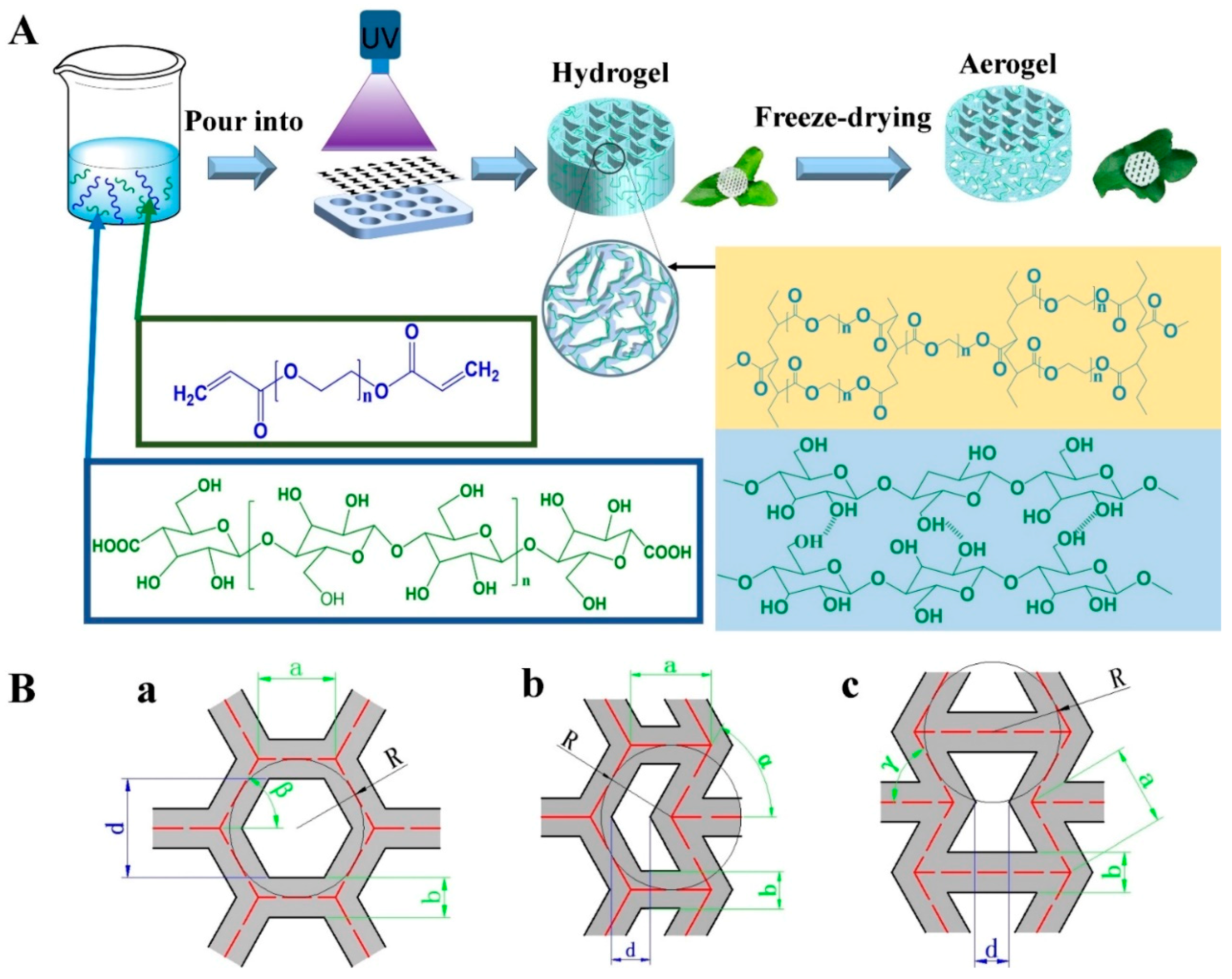
| Serial Number | Poisson’s Ratio | Each Letter Parameter Relation | Inscribed Radius, R (mm) | Side Length, a (mm) | Wall Thickness, b (mm) | Aperture, d (mm) |
|---|---|---|---|---|---|---|
| P-R1.0 | Positive | 1.0 | 1.155 | 1.2 | 0.824 | |
| Z-R1.0 | Zero | 1.0 | 1.155 | 0.86 | 0.489 | |
| N-R1.0 | Negative | 1.0 | 1.155 | 0.7 | 0.371 |
| Sample | PEGDA (wt%) | CNF (wt%) | Photoinitiator (wt%) | Deionized Water (wt%) |
|---|---|---|---|---|
| C-0 | 20.0 | 0 | 0.10 | 79.0 |
| C-1 | 20.0 | 1.0 | 0.10 | 78.9 |
| C-2 | 20.0 | 2.0 | 0.10 | 77.9 |
| I-0.05 | 20.0 | 1.0 | 0.05 | 78.95 |
| I-0.2 | 20.0 | 1.0 | 0.20 | 78.8 |
| P-10 | 10.0 | 1.0 | 0.10 | 88.9 |
| P-30 | 30.0 | 1.0 | 0.10 | 68.9 |
Publisher’s Note: MDPI stays neutral with regard to jurisdictional claims in published maps and institutional affiliations. |
© 2021 by the authors. Licensee MDPI, Basel, Switzerland. This article is an open access article distributed under the terms and conditions of the Creative Commons Attribution (CC BY) license (http://creativecommons.org/licenses/by/4.0/).
Share and Cite
Tang, A.; Ji, J.; Li, J.; Liu, W.; Wang, J.; Sun, Q.; Li, Q. Nanocellulose/PEGDA Aerogels with Tunable Poisson’s Ratio Fabricated by Stereolithography for Mouse Bone Marrow Mesenchymal Stem Cell Culture. Nanomaterials 2021, 11, 603. https://doi.org/10.3390/nano11030603
Tang A, Ji J, Li J, Liu W, Wang J, Sun Q, Li Q. Nanocellulose/PEGDA Aerogels with Tunable Poisson’s Ratio Fabricated by Stereolithography for Mouse Bone Marrow Mesenchymal Stem Cell Culture. Nanomaterials. 2021; 11(3):603. https://doi.org/10.3390/nano11030603
Chicago/Turabian StyleTang, Aimin, Jiaoyan Ji, Jiao Li, Wangyu Liu, Jufang Wang, Qiuli Sun, and Qingtao Li. 2021. "Nanocellulose/PEGDA Aerogels with Tunable Poisson’s Ratio Fabricated by Stereolithography for Mouse Bone Marrow Mesenchymal Stem Cell Culture" Nanomaterials 11, no. 3: 603. https://doi.org/10.3390/nano11030603
APA StyleTang, A., Ji, J., Li, J., Liu, W., Wang, J., Sun, Q., & Li, Q. (2021). Nanocellulose/PEGDA Aerogels with Tunable Poisson’s Ratio Fabricated by Stereolithography for Mouse Bone Marrow Mesenchymal Stem Cell Culture. Nanomaterials, 11(3), 603. https://doi.org/10.3390/nano11030603


