Synthesis and Stabilization of Support-Free Mesoporous Gold Nanoparticles

 , ,
, ,  ,
,  , ,
, ,  and
and
Abstract

1. Introduction
2. Materials and Methods
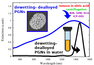
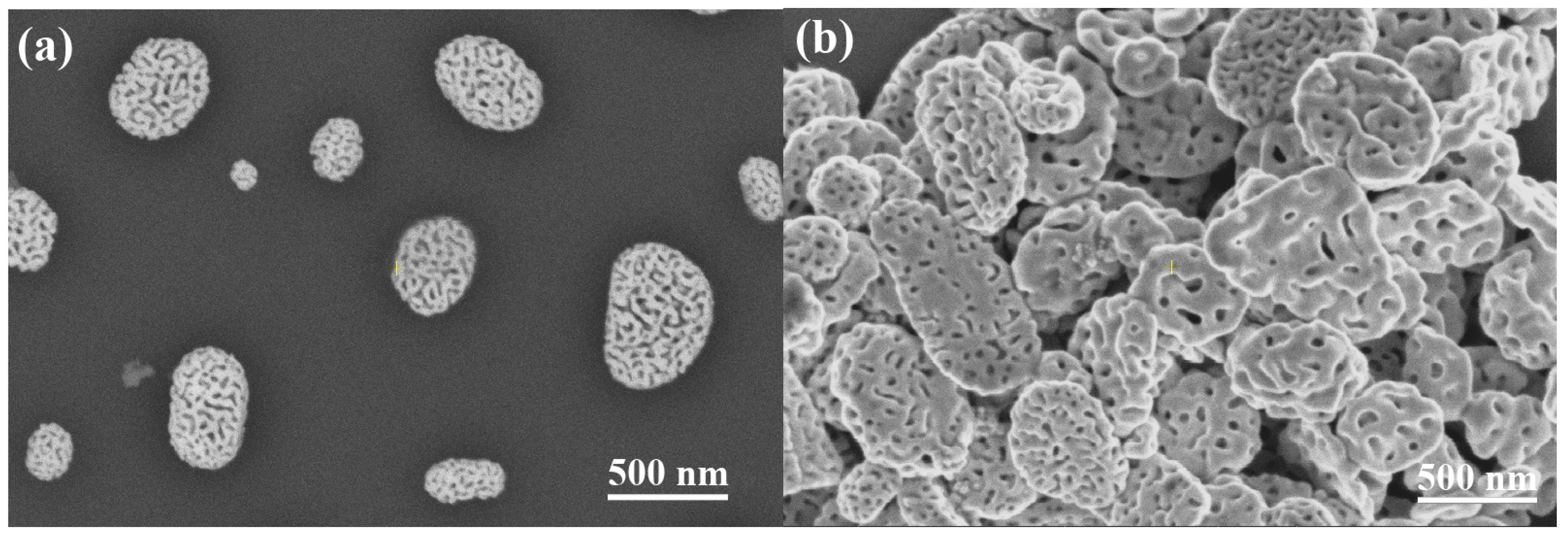
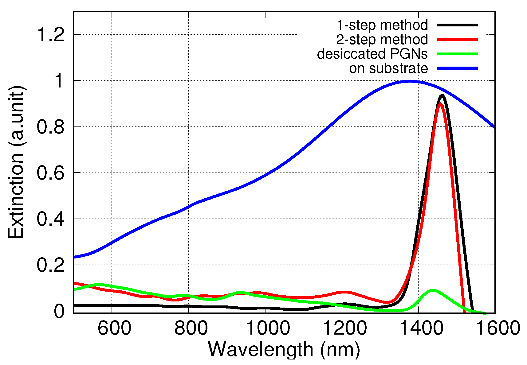
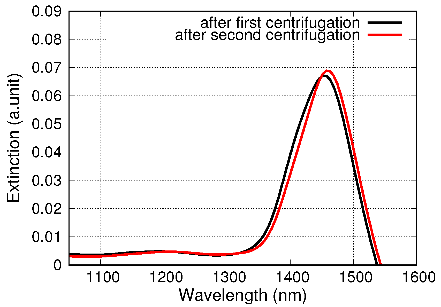
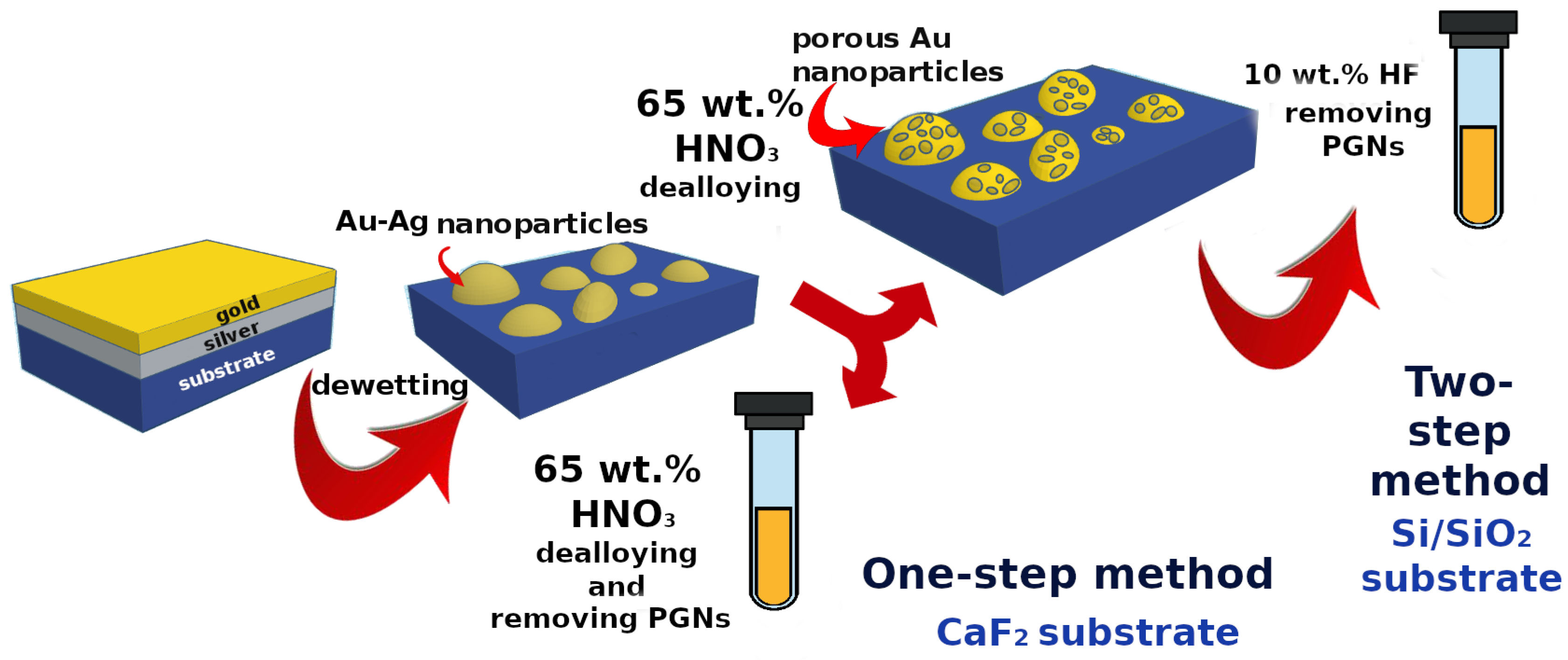
2.1. Sample Preparation
2.2. Sample Characterization
3. Results and Discussion
4. Conclusions
Author Contributions
Funding
Conflicts of Interest
References
- Wang, D.; Schaaf, P. Nanoporous gold nanoparticles. J. Mater. Chem. 2012, 22, 5344–5348. [Google Scholar] [CrossRef]
- Hodge, A.M.; Hayes, J.R.; Caro, J.A.; Biener, J.; Hamza, A.V. Characterization and mechanical behavior of nanoporous gold. Adv. Eng. Mater. 2006, 8, 853–857. [Google Scholar] [CrossRef]
- Glodán, G.; Cserháti, C.; Beszeda, I.; Beke, D.L. Production of hollow hemisphere shells by pure Kirkendall porosity formation in Au/Ag system. Appl. Phys. Lett. 2010, 97, 113109–113112. [Google Scholar] [CrossRef]
- Ron, R.; Haleva, E.; Salomon, A. Nanoporous Metallic Networks: Fabrication, Optical Properties, and Applications. Adv. Mater. 2018, 30, 1706755. [Google Scholar] [CrossRef] [PubMed]
- Ferreira, C.; Ribeiro, E.; Góes, A.; Silva, B. Current strategies for diagnosis of paracoccidioidomycosis and prospects of methods based on gold nanoparticles. Future Microbiol. 2016, 11. [Google Scholar] [CrossRef]
- Pasparakis, G. Light-Induced Generation of Singlet Oxygen by Naked Gold Nanoparticles and its Implications to Cancer Cell Phototherapy. Small 2013, 9, 4130–4134. [Google Scholar] [CrossRef]
- Kirui, D.; Rey, D.; Batt, C. Gold hybrid nanoparticles for targeted phototherapy and cancer imaging. Nanotechnology 2010, 21, 105105. [Google Scholar] [CrossRef]
- Hu, Y.; Wang, R.; Wang, S.; Ding, L.; Jingchao, L.; Luo, Y.; Wang, X.; Shen, M.; Shi, X. Multifunctional Fe3O4 @ Au core/shell nanostars: A unique platform for multimode imaging and photothermal therapy of tumors. Sci. Rep. 2016, 6, 28325. [Google Scholar] [CrossRef]
- Chen, Q.; Chen, Q.; Qi, H.; Ruan, L.; Ren, Y. Experimental Comparison of Photothermal Conversion Efficiency of Gold Nanotriangle and Nanorod in Laser Induced Thermal Therapy. Nanomaterials 2017, 7, 416. [Google Scholar] [CrossRef]
- Ahijado-Guzmán, R.; Sánchez-Arribas, N.; Martínez-Negro, M.; González-Rubio, G.; Santiago-Varela, M.; Pardo, M.; Piñeiro, A.; López-Montero, I.; Junquera, E.; Guerrero-Martínez, A. Intercellular Trafficking of Gold Nanostars in Uveal Melanoma Cells for Plasmonic Photothermal Therapy. Nanomaterials 2020, 10, 590. [Google Scholar] [CrossRef]
- Paul, M. Surface Plasmon Spectroscopy of Nanosized Metal Particles. Langmuir 1996, 12, 788–800. [Google Scholar] [CrossRef]
- Kelly, K.L.; Coronado, E.; Zhao, L.L.; Schatz, G.C. The Optical Properties of Metal Nanoparticles: The Influence of Size, Shape, and Dielectric Environment. J. Phys. Chem. B 2003, 107, 668–677. [Google Scholar] [CrossRef]
- Jain, P.K.; Lee, K.S.; El-Sayed, I.H.; El-Sayed, M.A. Calculated Absorption and Scattering Properties of Gold Nanoparticles of Different Size, Shape, and Composition: Applications in Biological Imaging and Biomedicine. J. Phys. Chem. B 2006, 110, 7238–7248. [Google Scholar] [CrossRef] [PubMed]
- Kosinova, A.; Wang, D.; Baradács, E.; Parditka, B.; Kups, T.; Klinger, L.; Erdélyi, Z.; Schaaf, P.; Rabkin, E. Tuning the nanoscale morphology and optical properties of porous gold nanoparticles by surface passivation and annealing. Acta Mater. 2017, 127, 108–116. [Google Scholar] [CrossRef]
- Rao, W.; Wang, D.; Kups, T.; Baradács, E.; Parditka, B.; Erdélyi, Z.; Schaaf, P. Nanoporous Gold Nanoparticles and Au/Al2O3 Hybrid Nanoparticles with Large Tunability of Plasmonic Properties. ACS Appl. Mater. Interfaces 2017, 9, 6273–6281. [Google Scholar] [CrossRef] [PubMed]
- Juhász, L.; Parditka, B.; Shenouda, S.; Kadoi, M.; Fukanaga, K.; Erdélyi, Z.; Cserháti, C. Morphologically Tuned Optical Properties of Titania Coated Porous Gold Nanoparticles. J. Appl. Phys. 2020. submitted. [Google Scholar]
- Juhász, L.; Parditka, B.; Petrik, P.; Cserháti, C.; Erdélyi, Z. Continuous tuning of the plasmon resonance frequency of porous gold nanoparticles by mixed oxide layers. J. Porous Mater. 2020. submitted. [Google Scholar]
- Kong, F.Y.; Zhang, J.W.; Li, R.F.; Wang, Z.X.; Wang, W.J.; Wang, W. Unique Roles of Gold Nanoparticles in Drug Delivery, Targeting and Imaging Applications. Molecules 2017, 22, 1445. [Google Scholar] [CrossRef]
- Daraee, H.; Eatemadi, A.; Abbasi, E.; Aval, S.F.; Kouhi, M.; Akbarzadeh, A. Application of gold nanoparticles in biomedical and drug delivery. Artif. Cells Nanomed. Biotechnol. 2016, 44, 410–422. [Google Scholar] [CrossRef]
- N’deh, K.P.U.; Kim, G.J.; Chung, K.H.; Shin, J.S.; Lee, K.S.; Choi, J.W.; Lee, K.J.; An, J.H. Surface-Modified Industrial Acrylonitrile Butadiene Styrene 3D Scaffold Fabrication by Gold Nanoparticle for Drug Screening. Nanomaterials 2020, 10, 529. [Google Scholar] [CrossRef]
- Rodrigues, C.; Raposo, L.; Cabral, R.; Paradinha, F.; Baptista, P.; Fernandes, A. Tumor Microenvironment Modulation via Gold Nanoparticles Targeting Malicious Exosomes: Implications for Cancer Diagnostics and Therapy. Int. J. Mol. Sci. 2017, 18, 162. [Google Scholar] [CrossRef] [PubMed]
- Beik, J.; Khademi, S.; Attaran, N.; Sarkar, S.; Shakeri-Zadeh, A.; Ghaznavi, H.; Ghadiri, H. A Nanotechnology- based Strategy to Increase the Efficiency of Cancer Diagnosis and Therapy: Folate-conjugated Gold Nanoparticles. Curr. Med. Chem. 2017, 24, 4399–4416. [Google Scholar] [CrossRef] [PubMed]
- Kong, L.; Qiu, J.; Sun, W.; Yang, J.; Shen, M.; Wang, L.; Shi, X. Multifunctional PEI-entrapped gold nanoparticles enable efficient delivery of therapeutic siRNA into glioblastoma cells. Biomater. Sci. 2017, 5, 258–266. [Google Scholar] [CrossRef] [PubMed]
- Mendes, R.; Fernandes, A.; Baptista, P. Gold Nanoparticle Approach to the Selective Delivery of Gene Silencing in Cancer—The Case for Combined Delivery? Genes 2017, 8, 94. [Google Scholar] [CrossRef] [PubMed]
- Shi, X.; Qiu, J.; Kong, L.; Cao, X.; Li, A.; Tan, H. Dendrimer-entrapped gold nanoparticles modified with β-cyclodextrin for enhanced gene delivery applications. RSC Adv. 2016, 6, 25633–25640. [Google Scholar] [CrossRef]
- Şologan, M.; Padelli, F.; Giachetti, I.; Aquino, D.; Boccalon, M.; Adami, G.; Pengo, P.; Pasquato, L. Functionalized Gold Nanoparticles as Contrast Agents for Proton and Dual Proton/Fluorine MRI. Nanomaterials 2019, 9, 879. [Google Scholar] [CrossRef]
- Masse, F.; Ouellette, M.; Lamoureux, G.; Boisselier, E. Gold nanoparticles in ophthalmology. Med. Res. Rev. 2019, 39, 302–327. [Google Scholar] [CrossRef]
- Kwon, K.C.; Ryu, J.; Lee, J.; Lee, E.; Kwon, I.; Kim, K.; Lee, J. Proteinticle/Gold Core/Shell Nanoparticles for Targeted Cancer Therapy without Nanotoxicity. Adv. Mater. 2014, 26, 6436–6441. [Google Scholar] [CrossRef]
- Zhao, J.; Lee, P.; Wallace, M.; Melancon, M. Gold Nanoparticles in Cancer Therapy: Efficacy, Biodistribution, and Toxicity. Curr. Pharm. Des. 2015, 21, 4240–4251. [Google Scholar] [CrossRef]
- Hu, J.; Jiang, R.; Zhang, H.; Guo, Y.; Wang, J.; Wang, J. Colloidal porous gold nanoparticles. Nanoscale 2018, 10, 18473–18481. [Google Scholar] [CrossRef]
- Pedireddy, S.; Lee, H.K.; Tjiu, W.W.; Phang, I.Y.; Tan, H.R.; Chua, S.Q.; Troadec, C.; Ling, X.Y. One-step synthesis of zero-dimensional hollow nanoporous gold nanoparticles with enhanced methanol electrooxidation performance. Nat. Commun. 2017, 5, 4947. [Google Scholar] [CrossRef] [PubMed]
- Park, J.; Kang, H.; Kim, Y.H.; Lee, S.W.; Lee, T.G.; Wi, J.S. Physically-synthesized gold nanoparticles containing multiple nanopores for enhanced photothermal conversion and photoacoustic imaging. Nanoscale 2016, 8, 15514–15520. [Google Scholar] [CrossRef] [PubMed]
- Liu, K.; Bai, Y.; Zhang, L.; Yang, Z.; Fan, Q.; Zheng, H.; Yin, Y.; Gao, C. Porous Au–Ag Nanospheres with High-Density and Highly Accessible Hotspots for SERS Analysis. Nano Lett. 2016, 16, 3675–3681. [Google Scholar] [CrossRef] [PubMed]
- Kosinova, A.; Wang, D.; Schaaf, P.; Kovalenko, O.; Klinger, L.; Rabkin, E. Fabrication of hollow gold nanoparticles by dewetting, dealloying and coarsening. Acta Mater. 2016, 102, 108–115. [Google Scholar] [CrossRef]
- Forgács, A.; Moldován, K.; Herman, P.; Baranyai, E.; Fábián, I.; Lente, G.; Kalmár, J. Kinetic Model for Hydrolytic Nucleation and Growth of TiO2 Nanoparticles. J. Phys. Chem. C 2018, 122, 19161–19170. [Google Scholar] [CrossRef]
- Hoo, C.M.; Starostin, N.; West, P.; Mecartney, M.L. A comparison of atomic force microscopy (AFM) and dynamic light scattering (DLS) methods to characterize nanoparticle size distributions. J. Nanopart. Res. 2008, 10, 89–96. [Google Scholar] [CrossRef]
- Germain, V.; Brioude, A.; Ingert, D.; Pileni, M.P. Silver nanodisks: Size selection via centrifugation and optical properties. J. Chem. Phys. 2005, 122, 124707. [Google Scholar] [CrossRef]
- Garcia, M.A. Surface plasmons in metallic nanoparticles: Fundamentals and applications. J. Phys. D Appl. Phys. 2011, 44, 283001. [Google Scholar] [CrossRef]
- Müller, A.; Peglow, S.; Karnahl, M.; Kruth, A.; Junge, H.; Brüser, V.; Scheu, C. Morphology, Optical Properties and Photocatalytic Activity of Photo- and Plasma-Deposited Au and Au/Ag Core/Shell Nanoparticles on Titania Layers. Nanomaterials 2018, 8, 502. [Google Scholar] [CrossRef]
- Kang, T.; Park, K.; Kwon, S.; Chae, W.S. Surface-engineered nanoporous gold nanoparticles for light-triggered drug release. Opt. Mater. 2020, 106, 109985. [Google Scholar] [CrossRef]
- Osica, I.; Melo, A.; Camargo Dalmatti Alves Lima, F.; Shiba, K.; Imamura, G.; Crespilho, F.; Betlej, J.; Kurzydłowski, K.; Yoshikawa, G.; Ariga, K. Nanomechanical Recognition and Discrimination of Volatile Molecules by Au Nanocages Deposited on Membrane-Type Surface Stress Sensors. ACS Appl. Nano Mater. 2020, 3, 4061–4068. [Google Scholar] [CrossRef]


| replications | 3 |
| pump speed | 12 rpm |
| uptake delay | 15 s |
| read time | 5 s |
| RF power | 1.20 kW |
| stabilization time | 15 s |
| viewing mode | axial |
| nebulizer flow | 0.70 L/min |
| plasma flow | 12.0 L/min |
| aux flow | 1.00 L/min |
| make up flow | 0.00 L/min |
| viewing height | 8 mm |
| Stage of Synthesis | Diameter (nm) |
|---|---|
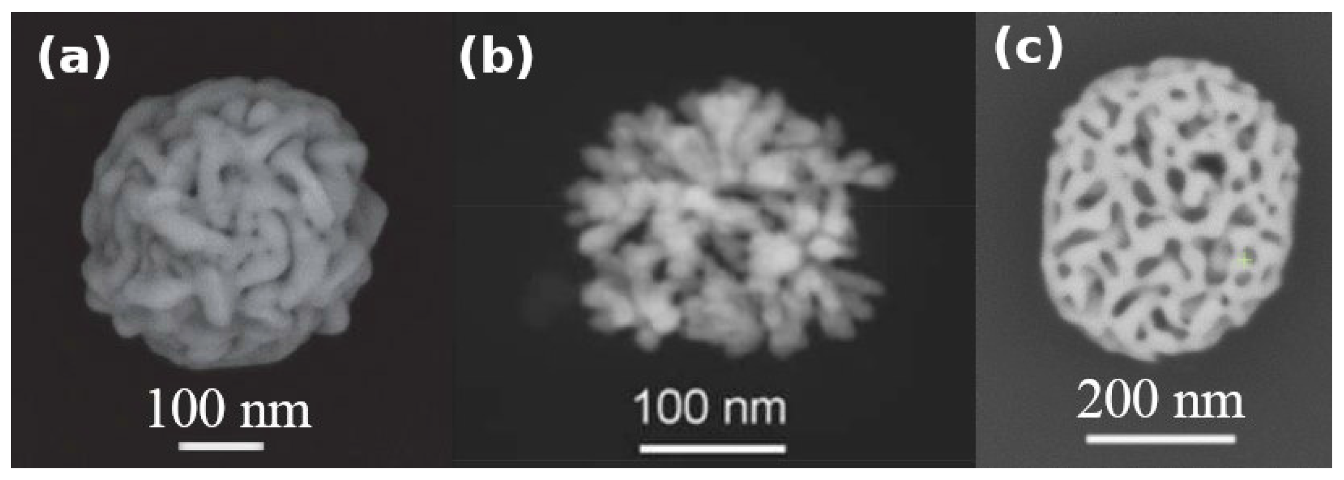
| Diameter of supported PGNs | 316 ± 136 (image analysis) |
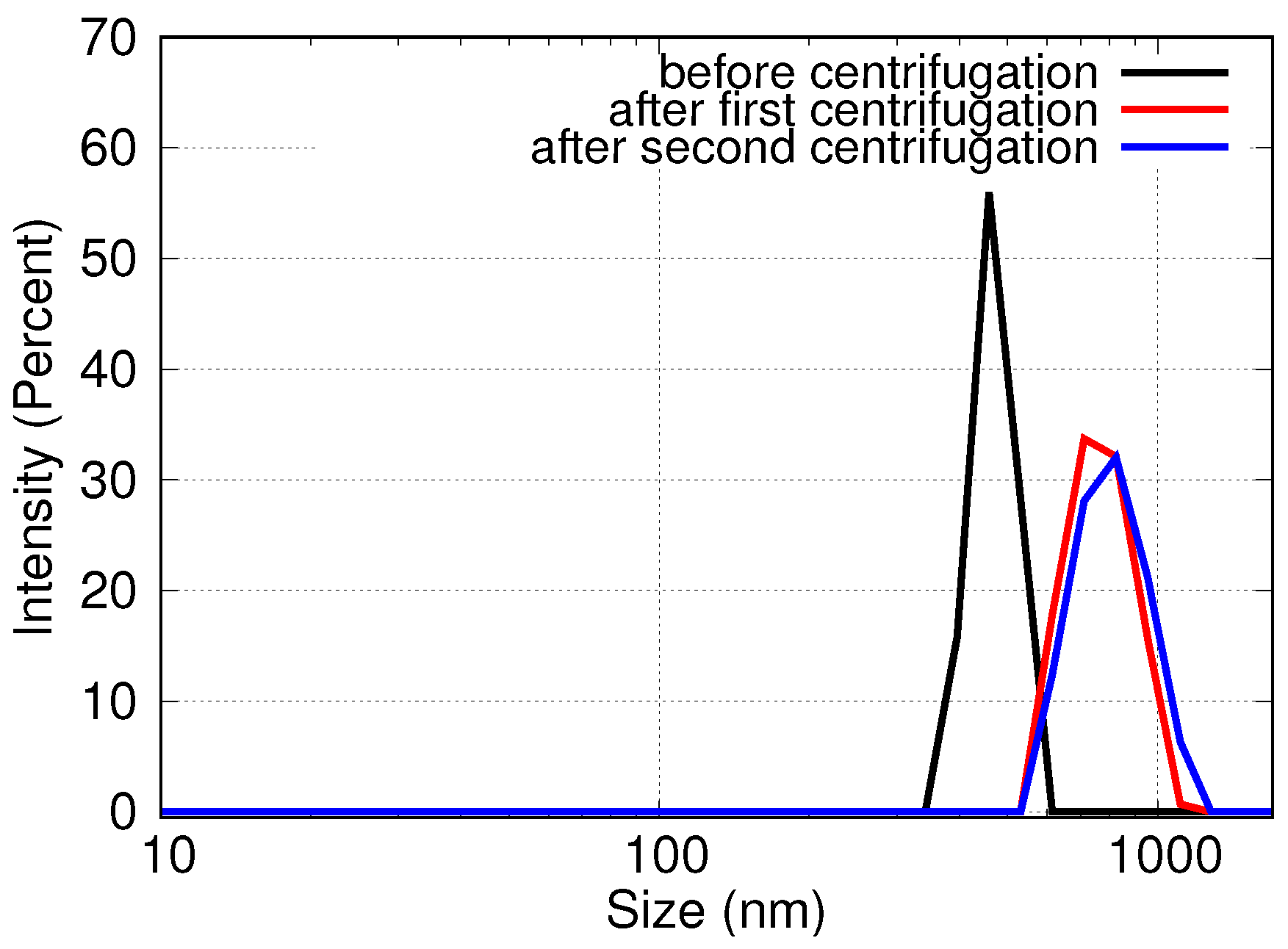
| Hydrodynamic diameter of support-free PGNs in the original suspension | 447 ± 64 (DLS) |
| Hydrodynamic diameter of support-free PGNs after 1 round of centrifugation | 765 ± 149 (DLS) |
| Hydrodynamic diameter of support-free PGNs after 2 rounds of centrifugations | 851 ± 110 (DLS) |
© 2020 by the authors. Licensee MDPI, Basel, Switzerland. This article is an open access article distributed under the terms and conditions of the Creative Commons Attribution (CC BY) license (http://creativecommons.org/licenses/by/4.0/).
Share and Cite
Juhász, L.; Moldován, K.; Herman, P.; Erdélyi, Z.; Fábián, I.; Kalmár, J.; Cserháti, C. Synthesis and Stabilization of Support-Free Mesoporous Gold Nanoparticles. Nanomaterials 2020, 10, 1107. https://doi.org/10.3390/nano10061107
Juhász L, Moldován K, Herman P, Erdélyi Z, Fábián I, Kalmár J, Cserháti C. Synthesis and Stabilization of Support-Free Mesoporous Gold Nanoparticles. Nanomaterials. 2020; 10(6):1107. https://doi.org/10.3390/nano10061107
Chicago/Turabian StyleJuhász, Laura, Krisztián Moldován, Petra Herman, Zoltán Erdélyi, István Fábián, József Kalmár, and Csaba Cserháti. 2020. "Synthesis and Stabilization of Support-Free Mesoporous Gold Nanoparticles" Nanomaterials 10, no. 6: 1107. https://doi.org/10.3390/nano10061107
APA StyleJuhász, L., Moldován, K., Herman, P., Erdélyi, Z., Fábián, I., Kalmár, J., & Cserháti, C. (2020). Synthesis and Stabilization of Support-Free Mesoporous Gold Nanoparticles. Nanomaterials, 10(6), 1107. https://doi.org/10.3390/nano10061107