Abstract
Among oxide semiconductors, p-type Mn3O4 systems have been exploited in chemo-resistive sensors for various analytes, but their use in the detection of H2, an important, though flammable, energy vector, has been scarcely investigated. Herein, we report for the first time on the plasma assisted-chemical vapor deposition (PA-CVD) of Mn3O4 nanomaterials, and on their on-top functionalization with Ag and SnO2 by radio frequency (RF)-sputtering, followed by air annealing. The obtained Mn3O4-Ag and Mn3O4-SnO2 nanocomposites were characterized by the occurrence of phase-pure tetragonal α-Mn3O4 (hausmannite) and a controlled Ag and SnO2 dispersion. The system functional properties were tested towards H2 sensing, yielding detection limits of 18 and 11 ppm for Mn3O4-Ag and Mn3O4-SnO2 specimens, three orders of magnitude lower than the H2 explosion threshold. These performances were accompanied by responses up to 25% to 500 ppm H2 at 200 °C, superior to bare Mn3O4, and good selectivity against CH4 and CO2 as potential interferents. A rationale for the observed behavior, based upon the concurrence of built-in Schottky (Mn3O4/Ag) and p-n junctions (Mn3O4/SnO2), and of a direct chemical interplay between the system components, is proposed to discuss the observed activity enhancement, which paves the way to the development of gas monitoring equipments for safety end-uses.
1. Introduction
The reliable detection of hazardous/flammable gases is of key importance in a variety of fields, encompassing disease diagnosis, environmental monitoring and human health protection [1,2,3,4,5,6]. In this broad scenario, a key role is played by the early recognition of molecular hydrogen (H2), a zero-emission and clean fuel, with a high energy density of ≈130 MJ×kg−1 [2,7], which has emerged as a future energy source for transportation, industrial and residential applications [8,9,10]. Nevertheless, since H2 is colorless, odorless and highly flammable, the detection of hydrogen leakages at concentrations lower than hazardous levels [11,12,13,14] is extremely critical towards the emergence of a future hydrogen economy [7,8,15,16,17,18,19].
Simple architecture, cost-effective fabrication, stability under the operating conditions, and high efficiency, are the main requirements and core features of advanced sensors needed for such applications [16]. Among the various active systems and devices [20,21,22,23,24], metal oxide nanostructures have been the subject of an increasing interest, thanks to their high carrier mobility, easy fabrication and excellent stability [9,25,26,27,28]. In particular, whereas n-type oxide semiconductors have been largely investigated as gas sensors [8,9,12,15], p-type ones have not yet been widely studied [4,17,29,30], since their responses are typically lower than those of n-type systems with comparable morphology [31,32,33]. Nonetheless, p-type oxide semiconductors have an important potential as gas sensors, and represent promising platforms for the development of devices exhibiting new functions [32], taking into account their appreciable activity as oxidation catalysts and the possibility of boosting their performances by tailoring their chemico-physical properties [2,31,34,35].
Among p-type systems, Mn3O4 has received significant attention due to its low cost, large natural abundance, environmentally friendly character and versatile chemico-physical properties, including the coexistence of mixed valence states [36,37]. Over the last decade, different studies have reported on Mn3O4-containing gas sensors for various analytes, including CH3CH2OH, CH3COCH3, NH3 and chemical warfare agent simulants [31,35,36,37,38,39,40,41]. Nonetheless, only two works on Mn3O4-based gas sensors for molecular hydrogen detection are available in the literature so far [35,42], and the implementation of H2 sensors endowed with improved sensitivity and selectivity undoubtedly requires additional research efforts [29,35,37,38].
Beside tailoring the system morphology [25,26,33,39], a proficient way to enhance the functionality of bare Mn3O4 gas sensors involves their sensitization with suitable metal/oxide agents [8,18,35,42,43,44]. The ultimate aim of this strategy is the exploitation of synergistical chemical and electronic effects, in order to obtain improved performances at moderate working temperatures, an issue of key importance for the development of low power consumption devices [4,37,41]. In this context, the present study is devoted to the fabrication of Mn3O4-based chemo-resistive sensors for H2 detection, sensitized through the on-top deposition of selected metal and metal oxide activators. As prototypes for the two categories, in this work our attention has been focused on the use of Ag, a potential catalyst promoting the reactions involved in the sensing process [45,46,47,48,49], and of SnO2, by far one of the most investigated metal oxides for gas sensing applications, endowed with high electron mobility and gas sensitivity [10,13,25,26,50]. In particular, the occurrence of Schottky (Mn3O4/Ag) or p-n (Mn3O4/SnO2) junctions between the system components can indeed enhance the modulations of the space charge region, and of the measured electrical resistance, ultimately yielding improved sensing performances thanks to electronic effects [18,26,33,43].
At variance with our previous studies, which have involved the fabrication of Mn3O4-based sensors by means of thermally activated chemical vapor deposition (CVD)-based processes [31,38,39,41], in this work a novel two-step plasma-assisted route was adopted for the preparation of the present materials. The fabrication procedure (Figure 1) involved: (i) the initial plasma assisted-CVD (PA-CVD) on alumina substrates of MnO2 from Mn(hfa)2TMEDA (Hhfa = 1,1,1,5,5,5-hexafluoro-2,4-pentanedione; TMEDA = N,N,N’,N’-tetramethylethylenediamine) [51,52], a molecular precursor never utilized so far for PA-CVD processes; (ii) the functionalization with Ag or SnO2 by means of radio frequency (RF)-sputtering, and (iii) a final thermal treatment in air to trigger the transformation of MnO2 into Mn3O4. The main focus of the present investigation was directed at elucidating the structural, compositional and morphological characteristics of the target materials and their interplay with the resulting sensing performances in hydrogen detection. The latter were investigated at a fixed humidity level as a function of the operating temperature, with particular regard to the role exerted by the formation of metal-oxide (Mn3O4/Ag) or oxide-oxide (Mn3O4/SnO2) junctions. The obtained results indicate that the proposed preparation method yields H2 sensors exhibiting favorable detection limits, promising responses at moderate temperature, as well as selectivity against carbon dioxide and methane as potential interferents. To the best of our knowledge, this is the first report on hydrogen gas sensing by Mn3O4-based composites prepared by a plasma-assisted route.
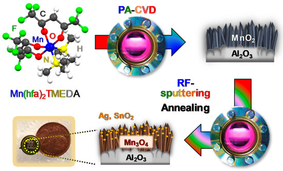
Figure 1.
Scheme of the route proposed in the present study for the fabrication of Mn3O4-Ag and Mn3O4-SnO2 nanomaterials.
2. Experimental Procedure
2.1. Material Preparation
MnO2 nanomaterials were initially deposited using a two-electrode plasmo-chemical instrumentation [53] equipped with a RF-generator (ν = 13.56 MHz), using Mn(hfa)2TMEDA [51,52] as the manganese molecular source. Depositions were performed on pre-cleaned [15,31,39] polycrystalline Al2O3 substrates (99.6%, Maruwa, Owariasahi, Japan; thickness = 0.25 mm), mounted on the grounded electrode, from Ar-O2 plasmas, using the following pre-optimized experimental settings: RF-power = 20 W; growth temperature = 300 °C; total pressure = 1.0 mbar). In a typical process, the precursor powders (weight = 0.15 g) were placed in an external glass reservoir heated at 70 °C, and their vapors were transported into the reactor using an electronic grade Ar flow (60 standard cubic centimeters per minute (sccm)) through gas lines maintained at 130 °C by means of external heating tapes. Additional Ar and O2 flows (15 and 5 sccm, respectively) were separately introduced into the reactor.
Deposition of Ag or SnO2 over the obtained systems was subsequently performed through RF-sputtering experiments, carried out using the above described instrumentation, and utilizing silver (Alfa Aesar®, Ward Hill, MA, USA; thickness = 0.3 mm, purity ≥ 99.95%) or tin targets (Neyco®, Vanves, France; thickness = 2.0 mm, purity = 99.99%). In each experiment, the used target and the Al2O3-supported manganese oxide deposits were mounted on the RF and grounded electrode, respectively. Depositions were carried out using an Ar flow rate of 10 sccm (total pressure = 0.3 mbar), a RF-power of 5 W, and a grounded electrode temperature of 60 °C. After a preliminary optimization, the deposition time was set at 45 and 90 min for silver and tin sputtering, respectively.
After preparation, ex-situ thermal treatments in air at a temperature of 400 °C for 1 h were carried out in order to ensure the conversion of MnO2 into phase-pure Mn3O4 [54] and to stabilize the obtained nanomaterials in view of gas sensing tests [19,48].
2.2. Material Characterization
X-ray diffraction (XRD) patterns were recorded at room temperature using a Bruker (Billerica, MA, USA) D8 Advance X-ray diffractometer with a Cu Kα X-ray source (λ = 1.54 Å) operated at 40 kV and 40 mA, employing an incidence angle of 1.0°. The average crystallite dimensions were calculated by means of the Scherrer equation [29,33,36,55].
X-ray photoelectron spectroscopy (XPS) analysis was carried out by means of a Perkin-Elmer (Chanhassen, MN, USA) Φ 5600ci instrument equipped with a hemispherical electron analyzer, using a standard Al Kα X-ray excitation source (hν = 1486.6 eV). The charging effect on the measured binding energies (BEs) was corrected by adjusting the position of the adventitious C1s signal to 284.8 eV [56]. Atomic percentage (at.%; uncertainty = ±2%) values were determined by peak integration using Φ V5.4A sensitivity factors, after a Shirley-type background subtraction. Silver and tin molar fractions were calculated as XM = [(M at.%)/(M at.% + Mn at.%) × 100], with M = Ag, Sn [38,46]. Spectra were fitted with mixed Gaussian–Lorentzian peak shapes using the XPSPEAK program [57]. Auger parameters α1 and α2 for silver were calculated according to the literature [56]. A Cameca (Gennevilliers CEDEX, France) IMS 4f spectrometer was used for in-depth secondary ion mass spectrometry (SIMS) analyses, performed at pressures lower than 1 × 10⁻¹⁰ mbar, using a Cs+ primary beam (14.5 keV, 20 nA, stability 0.2%) and negative secondary ion detection. Depth profiles were acquired using an electron gun for charge compensation in beam blanking mode and high mass resolution configuration. Elemental signals were recorded rastering over a 150 × 150 μm2 area and sampling secondary ions from an 8 × 8 μm2 region to avoid crater effects. The erosion time in the abscissa of the recorded profiles was converted into depth, based on thickness values measured by field emission-scanning electron microscopy (FE-SEM) measurements. The latter were carried out on a Zeiss (Oberkochen, Germany) SUPRA 40VP instrument at operating voltages of 5.00 kV. Sample thickness and nanoaggregate size values were obtained by analyzing cross-sectional and plane-view images with the ImageJ® software [58]. Atomic force microscopy (AFM) measurements were performed under normal air conditions in tapping mode, using a NT-MDT (Moscow, Russia) SPM Solver P47H-PRO apparatus. Root-mean-square (RMS) surface roughness values were obtained from 3 × 3 μm2 micrographs after plane fitting.
2.3. Gas Sensing Tests
To avoid any influence from the external environmental conditions, the used gas sensing test system is composed of a stainless-steel chamber located inside a temperature-stabilized climatic chamber, which was set at 20 °C for all measurements. The relative humidity level inside the stainless-steel chamber was constantly monitored and controlled to be exactly 40% at 20 °C. As the sensing devices are completely sealed inside the test chamber (dark conditions), there is no effect from external room illumination. Gas sensing properties of the fabricated systems were investigated using the flow-through method at atmospheric pressure. A constant synthetic air flow (300 sccm) was used as a carrier for the dispersion of H2 (and of CH4 and CO2 in the selectivity tests) at the desired concentrations. Pt interdigitated contacts and a Pt heater were deposited by sputtering (applied power = 30 W, Ar plasma, room temperature) on the active material surface and on the alumina substrate backside (lateral dimensions = 3 × 3 mm2), respectively. Resistance values were obtained by applying a bias voltage of 0.2 V, measuring the flowing current through a picoammeter. The measurements were performed in the 100–300 °C temperature interval, after pre-stabilization for 8 h at each temperature to reach a steady state. In line with our previous studies on Mn3O4-based gas sensors [31,38,39,41], no appreciable resistance variations upon gas exposure were obtained for working temperatures <150 °C, whereas the use of temperatures higher than 300 °C was intentionally avoided in order to prevent Mn3O4 thermal alterations during sensing tests.
No significant baseline resistance drift was detected after testing up to 12 h. The sensor response was calculated from the measured values of equilibrium resistances in air (RA), and in the presence of the target gas (RG), using the following relation [7,31,38,43]:
A detailed comparison of the present results with previous literature studies was performed by converting the reported response values into those defined by Equation (1). Repeated measurements under the same operating conditions on up to 10 identical sensors yielded stable and reproducible responses (maximum uncertainty = ±10%). The same variation was also estimated to be the response drift upon repeated tests up to 4 months, highlighting thus the system stability, an important issue in view of eventual real-world end uses [37,38,48].
The experimental response vs. concentration trends were fitted by the typical power law relation for metal oxide sensors [9,30,42]:
where C is the gas concentration, whereas a and b are constants dependent on the active material and the stoichiometry of the involved reactions [8,31,44]. Detection limits (maximum estimated uncertainty = ±1 ppm) were extrapolated at a response value of 3, assuming the validity of Equation (2) at low analyte concentrations.
3. Results and Discussion
3.1. Chemico-Physical Characterization
The fabrication process of the target materials is illustrated in Figure 1. Particular efforts were dedicated to elucidating the interplay between the adopted processing conditions and material chemical, physical and functional properties.
The system structure was investigated by XRD analyses (Figure 2). All the observed reflections located at 2θ = 18.0°, 28.9°, 31.0°, 32.3° and 36.1° could be indexed to the (101), (112), (200), (103) and (211) planes of tetragonal hausmannite (α-Mn3O4; a = 5.762 Å and c = 9.470 Å [31,38,59]). The occurrence of relatively weak and broad diffraction peaks suggested the formation of defective nanocrystallites [11], whose average dimensions were close to 25 nm for all the target specimens. A comparison of the signal relative intensities with those of the reference pattern [59] did not reveal any significant orientation/texturing effect, and no appreciable reflections from other Mn oxide polymorphs could be distinguished, highlighting the occurrence of phase-pure systems. Upon functionalization of Mn3O4 by RF-sputtering, no net variation in the recorded XRD patterns took place. The absence of noticeable diffraction peaks related to Ag or SnO2 was traced back to their low content and high dispersion into the Mn3O4 systems [19,41,44,46] (see also XPS and SIMS results).
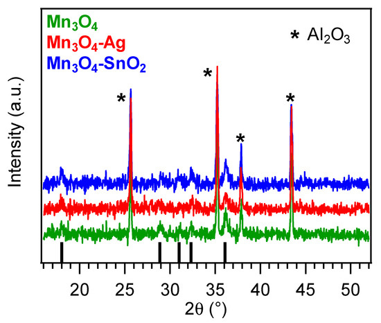
Figure 2.
X-ray diffraction (XRD patterns for bare and functionalized Mn3O4 nanosystems. Vertical black bars correspond to α-Mn3O4 signals [59].
The surface chemical state of the developed materials was analyzed by means of XPS. Figure 3a displays the survey spectra for the target specimens, that revealed the presence of oxygen, manganese and eventually, silver or tin signals, for the functionalized systems, together with a minor carbon contribution (<10 at.%) resulting from adventitious contamination. The detection of manganese signals even after RF-sputtering suggested only a partial coverage of Mn3O4 by the deposited silver- and tin-containing species. Accordingly, Ag and Sn molar fractions were evaluated to be 47.0% and 31.0%, respectively. For bare Mn3O4, the Mn2p3/2 component was located at BE = 641.8 eV (spin-orbit splitting (SOS) = 11.5 eV, Figure 3b), in accordance with previous literature data [31,35,38,41]. For the functionalized systems, a lower Mn2p3/2 BE was observed (641.7 eV, for Mn3O4-Ag, and 641.5 eV, for Mn3O4-SnO2). This finding suggested the formation of Schottky and p-n junctions for Mn3O4-Ag and Mn3O4-SnO2, respectively [33,37,38,41,42], resulting in an Ag → Mn3O4 and SnO2 → Mn3O4 electron transfer. This phenomenon, more pronounced for SnO2-containing samples, as testified by the higher BE decrease, exerted a favorable influence on the resulting gas sensing performances. As regards silver (Figure 3c), the Ag3d5/2 position (BE = 368.5 eV, SOS = 6.0 eV), as well as the pertaining Auger parameters (see also Figure S1; α1 = 719.7 eV and α2 = 725.6 eV), revealed a partial Ag surface oxidation, i.e., the coexistence of Ag(0) and Ag(I) oxide, as typically observed in similar cases [38,46,49,60]. Finally, the main tin photopeak (Figure 3d; BE(Sn3d5/2) = 486.9 eV; SOS = 8.4 eV) was located at higher energies than those reported for SnO2 [14,56,61], in line with the above mentioned charge transfer process. Taken together, these results highlighted the formation of nanocomposites in which the single components maintained their chemical identity, and enabled us to discard the formation of ternary phases, in line with XRD results. The deconvolution of O1s photopeaks (Figure S2) revealed the concurrence of two distinct bands at BE = 530.0 eV, resulting from lattice oxygen in Mn3O4, Ag(I) oxide (Mn3O4-Ag) or SnO2 (Mn3O4-SnO2) [14,31,38,60,61], and 531.6 eV, assigned to oxygen species adsorbed on surface O defects [4,41,51,52,55]. The contribution of the latter component to the overall O1s signal increased from ≈36.0%, for bare Mn3O4, to ≈58.0%, for the functionalized specimens, indicating a parallel increase of the oxygen defect content. The latter feature had a direct beneficial impact on the resulting gas sensing behavior.
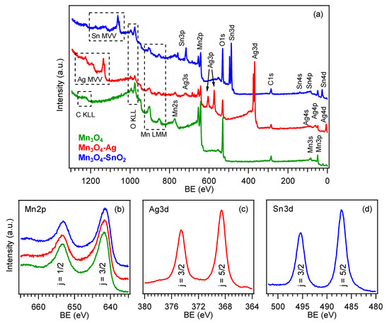
Figure 3.
(a) X-ray photoelectron spectroscopy (XPS) wide-scan spectra pertaining to bare Mn3O4, Mn3O4-Ag and Mn3O4-SnO2 samples. (b) Mn2p, (c) Ag3d and (d) Sn3d photoelectron peaks. The color code is reported in panel (a).
Complementary information on material chemical composition was obtained by SIMS in-depth profiling (Figure 4). Upon functionalization of Mn3O4, no significant variations in the overall deposit thickness took place (for all specimens, the average value was (400 ± 50) nm, as determined by cross-sectional FE-SEM analyses (see below and Figure 5)). The almost parallel trends of manganese and oxygen ionic yields suggested their common chemical origin, in line with the formation of phase-pure Mn3O4. Silver and tin trends could be described by an erfchian profile, such as in thermal diffusion processes [49]. For the Mn3O4-SnO2 sample, Sn yield underwent a progressive decrease throughout the outer 100 nm, subsequently followed by a plateau, whereas, for the Mn3O4-Ag specimen, the silver curve continuously declined even at higher depth values. In spite of these differences, a penetration of both Ag and Sn up to the interface with the alumina substrate was observed, and ascribed to the synergistical combination between the inherent RF-sputtering infiltration power and the Mn3O4 deposit open morphology [38,41,44,48] (see also Figure 5). This intimate contact between the system components is indeed an issue of key importance in order to benefit from their mutual electronic interplay, as discussed in detail below.
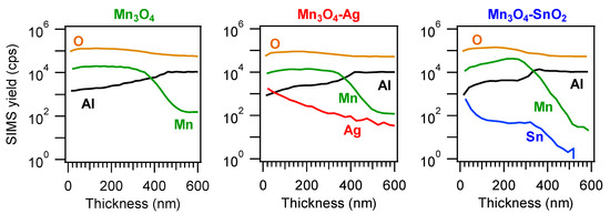
Figure 4.
Secondary ion mass spectrometry (SIMS) depth profiles for Mn3O4, Mn3O4-Ag and Mn3O4-SnO2 specimens.
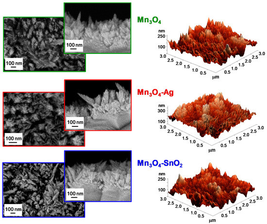
Figure 5.
Representative plane-view and cross-sectional field emission-scanning electron microscopy (FE-SEM) micrographs (left panels) and atomic force microscopy (AFM) images (right panels) for Mn3O4, Mn3O4-Ag and Mn3O4-SnO2 samples.
The system morphology was investigated by the complementary use of FE-SEM and AFM. FE-SEM micrographs (see Figure 5, left side) highlighted that bare Mn3O4 was characterized by the presence of elongated nanoaggregates (mean size = 100 nm), whose interconnection resulted in the formation of arrays with an open morphology. This feature is indeed favorable in view of gas sensing applications, since a higher area available for the interaction with the surrounding gases has a beneficial effect on the ultimate material functional performances [15,19,25,31,40]. After Ag and SnO2 introduction, no marked variations involving aggregate coalescence/collapse could be observed, validating the potential of the adopted synthetic route in functionalizing Mn3O4 nano-deposits without any undesired morphological alteration. AFM analyses (Figure 5, right side) confirmed the presence of the aforementioned aggregates uniformly protruding from the growth substrate, resulting in a crack-free and homogeneous granular topography, yielding an average RMS surface roughness of 40 nm for all the analyzed specimens.
3.2. Gas Sensing Performances
Figure 6 displays representative dynamical responses of the developed sensors towards square concentration pulses of gaseous hydrogen. All the target materials exhibited a p-type sensing behavior, as indicated by the resistance increase upon H2 exposure due to the reaction of the analyte with adsorbed oxygen species, resulting in a decrease of the major p-type carrier concentration [2,16,31,35,39]. This phenomenon is in agreement with the fact that Mn3O4 is the main system component, as indicated by structural and compositional characterization [44].
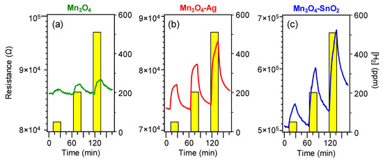
Figure 6.
Dynamical responses of Mn3O4 (a), Mn3O4-Ag (b) and Mn3O4-SnO2 (c) nanosystems vs. different H2 concentrations, at a fixed working temperature of 200 °C.
Remarkably, data in Figure 6 evidence that the on-top deposition of Ag and SnO2 was an effective mean to increase the electrical property modulation upon H2 exposure with respect to pure Mn3O4. For both composite systems, the measured resistance underwent a relatively sharp rise upon hydrogen exposure, and a subsequent slower increase up to the end of each gas pulse. This phenomenon suggested that the rate-limiting step in the resistance change was the chemisorption of molecular hydrogen on the sensor surface [15,31,41,48]. In spite of an incomplete baseline recovery after switching off hydrogen pulses, the measured resistance variations were almost proportional to the used hydrogen concentrations, enabling us to rule out significant saturation effects, an important starting point for eventual practical applications [15,38,39].
To account for the performance increase yielded by composite systems, it is necessary to consider the mechanism of hydrogen detection by the target p-type materials, which can be described as follows. Upon air exposure prior to contact with the target analyte, oxygen molecules undergo chemisorption processes, yielding the formation of various species [6,9,29,36,43,55], among which O− is the prevailing one in the present working temperature interval [14,17,30,33]:
O2 (g) ⇄ 2O− (ads) + 2h+
As a consequence, the formation of a low resistance hole accumulation layer (HAL) in the near surface Mn3O4 region takes place (Figure 7; HAL thickness = 20.6 nm, see the Supporting Information) [32,33,37,47]. The subsequent analyte chemisorption is accompanied by electron injection into the system conduction band [3,4,7,29,40,44,62]:
H2 (ads) + O− (ads) ⇄ H2O (g) + e−
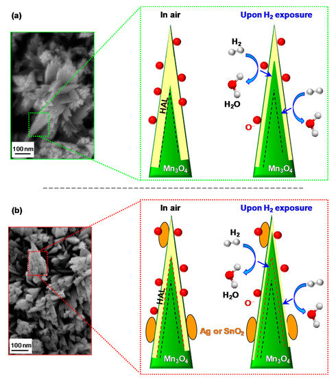
Figure 7.
Schematics of the hydrogen gas sensing mechanism and corresponding hole accumulation layer (HAL) modulation for nanosystems based on: (a) bare Mn3O4; (b) functionalized Mn3O4. The dashed black and red lines indicate the HAL boundaries in air in case of bare and functionalized Mn3O4, respectively. Red spheres, yellow areas, and orange ovals indicate adsorbed oxygen, HAL thickness, and functionalizing agent (Ag or SnO2), respectively. Blue arrows indicate electron flow due to H2 oxidation (see reaction 4).
A process which decreases the hole concentration and the HAL thickness, resulting, in turn, in an increase of the measured resistance [5,16,17,18,42,43]. Finally, upon switching off the gas pulse, the sensor surface is again in contact with air, and the original situation is restored, with a recovery of the pristine HAL width [30,31,41].
Since all the investigated systems have almost identical mean crystallite dimensions, grain size and RMS roughness values, a significant influence of these parameters on the different gas sensing performances can be reasonably ruled out. Indeed, the enhanced responses of composite sensors with respect to the pristine Mn3O4 can be first explained in terms of electronic effects occurring at the interface between Mn3O4 and the functionalizing agents, a key aspect to be considered for a deep understanding of gas sensing phenomena [26].
For Mn3O4-Ag sensors, these processes result from the formation of Mn3O4/Ag Schottky junctions, whose occurrence produces a Ag → Mn3O4 electron flow [38,63] (see also the above XPS data), and a consequent thinning of the HAL width in comparison with bare Mn3O4 (compare Figure 7a,b). As a consequence, HAL variations upon contact of the sensor with gaseous H2 produce higher responses by increasing the registered resistance modulations [38,44]. An analogous phenomenon occurs for Mn3O4-SnO2 systems (Figure 7b; HAL thickness = 12.4 nm, see the Supporting Information), although in this case the SnO2 → Mn3O4 electron flow is triggered by a different phenomenon, i.e., the presence of p-n Mn3O4/SnO2 junctions [32,33,37,41,42,43,55].
The latter effect can, in principle, result in enhanced variations of the HAL extension with respect to the case of Mn3O4-Ag sensors, since the occurrence of a partial silver oxidation (as evidenced by XPS analysis, see above) precludes a full exploitation of electron transfer effects resulting from the establishment of Mn3O4/Ag Schottky junctions [38,46].
Nonetheless, the enhanced hydrogen detection efficiency of Mn3O4-based nanocomposites with respect to bare manganese oxide is likely due to the concurrence of additional cooperative phenomena. For both Mn3O4-Ag and Mn3O4-SnO2 systems, the higher content of oxygen defects at the composite surface with respect to bare Mn3O4 (see the above XPS data and Figure S2), as well as the exposure of a high density of heterointerfaces, can in fact supply active sites for a more efficient chemisorption of both oxygen and analyte molecules, which, in turn, boosts the resulting gas responses [4,8,16,17,18,30,43]. In addition, the intimate component contact enabled by the adopted preparation route, yielding a good intergranular coupling, enables a proficient exploitation of their chemical interplay [38,44,48], related to the synergistical combination of materials with different catalytic activities [10,27,37,41,47]. Hence, the improved sensing performances of functionalized Mn3O4 systems can be related to the concomitance of electronic and catalytic effects.
Taken together, the above observations can account for the improved performances at lower working temperatures of SnO2-containing systems with respect to Ag-containing ones. This result is exemplified by an inspection of Figure 8, showing that, apart from the appreciable response enhancement, deposition of Ag and SnO2 onto Mn3O4 resulted in different trends as a function of the operating temperature. In the case of Mn3O4-Ag sensors, the progressive response rise indicated an enhanced extent of reaction (4) upon increasing the thermal energy supply [2,8,15,31,38,39]. Conversely, as concerns Mn3O4-SnO2, a maximum-like response behavior was observed, the best operating temperature being 200 °C. Such a response trend, in line with previous reports regarding H2 detection by other metal oxides [7,9,17,19,29,43,64], suggested the occurrence of a steady equilibrium between hydrogen adsorption and desorption at 200 °C, whereas an increase of the working temperature resulted in a predominant analyte desorption [27,33,55,64,65]. The lower value of the optimum operating temperature for Mn3O4-SnO2 in comparison to Mn3O4-Ag is in line with the more efficient SnO2 → Mn3O4 electron transfer (see the above XPS data).
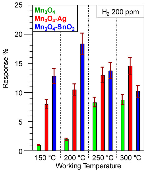
Figure 8.
Gas responses to a fixed H2 concentration (200 ppm) for the target Mn3O4-based sensors at different working temperatures.
The potential of the present results is highlighted by the fact that the best H2 responses obtained for Mn3O4-Ag (at 300 °C) and Mn3O4-SnO2 (at 200 °C) were higher than those reported for sensors based not only on Mn3O4 [35,42], but even on other p-type oxides, including CuO [3,29], NiO [2,30], BiFeO3 [7], Co3O4 [16,19], NixCo3-xO4 [16], as well as nanocomposites based on CuO-Pt [4] and CuO-WO3 [18]. In addition, the same responses compared favorably with those pertaining to various MnO2-based nanomaterials/thin films in the detection of the same analyte [1,64,65]. A comparison of selected representative data is reported in Table S1. It is also worthwhile highlighting that the optimal operating temperature for H2 detection by the present materials (200 °C for Mn3O4-SnO2 systems) was lower than the ones reported for Mn3O4 [35], MnO2 [8], CuO [3,17,29], Co3O4 [16], NiO [9], NiO-ZnO [43], NixCo3-xO4 [16], BiFeO3 [7] and CuO-WO3 sensors [18]. This result is of importance, not only to avoid dangerous temperature-triggered explosions, but also to implement sensing devices with a higher service life and a lower power consumption [11,25,38,41].
Gas responses were also analyzed as a function of H2 concentration (Figure S3). The obtained linear trends in the log-log scale confirmed the absence of appreciable saturation phenomena, an important prerequisite for a quantitative analyte detection [37,38,39,44,48]. The best detection limits obtained by fitting experimental data with equation (2) ((18 ± 1) ppm and (11 ± 1) ppm for Mn3O4-Ag and Mn3O4-SnO2 sensors, respectively) were close to those previously reported for MnO2 [8], CoO [6] and CuO-TiO2-Au [44] sensors, and inferior than those pertaining to ZnO ones [15]. It is also worth noticing that these values were nearly three orders of magnitude lower than the H2 lower explosion limit (LEL, 40000 ppm) [2,11,12,23,43], highlighting thus the detection efficiency of the present systems.
Beyond sensitivity, selectivity is an important parameter for the eventual utilization of gas sensing devices [6,11,19,27,30]. The responses towards a specific test gas are in fact required to be higher than those of other potential interferents, in order to avoid false alarms in real-time gas monitoring equipment [36,38,39]. In particular, the choice of CH4 and CO2 as potential interferents in real-time hydrogen leak detection is motivated by the fact that: (i) hydrogen and methane are common reducing gases, either stored, or used together [1,13]; (ii) the presence of carbon dioxide may hamper hydrogen recognition [5,21,43] in the case of fuel cells eliminating CO2 and producing electricity and H2 [66]. As shown in Figure S4, the present sensors yielded no responses towards CO2, and only weak signals upon exposure to CH4. In the latter case, the selectivity was estimated as the ratio between the responses to H2 and CH4 [32,41], yielding values of 22 and 24 for Mn3O4-Ag and Mn3O4-SnO2 at the best working temperatures (300 and 200 °C, respectively). Though preliminary in nature, the latter results are an attractive starting point for further studies aimed at implementing exclusive H2 sensors, which are highly required for practical applications [13].
4. Conclusions
In summary, in this work we have proposed an unprecedented fabrication route to Mn3O4-Ag and Mn3O4-SnO2 nanocomposites, consisting in: (i) the PA-CVD of MnO2 on alumina substrates; (ii) the subsequent introduction of Ag and SnO2, as prototypes of metal and oxide functional activators, by means of RF-sputtering; (iii) final thermal treatment in air. A thorough chemico-physical investigation revealed the formation of high purity nanocomposites, characterized by the presence of phase-pure hausmannite Mn3O4 featuring a close contact with Ag and SnO2. The successful obtainment of Schottky (Mn3O4/Ag) and p-n junctions (Mn3O4/SnO2) offered significant benefits in view of gas sensing applications, resulting in a nearly ten-fold enhancement of hydrogen responses in comparison to bare Mn3O4 (up to 25% to 500 ppm H2 at a working temperature as low as 200 °C). This improvement, reinforced by the concurrent chemical interplay between the system components, was accompanied by a good sensitivity (detection limits down to 11 ppm, significantly inferior than the H2 LEL of 40,000 ppm) and selectivity in the presence of CH4 and CO2 as potential interferents. Overall, these issues represent a step forward in the use of p-type Mn3O4-based nanocomposites for an efficient and early recognition of H2 leakages in low power consumption sensors.
Future developments of the present activities will concern the extension of the proposed preparation route to different multifunctional materials for (photo)electrochemical water splitting and solar-driven catalysis for air/water purification. In addition, more detailed selectivity studies, including other gaseous species and the evaluation of sensing performances under different environmental conditions, are undoubtedly important issues to be properly considered for the eventual real-world implementation of the developed materials.
Supplementary Materials
The following are available online at https://www.mdpi.com/2079-4991/10/3/511/s1: Figure S1: Surface silver Auger signal for the Mn3O4-Ag specimen, Figure S2: Deconvolution of surface O1s XP spectra for Mn3O4-based samples, Figure S3: Gas responses as a function of H2 concentration for bare and functionalized Mn3O4 sensors, Figure S4: Gas responses to fixed CO2, CH4, and H2 concentrations for Mn3O4-Ag and Mn3O4-SnO2 sensors, response time data, calculation of the HAL width.
Author Contributions
Conceptualization, L.B., C.M. and D.B.; Data curation, L.B.; Formal analysis, C.S.; Funding acquisition, A.G.; Investigation, L.B. and D.Z.; Project administration, C.M.; Resources, C.M.; Validation, D.Z. and E.C.; Writing—original draft, D.B.; Writing—review & editing, A.G., C.S. and E.C. All authors have read and agreed to the published version of the manuscript.
Funding
This research was funded by Padova University DOR 2016–2019 and P-DiSC #SENSATIONAL BIRD2016-UNIPD projects, and as well as by the INSTM Consortium (INSTMPD010—ISIDE).
Conflicts of Interest
The authors declare no conflict of interest.
References
- Zöpfl, A.; Lemberger, M.-M.; König, M.; Ruhl, G.; Matysik, F.-M.; Hirsch, T. Reduced graphene oxide and graphene composite materials for improved gas sensing at low temperature. Faraday Discuss. 2014, 173, 403–414. [Google Scholar] [CrossRef]
- Stamataki, M.; Tsamakis, D.; Brilis, N.; Fasaki, I.; Giannoudakos, A.; Kompitsas, M. Hydrogen gas sensors based on PLD grown NiO thin film structures. Phys. Status Solidi 2008, 205, 2064–2068. [Google Scholar] [CrossRef]
- Duc, L.D.; Le, D.T.T.; Duy, N.V.; Hoa, N.D.; Hieu, N.V. Single crystal cupric oxide nanowires: Length- and density-controlled growth and gas-sensing characteristics. Physica E 2014, 58, 16–23. [Google Scholar] [CrossRef]
- Sarıca, N.; Alev, O.; Arslan, L.Ç.; Öztürk, Z.Z. Characterization and gas sensing performances of noble metals decorated CuO nanorods. Thin Solid Film. 2019, 685, 321–328. [Google Scholar] [CrossRef]
- Tonezzer, M.; Le, D.T.T.; Iannotta, S.; Hieu, N.V. Selective discrimination of hazardous gases using one single metal oxide resistive sensor. Sens. Actuators B 2018, 277, 121–128. [Google Scholar] [CrossRef]
- Li, L.H.; Xiao, J.; Yang, G.W. Amorphization of cobalt monoxide nanocrystals and related explosive gas sensing applications. Nanotechnology 2015, 26, 415501. [Google Scholar] [CrossRef]
- Bala, A.; Majumder, S.B.; Dewan, M.; Roy Chaudhuri, A. Hydrogen sensing characteristics of perovskite based calcium doped BiFeO3 thin films. Int. J. Hydrogen Energy 2019, 44, 18648–18656. [Google Scholar] [CrossRef]
- Sanger, A.; Kumar, A.; Kumar, A.; Chandra, R. Highly sensitive and selective hydrogen gas sensor using sputtered grown Pd decorated MnO2 nanowalls. Sens. Actuators B 2016, 234, 8–14. [Google Scholar] [CrossRef]
- Kaur, N.; Comini, E.; Zappa, D.; Poli, N.; Sberveglieri, G. Nickel oxide nanowires: Vapor liquid solid synthesis and integration into a gas sensing device. Nanotechnology 2016, 27, 205701. [Google Scholar] [CrossRef] [PubMed]
- Lu, Z.; Zhou, Q.; Xu, L.; Gui, Y.; Zhao, Z.; Tang, C.; Chen, W. Synthesis and characterization of highly sensitive hydrogen (H2) sensing device based on Ag doped SnO2 nanospheres. Materials 2018, 11, 492. [Google Scholar] [CrossRef] [PubMed]
- Tian, X.Q.; Yang, L.; Qing, X.X.; Yu, K.; Wang, X.F. Trace level detection of hydrogen gas using birnessite-type manganese oxide. Sens. Actuators B 2015, 207, 34–42. [Google Scholar] [CrossRef]
- Lee, Y.T.; Lee, J.M.; Kim, Y.J.; Joe, J.H.; Lee, W. Hydrogen gas sensing properties of PdO thin films with nano-sized cracks. Nanotechnology 2010, 21, 165503. [Google Scholar] [CrossRef] [PubMed]
- Huang, H.; Gong, H.; Chow, C.L.; Guo, J.; White, T.J.; Tse, M.S.; Tan, O.K. Low-temperature growth of SnO2 nanorod arrays and tunable n–p–n sensing response of a ZnO/SnO2 heterojunction for exclusive hydrogen sensors. Adv. Funct. Mater. 2011, 21, 2680–2686. [Google Scholar] [CrossRef]
- Bigiani, L.; Zappa, D.; Maccato, C.; Comini, E.; Barreca, D.; Gasparotto, A. Quasi-1D MnO2 nanocomposites as gas sensors for hazardous chemicals. Appl. Surf. Sci. 2020, 512, 145667. [Google Scholar] [CrossRef]
- Barreca, D.; Bekermann, D.; Comini, E.; Devi, A.; Fischer, R.A.; Gasparotto, A.; Maccato, C.; Sberveglieri, G.; Tondello, E. 1D ZnO nano-assemblies by plasma-CVD as chemical sensors for flammable and toxic gases. Sens. Actuators B 2010, 149, 1–7. [Google Scholar] [CrossRef]
- Govindhan, M.; Sidhureddy, B.; Chen, A. High-temperature hydrogen gas sensor based on three-dimensional hierarchical-nanostructured nickel–cobalt oxide. ACS Appl. Nano Mater. 2018, 1, 6005–6014. [Google Scholar] [CrossRef]
- Choi, Y.-H.; Kim, D.-H.; Hong, S.-H.; Hong, K.S. H2 and C2H5OH sensing characteristics of mesoporous p-type CuO films prepared via a novel precursor-based ink solution route. Sens. Actuators B 2013, 178, 395–403. [Google Scholar] [CrossRef]
- Haviar, S.; Čapek, J.; Batková, Š.; Kumar, N.; Dvořák, F.; Duchoň, T.; Fialová, M.; Zeman, P. Hydrogen gas sensing properties of WO3 sputter-deposited thin films enhanced by on-top deposited CuO nanoclusters. Int. J. Hydrogen Energy 2018, 43, 22756–22764. [Google Scholar] [CrossRef]
- Barreca, D.; Comini, E.; Gasparotto, A.; Maccato, C.; Pozza, A.; Sada, C.; Sberveglieri, G.; Tondello, E. Vapor phase synthesis, characterization and gas sensing performances of Co3O4 and Au/Co3O4 nanosystems. J. Nanosci. Nanotechnol. 2010, 10, 8054–8061. [Google Scholar] [CrossRef]
- Fortunato, E.; Malik, A.; Martins, R. Photochemical sensors based on amorphous silicon thin films. Sens. Actuators B 1998, 46, 202–207. [Google Scholar] [CrossRef]
- Hashtroudi, H.; Atkin, P.; Mackinnon, I.D.R.; Shafiei, M. Low-operating temperature resistive nanostructured hydrogen sensors. Int. J. Hydrogen Energy 2019, 44, 26646–26664. [Google Scholar] [CrossRef]
- Hübert, T.; Boon-Brett, L.; Black, G.; Banach, U. Hydrogen sensors—A review. Sens. Actuators B 2011, 157, 329–352. [Google Scholar] [CrossRef]
- Penner, R.M. A nose for hydrogen gas: Fast, sensitive H2 sensors using electrodeposited nanomaterials. Acc. Chem. Res. 2017, 50, 1902–1910. [Google Scholar] [CrossRef] [PubMed]
- Meng, X.; Zhang, Q.; Zhang, S.; He, Z. The enhanced H2 selectivity of SnO2 gas sensors with the deposited SiO2 filters on surface of the sensors. Sensors 2019, 19, 2478. [Google Scholar] [CrossRef]
- Comini, E.; Baratto, C.; Concina, I.; Faglia, G.; Falasconi, M.; Ferroni, M.; Galstyan, V.; Gobbi, E.; Ponzoni, A.; Vomiero, A.; et al. Metal oxide nanoscience and nanotechnology for chemical sensors. Sens. Actuators B 2013, 179, 3–20. [Google Scholar] [CrossRef]
- Zappa, D.; Galstyan, V.; Kaur, N.; Munasinghe Arachchige, H.M.M.; Sisman, O.; Comini, E. “Metal oxide -based heterostructures for gas sensors”—A review. Anal. Chim. Acta 2018, 1039, 1–23. [Google Scholar] [CrossRef]
- Yang, Y.; Wang, X.; Yi, G.; Li, H.; Shi, C.; Sun, G.; Zhang, Z. Hydrothermal synthesis of Co3O4/ZnO hybrid nanoparticles for triethylamine detection. Nanomaterials 2019, 9, 1599. [Google Scholar] [CrossRef]
- Coll, M.; Fontcuberta, J.; Althammer, M.; Bibes, M.; Boschker, H.; Calleja, A.; Cheng, G.; Cuoco, M.; Dittmann, R.; Dkhil, B.; et al. Towards oxide electronics: A roadmap. Appl. Surf. Sci. 2019, 482, 1–93. [Google Scholar] [CrossRef]
- Hoa, N.D.; An, S.Y.; Dung, N.Q.; Van Quy, N.; Kim, D. Synthesis of p-type semiconducting cupric oxide thin films and their application to hydrogen detection. Sens. Actuators B 2010, 146, 239–244. [Google Scholar] [CrossRef]
- Zhao, S.; Shen, Y.; Zhou, P.; Zhang, J.; Zhang, W.; Chen, X.; Wei, D.; Fang, P.; Shen, Y. Highly selective NO2 sensor based on p-type nanocrystalline NiO thin films prepared by sol–gel dip coating. Ceram. Int. 2018, 44, 753–759. [Google Scholar] [CrossRef]
- Bigiani, L.; Maccato, C.; Carraro, G.; Gasparotto, A.; Sada, C.; Comini, E.; Barreca, D. Tailoring vapor-phase fabrication of Mn3O4 nanosystems: From synthesis to gas-sensing applications. ACS Appl. Nano Mater. 2018, 1, 2962–2970. [Google Scholar] [CrossRef]
- Kim, H.-J.; Lee, J.-H. Highly sensitive and selective gas sensors using p-type oxide semiconductors: Overview. Sens. Actuators B 2014, 192, 607–627. [Google Scholar] [CrossRef]
- Kim, J.-H.; Lee, J.-H.; Mirzaei, A.; Kim, H.W.; Kim, S.S. SnO2 (n)-NiO (p) composite nanowebs: Gas sensing properties and sensing mechanisms. Sens. Actuators B 2018, 258, 204–214. [Google Scholar] [CrossRef]
- Chen, Z.; Jiao, Z.; Pan, D.; Li, Z.; Wu, M.; Shek, C.-H.; Wu, C.M.L.; Lai, J.K.L. Recent advances in manganese oxide nanocrystals: Fabrication, characterization, and microstructure. Chem. Rev. 2012, 112, 3833–3855. [Google Scholar] [CrossRef]
- Na, C.W.; Park, S.-Y.; Chung, J.-H.; Lee, J.-H. Transformation of ZnO nanobelts into single-crystalline Mn3O4 nanowires. ACS Appl. Mater. Interfaces 2012, 4, 6565–6572. [Google Scholar] [CrossRef] [PubMed]
- John, N.; Thomas, P.; Divya, K.V.; Abraham, K.E. Enhanced room temperature gas sensing of aligned Mn3O4 nanorod assemblies functionalized by aluminum anodic membranes. Nanotechnology 2018, 29, 335503. [Google Scholar] [CrossRef]
- Zhou, T.; Liu, X.; Zhang, R.; Wang, L.; Zhang, T. Constructing hierarchical heterostructured Mn3O4/Zn2SnO4 materials for efficient gas sensing reaction. Adv. Mater. Interfaces 2018, 5, 1800115. [Google Scholar] [CrossRef]
- Bigiani, L.; Zappa, D.; Barreca, D.; Gasparotto, A.; Sada, C.; Tabacchi, G.; Fois, E.; Comini, E.; Maccato, C. Sensing nitrogen mustard gas simulant at the ppb scale via selective dual-site activation at Au/Mn3O4 interfaces. ACS Appl. Mater. Interfaces 2019, 11, 23692–23700. [Google Scholar] [CrossRef]
- Maccato, C.; Bigiani, L.; Carraro, G.; Gasparotto, A.; Sada, C.; Comini, E.; Barreca, D. Toward the detection of poisonous chemicals and warfare agents by functional Mn3O4 nanosystems. ACS Appl. Mater. Interfaces 2018, 10, 12305–12310. [Google Scholar] [CrossRef]
- Ben Said, L.; Inoubli, A.; Bouricha, B.; Amlouk, M. High Zr doping effects on the microstructural and optical properties of Mn3O4 thin films along with ethanol sensing. Spectrochim. Acta Part A 2017, 171, 487–498. [Google Scholar] [CrossRef]
- Bigiani, L.; Zappa, D.; Maccato, C.; Gasparotto, A.; Sada, C.; Comini, E.; Barreca, D. Mn3O4 nanomaterials functionalized with Fe2O3 and ZnO: Fabrication, characterization, and ammonia sensing properties. Adv. Mater. Interfaces 2019, 6, 1901239. [Google Scholar] [CrossRef]
- Kim, H.W.; Kwon, Y.J.; Na, H.G.; Cho, H.Y.; Lee, C.; Jung, J.H. One-pot synthesis of Mn3O4-decorated GaN nanowires for drastic changes in magnetic and gas-sensing properties. Microelectron. Eng. 2015, 139, 60–69. [Google Scholar] [CrossRef]
- Nakate, U.T.; Ahmad, R.; Patil, P.; Wang, Y.; Bhat, K.S.; Mahmoudi, T.; Yu, Y.T.; Suh, E.-K.; Hahn, Y.-B. Improved selectivity and low concentration hydrogen gas sensor application of Pd sensitized heterojunction n-ZnO/p-NiO nanostructures. J. Alloy. Compd. 2019, 797, 456–464. [Google Scholar] [CrossRef]
- Barreca, D.; Carraro, G.; Comini, E.; Gasparotto, A.; Maccato, C.; Sada, C.; Sberveglieri, G.; Tondello, E. Novel synthesis and gas sensing performances of CuO–TiO2 nanocomposites functionalized with Au nanoparticles. J. Phys. Chem. C 2011, 115, 10510–10517. [Google Scholar] [CrossRef]
- Acharyya, S.S.; Ghosh, S.; Sharma, S.K.; Bal, R. Fabrication of Ag nanoparticles supported on one-dimensional (1D) Mn3O4 spinel nanorods for selective oxidation of cyclohexane at room temperature. New J. Chem. 2016, 40, 3812–3820. [Google Scholar] [CrossRef]
- Carraro, G.; Gasparotto, A.; Maccato, C.; Gombac, V.; Rossi, F.; Montini, T.; Peeters, D.; Bontempi, E.; Sada, C.; Barreca, D.; et al. Solar H2 generation via ethanol photoreforming on ε-Fe2O3 nanorod arrays activated by Ag and Au nanoparticles. RSC Adv. 2014, 4, 32174–32179. [Google Scholar] [CrossRef]
- Rahaman, H.; Kundu, S.; Ghosh, S.K. Size-selective silver-induced evolution of Mn3O4-Ag nanocomposites for effective ethanol sensing. ChemistrySelect 2017, 2, 6991–6999. [Google Scholar] [CrossRef]
- Simon, Q.; Barreca, D.; Gasparotto, A.; Maccato, C.; Tondello, E.; Sada, C.; Comini, E.; Devi, A.; Fischer, R.A. Ag/ZnO nanomaterials as high performance sensors for flammable and toxic gases. Nanotechnology 2012, 23, 025502. [Google Scholar] [CrossRef]
- Carraro, G.; Barreca, D.; Comini, E.; Gasparotto, A.; Maccato, C.; Sada, C.; Sberveglieri, G. Controlled synthesis and properties of β-Fe2O3 nanosystems functionalized with Ag or Pt nanoparticles. CrystEngComm 2012, 14, 6469–6476. [Google Scholar] [CrossRef]
- Rahman, M.M.; Alam, M.M.; Asiri, A.M. Fabrication of an acetone sensor based on facile ternary MnO2/Gd2O3/SnO2 nanosheets for environmental safety. New J. Chem. 2017, 41, 9938–9946. [Google Scholar] [CrossRef]
- Barreca, D.; Carraro, G.; Fois, E.; Gasparotto, A.; Gri, F.; Seraglia, R.; Wilken, M.; Venzo, A.; Devi, A.; Tabacchi, G.; et al. Manganese(II) molecular sources for plasma-assisted CVD of Mn oxides and fluorides: From precursors to growth process. J. Phys. Chem. C 2018, 122, 1367–1375. [Google Scholar] [CrossRef]
- Maccato, C.; Bigiani, L.; Carraro, G.; Gasparotto, A.; Seraglia, R.; Kim, J.; Devi, A.; Tabacchi, G.; Fois, E.; Pace, G.; et al. Molecular engineering of MnII diamine diketonate precursors for the vapor deposition of manganese oxide nanostructures. Chem. Eur. J. 2017, 23, 17954–17963. [Google Scholar] [CrossRef] [PubMed]
- Barreca, D.; Gasparotto, A.; Tondello, E.; Sada, C.; Polizzi, S.; Benedetti, A. Nucleation and growth of nanophasic CeO2 thin films by plasma-enhanced CVD. Chem. Vap. Depos. 2003, 9, 199–206. [Google Scholar] [CrossRef]
- Mattelaer, F.; Bosserez, T.; Ronge, J.; Martens, J.A.; Dendooven, J.; Detavernier, C. Manganese oxide films with controlled oxidation state for water splitting devices through a combination of atomic layer deposition and post-deposition annealing. RSC Adv. 2016, 6, 98337–98343. [Google Scholar] [CrossRef]
- Liu, H.; Wang, F.; Hu, K.; Zhang, B.; He, L.; Zhou, Q. Superior hydrogen sensing property of porous NiO/SnO2 nanofibers synthesized via carbonization. Nanomaterials 2019, 9, 1250. [Google Scholar] [CrossRef]
- Briggs, D.; Seah, M.P. Practical Surface Analysis: Auger and X-ray Photoelectron Spectroscopy, 2nd ed.; John Wiley & Sons: Hoboken, NJ, USA, 1990. [Google Scholar]
- Available online: https://xpspeak.software.informer.com/4.1/ (accessed on 31 December 2019).
- Available online: http://imagej.nih.gov/ij/ (accessed on 31 January 2020).
- JCPDS card no. 024-0734 (2000).
- Bigiani, L.; Barreca, D.; Gasparotto, A.; Maccato, C. Mn3O4 thin films functionalized with Ag, Au, and TiO2 analyzed using X-ray photoelectron spectroscopy. Surf. Sci. Spectra 2018, 25, 014003. [Google Scholar] [CrossRef]
- NIST X-ray Photoelectron Spectroscopy Database. Available online: http://srdata.nist.gov/xps (accessed on 31 December 2019).
- Sharma, J.K.; Srivastava, P.; Ameen, S.; Akhtar, M.S.; Singh, G.; Yadava, S. Azadirachta Indica plant-assisted green synthesis of Mn3O4 nanoparticles: Excellent thermal catalytic performance and chemical sensing behavior. J. Colloid Interface Sci. 2016, 472, 220–228. [Google Scholar] [CrossRef]
- Zhang, Z.; Yates, J.T. Band bending in semiconductors: Chemical and physical consequences at surfaces and interfaces. Chem. Rev. 2012, 112, 5520–5551. [Google Scholar] [CrossRef]
- Zhang, C.; Boudiba, A.; Navio, C.; Olivier, M.-G.; Snyders, R.; Debliquy, M. Study of selectivity of NO2 sensors composed of WO3 and MnO2 thin films grown by radio frequency sputtering. Sens. Actuators B 2012, 161, 914–922. [Google Scholar] [CrossRef]
- Jung, D.; Yoon, Y.; Lee, G.S. Hydrogen sensing characteristics of carbon-nanotube sheet decorated with manganese oxides. Chem. Phys. Lett. 2013, 577, 96–101. [Google Scholar] [CrossRef]
- Fuel Cell-Based System Converts Atmospheric CO2 into Usable Electric Current. Available online: https://www.theengineer.co.uk/fuel-cell-based-system-co2/ (accessed on 31 December 2019).


© 2020 by the authors. Licensee MDPI, Basel, Switzerland. This article is an open access article distributed under the terms and conditions of the Creative Commons Attribution (CC BY) license (http://creativecommons.org/licenses/by/4.0/).