1. Introduction
An optical sensor, a classical sensor type based on optical principles, can sensitively monitor measured information and convert the information into optical signals or other forms of data according to certain rules [
1]. Owing to its advantages (e.g., non-contact and non-destructive measurement, little interference and high sensitivity), the optical sensor supports a wide range of applications in the realm of food safety [
2,
3], environmental monitoring [
4], drug testing [
5], medical analysis [
6,
7], biochemical tests [
8,
9] and so on. The realization and control means of high-sensitivity optical sensors, especially related optical sensor devices, play a key role in optical measurement and biosensors. In particular, a micro-nano optical sensor, the size of an integrated chip, is the key to information detection and monitoring. Therefore, the realization and testing methods of micro-nano optical sensors have become the center of attention in recent years. For example, scholars have conducted extensive research into carbon nanotubes [
10], photonic crystal [
11], and graphene/waveguide hybrid structures [
12]. In addition, due to the low photon energy in the terahertz band and the distinctive spectral signatures of most biomolecules in the terahertz band, optical sensors working in the terahertz band are also widely used. It is worth mentioning that surface plasmon resonance (SPR) is very sensitive to changes in any boundary environment owing to the boundary propagation of surface plasmon wave. For this reason, SPR-based optical sensors have also become a main focus of attention among researchers. Various high-sensitivity optical sensors have been proposed on the basis of SPR technology [
13,
14,
15]. Recently, graphene has started to play an active role in the realization of high-sensitivity optical sensors due to its excellent optoelectronic properties such as SPR support [
16], tunability of optical conductivity [
17], broadband [
18], etc. In this respect, graphene-based SPR sensors [
19,
20], hybrid graphene/gold plasmonic fiber-optic sensors [
21], mid-infrared plasmonic biosensing with graphene [
22], and multi-channel graphene sensors [
23], and graphene-based Bloch-like surface wave sensors [
24] have been proposed. Recently, Sun et al., reported the application of inorganic/polymer-graphene hybrid gel as a versatile electrochemical platform for an electrochemical capacitor and biosensor [
25]. Sun et al., demonstrated the sensitivity enhancement of SPR biosensor based on graphene and barium titanate layers [
26]. It can be predicted optimistically that graphene-based or 2D materials-based optical sensors will be one of the most promising application trends [
27,
28,
29]. Although the basic theory of optical sensors is relatively mature, the implementation of optical sensors with a simple structure, high sensitivity and dynamic controllability still remains challenging. Optical sensors with new materials or novel structures and working mechanisms have become the main direction of research in the optical sensor industry.
We know that the generation of optical resonance has a very positive effect on the realization of high-sensitivity sensor detection. At present, many optical-sensor schemes are mainly realized by the exciting of SPR. This is mainly due to the fact that SPR can produce a very obvious resonance peak, thus creating conditions for sensitive sensor detection. Recently, optical Tamm states (OTSs), a kind of surface wave confined on the contact surface of two different media, have attracted attention in the field of optical sensing [
30,
31]. It is essentially an interface state. Compared with SPR, OTSs can be excited without a specific angle of incidence, as well as being directly excited by transverse electric (TE) polarization. More importantly, the excitation of OTSs is very sensitive to the change of boundary environment [
32]. Therefore, the implementation of optical sensors based on OTSs is very attractive and promising. For example, Zhang et al., proposed a novel concept of a refractive index sensor based on a metal-distributed Bragg reflector [
33]. However, the excitation of conventional OTSs is mainly based on a metal-Bragg reflector structure, which does not have dynamic tunability, thus limiting its application in the terahertz band. Graphene not only has excellent tunability and broadband characteristics, but also presents some metal-like properties under certain conditions. This makes it possible to combine graphene and the typical OTS structure to realize a dynamically tunable THz sensor. For example, Ye et al., proposed a graphene-based composite structure to realize a tunable and highly sensitive optical biosensor by exciting OTSs [
34]. In this paper, we propose a novel terahertz sensor based on a graphene Bragg reflector composite structure to realize high sensitivity. We find that the high sensitivity of a terahertz sensor is a product of the abnormal reflectance peaks caused by optical resonance. In addition, the tunable conductivity of graphene provides a basis for the design of tunable sensing characteristics in the proposed structure. We believe this electronically-tunable terahertz sensor based on a vertically stacked structure with graphene could offer great potential for applications in the biosensor field.
2. Materials and Methods
We consider a terahertz sensor by inserting a sensing medium between a polymethylpenten (TPX)/
Distributed Bragg Reflector (DBR) and a monolayer graphene film in a vertically stacked structure, with a
DBR structure underneath and a monolayer graphene film on the top, as illustrated in
Figure 1. Graphene can be transferred to the silicon substrate with holes in consideration of the practically-possible device fabrication. A one-dimensional photonic crystal (1D PC) is formed by alternately stacking dielectric A and dielectric B at a period of
. The center wavelength
is set as
; the materials of dielectric A and B are selected respectively as
with refractive index
and
with refractive index
; the thickness of each 1D PC layer is
. The refractive index and original thickness of the sensing medium are, respectively, set as
and
;
is the change of refractive index of the sensing medium due to the absorption of biomolecules on the surface of graphene. The thickness of graphene, written as
, is neglected in our calculation;
stands for the number of graphene layers. Here, in order to obtain the physical mechanism more easily and to simplify the calculation, we assume that the refractive indexes of the above materials are dispersionless in the terahertz band. In practice, absorption in the sensing medium cannot be completely avoided, especially in the terahertz band. Therefore, in the next section, we will also briefly discuss the possible influence of the absorption in the sensing layer on the sensitivity performance of the sensor. Besides, the inter-conductivity of graphene is negligible under terahertz band and random phase approximation. According to a Drude-like formula, the conductivity of graphene can be approximately expressed as:
where
is the reduced Planck’s constant,
is the Fermi energy closely related to carrier density (
), and
(
represents the Fermi velocity of the electron). It creates conditions for us to adjust the conductivity of graphene by controlling the gate voltage.
is the angular frequency of the incident beam;
and
represent the elementary electric charge and the relaxation time, respectively. It can be noted that there are some similarities between the above composite structure and the model in Reference [
34]. However, the structure of the two is essentially different. In Ref. [
34], the incident light needs to pass through the 1D PC with band gap characteristics first, and then contact the sensing medium. The excitation of OTSs has more stringent requirements on the 1D PC. For example, the period of 1D PC cannot be too large, otherwise the incident light cannot penetrate the 1D PC. However, in this work, the incident light first acts on graphene and the sensing medium. At this time, the function of 1D PC is equivalent to the Bragg reflector, and its period needs to be set to a larger value. In addition, the sensing medium in Reference [
34] is under the 1D PC, so the influence of the absorption of the sensing medium on the sensitivity of the sensor is much smaller. In our structure, the absorption of the sensing medium has an influence on the sensing performance.
A transfer matrix is applied to calculate the reflectance of the proposed vertically stacked structure [
35]. For simplicity, we only consider transverse magnetic (TM) polarization. Then, the transfer matrix between air and the sensing medium can be expressed as:
where
and
;
and
are the wave vector components of light wave propagating in air and sensing medium, respectively. Combined with the propagation matrix of light in dielectric layer
(
is the thickness of the dielectric), the transfer matrix of the whole system can be expressed as:
, where
is the period of 1D PC. Thus the reflectance of the structure can be obtained by
. In this paper, the reflectance of this structure is strongly sensitive to the variation in the refractive index of the sensing medium. Therefore, the sensitivity of this structure can be expressed as:
where
represents the change of the resonance angle of the proposed structure, and it is caused by the variation in the refractive index of the sensing medium. In addition, the figure of merit (FOM) can be described as:
, where the quality factor (DA) is defined as
(full width at half maxima).
3. Results and Discussion
This section focuses on the sensing characteristics of the proposed structure. In essence, the configuration in
Figure 1 can be seen as an asymmetrical cavity consisting of a graphene layer on the left side and a Bragg reflector on the right side, with the sensing medium located inside the cavity. The Fabry–Perot mode can be excited at a certain structural parameter and there is a dip appearing in the reflectance spectrum owing to the excitation of the Fabry–Perot mode. The resonance angle of mode is sensitive to the change of ambient refractive index (
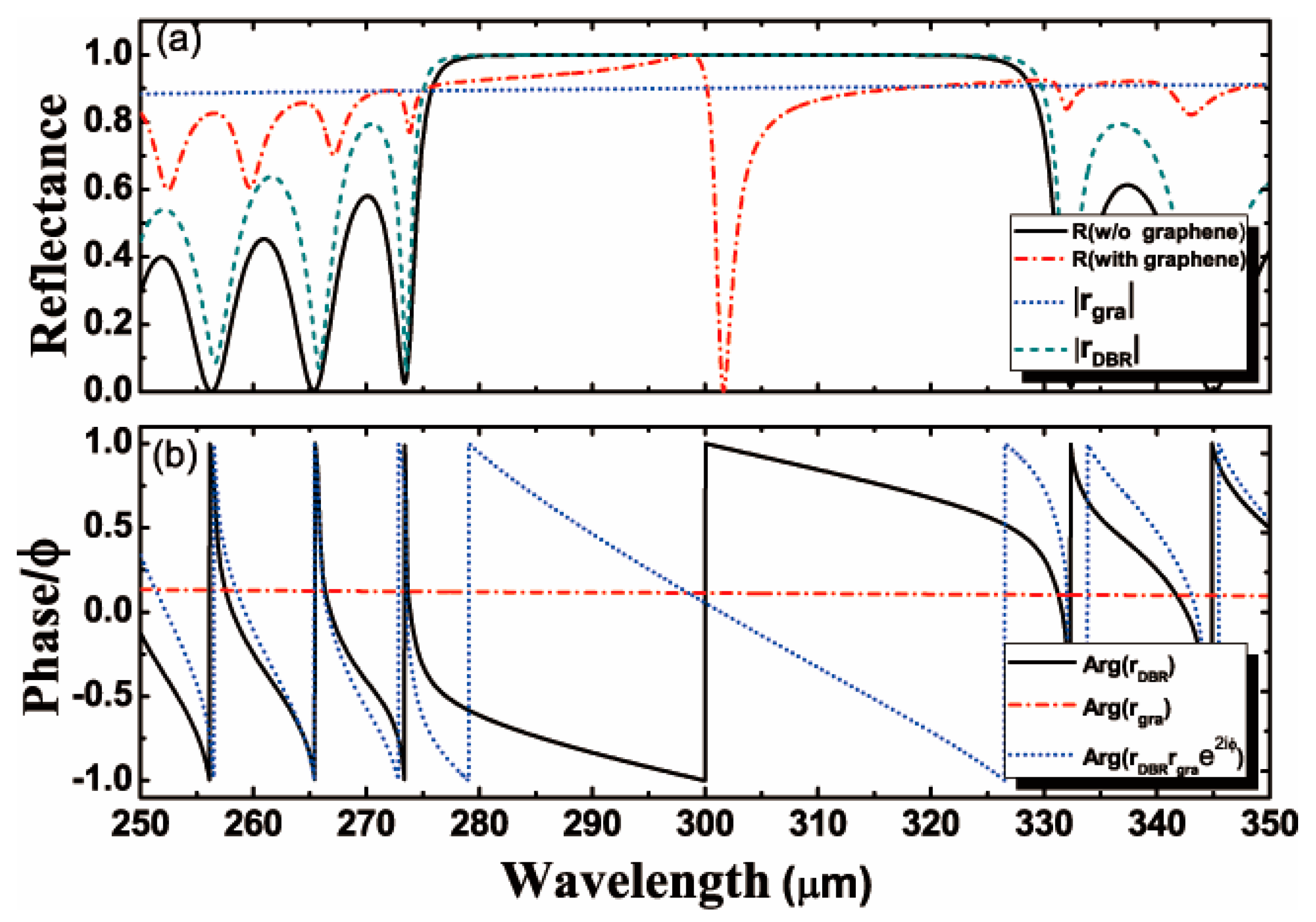
). Therefore, the sensitivity can be calculated by Formula (3). To better illustrate the physics mechanism, the reflectance of the structure with and without graphene are illustrated in
Figure 2a. It is found that there is a band gap at a wavelength in the range of
to
in the absence of graphene. However, when the sensing medium is coated with graphene, an obvious reflection dip appears at a wavelength of about
within the band gap. The physical origin of the optical resonance modes observed in
Figure 2a can be explained by using the Fabry–Perot mode. However, it is known that graphene is intrinsically a semimetal with some metallic properties under certain conditions. Therefore, the above optical resonance phenomenon can also be explained from the perspective of excitation of OTSs. It is well-known that the excitation of OTSs should satisfy
, where
is the reflection coefficient of the incident light beam on the sensing medium and the photonic crystal interface,
is the reflection coefficient of the incident light beam on the interface of graphene and the sensing medium, and
is the phase change of the light beam impinging at two interfaces of the proposed structure. The resonance frequency position of OTSs excitation can be estimated based on the above formula. Accordingly,
and
can be obtained around
by calculation, where
and
. When there is no graphene (namely
and
), it will be impossible to excite OTSs. In contrast, the introduction of graphene will bring about
and the formula
can be obtained. Furthermore, the excitation of OTSs should also satisfy
. As shown in
Figure 2b, we obtain
around
and this result coincides with the dip in
Figure 2a.
In order to further illustrate the relevance of graphene to the strong excitation of the resonance mode, we cover the sensing medium with monolayer graphene and draw the normalized electric field distributions in
Figure 3a. The normalized electric field distributions in the absence of graphene are plotted in
Figure 3b for comparison. The position of graphene is set as
. It is shown that the light beam impinges on the interface between the graphene and the sensing medium, and the sharp rise of the electric field near the graphene coincides precisely with the abnormal dip of reflectance in
Figure 2a. When the light beam penetrates the photonic crystal, the electric field decays rapidly with the increase of PC periods. The local field intensity is enhanced significantly in the structure coated with graphene. These results have proved that graphene plays a positive role in the excitation of resonance mode and the realization of a high sensitivity of the proposed structure.
To enhance the sensitivity and to expand the measuring range of the proposed terahertz refractive index sensor, we also take the major parameters of graphene, and the thickness and refractive index of the sensing medium into consideration. The variations in the reflectance at different levels of Fermi energy with respect to the incident angle are plotted, as shown in
Figure 4. For a sensing medium containing biomacromolecules, we select aqueous solution with a refractive index of 1.33;
and
are set to be
and 1, respectively. Graphene, in comparison with a metal surface, also shows strong and stable absorption to biomolecules. For the calculation of sensitivity, the change of the refractive index of the sensing medium is assumed to be
. The optical properties of graphene are represented by electrical conductivity, and the conductivity of graphene can be regulated by adjusting the Fermi energy and relaxation time. Furthermore, we regulate the Fermi energy of graphene by adjusting the external voltage. The sensitivity of the proposed structure is as high as
when
, as shown in
Figure 4d. Through research, the optimal Fermi energy of graphene should be
. It is worth mentioning that the above sensitivity performance is calculated without considering the absorption of the sensing medium. In fact, the influence of the absorption coefficient of the sensing medium on the sensing characteristics also needs to be considered, especially in the terahertz band. Therefore, based on the structure of
Figure 1, we further calculate the sensitivity performance of the terahertz refractive index sensor in three cases: (1) when the sensing medium is an aqueous solution, the dielectric function of water can be expressed by a triple Debye function:
, where
[
36]. It is found that the resonance phenomenon cannot occur obviously because the aqueous solution has a large absorption coefficient in the terahertz band. Therefore, it is difficult to use aqueous solution as sensing medium in terahertz band. (2) We also calculated the sensing characteristics when the sensing medium is a liquid with low absorption coefficient (for example, the absorption of nonpolar solution is generally low in the terahertz band). Take n-propanol as an example in the calculation (
, with other parameters having the same values as those in
Figure 4) [
37]. It is found that the resonance phenomenon can still be realized and the sensitivity of about
can be obtained. (3) Furthermore, when the sensing medium is gas (where the refractive index is close to 1 and absorption can be ignored), the structure in the manuscript can be further developed into a THz gas sensor. In the case of similar structural parameters as before, the resonance phenomenon is very obvious and the sensitivity above
can be obtained. The above results indicate that a high-sensitivity terahertz refractive index sensor based on this structure is feasible.
It can be seen from the formula of graphene conductivity that the relaxation time also has an obvious effect on conductivity. However, there are sometimes obstacles to regulating the relaxation time of graphene, because it is difficult to change the relaxation time once the graphene is prepared. Nevertheless, it is necessary to evaluate systematically the effects of relaxation time on the sensing properties of the proposed structure, as shown in
Figure 5. The variations of the sensitivity and FOM with respect to the relaxation time of graphene are plotted in
Figure 5a. It is found that the narrower the FHWM is, the higher the structural sensitivity would be. Meanwhile,
Figure 5b shows the variation of reflectance as a function of the incident angle when
and
. When
, the highest sensitivity of the proposed structure (
) is obtained. It can be observed that the sensitivity and FOM vary monotonously with the relaxation time, mainly because the increase of relaxation time has a strong influence on the real part of the conductivity but is barely effective for the imaginary part, and this is reflected in narrower and deeper reflectance curves. Consequently, the sensitivity and FOM of the proposed structure are affected. The FHWM of the reflectance at
is obviously smaller than that at
, thus producing the highest FOM (65 RIU
−1). In summary, the effect of relaxation time on sensitivity is more significant than that on Fermi energy.
The thickness of sensing medium is also an important factor in the sensitivity of the proposed structure. Therefore, we plot the curves of sensitivity according to the thickness of the sensing medium, as shown in
Figure 6a,b. From the point of view of OTSs mode, it is commonly known that the thickness of the sensing medium is very important to the excitation of OTSs. The thickness of the sensing medium and the excitation of OTSs should satisfy:
, where
is the minimum value of OTSs observed at
, and
is a natural number. When
, we can get the maximum sensitivity (
) of the proposed structure. Accordingly, the reflectance as a function of the incident angle is plotted. Since the resonance angle of OTSs excitation is highly sensitive to the thickness of sensing medium, we set
as
to ensure higher overall sensitivity and a feasible design of the structure. To conclude,
is no doubt an important factor in the design of the terahertz refractive index sensor. In order to expand the detection range of the refractive index sensor, we plot a curve to describe the relevance between the sensitivity of refractive index sensor and the refractive index of the sensing medium, as shown in
Figure 6c. It is found that the refractive index of the sensing medium shows the same trend as its thickness. Additionally, the sensitivity varies monotonously with
, thus ensuring a large measuring range of
. A sensitivity of
can be achieved even if the
of the sensing medium increases to 1.42.
Therefore, it is proved that proper selection of the sensing medium thickness () and the refractive index () can produce high sensitivity.
Next, the relationship between the number of graphene layers and the sensitivity of this structure is also investigated, as shown in
Figure 7. According to
Figure 7a, when the number of graphene layers increases from 1 to 5, the resonance angle tends to shift to a lower position. The variations of sensitivity and FOM as a function of the number of graphene layers are illustrated in
Figure 7b. The increase in the number of graphene layers could greatly enhance the local electric field at the interface between the graphene and the sensing medium, which would further change the position of the resonance angle as well as the depth and width of the reflectance, thus improving the sensitivity and FOM. The highest sensitivity (
) is obtained when the number of graphene layers increases to 5, and the corresponding FOM would reach 461.59 RIU
−1. However, it is noteworthy that a continuous increase in the number of graphene layers would lead to a drop in the depth of the reflectance curve, and this would cause difficulties in calculating resonance mode measurements.
We know that numerous approaches to creating a terahertz refractive index sensor have been reported. Lastly, in order to more intuitively reflect the sensing characteristics of the refractive index sensor in this paper, we compiled a table and compared our results with some typical and high-performance previous works, as shown in
Table 1. From the table we can see that the main advantages of our design compared to other refractive index sensor are that its sensitivity is higher (although its sensitivity is lower than the grating combining technique scheme of Koju et al. [
24], it is at a high level in many recent refractive index-sensitive sensor schemes) and its sensitivity characteristics are much more easily tuned. Also, it needs no phase-matching mechanisms as in the cases of conventional SPR structures. It is also observed that the proposed graphene-covered Bragg reflector structure sensor has a FOM with the same order of magnitude as the majority of schemes reported in the cited references, albeit not as high as structures exhibiting ultra-narrow resonances. Nowadays, the fabrication of 1D PCs and the transfer of graphene are mature technologies, and it is not hard to fabricate the proposed structure as shown in
Figure 1. Hence, the proposed structure is a feasible and simple terahertz refractive index sensor method.


 ,
, 


