Article

Treatment of Breast Cancer-Bearing BALB/c Mice with Magnetic Hyperthermia using Dendrimer Functionalized Iron-Oxide Nanoparticles

1
School of Physics and Astronomy, University of Exeter, Exeter EX4 4QL, UK
2
Department of Medical Physics and Biomedical Engineering, Tehran University of Medical Sciences, Tehran 1417613151, Iran
3
Research Center of Science and Technology in Medicine, Tehran University of Medical Sciences, Tehran 14185-615, Iran
4
Functional Neurosurgery Research Center, Shahid Beheshti University of Medical Sciences, Tehran 1989934148, Iran
5
Department of Medical Nanotechnology, School of Advanced Technologies in Medicine, Tehran University of Medical Sciences, Tehran 1417755469, Iran
*
Author to whom correspondence should be addressed.
Nanomaterials 2020, 10(11), 2310; https://doi.org/10.3390/nano10112310
Submission received: 23 October 2020 / Revised: 14 November 2020 / Accepted: 20 November 2020 / Published: 22 November 2020

Abstract

:
The development of novel nanoparticles for diagnostic and therapeutic applications has been one of the most crucial challenges in cancer theranostics for the last decades. Herein, we functionalized iron oxide nanoparticles (IONPs) with the fourth generation (G4) of poly amidoamine (PAMAM) dendrimers (G4@IONPs) for magnetic hyperthermia treatment of breast cancer in Bagg albino strain C (BALB/c)mice. The survival of breast cancer cells significantly decreased after incubation with G4@IONPs and exposure to an alternating magnetic field (AMF) due to apoptosis and elevation of Bax (Bcl-2 associated X)/Bcl-2(B-cell lymphoma 2) ratio. After intratumoral injection of G4@IONPs, tumor-bearing BALB/c mice were exposed to AMF for 20 min; this procedure was repeated three times every other day. After the last treatment, tumor size was measured every three days. Histopathological and Immunohistochemical studies were performed on the liver, lung, and tumor tissues in treated and control mice. The results did not show any metastatic cells in the liver and lung tissues in the treatment group, while the control mice tissues contained metastatic breast cancer cells. Furthermore, the findings of the present study showed that magnetic hyperthermia treatment inhibited tumor growth by increasing cancer cell apoptosis, as well as reducing the tumor angiogenesis.

1. Introduction

Breast cancer is the most common cancer in women both in developed and developing countries. In 2018, over 626,679 women died from breast cancer [1,2]. The most common treatment methods for breast cancer, e.g., radiation therapy, surgery, and chemotherapy, possess several side effects, such as secondary cancer, tumor recurrence, and normal tissue damage. Magnetic hyperthermia as a nanotechnology-based method for cancer treatment has attracted lots of attention in recent years [3,4,5,6,7].
In magnetic hyperthermia treatment, magnetic nanoparticles (MNPs) are injected into the tumor and subsequently exposed to an alternating magnetic field (AMF) to transform the magnetic field to heat through several physical mechanisms. Consequently, the temperature of the tumor tissue rises 5–7 °C above the normal temperature of the body [8]. In this situation and also considering that cancer cells are more sensitive to heat, these cells will be damaged while healthy cells can be spared [9,10].
The outcome of magnetic hyperthermia treatment depends on MNPs characteristics, such as saturation magnetization, polydispersity, and aggregation [11]. In this regard, iron oxide magnetic nanoparticles (IONPs) are suitable candidates for magnetic hyperthermia due to their high saturation magnetization. On the other hand, one of the major challenges in using IONPs in biomedical studies, in particular magnetic hyperthermia, is their tendency to agglomerate due to the high surface area to volume ratio [12,13]. To overcome this issue, it is necessary to use a polymeric coating that first prevents IONPs aggregation, and secondly possesses multiple surface-active functional groups for conjugation to the targeting, imaging, and therapeutic molecules.
Dendrimers are highly branched, star-shaped macromolecules with nanometer-scale architecture, and well suited for biological applications, such as gene delivery, drug delivery, photodynamic therapy, and imaging [4,13,14,15,16,17]. Dendrimers are composed of three components: a central core, an internal dendritic structure called generation, and an external surface with functional groups [18]. Monodisperse dendrimers are synthesized by step-wise chemical approaches (divergent and convergent) to give distinct generations of molecules with uniform size and shape, and multiple surface groups [19]. Poly amidoamine (PAMAM) dendrimers possess a condensed outer amine shell which can react with biomolecules and prevents the agglomeration of IONPs [20]. We have reported the synthesis method of functionalized IONPs with fourth-generation PAMAM dendrimers (G4@IONPs) in our previous studies [21]. The results showed that the dendrimer coating prevented IONPs aggregation and increase their colloidal stability.
Many studies were performed on magnetic hyperthermia using different functionalized MNPs on animal models of cancer [22,23,24,25,26,27,28,29,30,31,32,33,34,35] (Table 1). Generally speaking, their results were promising and showed a significant remission in the tumor size after treatment. Furthermore, several histological damages and destructions were detected in the treated tumor tissues. Regarding dendrimers are the powerful tools in theranostic applications and a lack of studies using dendrimer functionalized MNPs for magnetic hyperthermia, this study was performed to investigate the therapeutic effects of magnetic hyperthermia using G4@IONPs on breast cancer tumor.
In the present study, G4@IONPs were synthesized via a co-precipitated method and functionalized by adding methyl acrylate/ethylenediamine stepwise to the G0@IONPs solution. Subsequently, the polyethylene glycol coating (PEGylation) procedure was performed to increase MNPs stability. G4@IONPs were characterized by transmission electron microscope (TEM), Fourier-transform infrared spectroscopy (FTIR), and zeta potential measurements. The cytotoxicity of G4@IONPs in mice estrogen receptor-positive breast carcinoma cells (MC4L2) was evaluated by 3-[4, 5-dimethylthiazol-2yl]-2, 5 diphenyl tetrazolium bromide (MTT) assay. For magnetic hyperthermia treatment, MC4L2 were cultured with G4@IONPs for 2 h and then exposed to the AMF for 120 min. MTT assay, terminal deoxynucleotidyl transferase-mediated dUTP nick-end labeling (TUNEL) staining, and real-time polymerase chain reaction (PCR) were performed to investigate the efficiency of treatment. Breast cancer-bearing BALB/c mice were exposed to the AMF for 20 min after intratumoral G4@IONPs injection. The tumor volume and mortality of mice in all groups were monitored after the last treatment for four and eight weeks, respectively. Liver, lung, and tumor tissues were harvested from control and treated mice eight days after the last treatment and assessed by histopathological staining. Furthermore, apoptosis and angiogenesis in control and treated tumor tissues were studied by TUNEL and Immunohistochemistry assays, respectively (Figure 1). The results showed that magnetic hyperthermia using G4@IONPs could effectively demolish breast cancer cells by increasing apoptosis and inhibit tumor growth by reducing the tumor angiogenesis and perfusion.

2. Materials and Methods

2.1. Chemicals and Materials

Ferric chloride hexahydrate (FeCl3, 6H2O, 99% w/w), ferric sulfate heptahydrate (FeSO4·7H2O, 99% w/w), hydrochloric acid (HCl, 32% v/v), methanol (99.9% v/v), ammonia solution (NH3, 32% v/v), 3-aminopropyltriethoxysilane (NH2(CH2)3-Si-(OCH3)3, methyl acrylate (99.5% v/v), (3-Aminopropyl)triethoxysilane (APTS), ethanol (99.9% v/v), methoxy-PEG and ethylenediamine (99% v/v), fetal bovine serum (FBS), dimethyl sulfoxide (DMSO), Eagle’s minimal essential medium (DEMEM), and PenStrep were purchased from Sigma-Aldrich (Hamburg, Germany).

2.2. Magnetic Nanoparticles Synthesis

G4@IONPs synthesis and characterization were explained in detail in our previous studies [21,36]. Briefly, IONPs were synthesized by co-precipitation of 0.84 g of FeSO4 and 1.22 g of FeCl3 and then functionalized by PAMAM dendrimers with step by step addition of methyl acrylate and Ethylenediamine (Figure 2a). 50 mL methyl acrylate/methanol solution (20%, v/v) was added to the 10 mL ethanol solution of APTS coated IONPs (5 g/L); after 1h sonication and stirring, 15 mL ethylenediamine/methanol (50%, v/v) was added to the previous solution followed by 3 h sonication at room temperature. Subsequently, methoxypolyethylene glycol (mPEG) molecules (molecular weight = 4000 Da) having three times the mass of the iron were dissolved in ethanol and added to the G4@IONPs solution before 18 h reflux. Transmission electron microscopy (TEM) and Fourier transform infrared (FTIR) spectroscopy were applied to assess G4@IONPs size and presence of PAMAM bonds on the IONPs surface, respectively. In addition,, the surface charge of G4@IONPs was measured using a Zetasizer instrument (Malvern Panalytical, Malvern, UK).

2.3. Cell Culture

MC4L2 breast cancer cells were obtained from the Pasteur Institute (Tehran, Iran) and cultured in DMEM medium supplemented with 10% (v/v) FBS, and 1% Pen-Strep at 37 °C and 5% CO2. Cytotoxicity of G4@IONPs Cytotoxicity of G4@IONPs was evaluated by MTT assay. After 24 h incubation, MC4L2 cells were washed with PBS twice and treated with different G4@IONPs concentrations of 1500, 1000, 500, 100, 10, and 0/control µg/mL for 24 h. The culture media were then removed, and the MTT solution was added to each well for 4 h. Finally, 100 µL DMSO was added and the absorbance of wells was read using an ELISA (enzyme-linked immunosorbent assay) plate reader (Hyperion, microplate reader MPR4+) at 540 nm [37].

2.4. Magnetic Hyperthermia Treatment in Cancer Cells

MC4L2 cells were divided into four experimental groups: MNPs + AMF, AMF, MNPs, and control. In the group of MNPs + AMF, cells were cultured with 500 μg/mL G4@IONPs for 2 h and then exposed to the AMF (300 kHz and 12 kA/m; LABA, HT-1000W, Nanotechnology System Corporation (NATSYCO), Tehran, Iran) for 120 min. The other cells were only exposed to the AMF (AMF group) or cultured with 500 μg/mL G4@IONPs (MNPs group). Control cells received neither G4@IONPs nor AMF exposure. MTT assay was performed to assess the cellular viability in all group (Figure 3a).

2.5. G4@IONPs Cellular Uptake and Localization

Cellular uptake of G4@IONPs was evaluated by Prussian blue staining and measured by Inductively Coupled Plasma Mass Spectrometry (ICP-MS) [38,39]. Different concentrations of G4@IONPs (500, 250, 100, 50, and 0 (control) μg/mL) were added to cell culture media. After 2 h incubation, cells were fixed with 4% formalin and incubated with 4% potassium ferrocyanide and 4% hydrochloric acid (50%, v/v) for 20 min. Finally, the G4@IONPs cellular localization was observed by optical microscopy (Olympus, Tokyo, Japan). Furthermore, MC4L2 cells were trypsinized after 2 h incubation with G4@IONPs (500, 250, 100, 50, and 0 (control) μg/mL) and lysed by 2 mL 65% nitric acid; the quantity of cellular uptake of G4@IONPs was assessed using ICP-MS (Varian Inc, Palo Alto, CA, USA). To obtain the iron concentration per cell, the total iron concentration measured by ICP-MS was divided by the number of lysed cells.

2.6. Apoptotic Cell Death Assessment

Apoptosis in cancer cells after magnetic hyperthermia treatment was assessed by terminal deoxynucleotidyl transferase dUTP (2’-deoxyuridine-5’-triphosphate) nick end labeling (TUNEL) assay. Briefly, MC4L2 cells were fixed by 4% paraformaldehyde for 10 min and permeabilized with 0.2% Triton X-100 for 2 min on the ice and then incubated with TUNEL reaction mixture. For the positive and negative control, cells were treated with 5% ethanol and label solution, respectively [40]. The apoptotic index was the number of apoptotic cells divided by the total number of cells.

2.7. Effect of Magnetic Hyperthermia Treatment on the Expression of Apoptosis-related Genes

Expression of Bax and Bcl-2 in MC4L2 cells was measured with real-time PCR followed by magnetic hyperthermia treatment. Complementary DNA (cDNA) was synthesized using RevertAid First Strand cDNA Synthesis Kit (Fermentas, Germany) based on the manufacturer’s protocol. Quantification of gene expression was done with RotorGene 6000 detection system (Corbett Research, Australia). PCR solution (20 μL) was composed of 2 μL cDNA, 4 μL of master mix solution of 5 × HOT FIREPol® EvaGreen® qPCR Mix Plus kit (ROX), and 0.5 μL of each primer. The Bax/Bcl-2 level was normalized to the glyceraldehyde 3-phosphate dehydrogenase (GAPDH, housekeeping gene) transcript and calculated utilizing the 2−ΔΔCt method (Table 2) [41].

2.8. Ethical Statement and Animal Welfare

The animal experiments were approved by the Animal Ethics Committee of Tehran University of Medical Sciences (IR.TUMS. REC.28169); Applied Research Ethics National Association guidelines were administered for animal welfare. Six- to eight-week-old female BALB/c mice (25–30 g) were purchased from the Animal Center of Pasture Research Center. The groups of five were kept in the individual cages with unlimited access to water and food, and the circadian rhythm was 12 h in the light and 12 h in the dark.

2.9. Breast Tumor Induction in BALB/c Mice

MC4L2 cells (1 × 10⁶/0.1 mL) were injected into the right inguinal flank of the female mice under ketamine and xylazine anesthesia. The tumor size was measured regularly using a digital Vernier caliper (Mitutoyo, Kawasaki, Japan) and calculated using the following equation:
V (mm3) = (L × W2) × 0.5,
where V = tumor volume, L = Length, and W = Width.
Two weeks post cell injection, the mice with the tumor ≥ 50 mm3 were taken and randomly divided into four groups: control, G4@IONPs injection (MNPs), AMF exposure (AMF), and treatment (MNPs + AMF).

2.10. Magnetic Hyperthermia Treatment in BALB/c Mice

G4@IONPs (5mg/mL) were injected intratumorally under ketamine and xylazine anesthesia; then, the mouse was transferred into the magnetic coil and exposed to the AMF (300 kHz and 12 kA/m) for 20 min. The treatment procedure was repeated three times for each animal of the MNPs+AMF group, every other day.

2.11. Histopathological Studies in Liver, Lung, and Tumor Tissues

Three mice in each group were euthanized eight days post-treatment. Harvested tissues (liver, lung, and tumor) were fixed in the 10% NBF (neutral buffered formalin, pH 7.26) for 48 h, then processed and embedded in paraffin. The 5 µm thick sections were prepared and stained with Hematoxylin and eosin (H&E). The histological slides were evaluated by an independent reviewer, using light microscopy (Olympus, Tokyo, Japan). Histopathological evaluation was performed using the Nottingham histologic grading system (Elston-Ellis modification of the Scarff-Bloom-Richardson grading system) for breast cancer [42]. This scoring system grades the breast tumor malignancy between 3 and 9 based on the following features: the amount of gland formation (acinar or tubular differentiation), the nuclear features (pleomorphism), and the mitotic activity, which were scaled from 1–3. A tumor with a sum of 3–5 was considered as Grade 1 (well-differentiated). A tumor with a sum of 6–7 and 8–9 was considered as Grade 2 (moderately differentiated) and Grade 3 (poorly differentiated), respectively. Moreover, any changes, including metastasis, inflammatory response, coagulative necrosis, hemorrhage, and hyperemia, were assessed in different groups, comparatively.

2.12. Immunohistochemistry (IHC) Assay in Tumor Tissues

The angiogenesis index in tumor tissues was assessed using a monoclonal mouse anti-human cluster of differentiation 34 (CD34) antibody (ready to use, Biocare, Pacheco, CA, USA) in control and treatment mice. This index was defined by counting the positive staining for CD34 in five fields at 200× magnification, using computer software Image-Pro Plus®V.6 (Media Cybernetics, Inc., Silver Spring, MD, USA), and the results were expressed as the mean number of vessels ± standard error of the mean (SEM). The negative control sections were obtained by omitting the primary antibody for CD34.

2.13. Apoptosis in the Tumor Tissues (TUNEL Assay)

TUNEL assay (TUNEL Assay Kit-BrdU-Red, ab66110) was utilized to determine if treatment inhibited the tumor tissue growth. The number of apoptotic cells was counted in three high-power fields (40× magnification), and the mean percentage of apoptotic cells was reported.

2.14. Statistical Analysis

Results were obtained from three independent experiments and reported as the mean ± SEM. One-way analysis of variance (ANOVA) was used to compare the means in the groups. Statistical differences were significant when P < 0.05.

3. Results

3.1. Characterization of G4@IONPs

G4@IONPs characterization has been explained in detail in our previous paper [21]. Briefly, the results of TEM showed that the size of nanoparticles was 10 ± 4 nm (Figure 2b). The surface charge of G4@IONPs determined by zeta potential measurement was +35 mV at pH = 7 and 25 °C. Zeta potential is an indicator of surface charge that can be used to predict the MNPs solution stability and also is a crucial parameter for the interaction of G4@IONPs with biological systems in vivo. FTIR demonstrated the presence of Fe3O4 core, dendrimer coating, and mPEG molecules in the G4@IONPs structure. Magnetite core was detected by a strong peak at 570 cm−1 [43]. The peaks at 1450, 1490, 1570, and 1620 cm−1 confirmed the existence of –CO–NH– bonds related to PAMAM dendrimer on the surface of IONPs. In addition,, the absorption bond at 2888 cm−1 and 1110 cm−1 was attributed to the C–H and C–O bond of mPEG, respectively (Figure 2c) [44,45,46].

3.2. Cytotoxicity of G4@IONPs in Cancer Cells (MTT Assay)

The MTT assay was performed to investigate the toxic effect of G4@IONPs on MC4L2 cells at different concentrations. The results showed that G4@IONPs did not have significant cytotoxicity at concentrations up to 500 µg/mL. The cell viability reduced significantly at 1000 and 1500 µg/mL (66% and 31%, respectively) (Figure 3b).

3.3. Effect of Magnetic Hyperthermia Treatment on the Viability of Cancer Cells

MTT assay results showed that magnetic hyperthermia treatment (HT + MNPs) significantly decreased cancer cells viability (41.7 ± 2.3%). Furthermore, cell viability was 91.3 ± 1.1% and 97.8 ± 2.6% in groups of MNPs-HT and HT-MNPs, respectively (Figure 3c).

3.4. Cellular Apoptosis and Expression of Apoptosis-Related Genes after Magnetic Hyperthermia Treatment

The results of the TUNEL assay indicated that the number of apoptotic cells in the treatment group (MNPs + AMF) was significantly higher than that in the control group (apoptotic index = 86%). On the other hand, the number of apoptotic cells in groups of MNPs and AMF did not have a significant difference with the control group (14% and 27%, respectively) (Figure 3d–h). The expression of Bax in cancer cells increased significantly (P < 0.05) after magnetic hyperthermia treatment (MNPs + AMF) compared to that in control group; Furthermore, Bcl-2 expression decreased significantly (P < 0.05) in the group of MNPs + AMF (Figure 3i).

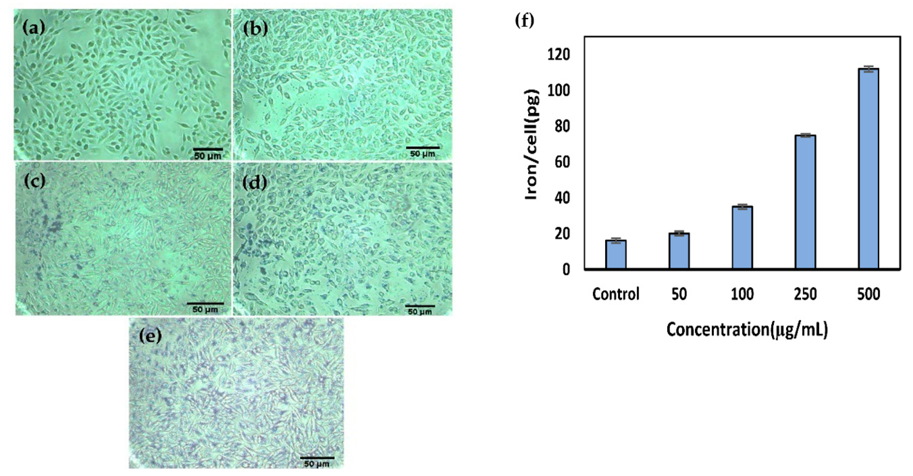
3.5. Cellular Uptake and Localization of G4@IONPs

Prussian blue staining was performed to demonstrate the cellular uptake of G4@IONPs after 2 h. The iron particles were seen as blue precipitates which increased in the cell cytoplasm with increasing the G4@IONPs concentration (Figure 4a–e). Consequently, ICP-MS results revealed that the iron concentration in the cells increased in a concentration-dependent manner. The amount of 16.1 ± 2.7 pg iron was detected in control cells (Figure 4f).

3.6. Histopathological Effects of Magnetic Hyperthermia Treatment on Liver and Lung Tissues

All H&E-stained lung and liver sections from control and treatment groups were evaluated histologically (Figure 5). Focal metastasis of breast cancer (stars) was seen in the control group (thick arrows). Moreover, hemorrhage, necrosis, and infiltration of inflammatory cells were detected in control tissues due to the invasion of tumor cells. The treatment group showed only mild edema in liver tissue. The histology of the lung tissue in the treatment group was normal without any significant histopathological change.

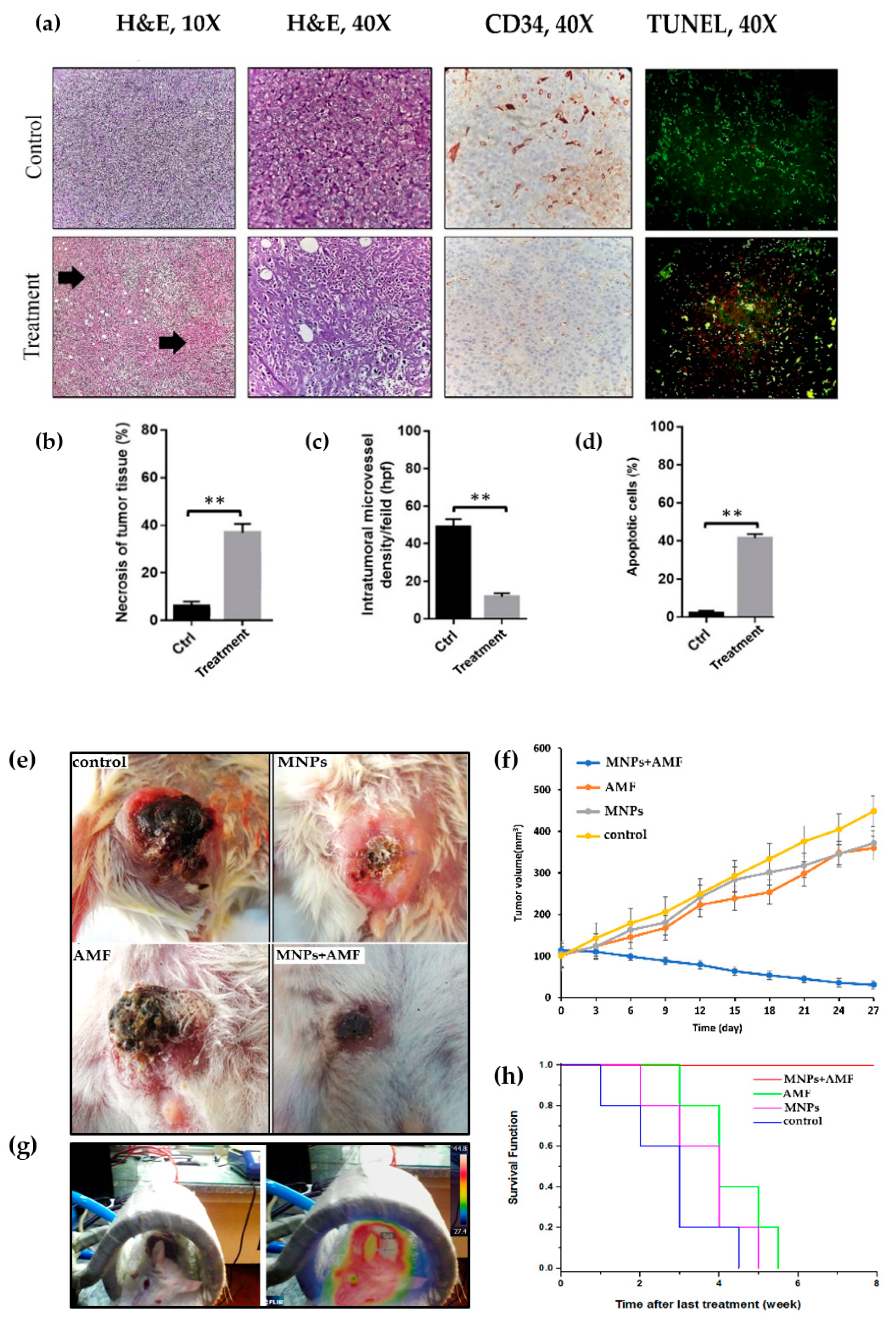
3.7. Histopathological Effects of Magnetic Hyperthermia Treatment on Tumor Tissue

Tumor sections from control and treated mice (MNPs + AMF) were graded histologically using the Nottingham histologic grading system. Many disproportionate tumor cells (anisocytosis), nuclear polymorphism (anisokaryosis, +3), and prominent nucleoli were seen in control animals. Moreover, in the control group, glandular (acinar/tubular) differentiation (GD) was low (+3), and the mitotic index in 10 HPF (400×) was +3. Overall, the tumor in the control group was considered as Grade 3 (poorly differentiated) (Figure 6a). In the treatment group, nuclear polymorphism (+1), GD (+2), and the mitotic index in 10 high power microscopic fields (HPF) (+1) were decreased in comparison to the control group. The tumor in the treatment group was considered as Grade 1 (well-differentiated); furthermore, massive necrosis was seen in breast cancer cells in the treatment group (Figure 6a,b).

3.8. Angiogenesis and Apoptosis in Tumor Tissue after Magnetic Hyperthermia Treatment

The microvessel density (angiogenesis) in treated tumors with magnetic hyperthermia was 13.4 ± 2.4 per HPF compared to that in control tumors (51.3 ± 4.5—P < 0.01) (Figure 6a,c). The proportion of apoptotic-positive cells in the treatment group was significantly higher than that in the control group (P < 0.01, Figure 6a,d).

3.9. Effect of Magnetic Hyperthermia Treatment on Tumor Volume

After the last treatment, tumor volume was measured in all groups every three days for four weeks. The final tumor volume in the group of treatment (30.83 mm3) was significantly less than that in the control group (448.11 mm3) (Figure 6e,f).

3.10. Kaplan–Meier Curve and Survival Rate of BALB/c Mice

Kaplan-Meier curve illustrated the survival rate of BALB/c mice over time in different groups. Five mice in each group were watched for eight weeks after the last treatment. There was no mortality in treated BALB/c mice during this period, while all other mice died during six weeks (Figure 6h).

4. Discussion

Dendrimers are developing polymeric multivalent system with multiple surface functional groups that have been used in many studies for drug delivery and imaging applications [16,47]. Regarding the potentials of dendrimers for theranostic applications, we functionalized IONPs (size of 10 ± 4 nm) with G4 PAMAM dendrimers and mPEG molecules for magnetic hyperthermia treatment of breast cancer. The amine-terminated PAMAM dendrimers attach to the negatively charged membrane of the cells via electrostatic interactions causing cytotoxicity. The results of this study showed that G4@IONPs cytotoxicity was negligible even at high concentrations, e.g., 500 μg/mL, that could be due to PEGylation of G4@IONPs [48]. PEGylation of MNPs has several advantages, such as additional stability, favorable pharmacokinetics, and enhanced therapeutic activity. Moreover, PEGylation can increase the retention time of nanoparticles [49,50]. The pharmacokinetics, stability, and retention time of G4@IONPs were assessed in our previous study [36].
The results of Prussian blue staining and ICP-MS showed that G4@IONPs entered into the cytoplasm of the MC4L2 cells in a concentration-dependent manner. The surface charge of G4@IONPs was +35 mV; cationic dendrimers possess a high tendency to interact with the bilayer lipid membrane of cells to consequently increase the cellular uptake [51]. PEGylation of G4@IONPs can also alter their cellular uptake via decreasing or preventing the protein adsorption (opsonization) on the G4@IONPs surface [52]. Indeed, higher cellular uptake of MNPs can increase the therapeutic effects of magnetic hyperthermia because more MNPs (heat sources) will be close to cell cytoplasmic proteins and nucleus [53]. Magnetic hyperthermia treatment enhanced the cytotoxic effect of G4@IONPs and decreased the MC4L2 cell proliferation; a slight decrease in cell viability in other experimental groups (MNPs and AMF) could be due to G4@IONPs incubation and AMF exposure, respectively.
Apoptosis is a genetically structured cellular death process activated by several internal and external signals. The intrinsic mitochondrial apoptosis pathway is triggered by intracellular stimuli that upregulate the pro-apoptotic Bcl-2 family of proteins, such as Bax, Bad (Bcl-2 associated agonist of cell death), and Bak (Bcl-2 homologous antagonist/killer), leading to the mitochondrial release of cytochrome C [54,55]. This intracellular stress (stimuli) should be strong enough to be able to trigger the apoptosis pathway in the cells; otherwise, some anti-apoptosis molecules would be activated and inhibit the cellular apoptosis procedure. In our study, the magnetic hyperthermia treatment was able to trigger the intrinsic apoptosis pathway and overcome the heat shock proteins (HSP) that cause thermal resistance in cancer cells [56]. TUNEL data approved the MTT viability results; the G4@IONPs entered the cancer cells by endocytosis and produced heat after AMF exposure in the group of MNPs + AMF. These small heat sources could then be the powerful intracellular stimuli to trigger the intrinsic apoptosis pathway in the cancer cell (Figure 3h). The results also showed some apoptosis increase (P > 0.05) in the group of AMF, which could be due to eddy current induced in the cell dish during the AMF exposure.
The survivability of treated cancer-bearing BALB/c mice was significantly higher than the untreated ones. This could be because of smaller tumor size in these mice, and, as a result, they could move better in the cage and access to more food and water. Besides that, general health was better in treated mice that helped them to survive longer. The body weight did not show any significant variation during the experimental period that showed our MNPs did not alter the body metabolism in mice. In a similar study, Malik et al. also reported that injecting PAMAM dendrimer (95 mg/kg) into B16F10 tumor-bearing mice (three times per day) did not alter their weight [57].
Our results showed that the tumor volume in treated mice decreased significantly to 23.7% of the initial volume (Vt0) over 27 days. This ratio (V/Vt0) was 448% in control animals. Furthermore, tumor volume reduction in other mice (groups of MNPs and AMF) compared to control could be due to intratumoral G4@IONPs injection or AMF exposure. Tumor volume is one of the most important parameters to demonstrate the efficacy of magnetic hyperthermia treatment; therefore, many studies reported the final tumor volume, as well as histopathological outcomes. For instance, Alexanian et al. injected superparamagnetic IONP (SPION) linked to PEG and folic acid (FA-PEG-SPION) into mice intravenously and placed them in an alternating current (AC) magnetic field (8 kA/m and 230 kHz) for 20 min. They reported that tumor volume in treated mice was one-tenth of control ones 35 days after the last treatment [52].
Tumor regression after magnetic hyperthermia treatment could be due to increasing the cancer cell apoptosis as both in vitro and H&E results showed that the number of apoptotic cells in the group of MNPs + AMF was significantly more than that in the control group. Furthermore, magnetic hyperthermia treatment decreased the intratumoral microvessel density, which could be another reason for tumor shrinkage in treated mice.
Regarding the effect of magnetic hyperthermia treatment on tumor vascularization, Kossatz et al. investigated the degree of angiogenesis in tumor tissues using the CD31 indicator [23]. To this end, IONPs (25 mg Fe/100 mm3) were injected intratumorally, and, after 24 h, nude mice were exposed to an AMF (15.4 kA/m and 435 kHz). Their results showed that the rate of angiogenesis in treated tumors decreased 28 days after the first treatment. We also obtained similar results regarding tumor angiogenesis suppression with a lower amount of iron injected into the tumor (5 mg/mL). In our previous study, we showed that G4@IONPs were highly capable of converting AMF energy to heat [21]. Therefore, to generate a sufficient amount of heat inside the tumor, a lesser amount of MNPs was used in our study.
The highest temperature (max 45 ℃), and also temperature uniformity, in the tumor were monitored by a FLIR thermal camera system (Figure 6g) and based on this data, the location and number of MNPs injection sites were determined. The uniformity of heat distribution in the tumor is a determining factor in magnetic hyperthermia treatment [58]. Hence, multiple injection sites (four sites) were applied to obtain a more homogenous MNPs distribution in the tumor. G4@IONPs (0.1 mL) were gently injected intratumoral using an insulin syringe (Ultra-Fine needle) and after 15 min, the animal was transferred to the AMF coil. No leakage of the MNPs solutions back out was seen after the syringe needle was removed from the tumor.
Our results revealed that treated breast cancer cells (MNPs + AMF) could not progress to invasive ones over time, which could be due to higher apoptosis and necrosis in these cells. Histopathological evaluations were performed by a single-blinded pathologist and Nottingham Histologic Scoring System was used to assess the breast tumor grade. The tumors in treated and control mice were in grades 1 and 3, respectively. In grade 1, the cancer cells look similar to normal cells and usually are well differentiated. Grade 3 breast cancer is invasive, tumors may be larger, and cancer has spread to the lymph nodes [59,60]. The treatment could induce differentiation in breast cancer cells, in addition to inhibiting tumor growth. Histopathological studies (H&E staining) also revealed a higher amount of necrotic cells at the center of treated tumors. Consequently, stem cells at the center of the tumor that are mainly responsible for metastasis and tumor progression would be necrotic after magnetic hyperthermia treatment. This can explain the lack of metastasis and invasive cancer cells in lung and liver tissues in treated animals. On the other hand, it has been indicated that extracellular HSPs trigger antitumor immunity during tumor cell necrosis. Such induced immunity also promotes tumor regression [61].
Overall, magnetic hyperthermia treatment using G4@IONPs increased the cellular apoptosis via the intrinsic apoptosis pathway and at the same time cut the tumor blood supply by decreasing the tumor angiogenesis. These factors inhibited the tumor growth and progression; consequently, cancer cells did not spread and develop metastasis in healthy tissues. The present study has some limitations regarding the MNPs distribution and quantity in the tumor after injection; first, the precise distribution of G4@IONPs could have investigated by magnetic resonance imaging (MRI), confocal microscopy, and Prussian blue staining; second, the iron content in the tumor was not measured after intratumoral G4@IONPs injection.

5. Conclusions

The nanocomposites presented in this study were well-suited for magnetic hyperthermia treatment of breast cancer. The toxicity assay revealed the biocompatibility of G4@IONPs even at high concentrations up to 500 µg/mL. Breast cancer cell viability decreased significantly after magnetic hyperthermia treatment. Furthermore, cellular apoptosis increased in treated cancer cells; real time-PCR results also showed that magnetic hyperthermia treatment was able to regulate the expression of the apoptosis-related genes. The dendrimer functionalized IONPs presented here also showed promising outcomes for breast cancer treatment in BALB/c mice. The magnetic hyperthermia treatment decreased tumor mammary gland growth via suppressing the tumor angiogenesis and increasing the cellular necrosis. All in all, G4@IONPs seem to be suitable nanostructures for use in magnetic hyperthermia cancer treatment due to their biocompatibility, stability, and unique structure for the conjugation of biomolecules. For future studies, anti-cancer drugs and imaging contrast agents can be conjugated to the dendrimer coating for cancer theranostics applications.

Author Contributions

Conceptualization, M.S. and R.S.; methodology, M.S.; validation, S.S. and R.S.; formal analysis, M.S.; investigation, M.S. and M.H.; resources, S.S.; data curation, S.S. and R.S.; writing—Original draft preparation, M.S.; writing—Review and editing, all authors; visualization, M.S.; supervision, S.S. and R.S.; project administration, S.S.; funding acquisition, M.S. All authors have read and agreed to the published version of the manuscript.

Funding

This research was funded by Tehran University of Medical Sciences, grant number 28169

Acknowledgments

The authors would like to acknowledge the help with histopathological experiments from Department of Pharmaceutics, Tehran University of Medical Sciences.

Conflicts of Interest

The authors declare no conflict of interest. The funders had no role in the design of the study; in the collection, analyses, or interpretation of data; in the writing of the manuscript, or in the decision to publish the results.

References

  1. Carioli, G.; Malvezzi, M.; Rodríguez, T.; Bertuccio, P.; Negri, E.; La Vecchia, C. Trends and predictions to 2020 in breast cancer mortality: Americas and Australasia. Breast 2018, 37, 163–169. [Google Scholar] [CrossRef] [PubMed] [Green Version]
  2. Bray, F.; Ferlay, J.; Soerjomataram, I.; Siegel, R.L.; Torre, L.A.; Jemal, A. Global cancer statistics 2018: GLOBOCAN estimates of incidence and mortality worldwide for 36 cancers in 185 countries. CA Cancer J. Clin. 2018, 68, 394–424. [Google Scholar] [CrossRef] [PubMed] [Green Version]
  3. Pan, J.; Hu, P.; Guo, Y.; Hao, J.; Ni, D.; Xu, Y.; Bao, Q.; Yao, H.; Wei, C.; Wu, Q.; et al. Combined Magnetic Hyperthermia and Immune Therapy for Primary and Metastatic Tumor Treatments. ACS Nano 2020, 14, 1033–1044. [Google Scholar] [CrossRef] [PubMed]
  4. Gawęda, W.; Osial, M.; Żuk, M.; Pękała, M.; Bilewicz, A.; Krysinski, P. Lanthanide-Doped SPIONs Bioconjugation with Trastuzumab for Potential Multimodal Anticancer Activity and Magnetic Hyperthermia. Nanomaterials 2020, 10, 288. [Google Scholar] [CrossRef] [PubMed] [Green Version]
  5. Mondal, S.; Manivasagan, P.; Bharathiraja, S.; Moorthy, M.S.; Nguyen, V.T.; Kim, H.H.; Nam, S.Y.; Lee, K.D.; Oh, J. Hydroxyapatite Coated Iron Oxide Nanoparticles: A Promising Nanomaterial for Magnetic Hyperthermia Cancer Treatment. Nanomaterials 2017, 7, 426. [Google Scholar] [CrossRef] [Green Version]
  6. Spirou, S.V.; Basini, M.; Lascialfari, A.; Sangregorio, C.; Innocenti, C. Magnetic Hyperthermia and Radiation Therapy: Radiobiological Principles and Current Practice. Nanomaterials 2018, 8, 401. [Google Scholar] [CrossRef] [Green Version]
  7. Brero, F.; Albino, M.; Antoccia, A.; Arosio, P.; Avolio, M.; Berardinelli, F.; Bettega, D.; Calzolari, P.; Ciocca, M.; Corti, M.; et al. Hadron Therapy, Magnetic Nanoparticles and Hyperthermia: A Promising Combined Tool for Pancreatic Cancer Treatment. Nanomaterials 2020, 10, 1919. [Google Scholar] [CrossRef]
  8. Lin, T.-C.; Lin, F.-H.; Lin, J.-C. In vitro feasibility study of the use of a magnetic electrospun chitosan nanofiber composite for hyperthermia treatment of tumor cells. Acta Biomater. 2012, 8, 2704–2711. [Google Scholar] [CrossRef]
  9. Sugahara, T.; Van Der Zee, J.; Kampinga, H.H.; Vujaskovic, Z.; Kondo, M.; Ohnishi, T.; Li, G.; Park, H.J.; Leeper, D.B.; Ostapenko, V.; et al. Kadota Fund International Forum 2004. Application of thermal stress for the improvement of health, 15–18 June 2004, Awaji Yumebutai International Conference Center, Awaji Island, Hyogo, Japan. Final Report. Int. J. Hyperth. 2008, 24, 123–140. [Google Scholar] [CrossRef] [Green Version]
  10. Takada, T.; Yamashita, T.; Sato, M.; Sato, A.; Ono, I.; Tamura, Y.; Sato, N.; Miyamoto, A.; Ito, A.; Honda, H.; et al. Growth Inhibition of Re-Challenge B16 Melanoma Transplant by Conjugates of Melanogenesis Substrate and Magnetite Nanoparticles as the Basis for Developing Melanoma-Targeted Chemo-Thermo-Immunotherapy. J. Biomed. Biotechnol. 2009, 2009, 1–13. [Google Scholar] [CrossRef]
  11. Etheridge, M.; Manuchehrabadi, N.; Franklin, R.; Bischof, J.; Minkowycz, W.; Sparrow, E.; Abraham, J. Superparamagnetic Iron Oxide Nanoparticle Heating. Comput. Phys. Process. Mech. Therm. Sci. 2012, 97–122. [Google Scholar] [CrossRef]
  12. Ansari, S.A.M.K.; Ficiarà, E.; Ruffinatti, F.A.; Stura, I.; Argenziano, M.; Abollino, O.; Cavalli, R.; Guiot, C.; D’Agata, F. Magnetic Iron Oxide Nanoparticles: Synthesis, Characterization and Functionalization for Biomedical Applications in the Central Nervous System. Materials 2019, 12, 465. [Google Scholar] [CrossRef] [PubMed] [Green Version]
  13. Demirer, G.S.; Okur, A.C.; Kizilel, S. Synthesis and design of biologically inspired biocompatible iron oxide nanoparticles for biomedical applications. J. Mater. Chem. B 2015, 3, 7831–7849. [Google Scholar] [CrossRef] [PubMed] [Green Version]
  14. Esfand, R.; Tomalia, D.A. Poly(amidoamine) (PAMAM) dendrimers: From biomimicry to drug delivery and biomedical applications. Drug Discov. Today 2001, 6, 427–436. [Google Scholar] [CrossRef]
  15. Dufes, C.; Uchegbu, I.F.; Schätzlein, A.G. Dendrimers in gene delivery. Adv. Drug Deliv. Rev. 2005, 57, 2177–2202. [Google Scholar] [CrossRef] [Green Version]
  16. Gillies, E.R.; Fréchet, J.M.J. Dendrimers and dendritic polymers in drug delivery. Drug Discov. Today 2005, 10, 35–43. [Google Scholar] [CrossRef]
  17. Luong, D.; Sau, S.; Kesharwani, P.; Iyer, A.K. Polyvalent Folate-Dendrimer-Coated Iron Oxide Theranostic Nanoparticles for Simultaneous Magnetic Resonance Imaging and Precise Cancer Cell Targeting. Biomacromolecules 2017, 18, 1197–1209. [Google Scholar] [CrossRef]
  18. Alfei, S.; Marengo, B.; Zuccari, G.; Turrini, F.; Domenicotti, C. Dendrimer Nanodevices and Gallic Acid as Novel Strategies to Fight Chemoresistance in Neuroblastoma Cells. Nanomaterials 2020, 10, 1243. [Google Scholar] [CrossRef]
  19. Abbasi, E.; Aval, S.F.; Akbarzadeh, A.; Milani, M.; Nasrabadi, H.T.; Joo, S.W.; Hanifehpour, Y.; Nejati-Koshki, K.; Pashaei-Asl, R. Dendrimers: Synthesis, applications, and properties. Nanoscale Res. Lett. 2014, 9, 247. [Google Scholar] [CrossRef] [Green Version]
  20. Uzun, K.; Çevik, E.; Şenel, M.; Sözeri, H.; Baykal, A.; Abasıyanık, M.F.; Toprak, M.S. Covalent immobilization of invertase on PAMAM-dendrimer modified superparamagnetic iron oxide nanoparticles. J. Nanoparticle Res. 2010, 12, 3057–3067. [Google Scholar] [CrossRef]
  21. Salimi, M.; Sarkar, S.; Saber, R.; Delavari, H.; Alizadeh, A.M.; Mulder, H.T. Magnetic hyperthermia of breast cancer cells and MRI relaxometry with dendrimer-coated iron-oxide nanoparticles. Cancer Nanotechnol. 2018, 9, 1–19. [Google Scholar] [CrossRef] [PubMed] [Green Version]
  22. Hayashi, K.; Nakamura, M.; Miki, H.; Ozaki, S.; Abe, M.; Matsumoto, T.; Sakamoto, W.; Yogo, T.; Ishimura, K. Magnetically Responsive Smart Nanoparticles for Cancer Treatment with a Combination of Magnetic Hyperthermia and Remote-Control Drug Release. Theranostics 2014, 4, 834–844. [Google Scholar] [CrossRef] [PubMed]
  23. Kossatz, S.; Grandke, J.; Couleaud, P.; Latorre, A.; Aires, A.; Crosbie-Staunton, K.; Ludwig, R.; Dähring, H.; Ettelt, V.; Lazaro-Carrillo, A.; et al. Efficient treatment of breast cancer xenografts with multifunctionalized iron oxide nanoparticles combining magnetic hyperthermia and anti-cancer drug delivery. Breast Cancer Res. 2015, 17, 1–17. [Google Scholar] [CrossRef] [PubMed] [Green Version]
  24. Paknikar, K.M.; Haghniaz, R.; Umrani, R.D. Hyperthermia mediated by dextran-coated La0.7Sr0.3MnO3 nanoparticles: In vivo studies. Int. J. Nanomed. 2016, 11, 1779–1791. [Google Scholar] [CrossRef] [PubMed] [Green Version]
  25. Lee, J.-H.; Jang, J.-T.; Choi, J.-S.; Moon, S.H.; Noh, S.-H.; Kim, J.-W.; Kim, J.-G.; Kim, I.-S.; Park, K.I.; Cheon, J. Exchange-coupled magnetic nanoparticles for efficient heat induction. Nat. Nanotechnol. 2011, 6, 418–422. [Google Scholar] [CrossRef] [PubMed]
  26. Li, T.-J.; Huang, C.-C.; Ruan, P.-W.; Huang, K.-J.; Shieh, D.-B.; Yeh, C.-S. In vivo anti-cancer efficacy of magnetite nanocrystal—based system using locoregional hyperthermia combined with 5-fluorouracil chemotherapy. Biomaterials 2013, 34, 7873–7883. [Google Scholar] [CrossRef] [PubMed]
  27. Rabias, I.; Tsitrouli, D.; Karakosta, E.; Kehagias, T.; Diamantopoulos, G.; Fardis, M.; Stamopoulos, D.; Maris, T.G.; Falaras, P.; Zouridakis, N.; et al. Rapid magnetic heating treatment by highly charged maghemite nanoparticles on Wistar rats exocranial glioma tumors at microliter volume. Biomicrofluidics 2010, 4, 024111. [Google Scholar] [CrossRef] [Green Version]
  28. Bae, K.H.; Park, M.; Do, M.J.; Lee, N.; Ryu, J.H.; Kim, G.W.; Kim, C.; Park, T.G.; Hyeon, T. Chitosan Oligosaccharide-Stabilized Ferrimagnetic Iron Oxide Nanocubes for Magnetically Modulated Cancer Hyperthermia. ACS Nano 2012, 6, 5266–5273. [Google Scholar] [CrossRef]
  29. Ling, Y.; Tang, X.; Wang, F.; Zhou, X.; Wang, R.; Deng, L.; Shang, T.; Liang, B.; Li, P.; Ran, H.; et al. Highly efficient magnetic hyperthermia ablation of tumors using injectable polymethylmethacrylate–Fe3O4. RSC Adv. 2017, 7, 2913–2918. [Google Scholar] [CrossRef] [Green Version]
  30. Arriortua, O.K.; Garaio, E.; De La Parte, B.H.; Insausti, M.; Lezama, L.; Plazaola, F.; García, J.A.; Aizpurua, J.M.; Sagartzazu, M.; Irazola, M.; et al. Antitumor magnetic hyperthermia induced by RGD-functionalized Fe3O4 nanoparticles, in an experimental model of colorectal liver metastases. Beilstein J. Nanotechnol. 2016, 7, 1532–1542. [Google Scholar] [CrossRef] [Green Version]
  31. Ohtake, M.; Umemura, M.; Sato, I.; Akimoto, T.; Oda, K.; Nagasako, A.; Kim, J.-H.; Fujita, T.; Yokoyama, U.; Nakayama, T.; et al. Hyperthermia and chemotherapy using Fe(Salen) nanoparticles might impact glioblastoma treatment. Sci. Rep. 2017, 7, 42783. [Google Scholar] [CrossRef] [PubMed]
  32. Sato, I.; Umemura, M.; Mitsudo, K.; Fukumura, H.; Kim, J.-H.; Hoshino, Y.; Nakashima, H.; Kioi, M.; Nakakaji, R.; Sato, M.; et al. Simultaneous hyperthermia-chemotherapy with controlled drug delivery using single-drug nanoparticles. Sci. Rep. 2016, 6, 24629. [Google Scholar] [CrossRef] [PubMed] [Green Version]
  33. Yang, Y.; Wang, F.; Zheng, K.; Deng, L.; Yang, L.; Zhang, N.; Xu, C.; Ran, H.; Wang, Z.; Wang, Z.; et al. Injectable PLGA/Fe3O4 implants carrying cisplatin for synergistic magnetic hyperthermal ablation of rabbit VX2 tumor. PLoS ONE 2017, 12, e0177049. [Google Scholar] [CrossRef] [PubMed] [Green Version]
  34. Zhang, Z.-Q.; Song, S.-C. Multiple hyperthermia-mediated release of TRAIL/SPION nanocomplex from thermosensitive polymeric hydrogels for combination cancer therapy. Biomaterials 2017, 132, 16–27. [Google Scholar] [CrossRef] [PubMed]
  35. Hayashi, K.; Nakamura, M.; Sakamoto, W.; Yogo, T.; Miki, H.; Ozaki, S.; Abe, M.; Matsumoto, T.; Ishimura, K. Superparamagnetic Nanoparticle Clusters for Cancer Theranostics Combining Magnetic Resonance Imaging and Hyperthermia Treatment. Theranostics 2013, 3, 366–376. [Google Scholar] [CrossRef] [PubMed] [Green Version]
  36. Salimi, M.; Sarkar, S.; Fathi, S.; Alizadeh, A.M.; Saber, R.; Moradi, F.; Delavari, H. Biodistribution, pharmacokinetics, and toxicity of dendrimer-coated iron oxide nanoparticles in BALB/c mice. Int. J. Nanomed. 2018, 13, 1483–1493. [Google Scholar] [CrossRef] [PubMed] [Green Version]
  37. Shirshahi, V.; Hatamie, S.; Tabatabaei, S.N.; Salimi, M.; Saber, R. Enhanced Thermal Stability and Biocompatibility of Gold Nanorods by Graphene Oxide. Plasmonics 2017, 13, 1585–1594. [Google Scholar] [CrossRef]
  38. Hsiao, I.-L.; Bierkandt, F.S.; Reichardt, P.; Luch, A.; Huang, Y.-J.; Jakubowski, N.; Tentschert, J.; Haase, A. Quantification and visualization of cellular uptake of TiO2 and Ag nanoparticles: Comparison of different ICP-MS techniques. J. Nanobiotechnol. 2016, 14, 1–13. [Google Scholar] [CrossRef] [Green Version]
  39. Wang, Z.; Cuschieri, A. Tumour Cell Labelling by Magnetic Nanoparticles with Determination of Intracellular Iron Content and Spatial Distribution of the Intracellular Iron. Int. J. Mol. Sci. 2013, 14, 9111–9125. [Google Scholar] [CrossRef] [Green Version]
  40. Hashemi, M.; Fallah, A.; Aghayan, H.R.; Arjmand, B.; Yazdani, N.; Verdi, J.; Ghodsi, S.M.; Miri, S.M.; Hadjighassem, M. A New Approach in Gene Therapy of Glioblastoma Multiforme: Human Olfactory Ensheathing Cells as a Novel Carrier for Suicide Gene Delivery. Mol. Neurobiol. 2015, 53, 5118–5128. [Google Scholar] [CrossRef]
  41. Livak, K.J.; Schmittgen, T.D. Analysis of relative gene expression data using real-time quantitative PCR and the 2−∆∆Ct method. Methods 2001, 25, 402–408. [Google Scholar] [CrossRef] [PubMed]
  42. Elston, C.; Ellis, I. pathological prognostic factors in breast cancer. I. The value of histological grade in breast cancer: Experience from a large study with long-term follow-up. Histopathology 1991, 19, 403–410. [Google Scholar] [CrossRef] [PubMed]
  43. Ufuk, G.; Unsoy, G.; Yalcın, S.; Gunduz, G.; Gunduz, U. PAMAM dendrimer-coated iron oxide nanoparticles: Synthesis and characterization of different generations. J. Nanoparticle Res. 2013, 15, 1–13. [Google Scholar] [CrossRef]
  44. Yamaura, M.; Camilo, R.; Sampaio, L.; Macêdo, M.; Nakamura, M.; Toma, H. Preparation and characterization of (3-aminopropyl)triethoxysilane-coated magnetite nanoparticles. J. Magn. Magn. Mater. 2004, 279, 210–217. [Google Scholar] [CrossRef]
  45. Tsubokawa, N.; Takayama, T. Surface modification of chitosan powder by grafting of ‘dendrimer-like’ hyperbranched polymer onto the surface. React. Funct. Polym. 2000, 43, 341–350. [Google Scholar] [CrossRef]
  46. Julian, J.M.; Brezinski, D.R. An Infrared Spectroscopy Atlas for the Coatings Industry; Federation of Societies for Paint Technology: Plymouth, PA, USA, 1991. [Google Scholar]
  47. Nanjwade, B.K.; Bechra, H.M.; Derkar, G.K.; Manvi, F.; Nanjwade, V.K. Dendrimers: Emerging polymers for drug-delivery systems. Eur. J. Pharm. Sci. 2009, 38, 185–196. [Google Scholar] [CrossRef]
  48. Kojima, C.; Turkbey, B.; Ogawa, M.; Bernardo, M.; Regino, C.A.S.; Bryant, L.H.; Choyke, P.L.; Kono, K.; Kobayashi, H. Dendrimer-based MRI contrast agents: The effects of PEGylation on relaxivity and pharmacokinetics. Nanomed. Nanotechnol. Biol. Med. 2011, 7, 1001–1008. [Google Scholar] [CrossRef] [Green Version]
  49. Mishra, P.; Nayak, B.; Dey, R.K. PEGylation in anti-cancer therapy: An overview. Asian J. Pharm. Sci. 2016, 11, 337–348. [Google Scholar] [CrossRef] [Green Version]
  50. Harris, J.M.; Chess, R.B. Effect of pegylation on pharmaceuticals. Nat. Rev. Drug Discov. 2003, 2, 214–221. [Google Scholar] [CrossRef]
  51. Mecke, A.; Majoros, I.J.; Patri, A.K.; Baker, J.J.R.; Holl, M.M.B.; Orr, B.G. Lipid Bilayer Disruption by Polycationic Polymers: The Roles of Size and Chemical Functional Group. Langmuir 2005, 21, 10348–10354. [Google Scholar] [CrossRef]
  52. Wood, A.J.; Alexanian, R.; Dimopoulos, M. The Treatment of Multiple Myeloma. N. Engl. J. Med. 1994, 330, 484–489. [Google Scholar] [CrossRef] [PubMed]
  53. Laurent, S.; Dutz, S.; Häfeli, U.O.; Mahmoudi, M. Magnetic fluid hyperthermia: Focus on superparamagnetic iron oxide nanoparticles. Adv. Colloid Interface Sci. 2011, 166, 8–23. [Google Scholar] [CrossRef] [PubMed]
  54. Linehan, W.M.; Walther, M.M.; Zbar, B. The Genetic Basis of Cancer of the Kidney. J. Urol. 2003, 170, 2163–2172. [Google Scholar] [CrossRef] [PubMed]
  55. You, Y.; Cheng, A.; Wang, M.-S.; Jia, R.-Y.; Sun, K.-F.; Yang, Q.; Wu, Y.; Zhu, D.; Chen, S.; Liu, M.-F.; et al. The suppression of apoptosis by α-herpesvirus. Cell Death Dis. 2017, 8, e2749. [Google Scholar] [CrossRef] [PubMed] [Green Version]
  56. Ikwegbue, P.C.; Masamba, P.; Oyinloye, B.E.; Kappo, A.P. Roles of Heat Shock Proteins in Apoptosis, Oxidative Stress, Human Inflammatory Diseases, and Cancer. Pharmaceuticals 2017, 11, 2. [Google Scholar] [CrossRef] [PubMed] [Green Version]
  57. Malik, N.; Evagorou, E.G.; Duncan, R. Dendrimer-platinate: A novel approach to cancer chemotherapy. Anti-Cancer Drugs 1999, 10, 767–776. [Google Scholar] [CrossRef]
  58. Du, Y.; Liu, X.; Liang, Q.; Liang, X.-J.; Tian, J. Optimization and Design of Magnetic Ferrite Nanoparticles with Uniform Tumor Distribution for Highly Sensitive MRI/MPI Performance and Improved Magnetic Hyperthermia Therapy. Nano Lett. 2019, 19, 3618–3626. [Google Scholar] [CrossRef]
  59. Giuliano, A.E.; Edge, S.B.; Hortobagyi, G.N. Eighth Edition of the AJCC Cancer Staging Manual: Breast Cancer. Ann. Surg. Oncol. 2018, 25, 1783–1785. [Google Scholar] [CrossRef]
  60. Singletary, S.E.; Allred, C.; Ashley, P.; Bassett, L.W.; Berry, D.; Bland, K.I.; Borgen, P.I.; Clark, G.; Edge, S.B.; Hayes, D.F.; et al. Revision of the American Joint Committee on Cancer Staging System for Breast Cancer. J. Clin. Oncol. 2002, 20, 3628–3636. [Google Scholar] [CrossRef]
  61. Zhu, J.; Zhang, Y.; Zhang, A.; He, K.; Liu, P.; Xu, L.X. Cryo-thermal therapy elicits potent anti-tumor immunity by inducing extracellular Hsp70-dependent MDSC differentiation. Sci. Rep. 2016, 6, 27136. [Google Scholar] [CrossRef]
Figure 1. Experimental workflow for magnetic hyperthermia treatment in breast cancer-bearing BALB/c mice.
Nanomaterials 10 02310 g001
Figure 2. (a) Schematic illustration of functionalization of iron oxide magnetic nanoparticles (IONPs) with G4 poly amidoamine (PAMAM) dendrimers, (b) transmission electron microscope (TEM) image of G4@IONPs, (c) Fourier-transform infrared spectroscopy (FTIR) spectra for PEGylated G4@IONPs.
Nanomaterials 10 02310 g002
Figure 3. (a) Schematic illustration of magnetic hyperthermia in vitro experiments. 2 × 10⁵ MC4L2 cells were seeded in a 35 mm culture dish overnight. 500 µg/mL G4@IONPs was added to cell culture media. After 2h incubation, cells were exposed to alternating magnetic field (AMF) for 120 min. MTT and TUNEL assays, and real-time polymerase chain reaction (PCR) were performed to assess cell viability, apoptosis and Bax/Bcl2 ratio immediately after treatment; (b) Cytotoxicity of G4@IONPs in MC4L2 cells (*P < 0.05); (c) MC4L2 cells viability percentage after magnetic hyperthermia treatment (*P < 0.05); TUNEL staining showed the apoptotic cancer cells in groups of: (d) control; (e) magnetic nanoparticles (MNPs); (f) AMF; (g) MNPs + AMF; (h) Apoptotic index in all groups (*P < 0.05); (i) Bax/Bcl-2 ratio in MC4L2 cells after magnetic hyperthermia treatment (*P < 0.05).
Nanomaterials 10 02310 g003
Figure 4. Prussian blue staining illustrated the density of iron oxide nanoparticles inside the MC4L2 cells after 2 h incubation with (a) control; (b) 50; (c) 100; (d) 250; (e) 500 µg/mL of G4@IONPs; the iron particles appeared as blue precipitates in the cell cytoplasm; (f) the cellular uptake of G4@IONPs at different concentrations was measured by Inductively Coupled Plasma Mass Spectrometry (ICP-MS).
Nanomaterials 10 02310 g004
Figure 5. Histopathological images of lung and liver tissues in control and treatment (MNPs + AMF) groups. Thick arrows show microscopic tumor metastasis in lung and liver, thin arrows represent tumor cells, and arrowheads: inflammatory cells.
Nanomaterials 10 02310 g005
Figure 6. (a) Histopathology, immunohistochemistry, and TUNEL assay of breast cancer tissues in control and treated BALB/c mice; (b) necrosis of tumor tissue (arrows in histopathological image); (c) Immunohistochemical analysis of the cluster of differentiation 34 (CD34) marker was performed to determine the angiogenesis in tumor tissues. The brown color represents positive staining of CD34 cell marker; (d) TUNEL assay was performed to assess the apoptotic cells in the experimental groups. The number of apoptotic-positive cells significantly increased in the treatment group compared to control, red florescent-stained cell nucleus represents apoptotic cells (**p < 0.01); (e) tumor shape and appearance in different experimental groups 27 days after the last treatment; (f) tumor volume versus time after the last treatment in different groups: magnetic hyperthermia using G4@IONPs, only AMF exposure, only G4@IONPs injection, and control; (g) the tumor temperature was monitored by the forward-looking infrared (FLIR) thermal camera; (h) Kaplan-Meier curve showing the survival rate of BALB/c mice over time (8 weeks).
Nanomaterials 10 02310 g006
Table 1. Different types of nanoparticles used in the magnetic hyperthermia treatment for cancer-bearing mice.
StudyMNPs CoreMNPs
Size
CoatingTreatment TimeMNPs Concentration/Type of InjectionTumor ModelAMFResults
Hayashi et al. [22]Fe3O410.5 nmPPy-PEG-FA
Dox
20 min5 mg/kg; intratumoral injectionMultiple myeloma8 kA/m
230 kHz
The combination of magnetic hyperthermia treatment and chemotherapy completely cured the tumor without any recurrence.
Kossatz et al. [23]SPIONs12 ± 3 nmN6L or/and DOX60 min0.25 mg Fe/100 mm3; intratumoral injectionBreast15.4 kA/m
435 kHz
Substantial tumor growth inhibition up to 40% and complete tumor regression were seen after magnetic hyperthermia treatment.
Haghniaz et al. [24]La0.7sr0.3MnO325–50 nmdextran20 min5 mg/100 μL saline;
intratumoral injection
Melanoma700 A
8000 W
365 kHz
Treatment inhibited tumor growth (84%) and increased animal survival (50%). In addition, levels of caspase-3 and caspase-6 also increased after treatment.
Lee et al. [25]CoFe2O415 nmMnFe2O410 min75 mg; intratumoral injectionGlioblastoma37.3 kA/m
500 kHz
The tumor was clearly eliminated in 18 days after treatment.
Li et al. [26]Fe3O422 nmanti-HER2, 5-FU and PEG15 min500 mg/mL iron; systematic injectionBladder carcinoma33 kA/m
1.3 MHz
Prominent tumor remission was seen after hyperthermia and chemotherapy.
Rabias et al. [27]Fe2O310–12 nmdextran20 min150 μL; intratumoral injectionGlioma11 kA/m
150 kHz
Significant tumor tissue damage and dissolution were seen after treatment.
Bae et al. [28]Fe3O430 nmChitosan-DOPA20 min375 μg Fe/kg; Intratumoral injectionLung carcinoma660 A/m
1 MHz
The tumor volume decreased substantially by about 70%.
Ling et al. [29]Fe3O420–50 nmPMMA3 min0.1 mL; intratumoral injectionBreast28.6 A
626 kHz
Tumor volume decreased within 15 days after treatment.
Arriortua et al. [30]Fe3O419 ± 2 nmRGD peptide>21 min1–1.5 mg Fe/mL; systemic injectionColon adenocarcinoma14 kA/m
606 kHz
Tumor necrosis was observed. Approximately whole tumor tissue was demolished in some animals, others showed very low damage in tumor tissue.
Ohtake et al. [31]Fe(Salen)200  nmSalen60 min0.12–0.60 mg/body; intratumoral injectionGlioblastoma335. 4 A
280 kHz
The tumor size was decreased, by 80–90%, in treatment group after 4 weeks.
Sato et al. [32]Fe(Salen)200 nm-30 min50 mM; intratumoral injectionTongue250 A
308 kHz
The tumor volume significantly decreased (223 ± 80.6%). The tumor almost completely disappeared after one week.
Yang et al. [33]Fe3O4-PLGA3 min100 μL;
intratumoral injection
Hepatic carcinoma28.6 A
626 kHz
Coagulative necrosis was seen in cancer tissues after treatment. In addition, anti-tumor immune system was activated in treated mice and promoted apoptosis in tumor cells.
Zhang et al. [34]Fe3O418 nmPPZ polymer60 min0.8 µL/mm3;
intratumoral injection
Glioblastoma13.3 kA/m
366 kHz
The tumor size was significantly smaller than the control 25 days after the last treatment. Pyknosis, karyorrhexis, and apoptosis were seen in treated tumor tissues.
Hayashi et al. [35]SPIONs7−9 nmPEG and FA20 min48 μmol Fe/kg; systematic injectionMultiple myeloma8 kA/m 230 kHzTumor volume in treated mice was one-tenth of control in 35 days after treatment
Abbreviations: SPIONs, superparamagnetic iron oxide nanoparticle; DOX, doxorubicin; HER2, human epidermal growth factor receptor; FU, fluorouracil; PPy, polypyrrole; FA, folic acid; DOPA, dihydroxyphenylalanine; PMMA, polymethyl methacrylate; RGD, arginylglycylaspartic acid; PLGA, poly(lactic-co-glycolic acid); PPZ, polyorganophosphazene.
Table 2. Oligonucleotide sequences of interest and reference genes.
GeneSequences (5′ → 3′)Product Size, bp
GAPDH-FAAGTTCAACGGCACAGTCAAGG22
GAPDH-RCATACTCAGCACCAGCATCACC22
Bax-FAGGGTGGCTGGGAAGGC17
Bax-RTGAGCGAGGCGGTGAGG17
Bcl2-FATCGCTCTGTGGATGACTGAGTAC24
Bcl2-RAGAGACAGCCAGGAGAAATCAAAC24
Publisher’s Note: MDPI stays neutral with regard to jurisdictional claims in published maps and institutional affiliations.

