Article

Preparation of Fe3+ Doped High-Ordered TiO2 Nanotubes Arrays with Visible Photocatalytic Activities

1
Shaanxi Province Key Laboratory of Thin Films Technology and Optical Test, School of Optoelectronic Engineering, Xi’an Technological University, Xi’an 710032, China
2
Belarusian State University of Informatics and Radioelectronics, Electronic Technique and Technology Department, Center 10.1, Thin Film Research Laboratory, 6 P. Brovka str., 220013 Minsk, Belarus
*
Author to whom correspondence should be addressed.
Nanomaterials 2020, 10(11), 2107; https://doi.org/10.3390/nano10112107
Submission received: 19 September 2020 / Revised: 21 October 2020 / Accepted: 21 October 2020 / Published: 23 October 2020

Abstract

:
In this paper, the Fe3+ doped rutile phase TiO2 nanotubes arrays (NTAs) were prepared in a low temperature water-assistant crystallization method. It is noteworthy that the Fe3+ doping hardly hinders either the crystallization of rutile TiO2 NTAs or the highly-ordered nanotubular morphologies. Moreover, Fe3+ did not form other compound impurities, which indicated that Fe3+ substitute Ti4+ into the lattice of TiO2. With the introduction of Fe3+, the light absorption range of TiO2 NTAs extends from the ultraviolet band to the visible light range. Photocatalytic testing results indicate that Fe3+ doped TiO2 NTAs can effectively improve the degradation rate of methyl orange aqueous solution in visible light, and the TiO2 NTAs with 0.2 mol/L Fe3+ doping exhibits the highest photocatalytic degradation efficiency.

1. Introduction

TiO2, as one of the most important derivatives of titanium, has been widely used in photocatalytic degradation of organic pollutants, hydrogen generation by water splitting, solar cells, lithium-ion batteries, sensors, and so on. This is due to its excellent photochemical activities, good electronic properties, low cost, and being non-toxic to environments [1,2,3,4,5,6,7]. Compared with TiO2 nanoparticles, TiO2 nanotube arrays (NTAs), as a fast electron transport channel, have attracted much attention in the fields of photocatalysis and solar cells [8,9,10]. However, as a photocatalyst, TiO2 nanotubes and nanoparticles share the same defects. Their wide band gap makes them insensitive to visible light, which greatly reduces the utilization efficiency of sunlight [11,12,13,14]. To solve this problem, various approaches have been proposed to improve the photo-response range of TiO2 materials [15,16,17,18,19], among which metal ion doping is one of the most common and promising methods [20,21,22,23,24,25,26,27,28].
Metal ion doping modification mainly includes 3d transition metal ions into the crystals of titanium dioxide, taking advantage of the instability of the outer electrons of 3d transition metals [29,30,31]. On one hand, when these unstable and unsaturated electrons are presented inside titanium dioxide, it helps to reduce the recombination rates of photogenerated electron-hole pairs on the surface of TiO2 and improve the utilization efficiency of photogenerated electrons [32,33,34,35]. On the other hand, some metal ions would form TiO2 electron traps in the band gap, and then expand the photo response of TiO2 NTAs to visible light [36,37]. Among various transition metal ions, Fe3+ has demonstrated excellent properties when doped with TiO2 NTAs [26,38,39]. However, preparation and usage of Fe3+ doped TiO2 NTAs reported in the literature does not achieve low power consumption, low cost and effective recycling. For example: Sun et al. used a high temperature annealing process to prepare TiO2 NTAs [26], Yu et al. adopted a high temperature alloy process to fabricate the Fe doped Ti foil [38], and Pang et al. prepared discrete Fe3+ doped TiO2 NTAs, which is not conducive to rapid recycling [39].
In order to facilitate recycling, we proposed a simple and low-cost preparation process of Fe3+-doped TiO2 NTAs on Ti foil under mild environment. In this study, anodic TiO2 NTAs were crystallized at low temperature induced by Fe3+ aqueous solution and the conditions and preparation process were optimized through multiple experiments. Various characterization and experimental results indicate that the prepared Fe3+ doped TiO2 NTAs have good photocatalytic activities for the degradation of organic substance under visible light irradiation.

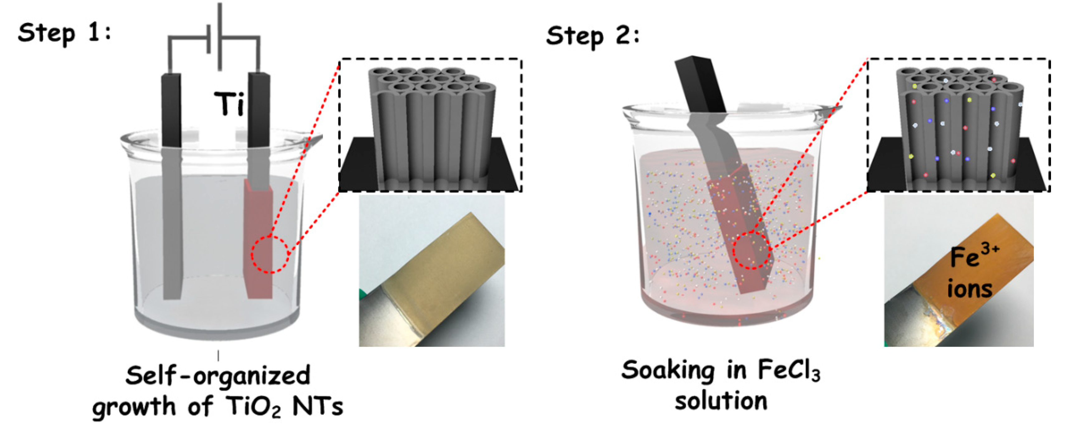
2. Materials and Methods

The initial amorphous TiO2 NTs were synthesized by electrochemical anodization of Ti foil (1.6 cm × 3 cm, >99.8%, 0.25 mm, Alfa Aesar, Shanghai, China) in glycol containing electrolyte (490 mL glycol (Alfa Aesar), 10 mL H2O and 1.66 g NH4F (Alfa Aesar)) at room temperature under a constant voltage of 50 V in a two-electrode configuration (a Ti foil as the working electrode and a platinum foil as the counter electrode), using a DC programmable power supply (Model IT6154, ITECH Electronics Co., Ltd. Nanjing, China). After anodization for 1.5 h, highly ordered TiO2 nanotube arrays on the Ti sheet substrate were obtained. Then, the as-prepared TiO2 NTs were thoroughly washed with a large amount of anhydrous alcohols and dried in air.
The Fe3+ doped TiO2 NTs were prepared by the water induced low temperature crystallization technology. The amorphous titanium dioxide nanotube films were immersed in mixed solutions which contained an equal volume of water and absolute alcohol, and 0.05 mol/L, 0.10 mol/L, 0.20 mol/L FeCl3 (Alfa Aesar) were added into the solvent, separately. Then, the mixed solutions were heated to 70 oC and held for 10 h to fully crystallize the TiO2 NTAs. After that, the samples were dried naturally at 20 oC room temperature to ensure the stability of the nanotube. It must be mentioned that we also tried to prepare higher doping concentration samples, but the TiO2 NTAs layers broke into pieces due to high internal stress caused by the excessive doping concentration.
X-ray diffraction (XRD) analysis (D8 ADVANCE, Bruker AXS, Karlsruhe, German) was employed to characterize the crystal structure and composition of the samples. The X-ray radiation source was Cu Kα, obtained at 40 kV, 30 mA, the scanning speed was 3.6°/min at a step of 0.03°, and the range of 2θ is 20–80°. The morphological property and energy dispersive spectrometry (EDS) were observed with a field-emission scanning electron microscopy (GeminiSEM 500, Zeiss, Aalen, German). Transmission electron microscopy (TEM) and EDS mapping were performed on a transmission electron microscope (JEOL JEM-2100). Raman testing and analysis were used a laser Raman spectroscope (LabRAM HR Evolution, HORIBA, Fukuoka, Japan) with a 532 nm line for the excitation. The absorption spectra were collected by using integrating sphere accessory of the UV−vis spectrometry (Ultrospec 2100 pro, Harvard Apparatus, Cambridge, MA, USA).
The photocatalytic activities of Fe3+ doped TiO2 NTAs were evaluated on the basis of the degradation of methyl orange (MO) as model organic pollutants in aqueous solutions and measured by the UV-vis spectrometer (JASCO V-570 UV/vis/NIR, JASCO, Tokyo, Japan). The surface area (3.0 × 1.6 cm2) of the samples were immersed in the 5 × 10⁻⁵ mol/L MO aqueous solution and irradiated with eight 4W UV bulbs (Toshiba, Black Light Blue, FL4W, Tokyo, Japan), eight 4W visible bulbs (Toshiba, Cool white, FL4W, Tokyo, Japan) with UV filter (>480 nm), for UV photoactivities test and visible photoactivities test, respectively. Before measurement, the samples were soaked for 30 min in the dark with magnetic stirring to reach adsorption/desorption equilibrium. The photo-degradation measurement was carried out in an open quartz photoreaction vessel with rapid stirring for 120 min under room temperature. The concentration of the residual MO was measured by the UV-vis spectrometer at about 460 nm. The photocatalytic performance data of each sample were tested for 10 times, and the mean value and error were calculated and plotted.

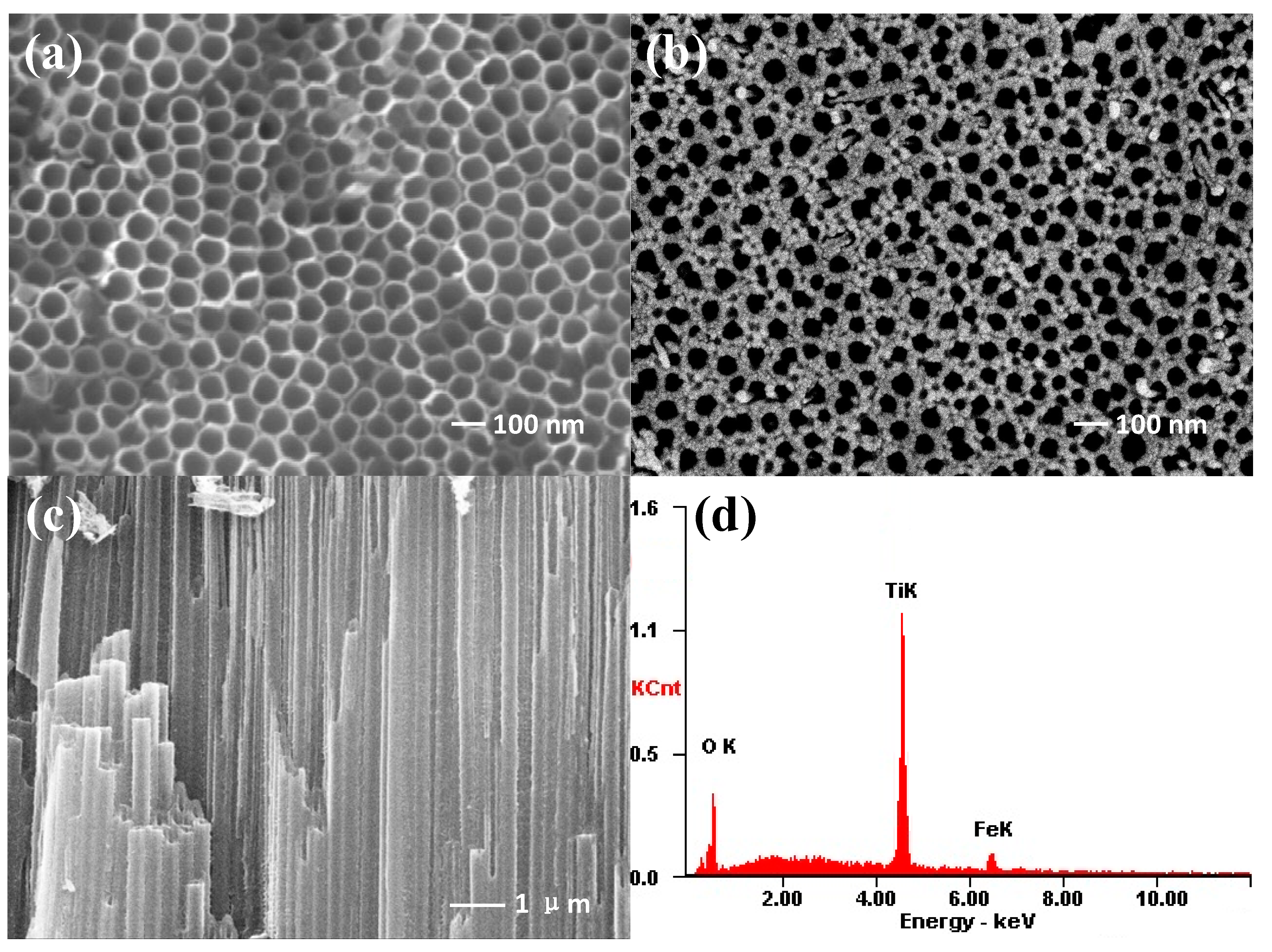
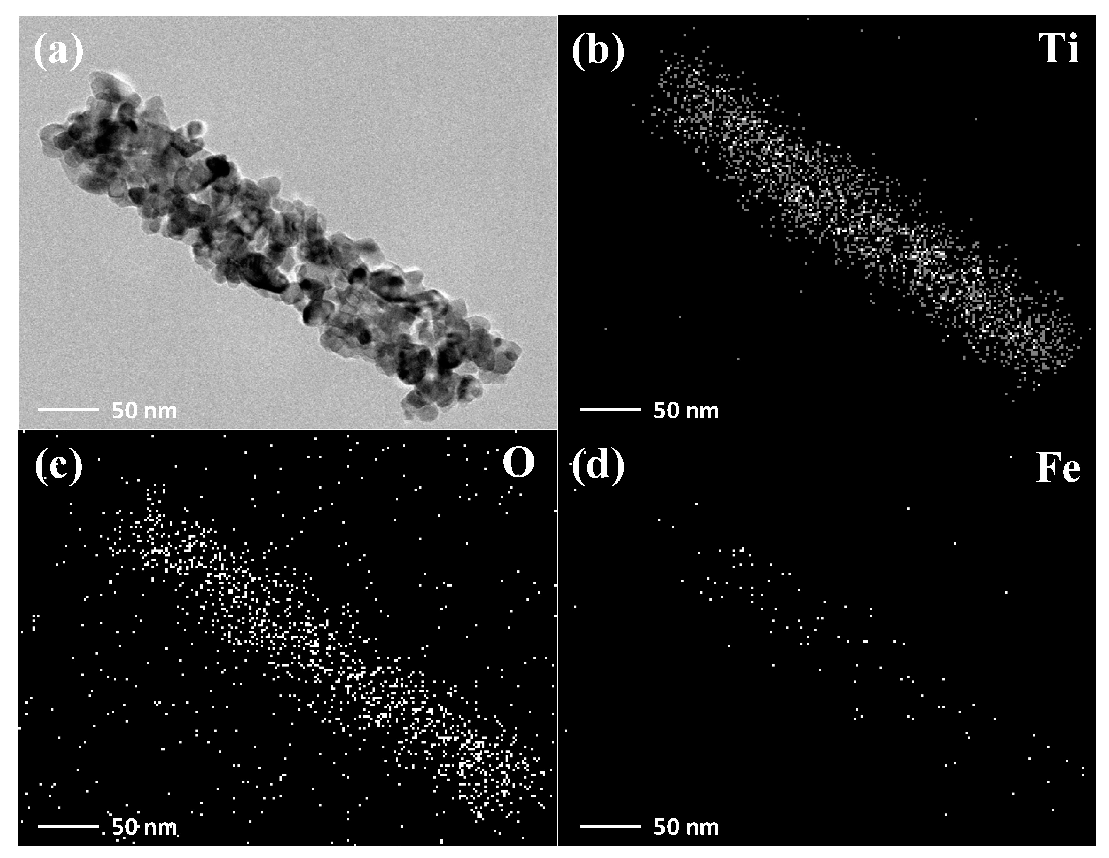
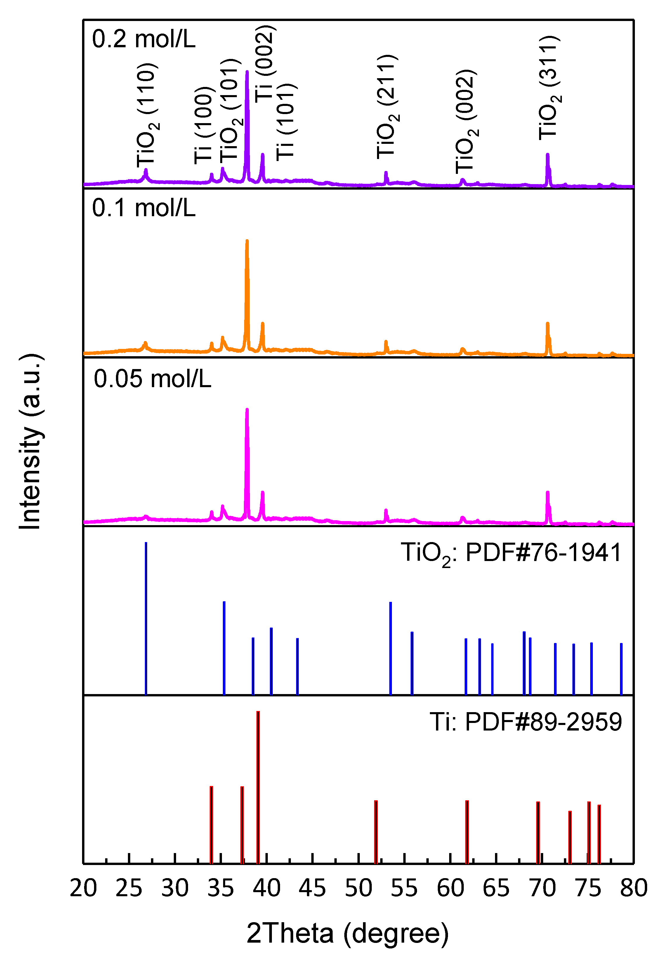
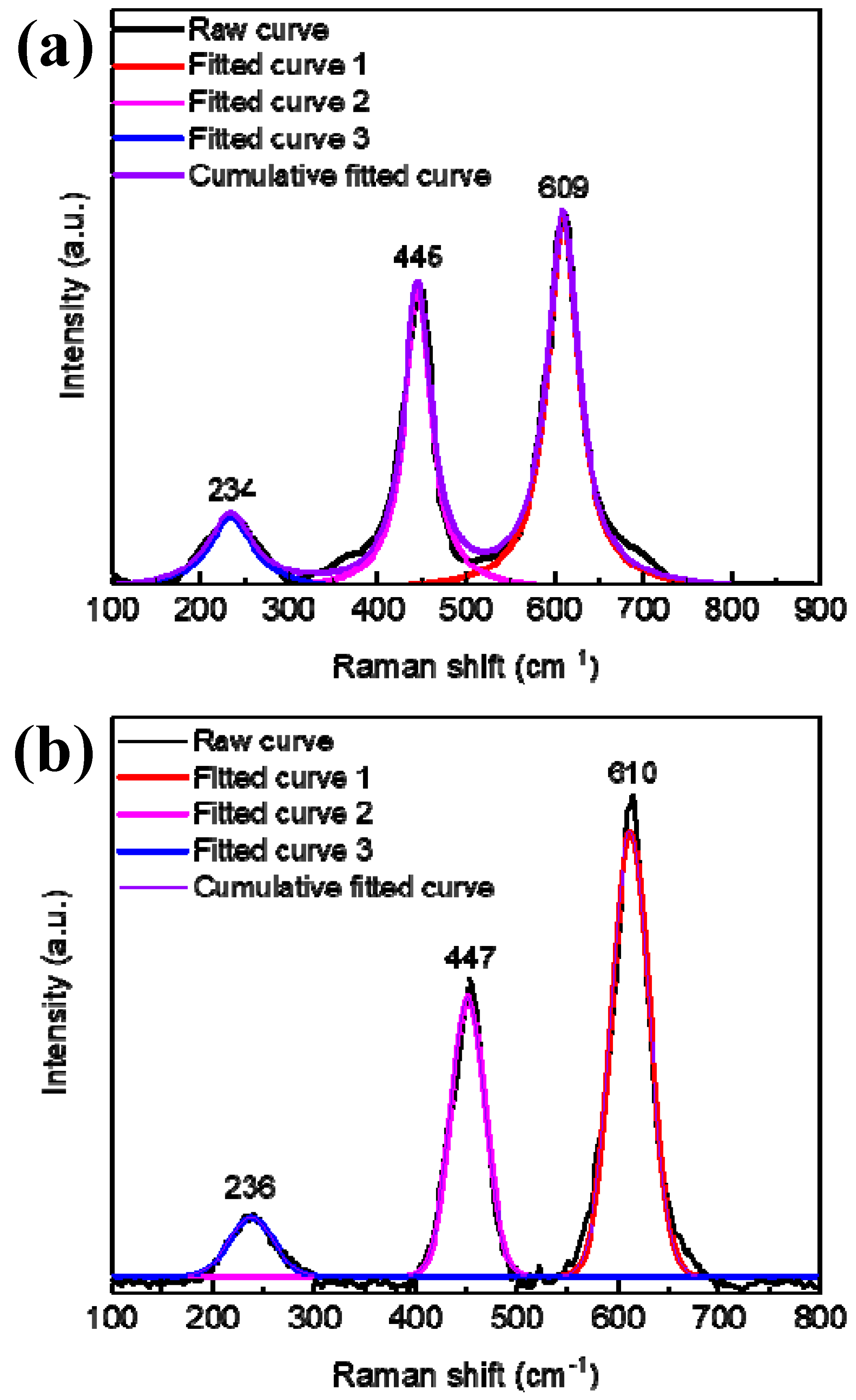
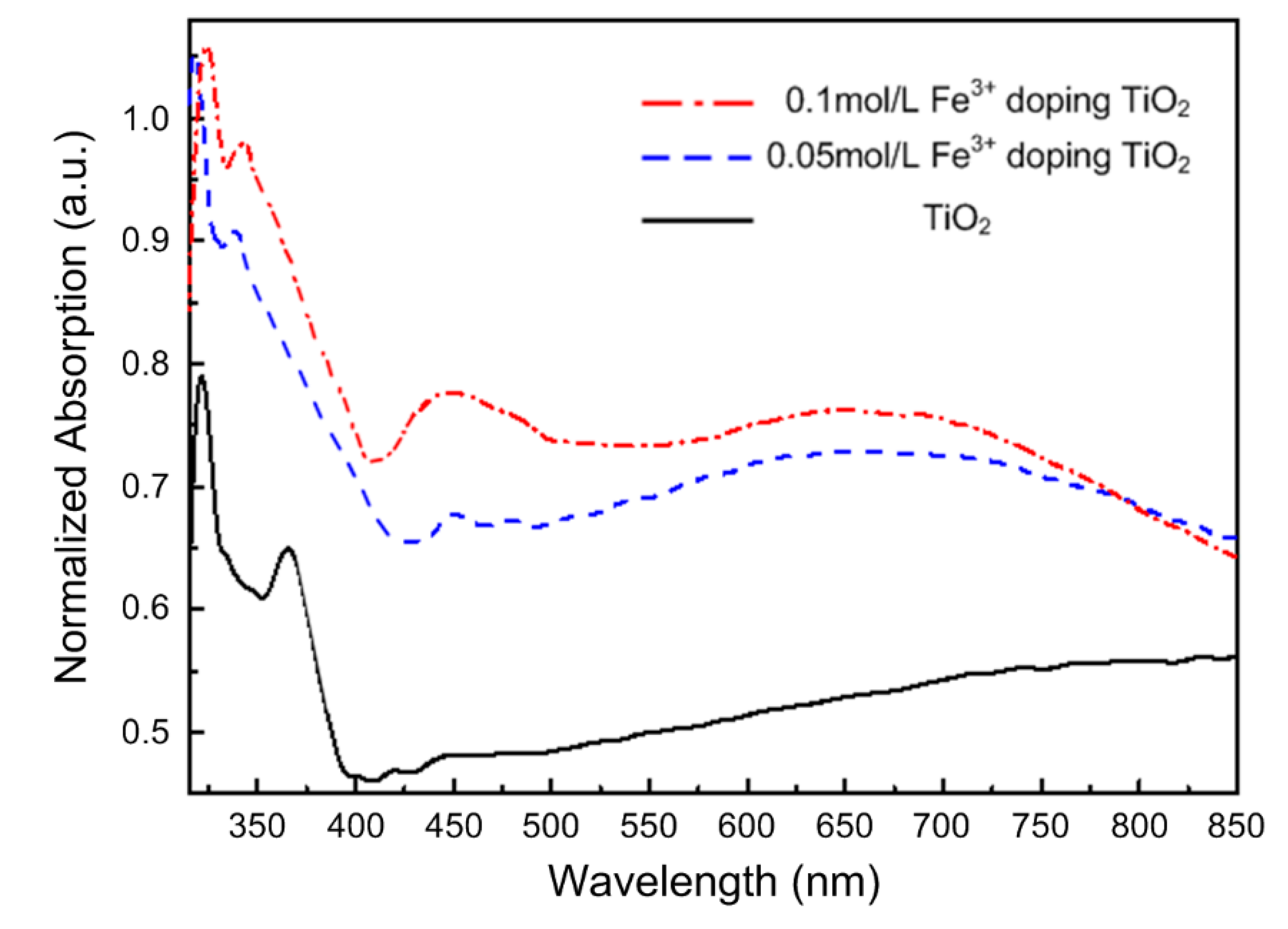
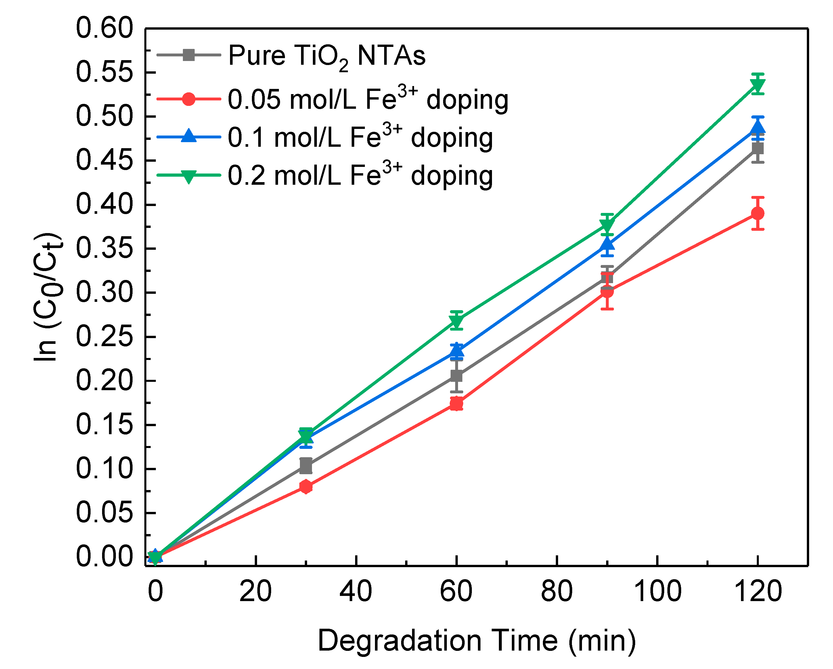
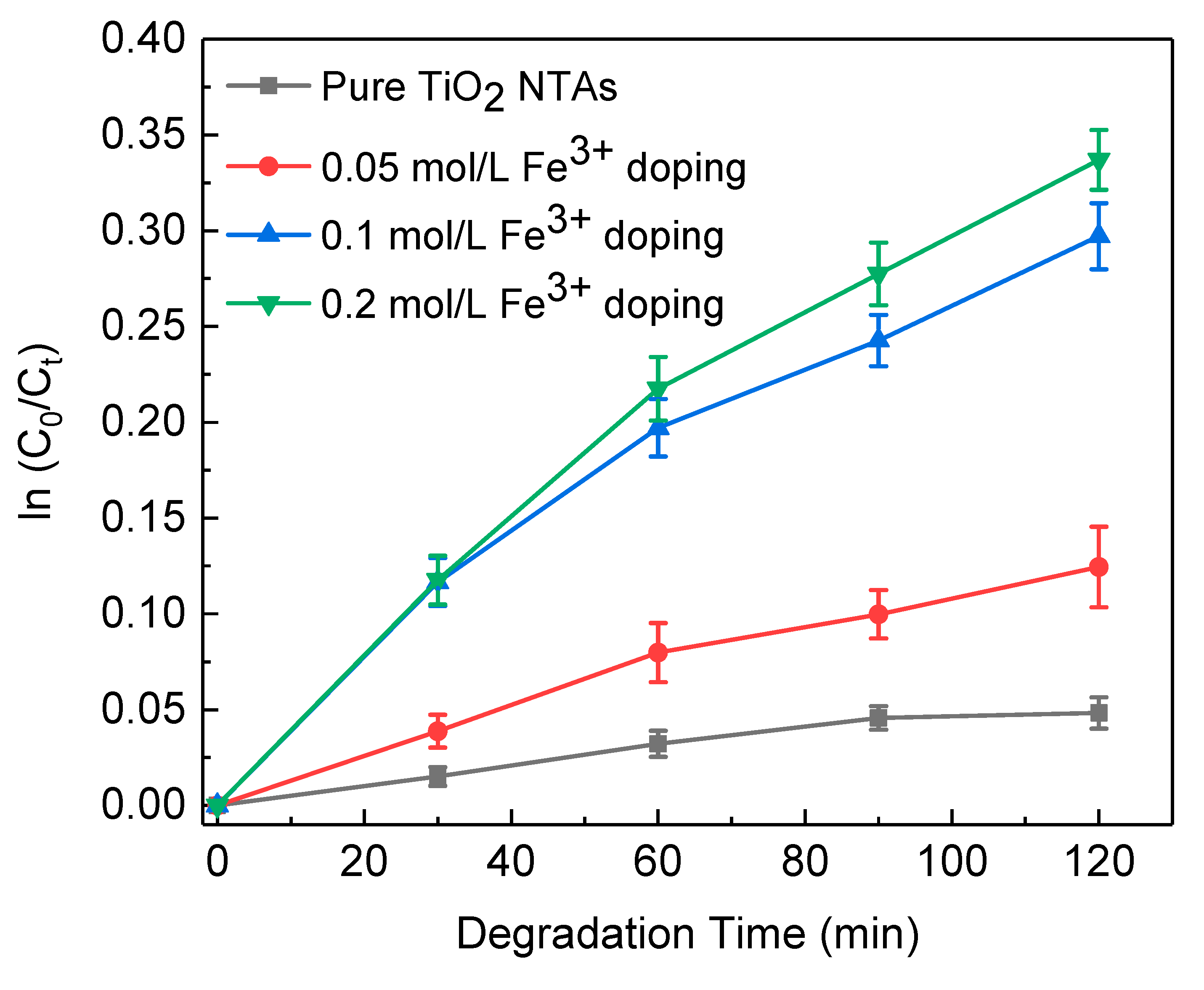
3. Results and Discussion

Figure 1 shows a schematic synthesis procedure of Fe3+ doped TiO2 NTAs. In step 1, self-organized TiO2 NTAs were firstly prepared by electrochemical anodization. A yellowish film could be observed, which is attributed to the amorphous TiO2 NTAs coated on the Ti foil. Then the as-prepared amorphous TiO2 NTAs film was immersed into aqueous solution of Fe3+ at 70 °C to induce crystallization and doping. After this step, the film turned a reddish color, which could be attributed to the doping of Fe3+ ion in TiO2 NTAs.
In order to detect the effect of Fe3+ doping on the morphology of TiO2 NTAs, SEM was used to observe and characterize the morphology of titanium dioxide nanotubes. As can be observed from Figure 2a, the TiO2 NTs are top-opened and have an inner diameter of about 80–90 nm with a wall thickness of ~20 nm. After doping with Fe3+, no significant difference in the nanotube diameter can be observed, while the wall thickness gets slightly increased about 2–5 nm (see Figure 2b). The cross section of Fe3+ doped TiO2 NTAs is exhibited in Figure 2c; it can be seen that the inner surface of the nanotubes is clean and not blocked by particles. The SEM test results show that the crystallization/doping process did not damage the top-open and highly oriented morphologies of TiO2 NTAs, which is advantageous for the organic solvent to be fully filled into the nanotubes during the photocatalytic process. The weight ratios of Ti, O, and Fe in samples with different doping concentrations were also tested by using EDS, and the results were presented in Table 1. It can be observed that with the increase of Fe3+ doping concentration, the Fe content gradually rises, the oxygen content basically remains unchanged, and the Ti content gradually decreases in these samples, indicating that the Fe3+ is likely substituting Ti4+ into the lattice of TiO2.
For the sake of observing the distribution of Fe3+ in TiO2 nanotube more intuitively, TEM and EDS mapping were used to test the distribution of Ti, O, and Fe elements in a single 0.2 mol/L Fe3+ doped TiO2 nanotube as shown in Figure 3. We can observe that there is no cladding layer and enrichment of the Fe element is not present on the surface of the TiO2 nanotube, and the Fe element uniformly distributes in the TiO2 nanotube, indicating that the Fe did not form compounds coated on the surface of TiO2 nanotube.
The XRD patterns of TiO2 NTAs samples with different Fe3+ doping concentrations are shown in Figure 4. It can be seen that the diffraction peaks of the three samples are quite similar. After comparison with standard PDF cards, we find that these main diffraction peaks well match (110), (101), (211), (002), (311) planes of rutile phase TiO2 and (100), (002), (101) planes of titanium. In addition, no other impurity peaks are found, and whether Fe3+ is doped into TiO2 nanotubes in alternative forms needs further discussion. The lattice parameters and interplanar crystal spacing (d values) of pure and Fe3+ doped TiO2 NTAs samples are outlined in Table 2. With the increase of Fe3+ doping concentration, the lattice constant and d values decrease slightly; the main reason is that the radius of Fe3+ (64 Å) is slightly smaller than that of Ti4+ (68 Å). This can also be proved from the side that Fe3+ replaces Ti4+ and enters into the lattice of TiO2.
To further verify that Fe3+ did not form a compound but entered into the TiO2 lattice, the Raman spectrum of the 0.2 mol/L Fe3+ doped sample was further investigated and analyzed. The Raman spectrum and peak fitting curves of the sample before photocatalytic experiments are shown in Figure 5a. After fitting the spectral line, we find that there are only three Raman peaks of rutile phase TiO2 (234 cm−1 (Eg mode), 445 cm−1 (Eg mode), and 609 cm−1 (A1g mode)) [26] and no other impurity peaks exist, which also confirmed the XRD test results that Fe was doped into the lattice of TiO2 and no impurity was formed.
Figure 6 shows the UV-Vis absorption spectra of the pure and Fe3+ doped TiO2 NTAs. As can be seen from the spectra, the TiO2 NTAs samples without Fe3+ doping shows an absorption characteristic only in the ultraviolet region (<380 nm), but not in the visible region. Once doped with Fe3+, the red shift of absorption edge can be observed clearly in the Figure 6. The absorption capacity of the ultraviolet region was also significantly enhanced. This result, along with the XRD and Raman analysis above, reveal that the Fe3+ ions were indeed introduced into the lattice of TiO2 and formed the impurity energy level, thus expanding the absorption spectrum range of TiO2 nanotubes.
The photocatalytic activities of samples were measured by degradation of methyl orange (MO). Figure 7 shows the concentration change of MO as a function of irradiation time under ultraviolet light for pure and doped TiO2 NTAs with a different amount of Fe3+. First, we find that the photocatalytic activity of the sample doped with Fe3+ in 0.05 mol/L precursor solution can only degrade MO about 33% in 2 h, which is much lower than that of pure titanium dioxide nanotubes. This is might due to the fact that the UV light has provided enough energy to generated electron-hole pairs, and a small amount doping of Fe3+ tends to act as a recombination center, which will decrease the photoactivities [40]. However, with the increase of the Fe3+ concentration, the catalytic activity increased gradually and reached the best level when the concentration was 0.2 mol/L. Under this condition, the major effect of the Fe3+ doping is to introduce impurity levels in the band gap of TiO2, and become a shallow trap of photogenerated electrons or holes, for reducing the recombination of electron-hole pairs [41].
For practical application, it is necessary to estimate the photocatalytic activity of these samples under visible light irradiation, which is shown in Figure 8. The results clearly revealed that doping with Fe3+ does greatly enhance the photochemical activity of TiO2 NTAs under visible light. Photogenerated charge carriers successfully flow to Fe3+ energy level and form an electron trap which shortens the distance between conduction band and valence band [42,43]. The successful doping of Fe ion into TiO2 makes it more liable for the visible radiation to excite the electrons in conduction band of TiO2. Along with the characterization and experiment above, it can be concluded that TiO2 NTAs have the best photocatalytic performance doped with 0.2 mol/L of Fe3+.
After photocatalytic experiments, the Raman spectrum and EDS of 0.2 mol/L Fe3+ doped TiO2 NTAs were also investigated. Figure 5b shows the Raman spectrum and peak fitting curves of the sample after photocatalytic experiments. Still, only three characteristic peaks located at 236, 447, and 610 cm−1 can be observed, which is consistent with the Raman results before the photocatalytic experiments. In addition, after photocatalytic experiments the weight ratios of Ti, O, and Fe in the sample are 41.01, 50.23, and 8.76% w/w, respectively, which also coincides with the original results. The above test results indicate that Fe3+ did not form other compound impurities and could not be released in the solution during the photoirradiation.

4. Conclusions

In summary, Fe3+ doped rutile phase TiO2 NTAs have been successfully prepared by a simple and low-cost method which combined electrochemical anodization and low temperature water-assistant crystallization. The results of test and analysis show that Fe3+ doping not only doesn’t change the crystallization of TiO2 NTAs, but also no Fe-containing compounds impurities are produced. The Fe3+ substitutes Ti4+ into the lattice of TiO2 and forms the impurity energy level, which expands the absorption range of TiO2 NTAs from ultraviolet to the visible band. It is noteworthy that Fe3+ doping significantly improves the photocatalytic activity of TiO2 NTAs in the visible light region, making it have a promising practical application prospect in the photocatalytic decomposition of organic pollutants.

Author Contributions

Conceptualization, J.Z.; Data curation, C.C.; Investigation, C.Y. and S.Z.; Methodology, S.L., Y.X., and S.M.; Project administration, W.L.; Validation, D.G.; Writing—original draft, J.Z. All authors have read and agreed to the published version of the manuscript.

Funding

This research received no external funding.

Acknowledgments

This work was supported by the International Science and Technology Innovation Cooperation between Governments Project of National Key Research and Development Program (2018YFE0199200); the Science and technology exchange programs between the governments of China and Belarus (CB02-19); the National R&D Program of China (2017YFA0207400); the Natural Science Basic Research Plan in Shaanxi Province of China (2017JM5022); the Open Fund of Shaanxi Province Key Laboratory of Thin Films Technology and Optical Test (ZSKJ201706); the Key Industry Innovation Chain Project of Shaanxi Provincial Science and the Technology Department (2018ZDCXL-GY-08-02-01); and the Xi’an Key Laboratory of Intelligent Detection and Perception (201805061ZD12CG45).

Conflicts of Interest

The authors declare no conflict of interest.

References

  1. Bakbolat, B.; Daulbayev, C.; Sultanov, F.; Beissenov, R.; Umirzakov, A.; Mereke, A.; Bekbaev, A.; Chuprakov, I. Recent Developments of TiO2-Based Photocatalysis in the Hydrogen Evolution and Photodegradation: A Review. Nanomaterials 2020, 10, 1790. [Google Scholar] [CrossRef]
  2. Gopalan, A.-I.; Lee, J.-C.; Saianand, G.; Lee, K.-P.; Sonar, P.; Dharmarajan, R.; Hou, Y.-L.; Ann, K.-Y.; Kannan, V.; Kim, W.-J. Recent Progress in the Abatement of Hazardous Pollutants Using Photocatalytic TiO2-Based Building Materials. Nanomaterials 2020, 10, 1854. [Google Scholar] [CrossRef] [PubMed]
  3. Carp, O.; Huisman, C.L.; Reller, A. Photoinduced reactivity of titanium dioxide. Prog. Solid State Chem. 2004, 32, 33–177. [Google Scholar] [CrossRef]
  4. Paulose, M.; Prakasam, H.E.; Varghese, O.K.; Peng, L.; Popat, K.C.; Mor, G.K.; Desai, T.A.; Grimes, C.A. TiO2 Nanotube Arrays of 1000 μm Length by Anodization of Titanium Foil:  Phenol Red Diffusion. J. Phys. Chem. C 2007, 111, 14992–14997. [Google Scholar] [CrossRef]
  5. Jaramillo-Fierro, X.; González, S.; Jaramillo, H.A.; Medina, F. Synthesis of the ZnTiO3/TiO2 Nanocomposite Supported in Ecuadorian Clays for the Adsorption and Photocatalytic Removal of Methylene Blue Dye. Nanomaterials 2020, 10, 1891. [Google Scholar] [CrossRef]
  6. Quan, X.; Yang, S.; Ruan, X.; Zhao, H. Preparation of Titania Nanotubes and Their Environmental Applications as Electrode. Environ. Sci. Technol. 2005, 39, 3770–3775. [Google Scholar] [CrossRef]
  7. Pant, B.; Ojha, G.P.; Kuk, Y.-S.; Kwon, O.H.; Park, Y.W.; Park, M. Synthesis and Characterization of ZnO-TiO2/Carbon Fiber Composite with Enhanced Photocatalytic Properties. Nanomaterials 2020, 10, 1960. [Google Scholar] [CrossRef]
  8. Yu, J.; Dai, G.; Huang, B. Fabrication and Characterization of Visible-Light-Driven Plasmonic Photocatalyst Ag/AgCl/TiO2 Nanotube Arrays. J. Phys. Chem. C 2009, 113, 16394–16401. [Google Scholar] [CrossRef]
  9. Macák, J.M.; Tsuchiya, H.; Schmuki, P. High-Aspect-Ratio TiO2 Nanotubes by Anodization of Titanium. Angew. Chem. Int. Ed. 2005, 44, 2100–2102. [Google Scholar] [CrossRef]
  10. Park, J.; Bauer, S.; Mark, K.; Schmuki, P. Nanosize and Vitality:  TiO2 Nanotube Diameter Directs Cell Fate. Nano Lett. 2007, 7, 1686–1691. [Google Scholar] [CrossRef]
  11. Roy, P.; Berger, S.; Schmuki, P. TiO2 Nanotubes: Synthesis and Applications. Angew. Chem. Int. Ed. 2011, 50, 2904–2939. [Google Scholar] [CrossRef] [PubMed]
  12. Shankar, K.; Basham, J.I.; Allam, N.K.; Varghese, O.K.; Mor, G.K.; Feng, X.; Paulose, M.; Seabold, J.A.; Choi, K.S.; Grimes, C.A. Recent Advances in the Use of TiO2 Nanotube and Nanowire Arrays for Oxidative Photoelectrochemistry. J. Phys. Chem. C 2009, 113, 6327–6359. [Google Scholar] [CrossRef]
  13. Park, J.H.; Kim, S.; Bard, A.J. Novel Carbon-Doped TiO2 Nanotube Arrays with High Aspect Ratios for Efficient Solar Water Splitting. Nano Lett. 2006, 6, 24–28. [Google Scholar] [CrossRef] [PubMed]
  14. Zheng, Q.; Zhou, B.; Bai, J.; Li, L.; Jin, Z.; Zhang, J.; Li, J.; Liu, Y.; Cai, W.; Zhu, X. Self-Organized TiO2 Nanotube Array Sensor for the Determination of Chemical Oxygen Demand. Adv. Mater. 2008, 20, 1044–1049. [Google Scholar] [CrossRef]
  15. He, Z.; Kim, C.; Jeon, T.H.; Choi, W. Hydrogenated heterojunction of boron nitride and titania enables the photocatalytic generation of H2 in the absence of noble metal catalysts. Appl. Catal. B-Environ. 2018, 237, 772–782. [Google Scholar] [CrossRef]
  16. He, Z.; Kim, C.; Lin, L.; Jeon, T.H.; Lin, S.; Wang, X.; Choi, W. Formation of heterostructures via direct growth CN on h-BN porous nanosheets for metal-free photocatalysis. Nano Energy 2017, 42, 58–68. [Google Scholar] [CrossRef]
  17. Klosek, S.; Raftery, D. Visible Light Driven V-Doped TiO2 Photocatalyst and Its Photooxidation of Ethanol. J. Phys. Chem. B 2001, 105, 2815–2819. [Google Scholar] [CrossRef]
  18. Liu, Z.; Pesic, B.; Raja, K.S.; Rangaraju, R.R.; Misra, M. Hydrogen generation under sunlight by self ordered TiO2 nanotube arrays. Int. J. Hydrogen Energy 2009, 34, 3250–3257. [Google Scholar] [CrossRef]
  19. Yang, Y.; Wang, H.; Li, X.; Wang, C. Electrospun mesoporous W6+-doped TiO2 thin films for efficient visible-light photocatalysis. Mater. Lett. 2009, 63, 331–333. [Google Scholar] [CrossRef]
  20. Asiltürk, M.; Sayılkan, F.; Arpaç, E. Effect of Fe3+ ion doping to TiO2 on the photocatalytic degradation of Malachite Green dye under UV and vis-irradiation. J. Photochem. Photobiol. A 2009, 203, 64–71. [Google Scholar] [CrossRef]
  21. Devi, L.G.; Kottam, N.; Murthy, B.N.; Kumar, S.G. Enhanced photocatalytic activity of transition metal ions Mn2+, Ni2+ and Zn2+ doped polycrystalline titania for the degradation of Aniline Blue under UV/solar light. J. Mol. Catal. A Chem. 2010, 328, 44–52. [Google Scholar] [CrossRef]
  22. Kuljanin-Jakovljević, J.; Radoičić, M.; Radetić, T.; Konstantinović, Z.; Šaponjić, Z.V.; Nedeljković, J. Presence of Room Temperature Ferromagnetism in Co2+ Doped TiO2 Nanoparticles Synthesized through Shape Transformation. J. Phys. Chem. C 2009, 113, 21029–21033. [Google Scholar] [CrossRef]
  23. Lin, H.J.; Yang, T.S.; Hsi, C.S.; Wang, M.C.; Lee, K.C. Optical and photocatalytic properties of Fe3+-doped TiO2 thin films prepared by a sol–gel spin coating. Ceram. Int. 2014, 40, 10633–10640. [Google Scholar] [CrossRef]
  24. Liu, G.; Zhang, X.; Xu, Y.; Niu, X.; Zheng, L.; Ding, X. The preparation of Zn2+-doped TiO2 nanoparticles by sol–gel and solid phase reaction methods respectively and their photocatalytic activities. Chemosphere 2005, 59, 1367–1371. [Google Scholar] [CrossRef] [PubMed]
  25. Anandan, S.; Miyauchi, M. Ce-doped ZnO (CexZn1−xO) becomes an efficient visible-light-sensitive photocatalyst by co-catalyst (Cu2+) grafting. Phys. Chem. Chem. Phys. 2011, 13, 14937–14945. [Google Scholar] [CrossRef]
  26. Sun, L.; Li, J.; Wang, C.L.; Li, S.F.; Chen, H.B.; Lin, C.J. An electrochemical strategy of doping Fe3+ into TiO2 nanotube array films for enhancement in photocatalytic activity. Sol. Energy Mater. Sol. Cells 2009, 93, 1875–1880. [Google Scholar] [CrossRef]
  27. Tong, T.; Zhang, J.; Tian, B.; Chen, F.; He, D. Preparation of Fe3+-doped TiO2 catalysts by controlled hydrolysis of titanium alkoxide and study on their photocatalytic activity for methyl orange degradation. J. Hazard. Mater. 2008, 155, 572–579. [Google Scholar] [CrossRef]
  28. Zhang, D.; Chen, J.; Deng, P.; Wang, X.; Li, Y.; Wen, T.; Li, Y.; Xiang, Q.; Liao, Y. Hydrogen evolution promotion of Au-nanoparticles-decorated TiO2 nanotube arrays prepared by dip-loading approach. J. Am. Ceram. Soc. 2019, 102, 5873–5880. [Google Scholar] [CrossRef]
  29. Buschow, K.H.J. Intermetallic compounds of rare-earth and 3d transition metals. Rep. Prog. Phys. 1977, 40, 1179. [Google Scholar] [CrossRef]
  30. Kitchin, J.R.; Nørskov, J.K.; Barteau, M.A.; Chen, J.G. Modification of the surface electronic and chemical properties of Pt (111) by subsurface 3d transition metals. J. Chem. Phys. 2004, 120, 10240–10246. [Google Scholar] [CrossRef] [Green Version]
  31. Hanada, N.; Ichikawa, T.; Fujii, H. Catalytic Effect of Nanoparticle 3d-Transition Metals on Hydrogen Storage Properties in Magnesium Hydride MgH2 Prepared by Mechanical Milling. J. Phys. Chem. B 2005, 109, 7188–7194. [Google Scholar] [CrossRef] [PubMed]
  32. Choudhury, B.; Dey, M.; Choudhury, A. Defect generation, d-d transition, and band gap reduction in Cu-doped TiO2 nanoparticles. Int. Nano Lett. 2013, 3, 25. [Google Scholar] [CrossRef] [Green Version]
  33. Mittal, M.; Sharma, M.; Pandey, O.P. UV–Visible light induced photocatalytic studies of Cu doped ZnO nanoparticles prepared by co-precipitation method. Sol. Energy 2014, 110, 386–397. [Google Scholar] [CrossRef]
  34. Lei, C.; Xiang, Q.; Liao, Y.; Zhang, H. CdS-Based photocatalysts. Energy Environ. Sci. 2018, 11, 1362–1391. [Google Scholar]
  35. Yin, X.; Guo, Y.; Xie, H.; Que, W.; Kong, L.B. Nickel Oxide as Efficient Hole Transport Materials for Perovskite Solar Cells. Sol. RRL 2019, 3, 1900001. [Google Scholar] [CrossRef]
  36. Alexandrescu, R.; Morjan, I.; Scarisoreanu, M.; Birjega, R.; Popovici, E.; Soare, I.; Gavrila-Florescu, L.; Voicu, I.; Sandu, I.; Dumitrache, F.; et al. Structural investigations on TiO2 and Fe-doped TiO2 nanoparticles synthesized by laser pyrolysis. Thin Solid Films 2007, 515, 8438–8445. [Google Scholar] [CrossRef]
  37. Vignesh, K.; Rajarajan, M.; Suganthi, A. Visible light assisted photocatalytic performance of Ni and Th co-doped ZnO nanoparticles for the degradation of methylene blue dye. J. Ind. Eng. Chem. 2014, 20, 3826–3833. [Google Scholar] [CrossRef]
  38. Yu, J.; Wu, Z.; Gong, C.; Xiao, W.; Sun, L.; Lin, C. Fe3+-Doped TiO2 Nanotube Arrays on Ti-Fe Alloys for Enhanced Photoelectrocatalytic Activity. Nanomaterials 2016, 6, 107. [Google Scholar] [CrossRef]
  39. Pang, Y.L.; Abdullah, A.Z. Fe3+ doped TiO2 nanotubes for combined adsorption–sonocatalytic degradation of real textile wastewater. Appl. Catal. B-Environ. 2013, 129, 473–481. [Google Scholar] [CrossRef]
  40. Ghicov, A.; Schmidt, B.; Kunze, J.; Schmuki, P. Photoresponse in the visible range from Cr doped TiO2 nanotubes. Chem. Phys. Lett. 2007, 433, 323–326. [Google Scholar] [CrossRef]
  41. Shen, X.Z.; Guo, J.; Liu, Z.C.; Xie, S.M. Visible-light-driven titania photocatalyst co-doped with nitrogen and ferrum. Appl. Surf. Sci. 2008, 254, 4726–4731. [Google Scholar] [CrossRef]
  42. Su, Y.L.; Xiao, Y.T.; Li, Y.; Du, Y.X.; Zhang, Y.L. Preparation, photocatalytic performance and electronic structures of visible-light-driven Fe–N-codoped TiO2 nanoparticles. Mater. Chem. Phys. 2011, 126, 761–768. [Google Scholar] [CrossRef]
  43. Xin, B.; Ren, Z.; Wang, P.; Liu, J.; Jing, L.; Fu, H. Study on the mechanisms of photoinduced carriers separation and recombination for Fe3+–TiO2 photocatalysts. Appl. Surf. Sci. 2007, 253, 4390–4395. [Google Scholar] [CrossRef]
Figure 1. Schematic synthesis diagram of the amorphous TiO2 and Fe3+-doped TiO2 nanotubes arrays (NTAs), involved the photographs of one sample.
Nanomaterials 10 02107 g001
Figure 2. Typical SEM images of (a) pure TiO2 nanotube arrays without doping, (b) Fe3+ doped TiO2 NTAs, (c) cross section of Fe3+ doped TiO2 NTAs, and (d) EDS spectrum of 0.2 mol/L Fe3+ doped TiO2 NTAs.
Nanomaterials 10 02107 g002
Figure 3. TEM image of 0.2 mol/L Fe3+ doped singleTiO2 nanotube (a), and EDS mapping of Ti (b), O (c), and Fe (d) in the singleTiO2 nanotube.
Nanomaterials 10 02107 g003
Figure 4. XRD patterns of Fe3+ doped TiO2 NTAs with various FeCl3 solution concentrations and standard PDF cards of rutile phase TiO2 and titanium.
Nanomaterials 10 02107 g004
Figure 5. Raman spectra and peak fitting curves of 0.2 mol/L Fe3+ doped TiO2 NTAs before (a) and after (b) photocatalytic experiments.
Nanomaterials 10 02107 g005
Figure 6. UV-Vis absorption spectra of pure and Fe3+ doped TiO2 NTAs.
Nanomaterials 10 02107 g006
Figure 7. Concentration change of methyl orange (MO) as a function of irradiation time under ultraviolet light for pure and Fe3+ doped TiO2 NTAs. C0, and Ct were the concentrations of MO before and after ultraviolet light irradiation, respectively.
Nanomaterials 10 02107 g007
Figure 8. Concentration change of MO as a function of irradiation time under visible light for pure and Fe3+ doped TiO2 NTAs.
Nanomaterials 10 02107 g008
Table 1. Weight ratios of Ti, O, and Fe in Fe3+ doped TiO2 nanotubes arrays (NTAs) with different doping concentrations.
SamplesTiOFe
0.05 mol/L Fe3+47.6450.022.34
0.1 mol/L Fe3+46.1849.364.46
0.2 mol/L Fe3+40.9850.498.53
Table 2. Lattice parameters and interplanar crystal spacing (d values) of pure and Fe3+ doped TiO2 NTAs samples.
Samplesa = bcd Value (Å)
(110)(101)(211)(002)(311)
Pure4.6452.9923.2852.5151.7061.4961.319
0.05 mol/L Fe3+4.6422.9913.2822.5141.7051.4961.318
0.1 mol/L Fe3+4.6402.9883.2812.5121.7041.4941.317
0.2 mol/L Fe3+4.6382.9873.2802.5111.7041.4941.317
Publisher’s Note: MDPI stays neutral with regard to jurisdictional claims in published maps and institutional affiliations.

