Review

Natural Polymers, Their Modifications and Composites with Synthetic Polymers for Tympanic Membrane Regeneration

1
Institute of Advanced Data Transfer Systems, ITMO University, Kronverkskiy Prospekt, 49, Bldg. A, Saint Petersburg 197101, Russia
2
Ministry of Education Key Laboratory for Cross-Scale Micro and Nano Manufacturing, Changchun University of Science and Technology, Changchun 130022, China
3
Medical Institute, Saint Petersburg State University, Universitetskaya Embankment, 7–9, Saint Petersburg 199034, Russia
4
Progressive Materials and Additive Technologies Center, Kabardino-Balkarian State University Named After H.M. Berbekov, St. Chernyshevsky, 173, Nalchik 360004, Russia
*
Author to whom correspondence should be addressed.
J. Funct. Biomater. 2025, 16(10), 384; https://doi.org/10.3390/jfb16100384
Submission received: 25 August 2025 / Revised: 28 September 2025 / Accepted: 10 October 2025 / Published: 13 October 2025
(This article belongs to the Special Issue Advanced Functional Biomaterials in Regenerative Medicine)

Abstract

The perforation of the tympanic membrane is a world-wide clinical problem resulting from trauma or infection and requiring effective regeneration methods. In recent years researchers have focused on natural polymers as promising materials for restoring the structure and function of the tympanic membrane. This review summarizes current advances in the use of natural polymers, such as silk fibroin, chitosan, hyaluronic acid, collagen, bacterial cellulose, alginates and others, for the treatment of tympanic membrane perforations. The key properties of these materials are discussed, including their biocompatibility, biodegradability, mechanical strength, and ability to support cell adhesion and proliferation. The review also covers the potential of natural polymers and their combinations in tympanic membrane regeneration and suggests the directions for future research.

Graphical Abstract

1. Introduction

The tympanic membrane (TM or eardrum) is a membrane located between the outer and middle ear. The TM plays a key role in the perception of sound vibrations and the protection of the middle ear cavity, as it separates the external auditory canal from the middle ear cavity and transmits sound vibrations from the external auditory canal to the ossicles. The TM can be anatomically divided into three layers: the epidermal outer layer, the fibrous middle layer, and the mucous inner layer, consisting mainly of keratinocytes, fibroblasts, and collagen [1]. The eardrum varies in thickness in different parts, but the greatest thickness does not exceed 150 µm [2]. This makes the eardrum vulnerable to perforation due to mechanical trauma or infection. Chronic otitis media is one of the main causes of eardrum perforation. Every year, 700 million cases of acute otitis and 31 million cases of chronic otitis are recorded worldwide [3]. And by 2050, it is predicted that 10% of the world’s population will have hearing problems [4]. The body is able to recover with a small perforation on its own within a few days, but if the perforation persists for three months, the patient is diagnosed with chronic perforation [5].
Autologous material is currently a popular material for TM restoration. For example, fat grafts are easy and quick to obtain with minimal risk of complications, demonstrate efficacy comparable to temporal fascia, and promote new vessel formation and tissue restoration through the release of angiogenic and growth factors [6,7]. However, fat grafts have a limited source: they are taken from the earlobe or subcutaneous tissue behind the ear, so abdominal fat is used as an alternative source. However, because surgical treatment is costly, causes side effects, and may be uncomfortable for patients, better techniques are being developed [8].
A significant number of studies have been devoted to solving the problem of tympanic membrane perforation and its regeneration [9,10,11]. For example, a silicone film with a layer of Soft Skin Adhesive (SSA) MG7-1010 has been tested on mouse eardrums in vivo [12]. This technology implies that the artificial structure will remain in the ear permanently, which requires additional investigation to verify the durability of such a film. Another approach is to use decellularized eardrums into which bone marrow stem cells have been placed [13]. In this case, the question arises about the source of the eardrums, since in clinical practice, human eardrums will be needed for this. Attempts to use autologous material have also been made. In one of the studies researchers collected 10 mL of blood from patients, isolated platelet-rich fibrin by the centrifugation, and used it to close the chronic perforation of the eardrum. The method showed excellent results, but some patients experienced inflammation and infection after the surgery. In addition, as the authors admit, this method is only suitable for treating small perforations [14]. There is still potential for research in the field of platelet-rich fibrin membrane use [15,16]. In addition, the use of stem cells alone for TM regeneration has been investigated, but the cells require a matrix for protection and to facilitate proliferation [17].
Natural polymers are promising materials for eardrum regeneration. They combine mechanical strength and the ability to control their properties through chemical modifications with biodegradability and tolerance for the immune system [18]. The use of natural polymers in combination with other materials can result in a synergic effect. In this review, we discussed the main studies on the use of natural polymers for TM regeneration carried out in recent years.

2. Natural Polymers Used for Tympanic Membrane Regeneration

Table 1 presents the key information about the studies that used natural polymers for tympanic membrane regeneration.

2.1. Silk Fibroin

Silk fibroin is a protein obtained from the cocoon of the silkworm Bombyx mori. Silk fibroin is widely used in the development of smart materials and medical materials due to its properties: mechanical strength, biocompatibility, and others [58]. In otolaryngology, silk fibroin is one of the most popular materials for treating tympanic membrane perforations. It is often used in in vivo experiments on animals [59].
Jin Kim and others conducted an in vivo experiment to compare the effectiveness of silk fibroin patches and paper patches for the treatment of large perforations of the tympanic membrane. Because of the advantages of silk fibroin and its ability to retain moisture, the tympanic membranes with fibroin patches recovered from perforation 2 times faster than those with paper patches. The authors believe that this promising method is also suitable for humans [19].
It is worth noting the biodegradability of silk fibroin, which guarantees safe metabolism of the implant. It was demonstrated how this occurs in a culture medium with the L929 cell line (mouse fibroblasts) and when fibroin membranes are implanted under the skin of rats. In the case of the cell line, the degradation of the films did not exceed 17% within 60 days. These results were confirmed by an in vivo experiment: after 19 months, the films had degraded by only 35%. Such implant stability is especially important in the first months after the surgery but may become unnecessary later on [20].
In addition, some studies confirmed that silk fibroin not only promotes the closure of the perforation of the TM but also helps to restore its functions. For example, Benjamin J. Allardyce and colleagues developed an in vitro model of the external auditory canal and middle ear to determine how the material copes with sound transmission and whether it can withstand pressure changes (Figure 1). The silk fibroin membrane performed well on both counts [60].
Yi Shen and colleagues also published two research papers. In the first, they demonstrated that silk fibroin-based scaffolds are significantly more effective than paper patch and the commercial gelatin-based material Gelfoam for TM regeneration [21]. In the second paper, the authors studied how the TM regenerated with different patches transmits sound. It was found that the hearing threshold in animals with fibroin patches returned to the preoperative level, while paper patches led to an increase in the hearing threshold, i.e., the sensitivity of the TM decreased [22].
Some studies have already been conducted on humans, confirming that fibroin patches are more effective in treating chronic TM perforation than standard perichondrial myringoplasty, as the use of fibroin patches is simpler and faster, does not require additional surgical invasions in the patient’s body, better restores sound conduction and promotes natural regeneration of the tympanic membrane. Anyway, further research is still needed to study the treatment of large tympanic membrane perforations on humans [23].
Several attempts have been made to use silk fibroin together with TM cells to create a bioengineered implant [24]. Cell adhesion and cell survival markers showed good results, but the use of cells together with natural polymers has not become one of the leading approaches in the field of TM regeneration.

2.2. Chitosan and Chitin

Chitosan is a natural aminopolysaccharide obtained by the deacetylation of chitin. Chitin is a nitrogen-containing polysaccharide that is chemically similar to cellulose. Both polymers are widely used in medicine and chemistry due to their biocompatibility, non-toxicity and other properties, especially chitosan in the field of drug delivery [25].
One simple method that does not require special equipment is the production of chitosan-based films, for example, from aqueous solutions of chitosan and glycerol, which was used as a plasticizer. The solutions were filtered, poured into Petri dishes and dried at 45 °C for 12 h. In an in vivo experiment on rats, the films were attached to the tympanic membrane at the site of a small perforation using an adhesive ointment, and the animals were observed. It was found that the films promote the perforation healing, as 100% of the rats in the experimental group recovered their eardrums within the first week after the experiment. However, in the group without the treatment, spontaneous regeneration within a week was observed in only 78% of the rats. The authors believe that these data will serve as a basis for further work, as it is necessary to find out how the films affect the healing of large perforations [61].
Thus, Hoon Seonwoo and others developed chitosan patches using the aforementioned technology: a solution of chitosan and glycerol was poured into Petri dishes and dried. In addition, the researchers added IGFBP2 to the solution, a protein that binds to insulin-like growth factor and stimulates epithelial progenitor cells, leading to improved proliferation and differentiation of stem cells. IGFBP2 release was 30% over 15 days in vitro. It was also found that patches with IGFBP2 significantly improve cell migration in the wound healing assay. It worth noting that in the in vivo experiment, TM perforations were significant in size: from 10 to 60% of the TM area. It was found that the size of the perforation is one of the key factors in TM regeneration. Perforations of 40% or less showed significant improvement over the 10 weeks of the experiment. Perforations with a larger area did not heal despite the use of patches. Nevertheless, the researchers developed a promising path as implants based on natural polymers: they included growth factors in their composition [28].
Another technology is developing chitosan scaffolds by freezing and lyophilising the polymer solution. The porous polymer mass is then cut into small pieces for further research. An in vitro test showed that the scaffolds support TM cell proliferation. In an in vivo test, TM perforations accounted for 35–50% of the TM area. The researchers compared the effect of the scaffolds with a paper patch. The scaffolds significantly accelerated TM regeneration, which was especially noticeable in the first week of the experiment. Hearing sensitivity was almost completely restored after the treatment with scaffolds, but the researchers did not provide any data on sensitivity in mice treated with paper patches, so it is unknown whether scaffolds are more effective than paper patches. In addition, the thickness of the TM after the treatment with scaffolds was several times thicker than the natural TM and significantly thicker than the TM after the paper patch. This is due to the porous structure of the material and its gradual degradation, which, according to the authors, results in the formation of voids in the TM. Although no bacterial infection was observed, such cavities could become a potential starting ground for pathogens. It is also unknown how such structure affects the mechanical strength of the regenerated TM and its ability to transmit sound vibrations [26].
More complex methods were used to produce films and scaffolds based on chitin, polyethylene glycol (PEG), poly (ethylene oxide terephthalate) and poly (butylene terephthalate) (Figure 2). It is noteworthy that the addition of chitin and PEG to the composite significantly increased mechanical strength and reduced the diameter of the fibers during the manufacture of scaffolds by electrospinning. In addition, films containing chitosan had low toxicity, which was tested on OC-k3 cells (inner ear of mice) and human mesenchymal stromal cells. Also, due to their large surface area, electrospun fibers proved to be a more suitable substrate for human mesenchymal stromal cells than films: cell survival on fibers was 153%, while on films it was less than 60%. However, due to the inclusion of synthetic polymers, the material’s biodegradability is low: about 8% over the course of a year. Low biodegradability is a disadvantage of such composites of natural and synthetic polymers [27].

2.3. Hyaluronic Acid

Hyaluronic acid (HA) is a natural glycosaminoglycan that is widely found in the bodies of many animals, including humans. It is found in the skin, joints, eyes and other organs. HA is widely used in tissue surgery and implantology because it is biocompatible and biodegradable, and can retain a significant amount of water, which ensures the hydration of the tissues and cells proliferation around the implant [62].
One of the first studies in this field was conducted by Sten Hellstrom and Claude Laurent. The researchers studied the effect of HA in different concentrations and molecular weights on the healing rate of TM perforations in comparison with PBS and a control group without the treatment. It turned out that the application of a simple HA solution on TM significantly accelerated the healing of it. However, the molecular weight of HA did not have a significant effect on the healing rate. The concentration of HA in the solution was more important: at the concentration of 1.9%, the healing occurred faster than at 1% [29].
Also a work about the effect of an HA solution on TM healing was published. They compared the effectiveness of regeneration when using a 1% HA solution and epidermal growth factor (EGF). The gelfoam particle was soaked in 1% HA solution and then, if necessary, in 400 µg/mL EGF and then applied to the TM perforation. Both substances accelerate TM regeneration in the perforated ear. Interestingly, in the second ear with the perforation, which was left for the control, the TM recovered 2 days faster in the EGF group than in the HA group. EGF is probably able to spread slightly throughout the body after being absorbed into the mucous membrane [30].
Implants based on modified HA or HA and combinations with other materials are also being investigated. It was reported that the combination of HA and the fat graft myringoplasty (FGM) method (i.e., harvesting autologous adipose tissue, usually from the earlobe, as the basis for the implant) promotes effective regeneration of the TM in patients (Figure 3). A fat graft was placed into the TM perforation to close it, and the external auditory canal side was closed with an HA epifilm. However, compared to the use of temporalis fascia or the tragal peri-chondrium (underlay and overlay technique) to close the perforation, the combination of HA and FGM did not show a statistically significant improvement. Nevertheless, the operation with HA plus FGM was completed in four times less time than with other autologous materials, and patients did not need to be hospitalized after the operation, which saves resources and time for patients and doctors [63].
Similar techniques were then tested on pediatric patients (aged from 4 to 16 years). The researchers confirmed their previous results: the combination of HA and FGM had the same therapeutic efficacy as the underlay and overlay techniques, but the operation was performed under local anesthesia rather than general anesthesia. In addition, the postoperative follow-up period for patients was several months shorter. These factors are particularly important in pediatric surgery [31].
Some studies are also investigating the effectiveness of existing surgical materials for TM regeneration. For example, it was reported that in rat models, Epifilm® (HA ether) and Vivosorb® (poly (DL-lactide-ε-caprolactone)) promoted effective TM regeneration within 14 days. An HA ether is a chemically modified form of hyaluronic acid, where the hydroxyl groups on the HA backbone have been modified to form ether linkages by reaction with alkyl groups or other substituents. This modification can change the physicochemical properties such as water solubility, viscosity, stability against enzymatic degradation. Poly (DL-lactide-ε-caprolactone) is a biodegradable copolymer obtained by copolymerization of DL-lactide and ε-caprolactone. It is an amorphous, soft, and elastic polyester copolymer that is used in many biomedical spheres. Commercial films were simply used to cover the TM perforation without any additional fixing or supporting materials. The effectiveness of perforation closure in the natural regeneration and Vivosorb® groups was 85.7% (6/7), while in the Epifilm® group it was 100% (7/7). Epifilm® promotes less neovascularisation and fibrosis than natural regeneration or Vivosorb® [32].
On the other hand, the effectiveness of Epifilm® in 20 patients with TM perforation was investigated, but the film had no effect on the first 5 patients. Films were used to cover the TM perforation also without any fixing. Six weeks after the surgery, the film biodegraded, but the size of the perforation did not change, so the research was stopped [33]. Additional research is still needed to transfer the success from animal models to patients.
A study suggests that HA ether may not always provide optimal conditions. Rats with perforations were either treated with a 1% HA solution daily or covered with MeroGel (surgical tampons made of esterified HA). After 7 days of the experiment, TM control and histological analysis were performed. Vascular endothelial growth factor, fibroblast growth factor, collagen and lymphocytes were observed in both groups. However, in the MeroGel group, the perforation healed in 11/12 rats (91.7%) compared to 12/12 rats (100%) in the HA group. Although this result is not statistically significant [34].
Seprafilm®, which contains HA and carboxymethylcellulose, showed a significantly better effect. The material was used without any additions, as in the above-mentioned papers. In the study on rats, the average healing time for perforation in the film group was 7.8 days, while in the natural regeneration group it was 14.9 days. Unfortunately, the authors do not report the area of the perforation created, but the needle diameter mentioned (1.3 mm) suggests that the perforation area was about 50% of the TM area [35].

2.4. Collagen

Collagen is a connective tissue protein that participates in the formation of many structures and organs, as well as in the regeneration process. Collagen is plastic, biocompatible, and biodegradable in the human body. Collagen is widely used in tissue engineering as a basis for implants and a structuring component [64].
Collagen combines well with other materials from the human body, even when extracted from other animals. For example, scaffolds were developed on a 3D printer from pig collagen with the inclusion of human umbilical cord serum. Such structures proved to be very effective in regenerating TM in guinea pigs [36]. In another study, membranes were obtained from bovine collagen by electrophoretic deposition. Implants in an experiment on chinchillas contributed to the complete restoration of the TM after the perforation and the return of hearing to normal [37]. Collagen isolated from duck feet also accelerated TM regeneration in rats. At the same time, the material degraded almost completely within 14 days of the experiment, and the collagen patch did not need to be removed, unlike the paper patch [65].
Also it was found out with cell viability and proliferation tests that collagen promote the healing of perforations of tympanic membrane [38]. Thus, the main advantages of collagen are its significant biocompatibility, biodegradability and plasticity, which are especially important for hearing restoration.

2.5. Bacterial Cellulose

Bacterial cellulose (BC) is a polymer produced by certain types of bacteria. It consists of microscopic fibers that form a three-dimensional network and has a number of unique properties, such as strength and absorbency. In medicine, it is used as a basis for scaffolds and wound dressings [66].
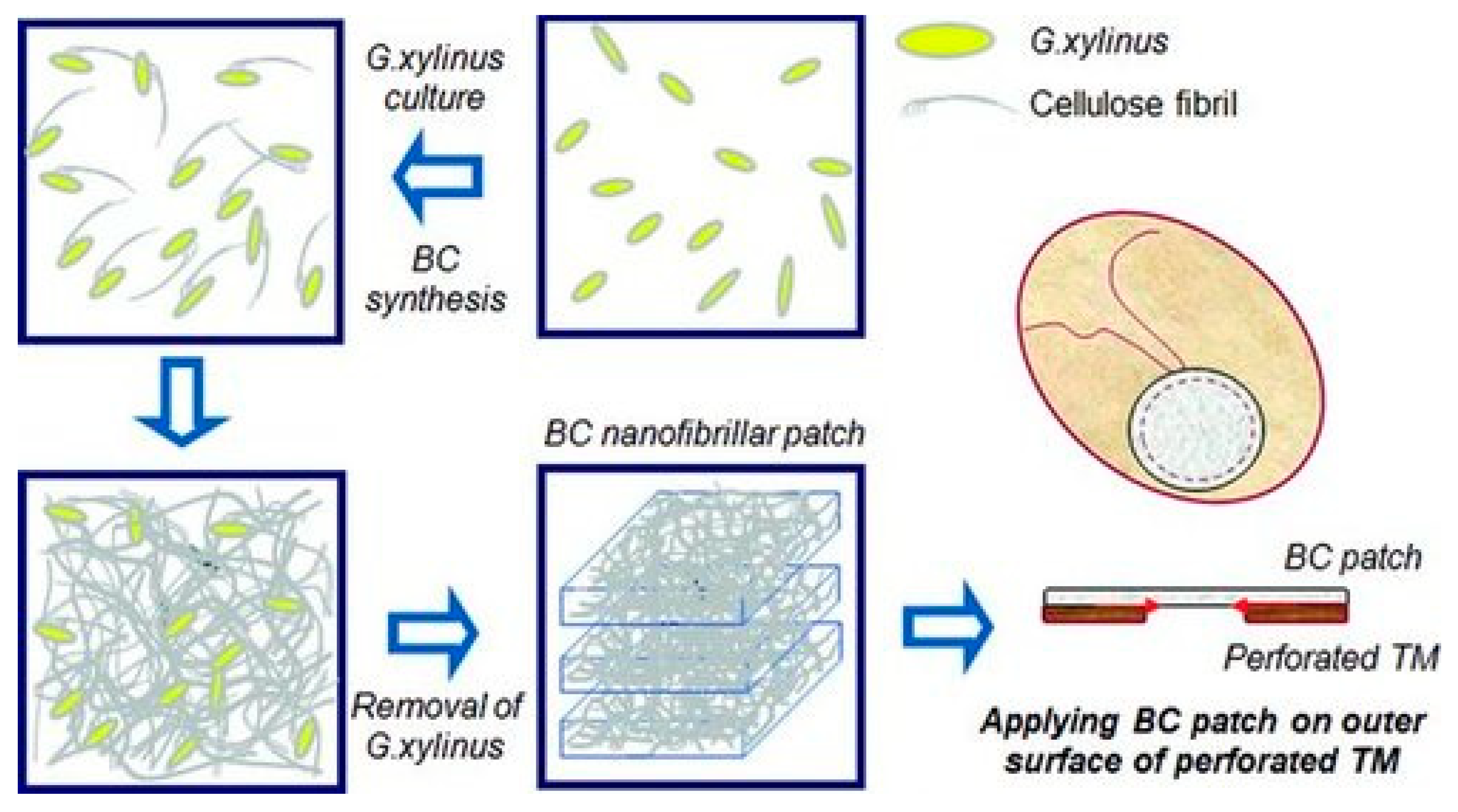
One of the most known works in this field is an article by Jangho Kim et al. Gluconacetobacter xylinus bacteria synthesized cellulose over a period of 14 days, after which the polymer was collected, purified and sterilized (Figure 4). In vivo, BC-based patches had no significant effect on TM during the first 5 days, but significantly improved regeneration thereafter. Compared to TM that healed naturally, TM healed with BC was closer to the original TM in terms of structure and composition. This study laid the foundation for further work and showed that BC can be a real alternative to other polymers in TM restoration [39].
At the same time, the effectiveness of BC films in comparison with classical methods (TM restoration with autologous temporal fascia and fat graft) was investigated. Each of the three experimental groups consisted of 40 people, of whom 20 had minor perforations and 20 had moderate perforations. In the BC film group, 37 people recovered, compared to 25 in the fat graft group and 30 in the fascia group. The operation time was 15, 35 and 65 min, respectively. Thus, BC-based films are not only better suited for TM regeneration, but also easier for doctors to use [40].
Other polymeric films were also studied. For example, the Bionext® film has been developed for wound closure and skin regeneration. Bionext® is bacterial cellulose film for wound treatment, used as a temporary skin substitute for burns, injuries, chronic and diabetic ulcers. It was investigated how this film affects TM healing in 24 patients. Immediately after applying the film, the patients’ well-being improved and their hearing sensitivity increased. A month later, the patients were invited for a follow-up examination, and most of those (83%) had fully recovered, while the rest recovered after another 2 months. BC showed excellent results, however more research is needed in clinical trials to monitor the recovery of all patients [41].
Another BC-based graft from Polisa™, biopolymer membrane obtained by polymer synthesis from sugarcane molasses, has successfully completed clinical trials on 40 patients. All patients were divided into two groups: those with a BC-based graft and those using autologous temporal fascia for TM restoration. In the BC group, regeneration was faster and more complete, the operation time did not exceed 15 min, and the cost of therapy per patient was 13 times lower than in the fascia group. However, the authors do not provide data on the composition and production process of the transplant. Therefore, additional research is needed to determine the components of successful BC application [42].
It can be concluded that BC is one of the most promising materials for TM regeneration, as it has an extremely positive effect on regeneration. In addition, BC production can potentially be scaled up, thereby making the method even more cost-effective.

3. Other Polymers

Many other natural polymers are also used for TM regeneration, but they are not as frequently encountered in research as the polymers mentioned above. In this section of the review, we will discuss the use of less popular polymers.

3.1. Poly(l-lactic acid) and Poly(lactic-co-glycolic acid)

Poly(l-lactic acid) (PLA) and poly(lactic-co-glycolic acid) (PLGA) are both biodegradable and biocompatible polymers used in various biomedical applications. PLA is a homopolymer of lactic acid, while PLGA is a copolymer of lactic acid and glycolic acid. The polymers are often used together as a base solution for electrospinning.
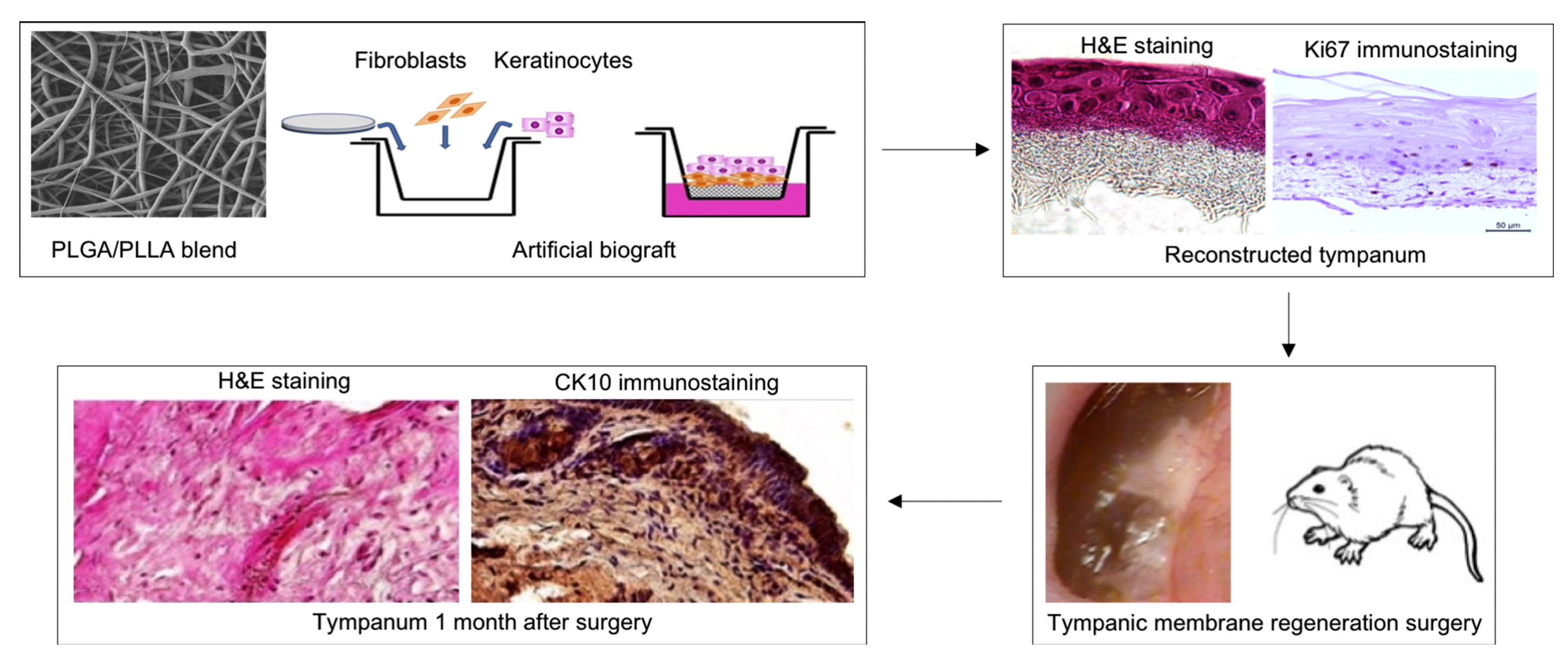
Electrospun scaffolds were used for TM regeneration. Scaffolds with immersed fibroblasts and fibroblasts with keratinocytes showed greater efficacy than the scaffolds alone (Figure 5). In rats with simple polymer matrices, rough traces of regeneration were observed, as well as side effects: cholesteatomas and granulomas. Scaffolds are necessary for such artificial tissues, as the cells themselves are too vulnerable and do not hold their shape during transplantation [43].
In addition to being used as a base for implants with cells, PLA is also used to create implants that are similar in acoustic properties to native TM. The matrix is also formed by electrospinning. Thus, in a series of experiments with different polymers with the addition of graphene oxide, the PLA-based implant showed the best acoustic properties. Further research is needed to investigate the biocompatibility and safety of this matrix [67].

3.2. Latex

Natural latex is a substance obtained from the Brazilian rubber tree (Hevea brasiliensis). Latex is a complex emulsion consisting of various components, including proteins and resins. Due to its elasticity and strength, latex is widely used in regenerative medicine for the restoration of bones in joints, cartilage, and also in dentistry [68].
Latex has potential for use in otosurgery, as some biocompatibility studies have already been conducted on cell and animal models [69,70]. Latex has also been tried for TM regeneration. In one work authors compared latex film with Sylastic® silicone film, a type of silicone implant material used in otorhinolaryngology surgery for a variety of purposes, when performing classic underlay myringoplasty technique surgery. In the group of patients treated with latex, 66% of people had complete TM healing, compared to 55% in the case without lining and 57% in the case of Sylastic®. Also, vascularisation was more active in patients treated with latex, which the authors attributed to the presence of growth factors in Hevea brasiliensis that could affect human tissue. Additional research remains to be performed in this area, as the percentage of healed TM is still too low, and it is known that latex can cause allergic reactions as it is a foreign polymer [44].

3.3. Gelatin

Gelatin is a product of partial hydrolysis of collagen. It is mainly used in cooking but also finds application in pharmaceuticals and cosmetology [71]. Gelfoam® (Pharmacia & Upjohn Company LLC, Kalamazoo, USA) foam has been developed on the basis of gelatin and is widely used for local control of bleeding during surgery. In fact, in many articles mentioned in the review, Gelfoam® is used in surgical invasions.
For TM regeneration, gelfoams without additional substances were tried and they showed excellent results in terms of both the degree of TM healing and the time of regeneration [45]. That is, such matrices themselves create favorable conditions for cell proliferation. However, such studies using only Gelfoam are very limited. Gelatin matrices are usually used as an auxiliary material or loaded with active substances. For example, an experiment with guinea pigs showed that when fibroblast growth factor was included in the gelatin matrix, the TM in all animals in the group recovered within 8 days after laser perforation. In addition, the structure of the restored TM is identical to native TM and includes epithelial, fibrous and mucous layers and blood vessels [46]. It is also important to note that gelatin is used to create fibers by electrospinning, using synthetic and natural cross-linkers. For example, modification of gelatin with methacrylate and subsequent cross-linking with tannic acid has a positive effect on the mechanical properties of the fibers, as the acid promotes the formation of numerous hydrogen bonds between hydrogen in hydroxyl and amino groups and oxygen. The strength of scaffolds, in turn, is important for imitating the acoustic properties of natural TM. In an experiment on TM regeneration in vivo, such scaffolds performed better than simple gelfoam, probably because the cross-linked structure is more suitable for cell proliferation than only gelatin itself [72]. It is also possible to crosslink the gelatin matrix using genipin. Researchers have investigated electrospun gelatin structures crosslinked with genipin as potential scaffolding materials for tympanic membrane regeneration, highlighting their non-toxic nature, favorable mechanical characteristics, biocompatibility and durability [47].
Gelatin matrices are an excellent basis for implants; they are widely used not only in surgical operations but also in combination with other polymers, as we will see later.

3.4. Polycaprolactone

Polycaprolactone (PCL) is a biodegradable and biocompatible polyester synthesized from ε-caprolactone. PCL, like PLA and PLGA, is widely used as a polymer for electrospinning, for the regeneration of vessels, bones and skin [73].
Electrospun fibers can be loaded with active substances and cell cultures, and the porosity of scaffolds promotes cell proliferation and implant ‘ingrowth’ into tissue. Therefore, PCL scaffolds have also been tried for TM regeneration. For example, in one study, electrospun scaffolds with epidermal growth factor significantly improved TM healing in vivo [48] (Figure 6). In a subsequent study, authors immersed insulin-like growth factor binding protein 2 (IGFBP2) in PCL fibers. The scaffolds helped speed up the healing of TM perforations, even though the maximum perforation area in the groups did not go over 20% [49].
It is important to note that in both studies, the authors used radially aligned electrospun fibers, meaning that in the TM they created, the fibers were arranged along the radius. Even without growth factor or IGFBP2, such matrices promoted regeneration, probably by directing cell proliferation along the fibers. In other words, the shape of the matrix can significantly affect the rate of regeneration.

3.5. Alginates

Alginates are salts of alginic acid, a natural polysaccharide obtained from brown seaweed. They are widely used in various fields, including the food industry, medicine, cosmetics, and dentistry. Alginates have unique properties, such as the ability to form gels, bind water, thicken and stabilize mixtures [74]. Alginate hydrogels are considered a promising material for creating implants, as they are highly compatible with tissues, easy to form into a gel, and inexpensive to produce [75].
The ability of alginates to retain water is used in transplantology to create patches and fibers that would maintain optimal conditions for TM regeneration. Thus, alginate-based patches were developed, which in an experiment with chinchillas showed significantly greater effectiveness than paper patches. The authors used calcium ions as a cross-linker for polymer molecules and found that a 4% ion concentration slightly impaired TM regeneration compared to a 2% concentration. However, patches with a 4% ion concentration were more durable and resistant to damage. Therefore, studies on both animals and patients are needed to determine the necessary crosslinker concentrations. This applies not only to ionic crosslinkers but also to other substances [50].
In addition to using alginate on its own, it is also combined with synthetic polymers. For example, electrospun fibers based on polyvinyl alcohol (PVA) and alginate (50:50) provided the best conditions for NIH/3T3 cells (mouse embryonic fibroblasts) in vitro. And in an experiment with rats, the PVA/alginate + Wharton’s Jelly (mucoid connective tissue that surrounds the umbilical cord vessels) composite showed excellent results in regeneration and hearing restoration. But in the case of such a non-standard donor material as Wharton’s Jelly, its source must be considered in advance when scaling up the method [51].

4. Composites Based on Several Polymers

As we have seen in previous sections, natural polymers have useful characteristics. To give implants optimal properties, composites are being developed that combine not only several natural polymers, but also natural and synthetic polymers. This makes it possible to achieve several useful properties at once.
As in the case of single polymers, different methods are used to manufacture TM from composites. One of the simplest is the Petri dish pouring method. This is how films based on silk fibroin and gelatin are obtained. The two polymers are cross-linked with genipin, and such films have shown excellent results in terms of both TM regeneration rate and biocompatibility [52]. The pouring method is also used to produce patches with a chitosan-collagen net structure, with glycerol used as a plasticizer [76]. In both cases, the polymers have the same charge or no charge at all, but their interaction is ensured by a cross-linking agent (genipin) or intertwining chains (chitosan and collagen). Another possible combination of polymers and cross-linking agent: polyvinyl alcohol and gelatin are cross-linked with maleic anhydride to form a gel with a stable structure. The gel showed excellent mechanical properties, but in an in vitro cytotoxicity test, the survival rate of L929 cells (mouse fibroblasts) decreased to almost 80%. This is probably due to the large amount of maleic anhydride compared to PVA (1:1). In the future, the authors should work on the biocompatibility of the gel and test it on animal models [77].
Among non-standard methods of polymer modification, there is a gel based on HA and chitosan, which is fixed with a CO2 laser. The gel was studied in comparison with EpiDisc (etherified HA) on chinchillas. Although more animals recovered in the gel group, the gel had to be reapplied to some animals, resulting in middle ear infection in 6/26 chinchillas. In the EpiDisc group, the TM closed in only half of the animals, but the TM thickness was closer to the initial value than in the gel group. So far, photocurable gel has shown insignificant advantages in therapy, so further research on its use is needed [53].
Electrospinning is often used to create scaffolds for TM regeneration. This method is also used to manufacture composites, often using cross-linkers to improve mechanical properties and increase material stability. The possibility of creating aligned patch using electrospinning rather than 3D printing was demonstrated. The printed fibers were cross-linked with glutaraldehyde and loaded with thyme essential oil to obtain antibacterial properties (Figure 7). The fibers showed a significant zone of inhibition of Pseudomonas aeruginosa (25 mm) and Escherichia coli (25 mm) in the disk diffusion method. However, in the cytotoxicity test, the survival rate of mesenchymal stem cells decreased to 81%. The authors conducted preliminary successful trials on six patients with TM perforation, but number of patients needs to be increased to obtain reliable data [54].
Fibers created by electrospinning can also be used as a component of implants. For example, PCL and gelatin fibers were used to create a composite with alginate hydrogel. The resulting sandwich structure promoted rapid healing of the TM, and the presence of exosomes in the alginate hydrogel promoted natural regeneration. Such an original implant has great potential, but it takes a significant amount of time to produce [55]. Another example of the use of human tissue in a composite is presented in the work of Hyeongjin Lee et al. The researchers developed an implant based on PCL and silk fibroin and included human umbilical cord serum as the active ingredient. The composite showed excellent results in in vivo and in vitro experiments compared to a paper patch. However, it would also be interesting to see the results of a comparison of fibers with and without human umbilical cord serum to assess the effect of cell factors and active substances [78].
It is also possible to combine collagen and poly (ε-caprolactone) structures to achieve properties similar to those of natural TM. Human mesenchymal stromal cells (hMSCs) were cultured on star-branched poly (ε-caprolactone) (*PCL) scaffolds. Under dynamic conditions, hMSCs showed fibroblast marker overexpression and produced collagens I, II, III, and IV. This approach demonstrates that *PCL scaffolds with appropriate mechanical properties can guide hMSCs toward TM-like fibroblast collagen production [79].
Another advantage of electrospinning is the ability to encapsulate a large amount of active substances in the fibers and then control their release by changing the morphology of the fibers. PLGA copolymers were first investigated for its mechanical properties [56]. Then one of the studies was focused on the use of poly (ethylene oxide terephthalate)/poly (butylene terephthalate) (PEOT/PBT) and poly (d,l-lactide-co-glycolide) (PLGA) to create fibers and incorporate the antibiotic ciprofloxacin into them. The solvents used were trichloromethane and absolute ethanol for PEOT/PBT and water with Span 80® for PLGA. Different polymer ratios were used to create different drug release profiles. It is also interesting that the addition of the antibiotic increased the mechanical strength of the fibers. The authors have successfully tested the fibers in vitro for restoration of TM. Experiments on animals are planned in future [80].
Another area of the application for composites is 3D printing of TM. This technology allows grafts to be produced with specified shape and structure parameters, which provides control over the properties of the transplant. Well-known polymers such as gelatin are used to create the basis for such structures. By combining gelatin and methacrylate-modified gelatin, it is possible to obtain layered structures and then wash out the gelatin layers. In this way, the graft for TM restoration was obtained. The graft showed excellent results in an in vivo toxicity test, and the authors conducted an experiment on animal models. In groups with perforation without the treatment, with a graft, and with a graft loaded with EGF, the following results were observed for TM restoration within 14 days: 1/4, 3/4, and 6/6, respectively. However, the small number of animals used in the experiment requires further research to confirm the effectiveness of such a graft [57]. The combination of the scaffold and EGF can significantly improve the healing rate of the eardrum. The factor mainly affects the epithelial cell layer, promotes keratinocyte migration and capillary regeneration, and increases collagen formation in the regenerating tissues [81].
Ideas inspired by nature are also used in the development of transplants. For example, one graft was printed in two approaches: first, a silk scaffold was printed, and then the surrounding area was filled with a fibrin-collagen composite hydrogel matrix [82]. Unfortunately, the authors only investigated the mechanical properties of the transplant. Such a structure may provide not only mechanical strength but also guide cell proliferation during regeneration, as we saw in the case of PCL scaffolds [49].
The most complex composites can contain more than three polymers. For example, in one study, scaffolds with different numbers of fibers (8 or 16) were made from PDMS, PLA and PCL and filled with a fibrin and collagen hydrogel. The grafts were then tested using laser and holographic methods. The resulting 3D structures showed more regular and stable oscillatory characteristics in response to sound compared to human fascia. In addition, their mechanical strength and load resistance were significantly higher [83].
Composites are also being developed that can release not only growth factors but also their receptor genes. Such structures can increase adhesion, proliferation, migration ability and viability of tympanic membrane cells. The matrix for factors and genes can be a polyethyleneimine (PEI)/chitosan composite, which combines the properties of natural and artificial substances [84].

5. Conclusions

The development of implants for TM regeneration is an important area in modern medicine, which involves many parameters. Natural polymers, which are already widely used in tissue surgery, and new polymers, which have rarely been used before, are used to create materials. At the same time, in each individual case, there are many parameters that can be varied to obtain the necessary properties of the implant: polymer molecular weight, polymer concentration, presence and concentration of cross-linkers, the manufacturing method, and others.
Many polymers demonstrate very perspective properties and have potential for use in otologic surgery, but several issues in this area still need to be resolved. For example, the biodegradation of polymers must be controlled, as rapid degradation prevents complete regeneration of the TM, while degradation time that is too long will cause discomfort to the patient. In addition, work needs to be performed on biocompatibility so that the stem cells placed in the implant remain alive for a long time. Another significant problem is the variability in the biological composition between batches of natural materials, which leads to inconsistent treatment results. Thus, natural polymers require modification or combination with synthetic materials to meet clinical requirements [85].
Research is already underway in a very promising area: the inclusion of cell cultures in grafts. It has been found that mesenchymal stem cells (MSCs) can differentiate into epithelial cells that produce many growth factors and cytokines that stimulate wound healing. These assumptions have been confirmed in in vivo experiments on animal models of rats and pigs [86]. In addition to MSCs, epidermal cells derived from the tympanic membrane with stem cell-like characteristics may also play a role in the regeneration of perforations [87].
It seems possible that in the future there will be two main directions for TM restoration: emergency restoration on site and deep regeneration. In the first case, the operation can be performed without prior preparation using simple films made of natural polymers to close small perforations. In the second case, complex implants with a special structure are used, created by electrospinning or 3D printing with the inclusion of stem cells and a set of growth factors, such as insulin and EGF [88]. In each of the two cases, materials with specific properties are required, which have yet to be developed and tested in clinical trials.

Author Contributions

Conceptualization, S.N.M. and P.P.S.; methodology, S.N.M. and P.P.S.; validation, P.G.S.; formal analysis, R.O.S.; investigation, R.O.S., J.Z., H.W. and Z.W.; resources, S.N.M.; data curation, S.N.M.; writing—original draft preparation, R.O.S. and P.G.S.; writing—review and editing, S.N.M. and P.P.S.; visualization, R.O.S. and P.G.S.; supervision, S.N.M. and P.P.S.; project administration, S.N.M.; funding acquisition, S.N.M. All authors have read and agreed to the published version of the manuscript.

Funding

This research was funded by the Russian Science Foundation, project number 25-73-20141. Link to information about the project, URL: https://rscf.ru/project/25-73-20141/ (accessed on 28 September 2025).

Institutional Review Board Statement

Not applicable.

Informed Consent Statement

Not applicable.

Data Availability Statement

No new data were created or analyzed in this study. Data sharing is not applicable to this article.

Conflicts of Interest

The authors declare no conflicts of interest.

References

  1. Sun, W.; Gregory, D.A.; Tomeh, M.A.; Zhao, X. Silk Fibroin as a Functional Biomaterial for Tissue Engineering. Int. J. Mol. Sci. 2021, 22, 1499. [Google Scholar] [CrossRef]
  2. Novozhilov, A.A.; Shilyagin, P.A.; Novozhilova, V.A.; Smirnova, D.D.; Slepanov, A.A.; Dilenjan, A.L.; Klimycheva, M.B.; Gelikonov, G.G.; Gelikonov, V.M.; Shakhov, A.V. Measuring the tympanic membrane thickness using optical coherence tomography. Opera Med. Physiol. 2023, 10, 106–112. [Google Scholar] [CrossRef]
  3. Monasta, L.; Ronfani, L.; Marchetti, F.; Montico, M.; Vecchi Brumatti, L.; Bavcar, A.; Grasso, D.; Barbiero, C.; Tamburlini, G. Burden of Disease Caused by Otitis Media: Systematic Review and Global Estimates. PLoS ONE 2012, 7, e36226. [Google Scholar] [CrossRef]
  4. Anand, S.; Danti, S.; Moroni, L.; Mota, C. Regenerative Therapies for Tympanic Membrane. Prog. Mater. Sci. 2022, 127, 100942. [Google Scholar] [CrossRef]
  5. Wang, A.Y.; Shen, Y.; Liew, L.J.; Wang, J.T.; von Unge, M.; Atlas, M.D.; Dilley, R.J. Rat Model of Chronic Tympanic Membrane Perforation: Ventilation Tube with Mitomycin C and Dexamethasone. Int. J. Pediatr. Otorhinolaryngol. 2016, 80, 61–68. [Google Scholar] [CrossRef] [PubMed]
  6. Acar, M.; Yazıcı, D.; San, T.; Muluk, N.B.; Cingi, C. Fat-Plug Myringoplasty of Ear Lobule vs Abdominal Donor Sites. Eur. Arch. Oto-Rhino-Laryngol. 2014, 272, 861–866. [Google Scholar] [CrossRef]
  7. Ozgursoy, O.B.; Yorulmaz, I. Fat Graft Myringoplasty: A Cost-Effective but Underused Procedure. J. Laryngol. Otol. 2005, 119, 277–279. [Google Scholar] [CrossRef] [PubMed]
  8. Maharajan, N.; Cho, G.W.; Jang, C.H. Application of Mesenchymal Stem Cell for Tympanic Membrane Regeneration by Tissue Engineering Approach. Int. J. Pediatr. Otorhinolaryngol. 2020, 133, 109969. [Google Scholar] [CrossRef]
  9. Villar-Fernandez, M.A.; Lopez-Escamez, J.A. Outlook for Tissue Engineering of the Tympanic Membrane. Audiol. Res. 2015, 5, 117. [Google Scholar] [CrossRef] [PubMed]
  10. Zhao, X.; Zhang, J.; Tian, P.; Cui, X. The Latest Progress of Tympanic Membrane Repair Materials. Am. J. Otolaryngol. 2022, 43, 103408. [Google Scholar] [CrossRef]
  11. Hong, P.; Bance, M.; Gratzer, P.F. Repair of Tympanic Membrane Perforation Using Novel Adjuvant Therapies: A Contemporary Review of Experimental and Tissue Engineering Studies. Int. J. Pediatr. Otorhinolaryngol. 2012, 77, 3–12. [Google Scholar] [CrossRef]
  12. Moreira Lana, G.; Sorg, K.; Wenzel, G.I.; Hecker, D.; Hensel, R.; Schick, B.; Kruttwig, K.; Arzt, E. Self-Adhesive Silicone Microstructures for the Treatment of Tympanic Membrane Perforations. Adv. NanoBiomed Res. 2021, 1, 2100057. [Google Scholar] [CrossRef]
  13. Lu, Y.; Li, J.; Hou, N.; Zhou, L.; Quan, X.; Tang, Y.; Luo, X.; Huang, S.; Ma, R. Decellularized Tympanic Membrane Scaffold with Bone Marrow Mesenchymal Stem Cells for Repairing Tympanic Membrane Perforation. Artif. Organs 2023, 47, 62–76. [Google Scholar] [CrossRef]
  14. Mohanty, R.; Harkut, R.; Mhashal, S.; Kadao, Y. Advancement in Tympanic Membrane Repair: Exploring the Potential of Platelet Rich Fibrin. Indian J. Otolaryngol. Head Neck Surg. 2024, 76, 2962–2967. [Google Scholar] [CrossRef]
  15. Gür, Ö.E.; Ensari, N.; Öztürk, M.T.; Boztepe, O.F.; Gün, T.; Selçuk, Ö.T.; Renda, L. Use of a Platelet-Rich Fibrin Membrane to Repair Traumatic Tympanic Membrane Perforations: A Comparative Study. Acta Oto-Laryngol. 2016, 136, 1017–1023. [Google Scholar] [CrossRef]
  16. Erkilet, E.; Koyuncu, M.; Atmaca, S.; Yarim, M. Platelet-Rich Plasma Improves Healing of Tympanic Membrane Perforations: Experimental Study. J. Laryngol. Otol. 2008, 123, 482–487. [Google Scholar] [CrossRef]
  17. Von Unge, M.; Dirckx, J.J.J.; Olivius, N.P. Embryonic Stem Cells Enhance the Healing of Tympanic Membrane Perforations. Int. J. Pediatr. Otorhinolaryngol. 2003, 67, 215–219. [Google Scholar] [CrossRef]
  18. Ranglani, D.; Agiwal, S.; More, N.; Parkale, R.; Shitole, V.; Hiray, A.R.; Kapusetti, G. Review on Tympanic Membrane and Auditory Canal Regeneration by Biomaterial Intervention. Mater. Highlights 2021, 2, 7. [Google Scholar] [CrossRef]
  19. Kim, J.; Kim, C.H.; Park, C.H.; Seo, J.-N.; Kweon, H.; Kang, S.W.; Lee, K.G. Comparison of Methods for the Repair of Acute Tympanic Membrane Perforations: Silk Patch vs. Paper Patch. Wound Repair Regen. 2010, 18, 132–138. [Google Scholar] [CrossRef]
  20. Lee, O.J.; Lee, J.M.; Kim, J.H.; Kim, J.; Kweon, H.; Jo, Y.Y.; Park, C.H. Biodegradation Behavior of Silk Fibroin Membranes in Repairing Tympanic Membrane Perforations. J. Biomed. Mater. Res. A 2012, 100A, 2018–2026. [Google Scholar] [CrossRef] [PubMed]
  21. Shen, Y.; Redmond, S.L.; Teh, B.M.; Yan, S.; Wang, Y.; Zhou, L.; Budgeon, C.A.; Eikelboom, R.H.; Atlas, M.D.; Dilley, R.J.; et al. Scaffolds for Tympanic Membrane Regeneration in Rats. Tissue Eng. Part A 2013, 19, 657–668. [Google Scholar] [CrossRef]
  22. Shen, Y.; Redmond, S.L.; Teh, B.M.; Yan, S.; Wang, Y.; Atlas, M.D.; Dilley, R.J.; Zheng, M.; Marano, R.J. Tympanic Membrane Repair Using Silk Fibroin and Acellular Collagen Scaffolds. Laryngoscope 2013, 123, 1976–1982. [Google Scholar] [CrossRef]
  23. Lee, J.H.; Kim, D.; Park, H.S.; Jeong, J.Y.; Yeon, Y.K.; Kumar, V.; Bae, S.H.; Lee, J.M.; Moon, B.M.; Park, C.H. A Prospective Cohort Study of the Silk Fibroin Patch in Chronic Tympanic Membrane Perforation. Laryngoscope 2016, 126, 2798–2803. [Google Scholar] [CrossRef]
  24. Ghassemifar, R.; Redmond, S.; Zainuddin; Chirila, T.V. Advancing Towards a Tissue-Engineered Tympanic Membrane: Silk Fibroin as a Substratum for Growing Human Eardrum Keratinocytes. J. Biomater. Appl. 2010, 24, 591–606. [Google Scholar] [CrossRef]
  25. Harugade, A.; Sherje, A.P.; Pethe, A. Chitosan: A Review on Properties, Biological Activities and Recent Progress in Biomedical Applications. React. Funct. Polym. 2023, 191, 105634. [Google Scholar] [CrossRef]
  26. Kim, J.; Kim, S.W.; Choi, S.J.; Lim, K.T.; Lee, J.B.; Seonwoo, H.; Choung, P.-H.; Park, K.; Cho, C.-S.; Choung, Y.-H.; et al. A Healing Method of Tympanic Membrane Perforations Using Three-Dimensional Porous Chitosan Scaffolds. Tissue Eng. Part A 2011, 17, 2763–2772. [Google Scholar] [CrossRef]
  27. Anand, S.; Azimi, B.; Lucena, M.; Ricci, C.; Candito, M.; Zavagna, L.; Astolfi, L.; Coltelli, M.-B.; Lazzeri, A.; Berrettini, S.; et al. Chitin Nanofibrils Modulate Mechanical Response in Tympanic Membrane Replacements. Carbohydr. Polym. 2023, 310, 120732. [Google Scholar] [CrossRef]
  28. Seonwoo, H.; Kim, S.W.; Shin, B.; Jang, K.-J.; Lee, M.; Choo, O.-S.; Choi, M.-J.; Kim, J.; Lim, K.-T.; Jang, J.H.; et al. Latent Stem Cell-Stimulating Therapy for Regeneration of Chronic Tympanic Membrane Perforations Using IGFBP2-Releasing Chitosan Patch Scaffolds. J. Biomater. Appl. 2019, 34, 198–207. [Google Scholar] [CrossRef]
  29. Hellstrom, S.; Laurent, C. Hyaluronan and Healing of Tympanic Membrane Perforations. An experimental study. Acta Otolaryngol. 1987, 442, 54–61. [Google Scholar] [CrossRef]
  30. Güneri, E.A.; Tekin, S.; Yilmaz, O.; Özkara, E.; Erdağ, T.K.; Ikiz, A.Ö.; Sarioğlu, S.; Güneri, A. The Effects of Hyaluronic Acid, Epidermal Growth Factor, and Mitomycin in an Experimental Model of Acute Traumatic Tympanic Membrane Perforation. Otol. Neurotol. 2003, 24, 371–376. [Google Scholar] [CrossRef]
  31. Saliba, I. Hyaluronic Acid Fat Graft Myringoplasty: An Office-Based Technique Adapted to Children. Arch. Otolaryngol. Head Neck Surg. 2011, 137, 1203. [Google Scholar] [CrossRef]
  32. Yilmaz, M.S.; Sahin, E.; Kaymaz, R.; Altunkaynak, B.Z.; Akidil, A.O.; Yanar, S.; Demir, D.; Guven, M. Histological Study of The Healing of Traumatic Tympanic Membrane Perforation After Vivosorb and Epifilm Application. Ear Nose Throat J. 2021, 100, 90–96. [Google Scholar] [CrossRef]
  33. Prior, M.; Gibbins, N.; John, G.; Rhys-Williams, S.; Scott, P. Hyaluronic Acid Ester in Myringoplasty. J. Laryngol. Otol. 2008, 122, e3. [Google Scholar] [CrossRef]
  34. Ozturk, K.; Yaman, H.; Cihat Avunduk, M.; Arbag, H.; Keles, B.; Uyar, Y. Effectiveness of MeroGel Hyaluronic Acid on Tympanic Membrane Perforations. Acta Otolaryngol. 2006, 126, 1158–1163. [Google Scholar] [CrossRef]
  35. Konakçi, E.; Koyuncu, M.; Ünal, R.; Tekat, A.; Uyar, M. Repair of Subtotal Tympanic Membrane Perforations with Seprafilm®. J. Laryngol. Otol. 2004, 118, 862–865. [Google Scholar] [CrossRef]
  36. Jang, C.H.; Cho, Y.B.; Yeo, M.; Lee, H.; Min, E.J.; Lee, B.H.; Kim, G.H. Regeneration of Chronic Tympanic Membrane Perforation Using 3D Collagen with Topical Umbilical Cord Serum. Int. J. Biol. Macromol. 2013, 62, 232–240. [Google Scholar] [CrossRef]
  37. Svistushkin, M.; Kotova, S.; Zolotova, A.; Fayzullin, A.; Antoshin, A.; Serejnikova, N.; Shekhter, A.; Voloshin, S.; Giliazova, A.; Istranova, E.; et al. Collagen Matrix to Restore the Tympanic Membrane: Developing a Novel Platform to Treat Perforations. Polymers 2024, 16, 248. [Google Scholar] [CrossRef]
  38. Li, L.; Zhang, W.; Huang, M.; Li, J.; Chen, J.; Zhou, M.; He, J. Preparation of Gelatin/Genipin Nanofibrous Membrane for Tympanic Member Repair. J. Biomater. Sci. Polym. Ed. 2018, 29, 2154–2167. [Google Scholar] [CrossRef]
  39. Kim, J.; Kim, S.W.; Park, S.; Lim, K.T.; Seonwoo, H.; Kim, Y.; Hong, B.H.; Choung, Y.; Chung, J.H. Bacterial Cellulose Nanofibrillar Patch as a Wound Healing Platform of Tympanic Membrane Perforation. Adv. Healthc. Mater. 2013, 2, 1525–1531. [Google Scholar] [CrossRef]
  40. Mandour, Y.M.H.; Mohammed, S.; Menem, M.o.A. Bacterial Cellulose Graft versus Fat Graft in Closure of Tympanic Membrane Perforation. Am. J. Otolaryngol. 2019, 40, 168–172. [Google Scholar] [CrossRef]
  41. Pinho, A.M.d.M.R.; Kencis, C.C.S.; Miranda, D.R.P.; Sousa Neto, O.M.d. Traumatic Perforations of the Tympanic Membrane: Immediate Clinical Recovery with the Use of Bacterial Cellulose Film. Braz. J. Otorhinolaryngol. 2020, 86, 727–733. [Google Scholar] [CrossRef]
  42. Silveira, F.C.A.; Pinto, F.C.M.; Caldas Neto, S.d.S.; Leal, M.d.C.; Cesário, J.; Aguiar, J.L.d.A. Treatment of Tympanic Membrane Perforation Using Bacterial Cellulose: A Randomized Controlled Trial. Braz. J. Otorhinolaryngol. 2016, 82, 203–208. [Google Scholar] [CrossRef]
  43. Immich, A.P.S.; Pennacchi, P.C.; Naves, A.F.; Felisbino, S.L.; Boemo, R.L.; Maria-Engler, S.S.; Catalani, L.H. Improved Tympanic Membrane Regeneration after Myringoplastic Surgery Using an Artificial Biograft. Mater. Sci. Eng. C 2017, 73, 48–58. [Google Scholar] [CrossRef]
  44. Araujo, M.M.; Massuda, E.T.; Hyppolito, M.A. Anatomical and Functional Evaluation of Tympanoplasty Using a Transitory Natural Latex Biomembrane Implant from the Rubber Tree Hevea Brasiliensis. Acta Cir. Bras. 2012, 27, 566–571. [Google Scholar] [CrossRef]
  45. Lou, Z.-C.; He, J.-G. A Randomised Controlled Trial Comparing Spontaneous Healing, Gelfoam Patching and Edge-approximation plus Gelfoam Patching in Traumatic Tympanic Membrane Perforation with Inverted or Everted Edges. Clin. Otolaryngol. 2011, 36, 221–226. [Google Scholar] [CrossRef]
  46. Hakuba, N.; Tabata, Y.; Hato, N.; Fujiwara, T.; Gyo, K. Gelatin Hydrogel With Basic Fibroblast Growth Factor for Tympanic Membrane Regeneration. Otol. Neurotol. 2014, 35, 540–544. [Google Scholar] [CrossRef]
  47. Han, S.; Zhang, Z.; Chen, J.; Li, J.; Zhou, M.; He, Z.; He, Z.; Li, L. Preparation of Antibacterial Gelatin/Genipin Nanofibrous Membrane for Tympanic Membrane Repair. Molecules 2022, 27, 2906. [Google Scholar] [CrossRef]
  48. Seonwoo, H.; Shin, B.; Jang, K.; Lee, M.; Choo, O.; Park, S.; Kim, Y.C.; Choi, M.; Kim, J.; Garg, P.; et al. Epidermal Growth Factor–Releasing Radially Aligned Electrospun Nanofibrous Patches for the Regeneration of Chronic Tympanic Membrane Perforations. Adv. Healthc. Mater. 2019, 8, e1801160. [Google Scholar] [CrossRef]
  49. Lee, J.; Park, S.; Shin, B.; Kim, Y.J.; Lee, S.; Kim, J.; Jang, K.-J.; Choo, O.-S.; Kim, J.; Seonwoo, H.; et al. Latent Stem Cell-Stimulating Radially Aligned Electrospun Nanofibrous Patches for Chronic Tympanic Membrane Perforation Therapy. Acta Biomater. 2024, 188, 212–222. [Google Scholar] [CrossRef]
  50. Weber, D.E.; Semaan, M.T.; Wasman, J.K.; Beane, R.; Bonassar, L.J.; Megerian, C.A. Tissue-Engineered Calcium Alginate Patches in the Repair of Chronic Chinchilla Tympanic Membrane Perforations. Laryngoscope 2006, 116, 700–704. [Google Scholar] [CrossRef]
  51. Najafi, R.; Yazdian, F.; Pezeshki-Modaress, M.; Aleemardani, M.; Chahsetareh, H.; Hassanzadeh, S.; Farhadi, M.; Bagher, Z. Fabrication and Optimization of Multilayered Composite Scaffold Made of Sulfated Alginate-Based Nanofiber/Decellularized Wharton’s Jelly ECM for Tympanic Membrane Tissue Engineering. Int. J. Biol. Macromol. 2023, 253, 127128. [Google Scholar] [CrossRef]
  52. Aleemardani, M.; Akbarnejad, Z.; Jalessi, M.; Chahsetareh, H.; Hajmohammadi, Z.; Hassanzadeh, S.; Najafi, R.; Alizadeh, R.; Farhadi, M.; Bagher, Z. Silk Fibroin-Gelatin Films Crosslinked by Genipin for Regenerating Tympanic Membrane Perforations. J. Drug Deliv. Sci. Technol. 2024, 95, 105642. [Google Scholar] [CrossRef]
  53. Rubinstein, B.J.; Ranney, J.D.; Khoshakhlagh, P.; Strasnick, B.; Horn-Ranney, E.L. A Novel Gel Patch for Minimally Invasive Repair of Tympanic Membrane Perforations. Int. J. Pediatr. Otorhinolaryngol. 2018, 115, 27–32. [Google Scholar] [CrossRef]
  54. Pourheydari-Barsari, Z.; Mirzadeh, H.; Farhadi, M.; Solouk, A.; Jalessi, M. Antibacterial Aligned Nanofibrous Chitosan/PVA Patch for Repairing Chronic Tympanic Membrane Perforations. Int. J. Biol. Macromol. 2023, 253, 126597. [Google Scholar] [CrossRef]
  55. Chahsetareh, H.; Yazdian, F.; Pezeshki-Modaress, M.; Aleemardani, M.; Hassanzadeh, S.; Najafi, R.; Simorgh, S.; Taghdiri Nooshabadi, V.; Bagher, Z.; Davachi, S.M. Alginate Hydrogel-PCL/Gelatin Nanofibers Composite Scaffold Containing Mesenchymal Stem Cells-Derived Exosomes Sustain Release for Regeneration of Tympanic Membrane Perforation. Int. J. Biol. Macromol. 2024, 262, 130141. [Google Scholar] [CrossRef]
  56. Mota, C.; Danti, S.; D’Alessandro, D.; Trombi, L.; Ricci, C.; Puppi, D.; Dinucci, D.; Milazzo, M.; Stefanini, C.; Chiellini, F.; et al. Multiscale Fabrication of Biomimetic Scaffolds for Tympanic Membrane Tissue Engineering. Biofabrication 2015, 7, 025005. [Google Scholar] [CrossRef]
  57. Kuo, C.-Y.; Wilson, E.; Fuson, A.; Gandhi, N.; Monfaredi, R.; Jenkins, A.; Romero, M.; Santoro, M.; Fisher, J.P.; Cleary, K.; et al. Repair of Tympanic Membrane Perforations with Customized Bioprinted Ear Grafts Using Chinchilla Models. Tissue Eng. Part A 2018, 24, 527–535. [Google Scholar] [CrossRef]
  58. Lu, H.; Xia, K.; Jian, M.; Liang, X.; Yin, Z.; Zhang, M.; Wang, H.; Wang, H.; Li, S.; Zhang, Y. Mechanically Reinforced Silkworm Silk Fiber by Hot Stretching. Research 2022, 2022, 9854063. [Google Scholar] [CrossRef]
  59. Levin, B.; Redmond, S.L.; Rajkhowa, R.; Eikelboom, R.H.; Atlas, M.D.; Marano, R.J. Utilising Silk Fibroin Membranes as Scaffolds for the Growth of Tympanic Membrane Keratinocytes, and Application to Myringoplasty Surgery. J. Laryngol. Otol. 2012, 127, S13–S20. [Google Scholar] [CrossRef]
  60. Allardyce, B.J.; Rajkhowa, R.; Dilley, R.J.; Xie, Z.; Campbell, L.; Keating, A.; Atlas, M.D.; von Unge, M.; Wang, X. Comparative Acoustic Performance and Mechanical Properties of Silk Membranes for the Repair of Chronic Tympanic Membrane Perforations. J. Mech. Behav. Biomed. Mater. 2016, 64, 65–74. [Google Scholar] [CrossRef]
  61. Kim, J.H.; Choi, S.J.; Park, J.-S.; Lim, K.T.; Choung, P.-H.; Kim, S.W.; Lee, J.B.; Chung, J.H.; Choung, Y.-H. Tympanic Membrane Regeneration Using a Water-Soluble Chitosan Patch. Tissue Eng. Part A 2010, 16, 225–232. [Google Scholar] [CrossRef] [PubMed]
  62. Knopf-Marques, H.; Pravda, M.; Wolfova, L.; Velebny, V.; Schaaf, P.; Vrana, N.E.; Lavalle, P. Hyaluronic Acid and Its Derivatives in Coating and Delivery Systems: Applications in Tissue Engineering, Regenerative Medicine and Immunomodulation. Adv. Healthc. Mater. 2016, 5, 2841–2855. [Google Scholar] [CrossRef]
  63. Saliba, I.; Woods, O. Hyaluronic Acid Fat Graft Myringoplasty: A Minimally Invasive Technique. Laryngoscope 2011, 121, 375–380. [Google Scholar] [CrossRef]
  64. Shekhter, A.B.; Fayzullin, A.L.; Vukolova, M.N.; Rudenko, T.G.; Osipycheva, V.D.; Litvitsky, P.F. Medical Applications of Collagen and Collagen-Based Materials. Curr. Med. Chem. 2019, 26, 506–516. [Google Scholar] [CrossRef]
  65. Kim, S.H.; Jeong, J.Y.; Park, H.J.; Moon, B.M.; Park, Y.R.; Lee, O.J.; Sultan, M.T.; Kim, D.-K.; Park, H.S.; Lee, J.H.; et al. Application of a Collagen Patch Derived from Duck Feet in Acute Tympanic Membrane Perforation. Tissue Eng. Regen. Med. 2017, 14, 233–241. [Google Scholar] [CrossRef]
  66. Picheth, G.F.; Pirich, C.L.; Sierakowski, M.R.; Woehl, M.A.; Sakakibara, C.N.; de Souza, C.F.; Martin, A.A.; da Silva, R.; de Freitas, R.A. Bacterial Cellulose in Biomedical Applications: A Review. Int. J. Biol. Macromol. 2017, 104, 97–106. [Google Scholar] [CrossRef]
  67. Milazzo, M.; Danti, S.; Livens, P.; Dirckx, J.; Scaffaro, R.; Gammino, M. Electrospun Graphene Oxide/Polymeric Nanocomposites for Eardrum Replacements. Compos. Commun. 2024, 51, 102048. [Google Scholar] [CrossRef]
  68. Marques, P.A.C.; Guerra, N.B.; dos Santos, L.S.; Mussagy, C.U.; Pegorin Brasil, G.S.; Burd, B.S.; Su, Y.; da Silva Sasaki, J.C.; Scontri, M.; de Lima Lopes Filho, P.E.; et al. Natural Rubber Latex-Based Biomaterials for Drug Delivery and Regenerative Medicine: Trends and Directions. Int. J. Biol. Macromol. 2024, 267, 131666. [Google Scholar] [CrossRef]
  69. Pegorin Brasil, G.S.; de Barros, P.P.; Miranda, M.C.R.; de Barros, N.R.; Junqueira, J.C.; Gomez, A.; Herculano, R.D.; de Mendonça, R.J. Natural Latex Serum: Characterization and Biocompatibility Assessment Using Galleria Mellonella as an Alternative in Vivo Model. J. Biomater. Sci. Polym. Ed. 2022, 33, 705–726. [Google Scholar] [CrossRef]
  70. Miranda, M.C.R.; Borges, F.A.; Barros, N.R.; Marques, M.P.; Galeane, M.C.; de Lacorte Singulani, J.; Guerra, N.B.; Brasil, G.S.P.; Mussagy, C.U.; Almeida, A.M.F.; et al. In Vitro and Alternative Animal Models to Evaluate the Biocompatibility of Natural Latex-Calcium Phosphate-Based Polymer. J. Polym. Environ. 2022, 30, 3368–3378. [Google Scholar] [CrossRef]
  71. Bello, A.B.; Kim, D.; Kim, D.; Park, H.; Lee, S.-H. Engineering and Functionalization of Gelatin Biomaterials: From Cell Culture to Medical Applications. Tissue Eng. Part B Rev. 2020, 26, 164–180. [Google Scholar] [CrossRef]
  72. Wang, B.; Xin, T.; Shen, L.; Zhang, K.; Zhang, D.; Zhang, H.; Liu, J.; Chen, B.; Cui, W.; Shu, Y. Acoustic Transmitted Electrospun Fibrous Membranes for Tympanic Membrane Regeneration. Chem. Eng. J. 2021, 419, 129536. [Google Scholar] [CrossRef]
  73. Janmohammadi, M.; Nourbakhsh, M.S. Electrospun Polycaprolactone Scaffolds for Tissue Engineering: A Review. Int. J. Polym. Mater. Polym. Biomater. 2019, 68, 527–539. [Google Scholar] [CrossRef]
  74. Hurtado, A.; Aljabali, A.A.A.; Mishra, V.; Tambuwala, M.M.; Serrano-Aroca, Á. Alginate: Enhancement Strategies for Advanced Applications. Int. J. Mol. Sci. 2022, 23, 4486. [Google Scholar] [CrossRef] [PubMed]
  75. Lee, K.Y.; Mooney, D.J. Alginate: Properties and Biomedical Applications. Prog. Polym. Sci. 2011, 37, 106–126. [Google Scholar] [CrossRef] [PubMed]
  76. Widiyanti, P.; Angtika, R.S.; Githanadi, B.; Kharisma, D.H.; Asyraf, T.O.; Wardani, A. Collagen-Chitosan-Glycerol Bio-Composite as Artificial Tympanic Membrane for Ruptured Inner Ear Organ. J. Phys. Conf. Ser. 2017, 853, 012029. [Google Scholar] [CrossRef]
  77. Joseph, J.; Ramesh, P.; Sayooj, K.; Unnikrishnan, M.; Unnikrishnan, G. Functionalized Polyvinyl Alcohol–Gelatin Graft for the Treatment of Tympanic Membrane Perforations. J. Biomed. Mater. Res. A 2025, 113, e37818. [Google Scholar] [CrossRef]
  78. Lee, H.; Jang, C.H.; Kim, G.H. A Polycaprolactone/Silk-Fibroin Nanofibrous Composite Combined with Human Umbilical Cord Serum for Subacute Tympanic Membrane Perforation; an in Vitro and in Vivo Study. J. Mater. Chem. B 2014, 2, 2703–2713. [Google Scholar] [CrossRef]
  79. Moscato, S.; Rocca, A.; D’Alessandro, D.; Puppi, D.; Gramigna, V.; Milazzo, M.; Stefanini, C.; Chiellini, F.; Petrini, M.; Berrettini, S.; et al. Tympanic Membrane Collagen Expression by Dynamically Cultured Human Mesenchymal Stromal Cell/Star-Branched Poly(ε-Caprolactone) Nonwoven Constructs. Appl. Sci. 2020, 10, 3043. [Google Scholar] [CrossRef]
  80. Anand, S.; Fusco, A.; Günday, C.; Günday-Türeli, N.; Donnarumma, G.; Danti, S.; Moroni, L.; Mota, C. Tunable Ciprofloxacin Delivery through Personalized Electrospun Patches for Tympanic Membrane Perforations. Bioact. Mater. 2024, 38, 109–123. [Google Scholar] [CrossRef]
  81. Lou, Z. The Effect of Epidermal Growth Factor on the Pseudo-Healing of Traumatic Tympanic Membrane Perforations. Braz. J. Otorhinolaryngol. 2019, 87, 53–58. [Google Scholar] [CrossRef]
  82. Rostam-Alilou, A.A.; Jafari, H.; Zolfagharian, A.; Serjouei, A.; Bodaghi, M. Using Fibrincollagen Composite Hydrogel and Silk for Bio-Inspired Design of Tympanic Membrane Grafts: A Vibro-Acoustic Analysis. Compos. Part C 2021, 6, 100205. [Google Scholar] [CrossRef]
  83. Kozin, E.D.; Black, N.L.; Cheng, J.T.; Cotler, M.J.; McKenna, M.J.; Lee, D.J.; Lewis, J.A.; Rosowski, J.J.; Remenschneider, A.K. Design, Fabrication, and In Vitro Testing of Novel Three-Dimensionally Printed Tympanic Membrane Grafts. Hear. Res. 2016, 340, 191–203. [Google Scholar] [CrossRef]
  84. Lee, M.C.; Seonwoo, H.; Garg, P.; Jang, K.J.; Pandey, S.; Park, S.B.; Kim, H.B.; Lim, J.; Choung, Y.H.; Chung, J.H. Chitosan/PEI Patch Releasing EGF and the EGFR Gene for the Regeneration of the Tympanic Membrane after Perforation. Biomater. Sci. 2017, 6, 364–371. [Google Scholar] [CrossRef] [PubMed]
  85. Danti, S.; Mota, C.; D’alessandro, D.; Trombi, L.; Ricci, C.; Redmond, S.L.; De Vito, A.; Pini, R.; Dilley, R.J.; Moroni, L.; et al. Tissue Engineering of the Tympanic Membrane Using Electrospun PEOT/PBT Copolymer Scaffolds: A Morphological in Vitro Study. Hear. Balance Commun. 2015, 13, 133–147. [Google Scholar] [CrossRef]
  86. Mittal, R.; Jung, H.D.; Mittal, J.; Eshraghi, A.A. A Perspective on Stem Cell Therapy for Ear Disorders. J. Cell. Physiol. 2017, 233, 1823–1824. [Google Scholar] [CrossRef] [PubMed]
  87. Liew, L.J.; Chen, L.Q.; Wang, A.Y.; Von Unge, M.; Atlas, M.D.; Dilley, R.J. Tympanic Membrane Derived STEM Cell-Like Cultures for Tissue Regeneration. Stem Cells Dev. 2018, 27, 649–657. [Google Scholar] [CrossRef]
  88. Eken, M.; Ates, G.; Sanlı, A.; Evren, C.; Bozkurt, S. The Effect of Topical Insulin Application on the Healing of Acute Tympanic Membrane Perforations: A Histopathologic Study. Eur. Arch. Oto-Rhino-Laryngol. 2007, 264, 999–1002. [Google Scholar] [CrossRef]
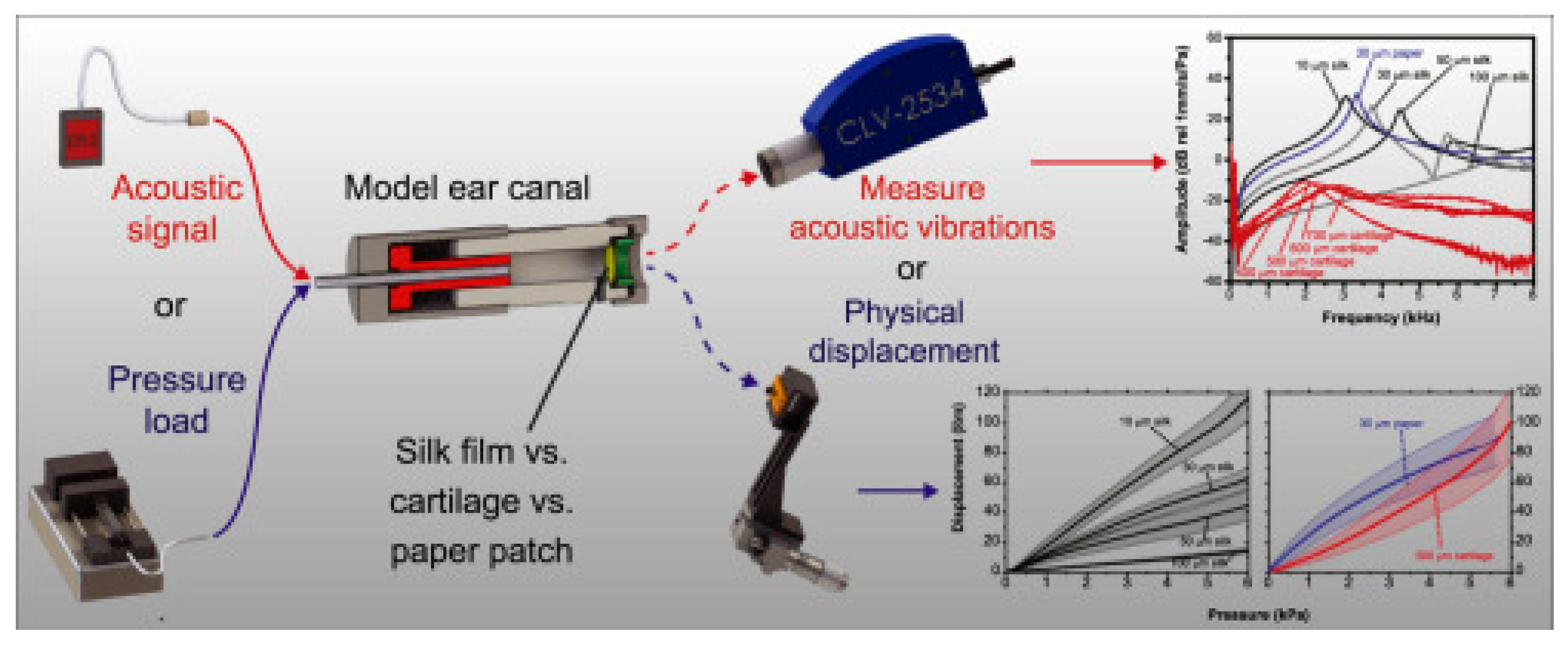
Figure 1. Comparison of the physical and acoustic characteristics of silk fibroin film compared to cartilage and paper patch. Reprinted with permission from Ref. [60] Copyright 2016 Elsevier.
Jfb 16 00384 g001
Figure 2. Manufacturing of chitosan-based fibers by electrospinning and investigation of their physical properties and biocompatibility on cell models. Reprinted from Ref. [27].
Jfb 16 00384 g002
Figure 3. Initial perforation of the tympanic membrane (A) and condition of the tympanic membrane 4 months after surgery using hyaluronic acid fat graft myringoplasty. (B) The perforation is completely closed and neovascularisation can be identified. Reprinted with permission from Ref. [63] Copyright 2011 Wiley.
Jfb 16 00384 g003
Figure 4. In vitro and in vivo studies demonstrate that the bacterial cellulose nanofibrillar patch promotes the TM healing speed and rate as well as recovering the function of the TM. Reprinted with permission from Ref. [39] Copyright 2013 Wiley.
Jfb 16 00384 g004
Figure 5. The process of creating and researching in vivo artificial grafts based on PLGA/PLLA with the inclusion of fibroblasts and keratinocytes for TM regeneration. Reprinted with permission from Ref. [43]. Copyright 2017 Elsevier.
Jfb 16 00384 g005
Figure 6. Electrospun radially aligned nanofibrous patches loaded with insulin-like growth factor binding protein 2 (IGFBP2) significantly improved TM regeneration in animal models. Reprinted from Ref. [28].
Jfb 16 00384 g006
Figure 7. Aligned nanofibers based on chitosan/PVA composite loaded with thyme essential oil have a significant antibacterial effect and good biocompatibility. Reprinted with permission from Ref. [54] Copyright 2023 Elsevier.
Jfb 16 00384 g007
Table 1. Main characteristics of the studied structures based on natural polymers.
Implant CompositionPolymer Molecular WeightPolymer Graft form/Active Inclusions and SubstancesSubject of Experiments% of Subjects with Restored TMReference
Cell LinesAnimals and Human
1Silk fibroinnaturally derivedcasted polymer patch/--rats93%[19]
2Silk fibroinnaturally derivedcasted polymer patch/-L929 cellsrats-[20]
3Silk fibroinnaturally derivedcasted polymer patch/--rats100%[21]
4Silk fibroinnaturally derivedcasted polymer patch/--guinea pigs100%[22]
5Silk fibroin Tympasil® patch-ready film/--human70%[23]
6Silk fibroinnaturally derivedcasted polymer patch/-human TM cells--[24]
7Chitosan100–200 kDacasted polymer patch/-human TM cellsrats100%[25]
8Chitosan200 kDafreezing and lyophilizing of polymer solution/-human TM cellsrats100%[26]
9PEOT/PBT/(Chitin/PEG)Chitin: naturally derived
PEOT/PBT: 93 kDa
PEG: 4000 g/mol
compression molding/-human mesenchymal stromal cells
OC-k3
HaCaT
PC12
--[27]
10Chitosan200 kDacasted polymer patch/IGFBP2human TM cellsrats44%[28]
11HA4 or 6 MDapolymer solution/--rats100%[29]
12HA-polymer solution/--rats100%[30]
13HA epidisc-fat graft myringoplasty -human93%[30]
14HA epidisc-fat graft myringoplasty -human (children)87%[31]
15HA ester Epifilm®-ready epidisc/--rats100%[32]
16HA ester Epifilm®-ready epidisc/--human0%[33]
17esterified HA MeroGel®-ready surgical tampons/--rats92%[34]
18Seprafilm®-ready film/--rats100%[35]
19Collagen-3D printing/umbilical cord serumNHDFsguinea pigs100%[36]
20Collagennaturally derivedelectrophoretic deposition/--chinchillas100%[37]
21Collagennaturally derivedlyophilisation of polymer solution/--rats100%[38]
22Bacterial cellulosenaturally derivednaturally formed/-rat TM cellsrats100%[39]
23Bacterial cellulosenaturally derivednaturally formed/myringoplasty-human92.5%[40]
24Bionext®-ready film/--human100% of examined patients[41]
25BC graft from Polisa™-ready film/--human90%[42]
26PLLA and PLGAPLLA: 216,000 g/mol
PLGA: 110,000 g/mol
electrospinning/fibroblasts and keratinocyteshuman fibroblastsrats100%[43]
27Latexnaturally derived-/underlay myringoplasty-human67%[44]
28Gelatin gelfoam--/--human97%[45]
29Gelatin-chemically cross-linked hydrogel/basic fibroblast growth factor (bFGF)-guinea pigs100%[46]
30Gelatin and methacrylic acid-electrospinning/-HEI-OC1 cellsguinea pigs100%[47]
31PCL80 kDaelectrospinning/epidermal growth factor (EGF)rat TM cellsrats100%[48]
32PCL80 kDaelectrospinning/IGFBP2rat TM cellsrats100%[49]
33Calcium alginate-injection molding technology/--chinchillas71%[50]
34Alginate sulfate and PVAAlginate sulfate: 140 kDa
PVA: 85,000–124,000 g/mol
electrospinning/Wharton’s JellyNIH 3T3rats100%[51]
35Silk fibroin and gelatinnaturally derivedcasted polymer patch/-human dermal fibroblast (HDF)rats100%[52]
36HA and chitosan-photocurable gel/--chinchillas100%[53]
37Chitosan and PVAChitosan: 600 g/mol
PVA: 88–98 kDa
electrospinning/thyme essential oilStaphylococcus aureus, Pseudomonas aeruginosa, Escherichia coli and Mesenchymal stem cellshuman100%[54]
38PCL, gelatin and sodium alginatePCL: 80,000 g/mol
sodium alginate: 200–300 kDa
composite structure/exosomes from human adipose-derived mesenchymal stem cellsNIH/3 T3rats100%[55]
39PCL and silk fibroinPCL: 80,000 g/mol
silk fibroin: naturally derived
composite structure/umbilical cord serumhuman dermal fibroblastsguinea pigs100%[56]
40Gelatin and gelatin modified with methacrylic anhydride-3D printing/EGFNIH/3T3chinchillas100%[57]
Disclaimer/Publisher’s Note: The statements, opinions and data contained in all publications are solely those of the individual author(s) and contributor(s) and not of MDPI and/or the editor(s). MDPI and/or the editor(s) disclaim responsibility for any injury to people or property resulting from any ideas, methods, instructions or products referred to in the content.