

Surface Functionalization of Titanium-Based Implants with a Nanohydroxyapatite Layer and Its Impact on Osteoblasts: A Systematic Review

 ,
,  ,
,  ,
,  and
and 
Abstract
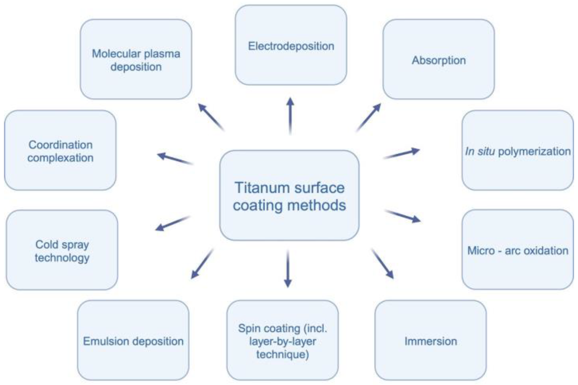
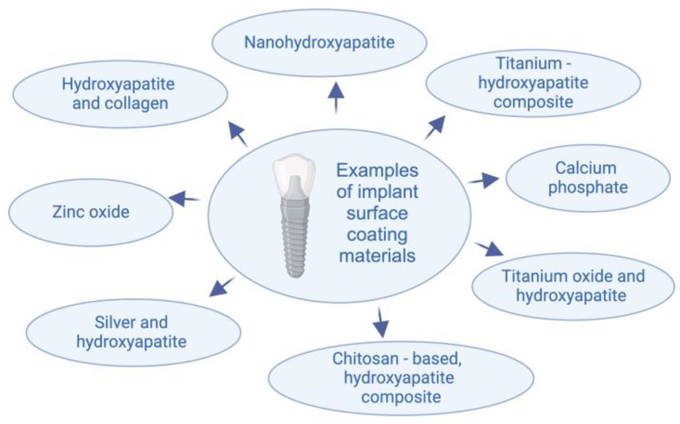
1. Introduction
2. Materials and Methods
2.1. Focused Question
2.2. Protocol
2.3. Eligibility Criteria
2.4. Information Sources, Search Strategy, and Study Selection
2.5. Data Collection and Data Items
2.6. Assessing Risk of Bias in Individual Studies
2.7. Quality Assessment
3. Results
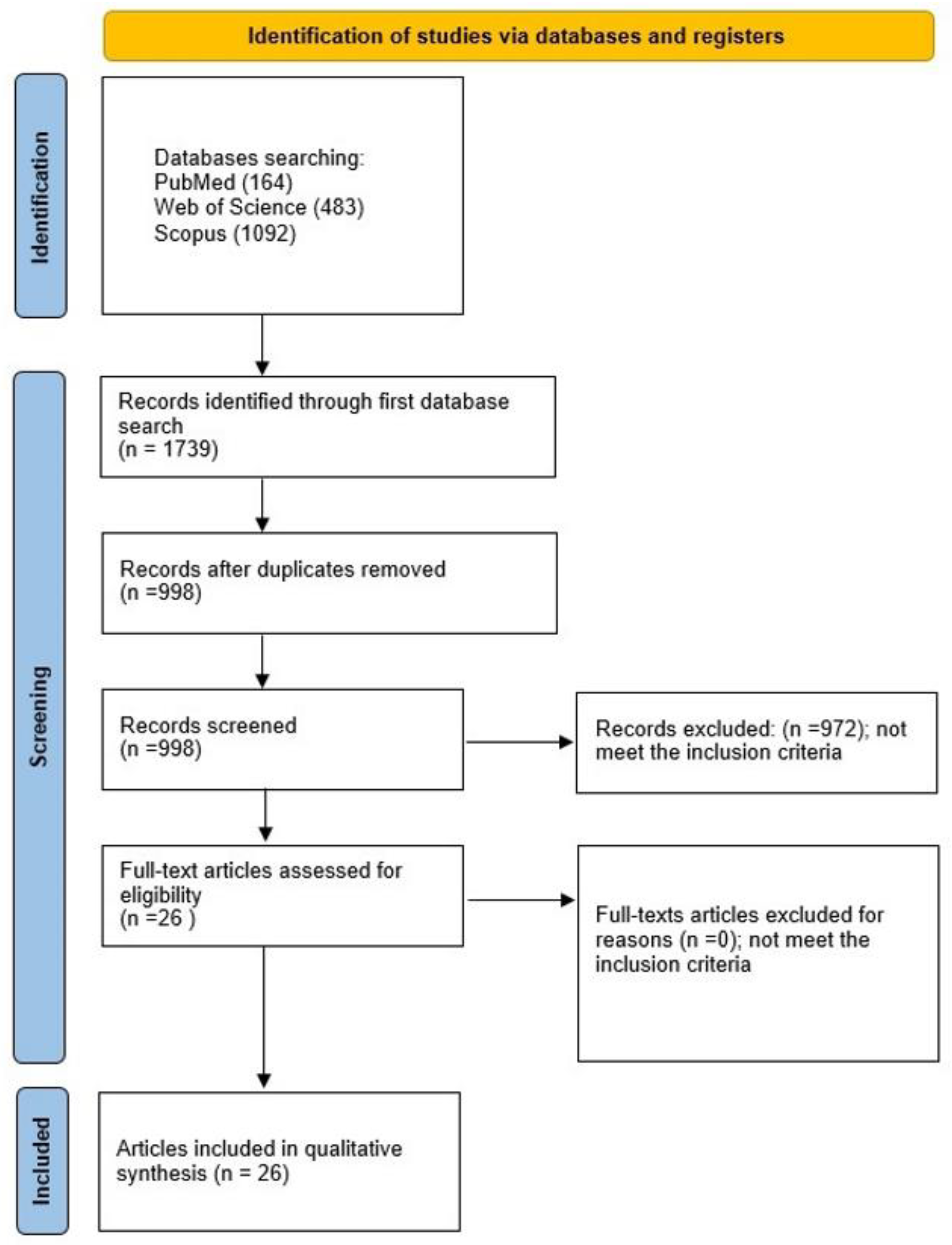
3.1. Study Selection
3.2. General Characteristics of the Included Studies
3.3. Main Study Outcomes
3.4. Quality Assessment
4. Discussion
5. Conclusions
Author Contributions
Funding
Institutional Review Board Statement
Informed Consent Statement
Data Availability Statement
Acknowledgments
Conflicts of Interest
References
- Wu, H.; Chen, X.; Kong, L.; Liu, P. Mechanical and Biological Properties of Titanium and Its Alloys for Oral Implant with Preparation Techniques: A Review. Materials 2023, 16, 6860. [Google Scholar] [CrossRef]
- MacBarb, R.F.; Lindsey, D.P.; Bahney, C.S.; Woods, S.A.; Wolfe, M.L.; Yerby, S.A. Fortifying the Bone-Implant Interface Part 1: An In Vitro Evaluation of 3D-Printed and TPS Porous Surfaces. Int. J. Spine Surg. 2017, 11, 15. [Google Scholar] [CrossRef]
- Morris, H.F.; Ochi, S. Hydroxyapatite-coated implants: A case for their use. J. Oral Maxillofac. Surg. 1998, 56, 1303–1311. [Google Scholar] [CrossRef]
- Matys, J.; Świder, K.; Flieger, R.; Dominiak, M. Assessment of the primary stability of root analog zirconia implants designed using cone beam computed tomography software by means of the Periotest® device: An ex vivo study. A preliminary report. Adv. Clin. Exp. Med. 2017, 26, 803–809. [Google Scholar] [CrossRef] [PubMed]
- Fernandes, C.J.C.; Bezerra, F.; Ferreira, M.R.; Andrade, A.F.C.; Pinto, T.S.; Zambuzzi, W.F. Nano hydroxyapatite-blasted titanium surface creates a biointerface able to govern Src-dependent osteoblast metabolism as prerequisite to ECM remodeling. Colloids Surf. B Biointerfaces 2018, 163, 321–328. [Google Scholar] [CrossRef]
- Pourmollaabbassi, B.; Karbasi, S.; Hashemibeni, B. Evaluate the growth and adhesion of osteoblast cells on nanocomposite scaffold of hydroxyapatite/titania coated with poly hydroxybutyrate. Adv. Biomed. Res. 2016, 5, 156. [Google Scholar] [CrossRef]
- Wang, J.; Chao, Y.; Wan, Q.; Zhu, Z.; Yu, H. Fluoridated hydroxyapatite coatings on titanium obtained by electrochemical deposition. Acta Biomater. 2009, 5, 1798–1807. [Google Scholar] [CrossRef] [PubMed]
- Seyedali Seyedmajidi, M.S. Fluorapatite: A Review of Synthesis, Properties and Medical Applications vs Hydroxyapatite. Iran. J. Mater. Sci. Eng. 2022, 19, 1–20. [Google Scholar] [CrossRef]
- Lin, Q.; Zhou, Y.; Yin, M.; Cheng, Y.; Wei, Y.; Hu, Y.; Liam, X.; Chen, W.; Huang, D. Hydroxyapatite/tannic acid composite coating formation based on Ti modified by TiO2 nanotubes. Colloids Surf. B Biointerfaces 2020, 196, 111304. [Google Scholar] [CrossRef]
- Salaie, R.N.; Besinis, A.; Le, H.; Tredwin, C.; Handy, R.D. The biocompatibility of silver and nanohydroxyapatite coatings on titanium dental implants with human primary osteoblast cells. Mater. Sci. Eng. C 2020, 107, 110210. [Google Scholar] [CrossRef]
- Cavalcanti, J.H.d.L.; Matos, P.C.; de Gouvêa, C.V.D.; Carvalho, W.; Calvo-Guirado, J.L.; Aragoneses, J.M.; Pérez-Díaz, L.; Gehrke, S.A. In vitro assessment of the functional dynamics of titanium with surface coating of hydroxyapatite nanoparticles. Materials 2019, 12, 840. [Google Scholar] [CrossRef]
- Balasundaram, G.; Storey, D.M.; Webster, T.J. Molecular plasma deposition: Biologically inspired nanohydroxyapatite coatings on anodized nanotubular titanium for improving osteoblast density. Int. J. Nanomed. 2015, 10, 527–535. [Google Scholar] [CrossRef]
- Shi, J.; Dong, L.L.; He, F.; Zhao, S.; Yang, G.L. Osteoblast responses to thin nanohydroxyapatite coated on roughened titanium surfaces deposited by an electrochemical process. Oral Surg. Oral Med. Oral Pathol. Oral Radiol. 2013, 116, e311–e316. [Google Scholar] [CrossRef] [PubMed]
- Zhang, L.; Chen, Y.; Rodriguez, J.; Fenniri, H.; Webster, T.J. Associate Professor of Engineering Biomimetic helical rosette nanotubes and nanocrystalline hydroxyapatite coatings on titanium for improving orthopedic implants. Int. J. Nanomed. 2008, 3, 323–334. [Google Scholar]
- Chien, C.Y.; Liu, T.Y.; Kuo, W.H.; Wang, M.J.; Tsai, W.B. Dopamine-assisted immobilization of hydroxyapatite nanoparticles and RGD peptides to improve the osteoconductivity of titanium. J. Biomed. Mater. Res. A 2013, 101A, 740–747. [Google Scholar] [CrossRef] [PubMed]
- Wang, X.; Mei, L.; Jiang, X.; Jin, M.; Xu, Y.; Li, J.; Li, X.; Meng, Z.; Zhu, J.; Wu, F. Hydroxyapatite-Coated Titanium by Micro-Arc Oxidation and Steam-Hydrothermal Treatment Promotes Osseointegration. Front. Bioeng. Biotechnol. 2021, 9, 625877. [Google Scholar] [CrossRef]
- Bezerra, F.; Ferreira, M.R.; Fontes, G.N.; Fernandes, C.J.d.C.; Andia, D.C.; Cruz, N.C.; da Silva, R.A.; Zambuzzi, W.F. Nano hydroxyapatite-blasted titanium surface affects pre-osteoblast morphology by modulating critical intracellular pathways. Biotechnol. Bioeng. 2017, 114, 1888–1898. [Google Scholar] [CrossRef]
- Kreller, T.; Sahm, F.; Bader, R.; Boccaccini, A.R.; Jonitz-Heincke, A.; Detsch, R. Biomimetic calcium phosphate coatings for bioactivation of titanium implant surfaces: Methodological approach and in vitro evaluation of biocompatibility. Materials 2021, 14, 3516. [Google Scholar] [CrossRef] [PubMed]
- Bai, L.; Du, Z.; Du, J.; Yao, W.; Zhang, J.; Weng, Z.; Liu, S.; Zhao, Y.; Liu, Y.; Zhang, X.; et al. A multifaceted coating on titanium dictates osteoimmunomodulation and osteo/angio-genesis towards ameliorative osseointegration. Biomaterials 2018, 162, 154–169. [Google Scholar] [CrossRef]
- Nakazawa, M.; Yamada, M.; Wakamura, M.; Egusa, H.; Sakurai, K. Activation of Osteoblastic Function on Titanium Surface with Titanium-Doped Hydroxyapatite Nanoparticle Coating: An In Vitro Study. Int. J. Oral Maxillofac. Implants 2017, 32, 779–791. [Google Scholar] [CrossRef]
- Koirala, M.B.; Nguyen, T.D.T.; Pitchaimani, A.; Choi, S.O.; Aryal, S. Synthesis and Characterization of Biomimetic Hydroxyapatite Nanoconstruct Using Chemical Gradient across Lipid Bilayer. ACS Appl. Mater. Interfaces 2015, 7, 27382–27390. [Google Scholar] [CrossRef]
- Zhu, X.; Eibl, O.; Scheideler, L.; Geis-Gerstorfer, J. Characterization of nano hydroxyapatite/collagen surfaces and cellular behaviors. J. Biomed. Mater. Res. A 2006, 79, 114–127. [Google Scholar] [CrossRef]
- Wu, Y.; Li, Q.; Xu, B.; Fu, H.; Li, Y. Nano-hydroxyapatite coated TiO2 nanotubes on Ti-19Zr-10Nb-1Fe alloy promotes osteogenesis in vitro. Colloids Surf. B Biointerfaces 2021, 207, 112019. [Google Scholar] [CrossRef]
- Vilardell, A.; Cinca, N.; Garcia-Giralt, N.; Dosta, S.; Cano, I.; Nogués, X.; Guilemany, J. Functionalized coatings by cold spray: An in vitro study of micro- and nanocrystalline hydroxyapatite compared to porous titanium. Mater. Sci. Eng. C 2018, 87, 41–49. [Google Scholar] [CrossRef]
- Chen, W.; Xu, K.; Tao, B.; Dai, L.; Yu, Y.; Mu, C.; Shen, X.; Hu, Y.; He, Y.; Cai, K. Multilayered coating of titanium implants promotes coupled osteogenesis and angiogenesis in vitro and in vivo. Acta Biomater. 2018, 74, 489–504. [Google Scholar] [CrossRef] [PubMed]
- Sato, M.; Aslani, A.; Sambito, M.A.; Kalkhoran, N.M.; Slamovich, E.B.; Webster, T.J. Nanocrystalline hydroxyapatite/titania coatings on titanium improves osteoblast adhesion. J. Biomed. Mater. Res. A 2008, 84, 265–272. [Google Scholar] [CrossRef] [PubMed]
- Abo-Elmahasen, M.M.F.; Dena, A.S.A.; Zhran, M.; Albohy, S.A.H. Do silver/hydroxyapatite and zinc oxide nano-coatings improve inflammation around titanium orthodontic mini-screws? In vitro study. Int. Orthod. 2023, 21, 100711. [Google Scholar] [CrossRef] [PubMed]
- Sato, M.; Sambito, M.A.; Aslani, A.; Kalkhoran, N.M.; Slamovich, E.B.; Webster, T.J. Increased osteoblast functions on undoped and yttrium-doped nanocrystalline hydroxyapatite coatings on titanium. Biomaterials 2006, 27, 2358–2369. [Google Scholar] [CrossRef] [PubMed]
- Man, K.; Brunet, M.Y.; Louth, S.; Robinson, T.E.; Fernandez-Rhodes, M.; Williams, S.; Federici, A.S.; Davies, O.G.; Hoey, D.A.; Cox, S.C. Development of a Bone-Mimetic 3D Printed Ti6Al4V Scaffold to Enhance Osteoblast-Derived Extracellular Vesicles’ Therapeutic Efficacy for Bone Regeneration. Front. Bioeng. Biotechnol. 2021, 9, 757220. [Google Scholar] [CrossRef] [PubMed]
- Yang, W.; Xi, X.; Si, Y.; Huang, S.; Wang, J.; Cai, K. Surface engineering of titanium alloy substrates with multilayered biomimetic hierarchical films to regulate the growth behaviors of osteoblasts. Acta Biomater. 2014, 10, 4525–4536. [Google Scholar] [CrossRef] [PubMed]
- Ma, X.-Y.; Feng, Y.-F.; Wang, T.-S.; Lei, W.; Li, X.; Zhou, D.-P.; Wen, X.-X.; Yu, H.-L.; Xiang, L.-B.; Wang, L. Involvement of FAK-mediated BMP-2/Smad pathway in mediating osteoblast adhesion and differentiation on nano-HA/chitosan composite coated titanium implant under diabetic conditions. Biomater. Sci. 2018, 6, 225–238. [Google Scholar] [CrossRef]
- Matys, J.; Dominiak, M. Assessment of Pain When Uncovering Implants with Er:YAG Laser or Scalpel for Second Stage Surgery. Adv. Clin. Exp. Med. 2016, 25, 1179–1184. [Google Scholar] [CrossRef]
- Ihde, S.; Pałka, Ł.; Janeczek, M.; Kosior, P.; Kiryk, J.; Dobrzyński, M. Bite Reconstruction in the Aesthetic Zone Using One-Piece Bicortical Screw Implants. Case Rep. Dent. 2018, 2018, 1–4. [Google Scholar] [CrossRef]
- Cordeiro, J.M.; Barão, V.A.R. Is there scientific evidence favoring the substitution of commercially pure titanium with titanium alloys for the manufacture of dental implants? Mater. Sci. Eng. C 2017, 71, 1201–1215. [Google Scholar] [CrossRef] [PubMed]
- Kensy, J.; Dobrzyński, M.; Wiench, R.; Grzech-Leśniak, K.; Matys, J. Fibroblasts Adhesion to Laser-Modified Titanium Surfaces-A Systematic Review. Materials 2021, 14, 7305. [Google Scholar] [CrossRef]
- Matys, J.; Flieger, R.; Dominiak, M. Assessment of Temperature Rise and Time of Alveolar Ridge Splitting by Means of Er:YAG Laser, Piezosurgery, and Surgical Saw: An Ex Vivo Study. Biomed. Res. Int. 2016, 2016, 9654975. [Google Scholar] [CrossRef]
- Matys, J.; Flieger, R.; Gedrange, T.; Janowicz, K.; Kempisty, B.; Grzech-Leśniak, K.; Dominiak, M. Effect of 808 nm Semiconductor Laser on the Stability of Orthodontic Micro-Implants: A Split-Mouth Study. Materials 2020, 13, 2265. [Google Scholar] [CrossRef]
- da Silva, R.A.; da Silva Feltran, G.; Ferreira, M.R.; Wood, P.F.; Bezerra, F.; Zambuzzi, W.F. The Impact of Bioactive Surfaces in the Early Stages of Osseointegration: An In Vitro Comparative Study Evaluating the HAnano® and SLActive® Super Hydrophilic Surfaces. Biomed. Res. Int. 2020, 2020, 3026893. [Google Scholar] [CrossRef]
- Janeczek, M.; Szymczyk, P.; Dobrzynski, M.; Parulska, O.; Szymonowicz, M.; Kuropka, P.; Rybak, Z.; Zywicka, B.; Ziolkowski, G.; Marycz, K.; et al. Influence of surface modifications of a nanostructured implant on osseointegration capacity–preliminary in vivo study. RSC Adv. 2018, 8, 15533–15546. [Google Scholar] [CrossRef] [PubMed]
- Pandey, A.; Midha, S.; Sharma, R.K.; Maurya, R.; Nigam, V.K.; Ghosh, S.; Balani, K. Antioxidant and antibacterial hydroxyapatite-based biocomposite for orthopedic applications. Mater. Sci. Eng. C 2018, 88, 13–24. [Google Scholar] [CrossRef] [PubMed]
- Sistanipour, E.; Meshkini, A.; Oveisi, H. Catechin-conjugated mesoporous hydroxyapatite nanoparticle: A novel nano-antioxidant with enhanced osteogenic property. Colloids Surf. B Biointerfaces 2018, 169, 329–339. [Google Scholar] [CrossRef] [PubMed]
- Ran, Q.; Yang, W.; Hu, Y.; Shen, X.; Yu, Y.; Xiang, Y.; Cai, K. Osteogenesis of 3D printed porous Ti6Al4V implants with different pore sizes. J. Mech. Behav. Biomed. Mater. 2018, 84, 1–11. [Google Scholar] [CrossRef] [PubMed]
- Lotz, E.M.; Berger, M.B.; Schwartz, Z.; Boyan, B.D. Regulation of osteoclasts by osteoblast lineage cells depends on titanium implant surface properties. Acta Biomater. 2018, 68, 296–307. [Google Scholar] [CrossRef] [PubMed]
- Costa, D.O.; Prowse, P.D.; Chrones, T.; Sims, S.M.; Hamilton, D.W.; Rizkalla, A.S.; Dixon, S.J. The differential regulation of osteoblast and osteoclast activity by surface topography of hydroxyapatite coatings. Biomaterials 2013, 34, 7215–7226. [Google Scholar] [CrossRef] [PubMed]
- Rubert, M.; Vetsch, J.R.; Lehtoviita, I.; Sommer, M.; Zhao, F.; Studart, A.R.; Müller, R.; Hofmann, S. Scaffold Pore Geometry Guides Gene Regulation and Bone-like Tissue Formation in Dynamic Cultures. Tissue Eng. Part A 2021, 27, 1192–1204. [Google Scholar] [CrossRef] [PubMed]
- Brennan, C.M.; Eichholz, K.F.; Hoey, D.A. The effect of pore size within fibrous scaffolds fabricated using melt electrowriting on human bone marrow stem cell osteogenesis. Biomed. Mater. 2019, 14, 065016. [Google Scholar] [CrossRef]
- Zhang, Y.; Sun, N.; Zhu, M.; Qiu, Q.; Zhao, P.; Zheng, C.; Bai, Q.; Zeng, Q.; Lu, T. The contribution of pore size and porosity of 3D printed porous titanium scaffolds to osteogenesis. Biomater. Adv. 2022, 133, 112651. [Google Scholar] [CrossRef]
- Deng, F.; Liu, L.; Li, Z.; Liu, J. 3D printed Ti6Al4V bone scaffolds with different pore structure effects on bone ingrowth. J. Biol. Eng. 2021, 15, 4. [Google Scholar] [CrossRef]
- Chien, C.-Y.; Tsai, W.-B. Poly(dopamine)-Assisted Immobilization of Arg-Gly-Asp Peptides, Hydroxyapatite, and Bone Morphogenic Protein-2 on Titanium to Improve the Osteogenesis of Bone Marrow Stem Cells. ACS Appl. Mater. Interfaces 2013, 5, 6975–6983. [Google Scholar] [CrossRef]
- Schliephake, H.; Scharnweber, D.; Dard, M.; Rö, S.; Sewing, A.; Hüttmann, C. Biological performance of biomimetic calcium phosphate coating of titanium implants in the dog mandible. J. Biomed. Mater. Res. A 2003, 64A, 225–234. [Google Scholar] [CrossRef]
- Urquia Edreira, E.R.; Wolke, J.G.C.; Aldosari, A.A.; Al-Johany, S.S.; Anil, S.; Jansen, J.A.; van den Beucken, J.J.J.P. Effects of calcium phosphate composition in sputter coatings on in vitro and in vivo performance. J. Biomed. Mater. Res. A 2015, 103, 300–310. [Google Scholar] [CrossRef] [PubMed]
- Wang, M.; Wang, C.; Zhang, Y.; Lin, Y. Controlled release of dopamine coatings on titanium bidirectionally regulate osteoclastic and osteogenic response behaviors. Mater. Sci. Eng. C 2021, 129, 112376. [Google Scholar] [CrossRef] [PubMed]
- García-Honduvilla, N.; Coca, A.; Ortega, M.A.; Trejo, C.; Román, J.; Peña, J.; Cabañas, M.V.; Regi, M.V.; Buján, J. Improved connective integration of a degradable 3D-nano-apatite/agarose scaffold subcutaneously implanted in a rat model. J. Biomater. Appl. 2018, 33, 741–752. [Google Scholar] [CrossRef]
- Herman, K.; Wujczyk, M.; Dobrzynski, M.; Diakowska, D.; Wiglusz, K.; Wiglusz, R.J. In Vitro Assessment of Long-Term Fluoride Ion Release from Nanofluorapatite. Materials 2021, 14, 3747. [Google Scholar] [CrossRef]
- Zhang, C.; Duan, G.; Li, J.; Xiao, D.; Shi, F.; Duan, K.; Guo, T.; Fan, X.; Weng, J. Hydrothermal growth of biomimetic calcium phosphate network structure on titanium surface for biomedical application. Ceram. Int. 2023, 49, 16652–16660. [Google Scholar] [CrossRef]

| Study | Aim of the Study | Material and Methods | Results | Conclusions |
|---|---|---|---|---|
| Fernandes et al. [5] | Creation of a biointerface, which can control Src-dependent osteoblast metabolism as a pre-requisite to ECM remodeling. | Three titanium discs were investigated, differing in surface properties identified as Machined (Mc; control), Dual Acid-Etched (DAE), and acid-etched nano-HAp-blasted (nHAp). A mouse pre-osteoblastic cell type, was utilized for this study. | Increased MMP activities in response to both DAE and nHAp. Significant increase in Src gene expression and in Integrin, FAK, and Src expression. | Molecular features that are associated with the pre-osteoblast interaction with distinct surface textures can direct Src-dependent osteoblast metabolism as a requirement for extracellular matrix remodeling. |
| Pourmollaabbassi et al. [6] | Generation of bioartificial bone tissues to overcome issues related to bone loss. Donor site morbidity and size limitations. | HAp powder was derived from bovine bone through thermal analysis at 900 °C. Subsequently, porous HAp with a 50% weight percentage was created using the polyurethane sponge replication method. The cellular scaffolds were divided into four groups for comparative analysis of their behaviour, specifically focusing on osteoblasts. Viability and attachment on the scaffold surfaces were assessed through Methylthiazole tetrazolium (MTT) and Trypan blue analyses as part of the phase studies. | The MTT analysis results of four scaffold groups revealed that Titanium oxide (TiO2) did not impact cell growth alone, and HAp was the primary factor for cell growth and osteoblast adhesion on the scaffold. Coating with poly-3-hydroxybutyrate helped retain the factors and position the osteoblasts in the pore. | The correlation between HAp and TiO2 is likely to enhance osteoblast adhesion and promote cell growth on the scaffold surface. |
| Qiaoxia] et al. [9] | Investigation of both the total antioxidant activity and osteoblast behaviour. | Ti0 foils served as substrates for growing TiO2 nanotube arrays film. The annealed Ti-TiO2 samples underwent immersion in a mixed aqueous solution of TA and CaCl2, denoted as the TA-CaCl2 solution. The morphologies of all coatings were observed using scanning electron microscopy. To assess the viability of mouse pre-osteoblasts on the surfaces of different samples, staining with Calcein-AM and EthD-1 was performed. For proliferation and adhesion experiments, cells (2 × 10⁴/mL) were co-cultured with sterilized samples in 24-well plates. After culturing for 1-, 4- and 7-day(s), cell proliferation was investigated using a Cell Counting Kit-8 (CCK-8) assay. | An antioxidant known as TA enhanced the antioxidative activity of Ti0 implants. | The composite coating exhibits antioxidant activity, with significant potential for scavenging ROS at the bone–implant interface. HAp/TA composite coating has superior cytocompatibility in terms of proliferation spreading and adhesion compared to the pure Ti4+ substrates. |
| Salaie et al. [10] | Improvement of the biocompatibility of titanium dental implants coated with Ag0 NPs and with HAp applied to their surface. | Titanium discs (Ti6Al4V) are suitably covered by: -Ag0 NPs, -Ag0 NPs plus nHAp, -Ag0 NPs plus mHAp. The coatings were examined for stability and tested for biocompatibility with primary human osteoblasts over a seven-day period. | With the addition of HAp, osteoblasts adhered well and maintained a normal morphology. Lactate dehydrogenase (LDH) leakage was negligible, while alkaline phosphatase (ALP) activity was observed. | The biocompatibility of implants coated with Ag+ nHAp was higher than that of those coated with Ag+ mHAp or Ag0 NPs alone. This indicates that the former may offer clinical advantages. |
| Cavalcanti et al. [11] | Presentation of valuable insights on implant surface evaluation and demonstration of potential effectiveness of nanostructured material surfaces in, focal cell adhesion, promoting cell viability and bone mineralization in vitro. | Mesenchymal stem cells were utilized for comparative analyses of two different implant topographies, focusing on their functional interaction with pre-osteoblasts directly seeded onto the implants. In vitro assay analysis was used to analyze the functionality of nanostructured implant surfaces. The machined surface of the titanium implant served as a control. It was compared with a surface implant activated by nanoparticle HAp. | Cell culture on the nano-group surface resulted in greater cell adhesion and viability of cultured osteoblasts than in the control group. Scanning electron microscope (SEM) images showed a stable interaction as evidenced by the development of focal cell adhesion. | Nano group was an excellent scaffold for bone–implant integration, together with positive mineralization assays. |
| Balasundaram et al. [12] | The aim of this in vitro investigation was to produce nHAp and apply it onto titanium (Ti0) using molecular plasma deposition (MPD). | nHAp was synthesized via a wet chemical process followed by hydrothermal treatment. Subsequently, nHAp coatings were processed at 500 °C, and micro-sized HAp was processed at 900 °C. SEM, atomic force microscopy, and X-ray diffraction were employed to characterize the coatings before and after sintering. | The results show that the post-MPD heat treatment up to 500 °C successfully restored the structural and topographical integrity of the nHAp. The nHAp-coated substrates supported a greater number of adherent cells compared to both the mHAp-coated and uncoated substrates. | MPD is an innovative method that can be utilized to deposit nHAp on anodized Ti0, resulting in higher osteoblast counts relative to untreated Ti0 and mHAp-coated Ti4+ substrates. MPD is inexpensive, fast, and effective. |
| MacBard et al. [2] | In vitro comparison of human osteoblasts proliferation on additive manufactured (AM) trabecular-like titanium implant surfaces and traditionally machined base material with titanium plasma spray (TPS)-coated surfaces. | TPS-coated groups: Ti6AL4V ELI machined into discs with diameter of 15 mm and 1.25 mm thick with a 0.75 mm thick TPS-coating on their surfaces. AM groups: discs with diameter of 15 mm and 2.0 mm thick were designed with a solid base (1.25 mm thick and a porous surface layer (0.75 mm thick) and printed by using Ti6Al4V ELI particles through electron beam melting technology. In groups that have HAp coating, a ~20 nm thick nanocrystalline HAp layer was applied on disc surfaces. Number of proliferated cells on discs was measured on day 2, 7, 14, and 21. | Surface of TPS-coated discs: Mean porosity: 59%. Mean pore size: 141.7. Tissue interface height: 743 μm. Estimated average available surface area: 649.39 mm2. Estimated surface contact angle: 105.2°. Surface of AM discs: Mean porosity of 60%. Mean pore size: 290.6 μm. Tissue interface height: 1051 μm. Estimated average available surface area: 1153.67 mm2. Estimated surface contact angle: 70.1°. Production of calcium on AM discs was 48% higher than on TPS discs. | Additive manufactured trabecular-like titanium implant surfaces encourage faster cell proliferation and higher calcium production than traditionally machined base material with titanium plasma spray coated surfaces. Moreover, nanocrystalline HAp did not enhance osteoblasts proliferation on titanium surfaces. |
| Shi et al. [13] | In vitro evaluation of osteoblasts proliferation and differentiation on porous titanium implant surfaces coated with nano HAp. | Plates of 10 mm × 10 mm × 1 mm were polished, sandblasted, washed, treated with HF and HNO3 solution, then treated with HCl and H2SO4 solution. Titanium plates were used as a cathode; platinum plates were used as an anode. Electrolytes were prepared by dissolution of Ca(NO3)2 (0.6 mmol/L) and NH4H2PO4 (0.36 mmol/L) in distilled water to a Ca/P ratio of 1.67. Pre-osteoblasts cells were seeded at a density of 1 × 10⁵ on titanium plates. | Cell proliferation was significantly higher than that in the control group. HAp-coated titanium enhanced alkaline phosphatase activity. Osteocalcin production released into cell culture medium was higher in cells that were grown on HAp surfaces than in those grown on control samples without Hap. | Deposition of nano HAp coating by the electrochemical process improved osteoblasts proliferation and differentiation. |
| Zhang et al. [14] | Evaluation of properties of helical rosette nanotubes (HRN) and nanocrystalline hydroxyapatite used as coatings on titanium implant surfaces. | Titanium plates 1 cm × 1 cm × 0.2 cm 250 mg of each type of nano HAp (small, middle, large sizes of HAp grains) was mixed with 5 mL of 70% ethanol and sonicated for 20 min. Then, 5 mL of 0.001 mg/mL HRN-K1 solution was added into all samples. Absorption of HRN-K1 and nano HA on titanium surfaces for 45 min. Group without HAp: 0.001 mg/mL HRN-K1 only was used as a titanium coating. Control group: uncoated titanium plates. | Better adhesion of osteoblasts to nanocrystalline HAp and HRN titanium coatings (especially small and middle grain sizes) when compared with uncoated titanium sample. small grains increased adhesion of osteoblast by 29.3%, middle grains by 36.3%. | Nanocrystalline HAp has been shown to have high affinity with HRN. Nanosized HAp and HRN used as a titanium coating promotes adhesion of osteoblasts. |
| Chien et al. [15] | Development of the procedure for immobilization of Hap nanoparticles and RGD peptides on titanium to improve the osteoconductivity of orthopedic and dental implants. | 3-hydroxytyramine hydrochloride was dissolved in 10 mM of Tris buffer to 2 mg/mL. HAp NPs were suspended in 0.02% polyacrylic acid solution. Equal volume of both solutions was mixed and then immediately added onto titanium discs and then incubated for 20 min. Immobilization of RGD: dopamine/HAp coated titanium discs were incubated with 1 mg/mL of RGD solution in phosphate-buffered saline for 24 h. | After 14 days of culture, small (~150 μm)-cell aggregates were found on titanium disc samples, but not on those coated with dopamine only. On the substrate deposited with dopamine/HAp, large (800 μm)-cell aggregates were formed. Dopamine deposition increased calcium amount from 31.6 nmol to 144.3 nmol. On the dopamine/HAp substrate calcium deposition increased to 517.6 nmol. | Co-deposition of dopamine on hydroxyapatite significantly enhances osteoblast mineralization, proliferation and adhesion. RGD peptides immobilized to dopamine/HAp-coated titanium promote osteoblasts adhesion and osteogenic differentiation. |
| Wang et al. [16] | Investigation of HAp coating of titanium by micro-arc oxidation (MAO). | Titanium surface was modified by MAO, and the sample was named Ti-M. Later, the sample was treated with SHT and placed in a Teflon reactor and then autoclaved at 250 degrees for 1, 4, and 8 h and named as Ti– M–H1, Ti–M–H4, and Ti–M–H8, respectively. | After 0.5 h of incubation the number of cells adhesive on Ti–M– H was significantly increased than that on Ti–M. After longer incubation for 1 h and 4 h, only Ti–M–H1 had a higher number of cells, while Ti–M, Ti–M–H4, and Ti–M–H8 had no significant differences. | MAO- and SHT-treated titanium promote osteogenesis and angiogenesis. It stimulates macrophages to regulate an immune response. |
| Bezerra et al. [17] | Evaluation of the behaviour of pre-osteoblast on nano HA-blasted titanium surface by examination of intracellular signal pathways. | Three different titanium surfaces were prepared: -machined (control group) -dual acid-etched (DEA) -acid-etched nHAp-blasted (nHAp) Mouse pre-osteoblastic cells were cultured and seeded at the density of 5 × 10⁴ cells/disc on different titanium surfaces. | Ras-Raf-Mek- p42/44 mapk signaling was examined and it was shown to be involved in response to titanium surfaces. Modified surfaces (DAE and nHAp) enhanced an increase in Raf (protein involvement with other signaling pathway). All 3 titanium surfaces stimulated ALP activity in osteoblasts cultured on them. | Different titanium surfaces enhanced crucial intracellular signaling pathways that are responsible for cell proliferation and adhesion. Nanosized HAp-blasted titanium surface promotes pre-osteoblast proliferation by activating crucial signaling pathways. |
| Kreller et al. [18] | In vitro evaluation of modifying titanium surface with calcium phosphate coating. | Biomimetic CaP coatings were generated by immersion in a modified BCP coating solution at 37 °C for 14 days. Chemically pre-treated titanium plates were soaked in a 1 M of CaCl2 solution at 37 °C for 24 h and then immersed in BCP coating solution. Substrates were transferred to BCPx1.5 for 14 days. BCP coating solutions were exchanged to new ones every second day. Control group: chemically untreated titanium surfaces without immersion in BCP coating solution. | Altered surface topographies were obtained due to titanium surface functionalization. | Functionalization of titanium surface with calcium phosphate coatings promotes its osteoconductive properties. |
| Bai et al. [19] | To describe a microporous TiO2 coating decorated with HAp nanoparticles that is generated by micro-arc oxidation of pure titanium and followed annealing | The specimens of a pure titanium were subjected to a micro-arc oxidation fabricating micro/nano-structured surfaces. The process was followed by annealing at different temperatures (250 °C, 450 °C, or 650 °C). Materials were evaluated in terms of osteogenic activity of osteoblasts, angiogenic activity of endothelial cells, macrophage response. | A coating produced with micro-arc oxidation with an annealing temperature of 650 °C exhibits numerous favourable physicochemical properties, such as hybrid micro-nano morphology, superhydrophilicity, and highly crystalline HAp nanoparticles. It supports proliferation and differentiation of osteoblasts and endothelial cells and also inhibits the inflammatory response of macrophages. | The coating annealed at 650 °C exhibited favourable physicochemical properties that synergistically regulated osteoimmunomodulation, osteo/angio-genesis and cross communications amongst immunoregulation, osteogenesis and angiogenesis to significantly enhance osseointegration. |
| Nakazawa et al. [20] | To assess the physicochemical properties of machined or micro-grooved titanium surfaces which have been coated with titanium-doped HAp nanoparticles and to investigate the subsequent impact on the function of osteoblasts cultured on these surfaces. | Titanium discs with commercially available surface topographies, such as machined or sandblasted, large-grit and acid-etched surfaces, were coated with titanium-doped hydroxyapatite. The discs with original or modified surfaces were evaluated for topography, wettability, and chemical composition. Rat femoral osteoblasts were cultured on the discs and evaluated for proliferation and differentiation. | Titanium hydroxyapatite coatings changed from hydrophobicity to hydrophilicity on both machined and acid-etched surfaces. There was no change in inherent surface topographies on both types of specimens. At day 4, osteoblastic proliferative activity was increased by a Ti0-HAp coating. | Titanium-doped hydroxyapatite coating enhances extracellular matrix formation on smooth and microrough titanium surfaces by increasing osteoblastic proliferative activity without compromising differentiation through hydrophilic and chemical functionalization. |
| Koirala et al. [21] | To synthesize and characterize biomimetic nanosized HAp, using a double emulsion technique combined with a chemical gradient across lipid bilayer for surface modification of a titanium implant. | The synthesized hydroxyapatite was evaluated by: dynamic light scattering, X-ray diffraction, transmission electron microscopy, Fourier-transform infrared spectroscopy, biocompatibility, in vitro proliferation efficacy using Normal Human Osteoblasts. | Nanosized HAp exhibited a diameter of approximately 200 nm with high aqueous stability. The synthesized hydroxyapatite nanoparticles exhibited excellent biocompatibility and provided the cellular proliferative environment. The synthesized material exhibited hierarchical nanostructures inside, filled with nanocrystallites of hydroxyapatite and were highly homogeneous. | Synthesized phospholipid bilayer-coated with hydroxyapatite nanosized construct provides a suitable environment for cell adhesion, particularly important for bone implants. The long-term sustainability of implants would be effectively supported by the incorporation of nanosized hydroxyapatite on the titanium substrate. |
| Zhu et al. [22] | To characterize synthesized nanocrystalline HAp and evaluate in vitro studies on nano HAp, nano HAp/collagen, and titanium surfaces. | In vitro studies on nanosized HAp, nano-HAp/collagen, and titanium surfaces were performed. Specimens were subjected to: transmission electron microscope evaluation, contact angle assessment, surface roughness evaluation, cell attachment and spreading on The SaOS-2 human osteoblast-like cell line. | Nanosized HAp/collagen was coated homogeneously on pure titanium or porous anodic oxides and exhibited higher wettability. Nano-HAp reduced cell attachment and spreading, and the combination of nano-HAp and collagen showed improved cell spreading of osteoblasts compared to nano-HAp. | To elucidate the reactions at the interface between surfaces and bone, cell responses to nano HAp, nano HAp/collagen, pure and porous titanium surfaces are considered crucial. |
| Wu et al. [1] | To assess osteogenesis on nano-hydroxyapatite coated TiO2 nanotubes on Ti-19Zr-10Nb-1Fe alloy in in vitro tests. | Samples of nano Hap-coated TiO2 nanotubes on Ti-19Zr-10Nb-1Fe alloy were characterized by SEM, FT-IR, XRD and assessed in terms of: - Mouse pre-osteoblasts proliferation and adhesion - Alkaline phosphatase and Osteocalcin content assay | TiO2 nanotubes with 87 ± 21 nm were successfully formed on Ti-19Zr-10Nb-1Fe alloy, and nanosized HAp coating was synthesized. The cells’ growth on the surface modified with hydroxyapatite was higher (30% or more) than the unmodified Ti-19Zr-10Nb-1Fe alloy in 7 days, as well as the expression of Alkaline phosphatase and Osteocalcin. | The results suggest that the introduction of nano-HAp improves the differentiation of osteoblasts and the production of local factors, as well as indicating the potential for improved osseointegration of implants. |
| Vilardell et al. [24] | To evaluate three different surface treatments on a Ti6Al4V alloy for possible application in cementless joint prosthesis in in vitro tests. | Cold Spray technology was used for deposition in all the evaluated methods of surface modifications: (i) an as-sprayed highly rough titanium, followed by the deposition of a thin hydroxyapatite layer with (ii) microcrystalline or (iii) nanocrystalline structure. Primary human osteoblasts, extracted from knee were used to assess cell viability by MTS and LIVE/DEAD assays. Alkaline phosphatase (ALP) was used for cell differentiation test and Phalloidin staining for cell morphology and quantification. All tests were performed at 1, 7, and 14 days of cell culture. | The titanium and HAp surfaces showed different cell morphologies. A higher cell viability was observed on titanium coating. From 7 days of culture, cells on HAp showed good attachment to surfaces and greatly increased proliferation, mostly on nanocrystals, achieving similar cell viability levels to titanium coatings. ALP levels were significantly higher on titanium. | The best cell function results were obtained on titanium coatings. Microcrystalline hydroxyapatite showed the worst cell parameters. Nevertheless, the results suggest that nanocrystalline hydroxyapatite layers could provide promising results for faster cell proliferation once cells have adhered to the surface. |
| Chen et al. [25] | Investigation of the interaction of osteogenesis and angiogenesis during bone healing in in vitro and in vivo tests using surface-modified titanium substrates with a multi-layered structure composed of chitosan–catechol, gelatine and hydroxyapatite nanofibers as a platform. | The adhesion, morphology, and migration of adipose-derived mesenchymal stem cells (Ad-MSCs) and human umbilical vein endothelial cells (HUVECs) grown on different titanium substrates were evaluated using combined techniques of Transwell co-culture, wound healing assay, enzyme-linked immunosorbent assay (ELISA), quantitative real-time polymerase chain reaction (qRT-PCR), Western blotting and immunohistochemical staining. | Multi-layered titanium substrates directly regulated cellular functions of Ad MSCs and angiogenic HUVEC, mediating intercellular communication through paracrine effects in vitro. The in vivo results showed that the altered microenvironment induced by surface-modified Ti implants promoted the adhesion, recruitment, and proliferation of MSCs and facilitated coupled osteogenesis and angiogenesis in bone healing. | It was proved that multi-layer film-coated titanium substrates positively mediated cellular biological function in vitro and improved bone healing in vivo. |
| Sato et al. [26] | To improve orthopedic implant performance, the objective of this in vitro study was to synthesize nanocrystalline hydroxyapatite (HA) powders to coat titanium. | HAp was synthesized through a wet chemical process. The precipitated powders were either sintered at 1100 °C for 1 h to produce UltraCap HAp (or microcrystalline size HAp) or were treated hydrothermally at 200 °C for 20 h to produce nanocrystalline HAp. The original HAp particles were characterized using X-ray diffraction (XRD), inductively coupled plasma–atomic emission spectroscopy (ICP–AES), BET, a laser particle size analyser and scanning electron microscopy (SEM). These powders were then deposited onto titanium by a novel room-temperature process, called IonTiteTM. | Results showed increased osteoblast adhesion on the nanocrystalline HAp IonTiteTM coatings compared to traditionally used plasma-sprayed HAp coatings. Results also demonstrated greater amounts of calcium deposition by osteoblasts cultured on Y-doped nanocrystalline HAp coatings compared to either UltraCap IonTiteTM coatings or plasma-sprayed HAp coatings. | These results encourage further studies on nanocrystalline IonTiteTM HAp coatings on titanium for improved orthopaedic applications. |
| Fathy Abo-Elmahasen et al. [27] | Evaluation of microbiological activities of the deposited nanomaterials (silver/hydroxyapatite nanoparticles (Ag0/HAp NPs) and zinc oxide nanoparticles (ZnO NPs)) in terms of microbial inhibition. | Ag/HAp NPs and ZnO NPs were built up onto the surface of titanium OMSs by electrochemical deposition. This electrochemical deposition was performed on 50 orthodontic mini screws. In addition, the microbiological activities of the deposited nanomaterials were explored in vitro in terms of microbial inhibition. Furthermore, the cytotoxicity and cytocompatibility were tested on four types of cells, namely, fibroblasts, osteocytes, osteoblasts, and oral epithelial cells. | ZnO NPs coated OMS had the highest antimicrobial activity than Ag0/HAp coated OMS against Gram-positive, Gram-negative, and fungal strains. After incubation, the decrease in the number of bacterial colonies was significant with ZnO and Ag0/HAp nanoparticles. ZnO NPs-coated OMSs showed a better cytocompatibility with oral epithelium, bone cells, and fibroblasts compared to Ag0/HAp NPs. | Suggested nanocoating is a promising strategy to overcome the development of an inflammatory zone around the fixed OMSs. |
| Sato et al. [28] | The objective of this in vitro study was to produce nanophase (i.e., materials with grain size less than 100 nm) HAp/titania coatings on titanium. | Nanocrystalline HAp powders were synthesized through wet chemistry and hydrothermal treatments at 200 °C. Nanocrystalline titania powders obtained commercially were mixed with the nanocrystalline HAp powders at various weight ratios. The mixed powders were then deposited on titanium utilizing a room-temperature coating process called IonTiteTM. | The number of osteoblasts adherent on the nanotitania/HAp composite were significantly greater compared with single-phase nanotitania coatings, currently used plasma-sprayed HAp coatings, and uncoated titanium. | These findings suggest that nanosized titania/HAp coatings on titanium should be further studied for improved orthopedic applications. |
| Man et al. [29] | Investigation of the potential of 3D-printed titanium scaffolds coated with hydroxyapatite to promote the therapeutic efficacy of osteoblast derived Extracellular Vesicles (EVs). | Ti6Al4V titanium scaffolds with different pore sizes (500 and 1000 μm) and shapes (square and triangle) were fabricated by selective laser melting. A bone-mimetic nanosized needle hydroxyapatite (nnHAp) coating was then applied. EVs were procured from scaffold-cultured osteoblasts over 2 weeks and vesicle concentration was determined using the CD63 ELISA. Osteogenic differentiation of human bone marrow stromal cells (hBMSCs) following treatment with primed EVs was evaluated by assessing alkaline phosphatase activity, collagen production and calcium deposition. | Triangle pore scaffolds significantly increased osteoblast mineralization (1.5-fold) when compared to square architectures. Osteoblast-derived EVs isolated from triangular pore scaffolds significantly increased hBMSCs mineralization when compared to EVs acquired from square pore scaffolds (1.7-fold) and 2D culture (2.2-fold) (p ≤ 0.001). | These findings demonstrate the potential of harnessing bone-mimetic culture platforms to enhance the production of pro-regenerative EVs as an acellular tool for bone repair. |
| Yang et al. [30] | Fabrication of a biomimetic lamellar structure on Ti6Al7Nb discs with HAp nanofibers as the intercalated materials and with the Gel and Chi as polyelectrolytes via spin-assisted Lbl assembly technique. | A hierarchical structure with osteoinduction potential was fabricated on titanium alloy (Ti6Al7Nb) substrates via a spin-assisted layer-by-layer assembly technique, with HAp nanofibers as the intercalated materials and gelatine and chitosan as the polycation and polyanion, respectively. The as-synthesized hydroxyapatite nanofibers were characterized using scanning electron microscopy (SEM), transmission electron microscopy, Fourier transform infrared spectroscopy and X-ray diffraction. | Osteoblasts cultured on the hierarchical structure deposited Ti0 alloy substrates displayed significantly higher cell viability (p < 0.01) and better adhesion, a higher production level of alkaline phosphatase, mineralization, genes expressions of osteocalcin and osteopontin (p < 0.01 or p < 0.05) compared to those of native Ti6Al7Nb substrates after culture for 4, 7 or 14 days. | The lamellar structure was beneficial for the biological functions of osteoblasts, establishing the basis for osseointegration of a titanium alloy implant. |
| Ma et al. [31] | Chitosan (CS)-based HAp composites have emerged as a novel strategy for promoting bone regeneration. | Here, nanophase HAp/CS composite-coated porous titanium implants (nCT) were fabricated, and their biological behaviour under diabetic conditions was investigated. | Rat osteoblasts were cultured on bare titanium implants (Ti0) and nCT, and subjected to normal serum (NS), diabetic serum (DS), DS + NAC (a potent ROS inhibitor), and DS + cytochalasin D (an actin polymerization inhibitor). | The study illustrated that the reactivation of the FAK-BMP-2/Smad pathway played a crucial role in enhancing osteoblast adhesion and differentiation when utilizing a nano-HAp/CS composite coating. |
| Authors | Titanium Surface Used in Study | Characteristics of Nanohydroxyapatite and Layer Deposition Technique | Sample Sterilization | Utilized Cell Line | Type of Study | Biological Activity |
|---|---|---|---|---|---|---|
| Fernandes et al. [5] | Machined titanium discs (control), dual acid-etched disc, acid-etched and nanoHAp-blasted discs. | Pro-mimic HAp nano-method (10 nm particles) | Gamma ray sterilization | MC3T3-E1, mouse pre-osteoblastic cells | In vitro | Surface topography affects Src-dependent osteoblast metabolism |
| Pourmollaabbassi et al. [6] | Nanocomposite scaffold of hydroxyapatite/titanium coated with poly hydroxybutyrate | Hydroxyapatite powder was prepared from bovine bone through thermal technique. Subsequently, a porous HAp material was produced using the polyurethane sponge replication method and coated with poly hydroxybutyrate. | The sample was treated with ethanol under ultraviolet radiation. | The human osteoblast cell lines, Saos-II | In vitro | Higher porosity and large pore sizes of HAp/TiO2 scaffold coated with P3HB are preferable for migration and proliferation of osteoblasts. |
| Qiaoxia et al. [9] | Titanium foils coated by TiO2 by electrochemical anodization. | Hydroxyapatite/tannic acid composite, immersion | Performed (no additional data) | The MC3T3-E1 cells | In vitro | HAp/TA coating exhibits antioxidant properties important in eliminating reactive oxygen species in bone/implant interfaces. |
| Salaie et al. [10] | Polished medical grade titanium alloy (Ti6Al4V) electroplated with Ag NP and HAp coating. | Sintering of the HAp dispersion on the titanium surface. | Gamma irradiation | Human osteoblast cells (HOb) | In vitro | No cytotoxicity in 7 days of cellular culture. |
| Cavalcanti et al. [11] | Smooth titanium disc and acid-etched with titanium disc with deposited HAp layer. | Pro-mimic nanosized HAp method. | Gamma radiation sterilization | MC3T3-E1 (ATCC 7594) murine osteoblastic cells | In vitro | Enhanced osteoblastic differentiation after exposure to HAp. |
| Balasundaram et al. [12] | Anodized Ti0 foil with and without nano HAp coating. | Molecular plasma deposition of nano HA on anodized Ti0. | 250 nm UV light exposure | CRL-11372 human osteoblasts | In vitro | Nanosized HAp-coated samples increased proliferation of osteoblasts in comparison with untreated samples and with micron-sized HA coating. |
| MacBard et al. [2] | Machined Ti6AL4V, TPS coated and with layer of nano HAp. | Dip, spin, and heat treatment technique. | Rinsing in ethanol | hFOB 1.19 fetal osteoblasts-like cells | In vitro | Nanocrystalline HAp coating on different Ti0 surfaces provides almost no advantage in generating an osteogenic environment. |
| Shi et al. [12] | Polished and acid-etched titanium plates covered with HAp coating | Electrolytic deposition of HAp coating. | No data | Murine preosteoblastic cell line (MC3T-E1) | In vitro | HAp coating enhanced both proliferation and differentiation of osteoblasts. |
| Zhang et al. [14] | Titanium coated with HRN and HAp. | Coating by means of adsorption. | No data | CRL-11372 human fetal osteoblasts | In vitro | HAp/HRN co-coating enhanced osteoblast adhesion |
| Chien et al. [15] | Ti-6A1-4V titanium alloy coated with HAp/dopamine | Dopamine-assisted deposition of HAp. | Sterilized in 70% ethanol | MG63 osteoblast-like cells | In vitro | HAp coating via polydopamine increased osteoconductivity of the Ti4+ substrate. Possibility of incorporation of the RGD peptide. |
| Wang et al. [16] | Ti0 foil with coating for in vitro evaluation and Ti0 rods with coating for in vivo evaluation | Micro-arc oxidation and steam-hydrothermal treatment to deposit HAp. | No data | MC3T-E1 preosteoblasts. | In vitro and in vivo | Coating induces osteo- and angiogenesis as well as macrophages’ polarization. |
| Bezerra et al. [17] | Titanium discs, dual acid-etched Ti0 discs and dual etched Ti0 discs with HAp coating. | Pro-mimic HAp nano-method. | Gamma irradiation. | MC3T3-E1 mouse preosteoblasts. | In vitro | Upregulation of RUNX2 and ALP biomarkers and promotion of osteoblast differentiation by nHA coating. |
| Kreller et al. [18] | Ti6Al4V plates coated with biomimetic CaP (HAp). | Immersion BCP technique. | No data | Primary human osteoblasts extracted from the femoral heads. | In vitro | Biomimetic coatings increased cell proliferation and change in concentration of VEGF and IL-6, IL-8, and CICP. |
| Bai et al. [19] | Ti0 sheets and Ti0 rods with HAp coating. | HAp deposition through micro-arc oxidation and thermal annealing. | No data | MC3T3-E1 preosteoblasts. | In vitro and in vivo | HAp coating annealed at 650 °C synergistically regulated osteoimmunomodulation, osteo- and angiogenesis to enhance osseointegration. |
| Nakazawa et al. [20] | Ti0 discs coated with Ti0/HAp particles. | Spin coating of sol-gel solution of Ti/HAp particles | No data | Rat bone marrow stromal cells extracted from femurs | In vitro | Ti/HAp coating enhanced proliferation and ECM mineralization. |
| Koirala et al. [21] | HAp-coated Ti0 foil | Drop casting and drying nano HA layer deposition | No data | NHOst cells | In vitro | HAp coating provides proliferative environment for the NHOst cells. |
| Zhu et al. [22] | Oxidized Ti0 plates with HAp and HAp/collagen coating. | Wet chemical synthesis of HAp. Deposition of HAp and HAp/collagen coating by immersion. | Ethylene oxide sterilization (42 °C, 12 h) | Human osteoblast-like SaOS-2 cells | In vitro | Addition of collagen to HAp coating enhanced cell spreading. |
| Wu et al. [1] | TZNF alloy—Ti-19Zr-10Nb-1Fe with HAp coating. | Microsphere-like nanohydroxyapatite crystals formed by double anodic oxidation | UV sterilization | Mouse pre-osteoblasts, MC3T3-E1 | In vitro | Promotion of osteoblast differentiation. |
| Vilardell et al. [24] | Titanium 6Al4V alloy, coated using a cold spray technology | Microcrystalline or nanocrystalline hydroxyapatite layer | Sterilization in ethanol | Primary human osteoblast cells (HOBs) extracted from knee trabecular bone | In vitro | Surface area affects cell attachment. Higher proliferation increased levels of ALP. |
| Chen et al. [25] | Multi-layered titanium structure fabricated with layer-by-layer technique using a spin coater. | Structure composed of chitosan–catechol, gelatine, and hydroxyapatite nanofibers. | Thermal sterilization at 121 °C for 20 min. | Adipose-derived mesenchymal stem cells and human umbilical vein endothelial cells. | In vitro and in vivo | Promotion of adhesion, recruitment, and proliferation of mesenchymal stem cells and facilitated coupled osteogenesis and angiogenesis in bone healing. |
| Sato et al. [26] | Titanium (no additional data) with HAp coating | Nanocrystalline hydroxyapatite synthesized hydrothermally and sintered microcrystalline size hydroxyapatite | No data | Human osteoblast-like cells, CRL- 11372. | In vitro | Increased osteoblast adhesion on the nanocrystalline hydroxyapatite. |
| Fathy Abo-Elmahasen et al. [27] | Titanium mini-screws, OMS system for skeletal anchoring coated with HAp. | Silver hydroxyapatite nanosized particle coated by electrodeposition. | No data | Normal primary cells isolated from a human jaw: fibroblasts, osteocytes, osteoblasts and oral epithelial cells. | In vitro | Decreased number of bacterial colonies. |
| Sato et al. [28] | Titanium (no additional data). | Nanocrystalline hydroxyapatite/titania synthesized by wet method and hydrothermal processes. Coated in room-temperature in a IonTite process. | Steam autoclave sterilization. | Human osteoblast-like cells, CRL- 11372. | In vitro | Promotion of osteoblast adhesion. |
| Man et al. [29] | 3D-printed (selective laser melting) titanium scaffolds with different pore sizes and shapes. | A bone-mimetic nano-needle hydroxyapatite. | UV and autoclave sterilization. | MC3T3 murine pre-osteoblasts and human bone marrow stromal cells. | In vitro | Some of the scaffolds (e.g., triangle pore) significantly increased osteoblast mineralization. |
| Yang et al. [30] | Titanium 6Al7Nb disks. | Hydroxyapatite nanofibers prepared by hydrothermal synthesis | No data | Osteoblasts isolated from the newborn rat’s cranium | In vitro | Beneficial for the biological functions of osteoblasts. |
| Ma et al. [31] | Porous Ti6Al4V frameworks. | Nanohydroxyapatite/chitosan composite coating. | No data | Primary rat osteoblasts. | In vitro and in vivo | Promotion of osteoblast adhesion and differentiation and eventual osteointegration was observed. |
| Authors | Group Size at Least 10 Subjects | Control Group | Sample Size Calculation | Detailed Description of Procedure, Manufacturer’s Data | Type of Osteoblast Line | Osteoblast Time Incubation | Total Points | Risk of Bias |
|---|---|---|---|---|---|---|---|---|
| Fernandes et al. [5] | 0 | 1 | 0 | 1 | 1 | 1 | 4 | Moderate |
| Pourmollaabbassi et al. [6] | 0 | 1 | 0 | 1 | 1 | 1 | 4 | Moderate |
| Qiaoxia et al. [9] | 0 | 1 | 0 | 1 | 1 | 1 | 4 | Moderate |
| Salaie et al. [10] | 0 | 1 | 0 | 1 | 1 | 1 | 4 | Moderate |
| Cavalcanti et al. [11] | 0 | 1 | 0 | 1 | 1 | 1 | 4 | Moderate |
| Balasundaram et al. [12] | 0 | 1 | 0 | 1 | 1 | 1 | 4 | Moderate |
| MacBard et al. [2] | 0 | 0 | 0 | 1 | 1 | 1 | 3 | Moderate |
| Shi et al. [13] | 0 | 1 | 0 | 1 | 1 | 1 | 4 | Moderate |
| Zhang et al. [14] | 0 | 1 | 0 | 1 | 1 | 1 | 4 | Moderate |
| Chien et al. [15] | 0 | 1 | 0 | 1 | 1 | 1 | 4 | Moderate |
| Wang et al. [16] | 0 | 1 | 0 | 1 | 1 | 1 | 4 | Moderate |
| Bezerra et al. [17] | 0 | 1 | 0 | 1 | 1 | 1 | 4 | Moderate |
| Kreller et al. [18] | 0 | 0 | 0 | 1 | 1 | 1 | 3 | Moderate |
| Bai et al. [19] | 0 | 1 | 0 | 1 | 1 | 1 | 4 | Moderate |
| Nakazawa et al. [20] | 0 | 1 | 0 | 1 | 1 | 1 | 4 | Moderate |
| Koirala et al. [21] | 0 | 1 | 0 | 1 | 1 | 0 | 3 | Moderate |
| Zhu et al. [22] | 0 | 1 | 0 | 1 | 1 | 1 | 4 | Moderate |
| Wu et al. [1] | 0 | 0 | 0 | 1 | 1 | 1 | 3 | Moderate |
| Vilardell et al. [24] | 0 | 1 | 0 | 1 | 1 | 1 | 4 | Moderate |
| Chen et al. [25] | 0 | 1 | 0 | 1 | 0 | 1 | 3 | Moderate |
| Sato et al. [26] | 0 | 0 | 0 | 1 | 1 | 1 | 3 | Moderate |
| Fathy Abo-Elmahasen et al. [27] | 1 | 0 | 0 | 1 | 1 | 0 | 3 | Moderate |
| Sato et al. [28] | 0 | 1 | 0 | 1 | 1 | 1 | 4 | Moderate |
| Man et al. [29] | 0 | 1 | 0 | 1 | 1 | 1 | 4 | Moderate |
| Yang et al. [30] | 0 | 1 | 0 | 1 | 1 | 1 | 4 | Moderate |
| Ma et al. [31] | 0 | 1 | 0 | 1 | 1 | 1 | 4 | Moderate |
Disclaimer/Publisher’s Note: The statements, opinions and data contained in all publications are solely those of the individual author(s) and contributor(s) and not of MDPI and/or the editor(s). MDPI and/or the editor(s) disclaim responsibility for any injury to people or property resulting from any ideas, methods, instructions or products referred to in the content. |
© 2024 by the authors. Licensee MDPI, Basel, Switzerland. This article is an open access article distributed under the terms and conditions of the Creative Commons Attribution (CC BY) license (https://creativecommons.org/licenses/by/4.0/).
Share and Cite
Homa, K.; Zakrzewski, W.; Dobrzyński, W.; Piszko, P.J.; Piszko, A.; Matys, J.; Wiglusz, R.J.; Dobrzyński, M. Surface Functionalization of Titanium-Based Implants with a Nanohydroxyapatite Layer and Its Impact on Osteoblasts: A Systematic Review. J. Funct. Biomater. 2024, 15, 45. https://doi.org/10.3390/jfb15020045
Homa K, Zakrzewski W, Dobrzyński W, Piszko PJ, Piszko A, Matys J, Wiglusz RJ, Dobrzyński M. Surface Functionalization of Titanium-Based Implants with a Nanohydroxyapatite Layer and Its Impact on Osteoblasts: A Systematic Review. Journal of Functional Biomaterials. 2024; 15(2):45. https://doi.org/10.3390/jfb15020045
Chicago/Turabian StyleHoma, Karolina, Wojciech Zakrzewski, Wojciech Dobrzyński, Paweł J. Piszko, Aleksandra Piszko, Jacek Matys, Rafal J. Wiglusz, and Maciej Dobrzyński. 2024. "Surface Functionalization of Titanium-Based Implants with a Nanohydroxyapatite Layer and Its Impact on Osteoblasts: A Systematic Review" Journal of Functional Biomaterials 15, no. 2: 45. https://doi.org/10.3390/jfb15020045
APA StyleHoma, K., Zakrzewski, W., Dobrzyński, W., Piszko, P. J., Piszko, A., Matys, J., Wiglusz, R. J., & Dobrzyński, M. (2024). Surface Functionalization of Titanium-Based Implants with a Nanohydroxyapatite Layer and Its Impact on Osteoblasts: A Systematic Review. Journal of Functional Biomaterials, 15(2), 45. https://doi.org/10.3390/jfb15020045