
Comparative Biocompatibility and Odonto-/Osteogenesis Effects of Hydraulic Calcium Silicate-Based Cements in Simulated Direct and Indirect Approaches for Regenerative Endodontic Treatments: A Systematic Review


Abstract

1. Introduction
2. Results and Discussion
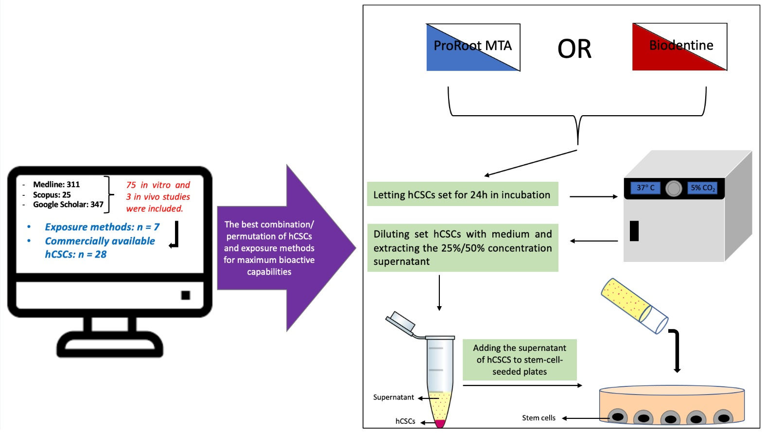
2.1. Study Selection
2.2. Study Characteristics
2.2.1. Types of hCSCs and Their Setting Times and Condition
2.2.2. Types of Cells

| Author/Year | Cements and Materials (Setting Times and Conditions) (Dilutions/Concentrations) | Cells/Interventions | Methods of Assessment | Results | ||
|---|---|---|---|---|---|---|
| Attachment (A)/ Migration (M) | Viability/ Proliferation | Odonto-/ Osteogenesis | ||||
| Youssef et al. [43]/2019 | 1. PRMTA (48 h set RT) 2. CH (48 h set RT) 3. EG 4. BD (48 h set RT) 5. NC | hDPSCs/ Direct1 | 1. Attachment, viability and proliferation: MTT (D 3) 2. Odonto-/Osteogenesis: RT-PCR (D 7 and 14) 3. Migration: NM | - A: NM - M: NM | D3: NC > EG >> PRMTA > CH > BD | 1. DSPP: 1.1. D7: EG >> CH > BD > PRMTA > NC 1.2. D14: CH >> BD > NC > EG > PRMTA 2.ALP: 2.1. D7: CH >> NC > EG > BD > PRMTA 2.2. D14: BD >> CH > PRMTA > NC > EG 3.OPN: 3.1. D7: CH >> PRMTA > EG > BD > NC 3.2. D14: BD >> CH > NC > PRMTA > EG |
| Sun et al. [103]/2020 | 1. NeoPutty 2.E RRM 3. NC 4. IRM (CP) | hDPSCs and hPDLSCs/ Indirect1 | 1. Attachment, viability and proliferation: MTT (D 3) 2. Odonto-/Osteogenesis: NM 3. Migration: NM | - A: NM - M: NM | D3: NC > NeoPutty > ERRM | NM |
| Lu et al. [44]/2019 | 1. iRBP (72 h set II and dried for 24 h) (0.02, 0.2, 1.0 and 2.0 mg/mL) 2. PRMTA (CP) (72 h set II and dried for 24 h) (2 mg/mL) 3. NC | rBMMSCs/ Indirect3 | 1. Attachment, viability and proliferation: CCK-8 (D 0, 1, 3, 5 and 7) 2. Odonto-/Osteogenesis: ALP (D 3, 5 and 7), Western blot (0, 15, 30 and 60 min), RT-PCR (D 0, 3 and 7) and ARS (D14) 3. Migration: NM | - A: NM - M: NM | At all-time points: BP iRBP (0.2 mg/mL) ≈ NC | 1.ALP activity: D3, D5 and D7: iRBP 0.2 mg/mL >> iRBP 0.02 mg/mL > NC > iRBP 2 mg/mL > iRBP 1 mg/mL 2.DSPP, OSX, OPN and ALP: 1.1. D0: NSD 1.2. D3 and D7: iRBP (0.2 mg/mL) >> NC 3. RUNX2: 2.1. D0: NC >> iRBP (0.2 mg/mL) 2.2. D3 and D7: iRBP (0.2 mg/mL) >> NC 4.ARS: iRBP >> NC |
| Tu et al. [46]/2020 | 1. PRMTA (24 h set II) 2. CAMTA: ProRoot MTA with TAF (24 h set II) 3. NC | hDPSCs and Raw 264.7 cells/Direct1 | 1. Attachment, viability and proliferation: Prestoblue and ELISA both at 12 h, D1 and D2 2. Odonto-/Osteogenesis: ARS (D 7 and 14) and ELISA (D 1 and 2) 3. Migration: NM | - A: 12 h, D1 and D2: CAMTA > PRMTA ≈ NC - M: NM | 12 h, D1 and D2: CAMTA > PRMTA ≈ NC | 1. DSPP and ALP: D7 and D14: CAMTA >> PRMTA > NC 2. ARS: D7 and D14: CAMTA >> PRMTA |
| Pedano et al. [47]/2018 | 1. Exp. cement: containing PPL (FM) (10%, 25%, 50% and 100% concentrations) 2. Nex-Cem MTA (FM) (10%, 25% and 50% concentrations) 3. BD (FM) (10%, 25% and 50% concentrations) 4. ZnOE (CP) (FM) (10%, 25% and 50% concentrations) 5. NC | hDPSCs/ Indirect1 | 1. Attachment, viability and proliferation: XTT (D 1, 4 and 7) 2. Odonto-/Osteogenesis: RT-PCR (D 4, 10 and 14) 3. Migration: WHA (D1) | - A: NM - M: 1. 10% concentration: the peak for each experiment; NexMTA > NC > Exp. > BD 2. NexMTA: 10%, 25% and 50% concentrations: NexMTA ≈ NC 3. Exp.: 10% and 25% concentrations: Exp. ≈ NC 4. BD: NC >> BD | 1. 10% concentration: 1.1. D1: Exp. > BD = NexMTA = NC > ZnOE 1.2. D4: NC > Exp. > BD > NexMTA > ZnOE 1.3. D7: BD > NC > Exp. > NexMTA > ZnOE 2. 25% concentration: 2.1. D1: NexMTA > Exp. > NC > BD > ZnOE 2.2. D4: NC > Exp. > BD > NexMTA > ZnOE 2.3. D7: NC > BD > Exp. > NexMTA > ZnOE 3. 50% concentration: 3.1. D1: NexMTA > Exp. > NC > BD > ZnOE 3.2. D4: NC > Exp. > NexMTA > BD > ZnOE 3.3. D7: NC > BD > Exp. > NexMTA > ZnOE | 1.DSPP: 1.1. D4: NC >> BD > Exp. > NexMTA 1.2. D10: BD >> NexMTA > NC > Exp. 1.3. D14: BD >> Exp. > NC > NexMTA 2.ALP: 2.1. D4: NC >> NexMTA > Exp. = BD 2.2. D10: BD > Exp. > NexMTA = NC 2.3. D14: Exp. >> NC > NexMTA > BD 3.OCN: 3.1. D4: NC > Exp. > BD > NexMTA 3.2. D10: BD >> NexMTA > NC > Exp. 3.3. D14: BD ≈ Exp. >> NexMTA > NC |
| Ali et al. [48]/2019 | 1. PRMTA (24 h set II) (1:2, 1:4,1:8 and 1:16 dilutions) 2. BD (24 h set II) (1:2, 1:4,1:8 and 1:16 dilutions) 3. TF (24 h set II) (1:2, 1:4,1:8 and 1:16 dilutions) 4. NC (1:2, 1:4,1:8 and 1:16 dilutions) | hBMSCs/ Indirect1 | 1. Attachment, viability and proliferation: MTT (D 1,3 and 7) 2. Odonto-/Osteogenesis: RT-PCR (6 h and D 1,3 and 7) and ELISA (D 1,3 and 7) 3. Migration: NM | - A: NM - M: NM | 1. D1, D3 and D7: 1:8 and 1:16 dilutions: NSD 2. D3 and D7: 1:2 and 1:4 dilutions: NC >> PRMTA ≈ BD ≈ TF | 1. ALP: 1.1. D7: TF = NC > PRMTA > BD 1.2. 6 h: BD >> NC ≈ PRMTA ≈ TF 2. COL1A: D7: PRMTA ≈ BD >> TF >> NC 3. OC: D1: BD >> NC ≈ PRMTA ≈ TF D7: NSD |
| Couto et al. [104]/2020 | 1. White MTA 2. CH 3. COP 4. MTA + COP 5. CH + COP 6. Cells in mineralizing medium (CP) 7. NC | hDPSCs/ Indirect3 | 1. Attachment, viability and proliferation: MTT (D 1, 2 and 3) 2. Odonto-/Osteogenesis: RT-PCR (D21) and ARS (D21) 3. Migration: WHA (12 h, D1 and D2) | - A: NM - M: 1.CH: no migration was observed 2. 12 h, D1 and D2: CH + COP > MTA + COP > NC > COP >> MTA 3. D1 and D2: CH + COP >> MTA + COP ≈ NC ≈ COP ≈ MTA | D3: NC > COP > CH + COP > MTA > MTA + COP >> CH | 1. DSPP and OCN: MTA + COP >> CH + COP ≈ NC ≈ COP ≈ MTA 2. ARS: COP >> CH + COP > MTA > MTA + COP > CH > CP > NC |
| Olcay et al. [49]/2019 | 1. PRMTA (72 h set II) 2. BD (72 h set II) 3. WRST (72 h set II) 4. Dycal (72 h set II) 5. NC | hDPSCs, hPDLSCs and hTGSCs/ Indirect2 | 1. Attachment, viability and proliferation: MTS (D 1, 3, 7, 10 and 14) 2. Odonto-/Osteogenesis: NM 3. Migration: NM | - A: NM - M: NM | 1. hDPSCs: D14: PRMTA > BD > WRST > NC >> Dycal 2. hTGSCs: D14: NC > WRST = BD > PRMTA >> Dycal 3. hPDLSCs: D7: PRMTA >> BD ≈ Dycal ≈ NC ≈ WRST | NM |
| Güven et al. [110]/2013 | 1. MTA Fillapex (24 h set II) 2. iRSP (24 h set II) 3. AH Plus Jet (24 h set II) 4. NC | hTGSCs/ Indirect2 | 1. Attachment, viability and proliferation: MTS and SEM both at D 1, 3, 7 and 14 2. Odonto-/Osteogenesis: NM 3. Migration: NM | - A: iRSP ≈ NC - M: NM | 1. D1: NC > AH > iRSP >> Fillapex 2. D3: NC > iRSP > AH >> Fillapex 3. D7: NC >> AH > iRSP >> Fillapex 4. D14: iRSP > NC > AH >> Fillapex | NM |
| Schneider et al. [50]/2014 | 1. PRMTA: with plain α-MEM (1 h set RT (FM) or 24 h set II): 2. PRMTA: with calcium-enriched media (3.0, 0.3 and 0.03 mmol dilutions of CaCl2) (1 h set RT (FM) and 24h set II): 3. PRMTA: with 2% FBS (1 h set RT (FM) and 24 h set II): 4. FBS (0%,2% and 10%) and CaCl2 media (NC) | SCAP/ Indirect2 | 1. Attachment, viability and proliferation: WST-1 (D 1, 3, 5, 7, 9, 11 and 14) 2. Odonto-/Osteogenesis: NM 3. Migration: TMA (0.5, 1, 3, 6, 12, 24, 48 and 72 h) | - A: NM - M: 1. 0.5 h to 6 h: Significantly higher in 24 h set PRMTA with plain α-MEM 2. 24, 48 and 72 h: significantly higher in PRMTA + CaCl2 3. FBS 2% and 10%: significantly induced early and short-term migration | 1. PRMTA with 0.3 and 0.03 mmol CaCl2 media: significant increase from D1 to D7 and decreased afterwards 2. PRMTA with 2% FBS: significantly lower than NC at D7 onwards | NM |
| Bortoluzzi et al. [37]/2015 | 1. MTA Angelus (FM or 24 h set II) 2. BD (FM or 24 h set II) 3. TCLC 4. IRM (CP) 5. NC | hDPSCs/ Indirect1 and Indirect2 | 1. Attachment, viability and proliferation: XTT (D3 for Indirect2 and D4 for Indirect1) and Flow cytometry (D3) 2. Odonto-/Osteogenesis: RT-PCR (D7) 3. Migration: NM | - A: NM - M: NM | 1. At the end of the fourth aging cycle: NC ≈ MTA ≈ BD >> TCLC 2. FM: all cements were cytotoxic | 1. DSPP and DMP1, ALP and BSP: BD ≈ MTA >> TCLC > NC 2. OCN, and Runx2: BD ≈ MTA >> NC > TCLC |
| Jun et al. [105]/2019 | 1. Ceria-incorporated MTA (CMTA: 2% and 4%) 2. NC 3. MTA (CP) | hDPSCs/ Indirect2 | 1. Attachment, viability and proliferation: MTS (D1) 2. Odonto-/Osteogenesis: RT-PCR (D7), ARS (D21) and ALP (D 7 and 14) 3. Migration: NM | - A: NM - M: NM | D1: CMTA >> MTA ≈ NC | ARS and ALP activity: CMTA ≈ MTA >> NC |
| Costa et al. [51]/2015 | 1.PRMTA (24 h set II) (1:2, 1:5, 1:10, and 1:20 dilutions) 2.MTA Plus (24 h set II) (1:2, 1:5, 1:10, and 1:20 dilutions) 3.MTA Fillapex (24 h set II) (1:2, 1:5, 1:10, and 1:20 dilutions) 4.BD (24 h set II) (1:2, 1:5, 1:10, and 1:20 dilutions) 5.NC | hBMSCs and hUVECs/ Indirect1 | 1.Attachment, viability and proliferation: Cell lysates (D 1, 7, 14 and 21) 2.Odonto-/Osteogenesis: ALP (D 7, 14, 21) 3.Migration: NM | - A: NM - M: NM | 1. D21: PRMTA (1:20) ≈ MTA Plus (1:20) >> Fillapex ≈ BD ≈ NC 2. At all time points: PRMTA (1:20) ≈ MTA Plus (1:20) ≈ NC >> MTA Fillapex ≈ BD (1:2) | ALP activity: D21: PRMTA (1:20) ≈ MTA Plus (1:20) >> control >> Fillapex ≈ BD (1:2 and 1:5) |
| D’Antò et al. [52]/2010 | 1. PRMTA (24 h set II) 2. PC (24 h set II) 3. NC 4. FBS 20% (CP) | hDPSCs/ Direct1 | 1. Attachment, viability and proliferation: Alamar blue (D 1, 3, 5, 7, 14, 21 and 28) and CLSM (D1) 2. Odonto-/Osteogenesis: NM 3. Migration: TMA (18 h) | - A: D1: PRMTA > PC ≈ NC - M: CP >> PRMTA >> PC ≈ NC | D14, D21 and D28: PRMTA >> PC ≈ NC | NM |
| Collado-González et al. [53]/2017 | 1. MTA Angelus (48 h set II) (1:1, 1:2 and 1:4 dilutions) 2. BD (48 h set II) (1:1, 1:2 and 1:4 dilutions) 3. TCLC (48 h set II) (1:1, 1:2 and 1:4 dilutions) 4. IRM (48 h set II) (CP) (1:1, 1:2 and 1:4 dilutions) 5. NC | hDPSCs/ Indirect1 | 1. Attachment, viability and proliferation: MTT (D 1,2 and 3) and SEM (D3) 2. Odonto-/Osteogenesis: NM 3. Migration: WHA (D 1 and 2) | - A: MTA ≈ BD ≈ NC >> TCLC ≈ IRM - M: 1.D1: 1.1. 1:1 dilution: NC ≈ BD >> MTA > IRM > TCLC 1.2. 1:2 dilution: TCLC >> BD > MTA > TCLC > IRM 1.3. 1:4 dilution: NC >> BD > TCLC > IRM > MTA 2.D2: 2.1. 1:1 dilution: NC = BD >> MTA > TCLC > IRM 2.2. 1:2 and 1:4 dilutions: NC ≈ BD >> MTA > IRM > TCLC | 1. 1:1 and 1:2 dilution at D3: BD >> NC >> IRM ≈ TCLC 2. 1:4 dilution at D3: BD >> NC ≈ TCLC ≈ IRM | NM |
| Agrafioti et al. [54]/2016 | 1. PRMTA (1 h or 24 h set RT) 2. BD (24 h set RT) 3. NC | hDPSCs/ Direct2 | 1. Attachment, viability and proliferation: MTT (D 4 and 7) 2. Odonto-/Osteogenesis: NM 3. Migration: NM | - A: NM - M: NM | 1. D4: BD >> PRMTA (24 h set) ≈ NC >> PRMTA (1 h set) 2. D7: PRMTA (24 h set) ≈ BD >> NC >> PRMTA (1 h set) | NM |
| Hasweh et al. [55]/2021 | 1. BD (15 min DH) (4 concentrations: 20 mg/mL, 2 mg/mL, 0.2 mg/mL and 0.02 mg/mL) 2. NC | SHED/ Indirect3 | 1. Attachment, viability and proliferation: MTT (D 1, 2, 3, 4, 5 and 6) and CAA (D1) 2. Odonto-/Osteogenesis: NM 3. Migration: WHA (D1) and TMA (D1) | - A: 0.2 mg/mL BD ≈ 0.02 mg/mL BD ≈ 2 mg/mL BD > NC - M: 0.2 mg/mL BD ≈ 0.02 mg/mL BD ≈ NC >> 2 mg/mL BD | 0.2 mg/mL BD ≈ 0.02 mg/mL BD > 2 mg/mL BD > NC >> 20 mg/mL BD | NM |
| Wang et al. [56]/2014 | 1. PRMTA (24 h DH) (0.002, 0.02, 0.2, 2, and 20 mg/mL concentrations) 2. NC | rDPSCs/ Indirect3 | 1. Attachment, viability and proliferation: MTT (D 1, 3, 5 and 7) and FCM 2. Odonto-/Osteogenesis: RT-PCR (D 3 and 7), Western blot (minutes 0, 15, 30 and 60), ARS and ALP (D 3 and 5) 3. Migration: NM | - A: NM - M: NM | 0.2 mg/mL PRMTA ≈ NC >> 2 mg/mL PRMTA > 20 mg/mL PRMTA | In 0.2 mg/mL MTA: 1. DSPP: PRMTA D7 > PRMTA D3 >> NC 2. ALP and OCN: PRMTA D7 > PRMTA D3 > NC 3. Runx2 and OSX: PRMTA D3 >> NC ≈ PRMTA D7 4. ARS and ALP activity: 0.2 mg/mL PRMTA >> NC |
| Widbiller et al. [57]/2015 | 1. PRMTA (24 h set II) 2. BD (24 h set II) 3. GIC (24 h set II) 4. Human dentin disks 5. NC | hDPSCs/ Direct1 | 1. Attachment, viability and proliferation: MTT (D 1, 3, 5 7, 10 and 14) and SEM (D1) 2. Odonto-/Osteogenesis: qRT-PCR (D 7, 14 and 21) and ALP (D 3, 7 and 14) 3. Migration: NM | - A: D1: Cell spreading and attachment was observed in BD - M: NM | 1. D14: BD ≈ PRMTA >> NC ≈ dentin disks 2. GIC: significantly cytotoxic | 1. ALP activity: dentin disks > BD >> PRMTA > NC 2. DSPP: D14: PRMTA >> BD ≈ dentin disks ≈ NC D21: BD >> PRMTA ≈ dentin disks ≈ NC 3. ALP: D3 and D14: dentin disks >> PRMTA ≈ NC >> BD 4. Runx2: NC >> PRMTA >> BD 5. COL1A1: D7: PRMTA ≈ BD >> NC |
| Athanasiadou et al. [38]/2018 | 1. BD (24 h set II) (1:1, 1:2, 1:4, 1:8, 1:16, 1:32, 1:64 and 1:128 dilutions) 2. NC | SHED/Direct1 (staining) and Indirect1 (MTT) | 1. Attachment, viability and proliferation: SEM (D3), MTT (D 1, 3 and 5) and LDA (D3) 2. Odonto-/Osteogenesis: RT-PCR (D 7 and 14), ARS (D14) 3. Migration: NM | - A: BD: Adhesion and spreading were observed - M: NM | D3: BD >> NC | 1. DSPP, ALP, Runx2 and BMP2: BD >> NC 2. ARS: NSD |
| Wang et al. [112]/2017 | 1. MTA (0.002, 0.02, 0.2, 2, 20 mg/mL concentrations) 2. Mineralization-inducing medium (MM) 3. MTA (2 mg/mL) + MM 4. Mouse IgG isotype antibodies (NC) 5. Gapdh (CP) | hPDLSCs/ Indirect3 | 1. Attachment, viability and proliferation: CCK-8 (D 1, 3, 5, 7 and 9) 2. Odonto-/Osteogenesis: RT-PCR (D 3 and 7), Western blot (0, 15, 30 and 60 min), ARS (D14) and ALP (D 3 and 5) 3. Migration: NM | - A: NM - M: NM | 2 mg/mL MTA: NSD | 1. RUNX2, OCN, OSX, COL-I, OPN, DMP1, ALP, and DSP: MTA >> NC 2. ARS and ALP activity: MTA + MM > MM > MTA >> NC |
| Matsumoto et al. [58]/2013 | 1.PRMTA (24 h set II) 2.NC | C2C12/ Indirect2 | 1.Attachment, viability and proliferation: CCK-8 (D 1, 3, 5 and 7) 2.Odonto-/Osteogenesis: RT-PCR (D 1, 3, 5 and 7) 3.Migration: NM | - A: NM - M: NM | 1. D7: PRMTA >> NC 2.D3: NSD | Runx2: PRMTA >> NC |
| Ajlan et al. [113]/2015 | 1. MTA (0.02, 0.2 and 2.0 mg/mL concentrations) 2. EMD (0.05, 0.1 and 0.2 mg/mL concentrations) 3. PDGF (0.000005, 0.00001and 0.00002 mg/mL concentrations) 4. NC 5. Cells in osteoinduction medium (OT) (reference control) | hDPSCs/ Indirect3 | 1. Attachment, viability and proliferation: - 2. Odonto-/Osteogenesis: ALP (D14) and ARS (D14) 3. Migration: NM | - A: NM - M: NM | NM | 1.ALP activity: 1.1. Lowest concentrations: MTA > EMD > PDGF 1.2. Middle concentrations: EMD >> PDGF > MTA 1.3. Highest concentrations: EMD >> PDGF >> MTA 2.ARS: EMD > MTA >> OT > PDGF > NC |
| Paranjpe et al. [59]/2010 | 1. PRMTA (48 h set II) 2. NC 3. BMP-4 (CP) 4. NAC (CP) | hDPSCs/ Direct1 | 1. Attachment, viability and proliferation: Flow cytometry (D1) 2. Odonto-/Osteogenesis: RT-PCR (D 1, 4 and 7) 3. Migration: NM | - A: NM - M: NM | D1: NSD | 1. Runx2: 1.1. D1 and D4: BMP-4 >> PRMTA > NC 1.2. D7: almost non-existent in all groups 2.DSPP: 2.1. D1: BMP-4 > PRMTA > NC 2.2. D4 and D7: PRMTA >> BMP-4 > NC 3.OCN: 3.1. D1: BMP-4 > PRMTA = NC 3.2. D4: BMP-4 >> PRMTA > NC 3.3. D7: PRMTA >> BMP-4 > NC 4. ALP: 4.1. D1 and D7: BMP-4 >> PRMTA > NC 4.2. D4: BMP-4 > PRMTA > NC |
| Araújo et al. [60]/2017 | 1. PRMTA 2. BD 3. CH 4. NC 5. Culture medium with 20% FBS (CP) | hDPSCs/ Indirect3 | 1. Attachment, viability and proliferation: MTT (D 1, 3, 5 and 7) and SRB (D 1, 3, 5 and 7) 2. Odonto-/Osteogenesis: RT-PCR (D 1, 7, 14 and 21) 3.Migration: Cell TrackerTM Green CMFDA (D1) | - A: NM - M: BD > PRMTA >> CH > CP > NC | 1. MTT: 1.1. D1 and D3: NSD 1.2. D5 and D7: BD >> PRMTA ≈ CH >> NC 2. SRB: NSD | DMP1: 1. D1: NSD 2. D7: PRMTA > CH >> NC >> BD 3. D14: PRMTA > CH > BD >> NC 4. D21: PRMTA > BD > CH >> NC |
| Tsai et al. [39]/2018 | 1. PRMTA (1 week set II) 2. NC | SHEDs/Direct1 and Indirect2 | 1. Attachment, viability and proliferation: WST-1 (D 1, 2 and 3) 2. Odonto-/Osteogenesis: NM 3. Migration: NM | - A: NC >> PRMTA - M: NM | 1. Direct: NC >> PRMTA 2. Indirect: D1 and D3: NC >> PRMTA D2: NSD | NM |
| Vanka et al. [61]/2019 | 1. PRMTA (24 h set II) 2. PRP (5% and 10% concentrations) 3.P RMTA combined with PRP 4. NC | hBMSCs/ Direct1 | 1. Attachment, viability and proliferation: MTT (D 3, 7 and 14) and CAA (D3) 2. Odonto-/Osteogenesis: ARS (D14) 3. Migration: NM | - A: PRMTA + 10%PRP >> PRMTA + 5%PRP > PRMTA > 10%PRP > 5% PRP > NC - M: NM | 1. D3, D7 and D14: NSD 2. D7 and D14: MTA ≈ PRMTA/5% PRP ≈ PRMTA/10%PRP >> NC | ARS: PRMTA + PRP 10% >> PRMTA + 5%PRP > 10%PRP > PRMTA = 5% PRP > NC |
| Kulan et al. [62]/2018 | 1. PRMTA with additives: (24 h set II) 1.1. Distilled water (DW) 1.2. Na2HPO4 2.5% 1.3. CaCl2 5% 2. PRMTA (24 h set II) (CP) 3. NC | hDPSCs/ Direct1 | 1. Attachment, viability and proliferation: MTS (D 1, 7 and 21) 2. Odonto-/Osteogenesis: RT-PCR (D 14 and 21) and ALP (D 7 and 14) 3. Migration: NM | - A: NM - M: NM | 1. D1: NSD 2. D7: NC > PRMTA + DW = PRMTA + CaCl2 > PRMTA + Na2HPO4 3. D21: NC >> PRMTA + CaCl2 > PRMTA + DW > PRMTA + Na2HPO4 | ALP activity: D7 and D14: PRMTA + CaCl2 >> PRMTA + Na2HPO4> NC > PRMTA + DW |
| Lee et al. [63]/2010 | 1. PRMTA (24 h set II) 2. Calcium phosphate cements (CPCs) 3. CPC-Ch (CPC with chitosan) 3. PC 4. NC | hDPSCs/ Direct1 | 1. Attachment, viability and proliferation: MTS (D 1, 7 and 14) and SEM (D7) 2. Odonto-/Osteogenesis: RT-PCR (D 1, 7 and 14) and ALP (D 1, 7 and 14) 3. Migration: NM | - A: NSD - M: NM | 1. D1 and D7: NC >> PC > CPC-Ch > CPC = PRMTA 2. D14: NC >> PC = PRMTA > CPC-Ch > CPC | 1. DSPP: 1.1. D1: PRMTA > CPC = CPC-Ch > PC > NC 1.2. D7: CPC > CPC-Ch > PRMTA > PC >> NC 1.3. D14: CPC-Ch > CPC > PC > PRMTA >> NC 2. DMP1: 2.1. D1: CPC-Ch > CPC > PC > PRMTA >> NC 2.2. D7: PRMTA > PC = CPC-Ch > CPC >> NC 2.3. D14: PRMTA > PC >> CPC > CPC-Ch > NC 3. ALP activity: D1, D7 and D14: PC > CPC-Ch > CPC > PRMTA >> NC 4. BSP: 4.1. D1: PRMTA > PC > CPC-Ch > CPC > NC 4.2. D7: PC > CPC > CPC-Ch >> PRMTA >> NC 4.3. D14: PC > CPC-Ch > CPC > PRMTA > NC 5. OPN: 5.1. D1: CPC-Ch > CPC > PRMTA > PC >> NC 5.2. D7: CPC-Ch > CPC >> PRMTA > PC > NC 5.3. D14: CPC > PC > CPC-Ch > PRMTA > NC 6. ON: 6.1. D1: PRMTA > CPC > CPC-Ch >> PC > NC 6.2. D7: CPC > CPC-Ch > PRMTA >> PC > NC 6.3. D14: PC = CPC > CPC-Ch > PRMTA > NC |
| Tomás-Catalá et al. [64]/2017 | 1. BD (48 h set II) (1:1, 1:2 and 1:4 dilutions) 2. NeoMTA Plus (48 h set II) (1:1, 1:2 and 1:4 dilutions) 3. MTA repair HP (48 h set II) (1:1, 1:2 and 1:4 dilutions) 4. NC | hDPSCs/ Indirect1 | 1. Attachment, viability and proliferation: MTT (D 1, 2 and 3) and SEM (D3) 2. Odonto-/Osteogenesis: NM 3. Migration: WHA (D 1 and 2) | - A: BD >> HP ≈ NeoMTA ≈ NC - M: 1. D1: 1.1. 1:1 dilution: BD > NeoMTA > HP > NC 1.2. 1:2 dilution: NeoMTA > BD > HP = NC 1.3. 1:4 dilution: BD > NeoMTA = NC > HP 2.D2: 2.1. 1:1 dilution: BD > NC >> NeoMTA > HP 2.2. 1:2 dilution: BD > NC > NeoMTA >> HP 2.3. 1:4 dilution: BD > NC > NeoMTA > HP | 1. D1: 1:1. 1:2 and 1:4 dilutions: NSD 2. D2: 2.1. 1:1 dilution: BD > HP > NeoMTA > NC 2.2. 1:2 dilution: BD > HP = NC > NeoMTA 2.3. 1:4 dilution: BD >> HP = NC > NeoMTA 3. D3: 3.1. 1:1 dilution: BD >> NeoMTA > HP > NC 3.2. 1:2 dilution: BD >> NeoMTA = HP = NC 3.3. 1:4 dilution: BD >> NC = NeoMTA > HP | NM |
| Guven et al. [114]/2011 | 1. MTA 2. Dycal 3. EMD 4. MTA + EMD 5. Dycal coated with EMD 6. NC 7. Regular tissue culture plate (TCP) | hTGSCs/ Direct1 | 1. Attachment, viability and proliferation: MTT (D2) and SEM (D14) 2. Odonto-/Osteogenesis: RT-PCR (D14) and ALP (D24) 3. Migration: NM | - A: EMD ≈ TCP >> MTA ≈ MTA + EMD ≈ NC >> Dycal - M: NM | 1. D2: EMD > MTA > Dycal + EMD > MTA + EMD > Dycal > NC 2. EMD coated Dycal: EMD coating significantly reduced Dycal’s cytotoxicity | 1. DSPP: EMD > MTA > NC 2. ALP activity: NC > EMD > TCP > MTA |
| Sun et al. [65]/2019 | 1. BD (set II) 2. iRFS (set II) 3.NC | hDPSCs/ Direct1 | 1. Attachment, viability and proliferation: SEM (D2) and LDA (D 1, 3 and 7) 2. Odonto-/Osteogenesis: RT-PCR (D 1, 3 and 7) 3. Migration: TMA (D7) | - A: NSD - M: D7: iRFS >> BD > NC | 1. D1 and D3: iRFS > BD = NC 2. D7: iRFS >> BD = NC | 1. ALP: 1.1. D1 and D7: NC >> iRFS > BD 1.2. D3: NC >> BD > iRFS 2. COL1: 2.1. D1: iRFS >> > BD 2.2. D3: iRFS > NC = BD 2.3. D7: BD > iRFS > NC 3. OCN: 3.1. D1: iRFS >> BD > NC 3.2. D3: NC >> BD > iRFS 3.3. D7: NC > iRFS > BD |
| Niu et al. [36]/2015 | 1. PRMTA (24 h set II) 2. Quick-set2 (experimental CS cement with oxide) (24 h set II) 3. IRM (CP) 4. NC | hDPSCs/ Indirect2 | 1. Attachment, viability and proliferation: MTT (D1), flow cytometry (D3) and CyQUANT (D3) 2. Odonto-/Osteogenesis: NM 3. Migration: NM | - A: NM - M: NM | 1. First cycle: Quick-set2 was significantly cytotoxic 2. Third cycle: NSD | NM |
| Zhao et al. [66]/2011 | 1. PRMTA (1 week set II) (20, 10, 2, 1, 0.2, 0.1, 0.02, and 0.002 mg/mL concentrations) 2. NC | hDPSCs/ Indirect1 | 1. Attachment, viability and proliferation: MTT (D 1, 3 and 5) 2. Odonto-/Osteogenesis: RT-PCR (6 h, 12 h, D1 and D2) 3. Migration: NM | - A: NM - M: NM | 1. In 10 and 20 mg/mL: cytotoxic at all time points 2. D1, D3 and D5: 2 mg/mL PRMTA = 1 mg/mL PRMTA > 0.2 mg/mL PRMTA > 0.1 mg/mL PRMTA > 0.02 mg/mL PRMTA > 0.002 mg/mL PRMTA > NC | 1. DSPP: PRMTA (0.2 mg/mL) D2 > D1 > 12 h > 6 h >> NC 2. BSP: PRMTA (0.2 mg/mL) 12 h > D1 > D2 >> 6 h > NC 3. OCN: PRMTA (0.2 mg/mL) D2 > D1 > 12 h >> 6 h > NC 4. COL1 and ALP: PRMTA (0.2 mg/mL) 12 h > D1 > D2 > 6 h >> NC |
| Yu et al. [67]/2016 | 1. Experimental cement: containing resin monomer (MAE-DB) and Portland cement (PC) 2. PRMTA (48 h set II) 3. MAE-DB 4. PC 5. NC 6. Cells cultured with osteogenic medium (CP) | hDPSCs/ Indirect1 | 1. Attachment, viability and proliferation: CCK-8 (D 1, 2 and 3) and CAA (1 h) 2. Odonto-/Osteogenesis: RT-PCR (D14), ARS (D14) and ALP (D 3, 5, 7 and 9) 3. Migration: WHA (D1) and TMA (D1) | - A: 1 h: PRMTA = PC > Exp. >> NC - M: 1.TMA: PRMTA >> PC > Exp. > NC 2.WHA: MTA ≈ PC > NC > Exp. | 1. D1: NC >> PRMTA = PC > Exp. > MAE-DB 2. D2: PRMTA = PC >> NC > Exp. >> MAE-DB 3. D3: NC = PRMTA = PC = Exp. >> MAE-DB | 1. ALP activity: 1.1. D3: NSD 1.2. D5: PRMTA > PC = Exp. >> CP > NC 1.3. D7 and D9: PRMTA = PC > Exp. >> CP > NC 2. ARS: PRMTA = PC >> Exp. > CP > NC 3. DSPP: PC > PRMTA > Exp. >> CP > NC 4. OCN and BMP1: PRMTA > PC > Exp. >> CP > NC 5. ON: PRMTA = PC > Exp. >> CP > NC 6. ALP: PC >> PRMTA > Exp. > CP > NC |
| Tomás-Catalá et al. [40]/2017 | 1. NeoMTA Plus (48 h set II) (1:1, 1:2 and 1:4 dilutions) 2. MTA Angelus (48 h set II) (1:1, 1:2 and 1:4 dilutions) 3. MTA Repair HP (48h set II) (1:1, 1:2 and 1:4 dilutions) 4. NC | hDPSCs/ Direct1 and Indirect1 | 1. Attachment, viability and proliferation: MTT (D 1, 2 and 3) and SEM (D3) 2. Odonto-/Osteogenesis: NM 3. Migration: WHA (D 1 and 2) | - A: NSD - M: 1.D1: NeoMTA ≈ NC >> HP ≈ Angelus 2.D2: NC > HP >> Angelus ≈ NeoMTA | 1. D1: NSD 2. D2: 2.1. 1:1 dilution: Angelus > HP >> NeoMTA > NC 2.2. 1:2 dilution: Angelus > HP > NeoMTA > NC 2.3. 1:4 dilution: Angelus >> HP > NC >> NeoMTA 3. D3: 3.1. 1:1 dilution: Angelus > HP > NeoMTA >> NC 3.2. 1:2 dilution: NSD 3.3. 1:4 dilution: NC = NeoMTA > Angelus >> HP | NM |
| Chen et al. [68]/2016 | 1. Newly developed bioceramic cement (RRM) (72 h set II) 2. PRMTA (72 h set II) 3. NC | hDPSCs, hBMSCs and hPDLSCs/ Direct1 | 1. Attachment, viability and proliferation: MTT (D 1, 3 and 5) and SEM (D3) 2. Odonto-/Osteogenesis: SEM (D3) 3. Migration: NM | - A: D3: NSD - M: NM | 1. hDPSCs: 1.1. D1: PRMTA > RRM > NC 1.2. D3: RRM > PRMTA > NC 1.3. D5: RRM >> PRMTA >> NC 2. hBMSCs: 2.1. D1: NC > PRMTA > RRM 2.2. D3: PRMTA > RRM > NC 2.3. D5: PRMTA > RRM >> NC 3. hPDLSCs: 3.1. D1: RRM > NC > PRMTA 3.2. D3: PRMTA = RRM >> NC 3.3. D5: RRM >> PRMTA >> NC | NM |
| Asgary et al. [69]/2014 | 1. PRMTA (24 h set II) 2. CEM (24 h set II) 3. Gapdh (CP) 4. Growth medium (GM) (NC) 5. Differentiation medium (DM) (NC) | hDPSCs/ Direct1 | 1. Attachment, viability and proliferation: SEM (D 1, 3, 7 and 14) 2. Odonto-/Osteogenesis: RT-PCR (D 1, 3, 7 and 14) and ARS (D14) 3. Migration: NM | - A: NM - M: NM | NSD | 1. ARS: PRMTA >> NC 2. DSPP: 2.1. D1: NSD 2.2. D3 and D7: PRMTA > CEM > DM >> GM 2.3. D14: PRMTA > DM > CEM >> GM 3. DMP1: 3.1. D1: PRMTA = CEM = DM > GM 3.2. D3: PRMTA > DM > CEM >> GM 3.3. D7: PRMTA > CEM > DM >> GM 3.4. D14: CEM > DM > PRMTA >> GM 4. ALP: 4.1. D1: NSD 4.2. D3, D7 and D14: DM >> PRMTA = CEM >> GM |
| Peters et al. [70]/2016 | 1. PRMTA (24 h set II) 2. BD (24 h set II) 3. NC 4. Cells with cobalt chloride (CP) | SCAP/ Direct1 | 1. Attachment, viability and proliferation: XTT (D 1, 3 and 7) and PCM (D 1 and 3) 2. Odonto-/Osteogenesis: NM 3. Migration: NM | - A: D1 and D3: NSD - M: NM | 1. D1: PRMTA > BD >> NC > CP 2. D3: PRMTA > NC > BD > CP 3. D7: PRMTA > BD > NC = CP | NM |
| Wongwatanasanti et al. [71]/2018 | 1. PRMTA (24 h set II) 2. RetroMTA (24 h set II) 3. BD (24 h set II) 4.NC 5. Odonto-/osteogenic induction medium (CP) | SCAP/ Indirect2 | 1. Attachment, viability and proliferation: MTT (D 1, 3, 7 and 14) 2. Odonto-/Osteogenesis: ARS (D 7, 14 and 21) 3. Migration: NM | - A: NM - M: NM | 1. D1: NSD 2. D3 and D7: BD > RetroMTA > PRMTA > NC 3.D14: RetroMTA = PRMTA > BD >> NC | ARS: BD ≈ CP >> PRMTA ≈ RetroMTA ≈ NC |
| Seo et al. [115]/2013 | 1. MTA 2. NC | hDPSCs/ Indirect2 | 1. Attachment, viability and proliferation: NM 2. Odonto-/Osteogenesis: RT-PCR (D14) 3. Migration: RT-PCR (D14) | - A: NM - M: NSD | NM | 1. DSPP: NSD 2. DMP1: MTA >> control |
| Sultana et al. [72]/2017 | 1. PRMTA (48 h set: 2 h to 3 h RT, and the rest II) 2. BD (48 h set: 2 h to 3 h RT, and the rest II) 3. ERRM (48 h set: 2 h to 3 h RT, and the rest II) 4. GIC (48 h set: 2 h to 3 h RT, and the rest II) 5. NC | hBMSCs/ Direct1 | 1. Attachment, viability and proliferation: MTT (D 1, 3, 5 and 7) and LDA (D 7 and 21) 2.O donto-/Osteogenesis: ALP (D21) 3. Migration: - | - A: D7 and D21: GIC >> BD ≈ ERRM >> NC - M: NM | 1. D1: NSD 2. D3: BD >> PRMTA = ERRM = NC > GIC 2. D5: NC >> ERRM > PRMTA > BD > GIC 3. D7: NC >> ERRM > PRMTA > BD > GIC | ALP activity: ERRM ≈ PRMTA >> GIC ≈ NC >> BD |
| Luo et al. [73]/2014 | 1. BD (4 concentrations: 0.02, 0.2, 2.0 and 20.0 mg/mL) 2. NC | hDPSCs/ Indirect3 | 1. Attachment, viability and proliferation: MTT (D 1, 3, 5 and 7) and BrdU (D1) 2. Odonto-/Osteogenesis: NM 3. Migration: WHA (D1) and TMA (D1) | - A: D1: 0.2 mg/mL BD >> 2 mg/mL BD > NC = 0.02 mg/mL BD > 20 mg/mL BD - M: WHA and TMA: 0.2 mg/mL BD >> NC | 1. D1: 0.02 mg/mL BD > 0.2 mg/mL BD > 2 mg/mL BD > NC > 20 mg/mL BD 2. D3, D5 and D7: 0.2 mg/mL BD >> 2 mg/mL BD > 0.02 mg/mL BD = NC > 20 mg/mL BD | NM |
| Luo et al. [74]/2014 | 1. BD (0.2 and 2.0 mg/mL concentrations) 2. Cells cultured in mineralization medium (CP) 3. NC | hDPSCs/ Indirect3 | 1. Attachment, viability and proliferation:- 2. Odonto-/Osteogenesis: ALP (D 1, 3, 7, 10 and 14) and qRT-PCR (D14) 3. Migration: NM | - A: NM - M: NM | NM | 1. ALP activity: 1.1. D1: NSD 1.2. D3, D7, D10 and D14: 0.2 mg/mL BD >> 2 mg/mL BD > CP > NC 2. DSPP, DMP1, OCN and BSP: 0.2 mg/mL BD >> NC |
| Yan et al. [75]/2014 | 1. PRMTA (24 h set DH) (0.0002, 0.002, 0.02, 0.2, 2.0 and 20 mg/mL concentrations) 2. Histone H1 and beta*-actin (internal controls) 3.NC | SCAP/ Indirect3 | 1. Attachment, viability and proliferation: IF (0, 0.25, 0.5, 1, and 3 h) and WB (D 1, 3, 5, 7 and 9) 2. Odonto-/Osteogenesis: RT-PCR (D 3 and 7), ALP (D 3 and 5) 3. Migration: NM | - A: NM - M: NM | At any time point: NSD | 1. ALP activity: 2 mg/mL PRMTA >> 20 mg/mL PRMTA > 0.2 mg/mL PRMTA > 0.02 mg/mL PRMTA > 0.002 mg/mL PRMTA > NC 2. DSPP, ALP, Runx2 and OCN: PRMTA >> NC |
| Wang et al. [76]/2013 | 1.PRMTA (24 h set DH) (0.002, 0.02, 0.2, 2.0 and 20 mg/mL concentrations) 2. NC 3. Gapdh (internal control) | rBMSCs/ Indirect3 | 1. Attachment, viability and proliferation: MTT (D 1, 3, 5 and 7) 2. Odonto-/Osteogenesis: ALP (D 3 and 5), ARS (D14), RT-PCR (D 3 and 7) and WB (D 3 and 7; each day at 0, 30, 60 and 90 min) 3. Migration: NM | - A: NM - M: NM | D3 and D5: 0.02 mg/mL PRMTA > NC > 0.002 mg/mL PRMTA > 0.2 mg/mL = 2 mg/mL PRMTA >> 20 mg/mL PRMTA | 1. ALP activity: 2.1. D3: 0.02 mg/mL PRMTA > 0.002 mg/mL PRMTA > NC > 0.2 mg/mL = 2 mg/mL PRMTA >> 20 mg/mL PRMTA 2.2. D5: 0.02 mg/mL PRMTA > 0.002 mg/mL PRMTA > NC > 0.2 mg/mL PRMTA > 2 mg/mL PRMTA >> 20 mg/mL PRMTA 2. DSPP, ALP, Runx2, OCN and OSX: PRMTA >> NC |
| Du et al. [108]/2020 | 1. MTA (0.02, 0.2, 2.0, 10 and 20 mg/mL concentrations) 2. NC | SCAP/ Indirect3 | 1. Attachment, viability and proliferation: CCK-8 (D 1, 3, 5 and 7) 2. Odonto-/Osteogenesis: ALP (D 3 and 5), RT-PCR (D5) and WB (0, 5, 15, 30, 60 and 120 min) 3.Migration: NM | - A: NM - M: NM | 1. D1: 2 mg/mL MTA > 0.2 mg/mL MTA > 0.02 mg/mL MTA > NC >> 10 mg/mL MTA > 20 mg/mL MTA 2. D3 and D7: 0.2 mg/mL MTA > 0.02 mg/mL MTA > 2 mg/mL MTA > NC >> 10 mg/mL MTA > 20 mg/mL MTA 3. D5: NC > 0.02 mg/mL = 0.2 mg/mL = 2 mg/mL MTA > 10 mg/mL MTA > 20 mg/mL MTA | 1. ALP activity: D3 and D5: 0.2 mg/mL MTA >> 2 mg/mL MTA > 0.02 mg/mL MTA > NC 2. DSPP and OCN: 0.2 mg/mL MTA >> 2 mg/mL MTA > 0.02 mg/mL MTA > NC 3. Runx2 and BSP: 0.2 mg/mL MTA > 2 mg/mL MTA >> 0.02 mg/mL MTA > NC |
| Lee et al. [77]/2014 | 1. MTA (24 h set II) (1:1, 1:2, 1:4, 1:10 and 1:50 dilutions) 2. BA (24 h set II) (1:1, 1:2, 1:4, 1:10 and 1:50 dilutions) 3. BD (24 h set II) (1:1, 1:2, 1:4, 1:10 and 1:50 dilutions) 4. NC | C3H10T1/2 cells/ Indirect1 | 1. Attachment, viability and proliferation: XTT (D5) 2. Odonto-/Osteogenesis: ALP (D 5 and 7) and RT-PCR (D 1, 2 and 3) 3. Migration: NM | - A: NM - M: NM | 1. In 1:1, 1:2 and 1:4 dilutions: BA ≈ MTA >> BD 2. In 1:10 and 1:50 dilutions: NSD | 1.ALP activity: 1.1. D5: BD > BA > MTA > NC 1.2. D7: MTA > BD > BA > NC 2. ALP: 2.1. D1 and D2: MTA >> BA > NC > BD 2.2. D3: BA > MTA >> NC > BD 3. OC: 3.1. D1: BA > MTA >> NC > BD 3.2. D2 and D3: MTA > BA >> NC > BD 4. BSP: 4.1. D1: BD >> NC > BA > MTA 4.2. D2: MTA >> BA > NC > BD 4.3. D3: BA >> MTA > BD > NC |
| Miller et al. [78]/2018 | 1. BD (12 h set II) 2. ERRM (12 h set II) 3. ERRM-FS (12 h set II) 4. PRMTA (12 h set II) 5. NC | SCAP/ Direct1 | 1. Attachment, viability and proliferation: OZBlue (D7) 2. Odonto-/Osteogenesis: ARS (D21) and RT-PCR (D21) 3. Migration: NM | - A: NM - M: NM | D7: ERRM >> BD > ERRM-FS = NC >> PRMTA | 1. DSPP: ERRM >> BD > ERRM-FS > PRMTA > NC 2. ALP: ERRM >> BD > PRMTA = NC > ERRM-FS 3. Runx2: PRMTA = BD = ERRM = ERRM-FS = NC 4. IBSP: PRMTA >> BD > ERRM > ERRM-FS > NC 5. ARS: BD > ERRM > PRMTA > NC >> ERRM |
| Natu et al. [79]/2015 | 1. PRMTA (with additive water/propylene glycol (PG) (100/0, 80/20 and 50/50) (24 h set II) 2. NC | hDPSCs/ Indirect1 | 1. Attachment, viability and proliferation: MTS (D 1, 3 and 5) 2. Odonto-/Osteogenesis: ARS (D 7 and 14) and RT-PCR (D 7 and 14) 3. Migration: NM | - A: NM - M: NM | 1. D1: NSD 2. D3: NC > 80/20 PRMTA > 50/50 PRMTA > 100/0 PRMTA 3. D5: NC >> 80/20 PRMTA > 100/0 PRMTA > 50/50 PRMTA | 1. ALP: 1.1. D7: NC >> 80/20 PRMTA > 100/0 PRMTA > 50/50 PRMTA 1.2. D14: 80/20 PRMTA > 100/0 PRMTA > 50/50 PRMTA >> NC 2. OCN: 2.1. D7: 100/0 PRMTA > NC > 50/50 PRMTA > 80/20 PRMTA 2.2. D14: 80/20 PRMTA > 100/0 PRMTA >> 50/50 PRMTA > NC 3. Runx2: 3.1. D7: 80/20 PRMTA > 100/0 PRMTA > 50/50 PRMTA > NC 3.2. D14: 100/0 PRMTA > 50/50 PRMTA >> 80/20 PRMTA > NC 4. DSPP: 4.1. D7: NC >> 50/50 PRMTA > 100/0 PRMTA > 80/20 PRMTA 4.2. D14: 100/0 PRMTA > 80/20 PRMTA > 50/50 PRMTA >> NC |
| Margunato et al. [80]/2015 | 1. PRMTA (48 h set II) 2. BD (48 h set II) 3. MM-MTA (48 h set II) 4. Dimethyl sulfoxide (DMSO) (CP) 5. NC | hBMSCs/ Indirect2 | 1. Attachment, viability and proliferation: MTS (D 1, 3, 7 and 14) 2. Odonto-/Osteogenesis: RT-PCR (D14) and ALP (D14) 3. Migration: NM | - A: NM - M: NM | 1. D1 and D3: NC > PRMTA >> MM-MTA > BD > CP 2. D7: MM-MTA >> PRMTA > BD = NC > CP 3. D14: BD >> MM-MTA > PRMTA > NC > CP | 1. ALP activity: PRMTA >> MM-MTA > CP > BD > NC 2. COL1A: PRMTA > BD > CP > MM-MTA >> NC 3. ON: CP >> PRMTA > BD > MM-MTA >> NC 4. Runx2: PRMTA > MM-MTA > BD > CP >> NC |
| Shi et al. [111]/2012 | 1. Polymeric powder coatings (PPC) 2. White PRMTA-enriched PPC (WMPPC) 3. Gray PRMTA-enriched PPC (GMPPC) 4.NC | hBMSCs/ Direct1 | 1. Attachment, viability and proliferation: MTT (D 1 and 3) and CAA (D 1 and 3) 2. Odonto-/Osteogenesis: NM 3. Migration: NM | - A: 1.D1: WMPPC > GMPPC > NC > PPC 2.D3: significantly higher in GMPPC; GMPPC > WMPPC > PPC > NC - M: NM | 1. D1: GMPPC = WMPPC > PPC > NC 2. D3: GMPPC > WMPPC >> PPC > NC | NM |
| Ong et al. [102]/2012 | 1. Accelerated-set white PRMTA (AWMTA) (24 h set RT) (1.5625, 3.125, 6.25, 12.5 and 25 mg/mL dilutions) 2. Accelerated-set Malaysian white PC (AMWPC) (24 h set RT) (1.5625, 3.125, 6.25, 12.5 and 25 mg/mL dilutions) 3. NC | SHED/ Indirect1 | 1. Attachment, viability and proliferation: MTT (D3) 2. Odonto-/Osteogenesis: NM 3. Migration: NM | - A: NM - M: NM | 1. 1.5625, 3.125 and 6.25 mg/mL: NC > AMWPC > AWMTA 2. 12.5 and 25 mg/mL: NC >> AWMTA > AMWPC | NM |
| Liu et al. [81]/2020 | 1. iRFS (2 mg/mL concentration) 2. PRMTA (CP) (2 mg/mL concentration) 2.NC | SCAP/ Indirect3 | 1. Attachment, viability and proliferation: BrdU (20 h) and MTT (D 1, 2, 3 and 4) 2. Odonto-/Osteogenesis: qRT-PCR (D6) and ARS (D28) 3. Migration: WHA (12 and 24 h) and TMA (24 h) | - A: NM - M: WHA and TMA: iRFS > PRMTA >> NC | 20 h, D1, D2, D3 and D4: NSD | 1. ARS: iRFS > PRMTA >> NC 2. ALP and DSPP: iRFS > PRMTA >> NC |
| López-García et al. [106]/2019 | 1. ERRM (48 h set II) (1:1, 1:2 and 1:4 dilutions) 2. Ceraseal (48 h set II) (1:1, 1:2 and 1:4 dilutions) 3. Endoseal MTA (48 h set II) (1:1, 1:2 and 1:4 dilutions) 4.NC | hPDLSCs/ Indirect1 | 1. Attachment, viability and proliferation: MTT (D 1, 2 and 3) and SEM (D3) 2. Odonto-/Osteogenesis: qRT-PCR (D 3, 7, 14 and 21) and ARS (D21) 3. Migration: WHA (D 1, 2 and 3) | - A: D3: ERRM ≈ Ceraseal >> NC >> Endoseal - M: 1.D1: 1.1. 1:1 dilution: Ceraseal > ERRM > NC >> Endoseal 1.2. 1:2 dilution: ERRM > Ceraseal > NC >> Endoseal 1.3. 1:4 dilution: ERRM > NC > Endoseal = Ceraseal 2. D2: 2.1. 1:1 dilution: Ceraseal >> ERRM > NC > Endoseal 2.2. 1:2 dilution: ERRM >> Ceraseal > NC > Endoseal 2.3. 1:4 dilution: ERRM >> NC > Ceraseal > Endoseal 3. D3: 3.1. 1:1 and 1:2 dilutions: ERRM >> Ceraseal > NC > Endoseal 3.2. 1:4 dilution: ERRM > NC > Ceraseal > Endoseal | 1. 1:1 dilution: 1.1. D1: ERRM > Ceraseal > NC >> Endoseal 1.2. D2: NC = Ceraseal > ERRM >> Endoseal 1.3. D3: Ceraseal > NC > ERRM >> Endoseal 2. 1:2 dilution: 2.1. D1: Ceraseal > ERRM > NC > Endoseal 2.2. D2: Ceraseal > NC > ERRM >> Endoseal 2.3. D3: ERRM > Ceraseal > NC >> Endoseal 3. 1:4 dilution: 3.1. D1: Ceraseal > ERRM > NC > Endoseal 3.2. D2: NC = Ceraseal > ERRM >> Endoseal 3.3. D3: Ceraseal > ERRM > NC >> Endoseal | 1. ARS: ERRM > Ceraseal >> NC > Endoseal 2. ALP: 1.1. D3 and D7: Ceraseal >> ERRM > NC 1.2. D14 and D21: Ceraseal >> NC = ERRM |
| Kim et al. [82]/2020 | 1. PRMTA (24 h set II) 2. BD (24 h set II) 3. TCLC (24 h set II) 4. Dycal (24 h set II) 5. NC | hDPSCs/ Indirect2 | 1. Attachment, viability and proliferation: MTT (D 1, 2, 3 and 5) 2. Odonto-/Osteogenesis: ALP (D14) and ARS (D 7, 14 and 21) 3. Migration: WHA (D 1, 2, 3 and 4) | - A: NM - M: 1. D1, D2 and D3: NC = BD > PRMTA >> TCLC > Dycal 2. D4: NC = BD = PRMTA >> TCLC > Dycal | 1. D1: NC >> PRMTA = BD > TCLC >> Dycal 2. D2, D3 and D5: NC >> BD > PRMTA > TCLC >> Dycal | 1. ALP activity: 1.1. D3 and D5: Dycal > TCLC >> BD > PRMTA > NC 1.2. D7: TCLC > Dycal >> BD > PRMTA > NC 1.3. D10: BD > Dycal > TCLC > NC > PRMTA 1.4. D14: BD > NC > Dycal > TCLC > PRMTA 2. ARS: 2.1. D7: Dycal > TCLC >> PRMTA > NC > BD 2.2. D14: Dycal > TCLC > PRMTA > BD >> NC 2.3. D21: TCLC > PRMTA > Dycal > BD >> NC |
| Petta et al. [83]/2020 | 1. MTA Angelus (24 h set II) (10% concentration) 2. BD (24 h set II) (10% concentration 3. Two paste calcium hydroxide cement (CHC) (24 h set II) (10% concentration 4. Mineralization medium (CP) 5. NC | hDPSCs/ Indirect1 | 1. Attachment, viability and proliferation: NM 2. Odonto-/Osteogenesis: ARS (D14) 3. Migration: NM | - A: NM - M: NM | NM | ARS: BD > MTA > CHC = CP >> NC |
| Omidi et al. [84]/2019 | 1. MTA Angelus (48 h set II) (1:1 dilution) 2. BD (48 h set II) (1:1 dilution) 3. CEM (48 h set II) (1:1 dilution) 4. TCLC (48 h set II) (1:2 dilution) 5. NC | hDPSCs/ Indirect1 | 1. Attachment, viability and proliferation: MTT (D 1, 2 and 3) 2. Odonto-/Osteogenesis: NM 3. Migration: TMA (D1) | - A: NM - M: D1: CEM > BD >> NC > TCLC > MTA | 1. D1: BD > CEM > TCLC > NC > MTA 2. D2: TCLC > CEM >> MTA > NC > BD 3. D3: TCLC > BD = CEM > MTA >> NC | NM |
| Collado-González et al. [107]/2017 | 1. GuttaFlow Bioseal (48 h set II) (1:1, 1:2 and 1:4 dilutions) 2. GuttaFlow2 (48 h set II) (1:1, 1:2 and 1:4 dilutions) 3. MTA Fillapex (48 h set II) (1:1, 1:2 and 1:4 dilutions) 4. AH Plus (48 h set II) (1:1, 1:2 and 1:4 dilutions) 5. NC | hPDLSCs/ Indirect1 | 1. Attachment, viability and proliferation: MTT (D 1, 2, 3 and 7) 2. Odonto-/Osteogenesis: NM 3. Migration: NM | - A: NM - M: NM | 1. 1:1 dilution: 1.1. D1 and D2: NC = Bioseal = GuttaFlow2 >> Fillapex = AH 1.2. D3: NC > Bioseal = GuttaFlow2 > Fillapex = AH 1.3. D7: Bioseal >> NC = GuttaFlow2 > Fillapex = AH 2. 1:2 dilution: 2.1. D1 and D2: NC = Bioseal = GuttaFlow2 = AH >> Fillapex 2.2. D3: NC > Bioseal > GuttaFlow2 > AH > Fillapex 2.3. D7: Bioseal >> NC > GuttaFlow2 >> Fillapex > AH 3. 1:4 dilution: 3.1. D1 and D2: NC = Bioseal = GuttaFlow2 = AH >> Fillapex 3.2. D3: NC > AH > Bioseal > GuttaFlow2 >> Fillapex 3.3. D7: Bioseal >> NC = GuttaFlow2 >> AH = Fillapex | NM |
| Çelik et al. [86]/2020 | 1. PRMTA (48 h set II) (with and without RSV) 2. BD (48 h set II) (with and without RSV) 3. TCLC (48 h set II) (with and without RSV) 4. CH (48 h set II) (with and without RSV) 5. Calcimol LC (resin modified calcium hydroxide) (48 h set II) (with and without RSV) 6. NC (with and without RSV) | hBMSCs/ Indirect2 | 1. Attachment, viability and proliferation: MTT (4 h) 2. Odonto-/Osteogenesis: NM 3. Migration: NM | - A: 4 h: NC > CP RSV > BD + RSV > PRMTA + RSV > TCLC + RSV > BD > TCLC > PRMTA > Calcimol + RSV >> Calcimol > CH + RSV > CH - M: NM | NM | NM |
| Sun et al. [87]/2017 | 1. iRFS (24 h set II) (0.2 and 2 mg/mL concentrations) 2. BD (24 h set II) (CP) (0.2 and 2 mg/mL concentrations) 3. NC | hDPSCs/ Indirect3 | 1. Attachment, viability and proliferation: CCK-8 (D 1, 3 and 7) 2. Odonto-/Osteogenesis: ALP (D 7 and 14), ARS (D21) and qRT-PCR (D 1, 7 and 14) 3. Migration: WHA (D1) and TMA (D1) | - A: NM - M: WHA and TMA: 0.2 mg/mL iRFS > 2 mg/mL iRFS >> NC > 0.2 mg/mL BD > 2 mg/mL BD | 1. D1 and D3: NSD 2. D7: 0.2 mg/mL BD > 0.2 mg/mL iRFS > 2 mg/mL BD = 2 mg/mL iRFS > NC | 1.ALP activity: D7 and D14: 0.2 mg/mL iRFS > 2 mg/mL iRFS = 0.2 mg/mL BD > 2 mg/mL BD >> NC 2. ARS: 0.2 mg/mL iRFS >> 0.2 mg/mL BD > 2 mg/mL iRFS > NC > 2 mg/mL BD 3. COL1: 3.1. D1: NC > 2 mgiRFS > 0.2 mg/mL BD > 0.2 mg/mL iRFS > 2 mg/mL BD 3.2. D7: NC >> 0.2 mg/mL iRFS > 0.2 mg/mL BD > 2 mg/mL BD > 2 mg/mL iRFS 3.3. D14: 0.2 mg/mL iRFS >> 0.2 mg/mL BD > 2 mg/mL BD > NC > 2 mg/mL iRFS 4. OCN: 4.1. D1: 2 mg/mL iRFS > 2 mg/mL BD > NC = 0.2 mg/mL iRFS = 0.2 mg/mL BD 4.2. D7: 0.2 mg/mL iRFS >> 2 mg/mL iRFS = NC > 2 mg/mL BD > 0.2 mg/mL BD 4.3. D14: 0.2 mg/mL iRFS > 2 mg/mL iRFS >> NC > 0.2 mg/mL BD > 2 mg/mL BD |
| Victoria-Escandell et al. [96]/2017 | 1. MTA Angelus (24 h or 48 h or 1 week or 15D or 30D set II) (1:2 dilution) 2. MTA Fillapex (24 h or 48 h or 1 week or 15D or 30D set II) (1:2 dilution) 3. AH Plus (24 h or 48 h or 1 week or 15D or 30D set II) (1:2 dilution) 4. NC | hDPSCs/ Indirect1 | 1. Attachment, viability and proliferation: SRB (D1) 2. Odonto-/Osteogenesis: NM 3. Migration: NM | - A: NM - M: NM | 1. D1: NC > Fillapex > Angelus > AH 2. D2, D7, D15 and D30: NC > Angelus > AH > Fillapex | NM |
| Collado-González et al. [88]/2019 | 1. PRMTA (1 week set II) 2. MTA Repair HP (1 week set II) 3. NC | hPDLSCs/ Indirect1 | 1. Attachment, viability and proliferation: MTT (D 1, 2 and 3) and SEM (D3) 2. Odonto-/Osteogenesis: NM 3.Migration: NM | - A: D3: NSD - M: NM | 1. 1:1 dilution: 1.1. D1: HP > PRMTA = NC 1.2. D2 and D3: NC = HP > PRMTA 2. 1:2 dilution: 2.1. D1 and D3: HP > PRMTA > NC 2.2. D2: HP > NC > PRMTA 3. 1:4 dilution: D1, D2 and D3: HP > PRMTA >> NC | NM |
| Wu et al. [116]/2021 | 1. iRSP (72 h set II) (0.02, 0.2, 2, 5 and 10 mg/mL concentrations) 2. NC | SCAP/ Indirect3 | 1. Attachment, viability and proliferation: CCK-8 (D 1, 3 and 5) 2. Odonto-/Osteogenesis: ALP (D 3, 7 and 14), ARS (3, 7, 14 and 21) and qRT-PCR (D 3 and 7) 3. Migration: WHA (12 h) | - A: NM - M: 12 h: 0.2 mg/mL iRSP > 0.02 mg/mL iRSP > 2 mg/mL iRSP >> NC | D1, D3 and D5: 0.2 mg/mL iRSP >> 2 mg/mL iRSP > 0.02 mg/mL iRSP > NC >> 5 mg/mL iRSP > 10 mg/mL iRSP | 1. ALP activity: 0.2 mg/mL iRSP > 2 mg/mL iRSP > 0.02 mg/mL iRSP >> NC 2. ARS: 0.2 mg/mL iRSP >> NC 3.OCN, OSX, Runx2 and DSPP: 0.2 mg/mL iRSP >> NC |
| Manaspon et al. [41]/2021 | 1. PRMTA (24 h set RT) (10%, 25%, 50% and 100% concentrations) 2. BD (24 h set RT) (10%, 25%, 50% and 100% concentrations) 3.TCLC (24 h set RT) (10%, 25%, 50% and 100% concentrations) 4. Dycal (24 h set RT) (10%, 25%, 50% and 100% concentrations) 5. NC | hDPSCs/ Direct1 (SEM) and Indirect1 (MTT) | 1. Attachment, viability and proliferation: MTT (D 1, 4 and 7) and SEM (3 h, 6 h, 24 h and 48 h) 2. Odonto-/Osteogenesis: ALP (D14), ARS (D14) and RT-PCR (D 4 and 10) 3. Migration: WHA (D1) | - A: PRMTA ≈ BD ≈ NC >> Dycal ≈ TCLC - M: PRMTA ≈ BD >> NC >> Dycal ≈ TCLC | 10%, 25%, 50% and 100% concentrations: PRMTA ≈ BD >> NC >> Dycal ≈ TCLC | 1. ALP activity and ARS: BD > PRMTA >> NC 2. Runx2: 2.1. D4: BD >> NC > PRMTA 2.2. D10: PRMTA > BD >> NC 3. DMP1: D4 and D10: BD > PRMTA >> NC 4. DSPP: 4.1. D4: PRMTA > BD >> NC 4.2. D10: BD > PRMTA >> NC 5. OCN: 5.1. D4: BD >> NC > PRMTA 5.2. D10: PRMTA > BD >> NC |
| Chung et al. [97]/2019 | 1. PRMTA + LPS (24 h set II) 2. Retro MTA + LPS (24 h set II) 3. BD + LPS (24 h set II) 4. Dycal + LPS (24 h set II) 5. NC 6. LPS | hDPSCs/ Indirect3 | 1. Attachment, viability and proliferation: CCK-8 (D 1 and 2) 2. Odonto-/Osteogenesis: qRT-PCR (12 h, D1 and D2) 3. Migration: NM | - A: NM - M: NM | 1.D1: NC >> Retro + LPS > BD + LPS > PRMTA + LPS > LPS = Dycal + LPS 2. D2: NC >> BD + LPS > Retro + LPS > LPS > PRMTA + LPS > Dycal + LPS | 1. ALP: 1.1. 12 h and D1: Dycal + LPS >> Retro + LPS > BD + LPS > PRMTA + LPS > LPS > NC 1.2. D2: Dycal + LPS >> NC > Retro + LPS > LPS > BD + LPS > PRMTA + LPS 2. OCN: 2.1. 12 h: BD + LPS > Retro + LPS > Dycal + LPS > PRMTA+ LPS > LPS > NC 2.2. D1: Dycal + LPS >> Retro + LPS > PRMTA + LPS > BD + LPS > NC 2.3. D2: NC >> Dycal + LPS > BD + LPS > Retro + LPS > LPS > PRMTA + LPS 3. Runx2: 3.1. 12 h: NC > Dycal + LPS >> BD + LPS > Retro + LPS > PRMTA + LPS > LPS 3.2. D1: Dycal + LPS >> NC > Retro + LPS > BD + LPS > PRMTA + LPS > LPS 3.3. D2: Dycal + LPS > NC >> Retro + LPS > LPS > BD + LPS > PRMTA + LPS |
| Birant et al. [89]/2020 | 1. PRMTA (24 h set II) 2. NeoMTA Plus (24 h set II) 3. BD (24 h set II) 4. NC | hDPSCs/ Indirect1 | 1. Attachment, viability and proliferation: FCM (D 1, 3 and 7) 2. Odonto-/Osteogenesis: NM 3. Migration: NM | - A: NM - M: NM | 1. D1: NC >> BD > NeoMTA > PRMTA 2. D3: NC >> BD > PRMTA > NeoMTA 3. D7: BD >> PRMTA > NeoMTA > NC | NM |
| Sanz et al. [90]/2021 | 1. BD (48 h set II) (1:1, 1:2 and 1:4 dilutions) 2. TCPT (48 h set II) (1:1, 1:2 and 1:4 dilutions) 3. TCLC (48 h set II) (1:1, 1:2 and 1:4 dilutions) 4. NC | hDPSCs/ Indirect1 | 1. Attachment, viability and proliferation: MTT (D 1, 2 and 3) and SEM (D3) 2. Odonto-/Osteogenesis: RT-PCR (D14) and ARS (D21) 3. Migration: WHA (D 1, 2 and 3) | - A: D3: BD >> NC = TCPT > TCLC - M: 1. 1:1 dilution: 1.1. D1: NC >> BD > TCPT > TCLC 1.2. D2 and D3: NC >> TCPT > BD > TCLC 2. 1:2 dilution: 2.1. D1 and D2: NC > BD > TCPT > TCLC 2.2. D3: NC = BD > TCPT > TCLC 3. 1:4 dilution: 3.1. D1: BD >> NC > TCPT > TCLC 3.2. D2: TCPT >> NC = BD > TCLC 3.3. D3: NC = BD = TCPT > TCLC | 1. 1:1 and 1:2 dilutions: NC >> BD > PT > LC 3. 1:4 dilution: 3.1. D1: TCPT > NC > BD > TCLC 3.2. D2: BD > NC > TCPT > TCLC 3.3. D3: NC > BD > TCPT > TCLC | 1. ARS: BD > TCPT >> TCLC = NC 2. DSPP: BD > TCPT >> NC 3. Runx2: TCPT > BD >> NC 4. ALP: NC >> TCPT > BD 5. COL1A1: NC > TCPT > BD 6. ON: TCPT >> NC > BD |
| Rahimi et al. [98]/2019 | 1. PC (24 h set II) 2. PC + ZnO (24 h set II) 3. PC + ZrO2 (24 h set II) 4. NC | hDPSCs/ Direct2 | 1. Attachment, viability and proliferation: MTT (D 7, 14 and 21) 2. Odonto-/Osteogenesis: ALP (D 7, 14 and 21) 3. Migration: NM | - A: NM - M: NM | 1. D7: PC + ZrO2 >> PC > NC = PC + ZnO 2. D14: PC >> PC + ZnO > PC + ZrO2 > NC 3. D21: PC + ZrO2 = PC + ZnO > PC > NC | ALP activity: 1. D7: PC + ZnO >> PC + ZrO2 = PC > NC 2. D14: PC = PC + ZnO >> PC + ZrO2 > NC 3.D21: NSD |
| Rodríguez-Lozano et al. [100]/2015 | 1. MTA Fillapex (48 h set II) (1:1, 1:2 and 1:4 dilutions) 2. AH Plus (48 h set II) (1:1, 1:2 and 1:4 dilutions) 3. TF BC (48 h set II) (1:1, 1:2 and 1:4 dilutions) 4. NC | hDPSCs/ Indirect1 | 1. Attachment, viability and proliferation: MTT (D 1, 2 and 3) and SEM (D4) 2. Odonto-/Osteogenesis: NM 3. Migration: WHA (D 1 and 2) | - A: 1:1, 1:2 and 1:4 dilutions: TF BC >> NC > AH > Fillapex - M: 1.D1: BC > AH >> NC >> Fillapex 2.D2: NC = AH = TF BC >> Fillapex | 1:1, 1:2 and 1:4 dilutions: 1. D1: TF BC = AH = NC >> Fillapex 2. D2 and D3: TF BC = NC >> AH > Fillapex | NM |
| Jaberiansari et al. [91]/2014 | 1. PRMTA (48 h set II) (1:2 dilution) 2. MTA Angelus (48 h set II) (1:2 dilution) 3. CEM (48 h set II) (1:2 dilution) 4. NC | hDPSCs/ Indirect1 | 1. Attachment, viability and proliferation: MTT (D 1, 2 and 3) 2. Odonto-/Osteogenesis: NM 3. Migration: WHA (D 1 and 2) | - A: NM - M: NM | 1. D1 and D2: CEM > Angelus > PRMTA >> NC 2. D3: PRMTA > CEM > Angelus >> NC | NM |
| Loison-Robert et al. [92]/2018 | 1. BD (24 h set II) 2. BioRoot RCS (24 h set II) 3. NC | hDPSCs/ Direct2 | 1. Attachment, viability and proliferation: MTT (D 1, 5 and 8) 2. Odonto-/Osteogenesis: ARS (D10) and qRT-PCR (D7) 3. Migration: WHA (D 1, 2 and 7) | - A: NM - M: D1 and D2: NC >> BD > RCS | D1, D5 and D8: NC >> BD = RCS | 1. ARS: BD ≈ RCS >> NC 2. ALP and OPN: NC >> BD ≈ RCS 3. Runx2: RCS > BD >> NC |
| Sun et al. [101]/2021 | 1. ERRM (48 h set II) 2. NeoPutty 3. NC | hDPSCs/ Indirect2 | 1. Attachment, viability and proliferation: NM 2. Odonto-/Osteogenesis: ALP (week 1, 2 and 3), ARS (weeks 1, 2 and 3) and qRT-PCR (week 1, 2 and 3) 3. Migration: NM | - A: NM - M: NM | NM | 1. ALP activity: weeks 1, 2 and 3: ERRM > NeoPutty >> NC 2. ARS: 2.1. week 1: NSD 2.2. weeks 2 and 3: NeoPutty > ERRM >> NC 3. Runx2: 3.1. weeks 1, 2: ERRM > NeoPutty >> NC 3.2. week 3: NeoPutty > ERRM >> NC 4. OSX: 4.1. weeks 1, 2: ERRM > NeoPutty >> NC 4.2. week 3: NC > NeoPutty > ERRM 5. DSPP: weeks 1, 2 and 3: ERRM > NeoPutty >> NC 6. OCN: 6.1. week 1: ERRM > NeoPutty >> NC 6.2. week 2: ERRM = NeoPutty >> NC 6.3. week 3: NeoPutty >> NC > ERRM 7. DMP1: 7.1. week 1: NeoPutty > NC > ERRM 7.2. week 2: NeoPutty > ERRM > NC 7.3. week 3: NeoPutty >> ERRM = NC 8. BSP: 8.1. week 1: NSD 8.2. week 2: ERRM >> NC > NeoPutty 8.3. week 3: ERRM > NeoPutty >> NC 9. ALP: 9.1. weeks 1 and 2: NeoPutty > ERRM >> NC 9.2. week 3: NeoPutty > NC > ERRM |
| Kim et al. [93]/2021 | 1. PRMTA (48 h set II) 2. BD (48 h set II) 3. TCLC (48 h set II) 4. Dycal (48 h set II) 5. NC | hDPSCs/ Indirect1 | 1. Attachment, viability and proliferation: CCK-8 (D 1, 2, 4 and 6) 2. Odonto-/Osteogenesis: ALP (D 3 and 6) and qRT-PCR (D 9 and 14) 3. Migration: NM | - A: NM - M: NM | 1. D1 and D2: NSD 2. D4 and D6: BD > PRMTA >> NC > TCLC >> Dycal | 1. ALP activity: TCLC = Dycal >> BD = PRMTA > NC 2.R unx2: TCLC > PRMTA > Dycal > BD >> NC 3. OCN: TCLC = PRMTA >> NC > Dycal > BD 4. OPN:NC >> Dycal > PRMTA > TCLC = BD 5. DMP1: 5.1. D9: PRMTA > Dycal > TCLC > BD >> NC 5.2. D14: NC > Dycal > PRMTA > TCLC = BD 6. DSPP: 6.1. D9: NC >> PRMTA = Dycal > TCLC = BD 6.2. D14: PRMTA > BD >> NC = Dycal = TCLC |
| Assadian et al. [92]/2022 | 1. Ortho MTA (OMTA) (24 h set II) (10%, 25%, 50% and 100% concentrations) 2. BD (24 h set II) (10%, 25%, 50% and 100% concentrations) 3. CEM (24 h set II) (10%, 25%, 50% and 100% concentrations) 4. NC | hDPSCs/ Indirect1 | 1. Attachment, viability and proliferation: MTT (D 1, 3 and 5) 2. Odonto-/Osteogenesis: RT-PCR (D 7 and 14) 3. Migration: NM | - A: NM - M: NM | D1, D3 and D5: NSD | 1. DSPP: OMTA >> BD > NC > CEM 2. DMP1: BD >> OMTA > CEM > NC |
| Author/Year | Cements and Materials (Setting Times and Conditions) (Dilutions/Concentrations) | Cells/ Interventions | Methods of Assessment | Results Attachment (A)/Viability/Odonto-/ Migration (M)Proliferation Osteogenesis | ||
|---|---|---|---|---|---|---|
| Jeanneau et al. [45]/2017 | 1. BD (FM) 2. TCLC (FM) 3. NC | hDPSCs/ Direct3 | 1. Attachment, viability and proliferation: MTT (D 1, 3 and 5) 2. Odonto-/Osteogenesis: NM 3. Migration: NM | - A: NM - M: NM | D1, D3 and D5: NC >> BD >> TCLC | NM |
| Abedi-Amin et al. [95]/2017 | 1. Experimental PC (Exp. PC) (24 h set II) 2. PC (24 h set II) (CP) 3. Two light curing cements: LC-CaP (24 h set II) and LC-Si/CaP (24 h set II) 4. NC | hDPSCs/ Direct4 | 1. Attachment, viability and proliferation: MTS (D 1, 2, 4 and 7) 2. Odonto-/Osteogenesis: NM 3. Migration: NM | - A: NM - M: NM | D1, D4 and D7: NC >> LC-CaP > LC-Si/CaP >> Exp. PC >> PC | ALP activity: Exp PC > PC >> LC-CaP > LC-Si/CaP |
| Birant et al. [85]/2021 | 1. PRMTA (FM or 24 h set RT) 2. MTA Fillapex (FM or 24 h set RT) 3. MTA Angelus (FM or 24 h set RT) 4. CEM (FM or 24 h set RT) 5. NC | hDPSCs/ Direct4 | 1. Attachment, viability and proliferation: SEM (D7) 2. Odonto-/Osteogenesis: NM 3. Migration: NM | - A: 1. CEM: Adhesion was seen in both 24 h set and FM groups 2. MTA Fillapex: Adhesion was seen only in the 24 h set group 3. MTA Angelus and PRMTA: Adhesion was seen only in the FM group - M: NM | NM | NM |
2.2.3. Types of Interventions
2.2.4. Methods of Assessment
Viability and Proliferation
Attachment
Migration
Odonto-/Osteogenesis
2.3. Results of Individual Studies
2.3.1. In Vivo Studies
2.3.2. Setting Times and Conditions In Vitro
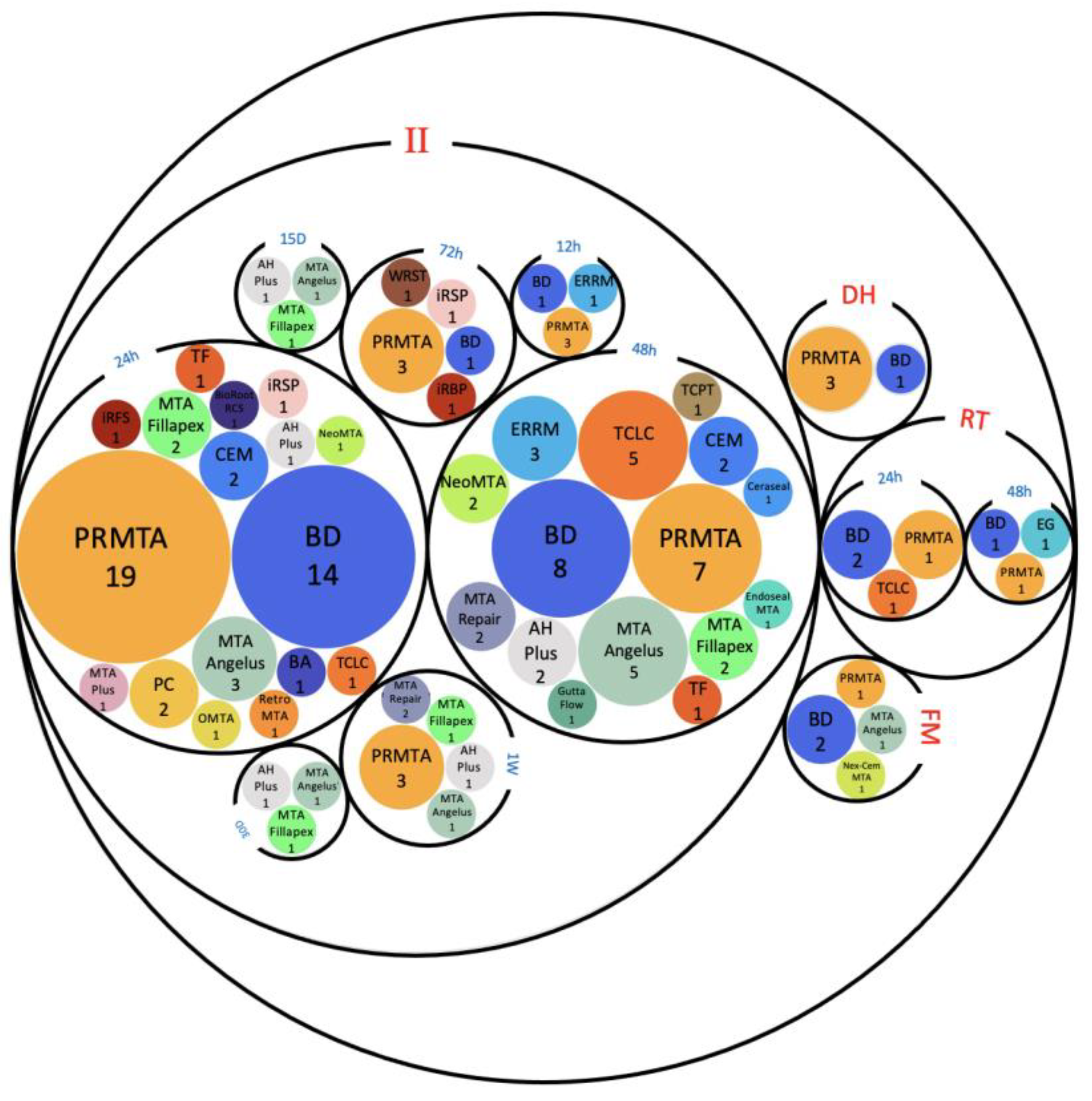
2.3.3. Comparison of Different hCSCs In Vitro
Viability/Proliferation, Migration and Attachment
Odontogenesis
Osteogenesis
2.3.4. Comparison of Different Exposure Methods In Vitro
Viability and Proliferation
Cellular Attachment
Cellular Migration
ALP Activity
Mineralization
ALP Expression
Runx2 Expression
DSPP Expression
DMP1 Expression
OCN Expression
COL1 Expression
BSP Expression
OPN Expression
ON Expression
2.4. Summary of Outcomes of In Vitro Studies
2.5. Risk of Bias Assessment
2.6. Discussion
2.6.1. hCSCs Differences
2.6.2. Setting Times and Conditions
2.6.3. Direct and Indirect Approaches In Vitro
2.6.4. Study Limitations and Suggestions
3. Conclusions
4. Materials and Methods
4.1. Eligibility Criteria
4.1.1. Types of Studies
4.1.2. Population
4.1.3. Intervention
4.1.4. Control
4.1.5. Types of Outcome Measures
4.2. Information Sources and Search Strategy
4.3. Study Selection and Data Collection
4.4. Data Items
4.5. Synthesis Methods
4.6. Risk of Bias Assessment
Supplementary Materials
Author Contributions
Funding
Data Availability Statement
Conflicts of Interest
References
- Mason, C.; Dunnill, P. A brief definition of regenerative medicine. Regen. Med. 2008, 3, 1–5. [Google Scholar] [CrossRef]
- Fischbach, G.D.; Fischbach, R.L. Stem cells: Science, policy, and ethics. J. Clin. Investig. 2004, 114, 1364–1370. [Google Scholar] [CrossRef] [PubMed]
- Rosa, V.; Dubey, N.; Islam, I.; Min, K.S.; Nör, J.E. Pluripotency of stem cells from human exfoliated deciduous teeth for tissue engineering. Stem Cells Int. 2016, 2016, 5957806. [Google Scholar] [CrossRef] [PubMed]
- Zhai, Q.; Dong, Z.; Wang, W.; Li, B.; Jin, Y. Dental stem cell and dental tissue regeneration. Front. Med. 2019, 13, 152–159. [Google Scholar] [CrossRef]
- Kisby, L. Vital pulp therapy in primary teeth: An update. Dent. Today 2016, 35, 112–113. [Google Scholar]
- Alongi, D.J.; Yamaza, T.; Song, Y.; Fouad, A.F.; Romberg, E.; Shi, S.; Tuan, R.S.; Huang, G.T.-J. Stem/progenitor cells from inflamed human dental pulp retain tissue regeneration potential. Regen. Med. 2010, 5, 617–631. [Google Scholar] [CrossRef] [PubMed]
- Prati, C.; Gandolfi, M.G. Calcium silicate bioactive cements: Biological perspectives and clinical applications. Dent. Mater. 2015, 31, 351–370. [Google Scholar] [CrossRef]
- Asgary, S.; Eghbal, M.J.; Parirokh, M.; Ghanavati, F.; Rahimi, H. A comparative study of histologic response to different pulp capping materials and a novel endodontic cement. Oral Surgery Oral Med. Oral Pathol. Oral Radiol. Endodontol. 2008, 106, 609–614. [Google Scholar] [CrossRef]
- Murray, P.E.; Windsor, L.J.; Smyth, T.W.; Hafez, A.A.; Cox, C.F. Analysis of pulpal reactions to restorative procedures, materials, pulp capping, and future therapies. Crit. Rev. Oral Biol. Med. 2002, 13, 509–520. [Google Scholar] [CrossRef]
- Washio, A.; Morotomi, T.; Yoshii, S.; Kitamura, C. Bioactive Glass-Based Endodontic Sealer as a Promising Root Canal Filling Material without Semisolid Core Materials. Materials 2019, 12, 3967. [Google Scholar] [CrossRef]
- Raszewski, Z.; Chojnacka, K.; Mikulewicz, M. Preparation and characterization of acrylic resins with bioactive glasses. Sci. Rep. 2022, 12, 16624. [Google Scholar] [CrossRef] [PubMed]
- Parirokh, M.; Torabinejad, M. Mineral trioxide aggregate: A comprehensive literature review—Part I: Chemical, physical, and antibacterial properties. J. Endod. 2010, 36, 16–27. [Google Scholar] [CrossRef] [PubMed]
- Parirokh, M.; Torabinejad, M. Mineral trioxide aggregate: A comprehensive literature review—Part III: Clinical applications, drawbacks, and mechanism of action. J. Endod. 2010, 36, 400–413. [Google Scholar] [CrossRef] [PubMed]
- Eskandar, R.F.; Al-Habib, M.A.; Barayan, M.A.; Edrees, H.Y. Outcomes of endodontic microsurgery using different calcium silicate–based retrograde filling materials: A cohort retrospective cone-beam computed tomographic analysis. BMC Oral Health 2023, 23, 70. [Google Scholar] [CrossRef] [PubMed]
- Gungor, A.; Durmus, E.; Kurt, B.; Kocyigit, A.; Dalkilic, E.; Arisu, H. Effects of Bioactive Pulp-capping Materials on Cell Viability, Differentiation, and Mineralization Behaviors of Human Dental Pulp Stem Cells In Vitro. Oper. Dent. 2023. [Google Scholar] [CrossRef]
- Rathinam, E.; Rajasekharan, S.; Chitturi, R.T.; Declercq, H.; Martens, L.; De Coster, P. Gene expression profiling and molecular signaling of various cells in response to tricalcium silicate cements: A systematic review. J. Endod. 2016, 42, 1713–1725. [Google Scholar] [CrossRef] [PubMed]
- Camilleri, J.; Ford, T.R.P. Mineral trioxide aggregate: A review of the constituents and biological properties of the material. Int. Endod. J. 2006, 39, 747–754. [Google Scholar] [CrossRef]
- Niu, L.-N.; Jiao, K.; Wang, T.-D.; Zhang, W.; Camilleri, J.; Bergeron, B.E.; Feng, H.-L.; Mao, J.; Chen, J.-H.; Pashley, D.H.; et al. A review of the bioactivity of hydraulic calcium silicate cements. J. Dent. 2014, 42, 517–533. [Google Scholar] [CrossRef] [PubMed]
- Han, L.; Kodama, S.; Okiji, T. Evaluation of calcium-releasing and apatite-forming abilities of fast-setting calcium silicate-based endodontic materials. Int. Endod. J. 2015, 48, 124–130. [Google Scholar] [CrossRef] [PubMed]
- Malik, Z.; Roth, D.M.; Eaton, F.; Theodor, J.M.; Graf, D. Mesenchymal Bmp7 controls onset of tooth mineralization: A novel way to regulate molar cusp shape. Front. Physiol. 2020, 11, 698. [Google Scholar] [CrossRef]
- Smith, A.J.; Duncan, H.F.; Diogenes, A.; Simon, S.; Cooper, P.R. Exploiting the bioactive properties of the dentin-pulp complex in regenerative endodontics. J. Endod. 2016, 42, 47–56. [Google Scholar] [CrossRef] [PubMed]
- Simon, S.; Smith, A.J.; Berdal, A.; Lumley, P.J.; Cooper, P.R. The MAP kinase pathway is involved in odontoblast stimulation via p38 phosphorylation. J. Endod. 2010, 36, 256–259. [Google Scholar] [CrossRef] [PubMed]
- Yoshioka, S.; Takahashi, Y.; Abe, M.; Michikami, I.; Imazato, S.; Wakisaka, S.; Hayashi, M.; Ebisu, S. Activation of the Wnt/?-catenin pathway and tissue inhibitor of metalloprotease 1 during tertiary dentinogenesis. J. Biochem. 2013, 153, 43–50. [Google Scholar] [CrossRef] [PubMed]
- Chang, H.-H.; Chang, M.-C.; Wu, I.-H.; Huang, G.-F.; Huang, W.-L.; Wang, Y.-L.; Lee, S.-Y.; Yeh, C.-Y.; Guo, M.-K.; Chan, C.-P.; et al. Role of ALK5/Smad2/3 and MEK1/ERK signaling in transforming growth factor beta 1–modulated growth, collagen turnover, and differentiation of stem cells from apical papilla of human tooth. J. Endod. 2015, 41, 1272–1280. [Google Scholar] [CrossRef]
- Shin, M.R.; Kang, S.K.; Kim, Y.S.; Lee, S.Y.; Hong, S.C.; Kim, E.-C. TNF-α and LPS activate angiogenesis via VEGF and SIRT1 signalling in human dental pulp cells. Int. Endod. J. 2014, 48, 705–716. [Google Scholar] [CrossRef]
- Zhang, J.; Zhu, L.; Cheng, X.; Lin, Y.; Yan, P.; Peng, B. Promotion of dental pulp cell migration and pulp repair by a bioceramic putty involving FGFR-mediated signaling pathways. J. Dent. Res. 2015, 94, 853–862. [Google Scholar] [CrossRef]
- Lee, S.-J.; Monsef, M.; Torabinejad, M. Sealing ability of a mineral trioxide aggregate for repair of lateral root perforations. J. Endod. 1993, 19, 541–544. [Google Scholar] [CrossRef]
- Lutfi, A.N.; Kannan, T.P.; Fazliah, M.N.; Jamaruddin, M.A.; Saidi, J. Proliferative activity of cells from remaining dental pulp in response to treatment with dental materials. Aust. Dent. J. 2010, 55, 79–85. [Google Scholar] [CrossRef]
- Agrawal, V. Calcium hydroxide: A miracle munition. Indian J. Dent. Res. Rev. 2011, 1, 16–18. [Google Scholar] [CrossRef]
- Malkondu, Ö.; Kazandağ, M.K.; Kazazoğlu, E. A review on biodentine, a contemporary dentine replacement and repair material. BioMed Res. Int. 2014, 2014, 160951. [Google Scholar] [CrossRef]
- Brookes, S.J.; Robinson, C.; Kirkham, J.; Bonass, W.A. Biochemistry and molecular biology of amelogenin proteins of developing dental enamel. Arch. Oral Biol. 1995, 40, 1–14. [Google Scholar] [CrossRef] [PubMed]
- Loushine, B.A.; Bryan, T.E.; Looney, S.W.; Gillen, B.M.; Loushine, R.J.; Weller, R.N.; Pashley, D.H.; Tay, F.R. Setting properties and cytotoxicity evaluation of a premixed bioceramic root canal sealer. J. Endod. 2011, 37, 673–677. [Google Scholar] [CrossRef] [PubMed]
- Ma, J.; Shen, Y.; Stojicic, S.; Haapasalo, M. Biocompatibility of two novel root repair materials. J. Endod. 2011, 37, 793–798. [Google Scholar] [CrossRef] [PubMed]
- Torabinejad, M.; Hong, C.-U.; Ford, T.R.P.; Kariyawasam, S.P. Tissue reaction to implanted super-EBA and mineral trioxide aggregate in the mandible of guinea pigs: A preliminary report. J. Endod. 1995, 21, 569–571. [Google Scholar] [CrossRef]
- Hirschman, W.R.; Wheater, M.A.; Bringas, J.S.; Hoen, M.M. Cytotoxicity comparison of three current direct pulp-capping agents with a new bioceramic root repair putty. J. Endod. 2012, 38, 385–388. [Google Scholar] [CrossRef] [PubMed]
- Niu, L.-N.; Watson, D.; Thames, K.; Primus, C.M.; Bergeron, B.E.; Jiao, K.; Bortoluzzi, E.A.; Cutler, C.W.; Chen, J.-H.; Pashley, D.H.; et al. Effects of a discoloration-resistant calcium aluminosilicate cement on the viability and proliferation of undifferentiated human dental pulp stem cells. Sci. Rep. 2015, 5, 17177. [Google Scholar] [CrossRef] [PubMed]
- Bortoluzzi, E.A.; Niu, L.-N.; Palani, C.D.; El-Awady, A.R.; Hammond, B.D.; Pei, D.-D.; Tian, F.-C.; Cutler, C.W.; Pashley, D.H.; Tay, F.R. Cytotoxicity and osteogenic potential of silicate calcium cements as potential protective materials for pulpal revascularization. Dent. Mater. 2015, 31, 1510–1522. [Google Scholar] [CrossRef]
- Athanasiadou, E.; Paschalidou, M.; Theocharidou, A.; Kontoudakis, N.; Arapostathis, K.; Bakopoulou, A. Biological interactions of a calcium silicate based cement (Biodentine™) with stem cells from human exfoliated deciduous teeth. Dent. Mater. 2018, 34, 1797–1813. [Google Scholar] [CrossRef]
- Tsai, C.-L.; Ke, M.-C.; Chen, Y.-H.; Kuo, H.-K.; Yu, H.-J.; Chen, C.-T.; Tseng, Y.-C.; Chuang, P.-C.; Wu, P.-C. Mineral trioxide aggregate affects cell viability and induces apoptosis of stem cells from human exfoliated deciduous teeth. BMC Pharmacol. Toxicol. 2018, 19, 21. [Google Scholar] [CrossRef]
- Tomás-Catalá, C.J.; Collado-González, M.D.M.; García-Bernal, D.; Oñate-Sánchez, R.E.; Forner, L.; Llena, C.; Lozano, A.; Castelo-Baz, P.; Moraleda, J.M.; Rodríguez-Lozano, F.J. Comparative analysis of the biological effects of the endodontic bioactive cements MTA?Angelus, MTA Repair HP and NeoMTA Plus on human dental pulp stem cells. Int. Endod. J. 2017, 50, e63–e72. [Google Scholar] [CrossRef]
- Manaspon, C.; Jongwannasiri, C.; Chumprasert, S.; Sa-Ard-Iam, N.; Mahanonda, R.; Pavasant, P.; Porntaveetus, T.; Osathanon, T. Human dental pulp stem cell responses to different dental pulp capping materials. BMC Oral Health 2021, 21, 209. [Google Scholar] [CrossRef]
- Page, M.J.; Moher, D.; Bossuyt, P.M.; Boutron, I.; Hoffmann, T.C.; Mulrow, C.D.; Shamseer, L.; Tetzlaff, J.M.; Akl, E.A.; Brennan, S.E.; et al. PRISMA 2020 explanation and elaboration: Updated guidance and exemplars for reporting systematic reviews. BMJ 2021, 372, n160. [Google Scholar] [CrossRef]
- Youssef, A.-R.; Emara, R.; Taher, M.M.; Al-Allaf, F.A.; Almalki, M.; Almasri, M.A.; Siddiqui, S.S. Effects of mineral trioxide aggregate, calcium hydroxide, biodentine and Emdogain on osteogenesis, Odontogenesis, angiogenesis and cell viability of dental pulp stem cells. BMC Oral Health 2019, 19, 133. [Google Scholar] [CrossRef] [PubMed]
- Lu, J.; Li, Z.; Wu, X.; Chen, Y.; Yan, M.; Ge, X.; Yu, J. iRoot BP Plus promotes osteo/odontogenic differentiation of bone marrow mesenchymal stem cells via MAPK pathways and autophagy. Stem Cell Res. Ther. 2019, 10, 222. [Google Scholar] [CrossRef]
- Jeanneau, C.; Laurent, P.; Rombouts, C.; Giraud, T.; About, I. Light-cured tricalcium silicate toxicity to the dental pulp. J. Endod. 2017, 43, 2074–2080. [Google Scholar] [CrossRef] [PubMed]
- Tu, M.-G.; Lee, A.K.-X.; Lin, Y.-H.; Huang, T.-H.; Ho, C.-C.; Shie, M.-Y. Caffeic Acid–coated Nanolayer on Mineral Trioxide Aggregate Potentiates the Host Immune Responses, Angiogenesis, and Odontogenesis. J. Endod. 2020, 46, 1455–1464. [Google Scholar] [CrossRef] [PubMed]
- Pedano, M.S.; Li, X.; Li, S.; Sun, Z.; Cokic, S.M.; Putzeys, E.; Yoshihara, K.; Yoshida, Y.; Chen, Z.; Van Landuyt, K.; et al. Freshly-mixed and setting calcium-silicate cements stimulate human dental pulp cells. Dent. Mater. 2018, 34, 797–808. [Google Scholar] [CrossRef] [PubMed]
- Ali, M.R.W.; Mustafa, M.; Bårdsen, A.; Bletsa, A. Tricalcium silicate cements: Osteogenic and angiogenic responses of human bone marrow stem cells. Eur. J. Oral Sci. 2019, 127, 261–268. [Google Scholar] [CrossRef]
- Olcay, K.; Taşli, P.N.; Güven, E.P.; Ülker, G.M.Y.; Öğüt, E.E.; Çiftçioğlu, E.; Kiratli, B.; Şahin, F. Effect of a novel bioceramic root canal sealer on the angiogenesis-enhancing potential of assorted human odontogenic stem cells compared with principal tricalcium silicate-based cements. J. Appl. Oral Sci. 2020, 28, e20190215. [Google Scholar] [CrossRef]
- Schneider, R.; Holland, G.R.; Chiego, D.; Hu, J.C.; Nör, J.E.; Botero, T.M. White mineral trioxide aggregate induces migration and proliferation of stem cells from the apical papilla. J. Endod. 2014, 40, 931–936. [Google Scholar] [CrossRef]
- Costa, F.; Gomes, P.S.; Fernandes, M.H. Osteogenic and angiogenic response to calcium silicate–based endodontic sealers. J. Endod. 2016, 42, 113–119. [Google Scholar] [CrossRef] [PubMed]
- D'Antò, V.; Di Caprio, M.P.; Ametrano, G.; Simeone, M.; Rengo, S.; Spagnuolo, G. Effect of mineral trioxide aggregate on mesenchymal stem cells. J. Endod. 2010, 36, 1839–1843. [Google Scholar] [CrossRef] [PubMed]
- Collado-González, M.; García-Bernal, D.; Oñate-Sánchez, R.E.; Ortolani-Seltenerich, P.S.; Álvarez-Muro, T.; Lozano, A.; Forner, L.; Llena, C.; Moraleda, J.M.; Rodríguez-Lozano, F.J. Cytotoxicity and bioactivity of various pulpotomy materials on stem cells from human exfoliated primary teeth. Int. Endod. J. 2017, 50, e19–e30. [Google Scholar] [CrossRef]
- Agrafioti, A.; Taraslia, V.; Chrepa, V.; Lymperi, S.; Panopoulos, P.; Anastasiadou, E.; Kontakiotis, E.G. Interaction of dental pulp stem cells with Biodentine and MTA after exposure to different environments. J. Appl. Oral Sci. 2016, 24, 481–486. [Google Scholar] [CrossRef] [PubMed]
- Awidi, A.; Hasweh, N.; Rajab, L.; Hiyasat, A.; Jafar, H.; Islam, N.; Hasan, M.; Abuarqoub, D. Characterization of the biological effect of BiodentineTM on primary dental pulp stem cells. Indian J. Dent. Res. 2018, 29, 787. [Google Scholar] [CrossRef]
- Wang, Y.; Yan, M.; Fan, Z.; Ma, L.; Yu, Y.; Yu, J. Mineral trioxide aggregate enhances the odonto/osteogenic capacity of stem cells from inflammatory dental pulps via NF-κB pathway. Oral Dis. 2014, 20, 650–658. [Google Scholar] [CrossRef]
- Widbiller, M.; Lindner, S.; Buchalla, W.; Eidt, A.; Hiller, K.-A.; Schmalz, G.H.; Galler, K.M. Three-dimensional culture of dental pulp stem cells in direct contact to tricalcium silicate cements. Clin. Oral Investig. 2016, 20, 237–246. [Google Scholar] [CrossRef]
- Matsumoto, S.; Hayashi, M.; Suzuki, Y.; Suzuki, N.; Maeno, M.; Ogiso, B. Calcium ions released from mineral trioxide aggregate convert the differentiation pathway of C2C12 cells into osteoblast lineage. J. Endod. 2013, 39, 68–75. [Google Scholar] [CrossRef]
- Paranjpe, A.; Zhang, H.; Johnson, J.D. Effects of mineral trioxide aggregate on human dental pulp cells after pulp-capping procedures. J. Endod. 2010, 36, 1042–1047. [Google Scholar] [CrossRef]
- Araújo, L.B.; Cosme-Silva, L.; Fernandes, A.P.; de Oliveira, T.M.; das Neves Cavalcanti, B.; Gomes Filho, J.E.; Sakai, V.T. Effects of mineral trioxide aggregate, Biodentine TM and calcium hydroxide on viability, proliferation, migration and differentiation of stem cells from human exfoliated deciduous teeth. J. Appl. Oral Sci. 2018, 26, e20160629. [Google Scholar] [CrossRef]
- Vanka, S.; Vanka, A.; Vishwakarma, S.; Bhat, M.K.; Wali, O.; Khan, A. Osteo/odontogenic differentiation of human mesenchymal stem cells with platelet-rich plasma and mineral trioxide aggregate. J. Contemp. Dent. Pract. 2019, 20, 1171–1178. [Google Scholar] [CrossRef]
- Kulan, P.; Karabiyik, O.; Kose, G.T.; Kargul, B. The effect of accelerated mineral trioxide aggregate on odontoblastic differentiation in dental pulp stem cell niches. Int. Endod. J. 2018, 51, 758–766. [Google Scholar] [CrossRef] [PubMed]
- Lee, S.-K.; Lee, S.-K.; Park, J.-H.; Jang, J.-H.; Kim, H.-W.; Kim, E.-C. Effect of calcium phosphate cements on growth and odontoblastic differentiation in human dental pulp cells. J. Endod. 2010, 36, 1537–1542. [Google Scholar] [CrossRef]
- Tomás-Catalá, C.J.; Collado-González, M.; García-Bernal, D.; Oñate-Sánchez, R.E.; Forner, L.; Llena, C.; Lozano, A.; Moraleda, J.M.; Rodríguez-Lozano, F.J. Biocompatibility of new pulp-capping materials NeoMTA Plus, MTA Repair HP, and Biodentine on human dental pulp stem cells. J. Endod. 2018, 44, 126–132. [Google Scholar] [CrossRef] [PubMed]
- Sun, Y.; Liu, J.; Luo, T.; Shen, Y.; Zou, L. Effects of two fast-setting pulp-capping materials on cell viability and osteogenic differentiation in human dental pulp stem cells: An in vitro study. Arch. Oral Biol. 2019, 100, 100–105. [Google Scholar] [CrossRef] [PubMed]
- Zhao, X.; He, W.; Song, Z.; Tong, Z.; Li, S.; Ni, L. Mineral trioxide aggregate promotes odontoblastic differentiation via mitogen-activated protein kinase pathway in human dental pulp stem cells. Mol. Biol. Rep. 2012, 39, 215–220. [Google Scholar] [CrossRef]
- Yu, F.; Dong, Y.; Yang, Y.-W.; Lin, P.-T.; Yu, H.-H.; Sun, X.; Sun, X.-F.; Zhou, H.; Huang, L.; Chen, J.-H. Effect of an experimental direct pulp-capping material on the properties and osteogenic differentiation of human dental pulp stem cells. Sci. Rep. 2016, 6, 34713. [Google Scholar] [CrossRef]
- Chen, I.; Salhab, I.; Setzer, F.; Kim, S.; Nah, H.-D. A new calcium silicate–based bioceramic material promotes human osteo-and odontogenic stem cell proliferation and survival via the extracellular signal-regulated kinase signaling pathway. J. Endod. 2016, 42, 480–486. [Google Scholar] [CrossRef]
- Asgary, S.; Nazarian, H.; Khojasteh, A.; Shokouhinejad, N. Gene expression and cytokine release during odontogenic differentiation of human dental pulp stem cells induced by 2 endodontic biomaterials. J. Endod. 2014, 40, 387–392. [Google Scholar] [CrossRef]
- Peters, O.A.; Galicia, J.; Arias, A.; Tolar, M.; Ng, E.; Shin, S.J. Effects of two calcium silicate cements on cell viability, angiogenic growth factor release and related gene expression in stem cells from the apical papilla. Int. Endod. J. 2016, 49, 1132–1140. [Google Scholar] [CrossRef]
- Wongwatanasanti, N.; Jantarat, J.; Sritanaudomchai, H.; Hargreaves, K.M. Effect of bioceramic materials on proliferation and odontoblast differentiation of human stem cells from the apical papilla. J. Endod. 2018, 44, 1270–1275. [Google Scholar] [CrossRef]
- Sultana, N.; Singh, M.; Nawal, R.R.; Chaudhry, S.; Yadav, S.; Mohanty, S.; Talwar, S. Evaluation of biocompatibility and osteogenic potential of tricalcium silicate–based cements using human bone marrow–derived mesenchymal stem cells. J. Endod. 2018, 44, 446–451. [Google Scholar] [CrossRef]
- Luo, Z.; Li, D.; Kohli, M.R.; Yu, Q.; Kim, S.; He, W.-X. Effect of Biodentine™ on the proliferation, migration and adhesion of human dental pulp stem cells. J. Dent. 2014, 42, 490–497. [Google Scholar] [CrossRef]
- Luo, Z.; Kohli, M.R.; Yu, Q.; Kim, S.; Qu, T.; He, W.-X. Biodentine induces human dental pulp stem cell differentiation through mitogen-activated protein kinase and calcium-/calmodulin-dependent protein kinase II pathways. J. Endod. 2014, 40, 937–942. [Google Scholar] [CrossRef]
- Yan, M.; Wu, J.; Yu, Y.; Wang, Y.; Xie, L.; Zhang, G.; Yu, J.; Zhang, C. Mineral trioxide aggregate promotes the odonto/osteogenic differentiation and dentinogenesis of stem cells from apical papilla via nuclear factor kappa B signaling pathway. J. Endod. 2014, 40, 640–647. [Google Scholar] [CrossRef]
- Wang, Y.; Li, J.; Song, W.; Yu, J. Mineral trioxide aggregate upregulates odonto/osteogenic capacity of bone marrow stromal cells from craniofacial bones via JNK and ERK MAPK signalling pathways. Cell Prolif. 2014, 47, 241–248. [Google Scholar] [CrossRef] [PubMed]
- Lee, B.-N.; Lee, K.-N.; Koh, J.-T.; Min, K.-S.; Chang, H.-S.; Hwang, I.-N.; Hwang, Y.-C.; Oh, W.-M. Effects of 3 endodontic bioactive cements on osteogenic differentiation in mesenchymal stem cells. J. Endod. 2014, 40, 1217–1222. [Google Scholar] [CrossRef] [PubMed]
- Miller, A.A.; Takimoto, K.; Wealleans, J.; Diogenes, A. Effect of 3 bioceramic materials on stem cells of the apical papilla proliferation and differentiation using a dentin disk model. J. Endod. 2018, 44, 599–603. [Google Scholar] [CrossRef]
- Natu, V.P.; Dubey, N.; Loke, G.C.L.; Tan, T.S.; Ng, W.H.; Yong, C.W.; Cao, T.; Rosa, V. Bioactivity, physical and chemical properties of MTA mixed with propylene glycol. J. Appl. Oral Sci. 2015, 23, 405–411. [Google Scholar] [CrossRef] [PubMed]
- Margunato, S.; Taşlı, P.N.; Aydın, S.; Kazandağ, M.K.; Şahin, F. In vitro evaluation of ProRoot MTA, Biodentine, and MM-MTA on human alveolar bone marrow stem cells in terms of biocompatibility and mineralization. J. Endod. 2015, 41, 1646–1652. [Google Scholar] [CrossRef] [PubMed]
- Kim, Y.; Lee, D.; Song, D.; Kim, H.-M.; Kim, S.-Y. Cell migration and osteo/odontogenesis stimulation of iRoot FS as a potential apical barrier material in apexification. Int. Endod. J. 2020, 53, 467–477. [Google Scholar] [CrossRef]
- Kim, Y.; Lee, D.; Song, D.; Kim, H.-M.; Kim, S.-Y. Biocompatibility and bioactivity of set direct pulp capping materials on human dental pulp stem cells. Materials 2020, 13, 3925. [Google Scholar] [CrossRef] [PubMed]
- Petta, T.D.M.; Pedroni, A.C.F.; Saavedra, D.F.; Faial, K.D.C.F.; Marques, M.M.; Couto, R.S.D. The effect of three different pulp capping cements on mineralization of dental pulp stem cells. Dent. Mater. J. 2020, 39, 222–228. [Google Scholar] [CrossRef] [PubMed]
- Omidi, S.; Bagheri, M.; Fazli, M.; Ahmadiankia, N. The effect of different pulp-capping materials on proliferation, migration and cytokine secretion of human dental pulp stem cells. Iran. J. Basic Med. Sci. 2020, 23, 768. [Google Scholar] [CrossRef] [PubMed]
- Aghazade, M.; Samiei, M.; Imani, M.; Aghazadeh, Z.; Alizadeh, E.; Rezaie, F. Evaluation of the adhesion of human dental pulp stem cells to different endodontic biomaterials before and after setting. J. Dent. Res. Dent. Clin. Dent. Prospect. 2020, 14, 97. [Google Scholar] [CrossRef]
- Çelik, N.; Yapar, M.I.; Taghizadehghalehjoughi, A.; Nalcı, K.A. Influence of resveratrol application with pulp-capping materials on the genetic expression levels of stem cells. Int. Endod. J. 2020, 53, 1253–1263. [Google Scholar] [CrossRef]
- Sun, Y.; Luo, T.; Shen, Y.; Haapasalo, M.; Zou, L.; Liu, J. Effect of iRoot Fast Set root repair material on the proliferation, migration and differentiation of human dental pulp stem cells in vitro. PloS ONE 2017, 12, e0186848. [Google Scholar] [CrossRef]
- Collado-González, M.; López-García, S.; García-Bernal, D.; Oñate-Sánchez, R.E.; Tomás-Catalá, C.J.; Moraleda, J.M.; Lozano, A.; Forner, L.; Rodríguez-Lozano, F.J. Biological effects of acid-eroded MTA Repair HP and ProRoot MTA on human periodontal ligament stem cells. Clin. Oral Investig. 2019, 23, 3915–3924. [Google Scholar] [CrossRef]
- Birant, S.; Gokalp, M.; Duran, Y.; Koruyucu, M.; Akkoc, T.; Seymen, F. Cytotoxicity of NeoMTA Plus, ProRoot MTA and Biodentine on human dental pulp stem cells. J. Dent. Sci. 2021, 16, 971–979. [Google Scholar] [CrossRef]
- Sanz, J.L.; Soler-Doria, A.; López-García, S.; García-Bernal, D.; Rodríguez-Lozano, F.J.; Lozano, A.; Llena, C.; Forner, L.; Guerrero-Gironés, J.; Melo, M. Comparative biological properties and mineralization potential of 3 endodontic materials for vital pulp therapy: Theracal PT, Theracal LC, and biodentine on human dental pulp stem cells. J. Endod. 2021, 47, 1896–1906. [Google Scholar] [CrossRef]
- Jaberiansari, Z.; Naderi, S.; Tabatabaei, F.S. Cytotoxic effects of various mineral trioxide aggregate formulations, calcium-enriched mixture and a new cement on human pulp stem cells. Iran. Endod. J. 2014, 9, 271. [Google Scholar] [PubMed]
- Loison-Robert, L.S.; Tassin, M.; Bonte, E.; Berbar, T.; Isaac, J.; Berdal, A.; Simon, S.; Fournier, B.P.J. In vitro effects of two silicate-based materials, Biodentine and BioRoot RCS, on dental pulp stem cells in models of reactionary and reparative dentinogenesis. PLoS ONE 2018, 13, e0190014. [Google Scholar] [CrossRef]
- Kim, Y.; Lee, D.; Kim, H.-M.; Kye, M.; Kim, S.-Y. Biological characteristics and odontogenic differentiation effects of calcium silicate-based pulp capping materials. Materials 2021, 14, 4661. [Google Scholar] [CrossRef]
- Assadian, H.; Khojasteh, A.; Ebrahimian, Z.; Ahmadinejad, F.; Boroojeni, H.S.H.; Bohlouli, M.; Nekoofar, M.H.; Dummer, P.M.; Nokhbatolfoghahaei, H. Comparative evaluation of the effects of three hydraulic calcium silicate cements on odontoblastic differentiation of human dental pulp stem cells: An in vitro study. J. Appl. Oral Sci. 2022, 30. [Google Scholar] [CrossRef]
- Abedi-Amin, A.; Luzi, A.; Giovarruscio, M.; Paolone, G.; Darvizeh, A.; Agulló, V.V.; Sauro, S. Innovative root-end filling materials based on calcium-silicates and calcium-phosphates. J. Mater. Sci. Mater. Med. 2017, 28, 31. [Google Scholar] [CrossRef]
- Victoria-Escandell, A.; Ibáñez-Cabellos, J.S.; de Cutanda, S.B.-S.; Berenguer-Pascual, E.; Beltrán-García, J.; García-López, E.; Pallardó, F.V.; García-Giménez, J.L.; Pallares-Sabater, A.; Zarzosa-López, I.; et al. Cellular responses in human dental pulp stem cells treated with three endodontic materials. Stem Cells Int. 2017, 2017, 8920356. [Google Scholar] [CrossRef]
- Chung, M.; Lee, S.; Chen, D.; Kim, U.; Kim, Y.; Kim, S.; Kim, E. Effects of different calcium silicate cements on the inflammatory response and odontogenic differentiation of lipopolysaccharide-stimulated human dental pulp stem cells. Materials 2019, 12, 1259. [Google Scholar] [CrossRef]
- Rahimi, S.; Salarinasab, S.; Ghasemi, N.; Rahbarghazi, R.; Shahi, S.; Milani, A.S.; Divband, B.; Davoudi, P. In vitro induction of odontogenic activity of human dental pulp stem cells by white Portland cement enriched with zirconium oxide and zinc oxide components. J. Dent. Res. Dent. Clin. Dent. Prospect. 2019, 13, 3. [Google Scholar] [CrossRef] [PubMed]
- López-García, S.; Myong-Hyun, B.; Lozano, A.; García-Bernal, D.; Forner, L.; Llena, C.; Guerrero-Gironés, J.; Murcia, L.; Rodríguez-Lozano, F.J. Cytocompatibility, bioactivity potential, and ion release of three premixed calcium silicate-based sealers. Clin. Oral Investig. 2020, 24, 1749–1759. [Google Scholar] [CrossRef]
- Rodríguez-Lozano, F.J.; García-Bernal, D.; Oñate-Sánchez, R.E.; Ortolani-Seltenerich, P.S.; Forner, L.; Moraleda, J.M. Evaluation of cytocompatibility of calcium silicate-based endodontic sealers and their effects on the biological responses of mesenchymal dental stem cells. Int. Endod. J. 2017, 50, 67–76. [Google Scholar] [CrossRef] [PubMed]
- Sun, Q.; Gustin, J.W.; Tian, F.-C.; Sidow, S.J.; Bergeron, B.E.; Ma, J.-Z.; Tay, F.R. Effects of pre-mixed hydraulic calcium silicate putties on osteogenic differentiation of human dental pulp stem cells in vitro. J. Dent. 2021, 108, 103653. [Google Scholar] [CrossRef] [PubMed]
- Couto, R.S.D.; Rodrigues, M.F.S.D.; Ferreira, L.S.; Diniz, I.M.A.; Silva, F.D.S.; Lopez, T.C.C.; Lima, R.R.; Marques, M.M. Evaluation of Resin-Based Material Containing Copaiba Oleoresin (Copaifera Reticulata Ducke): Biological Effects on the Human Dental Pulp Stem Cells. Biomolecules 2020, 10, 972. [Google Scholar] [CrossRef] [PubMed]
- Jun, S.-K.; Yoon, J.-Y.; Mahapatra, C.; Park, J.H.; Kim, H.-W.; Kim, H.-R.; Lee, J.-H.; Lee, H.-H. Ceria-incorporated MTA for accelerating odontoblastic differentiation via ROS downregulation. Dent. Mater. 2019, 35, 1291–1299. [Google Scholar] [CrossRef]
- Ajlan, S.A.; Ashri, N.Y.; Aldahmash, A.M.; Alnbaheen, M.S. Osteogenic differentiation of dental pulp stem cells under the influence of three different materials. BMC Oral Health 2015, 15, 132. [Google Scholar] [CrossRef]
- Seo, M.-S.; Hwang, K.-G.; Lee, J.; Kim, H.; Baek, S.-H. The effect of mineral trioxide aggregate on odontogenic differentiation in dental pulp stem cells. J. Endod. 2013, 39, 242–248. [Google Scholar] [CrossRef] [PubMed]
- Sun, Q.; Meng, M.; Steed, J.N.; Sidow, S.J.; Bergeron, B.E.; Niu, L.-N.; Ma, J.-Z.; Tay, F.R. Manoeuvrability and biocompatibility of endodontic tricalcium silicate-based putties. J. Dent. 2021, 104, 103530. [Google Scholar] [CrossRef]
- Collado-González, M.; Tomás-Catalá, C.J.; Oñate-Sánchez, R.E.; Moraleda, J.M.; Rodríguez-Lozano, F.J. Cytotoxicity of GuttaFlow Bioseal, GuttaFlow2, MTA Fillapex, and AH Plus on human periodontal ligament stem cells. J. Endod. 2017, 43, 816–822. [Google Scholar] [CrossRef]
- Wang, Y.; Zhou, Y.; Jin, L.; Pang, X.; Lu, Y.; Wang, Z.; Yu, Y.; Yu, J. Mineral trioxide aggregate enhances the osteogenic capacity of periodontal ligament stem cells via NF-κB and MAPK signaling pathways. J. Cell. Physiol. 2018, 233, 2386–2397. [Google Scholar] [CrossRef]
- Ong, R.M.; Luddin, N.; Ahmed, H.M.A.; Omar, N.S. Cytotoxicity of accelerated white MTA and Malaysian white Portland cement on stem cells from human exfoliated deciduous teeth (SHED): An in vitro study. Singap. Dent. J. 2012, 33, 19–23. [Google Scholar] [CrossRef]
- Guven, E.P.; Yalvac, M.; Kayahan, M.B.; Sunay, H.; Sahin, F.; Bayirli, G. Human tooth germ stem cell response to calcium-silicate based endodontic cements. J. Appl. Oral Sci. 2013, 21, 351–357. [Google Scholar] [CrossRef]
- Guven, E.P.; Yalvac, M.E.; Sahin, F.; Yazici, M.M.; Rizvanov, A.A.; Bayirli, G. Effect of dental materials calcium hydroxide–containing cement, mineral trioxide aggregate, and enamel matrix derivative on proliferation and differentiation of human tooth germ stem cells. J. Endod. 2011, 37, 650–656. [Google Scholar] [CrossRef] [PubMed]
- Sanz, J.L.; Guerrero-Gironés, J.; Pecci-Lloret, M.P.; Pecci-Lloret, M.R.; Melo, M. Biological interactions between calcium silicate-based endodontic biomaterials and periodontal ligament stem cells: A systematic review of in vitro studies. Int. Endod. J. 2021, 54, 2025–2043. [Google Scholar] [CrossRef] [PubMed]
- Du, J.; Lu, Y.; Song, M.; Yang, L.; Liu, J.; Chen, X.; Ma, Y.; Wang, Y. Effects of ERK/p38 MAPKs signaling pathways on MTA-mediated osteo/odontogenic differentiation of stem cells from apical papilla: A vitro study. BMC Oral Health 2020, 20, 50. [Google Scholar] [CrossRef] [PubMed]
- Maru, V.; Dixit, U.; Shetty, A. Biocompatibility, bioactivity and gene expression analysis of SHEDS cultured in various calcium silicate based cements: A systematic review and meta-analysis of in vitro studies. J. Clin. Pediatr. Dent. 2022, 46, 171–182. [Google Scholar] [CrossRef]
- Shi, W.; Mozumder, M.S.; Zhang, H.; Zhu, J.; Perinpanayagam, H. MTA-enriched nanocomposite TiO2-polymeric powder coatings support human mesenchymal cell attachment and growth. Biomed. Mater. 2012, 7, 055006. [Google Scholar] [CrossRef]
- Wu, L.; Xue, K.; Hu, G.; Du, H.; Gan, K.; Zhu, J.; Du, T. Effects of iRoot SP on osteogenic differentiation of human stem cells from apical papilla. BMC Oral Health 2021, 21, 407. [Google Scholar] [CrossRef]
- Sanz, J.L.; Forner, L.; Llena, C.; Guerrero-Gironés, J.; Melo, M.; Rengo, S.; Spagnuolo, G.; Rodríguez-Lozano, F.J. Cytocompatibility and bioactive properties of hydraulic calcium silicate-based cements (HCSCs) on stem cells from human exfoliated deciduous teeth (SHEDs): A systematic review of in vitro studies. J. Clin. Med. 2020, 9, 3872. [Google Scholar] [CrossRef]
- Bossù, M.; Iaculli, F.; Di Giorgio, G.; Salucci, A.; Polimeni, A.; Di Carlo, S. Different pulp dressing materials for the pulpotomy of primary teeth: A systematic review of the literature. J. Clin. Med. 2020, 9, 838. [Google Scholar] [CrossRef]
- Camilleri, J. Hydration mechanisms of mineral trioxide aggregate. Int. Endod. J. 2007, 40, 462–470. [Google Scholar] [CrossRef]
- Careddu, R.; Duncan, H. How does the pulpal response to Biodentine and ProRoot mineral trioxide aggregate compare in the laboratory and clinic? Br. Dent. J. 2018, 225, 743–749. [Google Scholar] [CrossRef]
- Marciano, M.A.; Camilleri, J.; Costa, R.M.; Matsumoto, M.A.; Guimarães, B.M.; Duarte, M.A.H. Zinc oxide inhibits dental discoloration caused by white mineral trioxide aggregate angelus. J. Endod. 2017, 43, 1001–1007. [Google Scholar] [CrossRef] [PubMed]
- Duarte, M.A.H.; Demarchi, A.C.C.D.O.; Yamashita, J.C.; Kuga, M.; Fraga, S.D.C. pH and calcium ion release of 2 root-end filling materials. Oral Surg. Oral Med. Oral Pathol. Oral Radiol. Endodontol. 2003, 95, 345–347. [Google Scholar] [CrossRef]
- Camilleri, J. Evaluation of the physical properties of an endodontic Portland cement incorporating alternative radiopacifiers used as root-end filling material. Int. Endod. 2010, 43, 231–240. [Google Scholar] [CrossRef]
- Attik, G.N.; Villat, C.; Hallay, F.; Pradelle-Plasse, N.; Bonnet, H.; Moreau, K.; Colon, P.; Grosgogeat, B. In vitro biocompatibility of a dentine substitute cement on human MG 63 osteoblasts cells: B iodentine™ versus MTA®. Int. Endod. J. 2014, 47, 1133–1141. [Google Scholar] [CrossRef]
- Camilleri, J.; Sorrentino, F.; Damidot, D. Investigation of the hydration and bioactivity of radiopacified tricalcium silicate cement, Biodentine and MTA Angelus. Dent. Mater. 2013, 29, 580–593. [Google Scholar] [CrossRef] [PubMed]
- Hayashi, Y.; Kawaki, H.; Hori, M.; Shintani, K.; Hasegawa, T.; Tanaka, M.; Kondoh, N.; Yoshida, T.; Kawano, S.; Tamaki, Y. Evaluation of the mechanical properties and biocompatibility of gypsum-containing calcium silicate cements. Dent. Mater. J. 2021, 40, 863–869. [Google Scholar] [CrossRef] [PubMed]


| Data Base | Date | Search Query | Result |
|---|---|---|---|
| PubMed | December 2022 | (“Dental Pulp stem cells” OR “Stem Cells”[Mesh]) AND (“Dental Cements”[Mesh] OR “mineral trioxide aggregate” [Supplementary Concept] OR “calcium-enriched mixture cement” [Supplementary Concept] OR “tricalcium silicate” [Supplementary Concept] OR “TheraCal” [Supplementary Concept] OR “iRoot BP Plus” [Supplementary Concept] OR “MTA Angelus” [Supplementary Concept] OR “MTA bio” [Supplementary Concept] OR “biodentine” OR “CEM” OR “MTA Plus” OR “MTA Fillapex” OR “endocem” [Supplementary Concept] OR “Neo MTA Plus” OR “MTA Repair HP” OR “Retro MTA” OR “Nex-cem MTA” OR “iRoot SP” [Supplementary Concept] OR “iRoot fast set” OR “well root ST” OR “AH Plus jet” OR “Portland cement” OR “accelerated Portland cement” [Supplementary Concept] OR “bioaggregate” [Supplementary Concept] OR “diaRoot bioaggregate” [Supplementary Concept] OR “NeoPutty” OR “es putty” OR “ERRM” OR “endosequence root repair material” [Supplementary Concept] OR “endosequence BC RRM putty”). | 311 |
| Scopus | December 2022 | TITLE-ABS-KEY (calcium silicate cement OR calcium silicate-based cement OR MTA OR Biodentine OR TheraCal OR Bioaggregate OR iRoot OR Portland cement OR well root OR AH Plus OR ERRM OR Neoputty OR ES Putty) AND (stem cells OR dental pulp stem cells) | 25 |
| Google Scholar | December 2022 | (calcium silicate cement OR calcium silicate-based cement OR MTA OR Biodentine OR TheraCal OR Bioaggregate OR iRoot OR Portland cement OR well root OR AH Plus OR ERRM OR Neoputty OR ES Putty) AND (stem cells OR dental pulp stem cells) | 347 |
| Intervention | In Vitro | In Vivo | Description | Number of Articles Featuring the Approach |
|---|---|---|---|---|
| Direct1 | * | Freshly mixed or fully set cements placed at the bottom of plates and cells placed directly on top of them | 17 [41,43,46,52,57,59,61,62,63,65,68,69,70,72,78,111,114] | |
| Direct2 | * | Cells placed at the bottom of the plates and freshly mixed or fully set cements placed directly on top of them | 3 [54,92,98] | |
| Direct3 | * | Direct pulp capping procedure in vivo; exposed pulp of teeth directly exposed to hCSCs | 1 [45] | |
| Direct4 | * | Root end filling procedure in vivo | 2 [85,95] | |
| Indirect1 | * | Fully set cements placed at the bottom of plates and then incubated with medium, then exposing cells to the supernatant of cements via the diluted medium | 24 [41,47,48,51,53,64,66,67,77,79,83,84,88,89,90,91,92,93,94,96,100,102,103,106,107] | |
| Indirect2 | * | Transwell plates containing cements placed above the cells and the gap between Transwell plates and cells filled with medium while the medium fully covers the cements. | 12 [36,49,50,58,71,79,82,86,101,105,110,115] | |
| Indirect3 | * | Freshly mixed cements immediately put in oven to completely dry and then ground into powder and mixed with medium. Filtering the medium and making different dilutions. Exposing cells to dilutions of the medium. | 16 [44,55,56,60,73,74,75,76,81,87,97,104,108,112,113,116] | |
| Direct1 + Indirect1 | * | Using both Direct1 and Indirect1 approaches simultaneously | 3 [38,40,41] | |
| Direct1 + Indirect2 | * | Using both Direct1 and Indirect2 approaches simultaneously | 1 [39] | |
| Indirect1 + Indirect2 | * | Using both Indirect1 and Indirect2 approaches simultaneously | 1 [37] |
| Different Experimented Abilities | >80% SH | 50–80% SH | 33–50% SH | <33% SH |
|---|---|---|---|---|
| Cellular attachment | Indirect3 | Indirect1 | - | Direct1 |
| Viability/proliferation | - | Indirect1 | Indirect2 > Indirect3 > Direct1 | Direct2 |
| Cellular migration | - | Direct1 > Indirect3 | Indirect1 > Indirect2 | - |
| ALP activity | Indirect2 = Indirect3 > Indirect1 | Direct1 | - | - |
| Mineralization (ARS) | Direct2 = Indirect2 = Indirect3 | Indirect1 > Direct1 | - | - |
| ALP gene expression | Indirect2 | Indirect1 > Indirect3 | - | Direct1 |
| Runx2 gene expression | Direct2 > Indirect1 ≅ Indirect2 | Indirect3 | Direct1 | - |
| DSPP gene expression | Indirect3 > Indirect1 > Indirect2 | Direct1 | - | - |
| DMP1 gene expression | Indirect1 = Indirect2 = Indirect3 | - | Direct1 | - |
| OCN gene expression | Indirect3 > Indirect1 > Indirect2 | Direct1 | - | - |
| COL1 and COL1A1 gene expression | Direct1 = Indirect2 = Indirect3 | Indirect1 | - | - |
| BSP gene expression | Indirect1 = Indirect2 = Indirect3 | - | - | - |
| OPN gene expression | Indirect3 | - | - | Direct1 |
| ON gene expression | Indirect2 | Indirect1 | - | - |
Disclaimer/Publisher’s Note: The statements, opinions and data contained in all publications are solely those of the individual author(s) and contributor(s) and not of MDPI and/or the editor(s). MDPI and/or the editor(s) disclaim responsibility for any injury to people or property resulting from any ideas, methods, instructions or products referred to in the content. |
© 2023 by the authors. Licensee MDPI, Basel, Switzerland. This article is an open access article distributed under the terms and conditions of the Creative Commons Attribution (CC BY) license (https://creativecommons.org/licenses/by/4.0/).
Share and Cite
Yousefi-Koma, A.-A.; Assadian, H.; Mohaghegh, S.; Nokhbatolfoghahaei, H. Comparative Biocompatibility and Odonto-/Osteogenesis Effects of Hydraulic Calcium Silicate-Based Cements in Simulated Direct and Indirect Approaches for Regenerative Endodontic Treatments: A Systematic Review. J. Funct. Biomater. 2023, 14, 446. https://doi.org/10.3390/jfb14090446
Yousefi-Koma A-A, Assadian H, Mohaghegh S, Nokhbatolfoghahaei H. Comparative Biocompatibility and Odonto-/Osteogenesis Effects of Hydraulic Calcium Silicate-Based Cements in Simulated Direct and Indirect Approaches for Regenerative Endodontic Treatments: A Systematic Review. Journal of Functional Biomaterials. 2023; 14(9):446. https://doi.org/10.3390/jfb14090446
Chicago/Turabian StyleYousefi-Koma, Amir-Ali, Hadi Assadian, Sadra Mohaghegh, and Hanieh Nokhbatolfoghahaei. 2023. "Comparative Biocompatibility and Odonto-/Osteogenesis Effects of Hydraulic Calcium Silicate-Based Cements in Simulated Direct and Indirect Approaches for Regenerative Endodontic Treatments: A Systematic Review" Journal of Functional Biomaterials 14, no. 9: 446. https://doi.org/10.3390/jfb14090446
APA StyleYousefi-Koma, A.-A., Assadian, H., Mohaghegh, S., & Nokhbatolfoghahaei, H. (2023). Comparative Biocompatibility and Odonto-/Osteogenesis Effects of Hydraulic Calcium Silicate-Based Cements in Simulated Direct and Indirect Approaches for Regenerative Endodontic Treatments: A Systematic Review. Journal of Functional Biomaterials, 14(9), 446. https://doi.org/10.3390/jfb14090446