


Fabrication and Characterization of Electrospun Chitosan/Polylactic Acid (CH/PLA) Nanofiber Scaffolds for Biomedical Application
 , and
, and 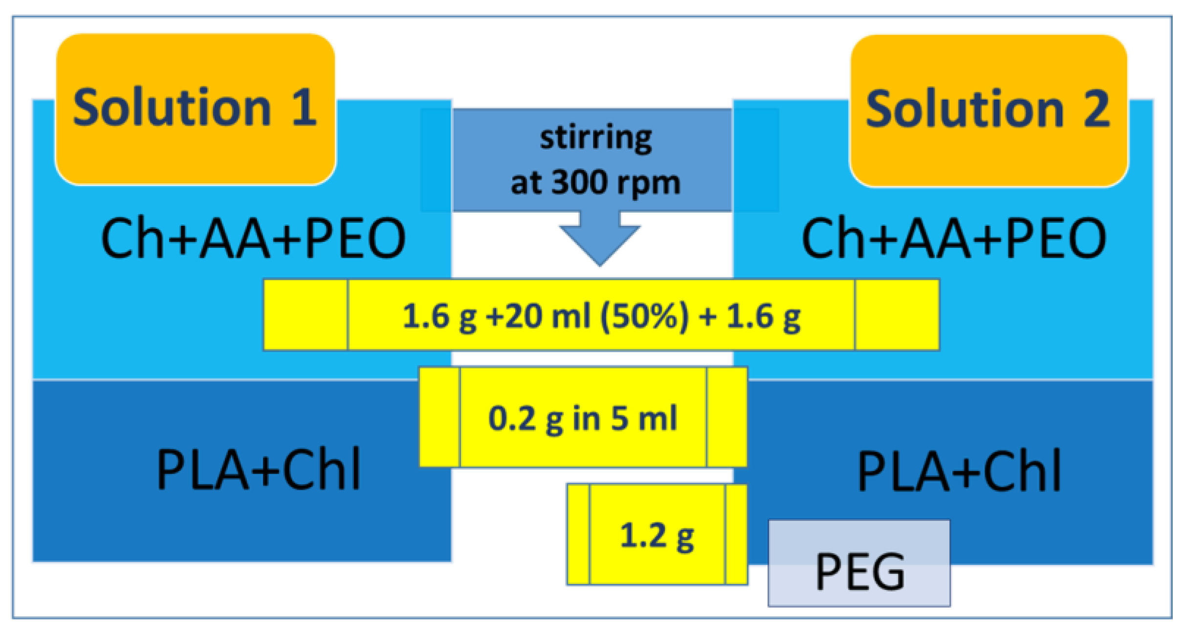
Abstract
1. Introduction
2. Materials and Methods
2.1. Materials
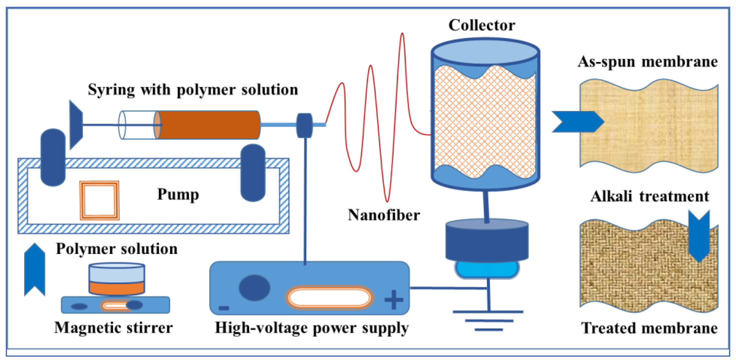
2.2. Chitosan Solutions Preparation, Electrospinning of Chitosan Nanofibrous Membranes, and Their Alkali Neutralization
2.3. Scanning Electron Microscopy (SEM)
2.4. Surface Characterization-Hydrophobicity
2.5. Fourier Transform Infrared Spectroscopy (FT-IR)
2.5.1. FT-IR
2.5.2. Adhesive Properties
2.6. Weight Loss (WL)
2.7. Bacteriological Experiment
2.7.1. Bacterial Reduction Rate
2.7.2. Disk Diffusion Method
2.7.3. Biofilm Formation Study
2.8. In Vitro Cell Culture Study
2.9. Statistics
3. Results
3.1. Material Characterization
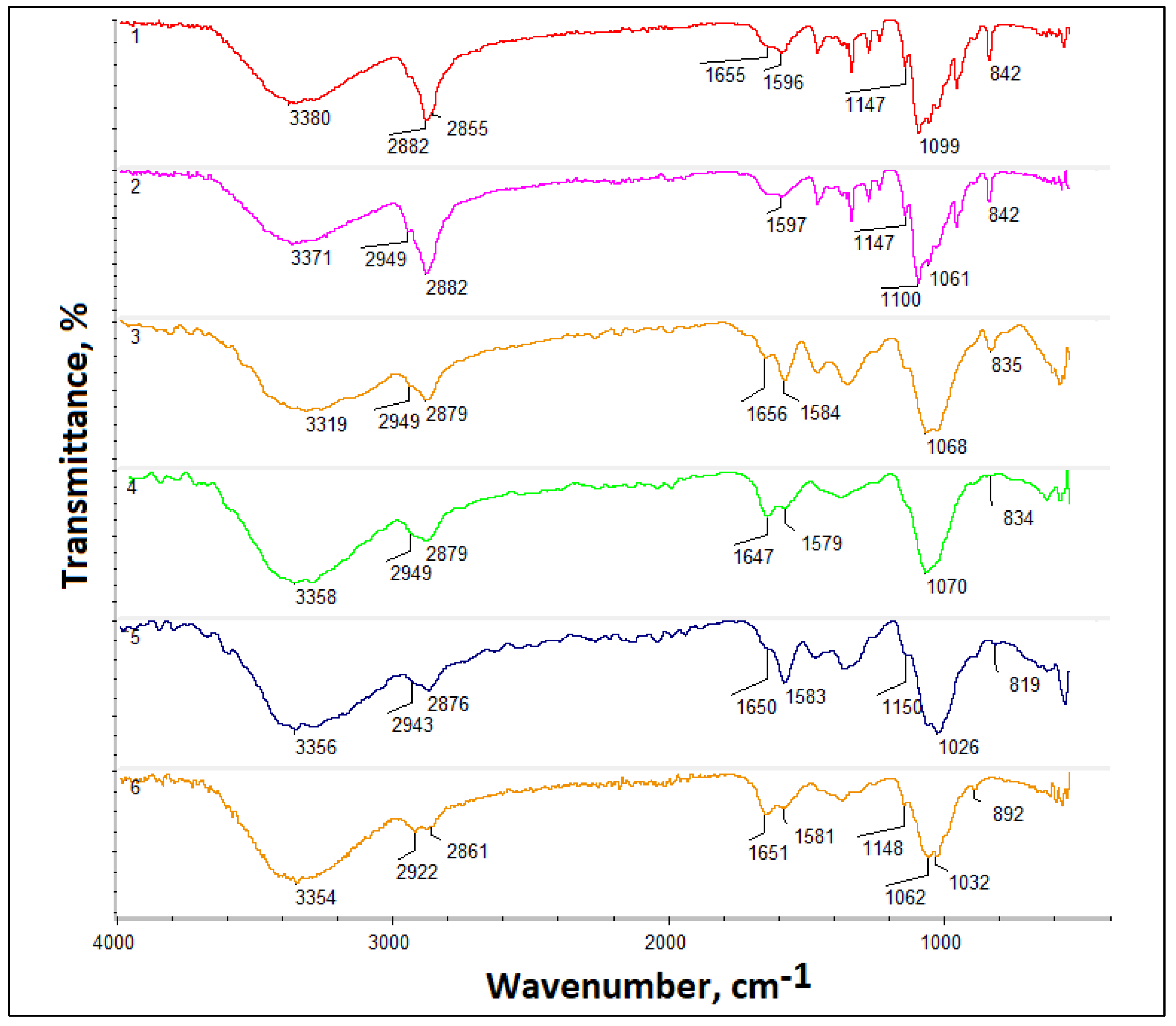
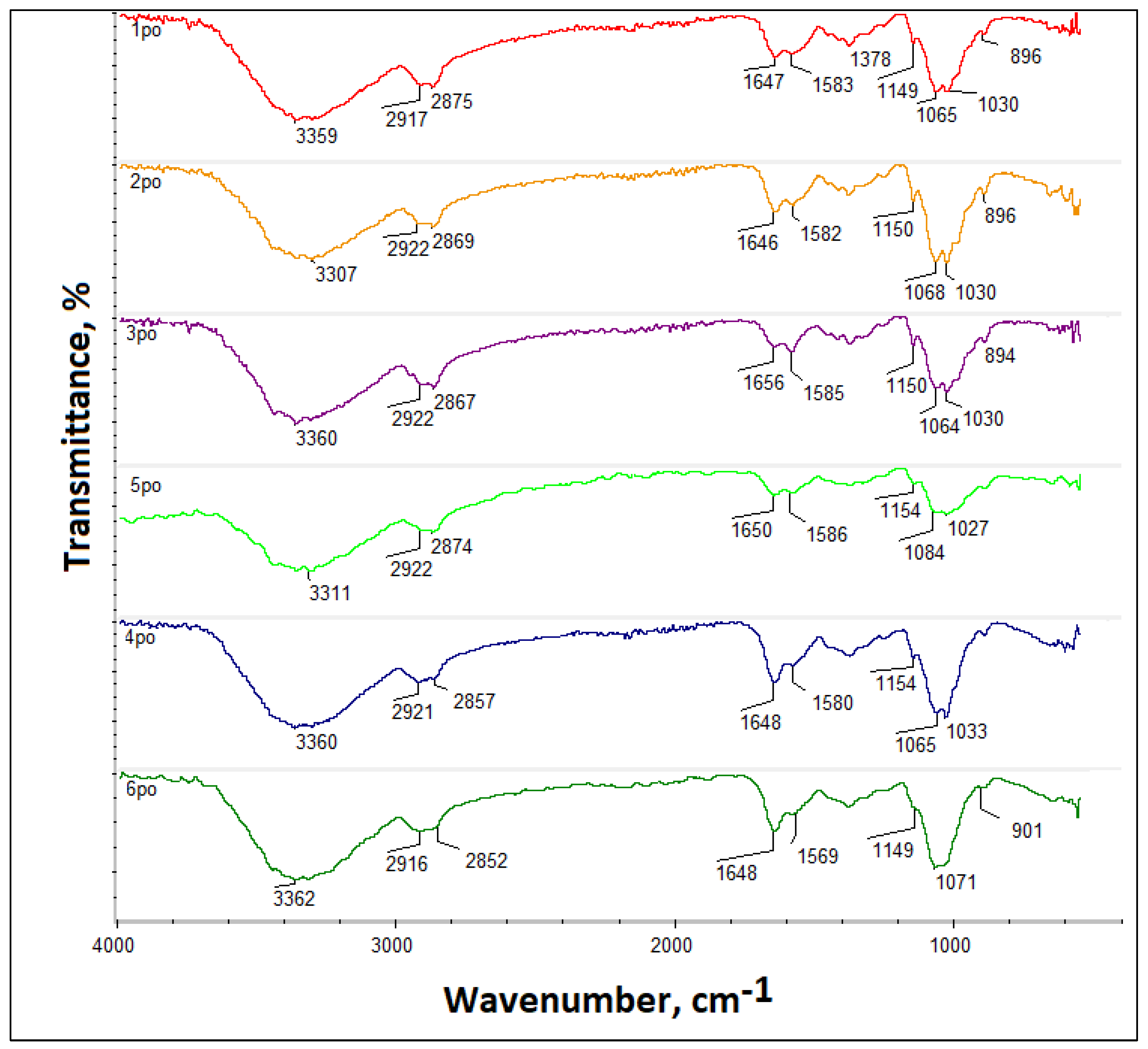
3.1.1. FT-IR Analysis
3.1.2. Adhesive Properties
3.2. SEM
3.3. Contact Angle Measurement
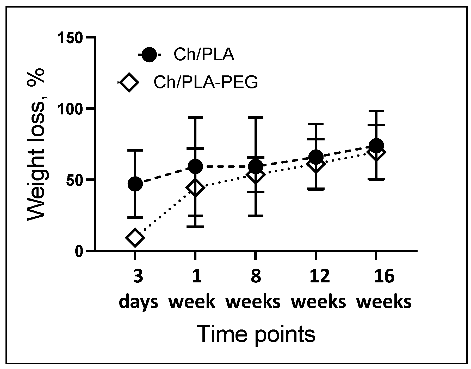
3.4. Weight Loss (WL)
3.5. Antibacterial Effects
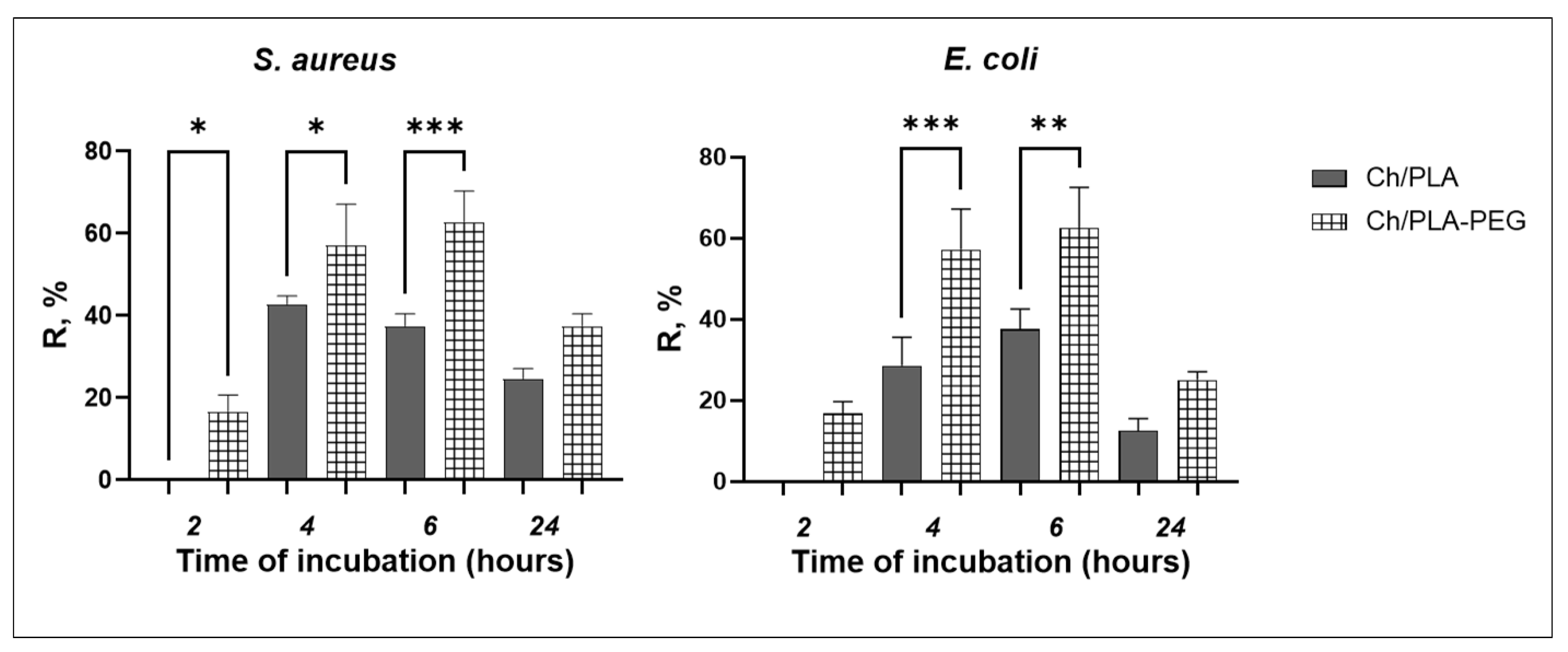
3.5.1. Bacterial Reduction Rate
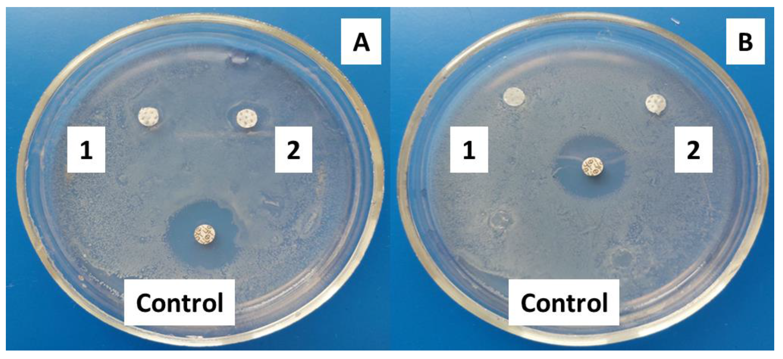
3.5.2. Antimicrobial Assay: Disk Diffusion Susceptibility Test
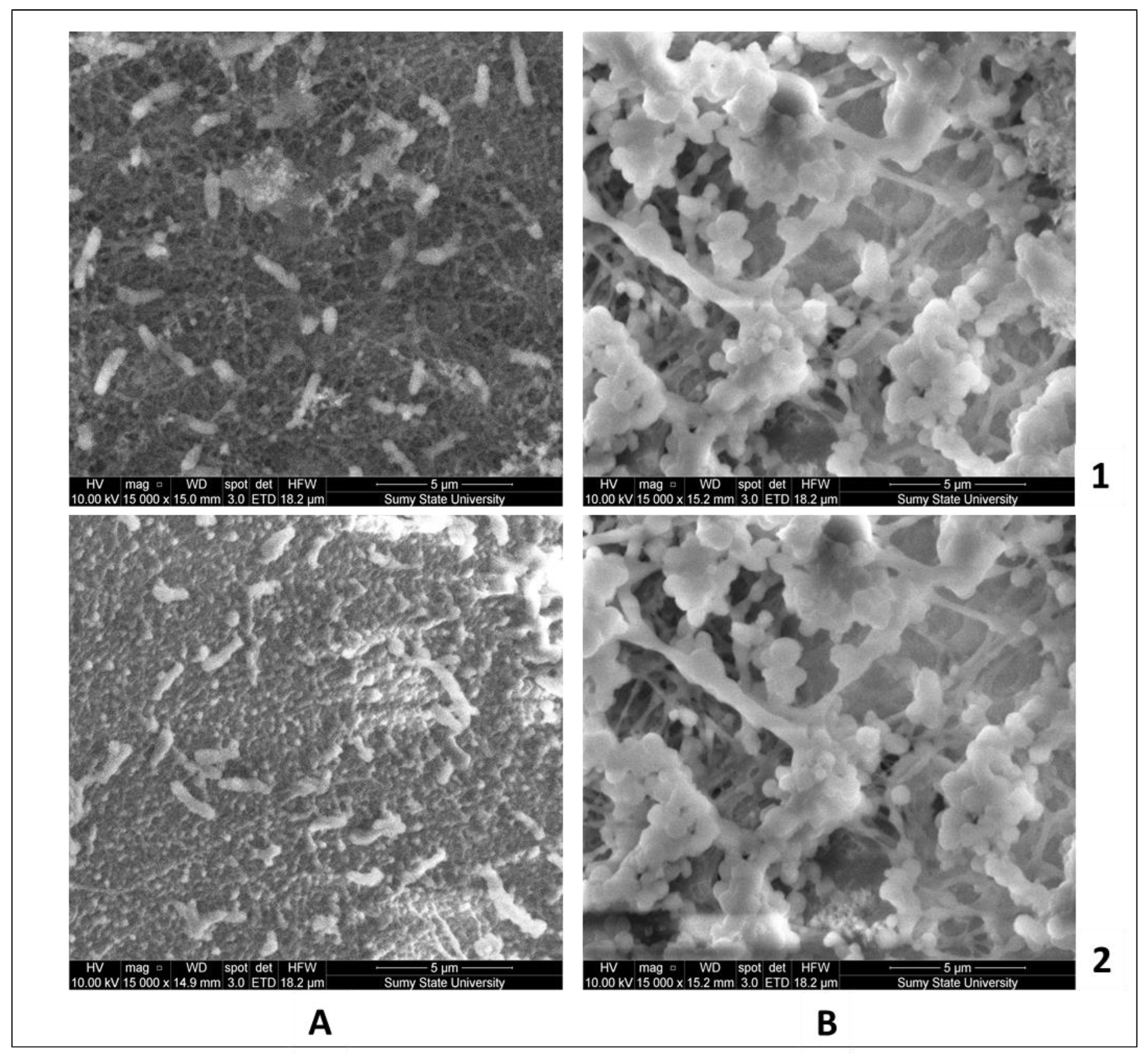
3.5.3. Morphology of the Bacteria Biofilm by SEM
3.6. In Vitro Cell Culture Study
4. Discussion
5. Conclusions
Author Contributions
Funding
Data Availability Statement
Conflicts of Interest
References
- Amna, R.; Ali, K.; Malik, M.I.; Shamsah, S.I. A brief review of electrospinning of polymer nanofibers: History and main applications. J. New Mater. Electrochem. Syst. 2020, 23, 151–163. [Google Scholar] [CrossRef]
- Bhagure, S.S.; Rao, D.A.R. A Review: Electrospinning and Electrospinning Nanofibre Technology, Process & Application. Int. J. Innov. Sci. Res. Technol. 2020, 5, 528–538. [Google Scholar] [CrossRef]
- Chen, K.; Li, Y.; Li, Y.; Pan, W.; Tan, G. Silk Fibroin Combined with Electrospinning as a Promising Strategy for Tissue Regeneration. Macromol. Biosci. 2023, 23, 2200380. [Google Scholar] [CrossRef] [PubMed]
- Ji, G.; Chen, Z.; Li, H.; Awuye, D.E.; Guan, M.; Zhu, Y. Electrospinning-Based Biosensors for Health Monitoring. Biosensors 2022, 12, 876. [Google Scholar] [CrossRef]
- Kianfar, P.; Bongiovanni, R.; Ameduri, B.; Vitale, A. Electrospinning of Fluorinated Polymers: Current State of the Art on Processes and Applications. Polym. Rev. 2023, 63, 127–199. [Google Scholar] [CrossRef]
- Cui, H.; Lu, J.; Li, C.; Rashed, M.M.A.; Lin, L. Antibacterial and physical effects of cationic starch nanofibers containing carvacrol@casein nanoparticles against Bacillus cereus in soy products. Int. J. Food Microbiol. 2022, 364, 109530. [Google Scholar] [CrossRef]
- Chen, Y.; Zhao, W.; Dai, H. Porous PLGA-PEG nerve conduit decorated with oriented electrospun chitosan-RGD nanofibre. J. Mater. Res. Technol. 2021, 15, 86–98. [Google Scholar] [CrossRef]
- Preet, G. A critical analysis on parameters affecting the formation of nano fibers using electrospinning technique. Int. J. Eng. Trends Technol. 2021, 69, 126–131. [Google Scholar] [CrossRef]
- Wei, L.; Yu, H.-N.; Sun, R.; Liu, C.; Chen, M.-Y.; Liu, H.; Xiong, J.; Qin, X. Experimental investigation of process parameters for the filtration property of nanofiber membrane fabricated by needleless electrospinning apparatus. J. Ind. Text. 2021, 50, 1528–1541. [Google Scholar] [CrossRef]
- Ranjbar-Mohammadi, M.; Shakoori, P.; Arab-Bafrani, Z.; Zabihi, E. Fibrous structures fabricated from polylactic acid and nanofibrillated chitosan/zinc oxide nanoparticles. Iran. J. Polym. Sci. Technol. 2021, 34, 174–191. [Google Scholar] [CrossRef]
- Maliszewska, I.; Czapka, T. Electrospun Polymer Nanofibers with Antimicrobial Activity. Polymers 2022, 14, 1661. [Google Scholar] [CrossRef]
- Maleki Dizaj, S.; Sharifi, S.; Jahangiri, A. Electrospun nanofibers as versatile platform in antimicrobial delivery: Current state and perspectives. Pharm. Dev. Technol. 2019, 24, 1187–1199. [Google Scholar] [CrossRef]
- Demchenko, V.; Rybalchenko, N.; Zahorodnia, S.; Naumenko, K.; Riabov, S.; Kobylinskyi, S.; Vashchuk, A.; Mamunya, Y.; Iurzhenko, M.; Demchenko, O.; et al. Preparation, Characterization, and Antimicrobial and Antiviral Properties of Silver-Containing Nanocomposites Based on Polylactic Acid–Chitosan. ACS Appl. Bio Mater. 2022, 5, 2576–2585. [Google Scholar] [CrossRef]
- Khazaeli, P.; Alaei, M.; Khaksarihadad, M.; Ranjbar, M. Preparation of PLA/chitosan nanoscaffolds containing cod liver oil and experimental diabetic wound healing in male rats study. J. Nanobiotechnol. 2020, 18, 176. [Google Scholar] [CrossRef] [PubMed]
- Osman, M.A.; Virgilio, N.; Rouabhia, M.; Mighri, F. Development and Characterization of Functional Polylactic Acid/Chitosan Porous Scaffolds for Bone Tissue Engineering. Polymers 2022, 14, 5079. [Google Scholar] [CrossRef] [PubMed]
- Ajith, G.; Tamilarasi, G.P.; Sabarees, G.; Gouthaman, S.; Manikandan, K.; Velmurugan, V.; Alagarsamy, V.; Solomon, V.R. Recent Developments in Electrospun Nanofibers as Delivery of Phytoconstituents for Wound Healing. Drugs Drug Candidates 2023, 2, 148–171. [Google Scholar] [CrossRef]
- Chen, S.; Tian, H.; Mao, J.; Ma, F.; Zhang, M.; Chen, F.; Yang, P. Preparation and application of chitosan-based medical electrospun nanofibers. Int. J. Biol. Macromol. 2023, 226, 410–422. [Google Scholar] [CrossRef]
- Parham, S.; Kharazi, A.Z.; Bakhsheshi-Rad, H.R.; Ghayour, H.; Ismail, A.F.; Nur, H.; Berto, F. Electrospun Nanofibers for biomedical and tissue engineering applications: A comprehensive review. Materials 2020, 13, 2153. [Google Scholar] [CrossRef]
- Ranjbar-Mohammadi, M.; Sa’di, V.; Moezzi, M.; Saghafi, R. Fabrication and Characterization of Antibacterial Suture Yarns Containing PLA/Tetracycline Hydrochloride-PVA/Chitosan Nanofibers. Fibers Polym. 2022, 23, 1538–1547. [Google Scholar] [CrossRef]
- Cui, C.; Sun, S.; Wu, S.; Chen, S.; Ma, J.; Zhou, F. Electrospun chitosan nanofibers for wound healing application. Eng. Regen. 2021, 2, 82–90. [Google Scholar] [CrossRef]
- Pakravan, M.; Heuzey, M.C.; Ajji, A. A fundamental study of chitosan/PEO electrospinning. Polymer 2011, 52, 4813–4824. [Google Scholar] [CrossRef]
- Yang, Q.; Guo, J.; Zhang, S.; Guan, F.; Yu, Y.; Feng, S.; Song, X.; Bao, D.; Zhang, X. Development of cell adhesive and inherently antibacterial polyvinyl alcohol/polyethylene oxide nanofiber scaffolds via incorporating chitosan for tissue engineering. Int. J. Biol. Macromol. 2023, 236, 124004. [Google Scholar] [CrossRef]
- Odili, C.; Sekunowo, I.O.; Ilomuanya, M.O.; Gbenebor, O.P.; Adeosun, S.O. Strength, Water Absorption, Thermal and Antimicrobial Properties of a Biopolymer Composite Wound Dressing. J. Cast. Mater. Eng. 2022, 6, 22–32. [Google Scholar] [CrossRef]
- Capuana, E.; Lopresti, F.; Ceraulo, M.; La Carrubba, V. Poly-L-Lactic Acid (PLLA)-Based Biomaterials for Regenerative Medicine: A Review on Processing and Applications. Polymers 2022, 14, 1153. [Google Scholar] [CrossRef]
- Deng, A.; Yang, Y.; Du, S. Tissue engineering 3D porous scaffolds prepared from electrospun recombinant human collagen (Rhc) polypeptides/chitosan nanofibers. Appl. Sci. 2021, 11, 5096. [Google Scholar] [CrossRef]
- Mania, S.; Partyka, K.; Pilch, J.; Augustin, E.; Cieślik, M.; Ryl, J.; Jinn, J.-R.; Wang, Y.-J.; Michałowska, A.; Tylingo, R. Obtaining and characterization of the PLA/chitosan foams with antimicrobial properties achieved by the emulsification combined with the dissolution of chitosan by CO2 saturation. Molecules 2019, 24, 4532. [Google Scholar] [CrossRef]
- Morariu, S.; Brunchi, C.E.; Honciuc, M.; Iftime, M.M. Development of Hybrid Materials Based on Chitosan, Poly(Ethylene Glycol) and Laponite® RD: Effect of Clay Concentration. Polymers 2023, 15, 841. [Google Scholar] [CrossRef] [PubMed]
- Wu, S.; Zhao, W.; Sun, M.; He, P.; Lv, H.; Wang, Q.; Zhang, S.; Wu, Q.; Ling, P.; Chen, S.; et al. Novel bi-layered dressing patches constructed with radially-oriented nanofibrous pattern and herbal compound-loaded hydrogel for accelerated diabetic wound healing. Appl. Mater. Today 2022, 28, 101542. [Google Scholar] [CrossRef]
- Kong, X.; Feng, M.; Wu, L.; He, Y.; Mao, H.; Gu, Z. Biodegradable gemcitabine-loaded microdevice with sustained local drug delivery and improved tumor recurrence inhibition abilities for postoperative pancreatic tumor treatment. Drug Deliv. 2022, 29, 1595–1607. [Google Scholar] [CrossRef]
- Du, J.; Hsieh, Y.L. PEGylation of chitosan for improved solubility and fiber formation via electrospinning. Cellulose 2007, 14, 543–552. [Google Scholar] [CrossRef]
- Yin, J.; Xu, L.; Ahmed, A. Batch Preparation and Characterization of Electrospun Porous Polylactic Acid-Based Nanofiber Membranes for Antibacterial Wound Dressing. Adv. Fiber Mater. 2022, 4, 832–844. [Google Scholar] [CrossRef]
- Pereao, O.; Uche, C.; Bublikov, P.; Bode-Aluko, C.; Rossouw, A.; Vinogradov, I.; Nechaev, A.; Opeolu, B.; Petrik, L. Chitosan/PEO nanofibers electrospun on metallized track-etched membranes: Fabrication and characterization. Mater. Today Chem. 2021, 20, 100416. [Google Scholar] [CrossRef]
- Kanani, N.; Rahmayetty; Wardhono, E.Y.; Wardalia. Preparation and characterization of blend film based on chitosan-poly lactic acid (PLA) composites. AIP Conf. Proc. 2021, 2370, 020026. [Google Scholar] [CrossRef]
- Santhanes, D.; Wilkins, A.; Zhang, H.; John Aitken, R.; Liang, M. Microfluidic formulation of lipid/polymer hybrid nanoparticles for plasmid DNA (pDNA) delivery. Int. J. Pharm. 2022, 627, 122223. [Google Scholar] [CrossRef] [PubMed]
- Ziel, R.; Haus, A.; Tulke, A. Quantification of the pore size distribution (porosity profiles) in microfiltration membranes by SEM, TEM and computer image analysis. J. Membr. Sci. 2008, 323, 241–246. [Google Scholar] [CrossRef]
- Ignatova, M.; Anastasova, I.; Manolova, N.; Rashkov, I.; Markova, N.; Kukeva, R.; Stoyanova, R.; Georgieva, A.; Toshkova, R. Bio-Based Electrospun Fibers from Chitosan Schiff Base and Polylactide and Their Cu2+ and Fe3+ Complexes: Preparation and Antibacterial and Anticancer Activities. Polymers 2022, 14, 5002. [Google Scholar] [CrossRef]
- Vlachopoulos, A.; Karlioti, G.; Balla, E.; Daniilidis, V.; Kalamas, T.; Stefanidou, M.; Bikiaris, N.D.; Christodoulou, E.; Koumentakou, I.; Karavas, E.; et al. Poly(Lactic Acid)-Based Microparticles for Drug Delivery Applications: An Overview of Recent Advances. Pharmaceutics 2022, 14, 359. [Google Scholar] [CrossRef]
- Arkoun, M.; Daigle, F.; Heuzey, M.C.; Ajji, A. Antibacterial electrospun chitosan-based nanofibers: A bacterial membrane perforator. Food Sci. Nutr. 2017, 5, 865–874. [Google Scholar] [CrossRef]
- Alavarse, A.C.; de Oliveira Silva, F.W.; Colque, J.T.; da Silva, V.M.; Prieto, T.; Venancio, E.C.; Bonvent, J.-J. Tetracycline hydrochloride-loaded electrospun nanofibers mats based on PVA and chitosan for wound dressing. Mater. Sci. Eng. C 2017, 77, 271–281. [Google Scholar] [CrossRef]
- Korniienko, V.; Husak, Y.; Radwan-Pragłowska, J.; Holubnycha, V.; Samokhin, Y.; Yanovska, A.; Varava, J.; Diedkova, K.; Janus, Ł.; Pogorielov, M. Impact of Electrospinning Parameters and Post-Treatment Method on Antibacterial and Antibiofilm Activity of Chitosan Nanofibers. Molecules 2022, 27, 3343. [Google Scholar] [CrossRef]
- Korniienko, V.; Husak, Y.; Yanovska, A.; Banasiuk, R.; Yusupova, A.; Savchenko, A.; Holubnycha, V.; Pogorielov, M. Functional and biological characterization of chitosan electrospun nanofibrous membrane nucleated with silver nanoparticles. Appl. Nanosci. 2022, 12, 1061–1070. [Google Scholar] [CrossRef]
- Korniienko, V.; Husak, Y.; Yanovska, A.; Altundal, Ş.; Diedkova, K.; Samokhin, Y.; Varava, Y.; Holubnycha, V.; Viter, R.; Pogorielov, M. Biological behaviour of chitosan electrospun nanofibrous membranes after different neutralisation methods. Prog. Chem. Appl. Chitin Its Deriv. 2022, 27, 135–153. [Google Scholar] [CrossRef]
- Xu, J.; Zhang, J.; Gao, W.; Liang, H.; Wang, H.; Li, J. Preparation of chitosan/PLA blend micro/nanofibers by electrospinning. Mater. Lett. 2009, 63, 658–660. [Google Scholar] [CrossRef]
- Sukum, P.; Punyodom, W.; Dangtip, S.; Poramapijitwat, P.; Daranarong, D.; Jenvoraphot, T.; Nisoa, M.; Kuensaen, C.; Boonyawan, D. Argon Plasma Jet-Treated Poly (Vinyl Alcohol)/Chitosan and PEG 400 Plus Mangifera indica Leaf Extract for Electrospun Nanofiber Membranes: In Vitro Study. Polymers 2023, 15, 2559. [Google Scholar] [CrossRef]
- Hendrick, E.; Frey, M. Increasing surface hydrophilicity in poly(lactic acid) electrospun fibers by addition of Pla-B-Peg copolymers. J. Eng. Fibers Fabr. 2014, 9, 155892501400900219. [Google Scholar] [CrossRef]
- Feng, P.; Jia, J.; Liu, M.; Peng, S.; Zhao, Z.; Shuai, C. Degradation mechanisms and acceleration strategies of poly (lactic acid) scaffold for bone regeneration. Mater. Des. 2021, 210, 110066. [Google Scholar] [CrossRef]
- Tighzert, W.; Habi, A.; Ajji, A.; Sadoun, T.; Daoud, F.B.-O. Fabrication and characterization of nanofibers based on poly(lactic acid)/chitosan blends by electrospinning and their functionalization with phospholipase A1. Fibers Polym. 2017, 18, 514–524. [Google Scholar] [CrossRef]
- Gao, Y.; Truong, Y.B.; Zhu, Y.; Louis Kyratzis, I. Electrospun antibacterial nanofibers: Production, activity, and in vivo applications. J. Appl. Polym. Sci. 2014, 131, 9041–9053. [Google Scholar] [CrossRef]
- Toncheva, A.; Mincheva, R.; Kancheva, M.; Manolova, N.; Rashkov, I.; Dubois, P.; Markova, N. Antibacterial PLA/PEG electrospun fibers: Comparative study between grafting and blending PEG. Eur. Polym. J. 2016, 75, 223–233. [Google Scholar] [CrossRef]
- Aijaz, M.O.; Yang, S.B.; Karim, M.R.; Othman, M.H.D.; Alnaser, I.A. Preparation and Characterization of Poly(Lactic Acid)/Poly (ethylene glycol)-Poly(propyl glycol)-Poly(ethylene glycol) Blended Nanofiber Membranes for Fog Collection. Membranes 2023, 13, 32. [Google Scholar] [CrossRef]
- Kaplan, J.B.; Mlynek, K.D.; Hettiarachchi, H.; Alamneh, Y.A.; Biggemann, L.; Zurawski, D.V.; Black, C.C.; Bane, C.E.; Kim, R.K.; Granick, M.S. Extracellular polymeric substance (EPS)-degrading enzymes reduce staphylococcal surface attachment and biocide resistance on pig skin in vivo. PLoS ONE 2018, 13, e0205526. [Google Scholar] [CrossRef] [PubMed]
- Liu, C.; Du, G.; Guo, Q.; Li, R.; Li, C.; He, H. Fabrication and Characterization of Polylactic Acid Electrospun Wound Dressing Modified with Polyethylene Glycol, Rosmarinic Acid and Graphite Oxide. Nanomaterials 2023, 13, 2000. [Google Scholar] [CrossRef] [PubMed]
- Moradkhannejhad, L.; Abdouss, M.; Nikfarjam, N.; Shahriari, M.H.; Heidary, V. The effect of molecular weight and content of PEG on in vitro drug release of electrospun curcumin loaded PLA/PEG nanofibers. J. Drug Deliv. Sci. Technol. 2020, 56, 101554. [Google Scholar] [CrossRef]
- Qian, Z.; Ni, P.; Fu, S.; Fan, M.; Guo, G.; Shi, S.; Peng, J. Preparation of poly(ethylene glycol)/polylactide hybrid fibrous scaffolds for bone tissue engineering. Int. J. Nanomed. 2011, 6, 3065–3075. [Google Scholar] [CrossRef][Green Version]
- Li, H.; Wang, Z.; Zhang, H.; Pan, Z. Nanoporous PLA/(Chitosan Nanoparticle) composite fibrous membranes with excellent air filtration and antibacterial per-formance. Polymers 2018, 10, 1085. [Google Scholar] [CrossRef] [PubMed]
- Maleki, H.; Azimi, B.; Ismaeilimoghadam, S.; Danti, S. Poly(lactic acid)-Based Electrospun Fibrous Structures for Biomedical Applications. Appl. Sci. 2022, 12, 3192. [Google Scholar] [CrossRef]


Disclaimer/Publisher’s Note: The statements, opinions and data contained in all publications are solely those of the individual author(s) and contributor(s) and not of MDPI and/or the editor(s). MDPI and/or the editor(s) disclaim responsibility for any injury to people or property resulting from any ideas, methods, instructions or products referred to in the content. |
© 2023 by the authors. Licensee MDPI, Basel, Switzerland. This article is an open access article distributed under the terms and conditions of the Creative Commons Attribution (CC BY) license (https://creativecommons.org/licenses/by/4.0/).
Share and Cite
Samokhin, Y.; Varava, Y.; Diedkova, K.; Yanko, I.; Husak, Y.; Radwan-Pragłowska, J.; Pogorielova, O.; Janus, Ł.; Pogorielov, M.; Korniienko, V. Fabrication and Characterization of Electrospun Chitosan/Polylactic Acid (CH/PLA) Nanofiber Scaffolds for Biomedical Application. J. Funct. Biomater. 2023, 14, 414. https://doi.org/10.3390/jfb14080414
Samokhin Y, Varava Y, Diedkova K, Yanko I, Husak Y, Radwan-Pragłowska J, Pogorielova O, Janus Ł, Pogorielov M, Korniienko V. Fabrication and Characterization of Electrospun Chitosan/Polylactic Acid (CH/PLA) Nanofiber Scaffolds for Biomedical Application. Journal of Functional Biomaterials. 2023; 14(8):414. https://doi.org/10.3390/jfb14080414
Chicago/Turabian StyleSamokhin, Yevhen, Yuliia Varava, Kateryna Diedkova, Ilya Yanko, Yevheniia Husak, Julia Radwan-Pragłowska, Oksana Pogorielova, Łukasz Janus, Maksym Pogorielov, and Viktoriia Korniienko. 2023. "Fabrication and Characterization of Electrospun Chitosan/Polylactic Acid (CH/PLA) Nanofiber Scaffolds for Biomedical Application" Journal of Functional Biomaterials 14, no. 8: 414. https://doi.org/10.3390/jfb14080414
APA StyleSamokhin, Y., Varava, Y., Diedkova, K., Yanko, I., Husak, Y., Radwan-Pragłowska, J., Pogorielova, O., Janus, Ł., Pogorielov, M., & Korniienko, V. (2023). Fabrication and Characterization of Electrospun Chitosan/Polylactic Acid (CH/PLA) Nanofiber Scaffolds for Biomedical Application. Journal of Functional Biomaterials, 14(8), 414. https://doi.org/10.3390/jfb14080414