

Cytotoxicity of Biodegradable Zinc and Its Alloys: A Systematic Review
 ,
, 
 and
and 
Abstract
1. Introduction
2. Materials and Methods
2.1. Quality Assurance and Criteria
2.2. Search Strategy
- Population (P): cells.
- Intervention (I): biodegradable Zn and its alloys.
- Comparison (C): nonbiodegradable metals, such as stainless steel, titanium, titanium alloy, and cobalt–chromium alloy; biodegradable polymers, such as polylactic acid; other biodegradable metals, such as Mg-based BMs.
- Outcome (O): cell viability.
- Study design (S): in vitro study.
2.3. Inclusion and Exclusion Criteria
2.4. Study Selection and Data Extraction
2.5. Assessment of Quality of Evidence
3. Results
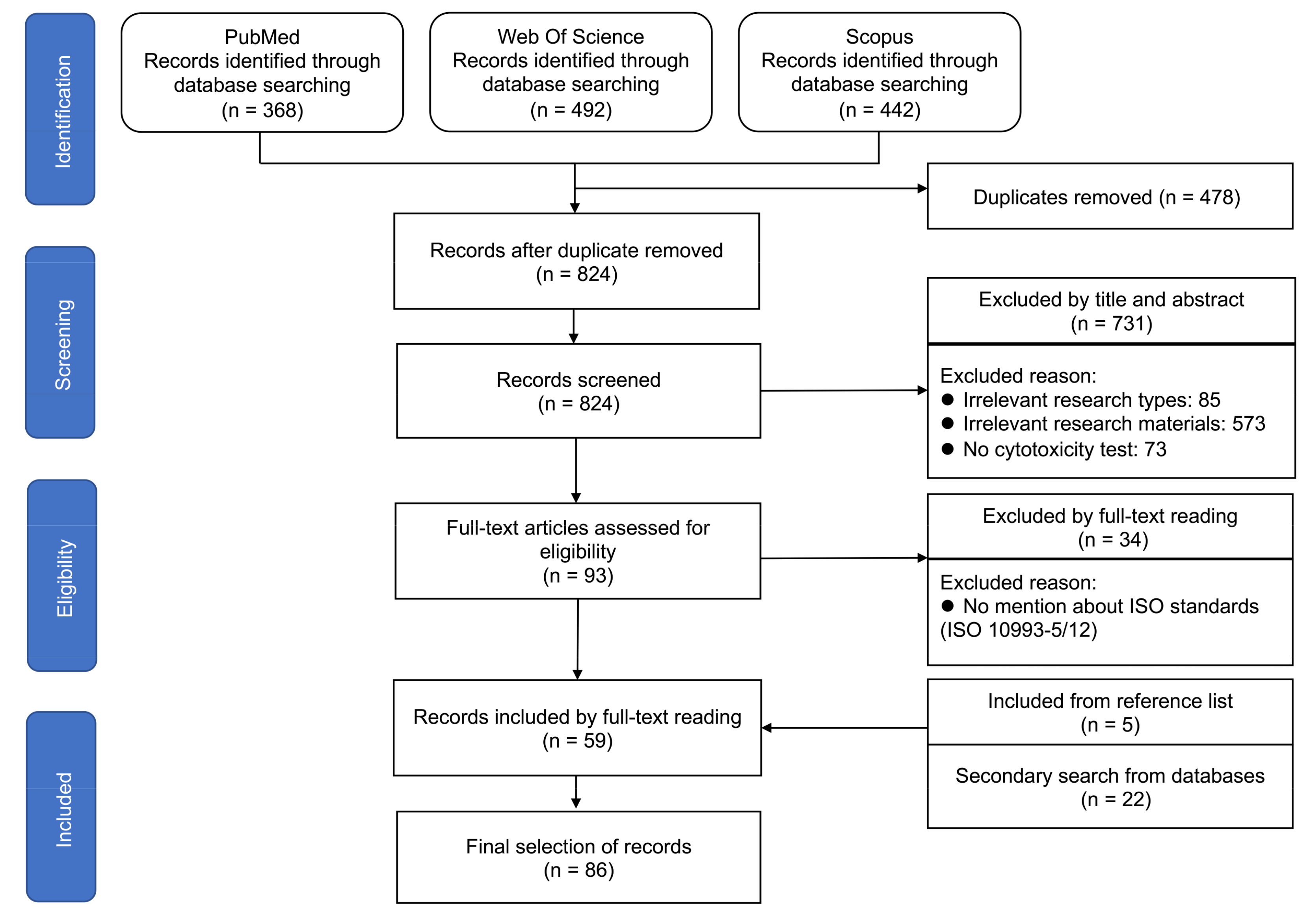
3.1. Included Studies
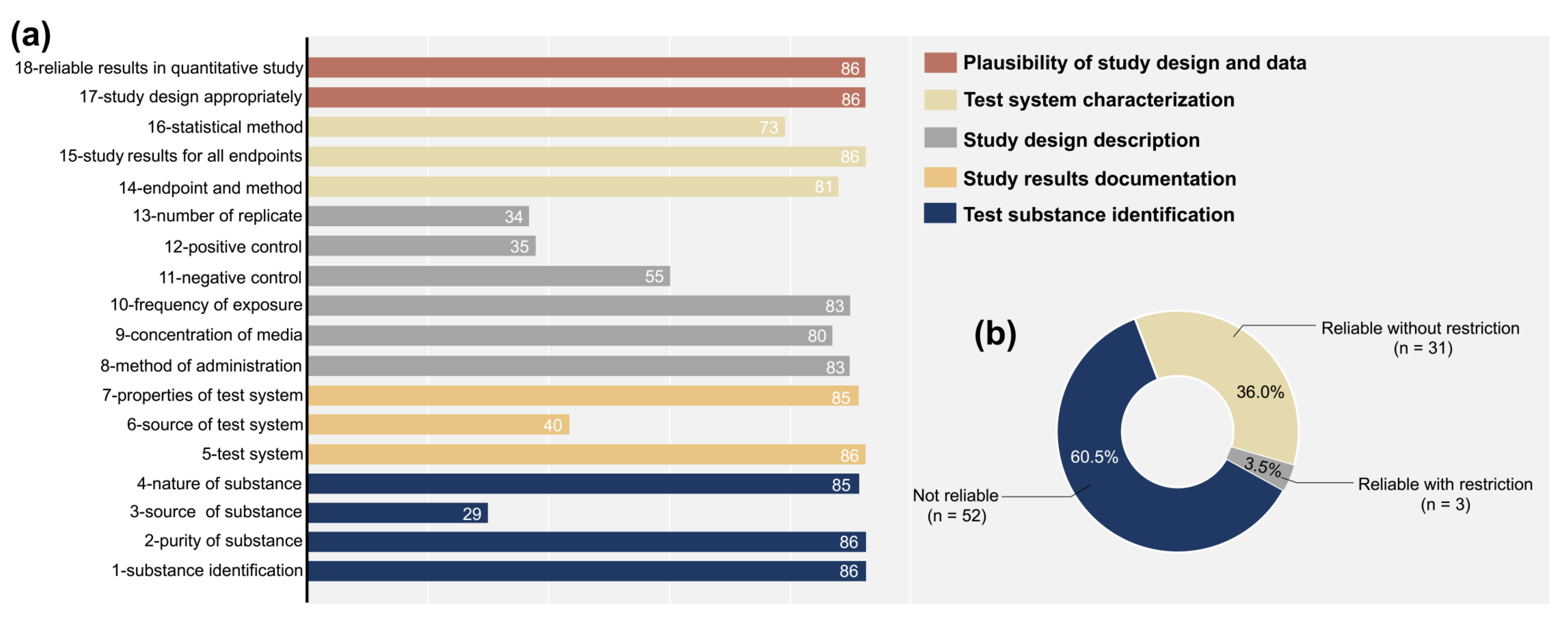
3.2. Quality Assessment According to the ToxRTool
3.3. Main Characteristics of the Included Articles
3.3.1. Materials and Processing
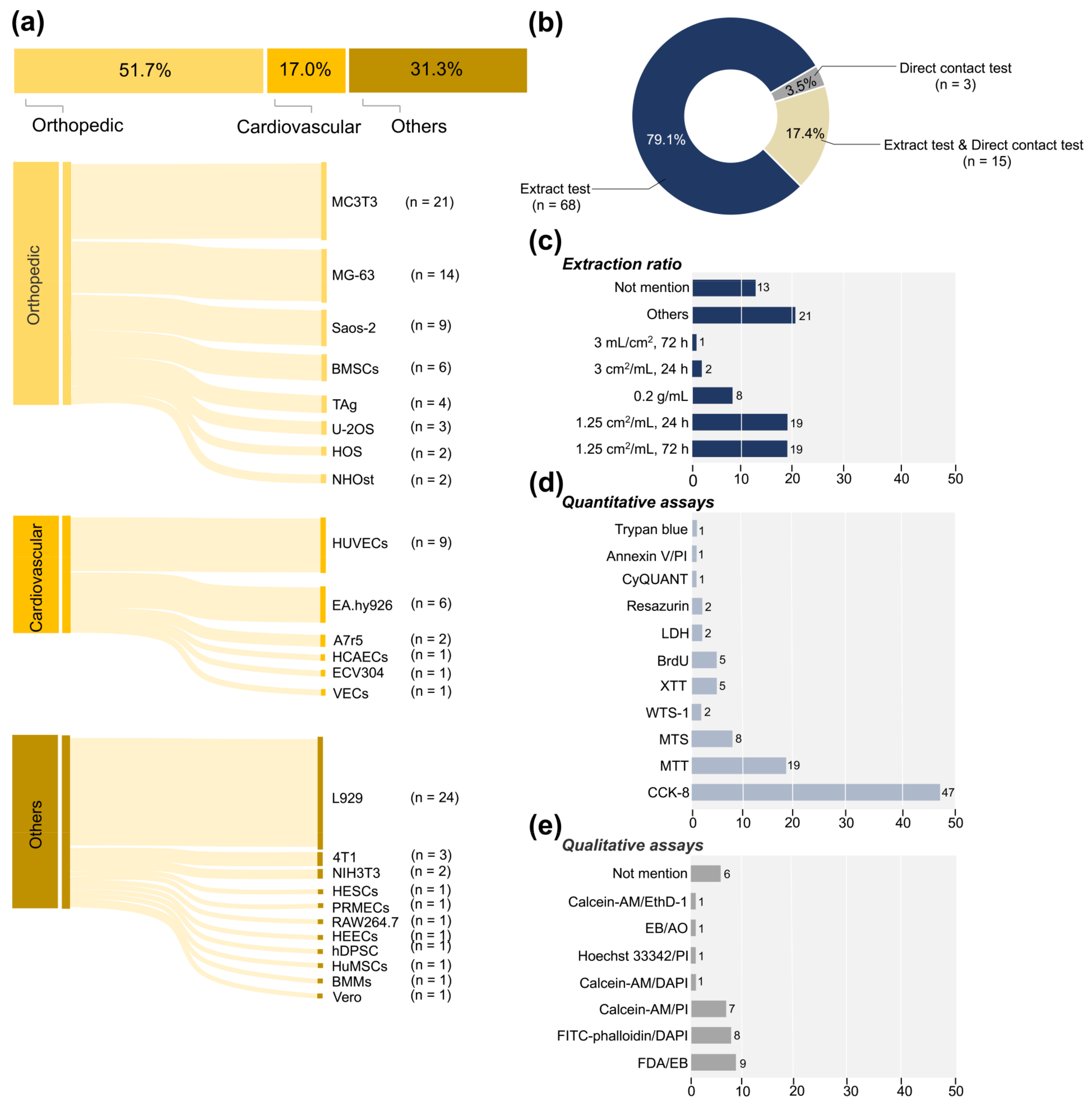
3.3.2. Tested Cell Types
3.3.3. Test System
3.3.4. Outcome
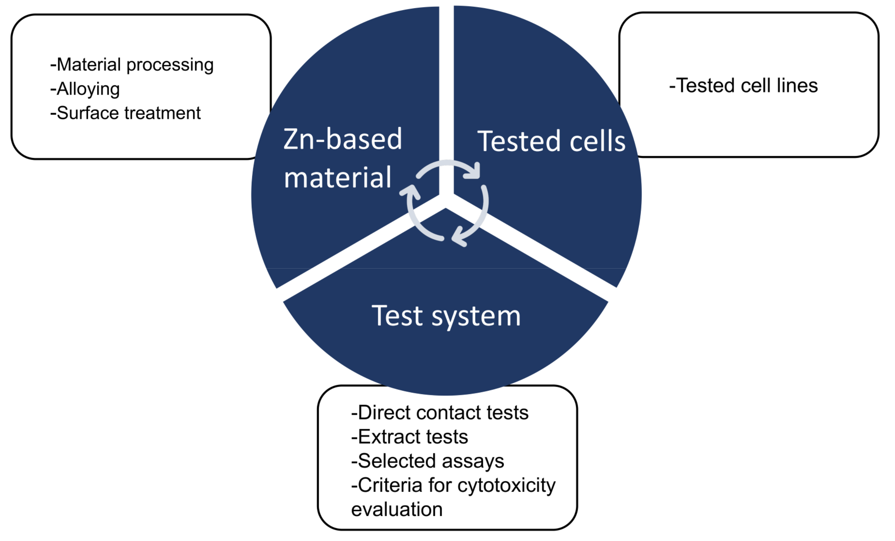
4. Discussion
4.1. Effects of the Materials on Cytotoxicity
4.1.1. Material Processing
4.1.2. Alloying and Its Micro-Galvanic Corrosion
4.1.3. Surface Treatment
4.2. Effects of Tested Cells on Cytotoxicity
4.3. Effects of Test System on Cytotoxicity
4.3.1. Parameters of the Extract Tests
4.3.2. Direct Contact Tests
4.3.3. Selected Assays
4.3.4. The Criteria of Cytotoxicity Evaluation
4.4. Strengths and Limitations of This Work
5. Conclusions
- High heterogeneity exists in the implementation of the included studies and the assessment results of the toxicity studies.
- The qualitative analysis demonstrated that biodegradable Zn and its alloys had conditionally cytotoxic effects, mainly dependent on the Zn-based materials, tested cells, and test systems.
- The material processing technologies and alloying elements had a potential effect on the toxicity of Zn-based BMs due to modifications in microstructure and corrosion characteristics.
- Endothelial cells had better tolerance to the toxic effects of Zn-based BMs than other tested cells.
- A standardized in vitro toxicity assessment system for biodegradable metals is still lacking, and further construction is required. In addition, researchers in this field need to comply with existing evaluation criteria and report test procedures in as much detail as possible to make the study data more informative and valuable to promote translational research and the long-term development of Zn-based BMs.
Supplementary Materials
Author Contributions
Funding
Institutional Review Board Statement
Informed Consent Statement
Data Availability Statement
Conflicts of Interest
References
- Han, H.S.; Loffredo, S.; Jun, I.; Edwards, J.; Kim, Y.C.; Seok, H.K.; Witte, F.; Mantovani, D.; Glyn-Jones, S. Current status and outlook on the clinical translation of biodegradable metals. Mater. Today 2019, 23, 57–71. [Google Scholar] [CrossRef]
- Liu, Y.; Zheng, Y.F.; Chen, X.H.; Yang, J.A.; Pan, H.B.; Chen, D.F.; Wang, L.N.; Zhang, J.L.; Zhu, D.H.; Wu, S.L.; et al. Fundamental theory of biodegradable metals—definition, criteria, and design. Adv. Funct. Mater. 2019, 29, 1805402. [Google Scholar] [CrossRef]
- Zhao, D.W.; Witte, F.; Lu, F.Q.; Wang, J.L.; Li, J.L.; Qin, L. Current status on clinical applications of magnesium-based orthopaedic implants: A review from clinical translational perspective. Biomaterials 2017, 112, 287–302. [Google Scholar] [CrossRef] [PubMed]
- Bowen, P.K.; Drelich, J.; Goldman, J. Zinc exhibits ideal physiological corrosion behavior for bioabsorbable stents. Adv. Mater. 2013, 25, 2577–2582. [Google Scholar] [CrossRef] [PubMed]
- Mostaed, E.; Sikora-Jasinska, M.; Drelich, J.W.; Vedani, M. Zinc-based alloys for degradable vascular stent applications. Acta Biomater. 2018, 71, 1–23. [Google Scholar] [CrossRef]
- Bowen, P.K.; Shearier, E.R.; Zhao, S.; Guillory, R.J., 2nd; Zhao, F.; Goldman, J.; Drelich, J.W. Biodegradable Metals for Cardiovascular Stents: From Clinical Concerns to Recent Zn-Alloys. Adv. Healthc. Mater. 2016, 5, 1121–1140. [Google Scholar] [CrossRef]
- Seitz, J.-M.; Durisin, M.; Goldman, J.; Drelich, J.W. Recent advances in biodegradable metals for medical sutures: A critical review. Adv. Healthc. Mater. 2015, 4, 1915–1936. [Google Scholar] [CrossRef]
- Venezuela, J.J.D.; Johnston, S.; Dargusch, M.S. The Prospects for Biodegradable Zinc in Wound Closure Applications. Adv. Healthc. Mater. 2019, 8, e1900408. [Google Scholar] [CrossRef]
- Xia, D.; Yang, F.; Zheng, Y.; Liu, Y.; Zhou, Y. Research status of biodegradable metals designed for oral and maxillofacial applications: A review. Bioact. Mater. 2021, 6, 4186–4208. [Google Scholar] [CrossRef]
- Chen, J.; Yang, R.; Shi, B.; Xu, Y.; Huang, H. Obturator Manufacturing for Oronasal Fistula after Cleft Palate Repair: A Review from Handicraft to the Application of Digital Techniques. J. Funct. Biomater. 2022, 13, 251. [Google Scholar] [CrossRef]
- Xu, Y.; Huang, H.; Wu, M.; Tian, Y.; Wan, Q.; Shi, B.; Hu, T.; Spintzyk, S. Rapid Additive Manufacturing of a Superlight Obturator for Large Oronasal Fistula in Pediatric Patient. Laryngoscope 2022. [Google Scholar] [CrossRef]
- Bedard, P.; Gauvin, S.; Ferland, K.; Caneparo, C.; Pellerin, E.; Chabaud, S.; Bolduc, S. Innovative Human Three-Dimensional Tissue-Engineered Models as an Alternative to Animal Testing. Bioengineering 2020, 7, 115. [Google Scholar] [CrossRef]
- Przekora, A.; Kazimierczak, P.; Wojcik, M. Ex vivo determination of chitosan/curdlan/hydroxyapatite biomaterial osseointegration with the use of human trabecular bone explant: New method for biocompatibility testing of bone implants reducing animal tests. Mater. Sci. Eng. C Mater. Biol. Appl. 2021, 119, 111612. [Google Scholar] [CrossRef]
- Jung, O.; Smeets, R.; Hartjen, P.; Schnettler, R.; Feyerabend, F.; Klein, M.; Wegner, N.; Walther, F.; Stangier, D.; Henningsen, A.; et al. Improved In Vitro Test Procedure for Full Assessment of the Cytocompatibility of Degradable Magnesium Based on ISO 10993-5/-12. Int. J. Mol. Sci. 2019, 20, 255. [Google Scholar] [CrossRef]
- Jablonska, E.; Kubasek, J.; Vojtech, D.; Ruml, T.; Lipov, J. Test conditions can significantly affect the results of in vitro cytotoxicity testing of degradable metallic biomaterials. Sci. Rep. 2021, 11, 6628. [Google Scholar] [CrossRef]
- Li, P.; Schille, C.; Schweizer, E.; Kimmerle-Muller, E.; Rupp, F.; Heiss, A.; Legner, C.; Klotz, U.E.; Geis-Gerstorfer, J.; Scheideler, L. Selection of extraction medium influences cytotoxicity of zinc and its alloys. Acta Biomater. 2019, 98, 235–245. [Google Scholar] [CrossRef]
- Zhang, K.; Ma, B.; Hu, K.; Yuan, B.; Sun, X.; Song, X.; Tang, Z.; Lin, H.; Zhu, X.; Zheng, Y.; et al. Evidence-based biomaterials research. Bioact. Mater. 2022, 15, 495–503. [Google Scholar] [CrossRef]
- Cheng, J.; Liu, B.; Wu, Y.H.; Zheng, Y.F. Comparative in vitro Study on Pure Metals (Fe, Mn, Mg, Zn and W) as Biodegradable Metals. J. Mater. Sci. Technol. 2013, 29, 619–627. [Google Scholar] [CrossRef]
- Dambatta, M.S.; Murni, N.S.; Izman, S.; Kurniawan, D.; Froemming, G.R.; Hermawan, H. In vitro degradation and cell viability assessment of Zn-3Mg alloy for biodegradable bone implants. Proc. Inst. Mech. Eng. H 2015, 229, 335–342. [Google Scholar] [CrossRef]
- Gong, H.; Wang, K.; Strich, R.; Zhou, J.G. In vitro biodegradation behavior, mechanical properties, and cytotoxicity of biodegradable Zn-Mg alloy. J. Biomed. Mater. Res. B Appl. Biomater. 2015, 103, 1632–1640. [Google Scholar] [CrossRef]
- Kubasek, J.; Vojtech, D.; Jablonska, E.; Pospisilova, I.; Lipov, J.; Ruml, T. Structure, mechanical characteristics and in vitro degradation, cytotoxicity, genotoxicity and mutagenicity of novel biodegradable Zn-Mg alloys. Mater. Sci. Eng. C Mater. Biol. Appl. 2016, 58, 24–35. [Google Scholar] [CrossRef] [PubMed]
- Murni, N.S.; Dambatta, M.S.; Yeap, S.K.; Froemming, G.R.A.; Hermawan, H. Cytotoxicity evaluation of biodegradable Zn-3Mg alloy toward normal human osteoblast cells. Mater. Sci. Eng. C Mater. Biol. Appl. 2015, 49, 560–566. [Google Scholar] [CrossRef] [PubMed]
- Tang, Z.; Huang, H.; Niu, J.; Zhang, L.; Zhang, H.; Pei, J.; Tan, J.; Yuan, G. Design and characterizations of novel biodegradable Zn-Cu-Mg alloys for potential biodegradable implants. Mater. Des. 2017, 117, 84–94. [Google Scholar] [CrossRef]
- Niu, J.; Tang, Z.; Huang, H.; Pei, J.; Zhang, H.; Yuan, G.; Ding, W. Research on a Zn-Cu alloy as a biodegradable material for potential vascular stents application. Mater. Sci. Eng. C Mater. Biol. Appl. 2016, 69, 407–413. [Google Scholar] [CrossRef]
- Jablonska, E.; Vojtech, D.; Fousova, M.; Kubasek, J.; Lipov, J.; Fojt, J.; Ruml, T. Influence of surface pre-treatment on the cytocompatibility of a novel biodegradable ZnMg alloy. Mater. Sci. Eng. C Mater. Biol. Appl. 2016, 68, 198–204. [Google Scholar] [CrossRef]
- Wang, C.; Yang, H.T.; Li, X.; Zheng, Y.F. In Vitro Evaluation of the Feasibility of Commercial Zn Alloys as Biodegradable Metals. J. Mater. Sci. Technol. 2016, 32, 909–918. [Google Scholar] [CrossRef]
- Shen, C.; Liu, X.; Fan, B.; Lan, P.; Zhou, F.; Li, X.; Wang, H.; Xiao, X.; Li, L.; Zhao, S.; et al. Mechanical properties, in vitro degradation behavior, hemocompatibility and cytotoxicity evaluation of Zn–1.2Mg alloy for biodegradable implants. RSC Adv. 2016, 6, 86410–86419. [Google Scholar] [CrossRef]
- Katarivas Levy, G.; Leon, A.; Kafri, A.; Ventura, Y.; Drelich, J.W.; Goldman, J.; Vago, R.; Aghion, E. Evaluation of biodegradable Zn-1%Mg and Zn-1%Mg-0.5%Ca alloys for biomedical applications. J. Mater. Sci. Mater. Med. 2017, 28, 174. [Google Scholar] [CrossRef]
- Tang, Z.; Niu, J.; Huang, H.; Zhang, H.; Pei, J.; Ou, J.; Yuan, G. Potential biodegradable Zn-Cu binary alloys developed for cardiovascular implant applications. J. Mech. Behav. Biomed. Mater. 2017, 72, 182–191. [Google Scholar] [CrossRef]
- Zhu, D.; Su, Y.; Young, M.L.; Ma, J.; Zheng, Y.; Tang, L. Biological Responses and Mechanisms of Human Bone Marrow Mesenchymal Stem Cells to Zn and Mg Biomaterials. ACS Appl. Mater. Interfaces 2017, 9, 27453–27461. [Google Scholar] [CrossRef]
- Ren, T.; Gao, X.; Xu, C.; Yang, L.; Guo, P.; Liu, H.; Chen, Y.; Sun, W.; Song, Z. Evaluation of as-extruded ternary Zn–Mg–Zr alloys for biomedical implantation material: In vitro and in vivo behavior. Mater. Corros. 2019, 70, 1056–1070. [Google Scholar] [CrossRef]
- Tong, X.; Zhang, D.; Zhang, X.; Su, Y.; Shi, Z.; Wang, K.; Lin, J.; Li, Y.; Lin, J.; Wen, C. Microstructure, mechanical properties, biocompatibility, and in vitro corrosion and degradation behavior of a new Zn-5Ge alloy for biodegradable implant materials. Acta Biomater. 2018, 82, 197–204. [Google Scholar] [CrossRef]
- Annonay, N.; Challali, F.; Labour, M.N.; Bockelee, V.; Garcia-Sanchez, A.; Tetard, F.; Besland, M.P.; Djemia, P.; Chaubet, F. Thin films of binary amorphous Zn-Zr alloys developed by magnetron co-sputtering for the production of degradable coronary stents: A preliminary study. Bioact. Mater. 2018, 3, 385–388. [Google Scholar] [CrossRef]
- Chen, Y.; Huang, P.; Chen, H.; Wang, S.; Wang, H.; Guo, J.; Zhang, X.; Zhang, S.; Yan, J.; Xia, J.; et al. Assessment of the Biocompatibility and Biological Effects of Biodegradable Pure Zinc Material in the Colorectum. ACS Biomater. Sci. Eng. 2018, 4, 4095–4103. [Google Scholar] [CrossRef]
- Xiao, C.; Wang, L.; Ren, Y.; Sun, S.; Zhang, E.; Yan, C.; Liu, Q.; Sun, X.; Shou, F.; Duan, J.; et al. Indirectly extruded biodegradable Zn-0.05wt%Mg alloy with improved strength and ductility: In vitro and in vivo studies. J. Mater. Sci. Technol. 2018, 34, 1618–1627. [Google Scholar] [CrossRef]
- Li, P.; Schille, C.; Schweizer, E.; Rupp, F.; Heiss, A.; Legner, C.; Klotz, U.E.; Geis-Gerstorfer, J.; Scheideler, L. Mechanical Characteristics, In Vitro Degradation, Cytotoxicity, and Antibacterial Evaluation of Zn-4.0Ag Alloy as a Biodegradable Material. Int. J. Mol. Sci. 2018, 19, 755. [Google Scholar] [CrossRef]
- Tong, X.; Shi, Z.; Xu, L.; Lin, J.; Zhang, D.; Wang, K.; Li, Y.; Wen, C. Degradation behavior, cytotoxicity, hemolysis, and antibacterial properties of electro-deposited Zn-Cu metal foams as potential biodegradable bone implants. Acta Biomater. 2020, 102, 481–492. [Google Scholar] [CrossRef]
- Zhang, Y.; Lu, Y.; Xu, X.; Chen, L.; Xiao, T.; Luo, X.; Yan, Y.; Li, D.; Dai, Y.; Yu, K. Microstructure, Corrosion Behaviors in Different Simulated Body Fluids and Cytotoxicity of Zn–Li Alloy as Biodegradable Material. Mater. Trans. 2019, 60, 583–586. [Google Scholar] [CrossRef]
- Li, Y.; Pavanram, P.; Zhou, J.; Lietaert, K.; Taheri, P.; Li, W.; San, H.; Leeflang, M.A.; Mol, J.M.C.; Jahr, H.; et al. Additively manufactured biodegradable porous zinc. Acta Biomater. 2020, 101, 609–623. [Google Scholar] [CrossRef]
- Guo, H.; Cao, R.H.; Zheng, Y.F.; Bai, J.; Xue, F.; Chu, C.L. Diameter-dependent in vitro performance of biodegradable pure zinc wires for suture application. J. Mater. Sci. Technol. 2019, 35, 1662–1670. [Google Scholar] [CrossRef]
- Shi, Z.Z.; Yu, J.; Liu, X.F.; Zhang, H.J.; Zhang, D.W.; Yin, Y.X.; Wang, L.N. Effects of Ag, Cu or Ca addition on microstructure and comprehensive properties of biodegradable Zn-0.8Mn alloy. Mater. Sci. Eng. C Mater. Biol. Appl. 2019, 99, 969–978. [Google Scholar] [CrossRef] [PubMed]
- Li, P.; Dai, J.; Schweizer, E.; Rupp, F.; Heiss, A.; Richter, A.; Klotz, U.E.; Geis-Gerstorfer, J.; Scheideler, L.; Alexander, D. Response of human periosteal cells to degradation products of zinc and its alloy. Mater. Sci. Eng. C Mater. Biol. Appl. 2020, 108, 110208. [Google Scholar] [CrossRef] [PubMed]
- Lin, S.; Ran, X.; Yan, X.; Wang, Q.; Zhou, J.G.; Hu, T.; Wang, G. Systematical evolution on a Zn-Mg alloy potentially developed for biodegradable cardiovascular stents. J. Mater. Sci. Mater. Med. 2019, 30, 122. [Google Scholar] [CrossRef] [PubMed]
- Li, P.; Zhang, W.; Dai, J.; Xepapadeas, A.B.; Schweizer, E.; Alexander, D.; Scheideler, L.; Zhou, C.; Zhang, H.; Wan, G.; et al. Investigation of zinccopper alloys as potential materials for craniomaxillofacial osteosynthesis implants. Mater. Sci. Eng. C Mater. Biol. Appl. 2019, 103, 109826. [Google Scholar] [CrossRef]
- Zhang, Y.; Yan, Y.; Xu, X.; Lu, Y.; Chen, L.; Li, D.; Dai, Y.; Kang, Y.; Yu, K. Investigation on the microstructure, mechanical properties, in vitro degradation behavior and biocompatibility of newly developed Zn-0.8%Li-(Mg, Ag) alloys for guided bone regeneration. Mater. Sci. Eng. C Mater. Biol. Appl. 2019, 99, 1021–1034. [Google Scholar] [CrossRef]
- Zhu, D.; Cockerill, I.; Su, Y.; Zhang, Z.; Fu, J.; Lee, K.W.; Ma, J.; Okpokwasili, C.; Tang, L.; Zheng, Y.; et al. Mechanical Strength, Biodegradation, and in Vitro and in Vivo Biocompatibility of Zn Biomaterials. ACS Appl. Mater. Interfaces 2019, 11, 6809–6819. [Google Scholar] [CrossRef]
- Shuai, C.; Xue, L.; Gao, C.; Peng, S.; Zhao, Z. Rod-like Eutectic Structure in Biodegradable Zn-Al-Sn Alloy Exhibiting Enhanced Mechanical Strength. ACS Biomater. Sci. Eng. 2020, 6, 3821–3831. [Google Scholar] [CrossRef]
- Chen, C.; Yue, R.; Zhang, J.; Huang, H.; Niu, J.; Yuan, G. Biodegradable Zn-1.5Cu-1.5Ag alloy with anti-aging ability and strain hardening behavior for cardiovascular stents. Mater. Sci. Eng. C Mater. Biol. Appl. 2020, 116, 111172. [Google Scholar] [CrossRef]
- Avior, O.; Ben Ghedalia-Peled, N.; Ron, T.; Vago, R.; Aghion, E. The Effect of Ca on In Vitro Behavior of Biodegradable Zn-Fe Alloy in Simulated Physiological Environments. Metals 2020, 10, 1624. [Google Scholar] [CrossRef]
- Shi, Z.Z.; Gao, X.X.; Chen, H.T.; Liu, X.F.; Li, A.; Zhang, H.J.; Wang, L.N. Enhancement in mechanical and corrosion resistance properties of a biodegradable Zn-Fe alloy through second phase refinement. Mater. Sci. Eng. C Mater. Biol. Appl. 2020, 116, 111197. [Google Scholar] [CrossRef]
- Jia, B.; Yang, H.; Han, Y.; Zhang, Z.; Qu, X.; Zhuang, Y.; Wu, Q.; Zheng, Y.; Dai, K. In vitro and in vivo studies of Zn-Mn biodegradable metals designed for orthopedic applications. Acta Biomater. 2020, 108, 358–372. [Google Scholar] [CrossRef]
- Xu, X.; Lu, Y.; Chu, X.; Yan, Y.; Liu, Y.; Xu, X.; Luo, X.; Chen, L.; Li, D.; Xiao, T.; et al. Microstructure, biodegradable behavior in different simulated body fluids, antibacterial effect on different bacteria and cytotoxicity of rolled Zn–Li–Ag alloy. Mater. Res. Express. 2020, 7, 055403. [Google Scholar] [CrossRef]
- Li, Y.; Pavanram, P.; Zhou, J.; Lietaert, K.; Bobbert, F.S.L.; Kubo, Y.; Leeflang, M.A.; Jahr, H.; Zadpoor, A.A. Additively manufactured functionally graded biodegradable porous zinc. Biomater. Sci. 2020, 8, 2404–2419. [Google Scholar] [CrossRef]
- Wang, K.; Tong, X.; Lin, J.; Wei, A.; Li, Y.; Dargusch, M.; Wen, C. Binary Zn–Ti alloys for orthopedic applications: Corrosion and degradation behaviors, friction and wear performance, and cytotoxicity. J. Mater. Sci. Technol. 2021, 74, 216–229. [Google Scholar] [CrossRef]
- Lin, J.; Tong, X.; Sun, Q.; Luan, Y.; Zhang, D.; Shi, Z.; Wang, K.; Lin, J.; Li, Y.; Dargusch, M.; et al. Biodegradable ternary Zn-3Ge-0.5X (X=Cu, Mg, and Fe) alloys for orthopedic applications. Acta Biomater. 2020, 115, 432–446. [Google Scholar] [CrossRef]
- Lin, J.; Tong, X.; Shi, Z.; Zhang, D.; Zhang, L.; Wang, K.; Wei, A.; Jin, L.; Lin, J.; Li, Y.; et al. A biodegradable Zn-1Cu-0.1Ti alloy with antibacterial properties for orthopedic applications. Acta Biomater. 2020, 106, 410–427. [Google Scholar] [CrossRef]
- Tong, X.; Zhang, D.; Lin, J.; Dai, Y.; Luan, Y.; Sun, Q.; Shi, Z.; Wang, K.; Gao, Y.; Lin, J.; et al. Development of biodegradable Zn-1Mg-0.1RE (RE = Er, Dy, and Ho) alloys for biomedical applications. Acta Biomater. 2020, 117, 384–399. [Google Scholar] [CrossRef]
- Yang, H.; Jia, B.; Zhang, Z.; Qu, X.; Li, G.; Lin, W.; Zhu, D.; Dai, K.; Zheng, Y. Alloying design of biodegradable zinc as promising bone implants for load-bearing applications. Nat. Commun. 2020, 11, 401. [Google Scholar] [CrossRef]
- Li, P.; Schille, C.; Schweizer, E.; Kimmerle-Muller, E.; Rupp, F.; Han, X.; Heiss, A.; Richter, A.; Legner, C.; Klotz, U.E.; et al. Evaluation of a Zn-2Ag-1.8Au-0.2V Alloy for Absorbable Biocompatible Materials. Materials 2019, 13, 56. [Google Scholar] [CrossRef]
- Yue, R.; Niu, J.; Li, Y.; Ke, G.; Huang, H.; Pei, J.; Ding, W.; Yuan, G. In vitro cytocompatibility, hemocompatibility and antibacterial properties of biodegradable Zn-Cu-Fe alloys for cardiovascular stents applications. Mater. Sci. Eng. C Mater. Biol. Appl. 2020, 113, 111007. [Google Scholar] [CrossRef]
- Li, Z.; Shi, Z.Z.; Hao, Y.; Li, H.F.; Zhang, H.J.; Liu, X.F.; Wang, L.N. Insight into role and mechanism of Li on the key aspects of biodegradable ZnLi alloys: Microstructure evolution, mechanical properties, corrosion behavior and cytotoxicity. Mater. Sci. Eng. C Mater. Biol. Appl. 2020, 114, 111049. [Google Scholar] [CrossRef] [PubMed]
- Guo, H.; Xia, D.; Zheng, Y.; Zhu, Y.; Liu, Y.; Zhou, Y. A pure zinc membrane with degradability and osteogenesis promotion for guided bone regeneration: In vitro and in vivo studies. Acta Biomater. 2020, 106, 396–409. [Google Scholar] [CrossRef] [PubMed]
- Xiao, C.; Su, Y.; Zhu, X.; Yu, W.; Cui, D.; Wei, X.; Zhang, X.; Li, J.; Wang, F.; Ren, Y.; et al. Mechanical performance and biocompatibility assessment of Zn-0.05wt%Mg-(0.5, 1 wt%) Ag alloys. J. Biomed. Mater. Res. B Appl. Biomater. 2020, 108, 2925–2936. [Google Scholar] [CrossRef] [PubMed]
- Deng, L.; Guo, P.; Li, F.; Yang, L.; Zhu, X.; Xu, C.; Zhang, Q.; Shi, Y.; Song, Z.; Sun, W.; et al. Ultrafine-grained Zn-0.45Li alloy with enhanced mechanical property, degradation behavior and cytocompatibility prepared by hot extrusion and multi-pass drawing. Materialwiss. Werkst. 2021, 52, 991–996. [Google Scholar] [CrossRef]
- Jia, B.; Yang, H.; Zhang, Z.; Qu, X.; Jia, X.; Wu, Q.; Han, Y.; Zheng, Y.; Dai, K. Biodegradable Zn-Sr alloy for bone regeneration in rat femoral condyle defect model: In vitro and in vivo studies. Bioact. Mater. 2021, 6, 1588–1604. [Google Scholar] [CrossRef]
- Wu, H.; Xie, X.; Wang, J.; Ke, G.; Huang, H.; Liao, Y.; Kong, Q. Biological properties of Zn–0.04Mg–2Ag: A new degradable zinc alloy scaffold for repairing large-scale bone defects. J. Mater. Res. Technol. 2021, 13, 1779–1789. [Google Scholar] [CrossRef]
- Farabi, E.; Sharp, J.A.; Vahid, A.; Fabijanic, D.M.; Barnett, M.R.; Gallo, S.C. Development of high strength and ductile Zn-Al-Li alloys for potential use in bioresorbable medical devices. Mater. Sci. Eng. C Mater. Biol. Appl. 2021, 122, 111897. [Google Scholar] [CrossRef]
- Yang, Y.; Yang, M.; He, C.; Qi, F.; Wang, D.; Peng, S.; Shuai, C. Rare earth improves strength and creep resistance of additively manufactured Zn implants. Compos. B Eng. 2021, 216, 108882. [Google Scholar] [CrossRef]
- Qu, X.; Yang, H.; Jia, B.; Wang, M.; Yue, B.; Zheng, Y.; Dai, K. Zinc alloy-based bone internal fixation screw with antibacterial and anti-osteolytic properties. Bioact. Mater. 2021, 6, 4607–4624. [Google Scholar] [CrossRef]
- Milenin, A.; Łukowicz, K.; Truchan, K.; Osyczka, A.M. In vitro cytotoxicity of biodegradable Zn-Mg surgical wires in tumor and healthy cells. Acta Bioeng. Biomech. 2021, 23. [Google Scholar] [CrossRef]
- Lin, J.; Tong, X.; Wang, K.; Shi, Z.; Li, Y.; Dargusch, M.; Wen, C. Biodegradable Zn–3Cu and Zn–3Cu–0.2Ti alloys with ultrahigh ductility and antibacterial ability for orthopedic applications. J. Mater. Sci. Technol. 2021, 68, 76–90. [Google Scholar] [CrossRef]
- Jan, P.; Španko, M.; Lacina, L.; Kubásek, J.; Ashcheulov, P.; Veřtát, P.; Školáková, A.; Kvítek, O.; Vojtěch, D.; Čapek, J. Influence of the pre-exposure of a Zn-0.8Mg-0.2Sr absorbable alloy in bovine serum albumin containing media on its surface changes and their impact on the cytocompatibility of the material. Mater. Today Commun. 2021, 28, 102556. [Google Scholar] [CrossRef]
- Zhang, W.; Li, P.; Shen, G.; Mo, X.; Zhou, C.; Alexander, D.; Rupp, F.; Geis-Gerstorfer, J.; Zhang, H.; Wan, G. Appropriately adapted properties of hot-extruded Zn-0.5Cu-xFe alloys aimed for biodegradable guided bone regeneration membrane application. Bioact. Mater. 2021, 6, 975–989. [Google Scholar] [CrossRef]
- Zhu, P.; Chen, J.; Li, P.; Xu, S. Limitation of Water-Soluble Tetrazolium Salt for the Cytocompatibility Evaluation of Zinc-Based Metals. Materials 2021, 14, 6247. [Google Scholar] [CrossRef]
- Li, P.; Zhang, W.; Spintzyk, S.; Schweizer, E.; Krajewski, S.; Alexander, D.; Dai, J.; Xu, S.; Wan, G.; Rupp, F. Impact of sterilization treatments on biodegradability and cytocompatibility of zinc-based implant materials. Mater. Sci. Eng. C Mater. Biol. Appl. 2021, 130, 112430. [Google Scholar] [CrossRef]
- Capek, J.; Kubasek, J.; Pinc, J.; Fojt, J.; Krajewski, S.; Rupp, F.; Li, P. Microstructural, mechanical, in vitro corrosion and biological characterization of an extruded Zn-0.8Mg-0.2Sr (wt%) as an absorbable material. Mater. Sci. Eng. C Mater. Biol. Appl. 2021, 122, 111924. [Google Scholar] [CrossRef]
- Avior, O.; Ben Ghedalia-Peled, N.; Ron, T.; Goldman, J.; Vago, R.; Aghion, E. Stress Corrosion Analysis and Direct Cell Viability of Biodegradable Zn-Fe-Ca Alloy in In-Vitro Conditions. Metals 2022, 12, 76. [Google Scholar] [CrossRef]
- Tong, X.; Zhu, L.; Wang, K.; Shi, Z.; Huang, S.; Li, Y.; Ma, J.; Wen, C.; Lin, J. Impact of gadolinium on mechanical properties, corrosion resistance, and biocompatibility of Zn-1Mg-xGd alloys for biodegradable bone-implant applications. Acta Biomater. 2022, 142, 361–373. [Google Scholar] [CrossRef]
- Jiang, J.; Qian, Y.; Huang, H.; Niu, J.; Yuan, G. Biodegradable Zn-Cu-Mn alloy with suitable mechanical performance and in vitro degradation behavior as a promising candidate for vascular stents. Biomater. Adv. 2022, 133, 112652. [Google Scholar] [CrossRef]
- Bao, G.; Wang, K.; Yang, L.; He, J.; He, B.; Xu, X.; Zheng, Y. Feasibility evaluation of a Zn-Cu alloy for intrauterine devices: In vitro and in vivo studies. Acta Biomater. 2022, 142, 374–387. [Google Scholar] [CrossRef]
- Ren, H.; Pan, C.; Liu, Y.; Liu, D.; He, X.; Li, X.; Sun, X. Fabrication, in vitro and in vivo properties of porous Zn–Cu alloy scaffolds for bone tissue engineering. Mater. Chem. Phys. 2022, 289, 126458. [Google Scholar] [CrossRef]
- Qin, Y.; Liu, A.; Guo, H.; Shen, Y.; Wen, P.; Lin, H.; Xia, D.; Voshage, M.; Tian, Y.; Zheng, Y. Additive manufacturing of Zn-Mg alloy porous scaffolds with enhanced osseointegration: In vitro and in vivo studies. Acta Biomater. 2022, 145, 403–415. [Google Scholar] [CrossRef] [PubMed]
- Xu, Y.; Xu, Y.; Zhang, W.; Li, M.; Wendel, H.P.; Geis-Gerstorfer, J.; Li, P.; Wan, G.; Xu, S.; Hu, T. Biodegradable Zn-Cu-Fe Alloy as a Promising Material for Craniomaxillofacial Implants: An in vitro Investigation into Degradation Behavior, Cytotoxicity, and Hemocompatibility. Front. Chem. 2022, 10, 860040. [Google Scholar] [CrossRef]
- Zeng, Y.; Guan, Z.; Linsley, C.S.; Pan, S.; Liu, J.; Wu, B.M.; Li, X. Experimental study on novel biodegradable Zn-Fe-Si alloys. J. Biomed. Mater. Res. B Appl. Biomater. 2022, 110, 2266–2275. [Google Scholar] [CrossRef] [PubMed]
- Liu, Y.; Fu, Z.; Chu, X.; Lu, Y.; Zhang, J.; Huang, J.; Liu, Y.; Yan, Y.; Yu, K. Fabrication and characterization of A Zn-0.5Fe alloy membrane by powder metallurgy route for guided bone regeneration. Mater. Res. Express. 2022, 9. [Google Scholar] [CrossRef]
- Palai, D.; Roy, T.; Prasad, P.S.; Hazra, C.; Dhara, S.; Sen, R.; Das, S.; Das, K. Influence of Copper on the Microstructural, Mechanical, and Biological Properties of Commercially Pure Zn-Based Alloys for a Potential Biodegradable Implant. ACS Biomater. Sci. Eng. 2022, 8, 1443–1463. [Google Scholar] [CrossRef]
- Gopal, N.; Palaniyandi, P.; Ramasamy, P.; Panchal, H.; Ibrahim, A.M.M.; Alsoufi, M.S.; Elsheikh, A.H. In Vitro Degradability, Microstructural Evaluation, and Biocompatibility of Zn-Ti-Cu-Ca-P Alloy. Nanomaterials 2022, 12, 1357. [Google Scholar] [CrossRef]
- Yang, N.; Balasubramani, N.; Venezuela, J.; Bielefeldt-Ohmann, H.; Allavena, R.; Almathami, S.; Dargusch, M. Microstructure refinement in biodegradable Zn-Cu-Ca alloy for enhanced mechanical properties, degradation homogeneity, and strength retention in simulated physiological condition. J. Mater. Sci. Technol. 2022, 125, 1–14. [Google Scholar] [CrossRef]
- Duan, J.; Li, L.; Liu, C.; Suo, Y.; Wang, X.; Yang, Y. Novel Zn-2Cu-0.2Mn-xLi (x = 0, 0.1 and 0.38) alloys developed for potential biodegradable implant applications. J. Alloys Compd. 2022, 916, 165478. [Google Scholar] [CrossRef]
- Zhu, X.; Ren, T.; Guo, P.; Yang, L.; Shi, Y.; Sun, W.; Song, Z. Strengthening mechanism and biocompatibility of degradable Zn-Mn alloy with different Mn content. Mater. Today Commun. 2022, 31, 103639. [Google Scholar] [CrossRef]
- Katarivas Levy, G.; Kafri, A.; Ventura, Y.; Leon, A.; Vago, R.; Goldman, J.; Aghion, E. Surface stabilization treatment enhances initial cell viability and adhesion for biodegradable zinc alloys. Mater. Lett. 2019, 248, 130–133. [Google Scholar] [CrossRef]
- Cockerill, I.; Su, Y.; Bitten, R.; Cloarec, B.; Aouadi, S.; Zhu, D.; Young, M.L. Salt Preform Texturing of Absorbable Zn Substrates for Bone-implant Applications. JOM 2020, 72, 1902–1909. [Google Scholar] [CrossRef]
- Li, P.; Qian, J.; Zhang, W.; Schille, C.; Schweizer, E.; Heiss, A.; Klotz, U.E.; Scheideler, L.; Wan, G.; Geis-Gerstorfer, J. Improved biodegradability of zinc and its alloys by sandblasting treatment. Surf. Coat. Technol. 2021, 405, 126678. [Google Scholar] [CrossRef]
- Tong, X.; Han, Y.; Zhou, R.; Jiang, W.; Zhu, L.; Li, Y.; Huang, S.; Ma, J.; Wen, C.; Lin, J. Biodegradable Zn-Dy binary alloys with high strength, ductility, cytocompatibility, and antibacterial ability for bone-implant applications. Acta Biomater. 2022, 155, 684–702. [Google Scholar] [CrossRef]
- Watroba, M.; Bednarczyk, W.; Szewczyk, P.K.; Kawalko, J.; Mech, K.; Grunewald, A.; Unalan, I.; Taccardi, N.; Boelter, G.; Banzhaf, M.; et al. In vitro cytocompatibility and antibacterial studies on biodegradable Zn alloys supplemented by a critical assessment of direct contact cytotoxicity assay. J. Biomed. Mater. Res. B Appl. Biomater. 2023, 111, 241–260. [Google Scholar] [CrossRef]
- Di, T.; Xu, Y.; Liu, D.; Sun, X. Microstructure, Mechanical Performance and Anti-Bacterial Activity of Degradable Zn-Cu-Ag Alloy. Metals 2022, 12, 1444. [Google Scholar] [CrossRef]
- Wang, Z.; Wang, W.; Zhang, X.; Cao, F.; Zhang, T.; Bhakta Pokharel, D.; Chen, D.; Li, J.; Yang, J.; Xiao, C.; et al. Modulation of Osteogenesis and Angiogenesis Activities Based on Ionic Release from Zn-Mg Alloys. Materials 2022, 15, 7117. [Google Scholar] [CrossRef]
- Ben Tzion-Mottye, L.; Ron, T.; Eliezer, D.; Aghion, E. The Effect of Mn on the Mechanical Properties and In Vitro Behavior of Biodegradable Zn-2%Fe Alloy. Metals 2022, 12, 1291. [Google Scholar] [CrossRef]
- Shi, Z.-Z.; Li, X.-M.; Yao, S.-L.; Tang, Y.-Z.; Ji, X.-J.; Wang, Q.; Gao, X.-X.; Wang, L.-N. 300 MPa grade biodegradable high-strength ductile low-alloy (BHSDLA) Zn-Mn-Mg alloys: An in vitro study. J. Mater. Sci. Technol. 2023, 138, 233–244. [Google Scholar] [CrossRef]
- Li, S.; Wang, X.; Ren, J.; Liu, C.; Hu, Y.; Yang, Y. Microstructure, mechanical property and corrosion behavior of biomedical Zn-Fe alloy prepared by low-temperature sintering. J. Alloys Compd. 2023, 934, 167812. [Google Scholar] [CrossRef]
- Sun, J.-L.; Feng, Y.; Shi, Z.-Z.; Xue, Z.; Cao, M.; Yao, S.-L.; Li, Z.; Wang, L.-N. Biodegradable Zn-0.5Li alloy rib plate: Processing procedure development and in vitro performance evaluation. J. Mater. Sci. Technol. 2023, 141, 245–256. [Google Scholar] [CrossRef]
- Xiang, E.; Gomez-Cerezo, M.N.; Ali, Y.; Ramachandra, S.S.; Yang, N.; Dargusch, M.; Moran, C.S.; Ivanovski, S.; Abdal-Hay, A. Surface Modification of Pure Zinc by Acid Etching: Accelerating the Corrosion Rate and Enhancing Biocompatibility and Antibacterial Characteristics. ACS Appl. Mater. Interfaces 2022, 14, 22554–22569. [Google Scholar] [CrossRef] [PubMed]
- Vojtech, D.; Kubasek, J.; Serak, J.; Novak, P. Mechanical and corrosion properties of newly developed biodegradable Zn-based alloys for bone fixation. Acta Biomater. 2011, 7, 3515–3522. [Google Scholar] [CrossRef] [PubMed]
- Zheng, Y.F.; Gu, X.N.; Witte, F. Biodegradable metals. Mater. Sci. Eng. R Rep. 2014, 77, 1–34. [Google Scholar] [CrossRef]
- Liu, X.; Sun, J.; Qiu, K.; Yang, Y.; Pu, Z.; Li, L.; Zheng, Y. Effects of alloying elements (Ca and Sr) on microstructure, mechanical property and in vitro corrosion behavior of biodegradable Zn–1.5Mg alloy. J. Alloys Compd. 2016, 664, 444–452. [Google Scholar] [CrossRef]
- Aghion, E.; Levy, G.; Ovadia, S. In vivo behavior of biodegradable Mg-Nd-Y-Zr-Ca alloy. J. Mater. Sci. Mater. Med. 2012, 23, 805–812. [Google Scholar] [CrossRef]
- Berterame, N.M.; Martani, F.; Porro, D.; Branduardi, P. Copper homeostasis as a target to improve Saccharomyces cerevisiae tolerance to oxidative stress. Metab. Eng. 2018, 46, 43–50. [Google Scholar] [CrossRef]
- Wang, J.; Witte, F.; Xi, T.; Zheng, Y.; Yang, K.; Yang, Y.; Zhao, D.; Meng, J.; Li, Y.; Li, W.; et al. Recommendation for modifying current cytotoxicity testing standards for biodegradable magnesium-based materials. Acta Biomater. 2015, 21, 237–249. [Google Scholar] [CrossRef]
- Chen, Z.; Zhu, X. Accumulation of rare earth elements in bone and its toxicity and potential hazard to health. J. Ecol. Rural Environ. 2008, 24, 88–91. [Google Scholar]
- Kirkpatrick, C.J.; Mittermayer, C. Theoretical and practical aspects of testing potential biomaterialsin vitro. J. Mater. Sci. Mater. Med. 1990, 1, 9–13. [Google Scholar] [CrossRef]
- Wang, D.; Han, X.; Luo, F.; Thieringer, F.M.; Xu, Y.; Ou, G.; Spintzyk, S. Adhesive Property of 3D-Printed PEEK Abutments: Effects of Surface Treatment and Temporary Crown Material on Shear Bond Strength. J. Funct. Biomater. 2022, 13, 288. [Google Scholar] [CrossRef]
- Hennig, B.; Toborek, M.; McClain, C.J. Antiatherogenic properties of zinc: Implications in endothelial cell metabolism. Nutrition 1996, 12, 711–717. [Google Scholar] [CrossRef]
- Beaussant Törne, K.; Örnberg, A.; Weissenrieder, J. Characterization of the protective layer formed on zinc in whole blood. Electrochim. Acta 2017, 258, 1476–1483. [Google Scholar] [CrossRef]
- Livingstone, C. Zinc: Physiology, deficiency, and parenteral nutrition. Nutr. Clin. Pract. 2015, 30, 371–382. [Google Scholar] [CrossRef]
- Karunakaran, R.; Ortgies, S.; Tamayol, A.; Bobaru, F.; Sealy, M.P. Additive manufacturing of magnesium alloys. Bioact. Mater. 2020, 5, 44–54. [Google Scholar] [CrossRef]
- Bushnell, P.J.; Kavlock, R.J.; Crofton, K.M.; Weiss, B.; Rice, D.C. Behavioral toxicology in the 21st century: Challenges and opportunities for behavioral scientists. Summary of a symposium presented at the annual meeting of the neurobehavioral teratology society, June, 2009. Neurotoxicol. Teratol. 2010, 32, 313–328. [Google Scholar] [CrossRef]


| Author /Year Ref. | Composition and Processing | Cell Line | Test Type (E, D) | Setup (SA: V, Time) and Extract Concentration (%) | Exposure Time | Negative Control | Positive Control | Assays | Others | Outcome |
|---|---|---|---|---|---|---|---|---|---|---|
| J. Cheng /2013 [18] | Zn (NA) | L929 ECV-304 | E | 1.25 cm2/mL, 72 h NA | E: 1, 2, 4 days | CCM | CCM with 10% DMSO | MTT | SF | Zn showed no cytotoxicity toward ECV304 cells, but could significantly reduce the cell viability of L929 cells. |
| M.S. Dambatta /2015 [19] | Zn Zn–3Mg (AC) | NHOst (P7) | E | 0.1, 0.5, 1.0, 2.0 mg/mL, 72 h NA | E: 1, 3, 7 days | CCM | NA | MTS | Filtered | The alloy’s extract toward NHOst cells at low concentrations was cytocompatible (<0.5 mg/mL). |
| H. Gong /2015 [20] | Zn–1Mg (HE) | L929 | E | Radio: NA, 72 h 6.25% | E: 24, 72 h | CCM | NA | MTS | SF, Filtered | Zn–1Mg alloy was biocompatible. |
| J. Kubásek /2015 [21] | Pure Zn Zn–0.8Mg Zn–1.6Mg (AC, HE) | U-2 OS L929 (P3) | E | NA 75%, 50%, 25% | E: 24 h | CCM with 5% FBS | CCM with 5% FBS and 0.64% phenol | WST-1 | The maximum safe concentrations of Zn2+ for the U-2OS and L929 cells were 120 μM and 80 μM, respectively. | |
| N.S. Murni /2015 [22] | Zn–3Mg Zn (AC) | NHOst primary cells | E | 0.75 mg/mL, 72 h NA | E: 1, 3, 7 days | CCM | NA | MTS Annexin V/PI FITC–phalloidin | Zn–3Mg alloy extract exhibited adjustable cytotoxic effects on normal human osteoblast cells at the concentration of 0.75 mg/mL. | |
| Z. Tang /2016 [23] | Zn Zn–3Cu–xMg (x = 0, 0.1, 0.5, 1.0 wt.%) (AC) | EA.hy926 | E | 1.25 cm2/mL, 72 h 10%, 50%, 100% | E: 1, 3, 5 days | NA | NA | CCK-8 | Zn–3Cu–xMg alloys were biocompatible. | |
| J. Niu /2016 [24] | Zn–4wt.%Cu (AC, HE) | EA.hy926 | E | 1.25 cm2/mL, 72 h 10%, 50%, 100% | E: 1, 3, 5 days | Ti | NA | CCK-8 | Zn–4Cu presented acceptable toxicity toward human endothelial cells. | |
| E. Jablonská /2016 [25] | Zn–1.5Mg (AC) | L929 U-2 OS | E & D | 87.5 cm2/mL, 24 h 100%, 50% | E: 1 day D: 24 h | E: CCM D: Untreated sample | NA | WST-1 DAPI | Pre-incubation | Pre-incubation significantly increased metabolic activity of L929 in indirect test, as well as number of U-2OS cells adhered to the surface of the alloy. |
| C. Wang /2016 [26] | Zn ZA4-1 ZA4-3 ZA6-1 (HE) | HUVECs | E | 1.25 mL/cm2, 24 h 100%, 50% | E: 1, 2, 4 days | CCM | NA | CCK-8 | SF | Cytotoxic effect was found in 100% extracts of both pure Zn and Zn alloys, while no cytotoxicity was observed after dilution. |
| C. Shen /2016 [27] | Zn–1.22%Mg (AC, HE) | HOS MG-63 | E | 1.25 cm2/mL, 48 h 100%, 75%, 50%, 25%, 12.5% | E: 3 days | CCM | CCM with 5% DMSO | MTT | The as-extruded alloy had no potential cytotoxicity and tolerance in cellular applications. | |
| G. Levy /2017 [28] | Zn–1%Mg Zn–1%Mg–0.5%Ca (AC) | Saos-2 | E | 1.25 cm2/mL, 24 h NA | E: 24, 48 h | Cells in CCM | CCM with 10% DMSO | CCK-8 | Pre-incubation | The safety of all the tested zinc alloys was established in terms of their toxic effect on cells. |
| Z. Tang /2017 [29] | Zn Zn–xCu (x = 1, 2, 3, 4 wt.%) (AC, HE) | EA.hy926 | E | 1.25 cm2/mL, 72 h 10%, 50%, 100% | E: 1, 3, 5 days | NA | NA | CCK-8 | Zn–xCu alloys were cytocompatible with human endothelial cells. | |
| D. Zhu /2017 [30] | Pure Zn (NA) | hMSCs | E & D | 1.25 mL/cm2, 7 days Zn ion (20−30 μM) | E: 1, 7, 14 days D: 14 days | Cells in CCM | No cells in CCM | MTT Calcein- AM | Cell motility was higher on Zn than on AZ31. | |
| T. Ren /2018 [31] | Zn–xMg0.5Zr (x = 0.5, 1, 1.5 wt.%) Zn (AC) | L929 | E | Ratio: NA, 24 h 100%, 50%, 25% | E: 1, 2, 3 days | NA | NA | MTT | The Zn–Mg–Zr alloys showed nontoxicity through in vitro cytotoxicity tests. | |
| X. Tong /2018 [32] | Zn Zn–Ge (HE, HR) | MC3T3-E1 | E | 1.25 mL/cm2, 72 h 100%, 50%, 25%, 12.5% | E: 3 days | NA | NA | CCK-8 | The <12.5% extracts of both the as-cast Zn–5Ge alloy and pure Zn showed grade 0 cytotoxicity. | |
| N. Annonay /2018 [33] | Zn ZnZr (RF magnetron co-sputtering) | HUVECs | D | NA | E: 72 h | NA | NA | MTT Resazurin | Human endothelial cells indicated good cytocompatibility of both amorphous and crystalline films with zinc content above 80% at such thin metallic glass layers. | |
| Y. Chen /2018 [34] | Pure Zn (AE) | PRMECs | E | 1.25 cm2/mL, 24 h 100%, 80%, 60%, 40%, 20% | NA | Ti | NA | CCK-8 Calcein-AM/PI | 100% and 80% pure zinc extracts were Grade 1, while 60%, 40%, and 20% extracts were Grade 0. | |
| C. Xiao /2018 [35] | Zn Zn–0.05Mg (AC, AE) | L929 | E | 1.25 mL/cm2, 72 h 100%, 50%, 10% | E: 1, 3, 5 days | CCM | CCM with 0.64% phenol | MTT | Zn and Zn–0.05Mg alloy were safe for cellular applications with a cytotoxicity grade of 0–1 to L929 cells. | |
| P. Li /2018 [36] | Zn–4.0Ag Pure Zn (AC) | L929 Saos-2 | E | 3 mL/cm2, NA 10%, 16.7%, 33.3%, 100% | E: 24, 48 h | Ti | Cu | XTT BrdU | A cytotoxic effect that decreased the viability and proliferation of L929 and Saos-2 cells was only observed in the undiluted extracts of the Zn–4Ag alloy. | |
| X. Tong /2019 [37] | Zn–Cu foam (ED) | MC3T3-E1 | E | 0.2 g/mL, 72 h 100%, 50%, 25%, 12.5% | E: 1, 3, 5 days | NA | NA | CCK-8 | The 100% and 50% concentrations of the extract showed clear cytotoxicity. | |
| Y. Zhang /2019 [38] | Zn 0.5%Li (AC, HE) | BMSCs | E | Ratio: NA, 72 h 100%, 50%, 10% | NA | CCM | NA | CCK-8 | The alloy was not toxic to BMSCs. | |
| Y. Li /2019 [39] | Porous Zn (AM) | MG-63 | D & E | 0.2 g/mL, 72 h 10% | E: 0, 24, 48, 72 h D: 24 h | Ti | 20% DMSO | MTS | Filtered | The AM porous Zn exhibited good biocompatibility in vitro. |
| H. Guo /2019 [40] | Pure Zn (HE, CD) | HUVECs | E | 1.25 cm2/mL, 24 h 100%, 50%, 10% | E: 1, 3, 5 days | CCM | NA | CCK-8 | SF | The ф 0.3 mm pure Zn wire presented benign cytocompatibility in 100% concentration extract, whereas the ф 3.0 mm pure Zn wire exhibited higher cytotoxicity in 100% concentration extract. |
| Z. Shi /2019 [41] | Zn–0.8Mn Zn–0.8Mn–0.4X (X = Ag, Cu, Ca) (AC) | L929 | E | 0.2 g/mL 100%, 80%, 60%, 40% 20% | E: 48 h | 100% HDPE extract | The medium with 10% FBS and 10% DMSO | MTT | The addition of Cu or Ca obviously alleviated the cytotoxic potential of Zn-0.8Mn alloy. | |
| P. Li /2019 [42] | Zn–4Ag Zn (AC) | TAg | E | 1.25 cm2/mL, NA 100%, 50%, 25%, 10%, 5%, 2% | E: 2, 6, 12 days | CCM and in osteogenic media | NA | CCK-8 | Compared with pure Zn, the Zn–4Ag alloy seemed to exhibit no adverse cytotoxic effects on TAg cells. | |
| P. Li /2019 [16] | Zn Zn–4Ag Zn–2Ag–1.8Au–0.2V (NA) | L929 Saos-2 | E | 3 cm2/mL, NA NA | E: 24 h | Ti | Cu | FDA/EB CCK-8 | Decreased cytotoxicity was observed in the extract media without FBS. | |
| S. Lin /2019 [43] | Pure Zn Zn–0.02Mg (AC) | HUVECs | E | 1.25 cm2/mL, 72 h NA | E: 1, 3 days | NA | NA | MTS | Zn–0.02Mg alloy extracts promoted HUVEC activity after 1 and 3 days of incubation. | |
| P. Li /2019 [44] | Zn Zn–xCu (x = 1, 2, 4 wt.%) (AC) | L929 Tag Saos-2 | E | 1.25 cm2/mL, 24 h NA | E: 24 h | Ti | Cu | FDA/EB CCK-8 BrdU | As-rolled Zn–4Cu alloy exhibited no apparent cytotoxic effect toward L929, TAg, or Saos-2 cells. | |
| Y. Zhang /2019 [45] | Zn–0.8%Li Zn–0.8%Li–0.2%X (X = Li, Ag) (AC) | L929 BMSCs | E | Ratio: NA, 72 h 100%, 50%, 10% | E: 1, 3, 5 days | CCM | CCM containing 0.64% phenol | CCK-8 | The cytotoxicity of these extracts of Zn–Li–Ag alloy was of Grade 0–1. | |
| D. Zhu /2019 [46] | Pure Zn Zn−1.5%Sr Zn−1.5%Mg (AC, HR, AE) | HCAECs (P4-6 HOBs hMSCs | D & E | NA 10%, 25%, 50% | E: 5 days D: 5 days | CCM | NA | MTT CyQUANT | The measured cell viability and proliferation of three different human primary cells fared better for Zn biomaterials than AZ31. | |
| C. Shuai /2020 [47] | Zn–Al Zn–Al–2Sn (SLM) | MG-63 | D & E | 1.25 cm2/mL, 72 h NA | E: 1, 3, 5 days D: 24 h | NA | NA | CCK-8 | Zn–Al–2Sn alloy had acceptable cytocompatibility. | |
| C. Chen /2020 [48] | Zn–1.5Cu–1.5Ag Zn (AC, AE) | EA.hy926 | E | 1.25 cm2/mL, 72 h 20%, 50% | E: 1, 2, 3 days | NA | NA | CCK-8 | The as-extruded alloy exhibited good biocompatibility at cellular level. | |
| O. Avior /2020 [49] | Zn Zn–2%Fe Zn–2%Fe–xCa (x = 0.3, 0.6, 1, 1.6 wt.%) (AC) | 4T1 | E | 1.25 cm2/mL, 24 h NA | E: 24, 48 h | CCM | CCM with 90% DMEM and 10% DMSO | XTT | Filtered | All the tested alloys can be noncytotoxic substances regarding 4T1 cells. |
| Z. Zhang /2020 [50] | Zn–0.3Fe (AC, BCWC) | HUVECs | E | 1/3 mL/cm2, 24 h 25%–100% | E: 24 h | CCM | NA | CCK-8 | Both the alloys exhibited no cytotoxicity. | |
| B. Jia /2020 [51] | Pure Zn Zn–xMn (x = 0.1, 0.4, 0.8 wt.%) (AE) | MC3T3-E1 | E | 1.25 mL/cm2, 24 h 25%, 50% | CCK-8: 1, 3, 5, 7 days Live/dead: 3 days | NA | NA | CCK-8 DAPI/FITC–phalloidin Live/dead | Filtered | The addition of Mn significantly improved the cytocompatibility properties of pure Zn. |
| X. Xu /2020 [52] | Zn–0.8Li–0.2Ag (HR) | BMSCs | E | 20 mL/cm2 100%, 50%,10% | E: 1, 3, 5 days | CCM | CCM with 0.64% phenol | CCK-8 | Zn–0.8Li–0.2Ag alloy showed no toxicity toward BMSCs in cytotoxicity test. | |
| Y. Li /2020 [53] | Porous Zn (AM) | MG-63 | D & E | 0.2 g/mL, 72 h 10% | E: 0, 24, 48, 72 h D: 24 h | Ti | 20% DMSO | MTS Live/dead | Filtered | The AM porous Zn exhibited good biocompatibility in vitro. |
| K. Wang /2020 [54] | Zn–xTi (x = 0.05, 0.1, 0.2, 0.3 wt.%) (AC, HR) | MG-63 | E | 1.25 cm2/mL, 3 days 100%, 25%, 12.5% | E: 1 day | NA | NA | CCK-8 | The extracts of both AC and HR Zn–xTi alloys at concentrations of ≤25% showed no cytotoxicity toward MG-63 cells. | |
| J. Lin /2020 [55] | Zn–3Ge Zn–3Ge–0.5X (X = Cu, Mg, Fe) (AC, HR) | MG-63 | E | 1.25 cm2/mL, 3 days 100%, 25%, 12.5% | E: 5 days | NA | NA | CCK-8 | The cell viability of MG-63 cells in the extracts of all the Zn alloys at a concentration of 12.5% exceeded 90%. | |
| J. Lin /2020 [56] | Zn–1Cu–0.1Ti Pure Zn (AC) | MC3T3-E1 MG-63 | D & E | 1.25 cm2/mL, 72 h 100%, 25%, 12.5% | D: 24, 48 h E: 1, 3, 5 days | NA | NA | CCK-8 | The extract of AC Zn–1Cu–0.1Ti alloy at a concentration ≤25% showed no significant cytotoxicity toward MC3T3-E1 and MG-63 cells. | |
| X. Tong /2020 [57] | Zn–1Mg Zn–1Mg–0.1RE (RE = Er, Dy, Ho) (AC, HR) | MC3T3-E1 MG-63 | D & E | 1.25 cm2/mL, 72 h 100%, 25%, 12.5% | E: 1, 3, 5 days D: 24, 48 h | CCM | NA | CCK-8 | The 12.5% concentration extracts of the HR Zn–1Mg and Zn–1Mg–0.1RE alloys showed good cell proliferation and growth of MG-63 without cytotoxicity. | |
| H. Yang /2020 [58] | Zn–xMg Zn–xCa Zn–xSr Zn–xLi Zn–xMn Zn–xFe Zn–xCu Zn–xAg Zn (HE) | MC3T3-E1 HUVEC | D & E | 1.25 mL/cm2, 24 h 100%, 50% | E: 1, 2, 4 days D: 12 h | CCM | CCM with 10% DMSO | CCK-8 DAPI/FITC–phalloidin | SF | E: Pure Zn and other binary Zn alloys exhibited severe cytotoxicity except for Zn–0.8Ca and Zn–0.1Sr. D: MC3T3-E1 cell displayed a round and unhealthy shape on materials with good cytocompatibility. |
| P. Li /2020 [59] | Zn–2Ag–1.8Au–0.2V (AC) | L929 Saos-2 | E | 3 cm2/mL, 24 h 33.3%, 16.7%, 10% | E: 24 h | Ti | Cu | XTT FDA/EB BrdU | It showed acceptable toxicity in the results obtained with cells exposed to 10% and 16.7% extracts and notable toxic effects in undiluted extracts. | |
| R. Yue /2020 [60] | Zn Zn–3Cu Zn–3Cu–0.2Fe Zn–3Cu–0.5Fe (AE) | EA.hy926 A7r5 | D & E | 1.25 cm2/mL, 3 days 10%, 50%, 75%, 100% | E: 3 days D: 12 h | No cells in CCM | Cells in CCM | CCK-8 LDH Live/Dead | Filtered | EA.hy926 cells were more tolerant than A7r5 cells to the extracts of Zn–3Cu–xFe alloys. |
| Z. Li /2020 [61] | Zn Zn–xLi (x = 0.2–1.4 wt.%) (AC) | L929 | E | 0.2 g/mL, 24 h 10%, 20%, 40%, 60%, 80%, 100% | E: 1 day | CCM | DMEM with 15% DMSO | MTT | The 10% extracts of Zn–Li alloys exhibited no cytotoxicity. | |
| H. Guo /2020 [62] | Pure Zn (LC) | MC3T3-E1 | E | 1.25 cm2/mL, 24 h 10%, 50%, 100% | E: 1, 3, 5 days | CCM | CCM with 10% DMSO | Calcein-AM/PI CCK-8 | SF | Pure zinc membrane with 300 μm pores displayed acceptable MC3T3-E1 cytocompatibility in vitro. |
| C. Xiao /2020 [63] | Zn–0.05Mg–xAg (x = 0.5, 1.0 wt.%) (AC) | L929 | E | 3 mL/cm2, 72 h 100%, 50%, 10% | E: 1, 3, 5 days | CCM | CCM with 0.64% phenol | MTT | L929 cells grew normally after culturing for 1, 3, and 5 days in the extracts of the alloys. | |
| L. Deng /2021 [64] | Zn–0.45Li Zn–2Li (AC, AE, AD) | L929 | E | NA 25%, 100% | E: 24, 48, 72 h | NA | NA | MTT DAPI/FITC–phalloidin | The MTT cytotoxicity assay suggested a low corrosion rate and good cytocompatibility of the Zn–0.45Li alloys. | |
| B. Jia /2021 [65] | Pure Zn Zn–xSr (x = 0, 0.1, 0.4, 0.8 wt.%) (HE) | MC3T3-E1 | E | 1.25 mL/cm2, 24 h 50%, 25% | E: 1, 3, 5, 7 days | CCM | NA | CCK-8 Live/dead DAPI/FITC–phalloidin | Filtered | Pure Zn was mildly cytotoxic to MC3T3-E1 cells but Zn–Sr alloys could significantly improve cytocompatibility. |
| H. Wu /2021 [66] | Pure Zn Zn–Ag Zn–Mg–Ag (AC) | MC3T3 | E | Ratio: NA 12.5% | E: 24, 48, 72, 96 h | NA | NA | CCK-8 DAPI/FITC–phalloidin | The Zn–0.04Mg–2Ag porous scaffold had excellent mechanical properties and biocompatibility. | |
| E. Farabi /2021 [67] | Zn–Al–Li (AC, AE) | HuMSCs L929 | E | 2 mL, 21 days 50%, 100% | E: 3 h | NA | NA | MTS | The developed Zn–4Al–0.6Li and Zn–6Al–0.4Li alloys appeared to be cytocompatible with HuMSCs and L929 cells. | |
| Y. Yang /2021 [68] | Zn Zn–xCe (x = 1, 2, 3 wt.%) (LPBF) | MG-63 | E | 1.25 cm2/mL, 72 h NA | E: 1, 3, 7 days | CCM | NA | Calcein-AM CCK-8 | Zn–Ce exhibited no obvious cell cytotoxicity. | |
| X. Qu /2021 [69] | Pure Zn Zn–xAg (x = 0.5, 1, 2 wt.%) (HE) | MC3T3-K BMMs | E | 1.25 cm2/mL, 24 h 50%, 33.3%, 25%, 20% | E: 24, 72 h | NA | NA | CCK-8 | Filtered | Zn–2Ag alloy significantly inhibited osteoclastic differentiation of BMMs cells in vitro. |
| A. Milenin /2021 [70] | Zn Zn–Mg (Properzi method) | hDPSC Saos-2 | E | 0.2 g/mL; 0.04 g/mL, NA NA | E: 24 h | TCP | NA | MTS | Mg content of 0.0026 wt.% in the Zn-based wire provided extracts that are toxic to cancer cells and nontoxic to healthy cells. | |
| J. Lin /2021 [71] | Zn–3Cu Zn–3Cu–0.2Ti (AC, HR, CR) | MG-63 | E | 1.25 cm2/mL, 3 days 100%, 25%, 12.5% | E: 1 day | NA | NA | CCK-8 | The extracts of both HR + CR Zn–3Cu and Zn–3Cu–0.2Ti alloys at a concentration of ≤25% showed no cytotoxicity toward MG-63 cells, and the Zn–3Cu–0.2Ti alloy exhibited higher cytocompatibility than Zn–3Cu. | |
| J. Pinc /2021 [72] | Zn–0.8Mg–0.2Sr (HE) | NIH 3T3 | E | 1 mL/cm2, 24 h 33.3%, 6.67% | E: 24, 48 h | Cells in CCM | NA | MTT Trypan blue | Pre-incubation | Poor cell viability in sample eluates was caused by the high Zn2+ ion release. |
| E. Jablonská /2021 [15] | Zn–0.8Mg (SPS) | U-2 OS L929 (P3-P20) | E | 87.5 mm2/mL, 24 h NA | E: 24 h | NA | NA | Resazurin | 5%, 10%, or without FBS | The type of medium, the concentration of FBS, mode of exposition, and cell type all influenced the cytotoxicity of the extracts. |
| W. Zhang /2021 [73] | Zn Zn–0.5%Cu–xFe (x = 0, 0.1, 0.2, 0.4 wt.%) (AC) | L929 Saos-2 TAg | E & D | 1.25 cm2/mL, 24 h 100% | E: 24 h D: NA | Ti/CCM | Cu | CCK-8 FDA/EB | The extracts of Zn–0.5Cu–Fe (0.2 wt.%) alloys showed no cytotoxic effects toward tested cells. | |
| P. Zhu /2021 [74] | Pure Zn (NA) | L929 | D | - | D: 24 h | Ti | Cu | FDA/EB XTT BrdU | Pre-incubation | The direct cells cultured on Zn-based surfaces led to apparent misleading cytotoxicity with the CCK-8 assay. |
| P. Li /2021 [75] | Pure Zn Zn–3Cu (AC) | L929 | E | 1.25 cm2/mL, 24 h NA | E: 24 h | Ti | Cu | FDA/EB CCK-8 | The extract test indicated that gamma irradiation or H2O2 gas plasma sterilization did not induce cytotoxic effects toward L929 fibroblasts on Zn and Zn–Cu alloy. | |
| J. Capek /2021 [76] | Zn–0.8Mg–0.2Sr Zn (AC, HE) | L929 Saos-2 Tag | E | 1.25 cm2/mL, 24 h 100%, 50%, 25% | E: 24 h | Ti | Cu | FDA/EB CCK-8 BrdU | The 25% extracts of the Zn–0.8Mg–0.2Sr alloys had no apparent adverse effects on the cell viability and proliferation of L929, Tag, and Saos-2 cells. | |
| O. Avior /2022 [77] | Zn–2%Fe–0.6%Ca (AC) | 4T1 | D | 1.25 mL/cm2, 24 h NA | D: 24 h, 48 h | Ti | NA | Live/dead | Pre-incubation | The tested alloy was suitable for cell growth under in vitro conditions, as seeded cells were adherent and viable on the alloy surface. |
| X. Tong /2022 [78] | Zn–1Mg–xGd (x = 0.1, 0.2, 0.3 wt.%) (AC, HR) | MG-63 | E | 1.25 cm2/mL, 3 days 12.5%, 25%, 50%, 100% | E: 24 h | NA | NA | CCK-8 | High-concentration (≥50%) extracts of Zn–1Mg–0.3Gd had clear inhibitory effects on MG-63 cells. | |
| J. Jiang /2022 [79] | Pure Zn Zn–2.2wt.% Cu–xMn (x = 0, 0.4, 0.7, 1 wt.%) (AC) | EA.hy926 A7r5 | E | 1.25 cm2/mL, 3 days 100%, 50%, 10% | E: 1, 3 days | No cells in CCM | Cells in CCM | CCK-8 | Zn–2.2Cu–0.4Mn alloy exhibited acceptable in vitro cytocompatibility, comparable with pure Zn. | |
| G. Bao /2022 [80] | Zn Zn–0.5Cu Zn–1Cu (NA) | HEECs HESCs | E | 1.25 cm2/mL, 24 h 100%, 50%, 10% | E: 1, 3, 5 days | CCM | CCM with 10% DMSO | CCK-8 | SF | The Zn–0.5Cu exhibited slightly higher-level cell viability than Cu, however, it was much lower than pure Zn and Zu–1Cu. |
| H. Ren /2022 [81] | Zn Porous Zn–xCu (x = 0, 1, 2, 3) (APIM) | MC3T3-E1 L929 | E | Ratio: NA, 24 h NA | E: 1, 2 days | NA | NA | MTT | The alloy exhibited good cytocompatibility at a low extract concentration. | |
| Y. Qin /2022 [82] | Zn–xMg (x = 1, 2, 5 wt.%) (AC) | MC3T3-E1 | E | 1.25 cm2/mL, 24 h 100%, 50%, 10% | E: 1, 3, 5 days | CCM | CCM with 10% DMSO | CCK-8 Calcein AM/PI | The cell viability increased with increasing Mg content. | |
| Y. Xu /2022 [83] | Zn–0.5Cu–0.2Fe Zn (AC) | HUVEC RAW264.7 MC3T3-E1 | E | 1.25 cm2/mL, 72 h 50, 25, 12.5% | E: 24 h | Ti | Cu | LDH FDA/EB | The hot extruded Zn–Cu–Fe alloy exhibited good performance in terms of cytocompatibility. | |
| Y. Zeng /2022 [84] | Zn–Fe–Si (AC) | HUVEC | E | 1.25 cm2/mL, 72 h 6.25% | E: 24, 72 h | CCM | NA | MTT | The biocompatibility of the test alloy was acceptable. | |
| Y. Liu /2022 [85] | Zn–0.5Fe (AS) | MC3T3-E1 | E | 6 cm2/mL, 24 h 12.5%, 25%, 50% | E: 1, 3, 5 days | CCM | NA | CCK-8 | The Zn–0.5Fe alloy membrane had adequate biocompatibility. | |
| D. Palai /2022 [86] | Zn Zn–xCu (x = 1, 2, 3 wt.%) (AC) | 3T3 fibroblasts | E | Ratio: NA, 72 h 50% | E: 1,3, 5 days | NA | NA | MTT | The Zn–2Cu and Zn–3Cu alloys exhibited better cytocompatibility compared to pure Zn. | |
| N.A. Gopal /2022 [87] | Zn–Ti–Cu–Ca–P (AS) | Vero cell | E | NA | E: 24, 48, 72 h | NA | NA | MTT EB/AO | The presented material can be used as a bio-implant. | |
| N. Yang /2022 [88] | Zn–Cu–Ca Zn (AC, HR) | HUVEC L929 | E | 1.25 cm2/mL, 24 h 100%, 50%, 25%, 12.5% | E: 1, 3 days | Cell in CCM | NA | CCK-8 | The alloys had good cytocompatibility for the tested cell lines. | |
| J. Duan /2022 [89] | Zn–2Cu–0.2Mn–xLi (x = 0, 0.1, 0.38 wt.%) (AC, HE) | MC3T3-E1 | 1.25 mL/cm2, 24 h 100%, 50%, 25% | E: 1, 2, 3 days | CCM | NA | CCK-8 Calcein-AM/EthD-1 DAPI/FITC–phalloidin | MC3T3-E1 cells exhibited over 95% viability in the 25% extracts of all as-extruded alloys. | ||
| X. Zhu /2022 [90] | Zn–Mn Pure Zn (AC, HE) | L929 | E | 1.5 cm2/mL, 24 h NA | E: 24, 48, 72 h | NA | NA | MTT | The concentration of Zn2+ in the 100% concentration extract exceeded the safety threshold, causing the relative growth rate of cells to be lower than 100%. | |
| G.K. Levy /2019 [91] | Zn–1Mg Zn–1Mg–0.5Ca (DC) | MSCs | D&E | Ratio: NA, 24 h NA | E: 24, 48, 72 h D: 24 h | CCM | 10% DMSO | CCK-8 Live/Dead | A short and simple 1 day surface stabilization treatment in cell growth medium significantly improved cell adhesion and viability. | |
| I. Cockerill /2019 [92] | Zn (AC) | MC3T3-E1 | D&E | 1.25 cm2/mL, 72 h 10% | D: 24 h E: 1, 3, 5 days | CCM | NA | D: SEM E: MTT | The textured Zn samples supported the adhesion of pre-osteoblasts that exhibited flat morphologies with numerous cytoplasmic extensions, and cytocompatibility tests showed >75% cell viability in 10% extracts. | |
| P. Li/2020 [93] | Pure Zn Zn–4Ag Zn–2Ag–1.8Au–0.2V (AC) | Saos-2 | E | 1.25 cm2/mL, 24 h NA | E: 24 h | Ti | Cu | FDA/EB CCK-8 | Samples treated with 250 µm sandblasting particles caused a mean decrease in viability below 70% of the control, i.e., classified as an apparent cytotoxic effect. | |
| X. Tong /2022 [94] | Zn–xDy (x = 1, 3, 5 wt.%) Zn HR | MC3T3-E1 | E | 1.25 cm2/mL, 48 h 100%, 25%, 12.5% | E: 3 days | CCM | NA | CCK-8 Calcein-AM/PI | The HR Zn–3Dy extract with 12.5% concentration showed the highest cell viability of ∼102.1% toward MC3T3-E1 cells among all samples tested. | |
| M. Wątroba /2022 [95] | Zn Zn–3Ag Zn–3Ag–0.5Mg (AC) | MG-63 | E&D | 1.25 cm2/mL, 24 h 100%, 50%, 25%, 12.5%, 5% | E: 24 h D: 24 h | NA | CCM | WST-8 LDH Calcein-AM/DAPI | Cytotoxicity tests showed almost no significant differences between pure Zn and Zn alloys. | |
| T. Di/2022 [96] | Zn–1Cu–xAg (x = 0.5, 1 wt.%) (HE) | MC3T3-E1 | E&D | Ratio: NA, 24 h 100%, 50%, 25%, 12.5%, 6.25% | E: 1, 2, 3 days | CCM | NA | Hoechst 33342/PI MTT | The cytotoxicity grade of the twofold diluted extracts of Zn–1Cu–xAg alloy was 0–1, and the cytocompatibility met the requirements for orthopedic application. | |
| Z. Wang /2022 [97] | Pure Zn Zn–Mg (HE) | MC3T3-E1 VEC | E | 1.25 cm2/mL, 24 h NA | E: 1, 3, 5 days | NA | NA | CCK-8 | Zn–Mg alloys examined in this study exhibited good cytocompatibility in vitro with osteoblasts and endothelial cells. | |
| L.B. Tzion-Mottye /2022 [98] | Zn–2%Fe (AC) | Mus musculus 4T1 | E | 1.25 cm2/mL, 24 h 10% | E: 24 h, 48 h | Ti | NA | XTT | Indirect cell viability assessment showed that the addition of Mn tended to increase cell viability in vitro. | |
| Z. Zhang /2022 [99] | Zn–0.60Mn–0.064Mg Zn–0.81Mn–0.049Mg (HE) | MC3T3-E1 | E | 1.25 cm2/mL, NA 100%, 25%–75% | E: 1, 3 days | CCM | Cells in CCM | CCK-8 Live/dead | Both alloys had biocompatibility. | |
| L. Sheng /2022 [100] | Zn–1.5Fe (SPS) | MG-63 | E | 1.25 cm2/mL, 24 h NA | E: 3, 5, 7 days | NA | NA | CCK-8 | The viability of MG-63 on Zn–1.5Fe alloys was over 85%. | |
| L. Jin /2022 [101] | Zn Zn–0.5Li (HR) | MC3T3-E1 | E | 1.25 cm2/mL, 24 h 100%, 50%, 25% | E: 1, 3, 5 days | CCM | NA | CCK-8 Calcein-AM/PI | SF | The biocompatibility of Zn–0.5Li was higher than that of pure Zn. |
Disclaimer/Publisher’s Note: The statements, opinions and data contained in all publications are solely those of the individual author(s) and contributor(s) and not of MDPI and/or the editor(s). MDPI and/or the editor(s) disclaim responsibility for any injury to people or property resulting from any ideas, methods, instructions or products referred to in the content. |
© 2023 by the authors. Licensee MDPI, Basel, Switzerland. This article is an open access article distributed under the terms and conditions of the Creative Commons Attribution (CC BY) license (https://creativecommons.org/licenses/by/4.0/).
Share and Cite
Liu, Q.; Li, A.; Liu, S.; Fu, Q.; Xu, Y.; Dai, J.; Li, P.; Xu, S. Cytotoxicity of Biodegradable Zinc and Its Alloys: A Systematic Review. J. Funct. Biomater. 2023, 14, 206. https://doi.org/10.3390/jfb14040206
Liu Q, Li A, Liu S, Fu Q, Xu Y, Dai J, Li P, Xu S. Cytotoxicity of Biodegradable Zinc and Its Alloys: A Systematic Review. Journal of Functional Biomaterials. 2023; 14(4):206. https://doi.org/10.3390/jfb14040206
Chicago/Turabian StyleLiu, Qian, An Li, Shizhen Liu, Qingyun Fu, Yichen Xu, Jingtao Dai, Ping Li, and Shulan Xu. 2023. "Cytotoxicity of Biodegradable Zinc and Its Alloys: A Systematic Review" Journal of Functional Biomaterials 14, no. 4: 206. https://doi.org/10.3390/jfb14040206
APA StyleLiu, Q., Li, A., Liu, S., Fu, Q., Xu, Y., Dai, J., Li, P., & Xu, S. (2023). Cytotoxicity of Biodegradable Zinc and Its Alloys: A Systematic Review. Journal of Functional Biomaterials, 14(4), 206. https://doi.org/10.3390/jfb14040206