Abstract
In this study, some physicochemical and antibacterial properties of three root canal filling materials for primary teeth, Calplus “CP” (Prevest DenPro, Lewes, DE, USA), Bio-C Pulpecto “Bio-CP” (Angelus, Basil, Londrina, Paraná, Brazil), and Zinc Oxide and Eugenol “ZOE” (Prevest DenPro, Lewes, DE, USA) were compared. For each material, the pH, solubility, contact angle, and crystalline microstructure under SEM were evaluated. Their antibacterial activity against Enterococcus faecalis was determined through direct tests. The Kruskal–Wallis test was used to analyze the results using a one-way analysis of variance on ranks. All the materials had an alkaline pH at 3, 24, and 72 h, with CalPlus having the highest (p < 0.05). Bio-CP was more soluble during the evaluation period (24 h) than ZOE and CalPlus (p < 0.05). Bio-CP and ZOE demonstrated the creation of crystallite structures on their surfaces after immersion in PBS at 37 °C, whereas CalPlus showed none. The lowest contact angle was observed for Bio-CP (53 ± 1.5°); contact angles of (86 ± 4°) and (96 ± 1°), respectively, were observed after 10 s of the deposition of the water drop for CalPlus and ZOE. In conclusion, according to this study, there is still a need to develop new filling materials for primary teeth. ZOE, CalPlus and Bio-CP demonstrated different physicochemical and antibacterial properties, but none of the materials had optimal properties and could be considered the most suitable filling material for primary teeth pulpectomy. Bioceramics in their current state are not an alternative. The physicochemical and antibacterial properties still need improvement to fit the intricate anatomy of primary teeth.
1. Introduction
When faced with an irreversibly infected or necrotic pulp, pulpectomy aims to retain primary teeth in a functional, healthy state until their physiological exfoliation [1]. The ultimate root canal filling material should be antibacterial, and resorbable at the same rate as the primary roots or if extruded. Moreover, it should bond to canal walls without shrinking, and cause no harm to the periapical area or the developing tooth germ [2].
A plethora of materials have been proposed over the years like zinc oxide eugenol alone or with formocresol, iodoform, and camphorated phenol. Pastes containing calcium hydroxide, iodoform, or a mixture of both have also been suggested [3,4,5,6].
Zinc oxide eugenol (ZOE) cement, a combination of zinc oxide (powder) and eugenol (liquid) has been, until recently, the most common filling material used [7]. However, it sets into a thick mass that resists resorption, may irritate periapical tissues, and can cause deviation of the permanent tooth bud [8].
Oil-based calcium hydroxide pastes containing iodoform, such as CalPlus (DenPro, Prevest, USA), have also been advocated as filling materials for primary teeth due to their antibacterial and healing properties, and easy handling [9]. When compared to the conventional filling materials (ZOE), this product is premixed and pre-dosed. It does not require any manual mixing procedure [10]. Their main disadvantage is potential external resorption and intracanal resorption, which can ultimately lead to failure [11].
New endodontic types of cement, such as bioceramics, have been gaining in popularity because of their physicochemical and biological properties, including alkaline pH, shrink-free property, chemical stability in the biological environment, biocompatibility, and bioactivity [12]. Bioceramics are composed mainly of calcium silicate materials and are a substitute for the traditionally used calcium hydroxide [13]. These bioactive materials are used in permanent teeth in different applications such as pulpotomy, pulp capping, resorption, perforation repair, and root canal filling [14,15,16,17]. They have also been recently advocated in pediatric dentistry [18].
Bio-C Pulpecto (Bio-CP) (Angelus, Basil, Londrina, Paraná, Brazil) is the first resorbable bioceramic root canal filling material for primary teeth. It is composed of titanium oxide, ester glycol salicylate, silicon dioxide, calcium tungstate, toluene sulphonamide, and calcium silicate [19]. To date, there is no comparative study between Bio-CP, ZOE and CalPlus paste regarding their physicochemical and antibacterial properties.
The obturation technique of primary teeth relies solely on the filling material, hence the importance of the antibacterial activity of the filling material. One of the most prevalent species resistant to mechanical preparation and irrigation protocols identified in human primary teeth is Enterococcus faecalis [20,21,22]. Moreover, when opting for a filling material, there should be a complete understanding of the physical and chemical behavior of the material in order to choose the most appropriate for every clinical situation.
The aim of this study was to evaluate, in vitro, some physicochemical properties and the antibacterial activity of ZOE, CalPlus, and Bio-C Pulpecto as resorbable filling materials to determine the most appropriate filling material for primary teeth pulpectomy. The null hypothesis was that there would be no significant difference in the physicochemical and/or antibacterial characteristics between the three tested materials.
2. Materials and Methods
2.1. Materials
Calplus “CP” (DenPro, Prevest, USA), Bio-C Pulpecto “Bio-CP” (Angelus, Basil, Londrina, Paraná, Brazil), and Zinc Oxide and Eugenol “ZOE” (DenPro, Prevest, USA) were used in the present study following the manufacturer’s instructions (Table 1). All specimens were conserved in a dark container for 48 h at 37 °C to accomplish a complete setting time [23].

Table 1.
Manipulation and manufacturer and of tested materials.
2.2. pH Measurements of the Aqueous Solution in Contact with the Cement
For each group, five samples were prepared using Teflon molds (3 mm in diameter and 3.8 in height). Each sample was put in contact with 10 mL of distilled water at 37 °C. A pH meter “CyberScan pH 510” (Thermo Scientific, Waltham, MA, USA) was used to measure the pH of water in contact with each sample at 3, 24, and 72 h. Distilled water was flushed over the pH meter electrode to eliminate contamination from the previous solution.
2.3. Solubility
Three samples (20 mm in diameter and 2 mm in height) of each group were prepared and analyzed following a previous study [24]. Using a digital system (accuracy ± 0.0001 g), the samples were weighed three times before the 24 h of the immersion period in 50 mL of distilled water at 37 °C. After the aging period, the samples were taken out from distilled water and then dried at 37 °C for 24 h. Finally, the samples were weighed again three times and averaged to obtain the final weight. For each material, the solubility percentage was attained from the difference in mass between the final and the initial weight.
2.4. Scanning Electron Microscope (SEM) of Crystallites Creation
Twelve samples for each material were prepared as described in Section 2.2. From each group, three samples were stored in hermetic boxes and kept in dry conditions. The remaining samples from each group were put in 10 mL of phosphate-buffered saline (PBS10×, Dominique Dutscher, Bernolsheim, France) at 37 °C. Three periods were assigned (24, 72, and 168 h) to investigate the morphological changes of the cement surfaces using SEM analysis. After each period, distilled water was poured over the samples for 5 min. These were mounted on SEM stubs and sputter-coated with gold–palladium (20/80), then analyzed using an SEM (FEI Company, Eindhoven, The Netherlands, 10 kV) at a magnification of ×5000 with a working distance of 10 mm [25].
2.5. Water Sorption Tests
From each group, three samples were prepared using Teflon molds (10 mm in diameter and 2 mm in height). After the setting time, the samples were kept dry in the fume hood overnight. To evaluate the sorption time of a 5 µL drop of distilled water into the cement surface, a contact angle device (Biolin Scientific, Espoo, Finland) was used [14]. Using a horizontal camera, a movie was recorded to track the profile of the water drop and its absorption time.
2.6. Antimicrobial Activity
Enterococcus faecalis (E. faecalis, ATCC 29212) was cultured in a Brain Heart Infusion medium (BHI) (Darmstadt, Germany). The turbidity of the bacterial medium containing E. faecalis was adapted at OD600(nm) = 0.3. To evaluate the antibacterial activity of these against E. faecalis, a direct contact test (DCT) was used. Each sample was placed in a well (24-well culture plate) (in triplicate). One mL of the bacterial medium was added to each well and incubated in anaerobic conditions at 37 °C for 24 h under constant stirring at 450 rpm [26]. The bacterial medium without the tested filling materials was used as a control group. After 24 h, on each specimen from each group, 10-fold serial dilutions up to 106 in BHI were performed. A volume of 100 µL of each diluted medium was added onto a BHI agar plate, then homogeneously spread and incubated at 37 °C for 24 h. Manual counting was used to measure the E. faecalis concentration by counting the colonies on the plate, and their CFU/mL (colony forming units/mL) was determined.
2.7. Statistical Analysis
The SigmaPlot release 11.2 (Systat Software, Inc., San Jose, CA, USA) was used to analyze the data. The normality of the data was verified with the Shapiro–Wilk test. To determine whether significant differences existed in the antibacterial activity, pH values, solubility, and angle contact, the Kruskal–Wallis test (one-way analysis of variance on ranks) including multiple comparison procedures (Tukey Test) was used. A statistical significance level was set at α = 0.05.
3. Results
3.1. pH Analysis
The pH of the solution in contact with the different types of cement was described for 72 h in Figure 1. All three types of cement demonstrated an alkaline pH for the solution in contact at 3, 24, and 72 h. CalPlus demonstrated the highest alkaline pH during 72 h compared to the two other types of cement. At 3 h, CalPlus has higher alkaline pH than ZOE (p < 0.05). At 24 h, a significant difference was found between the three tested materials (p < 0.05). Finally, at 72 h, ZOE has a higher pH than Bio-CP with no difference compared to CalPlus.
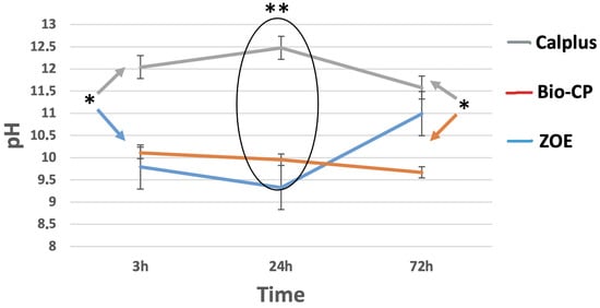
Figure 1.
pH changes of the water in contact with the different tested types of cement after 3, 24, and 72 of immersion in distilled water at 37 °C. Calplus (CP), Bio-C Pulpecto (Bio-CP), and Zinc Oxide and Eugenol (ZOE). (* p = 0.002; ** p ≤ 0.001).
3.2. Solubility
For each tested material, the mean and standard deviation of solubility (wt.%) values are presented in Figure 2. Bio-CP was more soluble during the evaluation period (24 h) than ZOE and CalPlus (p < 0.05).
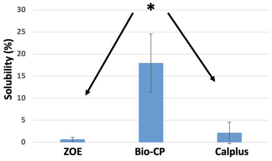
Figure 2.
Solubility percentages of the different types of cement after immersion in 50 mL of distilled water at 37 °C for 24 h. * p < 0.05.
3.3. Scanning Electron Microscope (SEM)
The crystalline structures of the three types of cement are shown in Figure 3. Bio-CP and ZOE demonstrated the creation of crystallite structures on their surfaces after immersion in PBS at 37 °C. Some zones of ZOE surface demonstrated cubical crystalline structures, whilst Bio-CP showed an urchin-like crystallite structure. CalPlus did not show any crystallite creation.
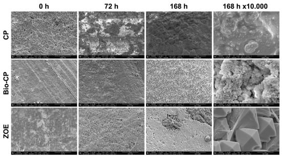
Figure 3.
Scanning electron microscope images (×1000 and ×10,000 magnifications) demonstrate the morphological changes of each material at different time points of immersion in phosphate-buffered solution at 37 °C.
3.4. Water Sorption Tests
Bio-CP demonstrated the highest hydrophilicity for 5 uL of a drop of distilled water compared to ZOE and CalPlus. The lowest contact angle was observed for Bio-CP (53 ± 1.5°). Contact angles of (86 ± 4°) and (96 ± 1°), respectively were observed after 10 s of deposition of the water drop for CalPlus and ZOE (Figure 4, Table 2).
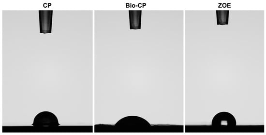
Figure 4.
Contact angles after 5 μL of water dropped on the different cement surfaces after 10 s of deposition. Calplus (CP), Bio-C Pulpecto (Bio-CP), and Zinc Oxide and Eugenol (ZOE).
Table 2.
Contact angles of 5 µL of distilled water on the different material surfaces after 10 s of deposition. Statistical significance (p < 0.05) is indicated with superscript letters a, b and c. Calplus (CP), Bio-C Pulpecto (Bio-CP), and Zinc Oxide and Eugenol (ZOE).
3.5. Antimicrobial Activity
Significant bacterial growth was observed after 24 h between all the groups (p < 0.05). ZOE is the most effective, killing 100% of bacteria, followed by Bio-CP, which eliminates more than 75%, and finally CalPlus (more than 50% of bacteria) (p < 0.05) (Figure 5). In addition, all three types of cement have antibacterial activity against E. faecalis and the best-performing cement was ZOE.
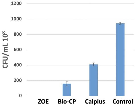
Figure 5.
The number of colonies forming units/mL of Enterococcus faecalis for the medium in contact with Calplus “CP”, Bio-C Pulpecto “Bio-CP”, Zinc Oxide, and Eugenol “ZOE” and the control group “C” (bacterial medium) after 24 h at 37 °C in anaerobic conditions.
4. Discussion
The obturation of primary teeth relies completely on the filling material [27]. ZOE has been, until 2008, the only material explicitly recommended by the American Academy of Pediatric Dentistry (AAPD) [28]. However, it cannot be regarded as an ideal root canal filling due to its resorption rate and negative effects when extruded [3]. Calcium hydroxide and iodoform pastes were recently recommended and showed a high success rate in randomized clinical trials [29]. Nonetheless, in a recent systematic review and meta-analysis, an unclear or high risk of bias was proven in most of the studies and the overall certainty of the evidence ranged from low to very low, denoting that there is still no conclusion to be drawn as to the best pulpectomy material [9].
A resorbable Bioceramic paste, like Bio-CP, could be a promising alternative if the material possessed physicochemical and antibacterial properties suitable for the filling of the intricate root canal system of primary teeth [30]. This was the first in vitro study in the pediatric endodontics literature to focus on the pH, solubility, contact angle, crystallographic changes using SEM images after immersion in PBS, and antibacterial activity of three filling materials for primary teeth. The results of this study showed significant differences between ZOE, CalPlus, and Bio-CP for all evaluated criteria; thus, the null hypothesis was rejected.
The pH analysis indicated an alkaline pH for all tested materials in contact with distilled water at 24 h. An alkaline pH plays a role in the deposition of mineralized tissue and neutralizing the lactic acid from the osteoclast, and promoting healing [31]. Higher pH values were observed for CalPlus compared to ZOE and Bio-CP (p < 0.05), which can be attributed to its dissociation into Ca2+ and OH− ions and has already been validated in many previous studies [32,33]. However, the main disadvantage of this type of material is intracanal resorption and accelerated external root resorption reported in several clinical studies [27].
A set endodontic sealer should present solubility of less than 3% according to ISO standards [23]. In our study, Bio-CP exhibited very high solubility exceeding 3%, demonstrating a high level of Ca2+ and bioactivity. However, high solubility also means that the material dissolves quickly, creating gaps in the canals and generating reinfection [34]. This was also observed in other studies about bioceramic sealers [35] and could be explained by the presence of fine hydrophilic particles in the composition of these sealers, which generate an increase in surface area that may increase sealer solubility when in contact with water/moisture, [36]. Therefore, it is essential to determine clinically the rate at which Bio-CP dissolves, since there is no Gutta Percha to complement the sealer in the root canal treatment of primary teeth. Moreover, the lifespan of a root canal treatment varies greatly depending on the child’s age and physiologic stage of the roots. In the only other study about Bio-CP, the authors highlighted that Bio-CP exhibited sufficient physicochemical properties, disclosed cytocompatibility, and indicated the potential to stimulate mineralization, but lacked clinical support [19].
Using SEM analysis, the microstructural crystalline formation during the initial setting time in 95% humidity and after 3 and 7 days in water was observed. CalPlus did not demonstrate a clear crystallite formation on its surface (Figure 3). This could be because this material is a non-setting paste and the acquisition of such an image was not possible. This result could not be compared to other studies since, to the best of our knowledge, there has not been a publication about the microstructure of calcium hydroxide iodoform-based materials.
The formation of a crystalline structure stipulates that remineralization can occur [23]. In contrast, ZOE had some zones with cubical structures, whereas Bio-CP had an urchin-like structure on its surface. This aspect was extensively described in many studies and was attributed to the extraction of hydroxyapatite from natural resources [37,38].
Contact angle measurements are a reliable tool to better understand the interactions between solids and liquids [39]. These interactions play a major role in explaining not only material wettability, but also wetting, spreading, and adsorption of liquids [39]. In this study, Bio-CP had the lowest contact angle (53 ± 1.5°), demonstrating a good wetting ability, and the capacity to spread faster on substrates such as dentin [40]. CalPlus and ZOE both exhibited poor surface wetting with high contact angles of (86 ± 4°) and (96 ± 1°), respectively. Tummala et al. also found a high contact angle for ZOE and credited this to the increased viscosity of the sealer [41].
The antibacterial potential of each material against E. faecalis was evaluated using a direct contact test per previous studies [42]. Anaerobic bacteria were found scattered within the whole root canal system of primary teeth (accessory canals, dentinal tubules, secondary canals, apical foramen) as well as on the physiological resorptive zones, hence the crucial role of the antibacterial property of the filling paste [43]. This species of bacteria may persist even after biomechanical preparation and use of intracanal irrigants [44]. According to this study, ZOE, CalPlus, and Bio-CP possess antibacterial activity compared to the bacterial medium (p < 0.05). ZOE is the most efficient (p < 0.05), corroborating the results of previous studies [45]. Eugenol-based root canal filling materials possess antibacterial activity due to the action of eugenol, triggering protein denaturation and rendering the microorganisms non-functional [46]. However, in some studies, ZOE demonstrated the smallest zones of bacterial growth inhibition against E. faecalis [43], and higher antibacterial activity for iodoform pastes was observed [47].
CalPlus iodoform paste did not set; therefore, compressive tests were not carried out. Furthermore, the filling ability, flow, radio-opacity and biological properties of each material were omitted [23], and they are important factors to consider. Further in-vitro studies should be conducted to better understand the physical and chemical properties of filling materials for primary teeth. Ageing of Bio-CP in simulated body fluid could provide interesting data about the resorption rate compared to that of the roots of primary teeth [48].
The results obtained in this study are encouraging for the use of resorbable bioceramics in pediatric endodontics. Bio-CP contains calcium silicate and can be classified as a bioactive material. It is capable of forming hydroxyapatite or carbonated apatite on its surface and inducing osteogenesis [48].
However, the results of this study are not conclusive and there is still a need to conduct more well-designed studies to better understand the credentials of an ideal filling material. For the Bioceramics, there is still a need to improve their properties, mainly the excessive solubility, and to increase their antibacterial effect. Moreover, due to the continuous root resorption occurring on primary teeth, there is a need for randomized long-term clinical trials to assess the clinical behavior of this type of material.
5. Conclusions
Within the limitations of this in-vitro study, it could be concluded that there is still a need to develop new filling materials for the root canal treatment of primary teeth. Given the results of this study, ZOE, CalPlus and Bio-CP demonstrated different physicochemical and antibacterial properties, but none of the materials had optimal properties and could be considered the most suitable filling material for primary teeth pulpectomy. The properties of bioceramics such as bioactivity, solubility in fluids, and adhesiveness would provide a crucial step in increasing the success rate of root canal treatment on primary teeth and developing more performant materials. More research should be conducted to optimize clinical protocols in pediatric endodontics.
Author Contributions
Conceptualization, C.E.H. and N.K.; methodology, N.G. and H.S.; validation, D.M. and Y.H.; writing—original draft preparation, C.E.H. and M.K.K.; writing—review and editing, J.C.A.C., N.K. and W.N.; supervision, N.K. All authors have read and agreed to the published version of the manuscript.
Funding
This research received no external funding.
Institutional Review Board Statement
Not applicable.
Informed Consent Statement
Not applicable.
Data Availability Statement
Not applicable.
Acknowledgments
We acknowledge INSERM UMR_S 1121 laboratory for providing the experimental setups.
Conflicts of Interest
The authors declare no conflict of interest.
References
- Coll, J.A.; Vargas, K.; Marghalani, A.A.; Chen, C.-Y.; AlShamali, S.; Dhar, V.; Crystal, Y.O. A Systematic Review and Meta-Analysis of Nonvital Pulp Therapy for Primary Teeth. Pediatr. Dent. 2020, 42, 256–461. [Google Scholar] [PubMed]
- Shindova, M. Root canal filling materials in primary teeth—Review. Folia Med. 2021, 63, 657–662. [Google Scholar] [CrossRef]
- Huang, T.-H.; Hung, C.-J.; Chen, Y.-J.; Chien, H.-C.; Kao, C.-T. Cytologic effects of primary tooth endodontic filling materials. J. Dent. Sci. 2009, 4, 18–24. [Google Scholar] [CrossRef][Green Version]
- Subramaniam, P.; Gilhotra, K. Endoflas, Zinc Oxide Eugenol and Metapex as Root Canal Filling Materials in Primary Molars—A Comparative Clinical Study. J. Clin. Pediatr. Dent. 2011, 35, 365–370. [Google Scholar] [CrossRef] [PubMed]
- Reddy, S.; Ramakrishna, Y. Evaluation of antimicrobial efficacy of various root canal filling materials used in primary teeth: A microbiological study. J. Clin. Pediatr. Dent. 2007, 31, 193–198. [Google Scholar] [CrossRef]
- Pilownic, K.J.; Gomes, A.P.N.; Wang, Z.J.; Almeida, L.H.S.; Romano, A.R.; Shen, Y.; Felix, A.d.O.C.; Haapasalo, M.; Pappen, F.G. Physicochemical and Biological Evaluation of Endodontic Filling Materials for Primary Teeth. Braz. Dent. J. 2017, 28, 578–586. [Google Scholar] [CrossRef]
- Barcelos, R.; Santos, M.P.A.; Primo, L.G.; Luiz, R.R.; Maia, L.C. ZOE paste pulpectomies outcome in primary teeth: A systematic review. J. Clin. Pediatr. Dent. 2011, 35, 241–248. [Google Scholar] [CrossRef]
- Trairatvorakul, C.; Chunlasikaiwan, S. Success of Pulpectomy With Zinc Oxide-Eugenol Vs Calcium Hydroxide/Iodoform Paste in Primary Molars: A Clinical Study. Pediatr. Dent. 2008, 30, 303–308. [Google Scholar]
- Silva Junior, M.F.; Wambier, L.M.; Gevert, M.V.; Chibinski, A.C.R. Effectiveness of iodoform-based filling materials in root canal treatment of deciduous teeth: A systematic review and meta-analysis. Biomater. Investig. Dent. 2022, 9, 52–74. [Google Scholar] [CrossRef]
- Eid, A.; Davide, M.; Joudi, F.; Rekab, M.; Layous, K.; Hamadah, O.; Haikel, Y.; Kharouf, N. Comparative Evaluation Of The Apical Sealing Ability Of BioRoot And Ahplus Sealers: An In Vitro Study. Int. J. Dent. Oral Sci. 2021, 8, 2309–2313. [Google Scholar] [CrossRef]
- Moskovitz, M.; Tickotsky, N.; Ashkar, H.; Holan, G. Degree of root resorption after root canal treatment with iodoform-containing filling material in primary molars. Quintessence Int. Berl. Ger. 1985 2012, 43, 361–368. [Google Scholar]
- Marciano, M.A.; Duarte, M.A.H.; Camilleri, J. Calcium silicate-based sealers: Assessment of physicochemical properties, porosity and hydration. Dent. Mater. 2016, 32, e30–e40. [Google Scholar] [CrossRef] [PubMed]
- Hardan, L.; Mancino, D.; Bourgi, R.; Alvarado-Orozco, A.; Rodríguez-Vilchis, L.E.; Flores-Ledesma, A.; Cuevas-Suárez, C.E.; Lukomska-Szymanska, M.; Eid, A.; Danhache, M.-L.; et al. Bond Strength of Adhesive Systems to Calcium Silicate-Based Materials: A Systematic Review and Meta-Analysis of In Vitro Studies. Gels Basel Switz. 2022, 8, 311. [Google Scholar] [CrossRef]
- Kharouf, N.; Zghal, J.; Addiego, F.; Gabelout, M.; Jmal, H.; Haikel, Y.; Bahlouli, N.; Ball, V. Tannic acid speeds up the setting of mineral trioxide aggregate cements and improves its surface and bulk properties. J. Colloid Interface Sci. 2021, 589, 318–326. [Google Scholar] [CrossRef]
- Kharouf, N.; Haïkel, Y.; Mancino, D. Unusual Maxillary First Molars with C-Shaped Morphology on the Same Patient: Variation in Root Canal Anatomy. Case Rep. Dent. 2019, 2019, 1857289. [Google Scholar] [CrossRef] [PubMed]
- Eid, A.; Mancino, D.; Rekab, M.S.; Haikel, Y.; Kharouf, N. Effectiveness of Three Agents in Pulpotomy Treatment of Permanent Molars with Incomplete Root Development: A Randomized Controlled Trial. Healthc. Basel Switz. 2022, 10, 431. [Google Scholar] [CrossRef] [PubMed]
- Kassab, P.; El Hachem, C.; Habib, M.; Nehme, W.; Zogheib, C.; Tonini, R.; Kaloustian, M.K. The Pushout Bond Strength of Three Calcium Silicate-based Materials in Furcal Perforation Repair and the Effect of a Novel Irrigation Solution: A Comparative In Vitro Study. J. Contemp. Dent. Pract. 2022, 23, 289–294. [Google Scholar]
- Stringhini Junior, E.; Dos Santos, M.G.C.; Oliveira, L.B.; Mercadé, M. MTA and biodentine for primary teeth pulpotomy: A systematic review and meta-analysis of clinical trials. Clin. Oral Investig. 2019, 23, 1967–1976. [Google Scholar] [CrossRef] [PubMed]
- Ochoa Rodríguez, V.M.; Tanomaru-Filho, M.; Rodrigues, E.M.; Bugança, E.d.O.; Guerreiro-Tanomaru, J.M.; Faria, G. Physicochemical properties and effect of bioceramic root canal filling for primary teeth on osteoblast biology. J. Appl. Oral Sci. 2021, 29, e20200870. [Google Scholar] [CrossRef]
- Cancio, V.; Carvalho Ferreira, D.D.; Cavalcante, F.S.; Rosado, A.S.; Teixeira, L.M.; Braga Oliveira, Q.; Barcelos, R.; Gleiser, R.; Santos, H.F.; dos Santos, K.R.N.; et al. Can the Enterococcus faecalis identified in the root canals of primary teeth be a cause of failure of endodontic treatment? Acta Odontol. Scand. 2017, 75, 423–428. [Google Scholar] [CrossRef]
- Forghani, M.; Afshari, E.; Parisay, I.; Garajian, R. Effect of a passive sonic irrigation system on elimination of Enterococcus faecalis from root canal systems of primary teeth, using different concentrations of sodium hypochlorite: An in vitro evaluation. J. Dent. Res. Dent. Clin. Dent. Prospect. 2017, 11, 177–182. [Google Scholar] [CrossRef] [PubMed][Green Version]
- Cogulu, D.; Uzel, A.; Oncag, O.; Eronat, C. PCR-based identification of selected pathogens associated with endodontic infections in deciduous and permanent teeth. Oral Surg. Oral Med. Oral Pathol. Oral Radiol. Endodontol. 2008, 106, 443–449. [Google Scholar] [CrossRef] [PubMed]
- Kharouf, N.; Arntz, Y.; Eid, A.; Zghal, J.; Sauro, S.; Haikel, Y.; Mancino, D. Physicochemical and Antibacterial Properties of Novel, Premixed Calcium Silicate-Based Sealer Compared to Powder–Liquid Bioceramic Sealer. J. Clin. Med. 2020, 9, 3096. [Google Scholar] [CrossRef] [PubMed]
- Kharouf, N.; Mancino, D.; Zghal, J.; Helle, S.; Jmal, H.; Lenertz, M.; Viart, N.; Bahlouli, N.; Meyer, F.; Haikel, Y.; et al. Dual role of tannic acid and pyrogallol incorporated in plaster of Paris: Morphology modification and release for antimicrobial properties. Mater. Sci. Eng. C Mater. Biol. Appl. 2021, 127, 112209. [Google Scholar] [CrossRef] [PubMed]
- Kharouf, N.; Sauro, S.; Jmal, H.; Eid, A.; Karrout, M.; Bahlouli, N.; Haikel, Y.; Mancino, D. Does Multi-Fiber-Reinforced Composite-Post Influence the Filling Ability and the Bond Strength in Root Canal? Bioeng. Basel Switz. 2021, 8, 195. [Google Scholar] [CrossRef]
- Kharouf, N.; Sauro, S.; Hardan, L.; Fawzi, A.; Suhanda, I.E.; Zghal, J.; Addiego, F.; Affolter-Zbaraszczuk, C.; Arntz, Y.; Ball, V.; et al. Impacts of Resveratrol and Pyrogallol on Physicochemical, Mechanical and Biological Properties of Epoxy-Resin Sealers. Bioeng. Basel Switz. 2022, 9, 85. [Google Scholar] [CrossRef]
- Najjar, R.S.; Alamoudi, N.M.; El-Housseiny, A.A.; Al Tuwirqi, A.A.; Sabbagh, H.J. A comparison of calcium hydroxide/iodoform paste and zinc oxide eugenol as root filling materials for pulpectomy in primary teeth: A systematic review and meta-analysis. Clin. Exp. Dent. Res. 2019, 5, 294–310. [Google Scholar] [CrossRef]
- American Academy of Pediatric Dentistry Clinical Affairs Committee—Pulp Therapy Subcommittee; American Academy of Pediatric Dentistry Council on Clinical Affairs. Guideline on pulp therapy for primary and young permanent teeth. Pediatr. Dent. 2005, 27, 130–134. [Google Scholar]
- Al-Attiya, H.; Schmoeckel, J.; Mourad, M.S.; Splieth, C.H. One year clinical success of pulpectomy in primary molars with iodoform-calcium hydroxide paste. Quintessence Int. Berl. Ger. 1985 2021, 52, 528–537. [Google Scholar] [CrossRef]
- El Hachem, C.; Kaloustian, M.K.; Nehme, W.; Ghosn, N.; Abou Chedid, J.C. Three-dimensional modeling and measurements of root canal anatomy in second primary mandibular molars: A case series micro CT study. Eur. Arch. Paediatr. Dent. Off. J. Eur. Acad. Paediatr. Dent. 2019, 20, 457–465. [Google Scholar] [CrossRef]
- Paz, L.C.d. Redefining the Persistent Infection in Root Canals: Possible Role of Biofilm Communities. J. Endod. 2007, 33, 652–662. [Google Scholar] [CrossRef] [PubMed]
- Tronstad, L.; Andreasen, J.O.; Hasselgren, G.; Kristerson, L.; Riis, I. pH changes in dental tissues after root canal filling with calcium hydroxide. J. Endod. 1981, 7, 17–21. [Google Scholar] [CrossRef]
- Foreman, P.C.; Barnes, I.E. A review of calcium hydroxide. Int. Endod. J. 1990, 23, 283–297. [Google Scholar] [CrossRef] [PubMed]
- Torres, F.F.E.; Zordan-Bronzel, C.L.; Guerreiro-Tanomaru, J.M.; Chávez-Andrade, G.M.; Pinto, J.C.; Tanomaru-Filho, M. Effect of immersion in distilled water or phosphate-buffered saline on the solubility, volumetric change and presence of voids within new calcium silicate-based root canal sealers. Int. Endod. J. 2020, 53, 385–391. [Google Scholar] [CrossRef]
- Poggio, C.; Dagna, A.; Ceci, M.; Meravini, M.-V.; Colombo, M.; Pietrocola, G. Solubility and pH of bioceramic root canal sealers: A comparative study. J. Clin. Exp. Dent. 2017, 9, e1189–e1194. [Google Scholar] [CrossRef] [PubMed]
- Mann, A.; Zeng, Y.; Kirkpatrick, T.; van der Hoeven, R.; Silva, R.; Letra, A.; Chaves de Souza, L. Evaluation of the Physicochemical and Biological Properties of EndoSequence BC Sealer HiFlow. J. Endod. 2022, 48, 123–131. [Google Scholar] [CrossRef]
- Eliaz, N.; Metoki, N. Calcium Phosphate Bioceramics: A Review of Their History, Structure, Properties, Coating Technologies and Biomedical Applications. Materials 2017, 10, 334. [Google Scholar] [CrossRef]
- Akram, M.; Ahmed, R.; Shakir, I.; Ibrahim, W.A.W.; Hussain, R. Extracting hydroxyapatite and its precursors from natural resources. J. Mater. Sci. 2014, 49, 1461–1475. [Google Scholar] [CrossRef]
- Kontakiotis, E.G.; Tzanetakis, G.N.; Loizides, A.L. A comparative study of contact angles of four different root canal sealers. J. Endod. 2007, 33, 299–302. [Google Scholar] [CrossRef]
- Prado, M.; de Assis, D.F.; Gomes, B.P.F.A.; Simão, R.A. Effect of Disinfectant Solutions on the Surface Free Energy and Wettability of Filling Material. J. Endod. 2011, 37, 980–982. [Google Scholar] [CrossRef]
- Tummala, M.; Chandrasekhar, V.; Rashmi, A.S.; Kundabala, M.; Ballal, V. Assessment of the wetting behavior of three different root canal sealers on root canal dentin. J. Conserv. Dent. JCD 2012, 15, 109–112. [Google Scholar] [CrossRef] [PubMed]
- Zhang, H.; Shen, Y.; Ruse, N.D.; Haapasalo, M. Antibacterial Activity of Endodontic Sealers by Modified Direct Contact Test Against Enterococcus faecalis. J. Endod. 2009, 35, 1051–1055. [Google Scholar] [CrossRef] [PubMed]
- Queiroz, A.M.D.; Nelson-Filho, P.; Silva, L.A.B.D.; Assed, S.; Silva, R.A.B.D.; Ito, I.Y. Antibacterial activity of root canal filling materials for primary teeth: Zinc oxide and eugenol cement, Calen paste thickened with zinc oxide, Sealapex and EndoREZ. Braz. Dent. J. 2009, 20, 290–296. [Google Scholar] [CrossRef] [PubMed][Green Version]
- Moradi, F.; Haghgoo, R. Evaluation of Antimicrobial Efficacy of Nanosilver Solution, Sodium Hypochlorite and Normal Saline in Root Canal Irrigation of Primary Teeth. Contemp. Clin. Dent. 2018, 9, S227–S232. [Google Scholar] [CrossRef] [PubMed]
- Pimenta, H.C.; Borges, Á.H.; Bandeca, M.C.; Neves, A.T.S.; Fontes, R.G.; da Silva, P.V.; Aranha, A.M.F. Antimicrobial Activity of Filling Materials Used in Primary Teeth Pulpotomy. J. Int. Oral Health 2015, 7, 54–57. [Google Scholar]
- Verma, R.; Sharma, D.; Pathak, A. Antibacterial Efficacy of Pastes Against E Faecalis in Primary Root Dentin: A Confocal Microscope Study. J. Clin. Pediatr. Dent. 2015, 39, 247–254. [Google Scholar] [CrossRef]
- Estrela, C.; Estrela, C.R.D.A.; Hollanda, A.C.B.; Decurcio, D.D.A.; Pécora, J.D. Influence of iodoform on antimicrobial potential of calcium hydroxide. J. Appl. Oral Sci. 2006, 14, 33–37. [Google Scholar] [CrossRef]
- Elsayed, M.A.; Hassanien, E.E.; Elgendy, A.A.E. Ageing of TotalFill BC Sealer and MTA Fillapex in Simulated Body Fluid. Eur. Endod. J. 2021, 6, 183–188. [Google Scholar] [CrossRef]
Publisher’s Note: MDPI stays neutral with regard to jurisdictional claims in published maps and institutional affiliations. |
© 2022 by the authors. Licensee MDPI, Basel, Switzerland. This article is an open access article distributed under the terms and conditions of the Creative Commons Attribution (CC BY) license (https://creativecommons.org/licenses/by/4.0/).