


Curcumin-Encapsulated Nanomicelles Improve Cellular Uptake and Cytotoxicity in Cisplatin-Resistant Human Oral Cancer Cells

 , , , , , , ,
  , , and
Abstract
1. Introduction
2. Materials and Methods
2.1. Materials
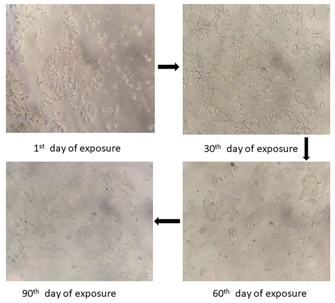
2.2. Drug-Resistant Cell Lines Development
2.3. Preparation of Curcumin Nanomicelles
2.4. Characterization of Curcumin Nanomicelles
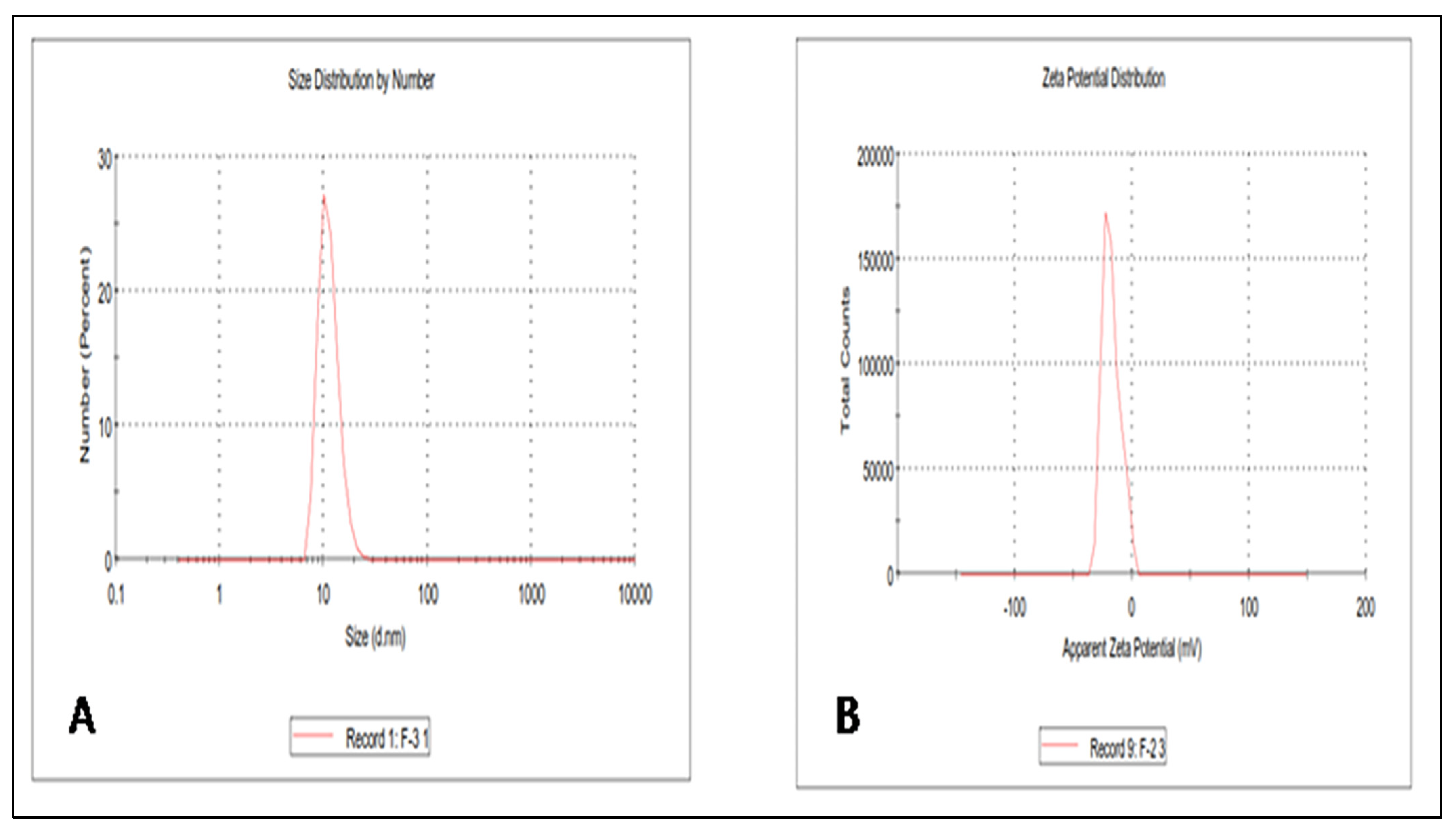
2.4.1. Curcumin Nanomicelle Size, Size Distribution, and Zeta Potential
2.4.2. Morphology and Structure—Transmission Electron Microscopy (TEM)
2.4.3. Entrapment Efficiency
- S is the quantity of CUR existing in the supernatant,
- T − S is the quantity of CUR existing within the micelles.
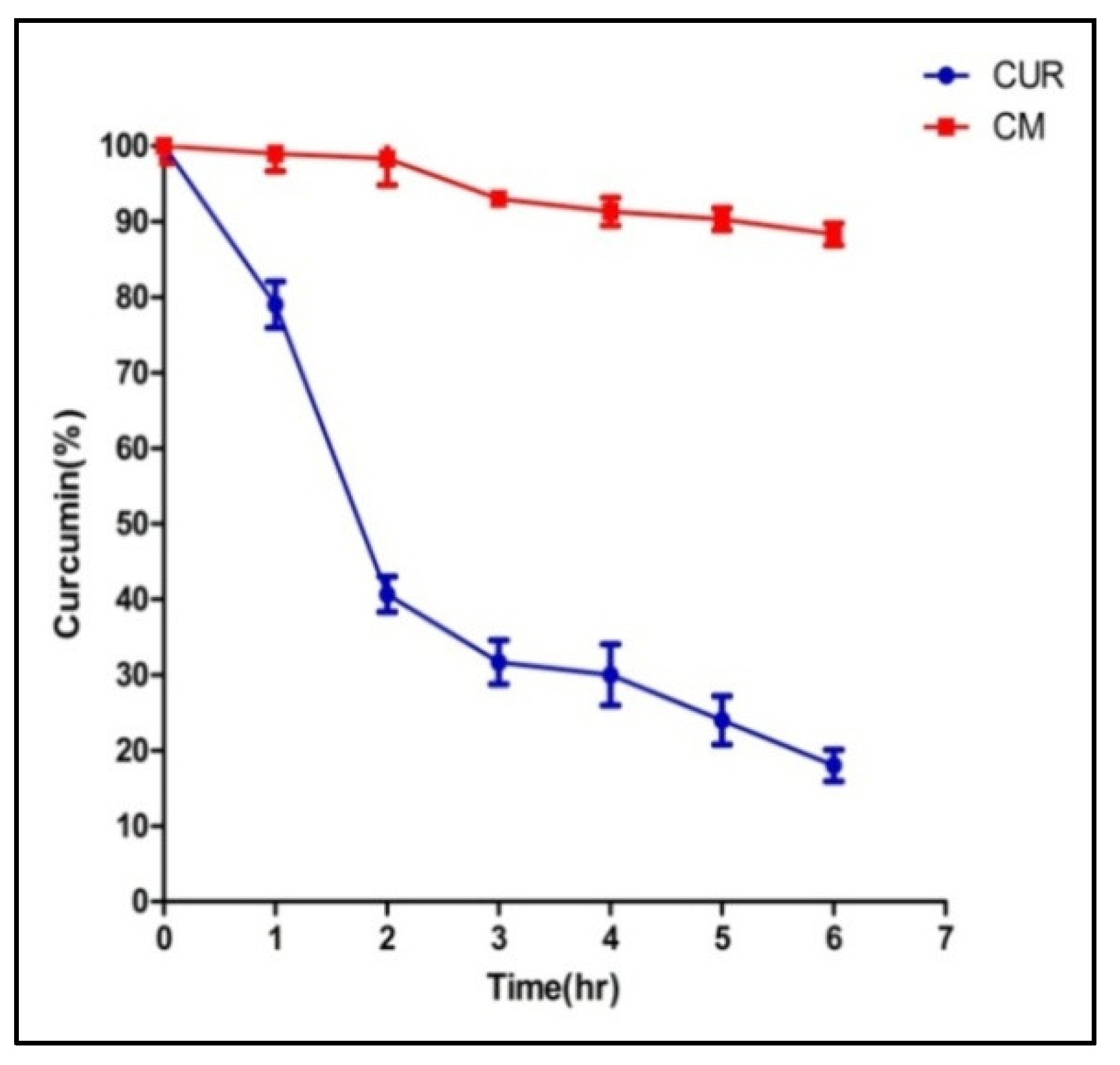
2.5. In Vitro Release Study
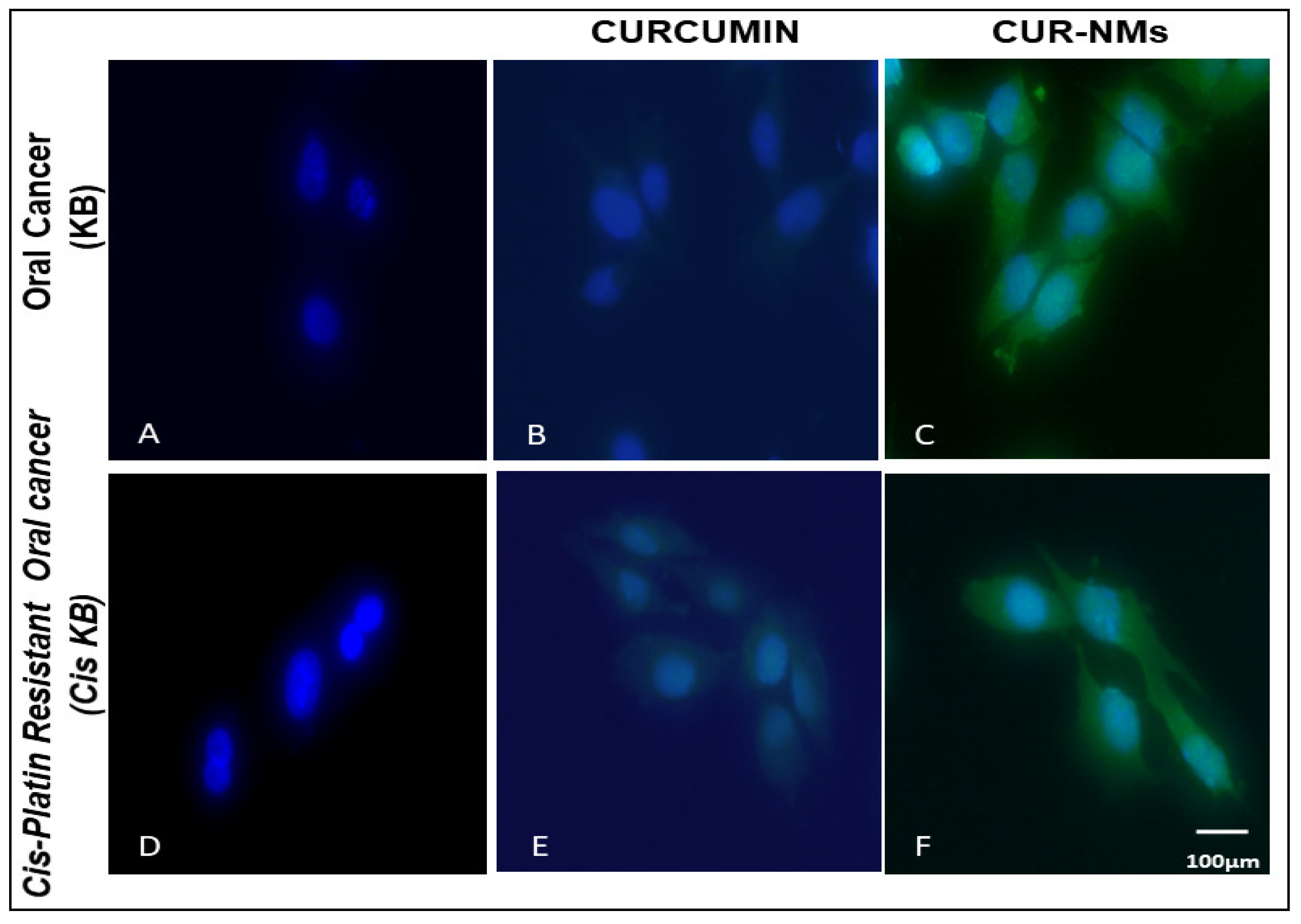
2.6. Cell Uptake Study
2.7. Cytotoxicity
2.8. Detection of Mitochondrial Transmembrane Potential
2.9. Dual Staining Assay (Acridine Orange/Ethidium Bromide)
2.10. 4′,6-diamidino-2-phenylindole (DAPI) Staining
2.11. Apoptosis by Flow Cytometer
2.12. Biocompatibility Assay
2.13. Statistical Analysis
3. Results
3.1. Drug-Resistant Cell Lines Development
3.2. Determination of the Size, Size Distribution, and Zeta Potential of CUR Nanomicelles (CUR-NMs)
3.3. Structure and Morphology of CUR-NMs by Transmission Electron Microscopy (TEM)
3.4. Entrapment Efficiency
3.5. In Vitro Release Study
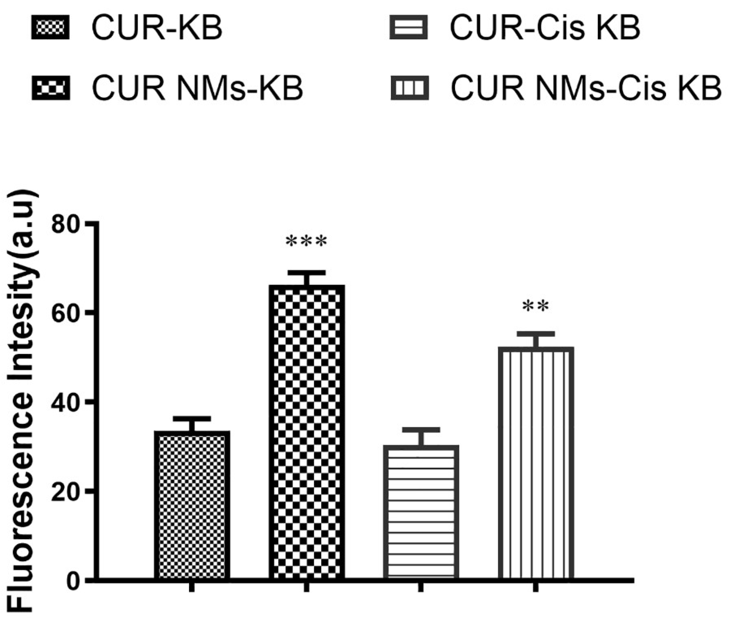
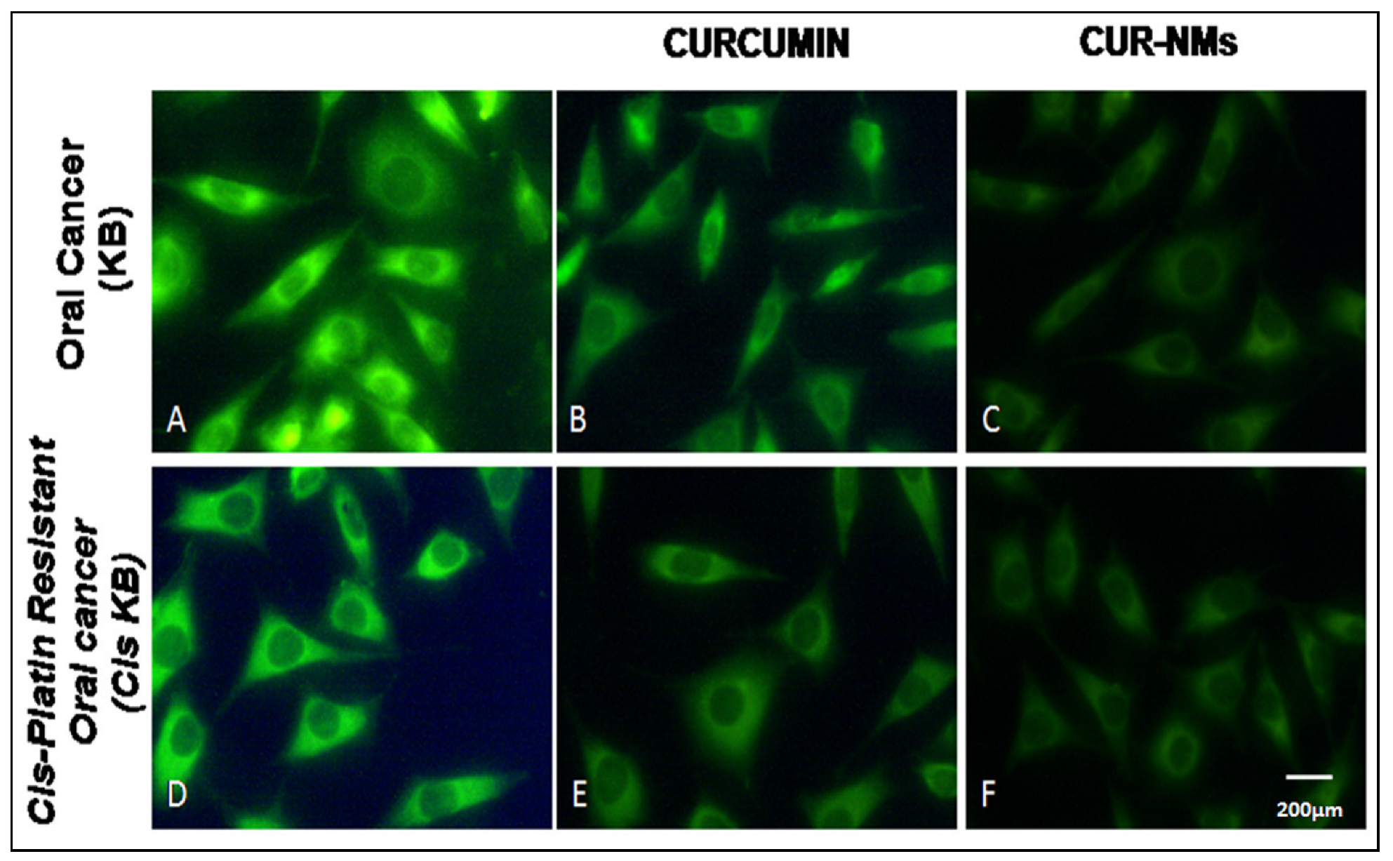
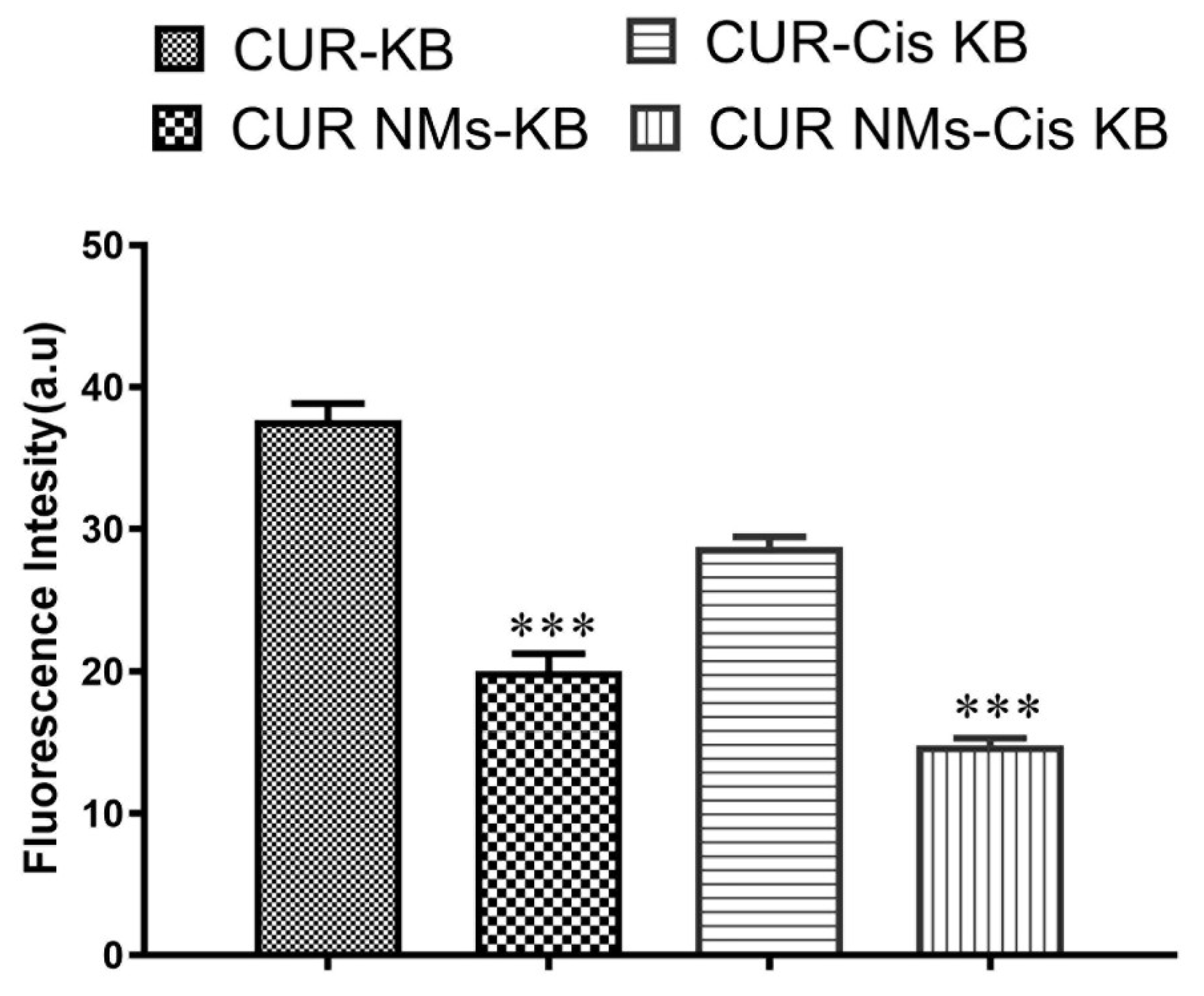
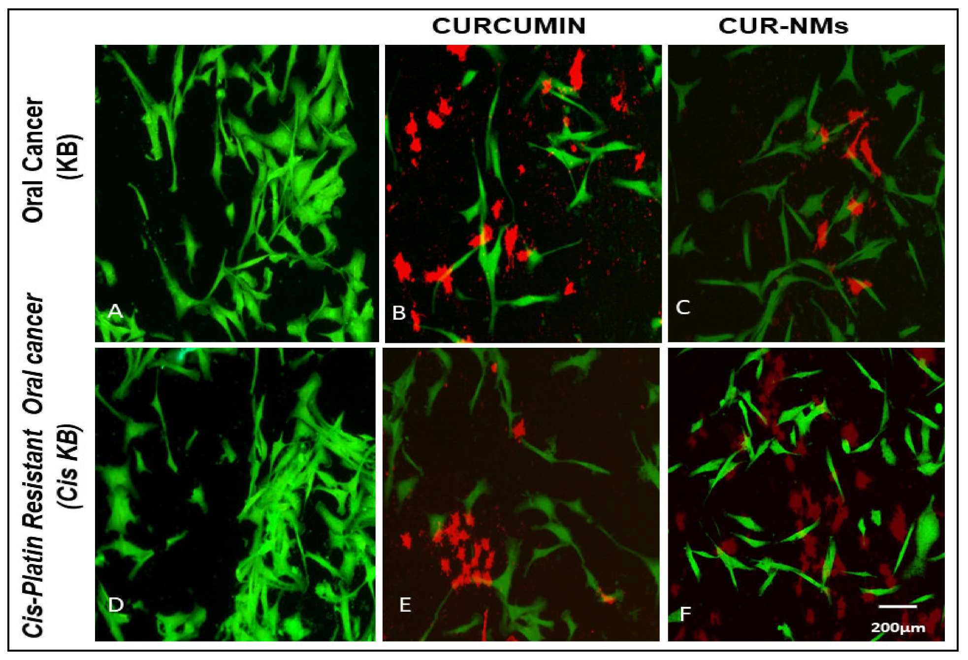
3.6. Cell Uptake Study
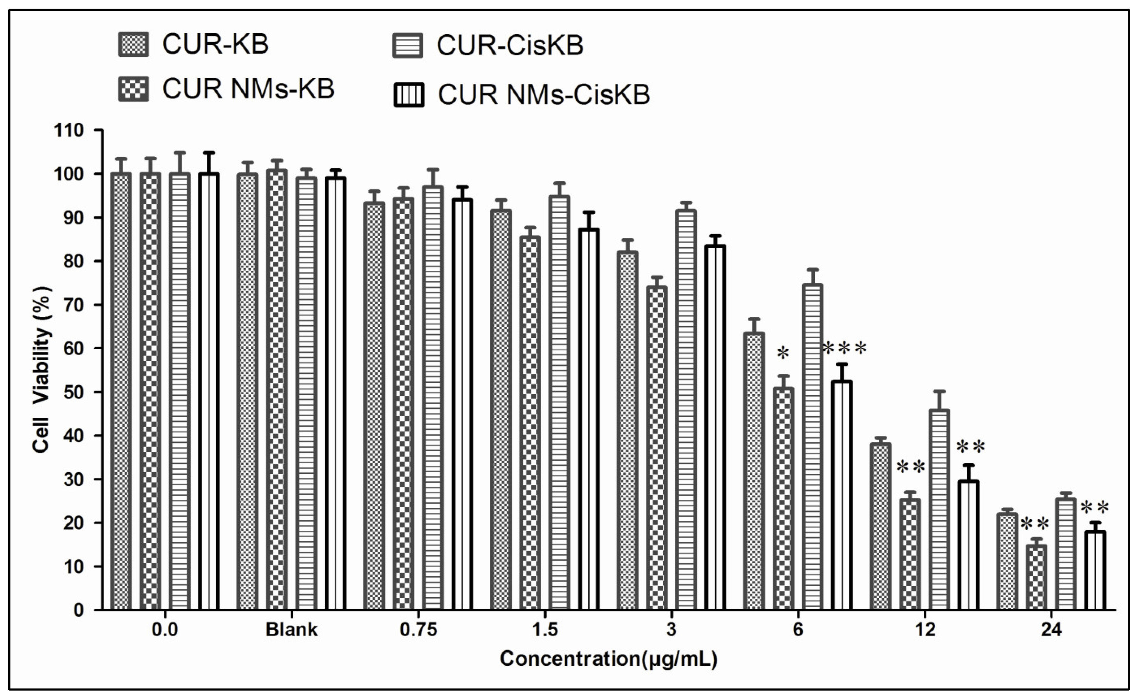
3.7. Cytotoxicity
3.8. Effects of Curcumin on Mitochondrial Membrane Potential (ΔΨ)
3.9. Double Staining (Acridine Orange/Ethidium Bromide)
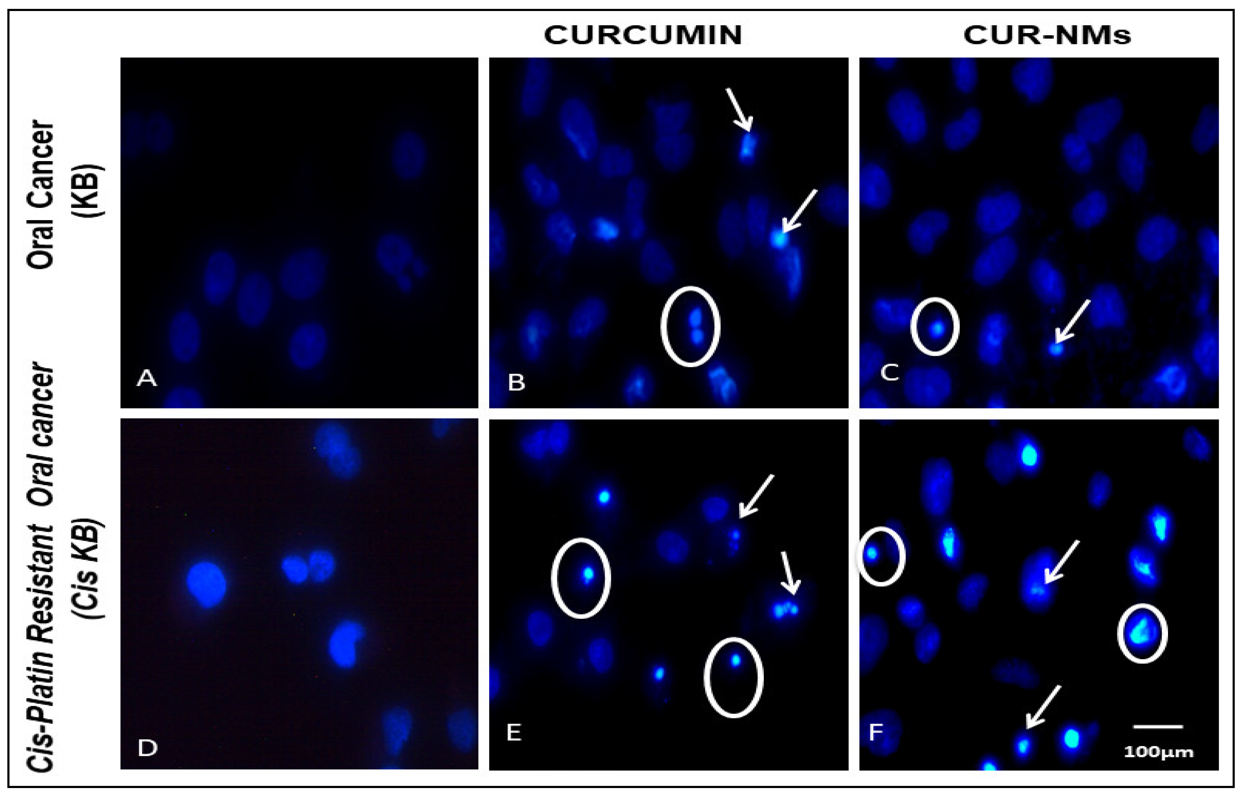
3.10. DAPI
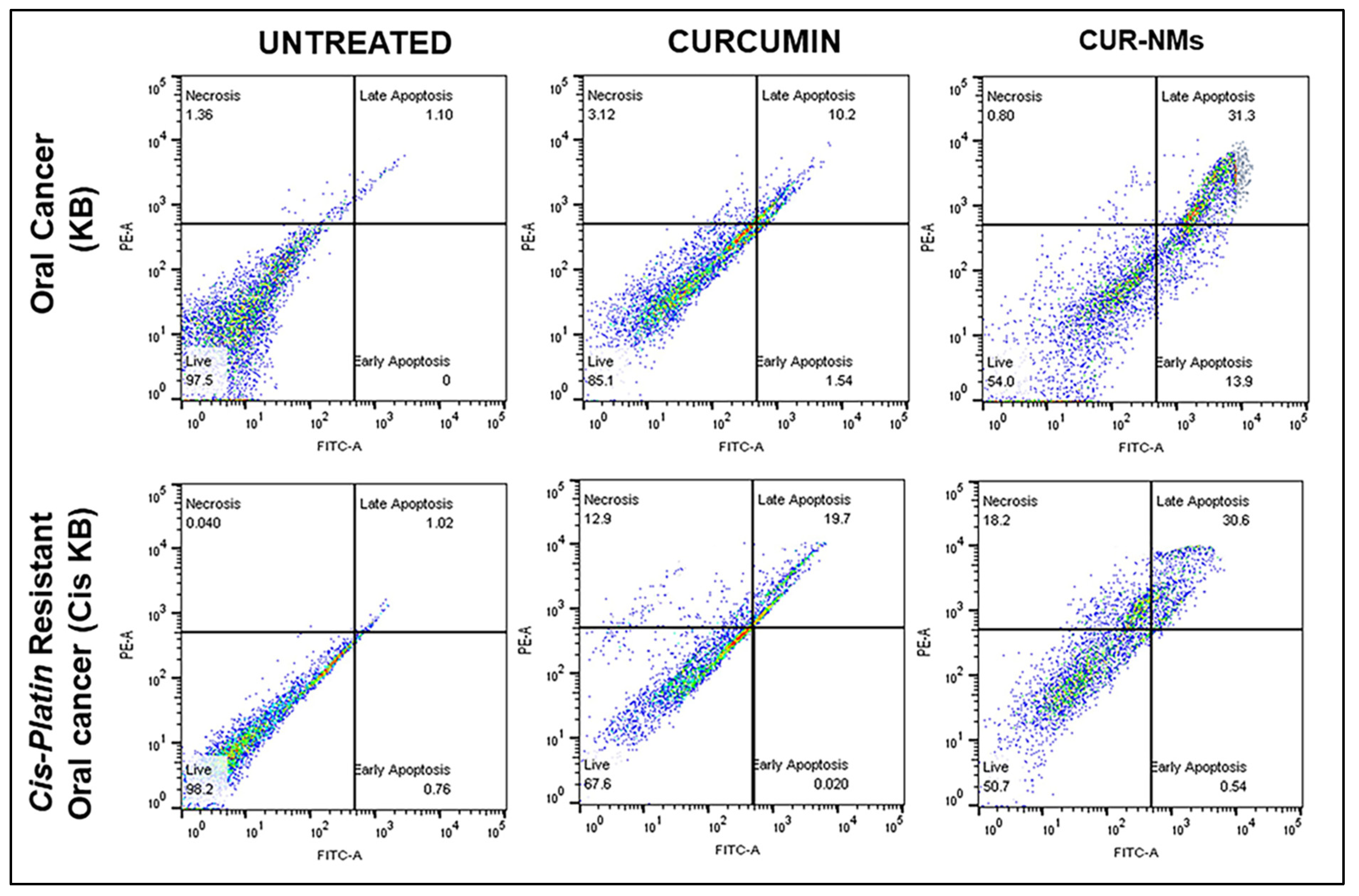
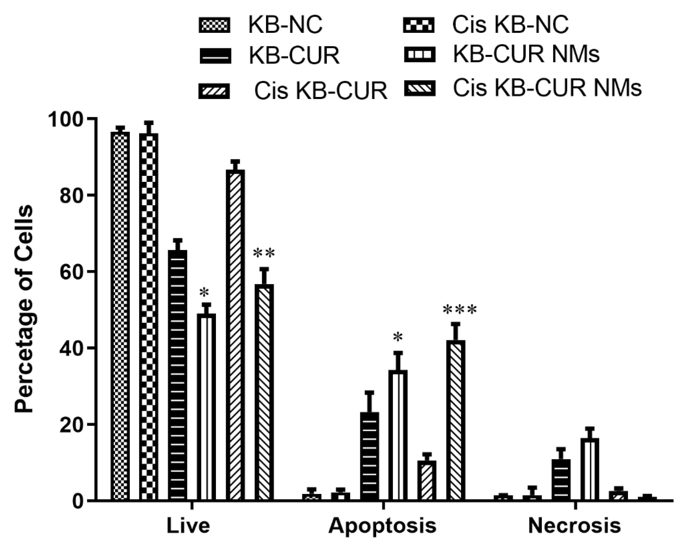
3.11. Apoptosis
3.12. Biocompatibility Assay
4. Discussion
4.1. Drug-Resistant Cell Lines Development
4.2. Characterization of Curcumin Nanomicelles (Curcumin Nanomicelle Size, Size Distribution, and Zeta Potential)
4.3. Entrapment Efficiency
4.3.1. In Vitro Release Study
4.3.2. In Vitro Cytotoxicity Assay
4.4. Cell Uptake Study
4.5. Double Staining (Acridine Orange/Ethidium Bromide)
4.6. DAPI
4.7. Apoptosis by Flow Cytometry
4.8. Biocompatibility Assay
5. Conclusions
Author Contributions
Funding
Institutional Review Board Statement
Informed Consent Statement
Data Availability Statement
Acknowledgments
Conflicts of Interest
Abbreviations
| Polymeric nanoparticles | NPs |
| Nanoemulsions | NEs |
| Polyion complex vesicle | PICsome |
| 1,2-Distearoyl-sn-glycero-3-phosphoethanolamine-N-methoxy-[poly(ethylene glycol); PEG MW2,000 | DSPE-PEG-2000 |
| 3-(4,5-dimethylthiazol-2-yl)-2,5-diphenyltetrazolium bromide | MTT |
| 6-diamidino-2-phenylindole | DAPI |
| Dimethyl sulfoxide | DMSO |
| Acridine orange and Ethidium bromide | AO and ETBR |
| Cisplatin-resistant oral cancer sublines | Cis-KB |
| Curcumin Nano micelles | CUR-NMs |
| Half-maximal inhibitory concentration | IC50 |
| Resistance Index | RI |
| Zeta Potential | ZP |
| Transmission Electron Microscopy | TEM |
| High-Performance Liquid Chromatography | HPLC |
| Entrapment Efficiency | EE |
| Dulbecco’s Phosphate Buffered Saline | DPBS |
| Human periodontal fibroblasts | |
| enhanced permeability and retention effect | EPR |
| dynamic light scattering | DLS |
| mitochondrial membrane potential | MMP |
| vulval, vaginal and gingival (VVG)- | VVG |
| Cancer Stem Cells | CSCs |
References
- Sung, H.; Ferlay, J.; Siegel, R.L.; Laversanne, M.; Soerjomataram, I.; Jemal, A.; Bray, F. Global Cancer Statistics 2020: GLOBOCAN Estimates of Incidence and Mortality Worldwide for 36 Cancers in 185 Countries. CA Cancer J. Clin. 2021, 71, 209–249. [Google Scholar] [CrossRef]
- Bray, F.; Ferlay, J.; Soerjomataram, I.; Siegel, R.L.; Torre, L.A.; Jemal, A. Global cancer statistics 2018: GLOBOCAN estimates of incidence and mortality worldwide for 36 cancers in 185 countries. CA Cancer J. Clin. 2018, 68, 394–424. [Google Scholar] [CrossRef] [PubMed]
- Vigneswaran, N.; Williams, M.D. Epidemiologic trends in head and neck cancer and aids in diagnosis. Oral Maxillofac. Surg. Clin. N. Am. 2014, 26, 123–141. [Google Scholar] [CrossRef]
- Jethwa, A.R.; Khariwala, S.S. Tobacco-related carcinogenesis in head and neck cancer. Cancer Metastasis Rev. 2017, 36, 411–423. [Google Scholar] [CrossRef]
- Ketabat, F.; Pundir, M.; Mohabatpour, F.; Lobanova, L.; Koutsopoulos, S.; Hadjiiski, L.; Chen, X.; Papagerakis, P.; Papagerakis, S. Controlled Drug Delivery Systems for Oral Cancer Treatment—Current Status and Future Perspectives. Pharmaceutics 2019, 11, 302. [Google Scholar] [CrossRef] [PubMed]
- Pochet, S.; Lechon, A.-S.; Lescrainier, C.; De Vriese, C.; Mathieu, V.; Hamdani, J.; Souard, F. Herb-anticancer drug interactions in real life based on VigiBase, the WHO global database. Sci. Rep. 2022, 12, 14178. [Google Scholar] [CrossRef] [PubMed]
- Greenwell, M.; Rahman, P.K. Medicinal Plants: Their Use in Anticancer Treatment. Int. J. Pharm. Sci. Res. 2015, 6, 4103–4112. [Google Scholar] [CrossRef]
- Garcia, S. Pandemics and Traditional Plant-Based Remedies. A Historical-Botanical Review in the Era of COVID19. Front. Plant Sci. 2020, 11, 571042. [Google Scholar] [CrossRef]
- Singh, S.; Sharma, B.; Kanwar, S.S.; Kumar, A. Lead Phytochemicals for Anticancer Drug Development. Front. Plant Sci. 2016, 7, 1667. [Google Scholar] [CrossRef]
- Amalraj, A.; Pius, A.; Gopi, S.; Gopi, S. Biological activities of curcuminoids, other biomolecules from turmeric and their derivatives—A review. J. Tradit. Complement. Med. 2016, 7, 205–233. [Google Scholar] [CrossRef] [PubMed]
- Golonko, A.; Lewandowska, H.; Świsłocka, R.; Jasińska, U.; Priebe, W.; Lewandowski, W. Curcumin as tyrosine kinase inhibitor in cancer treatment. Eur. J. Med. Chem. 2019, 181, 111512. [Google Scholar] [CrossRef] [PubMed]
- Wang, M.; Jiang, S.; Zhou, L.; Yu, F.; Ding, H.; Li, P.; Zhou, M.; Wang, K. Potential Mechanisms of Action of Curcumin for Cancer Prevention: Focus on Cellular Signaling Pathways and miRNAs. Int. J. Biol. Sci. 2019, 15, 1200–1214. [Google Scholar] [CrossRef]
- Ismail, N.I.; Othman, I.; Abas, F.; Lajis, N.H.; Naidu, R. Mechanism of Apoptosis Induced by Curcumin in Colorectal Cancer. Int. J. Mol. Sci. 2019, 20, 2454. [Google Scholar] [CrossRef]
- Shaikh, S.; Shaikh, J.; Naba, Y.S.; Doke, K.; Ahmed, K.; Yusufi, M. Curcumin: Reclaiming the lost ground against cancer resistance. Cancer Drug Resist. 2021, 4, 298–4320. [Google Scholar] [CrossRef]
- Qamar, Z.; Qizilbash, F.F.; Iqubal, A.; Ali, A.; Narang, J.K.; Ali, J.; Baboota, S. Nano-Based Drug Delivery System: Recent Strategies for the Treatment of Ocular Disease and Future Perspective. Recent Patents Drug Deliv. Formul. 2019, 13, 246–254. [Google Scholar] [CrossRef]
- Ahmad, M.Z.; Alkahtani, S.A.; Akhter, S.; Ahmad, F.J.; Ahmad, J.; Akhtar, M.S.; Mohsin, N.; Abdel-Wahab, B.A. Progress in nanotechnology-based drug carrier in designing of curcumin nanomedicines for cancer therapy: Current state-of-the-art. J. Drug Target. 2016, 24, 273–293. [Google Scholar] [CrossRef] [PubMed]
- Lin, W.; Ma, G.; Kampf, N.; Yuan, Z.; Chen, S. Development of Long-Circulating Zwitterionic Cross-Linked Micelles for Active-Targeted Drug Delivery. Biomacromolecules 2016, 17, 2010–2018. [Google Scholar] [CrossRef]
- Goto, A.; Yen, H.C.; Anraku, Y.; Fukushima, S.; Lai, P.S.; Kato, M.; Kishimura, A.; Kataoka, K. Facile preparation of delivery platform of water-soluble low-molecular-weight drugs based on polyion complex vesicle (PICsome) encapsulating mesoporous silica nanoparticle. ACS Biomater. Sci. Eng. 2017, 3, 807–815. [Google Scholar] [CrossRef]
- Baumann, K.N.; Piantanida, L.; García-Nafría, J.; Sobota, D.; Voïtchovsky, K.; Knowles, T.P.; Hernández-Ainsa, S. Coating and stabilization of liposomes by clathrin-inspired DNA self-assembly. ACS Nano 2020, 14, 2316–2323. [Google Scholar] [CrossRef]
- Kulthe, S.S.; Choudhari, Y.M.; Inamdar, N.N.; Mourya, V. Polymeric micelles: Authoritative aspects for drug delivery. Des. Monomers Polym. 2012, 15, 465–521. [Google Scholar] [CrossRef]
- Che, J.; Okeke, C.; Hu, Z.-B.; Xu, J. DSPE-PEG: A Distinctive Component in Drug Delivery System. Curr. Pharm. Des. 2015, 21, 1598–1605. [Google Scholar] [CrossRef]
- Govindan, S.V.; Kulsum, S.; Pandian, R.S.; Das, D.; Seshadri, M.; Hicks, W., Jr.; Kuriakose, M.A.; Suresh, A. Establishment and characterization of triple drug-resistant head and neck squamous cell carcinoma cell lines. Mol. Med. Rep. 2015, 12, 3025–3032. [Google Scholar] [CrossRef]
- Gülçür, E.; Thaqi, M.; Khaja, F.; Kuzmis, A.; Önyüksel, H. Curcumin in VIP-targeted sterically stabilized phospholipid nanomicelles: A novel therapeutic approach for breast cancer and breast cancer stem cells. Drug Deliv. Transl. Res. 2013, 3, 562–574. [Google Scholar] [CrossRef] [PubMed]
- Faisal, W.; Soliman, G.M.; Hamdan, A. Enhanced skin deposition and delivery of voriconazole using ethosomal preparations. J. Liposome Res. 2016, 28, 14–21. [Google Scholar] [CrossRef]
- Peram, M.R.; Jalalpure, S.S.; Kumbar, V.M.; Patil, S.R.; A Joshi, S.; Bhat, K.G.; Diwan, P.V. Factorial design based curcumin ethosomal nanocarriers for the skin cancer delivery: In vitro evaluation. J. Liposome Res. 2019, 29, 291–311. [Google Scholar] [CrossRef]
- Peram, M.R.; Jalalpure, S.S.; Joshi, S.A.; Palkar, M.B.; Diwan, P.V. Single robust RP-HPLC analytical method for quantification of curcuminoids in commercial turmeric products, Ayurvedic medicines, and nanovesicular systems. J. Liq. Chromatogr. Relat. Technol. 2017, 40, 487–498. [Google Scholar] [CrossRef]
- Kumbar, V.M.; Peram, M.R.; Kugaji, M.S.; Shah, T.; Patil, S.P.; Muddapur, U.M.; Bhat, K.G. Effect of curcumin on growth, biofilm formation and virulence factor gene expression of Porphyromonas gingivalis. Odontology 2021, 109, 18–28. [Google Scholar] [CrossRef]
- Annamalai, G.; Kathiresan, S.; Kannappan, N. [6]-Shogaol, a dietary phenolic compound, induces oxidative stress mediated mitochondrial dependant apoptosis through activation of proapoptotic factors in Hep-2 cells. Biomed. Pharmacother. 2016, 82, 226–236. [Google Scholar] [CrossRef]
- Bhagwat, D.A.; Swami, P.A.; Nadaf, S.J.; Choudhari, P.B.; Kumbar, V.M.; More, H.N.; Killedar, S.G.; Kawtikwar, P.S. Capsaicin Loaded Solid SNEDDS for Enhanced Bioavailability and Anticancer Activity: In-Vitro, In-Silico, and In-Vivo Characterization. J. Pharm. Sci. 2020, 110, 280–291. [Google Scholar] [CrossRef]
- Di Stasio, D.; Lauritano, D.; Gritti, P.; Migliozzi, R.; Maio, C.; Minervini, G.; Petruzzi, M.; Serpico, R.; Candotto, V.; Lucchese, A. Psychiatric disorders in oral lichen planus: A preliminary case control study. J. Biol. Regul. Homeost. Agents 2018, 32 (Suppl. S1), 97–100. [Google Scholar]
- Lucchese, A.; Dolci, A.; Salerno, C.; Di Stasio, D.; Minervini, G.; Laino, L.; Silvestre, F.; Serpico, R. Vulvovaginal gingival lichen planus: Report of two cases and review of literature. Oral Implantol. 2016, 9, 54–60. [Google Scholar]
- Rizvi, M.M.; Ahmad, T.; Khan, I.; Saalim, M.; Manzoor, N.; Sultana, A. An overview of effect of lycopene and curcumin in oral leukoplakia and oral submucous fibrosis. Natl. J. Maxillofac. Surg. 2021, 12, 316. [Google Scholar] [CrossRef] [PubMed]
- Basu, R.; Mandal, S.; Ghosh, A.; Poddar, T.K. Role of tobacco in the development of head and neck squamous cell carcinoma in an eastern Indian population. Asian Pac. J. Cancer Prev. 2008, 9, 381–386. [Google Scholar]
- Elkashty, O.A.; Ashry, R.; Tran, S.D. Head and neck cancer management and cancer stem cells implication. Saudi Dent. J. 2019, 31, 395–416. [Google Scholar] [CrossRef]
- Rosa, R.; Monteleone, F.; Zambrano, N.; Bianco, R. In Vitro and In Vivo Models for Analysis of Resistance to Anticancer Molecular Therapies. Curr. Med. Chem. 2014, 21, 1595–1606. [Google Scholar] [CrossRef]
- Huang, X.; Huang, J.; Leng, D.; Yang, S.; Yao, Q.; Sun, J.; Hu, J. Gefitinib-loaded DSPE-PEG2000 nanomicelles with CD133 aptamers target lung cancer stem cells. World J. Surg. Oncol. 2017, 15, 167. [Google Scholar] [CrossRef]
- Koo, O.M.; Rubinstein, I.; Onyuksel, H. Camptothecin in sterically stabilized phospholipid micelles: A novel nanomedicine. Nanomed. Nanotechnol. Biol. Med. 2005, 1, 77–84. [Google Scholar] [CrossRef]
- Lim, S.B.; Rubinstein, I.; Sadikot, R.T.; Artwohl, J.E.; Önyüksel, H. A novel peptide nanomedicine against acute lung injury: GLP-1 in phospholipid micelles. Pharm. Res. 2010, 28, 662–672. [Google Scholar] [CrossRef]
- Ji, S.; Lin, X.; Yu, E.; Dian, C.; Yan, X.; Li, L.; Zhang, M.; Zhao, W.; Dian, L. Curcumin-Loaded Mixed Micelles: Preparation, Characterization, and In Vitro Antitumor Activity. J. Nanotechnol. 2018, 2018, 9103120. [Google Scholar] [CrossRef]
- Allen, C.; Dos Santos, N.; Gallagher, R.; Chiu, G.N.C.; Shu, Y.; Li, W.M.; Johnstone, S.A.; Janoff, A.S.; Mayer, L.D.; Webb, M.S.; et al. Controlling the Physical Behavior and Biological Performance of Liposome Formulations Through Use of Surface Grafted Poly(ethylene Glycol). Biosci. Rep. 2002, 22, 225–250. [Google Scholar] [CrossRef]
- Cheng, Y.; Liu, M.; Hu, H.; Liu, D.; Zhou, S. Development, Optimization, and Characterization of PEGylated Nanoemulsion of Prostaglandin E1 for Long Circulation. AAPS PharmSciTech 2016, 17, 409–417. [Google Scholar] [CrossRef] [PubMed]
- Mohanty, C.; Sahoo, S.K. The in vitro stability and in vivo pharmacokinetics of curcumin prepared as an aqueous nanoparticulate formulation. Biomaterials 2010, 131, 6597–6611. [Google Scholar] [CrossRef] [PubMed]
- Chang, P.-Y.; Peng, S.-F.; Lee, C.-Y.; Lu, C.-C.; Tsai, S.-C.; Shieh, T.-M.; Wu, T.-S.; Tu, M.-G.; Chen, M.Y.; Yang, J.-S. Curcumin-loaded nanoparticles induce apoptotic cell death through regulation of the function of MDR1 and reactive oxygen species in cisplatin-resistant CAR human oral cancer cells. Int. J. Oncol. 2013, 43, 1141–1150. [Google Scholar] [CrossRef] [PubMed]
- Chen, C.; Lu, C.; Chiang, J.; Chiu, H.; Yang, J.; Lee, C.; Way, T.; Huang, H. Synergistic inhibitory effects of cetuximab and curcumin on human cisplatin-resistant oral cancer CAR cells through intrinsic apoptotic process. Oncol. Lett. 2018, 16, 6323–6330. [Google Scholar] [CrossRef] [PubMed]
- Huang, J.; Wang, K.; Liu, L.; Wang, X.; Wu, P.; Chen, Z.; Ni, C.; Zhang, T.; Hu, F.; Huang, J. Novel micelle formulation of curcumin for enhancing antitumor activity and inhibiting colorectal cancer stem cells. Int. J. Nanomed. 2012, 7, 4487–4497. [Google Scholar] [CrossRef] [PubMed]
- Ly, J.D.; Grubb, D.R.; Lawen, A. The mitochondrial membrane potential (deltapsi(m)) in apoptosis: An update. Apoptosis 2003, 8, 115–128. [Google Scholar] [CrossRef]
- Wang, J.-B.; Qi, L.-L.; Zheng, S.-D.; Wu, T.-X. Curcumin induces apoptosis through the mitochondria-mediated apoptotic pathway in HT-29 cells. J. Zhejiang Univ. Sci. B 2009, 10, 93–102. [Google Scholar] [CrossRef]
- Kulhari, H.; Pooja, D.; Singh, M.K.; Kuncha, M.; Adams, D.J.; Sistla, R. Bombesin-conjugated nanoparticles improve the cytotoxic efficacy of docetaxel against gastrin-releasing but androgen-independent prostate cancer. Nanomedicine 2015, 10, 2847–2859. [Google Scholar] [CrossRef]
- Tang, X.Q.; Bi, H.; Feng, J.Q.; Cao, J.G. Effect of curcumin on multidrug resistance in resistant human gastric carcinoma cell line SGC7901/VCR. Acta Pharmacol. Sin. 2005, 26, 1009–1016. [Google Scholar] [CrossRef]
- Rahman, M.; Hussain, A. Anti-Cancer Activity and Apoptosis Inducing Effect of Methanolic Extract of Cordia Dichotoma against Human Cancer Cell Line. Bangladesh J. Pharmacol. 2015, 10, 27–34. [Google Scholar] [CrossRef]
- Elmore, S. Apoptosis: A review of programmed cell death. Toxicol. Pathol. 2007, 35, 495–516. [Google Scholar] [CrossRef] [PubMed]


Publisher’s Note: MDPI stays neutral with regard to jurisdictional claims in published maps and institutional affiliations. |
© 2022 by the authors. Licensee MDPI, Basel, Switzerland. This article is an open access article distributed under the terms and conditions of the Creative Commons Attribution (CC BY) license (https://creativecommons.org/licenses/by/4.0/).
Share and Cite
Kumbar, V.M.; Muddapur, U.; Bin Muhsinah, A.; Alshehri, S.A.; Alshahrani, M.M.; Almazni, I.A.; Kugaji, M.S.; Bhat, K.; Peram, M.R.; Mahnashi, M.H.; et al. Curcumin-Encapsulated Nanomicelles Improve Cellular Uptake and Cytotoxicity in Cisplatin-Resistant Human Oral Cancer Cells. J. Funct. Biomater. 2022, 13, 158. https://doi.org/10.3390/jfb13040158
Kumbar VM, Muddapur U, Bin Muhsinah A, Alshehri SA, Alshahrani MM, Almazni IA, Kugaji MS, Bhat K, Peram MR, Mahnashi MH, et al. Curcumin-Encapsulated Nanomicelles Improve Cellular Uptake and Cytotoxicity in Cisplatin-Resistant Human Oral Cancer Cells. Journal of Functional Biomaterials. 2022; 13(4):158. https://doi.org/10.3390/jfb13040158
Chicago/Turabian StyleKumbar, Vijay M., Uday Muddapur, Abdullatif Bin Muhsinah, Saad Ali Alshehri, Mohammed Merae Alshahrani, Ibrahim Abdullah Almazni, Manohar S. Kugaji, Kishore Bhat, Malleswara Rao Peram, Mater H. Mahnashi, and et al. 2022. "Curcumin-Encapsulated Nanomicelles Improve Cellular Uptake and Cytotoxicity in Cisplatin-Resistant Human Oral Cancer Cells" Journal of Functional Biomaterials 13, no. 4: 158. https://doi.org/10.3390/jfb13040158
APA StyleKumbar, V. M., Muddapur, U., Bin Muhsinah, A., Alshehri, S. A., Alshahrani, M. M., Almazni, I. A., Kugaji, M. S., Bhat, K., Peram, M. R., Mahnashi, M. H., Nadaf, S. J., Rooge, S. B., Khan, A. A., & Shaikh, I. A. (2022). Curcumin-Encapsulated Nanomicelles Improve Cellular Uptake and Cytotoxicity in Cisplatin-Resistant Human Oral Cancer Cells. Journal of Functional Biomaterials, 13(4), 158. https://doi.org/10.3390/jfb13040158