Perspective

Quantum Mechanics/Molecular Mechanics Simulations for Chiral-Selective Aminoacylation: Unraveling the Nature of Life

by
Tadashi Ando
1,2,* and
Koji Tamura
2,3,*
1
Department of Applied Electronics, Tokyo University of Science, 6-3-1 Niijuku, Katsushika-ku 125-8585, Tokyo, Japan
2
Research Institute for Science and Technology, Tokyo University of Science, 2641 Yamazaki, Noda 278-8510, Chiba, Japan
3
Department of Biological Science and Technology, Tokyo University of Science, 6-3-1 Niijuku, Katsushika-ku 125-8585, Tokyo, Japan
*
Authors to whom correspondence should be addressed.
Computation 2024, 12(12), 238; https://doi.org/10.3390/computation12120238
Submission received: 29 October 2024 / Revised: 25 November 2024 / Accepted: 28 November 2024 / Published: 2 December 2024
(This article belongs to the Section Computational Biology)

Abstract

:
Biological phenomena are chemical reactions, which are inherently non-stopping or “flowing” in nature. Molecular dynamics (MD) is used to analyze the dynamics and energetics of interacting atoms, but it cannot handle chemical reactions involving bond formation and breaking. Quantum mechanics/molecular mechanics (QM/MM) umbrella sampling MD simulations gives us a significant clue about transition states of chemical reactions and their energy levels, which are the pivotal points in understanding the nature of life. To demonstrate the importance of this method, we present here the results of our application of it to the elucidation of the mechanism of chiral-selective aminoacylation of an RNA minihelix considered to be a primitive form of tRNA. The QM/MM MD simulation, for the first time, elucidated the “flowing” atomistic mechanisms of the reaction and indicated that the L-Ala moiety stabilizes the transition state more than D-Ala, resulting in L-Ala preference in the aminoacylation reaction in the RNA. The QM/MM method not only provides important clues to the elucidation of the origin of homochirality of biological systems, but also is expected to become an important tool that will play a critical role in the analysis of biomolecular reactions, combined with the development of artificial intelligence.

Graphical Abstract
Biological processes result from the interactions between many molecules, including proteins, nucleic acids, etc. Regardless of how strange the phenomena of life may be, they all obey the laws of physics, and, in the end, it is extremely important to properly analyze the physicochemical interactions between the atoms that make up biomolecules. X-ray crystallography [1,2,3,4,5] and nuclear magnetic resonance [6,7,8,9] have played major roles as analytical techniques for biomolecule analysis. Recently, cryo-electron microscopy has also made major breakthroughs in biomolecule analysis [10,11,12]. However, biological phenomena are chemical reactions, which are inherently “flowing” in nature. In other words, we want to take a snapshot of the critical point in a phenomenon, but the actual reaction does not stop there; it is destined to flow. To overcome this, commonly used molecules are the “reaction intermediate analogs”—molecules with the same skeletons as the actual reactants—but beyond a certain point, the reaction proceeds no further and they have been used for structural analysis.
Ribonucleic acid (RNA) consists of sequences of the four nucleotide bases and are essential for most biological functions. Early in the history of life on Earth, an “RNA world” could have existed in which RNA served as a storage method for genetic information as well as catalytic functions [13]. Transfer RNA (tRNA) is an adaptor molecule composed of RNA and functions as the physical link between three-letter combinations of nucleotides and amino acid sequences of proteins [14]. The combinations are known as the genetic code, and the specific aminoacylation of tRNA by aminoacyl-tRNA synthetase (aaRS) is an important phenomenon because it determines the genetic code and natural proteins produced in the ribosome, with a chain of L-amino acids derived from aminoacyl-tRNAs [15,16,17]. tRNA has an L-shaped three-dimensional structure [18,19] with distinct functions corresponding to its two helical arms. The RNA oligonucleotide helix, known as “minihelix”, which recapitulates the acceptor stems of tRNAs, retains its native functions in aminoacylation [20,21,22,23]. This suggests that the RNA minihelix may have been part of ancient tRNA [24,25,26]. The RNA minihelix and the corresponding minimal primordial aaRS may have evolved into the current system by adding another arm of tRNA and the corresponding aaRS moiety (Figure 1) [24,27].
Aminoacylation by aaRS is basically performed by the activation of an amino acid (aminoacyl adenylate (aa-AMP) formation) followed by its transfer to a cognate tRNA [29]. However, the reaction intermediate (aa-AMP) is a high-energy compound, and it is extremely difficult for it to stably exist in aqueous solvents. Therefore, structural analysis has been performed in many cases using aminoacyl-sulfamoyl adenosine, an analog of aa-AMP (Figure 2) [30]. This molecule is well accommodated in the pocket of the enzyme and functions as an analog, but the electric potential is quite different (Figure 2).
The origin of amino acid homochirality in biological systems has long been an unsolved mystery that continues to intrigue many researchers [32,33]. There are arguments that the L-amino acids carried by comets, the breaking of symmetry observed in the interactions between elementary particles [34], the influence of circularly polarized light in space [35,36], and chemical asymmetric self-propagation influence the homochirality of amino acids [37]. However, a slight enantiomeric excess of amino acids may have been degraded by racemization. Considering the speed of racemization, it is unlikely that a slight excess of L-amino acids would lead to homochirality [33,38]. In that sense, the transition from the putative “RNA world” to the “protein world” could be the key step in the establishment of homochiral life on Earth [15], and tRNA aminoacylation is the key reaction in the step. In 2004, Tamura and Schimmel designed a model system to achieve aminoacylation of an RNA minihelix without any help of enzymes but with an aminoacyl phosphate oligonucleotide (5′-Ala-p-dT6dA2) and a bridging oligonucleotide (Figure 3) [39], which possesses the same binding mode as aa-AMP. In this system, the formation of an L-aminoacyl-minihelix was preferred over that of a D-aminoacyl-minihelix, which is an important discovery directed toward the origin of proteins with a chain of L-amino acids [39]. However, the mechanism of chiral selectivity in this reaction has not been fully elucidated. Furthermore, the aminoacyl moiety of the aminoacyl phosphate oligonucleotide would be freely located in the solution because there are no additional molecules to accommodate the moiety, such as enzymes. Therefore, there is a concern that the use of its analog in the structural analysis will not necessarily produce adequate results since it may behave differently than if it were incorporated into the enzyme.
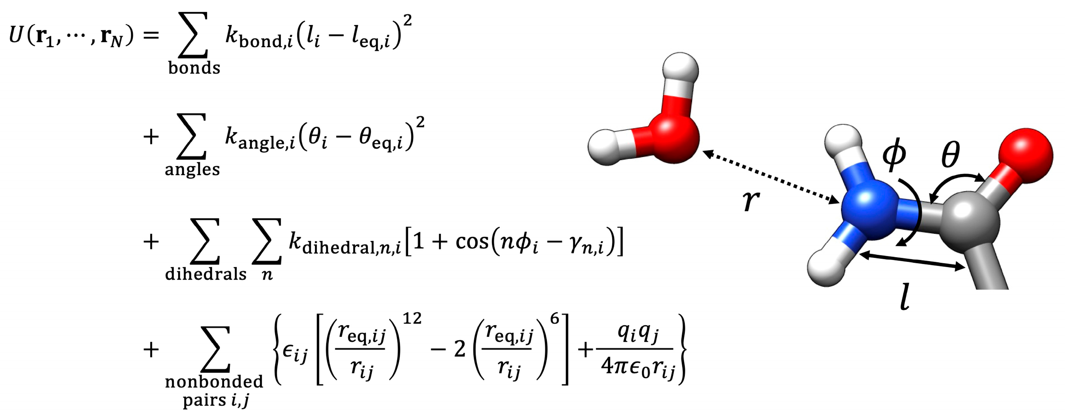
Molecular dynamics (MD) is a computational approach that analyzes the dynamics and energetics of interacting atoms and is a potential tool for studying the chiral-selective aminoacylation reaction of an RNA minihelix with an aminoacyl phosphate oligonucleotide and a bridging oligonucleotide. In classical MD simulations, successive configurations of a given system are generated by integrating Newton’s equations of motion, where the forces on the atoms are calculated using empirical molecular mechanics (MM) energy functions with force field parameters optimized to reasonably approximate the quantum mechanical potential energy surface (Figure 4) [40].
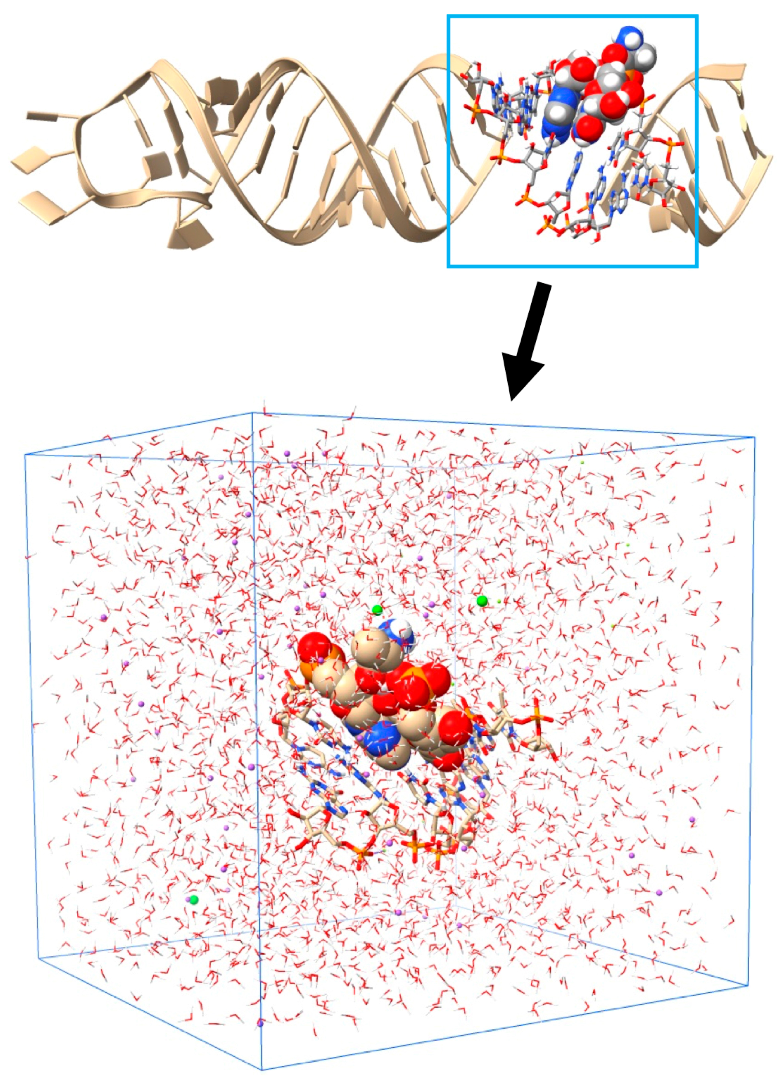
Classical MD with the MM potential has generally simulated the microsecond-long dynamics of a nucleic acid or a protein system in recent years. In 2018, we employed the classical MD method to investigate the possible mechanisms that determine chiral selectivity in the aminoacylation of RNA minihelices [41]. An atomistic model of six base pairs centered at the reaction site composed of the RNA minihelix, a bridging oligonucleotide, and 5′-Ala-p-dT6dA2 was used for the MD study, where L/D-alanine (Ala) was covalently attached at 5′-phosphate via an acyl phosphate linkage (Figure 5). In the study, Amber force fields OL15 [42,43,44], OL3 [45,46], and ff14SB [47] were used for RNA, DNA, and amino acids, respectively.
The classical MD study showed that the simulation system adopted the geometry required for the chemical reaction to occur more frequently with L-Ala than with D-Ala. For L-Ala, the structure with this geometry was formed by a combination of stable dihedral angles along the alanyl phosphate backbone with a canonical RNA structure, where the methyl group of alanine was placed on the opposite side of the approaching 3′-hydroxyl group concerning the carbonyl plane (Figure 6). For D-Ala, the methyl group and the 3′-hydroxyl group were placed on the same side concerning the carbonyl plane, which significantly decreased its ability to approach 3′-oxygen close to the carbonyl carbon compared with L-Ala (Figure 6). The chiral-selective aminoacylation mechanism suggested by the classical MD simulations was a possible scenario to explain the experimental results. For the deep understanding of a chemical reaction, revealing the energetics and structures along its reaction pathway, including the transition state, is essential in general. However, since simulating chemical reactions involving bond formation and breaking requires quantum mechanics (QM) calculations, our classical MD simulation study with the MM potentials gave us no clue about the transition state for the chiral-selective aminoacylation reactions of the primordial RNA.
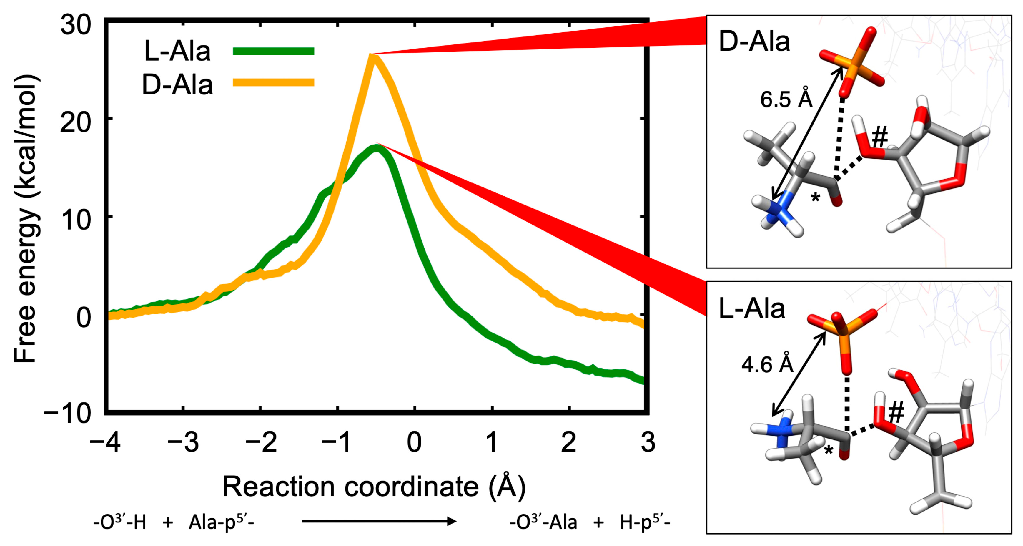
In 2023, to overcome these defects, we performed MD simulations where residues directly involving the aminoacylation reaction were treated using the QM approach and the environment around these residues were treated classically, or the so-called QM/MM method, to gain further insight into the mechanisms of chiral-selective aminoacylation in the RNA minihelix [48]. In the QM/MM MD simulations, we also introduced an umbrella sampling method to measure free-energy profiles along the reaction. For the first time, the details of the reaction pathway as to why L-amino acids are selected were revealed: (1) the height of the free-energy barrier for the L-Ala aminoacylation reaction was 9 kcal/mol lower than that for the D-Ala system. (2) At the transition state, the distance between the negatively charged 3′-oxygen and the positively charged amino group of L-Ala was shorter than that of D-Ala, which was caused by the chirality difference of the amino acid (Figure 7).
The following section describes the efforts and innovations made in our system to achieve this important result. We also present the simulation results using easy-to-understand diagrams so that the general public can understand the significance of the results.
The QM method can be more accurate for the energy calculation of simulation systems than the MM method [49]. However, the computational cost of the QM calculation is usually more expensive than the MM calculation. For an N-atom system with primitive calculation techniques, computational complexities for the MM and QM method are approximately O(N2) and O(N3), respectively [50]. The former requires calculations of nonbonded interactions (van der Waals and electrostatic interactions involving all atom pairs) (see Figure 4). The latter requires the diagonalization of the Hamiltonian matrix with a size of approximately N × N. In the hybrid QM/MM method, only a particular region of interest (typically a chemical reaction site) is treated with QM potentials, whereas the surrounding environment is evaluated with MM potentials, enabling the handling of chemical reactions in a computationally efficient manner [49,51]. Currently, more computationally efficient methods are well developed for both approaches. In the MM calculation, particle-mesh-Ewald and fast multipole methods reduce computational complexities to O(NlogN) and O(N), respectively, for large systems [50]. In the QM calculation, semiempirical molecular orbital theory and density function based tight binding (DFTB) theory scale O(N)~O(N2) [52]. If a more accurate and higher level of theory are required in the QM calculation, the computational cost increases significantly, up to ~O(N7) [52].
QM/MM MD (and classical MD) computes the potential energy of the simulated system at every time step. However, computing free-energy surfaces using these methods is difficult because of the limited sampling space. Umbrella sampling is used to calculate the free-energy profile along a predefined reaction coordinate (RC), also called the potential of mean force (PMF) [53]. Umbrella sampling attempts to efficiently sample energetically separated regions in phase space by adding a biasing potential to the original potential function. After a series of umbrella sampling simulations were performed along the RC, the PMF was recovered by reweighing the entire simulation period.
For our QM/MM MD simulation study, the same six-base RNA minihelix used in the classical MD simulation was used (Figure 5). The QM region comprises 79 atoms in Ala-dT and 3′-A, whose O3′ attacks the carbonyl carbon Ccarb in the acyl phosphate linkage. The QM calculations were performed using the self-consistent charge DFTB (SCC-DFTB) method, including the third-order term of the Taylor series expansion of the total energy, known as DFTB3 [54]. For the MM region, the same force fields were used as in the classical MD simulations. For umbrella sampling, an RC was selected as the difference in the distances between H and O3′, d(H3′…O3′) and between Ccarb and O3′, d(Ccarb…O3′): RC = d(H3′…O3′) − d(Ccarb…O3′). The initial and final coordinates for QM/MM umbrella sampling MD corresponded to the RC of −4.0 and 3.0 Å, respectively. The RC was divided into 101 windows, and umbrella sampling QM/MM MD simulations were performed for each window. For each window, 10 ps of equilibration and 20 ps of the production run with harmonic restraints were performed with a time step of 0.5 fs. A weighted histogram analysis method was used to compute the PMF from the simulation data for all windows. Thus, the aminoacylation of an RNA minihelix could determine the homochirality of amino acids in biological systems [39]. Once L-amino acids are selected in the primitive aminoacylation system, proteins generated from L-amino acid-bearing tRNA are inevitably composed of L-amino acids. QM/MM MD simulation techniques, for the first time, elucidated the “flowing” atomistic mechanisms of the chiral-selective aminoacylation and indicated that the L-Ala moiety stabilizes the transition state more than D-Ala, resulting in L-Ala preference in the aminoacylation reaction in the RNA [48]. The QM/MM method is expected to become an important strategy for analyzing biomolecular reactions in the future. In particular, using the QM/MM method will be greatly respected for reaction processes in general and transition states in particular, which cannot be clarified by experimental structural analysis in many cases. Additionally, its usefulness in simple systems that mimic the origin of life, such as those revealed in this study, is immeasurable.
The 2024 Nobel Prize in Chemistry was awarded for research on designing completely new proteins and for predicting the steric structure of proteins with high precision [55,56,57]. The role played by artificial intelligence in these studies was extremely significant, suggesting that it has become indispensable for discovering things that will lead to innovation in the future. The QM/MM method will continue to develop, riding on this trend and contributing to a deeper understanding of life phenomena. In fact, machine learning (ML) techniques based on the work awarded by the 2024 Nobel Prize in Physics are employed to improve the accuracy of QM calculations with low computational cost, which have made significant progress in recent years [58]. In the near future, the QM/MM method will make a significant contribution to the elucidation of the mechanisms of enzymatic reactions and chemical reactions involving electronic states, such as metalloenzymes and photosynthesis-related proteins. The method will also play important roles in drug design and in the prediction of the active site and the residues closely related to the activity, thereby fundamentally revolutionizing life science. It would be meaningful not only for specialists but also for general biologists to effectively utilize methods like the ones we have used.

Author Contributions

Conceptualization, T.A. and K.T.; data curation, T.A.; funding acquisition, T.A. and K.T.; investigation, T.A.; project administration, T.A.; supervision, K.T.; writing–original draft, T.A. and K.T.; writing–review and editing, T.A. and K.T. All authors have read and agreed to the published version of the manuscript.

Funding

This work was supported by JSPS KAKENHI Grant Numbers JP20K06592 (to T.A.) and JP21K06293 (to K.T.).

Data Availability Statement

The data presented in this study are available from the corresponding authors upon reasonable request.

Conflicts of Interest

The authors declare no conflict of interest.

References

  1. Perutz, M.F.; Rossmann, M.G.; Cullis, A.F.; Muirhead, H.; Will, G.; North, A.C.T. Structure of hæmoglobin: A three-dimensional Fourier synthesis at 5.5-Å. resolution, obtained by X-ray analysis. Nature 1960, 185, 416–422. [Google Scholar] [CrossRef] [PubMed]
  2. Kendrew, J.C.; Dickerson, R.E.; Strandberg, B.E.; Hart, R.G.; Davies, D.R.; Phillips, D.C.; Shore, V.C. Structure of myoglobin: A three-dimensional Fourier synthesis at 2 Å. resolution. Nature 1950, 185, 422–427. [Google Scholar] [CrossRef] [PubMed]
  3. Hodgkin, D.C. The X-ray analysis of complicated molecules. Science 1965, 150, 979–988. [Google Scholar] [CrossRef] [PubMed]
  4. Blundell, T.L. The first resolution revolution in protein structure analysis: X-ray diffraction of polypeptide conformations and globular protein folds in 1950s and 1960s. Prog. Biophys. Mol. Biol. 2021, 167, 32–40. [Google Scholar] [CrossRef] [PubMed]
  5. Henkel, A.; Oberthür, D. A snapshot love story: What serial crystallography has done and will do for us. Acta. Crystallogr. D Struct. Biol. 2024, 80, 563–579. [Google Scholar] [CrossRef]
  6. Aue, W.P.; Bartholdi, E.; Ernst, R.R. Two-dimensional spectroscopy. Application to nuclear magnetic resonance. J. Chem. Phys. 1976, 64, 2229–2246. [Google Scholar] [CrossRef]
  7. Wagner, G.; Wüthrich, K. Dynamic model of globular protein conformations based on NMR studies in solution. Nature 1978, 275, 247–248. [Google Scholar] [CrossRef]
  8. Yao, S.; Keizer, D.W.; Babon, J.J.; Separovic, F. NMR measurement of biomolecular translational and rotational motion for evaluating changes of protein oligomeric state in solution. Eur. Biophys. J. 2022, 51, 193–204. [Google Scholar] [CrossRef]
  9. Han, B.; Yang, J.; Zhang, Z. Selective methods promote protein solid-state NMR. J. Phys. Chem. Lett. 2024, 15, 11300–11311. [Google Scholar] [CrossRef]
  10. Shen, P.S. The 2017 Nobel Prize in Chemistry: Cryo-EM comes of age. Anal. Bioanal. Chem. 2018, 410, 2053–2057. [Google Scholar] [CrossRef] [PubMed]
  11. Cabral, A.; Cabral, J.E.; McNulty, R. Cryo-EM for small molecules. Curr. Protoc. 2022, 2, e632. [Google Scholar] [CrossRef] [PubMed]
  12. Mazal, H.; Wieser, F.F.; Sandoghdar, V. Insights into protein structure using cryogenic light microscopy. Biochem. Soc. Trans. 2023, 51, 2041–2059. [Google Scholar] [CrossRef] [PubMed]
  13. Gilbert, W. Origin of life: The RNA world. Nature 1986, 319, 618. [Google Scholar] [CrossRef]
  14. Crick, F.H.C. On Degenerate Templates and the Adapter Hypothesis. 1955. Available online: https://collections.nlm.nih.gov/catalog/nlm:nlmuid-101584582X73-doc (accessed on 25 November 2024).
  15. Tamura, K. Origins and early evolution of the tRNA molecule. Life 2015, 5, 1687–1699. [Google Scholar] [CrossRef]
  16. Giegé, R.; Eriani, G. The tRNA identity landscape for aminoacylation and beyond. Nucleic Acids Res. 2023, 51, 1528–1570. [Google Scholar] [CrossRef]
  17. Tennakoon, R.; Cui, H. Aminoacyl-tRNA synthetases. Curr. Biol. 2024, 34, R884–R888. [Google Scholar] [CrossRef]
  18. Kim, S.H.; Suddath, F.L.; Quigley, G.J.; McPherson, A.; Sussman, J.L.; Wang, A.H.; Seeman, N.C.; Rich, A. Three-dimensional tertiary structure of yeast phenylalanine transfer RNA. Science 1974, 185, 435–440. [Google Scholar] [CrossRef]
  19. Robertus, J.D.; Ladner, J.E.; Finch, J.T.; Rhodes, D.; Brown, R.S.; Clark, B.F.; Klug, A. Structure of yeast phenylalanine tRNA at 3 Å resolution. Nature 1974, 250, 546–551. [Google Scholar] [CrossRef] [PubMed]
  20. Francklyn, C.; Schimmel, P. Aminoacylation of RNA minihelices with alanine. Nature 1989, 337, 478–481. [Google Scholar] [CrossRef]
  21. Frugier, M.; Florentz, C.; Giegé, R. Efficient aminoacylation of resected RNA helices by class II aspartyl-tRNA synthetase dependent on a single nucleotide. EMBO J. 1994, 13, 2219–2226. [Google Scholar] [CrossRef]
  22. Martinis, S.A.; Schimmel, P. Small RNA oligonucleotide substrates for specific aminoacylations. In tRNA: Structure, Biosynthesis, and Function; ASM Press: Washington, DC, USA, 1997; pp. 349–370. [Google Scholar]
  23. Musier-Forsyth, K.; Schimmel, P. Atomic determinants for aminoacylation of RNA minihelices and relationship to genetic code. Acc. Chem. Res. 1999, 32, 368–375. [Google Scholar] [CrossRef]
  24. Schimmel, P.; Giegé, R.; Moras, D.; Yokoyama, S. An operational RNA code for amino-acids and possible relationship to genetic-code. Proc. Natl. Acad. Sci. USA 1993, 90, 8763–8768. [Google Scholar] [CrossRef]
  25. Lei, L.; Burton, Z.F. The 3 31 nucleotide minihelix tRNA evolution theorem and the origin of life. Life 2023, 13, 2224. [Google Scholar] [CrossRef] [PubMed]
  26. Tang, G.Q.; Hu, H.; Douglas, J.; Carter, C.W., Jr. Primordial aminoacyl-tRNA synthetases preferred minihelices to full-length tRNA. Nucleic Acids Res. 2024, 52, 7096–7111. [Google Scholar] [CrossRef]
  27. Schimmel, P.; Ribas de Pouplana, L. Transfer RNA: From minihelix to genetic code. Cell 1995, 81, 983–986. [Google Scholar] [CrossRef]
  28. Sarzynska, J.; Popenda, M.; Antczak, M.; Szachniuk, M. RNA tertiary structure prediction using RNAComposer in CASP15. Proteins 2023, 91, 1790–1799. [Google Scholar] [CrossRef] [PubMed]
  29. Schimmel, P. Aminoacyl tRNA synthetases: General scheme of structure-function relationships in the polypeptides and recognition of transfer RNAs. Annu. Rev. Biochem. 1987, 56, 125–158. [Google Scholar] [CrossRef]
  30. Lux, M.C.; Standke, L.C.; Tan, D.S. Targeting adenylate-forming enzymes with designed sulfonyladenosine inhibitors. J. Antibiot. 2019, 72, 325–349. [Google Scholar] [CrossRef]
  31. Pettersen, E.F.; Goddard, T.D.; Huang, C.C.; Couch, G.S.; Greenblatt, D.M.; Meng, E.C.; Ferrin, T.E. UCSF chimera—A visualization system for exploratory research and analysis. J. Comput. Chem. 2004, 25, 1605–1612. [Google Scholar] [CrossRef]
  32. Atencio, A. Separation of chiral molecules: A way to homochirality. Orig. Life Evol. Biosph. 2012, 42, 55–73. [Google Scholar] [CrossRef]
  33. Tamura, K. Perspectives on the origin of biological homochirality on Earth. J. Mol. Evol. 2019, 87, 143–146. [Google Scholar] [CrossRef] [PubMed]
  34. Hegstrom, R.A. Parity violation and chiral symmetry breaking of a racemic mixture. Biosystems 1987, 20, 49–56. [Google Scholar] [CrossRef] [PubMed]
  35. Bonner, W.A. Parity violation and the evolution of biomolecular homochirality. Chirality 2000, 12, 114–126. [Google Scholar] [CrossRef]
  36. Fukue, T.; Tamura, M.; Kandori, R.; Kusakabe, N.; Hough, J.H.; Bailey, J.; Whittet, D.C.; Lucas, P.W.; Nakajima, Y.; Hashimoto, J. Extended high circular polarization in the Orion massive star forming region: Implications for the origin of homochirality in the solar system. Orig. Life Evol. Biosph. 2010, 40, 335–346. [Google Scholar] [CrossRef]
  37. Soai, K.; Shibata, T.; Morioka, H.; Choji, K. Asymmetric autocatalysis and amplification of enantiomeric excess of a chiral molecule. Nature 1995, 378, 767–768. [Google Scholar] [CrossRef]
  38. Bada, J.L.; Miller, S.L. Racemization and the origin of optically active organic compounds in living organisms. Biosystems 1987, 20, 21–26. [Google Scholar] [CrossRef]
  39. Tamura, K.; Schimmel, P. Chiral-selective aminoacylation of an RNA minihelix. Science 2004, 305, 1253. [Google Scholar] [CrossRef]
  40. Karplus, M.; McCammon, J.A. Molecular dynamics simulations of biomolecules. Nat. Struct. Biol. 2002, 9, 646–652. [Google Scholar] [CrossRef]
  41. Ando, T.; Takahashi, S.; Tamura, K. Principles of chemical geometry underlying chiral selectivity in RNA minihelix aminoacylation. Nucleic Acids Res. 2018, 46, 11144–11152. [Google Scholar] [CrossRef]
  42. Krepl, M.; Zgarbová, M.; Stadlbauer, P.; Otyepka, M.; Banáš, P.; Koča, J.; Cheatham, T.E., 3rd; Jurečka, P.; Šponer, J. Reference simulations of noncanonical nucleic acids with different chi variants of the AMBER force field: Quadruplex DNA, quadruplex RNA and Z-DNA. J. Chem. Theory Comput. 2012, 8, 2506–2520. [Google Scholar] [CrossRef]
  43. Zgarbová, M.; Luque, F.J.; Šponer, J.; Cheatham, T.E., 3rd; Otyepka, M.; Jurečka, P. Toward improved description of DNA backbone: Revisiting epsilon and zeta torsion force field parameters. J. Chem. Theory Comput. 2013, 9, 2339–2354. [Google Scholar] [CrossRef] [PubMed]
  44. Zgarbová, M.; Šponer, J.; Otyepka, M.; Cheatham, T.E., 3rd; Galindo-Murillo, R.; Jurečka, P. Refinement of the sugar-phosphate backbone torsion beta for AMBER force fields improves the description of Z- and B-DNA. J. Chem. Theory Comput. 2015, 11, 5723–5736. [Google Scholar] [CrossRef] [PubMed]
  45. Pérez, A.; Marchán, I.; Svozil, D.; Sponer, J.; Cheatham, T.E., 3rd; Laughton, C.A.; Orozco, M. Refinement of the AMBER force field for nucleic acids: Improving the description of alpha/gamma conformers. Biophys. J. 2007, 92, 3817–3829. [Google Scholar] [CrossRef]
  46. Zgarbová, M.; Otyepka, M.; Šponer, J.; Mládek, A.; Banáš, P.; Cheatham, T.E., 3rd; Jurečka, P. Refinement of the Cornell et al. nucleic acids force field based on reference quantum chemical calculations of glycosidic torsion profiles. J. Chem. Theory Comput. 2011, 7, 2886–2902. [Google Scholar] [CrossRef] [PubMed]
  47. Maier, J.A.; Martinez, C.; Kasavajhala, K.; Wickstrom, L.; Hauser, K.E.; Simmerling, C. ff14SB: Improving the accuracy of protein side chain and backbone parameters from ff99SB. J. Chem. Theory Comput. 2015, 11, 3696–3713. [Google Scholar] [CrossRef]
  48. Ando, T.; Tamura, K. Mechanism of chiral-selective aminoacylation of an RNA minihelix explored by QM/MM free-energy simulations. Life 2023, 13, 722. [Google Scholar] [CrossRef]
  49. Kubař, T.; Elstner, M.; Cui, Q. Hybrid quantum mechanical/molecular mechanical methods for studying energy transduction in biomolecular machines. Annu. Rev. Biophys. 2023, 52, 525–551. [Google Scholar] [CrossRef]
  50. Schlick, T. Molecular Modeling and Simulation: An Interdisciplinary Guide, 2nd ed.; Springer: New York, NY, USA, 2010. [Google Scholar]
  51. Brunk, E.; Rothlisberger, U. Mixed quantum mechanical/molecular mechanical molecular dynamics simulations of biological systems in ground and electronically excited states. Chem. Rev. 2015, 115, 6217–6263. [Google Scholar] [CrossRef]
  52. Kar, R.K. Benefits of hybrid QM/MM over traditional classical mechanics in pharmaceutical systems. Drug Discov. Today 2023, 28, 103374. [Google Scholar] [CrossRef]
  53. Torrie, G.M.; Valleau, J.P. Nonphysical sampling distributions in Monte Carlo free-energy estimation: Umbrella sampling. J. Comput. Phys. 1977, 23, 187–199. [Google Scholar] [CrossRef]
  54. Gaus, M.; Cui, Q.; Elstner, M. DFTB3: Extension of the self-consistent-charge density-functional tight-binding method (SCC-DFTB). J. Chem. Theory Comput. 2012, 7, 931–948. [Google Scholar] [CrossRef] [PubMed]
  55. Service, R.F. AI tools set off an explosion of designer proteins. Science 2024, 386, 260–261. [Google Scholar] [CrossRef] [PubMed]
  56. Wang, J.; Watson, J.L.; Lisanza, S.L. Protein design using structure-prediction networks: AlphaFold and RoseTTAFold as protein structure foundation models. Cold Spring Harb. Perspect. Biol. 2024, 16, a041472. [Google Scholar] [CrossRef] [PubMed]
  57. Chen, L.; Li, Q.; Nasif, K.F.A.; Xie, Y.; Deng, B.; Niu, S.; Pouriyeh, S.; Dai, Z.; Chen, J.; Xie, C.Y. AI-driven deep learning techniques in protein structure prediction. Int. J. Mol. Sci. 2024, 25, 8426. [Google Scholar] [CrossRef]
  58. Cui, Q.; Pal, T.; Xie, L. Biomolecular QM/MM simulations: What are some of the “burning issues”? J. Phys. Chem. B 2021, 125, 689–702. [Google Scholar] [CrossRef]
Figure 1. Evolution of aminoacylation of tRNA by aminoacyl-tRNA synthetase (aaRS). The earlier RNA minihelix and primitive aaRS (left) could have been evolved to current tRNA and aaRS (right). L-aa indicates L-amino acid. The 3-dimensional structure of the RNA minihelix was modeled with RNAComposer [28] and that of tRNA was based on PDB ID 1EHZ.
Computation 12 00238 g001
Figure 2. The structures of L-alanyl-AMP and L-alanyl-sulfamoyl adenosine (left) and the electrostatic potential maps of these molecules (right). The maps were calculated and visualized by using Coulombic Surface Coloring tool in UCSF Chimera version 1.18 [31]. The potentials are on a [−10, 10] red–white–blue color map in units of kcal/mol/e. Molecules are shown in two views, 180° apart.
Computation 12 00238 g002
Figure 3. Schematic presentation of aminoacylation reaction in an RNA minihelix with an aminoacyl phosphate oligonucleotide (light green) and a bridging oligonucleotide (light blue). The reactions clearly showed that the formation of an L-aminoacyl-minihelix was preferred over that of a D-aminoacyl-minihelix [39].
Computation 12 00238 g003
Figure 4. Molecular mechanics energy functions. Total potential energy of an N-atom’s molecular model U is typically constructed as the sum of contributions from bond length, bond angle, torsion angle, van der Waals (Lennard-Jones), and electrostatic Coulombic potentials. i and j are indices of atom, bond, angle, or dihedral. The subscript eq’s refer to equilibrium value. r is position vector of atom. l, θ, and φ represent bond length, bond angle, and dihedral angle, respectively. kbond, kangle, and kdihedral are force constants for bond length, bond angle, and dihedral angle potentials, respectively. n and γ are periodicity of the rotational barrier and reference angle, respectively. r is the distance between two atoms. ε is the energy depth of Lennard-Jones potential. q and ε0 are the charge and the dielectric constant in vacuo, respectively.
Computation 12 00238 g004
Figure 5. Structural model of the RNA minihelix (upper) and a simulation system for classical and quantum mechanics/molecular mechanics (QM/MM) MD studies of chiral-selective aminoacylation reaction of the RNA minihelix (lower). The six base pairs used for MD simulations are represented by a ball and stick model and indicated by a rectangular box in the upper figure. The aminoacylation reaction occurs between Ala-dT and 3′-A, which are drawn with van der Walls spheres, and these residues are handled with the QM method in the QM/MM MD simulations. In the simulation system, the RNA is solvated with water molecules and neutralized by adding Mg2+, Cl, and Na+ ions in a box to mimic experimental conditions. The total number of atoms was 9300.
Computation 12 00238 g005
Figure 6. Representative structures with the 3′-oxygen of A3 ribose approaching the carbonyl carbon in the acyl phosphate linkage for the L-Ala (left) and D-Ala (right) systems obtained by the classical MD simulations. Ribose of A3, phosphate, and alanine moiety of dT4 are shown by a stick model, and the others in nucleotides are shown by a wire frame model with transparent color. The elements are colored according to the standard CPK rules: carbon, gray; hydrogen, white; oxygen, red; nitrogen, blue; phosphorus, orange. Carbonyl carbon (Ccarb) and 3′-oxygen (O3′) are marked by * and #, respectively. Arrows represent O3′ attacking Ccarb. A distinct difference between the two structures was the position of the amino group of alanine relative to the O3′ nucleophile and the carbonyl group. For the L-Ala system, the amino group and O3′ atom were placed on the same side with respect to the carbonyl plane. For the D-Ala system, the amino group was placed on the opposite side with respect to the carbonyl plane, and the methyl group of alanine was placed close to the O3′ atom.
Computation 12 00238 g006
Figure 7. Free-energy profiles along the reaction coordinate and structures of the reaction site at the transition states obtained by the QM/MM umbrella sampling MD of D/L-Ala aminoacylation reaction in the RNA minihelix. At the transition states, the distance between the negatively charged 3ʹ-oxygen and the positively charged amino group of L-Ala is shorter than that of D-Ala. The representation style for the structures is the same as that in Figure 6. Distances between the amino nitrogen atom of Ala and the phosphorus atom of phosphate group are shown. Broken lines represent bonds breaking and forming during the reaction. The elements are colored according to the standard CPK rules: carbon, gray; hydrogen, white; oxygen, red; nitrogen, blue; phosphorus, orange. Carbonyl carbon (Ccarb) and 3′-oxygen (O3′) are marked by * and #, respectively.
Computation 12 00238 g007
Disclaimer/Publisher’s Note: The statements, opinions and data contained in all publications are solely those of the individual author(s) and contributor(s) and not of MDPI and/or the editor(s). MDPI and/or the editor(s) disclaim responsibility for any injury to people or property resulting from any ideas, methods, instructions or products referred to in the content.