Article

Test–Retest Reliability of Deep Learning Analysis of Brain Volumes in Adolescent Brain

by
Anna-Maria Kasparbauer
1,
Heidrun Lioba Wunram
1,2,
Fabian Abuhsin
3,
Friederike Körber
4,
Eckhard Schönau
5,
Stephan Bender
1 and
Ibrahim Duran
2,5,*
1
Department of Child and Adolescent Psychiatry, Psychosomatics and Psychotherapy, Faculty of Medicine and University Hospital Cologne, University of Cologne, 50931 Cologne, Germany
2
Department of Pediatrics, Medical Faculty, University Hospital, University of Cologne, 50931 Cologne, Germany
3
Department of General Pediatrics, Neonatology and Pediatric Cardiology, Medical Faculty, University Hospital, 40255 Düsseldorf, Germany
4
Department of Pediatric Radiology, Medical Faculty, University Hospital, 50931 Cologne, Germany
5
Center of Prevention and Rehabilitation, Medical Faculty, University Hospital, University of Cologne, UniReha, 50931 Cologne, Germany
*
Author to whom correspondence should be addressed.
Information 2024, 15(12), 748; https://doi.org/10.3390/info15120748
Submission received: 26 September 2024 / Revised: 5 November 2024 / Accepted: 9 November 2024 / Published: 25 November 2024
(This article belongs to the Special Issue Real-World Applications of Machine Learning Techniques)

Abstract

:
Magnetic resonance imaging (MRI) is essential for studying brain development and psychiatric disorders in adolescents. However, the imaging consistency remains challenging, highlighting the need for advanced methodologies to improve the diagnostic and research reliability in this unique developmental period. Adolescence is marked by significant neuroanatomical changes, distinguishing adolescent brains from those of adults and making age-specific imaging research crucial for understanding the neuropsychiatric conditions in youth. This study examines the test–retest reliability of anatomical brain MRI scans in adolescents diagnosed with depressive disorders, emphasizing a developmental perspective on neuropsychiatric disorders. Using a sample of 42 adolescents, we assessed the consistency of structural imaging metrics across 95 brain regions with deep learning-based neuroimaging analysis pipelines. The results demonstrated moderate to excellent reliability, with the intraclass correlation coefficients (ICC) ranging from 0.57 to 0.99 across regions. Notably, regions such as the pallidum, amygdala, entorhinal cortex, and white matter hypointensities showed moderate reliability, likely reflecting the challenges in the segmentation or inherent anatomical variability unique to this age group. This study highlights the necessity of integrating advanced imaging technologies to enhance the accuracy and reliability of the neuroimaging data specific to adolescents. Addressing the regional variability and strengthening the methodological rigor are essential for advancing the understanding of brain development and psychiatric disorders in this distinct developmental stage. Future research should focus on larger, more diverse samples, multi-site studies, and emerging imaging techniques to further validate the neuroimaging biomarkers. Such advancements could improve the clinical outcomes and deepen our understanding of the neuropsychiatric conditions unique to adolescence.

Graphical Abstract

1. Introduction

The investigation of psychiatric disorders has increasingly recognized the importance of a developmental perspective, as evidenced by the diverse studies from the biological, imaging, genetic, and clinical fields [1]. This approach is particularly crucial given that the peak incidence of many psychiatric conditions, such as depression, typically coincides with the critical transition from childhood and adolescence to adulthood. Research indicates that the onset of the first mental disorder occurs before the age of 14 in one-third of individuals, before the age of 18 in almost half (48.4%), and before the age of 25 in 62.5%, with the peak age at onset being 14.5 years and the median age of 18 years old across all mental disorders [2,3].
This early onset aligns with significant developmental changes in the brain structure and function across adolescence, distinguishing adolescent brains from those of adults. Adolescence is marked by dynamic growth in gray and white matter, both of which develop in distinct, non-linear trajectories. The gray matter volume typically peaks before puberty, followed by a reduction due to synaptic pruning, which refines the neural connections in response to environmental experiences. In contrast, the white matter volume peaks later, during young adulthood, as myelination continues to increase, enhancing the speed and efficiency of the neural communication. These ongoing changes are especially pronounced in association cortices and the prefrontal cortex, which underlie complex cognitive functions such as decision making and impulse control—functions that reach maturity only in adulthood.
Additionally, adolescence involves marked shifts in the neurochemical systems and cerebral metabolic activity, particularly in regions like the amygdala, which is essential for emotional processing. In adulthood, these systems tend to stabilize, supporting more consistent emotional and cognitive regulation. The interplay between these developmental processes and the early onset of psychiatric disorders underscore the need for age-specific research that considers the unique neurodevelopmental trajectories of adolescence [3,4].
Together, these findings emphasize that early neurodevelopmental stages are crucial, with many mental disorders manifesting before the age of 18 and often persisting into adulthood [5,6]. This highlights the need for research and clinical approaches tailored to the neurodevelopmental profiles characteristic of adolescence. The existing divide between child/adolescent and adult psychiatry in both research and clinical settings presents a significant barrier to the integration of the developmental insights into psychiatric practice. This separation often leads to fragmented care and overlooks the continuous developmental trajectory that spans from childhood through adolescence and into adulthood. As a result, the identification of clinically useful biomarkers becomes crucial. These biomarkers can serve as early indicators of neurological or psychiatric disorders, facilitating timely and accurate diagnosis and intervention [7,8,9]. Reliable neuroimaging biomarkers are essential for the early detection of these conditions. Such biomarkers not only aid in the early identification of at-risk individuals but also provide critical information for tailoring therapeutic strategies, potentially altering the course of psychiatric disorders and improving the long-term patient outcomes. Integrating, for example, structural brain biomarkers into clinical practice could bridge the gap between the different stages of psychiatric care and enhance our ability to intervene earlier and more effectively across the lifespan [10].
In the context of depressive disorders, for instance, brain volume measurements in key regions such as the anterior cingulate cortex, hippocampus, and amygdala contribute significantly to understanding the pathophysiology, progression, and treatment of depression [7,8,9]. These measurements offer insights into emotional regulation and cognitive control, which are often disrupted in depression, and can inform both diagnostic processes and treatment planning. By utilizing brain volumetry as biomarkers to monitor the developmental changes, clinicians can adopt a more personalized approach, adjusting interventions to better support the individual’s neurodevelopmental trajectory, ultimately improving the outcomes and providing a continuity of care from adolescence into adulthood.
To enhance the accuracy of these brain volume measurements, accurate brain segmentation is critical for studying the brain development and neurological disorders. Supervised deep learning approaches, particularly fully convolutional neural networks (F-CNNs), offer a faster alternative to the traditional methods using pattern recognition approaches due to their ability to learn feature representations directly from images [11,12]. These methods, which can be parallelized on graphic processing units (GPUs) for significant speed improvements, often outperform the traditional techniques in terms of accuracy, making them popular for segmentation tasks. For this study, we chose FastSurferCNN [13,14,15,16], a deep learning model capable of segmenting the whole brain into 95 classes in just one minute on a GPU. Inspired by QuickNAT [17], FastSurferCNN incorporates improvements such as competitive dense blocks and spatial information aggregation to enhance segmentation accuracy, especially for cortical gray matter. The model is trained across multiple adult datasets (age range 18–96 years old) and scanner types (1.5 T and 3 T), balancing the anatomical diversity for generalizability [16].
It demonstrates high test–retest reliability in adult samples, yet reliability in the developing brain poses distinct challenges due to the ongoing maturation processes, individual variability, and structural complexity. As aforementioned, adolescent brains undergo significant changes in cortical thickness, white matter growth, and synaptic pruning, which vary across individuals and age groups [4,18]. Additionally, adolescent MRI data often suffer from a lower tissue contrast between gray and white matter, further hindering the accurate delineation of structures [19]. Motion artifacts are more common in pediatric populations, resulting in degraded image quality [20]. Studies on the test–retest reliability of brain volume measurements in adolescents are limited, and there has been evidence that automated segmentation overestimates, for example, the total hippocampus and amygdala volumes in comparison to the manual segmentation protocol, most likely due to partial volume effects [21]. Nonetheless, a study investigating the test–retest reliability in a sample of 50 adolescents (interscan interval of approximately 2.7 weeks) reported a high reliability for the traditional automated segmentation pipeline [22].
The challenges due to the developmental variations complicate the application of brain segmentation algorithms in adolescents, and deep learning approaches are typically trained on adult populations [11,23]. It is yet to be determined whether deep learning methods such as FastSurfer can effectively address these challenges such as a low tissue contrast, motion artifacts, and individual variability, ultimately leading to more accurate, reliable, and age-specific brain segmentation outcomes in adolescent populations.
To realize the potential of structural brain biomarkers, it is crucial to assess their reliability, which refers to the ability to consistently measure the results under similar circumstances. Reliability is a fundamental aspect of ensuring the accuracy and utility of neuroimaging data. Achieving reliable MRI results presents significant challenges, particularly due to the presence of imaging artifacts and the variations in scanning protocols [24].
The current study examined the test–retest reliability of structural MRI-based biomarkers in adolescents with depressive disorder, assessing the consistency of these biomarkers over an 8-week interval. There were no specific a priori hypotheses in this study, as there is a lack of comparable research on the test–retest reliability of structural MRI-based biomarkers in adolescents with depressive disorder. This study was therefore exploratory, aiming to establish foundational insights into the biomarker consistency within this population. We analyzed anatomical brain MRI scans from a sample of adolescents diagnosed with depressive disorder, each scanned twice with an 8-week interval. Brain segmentation was performed using the FastSurfer pipeline, and the regional test–retest reliability across 95 brain regions was assessed through intra-class coefficient (ICC) analysis. By comparing our findings to existing estimates from studies involving both depressive and healthy populations, we aimed to provide a comprehensive evaluation of the reliability of structural MRI-based biomarkers in adolescent clinical populations. This comparison is essential for validating the clinical utility of these biomarkers and exploring their potential implications in the early diagnosis and treatment of depressive disorders.

2. Materials and Methods

2.1. Study Design

The study employed a semi-randomized controlled trial design to evaluate the effects of physical activity on the brain structure in adolescents diagnosed with major depressive disorder. The detailed methodology can be referenced in Wunram et al. [25]. The participants were randomly assigned to one of three groups: two intervention groups engaged in additional physical activity and one control group which received no extra physical intervention beyond the standard treatment. Physical measures, Spiroergometry, Jump mechanography, Caliperometry, BMI, Depression questionnaires (Beck Depression Inventory BDI-II [26]), Sport questionnaires and structural whole-brain imaging were conducted during three time-points: baseline (t0), post (t1, after a six-week period with intervention except for the TAU group) and follow-up (t2, 8 weeks after t1 without any intervention).
The intervention involved moderate physical activity conducted 3–5 days a week for 30 min per session over a six-week period. This exercise regimen was supplementary to the treatment as usual (TAU) provided to all the participants, which included the standard therapeutic approaches for depression. The control group continued with the TAU without any added physical activity. The study protocol received approval from the University of Cologne Ethics Committee and was registered in the German Clinical Trials Register under the identification number DRKS00005120. Additionally, the Clinical Trials Center of the University of Cologne provided support for this study.

2.2. Participant Recruitment

The participants were recruited from the inpatient units at the Department of Child and Adolescent Psychiatry, Psychosomatic and Psychotherapy at the University Hospital of Cologne. The recruitment methods included distributing flyers and providing information on the clinic’s website. Adolescents and their parents were informed about the study and screened for eligibility upon admission to the inpatient treatment. Informed consent was obtained from the parents and adolescents involved in the study.
The inclusion criteria required the participants to be aged between 12 and 18 years old, have a diagnosis of non-psychotic major depressive disorder based on the DSM-IV/-5 and ICD-10 criteria, and score at least 18 raw points on a standardized depression questionnaire designed for children and adolescents (“Depressionsinventar für Kinder und Jugendliche”, DIKJ [27]). Additional requirements included normal intelligence, proficiency in the German language, and physical aptitude for sports activities. The exclusion criteria encompassed the presence of other psychiatric disorders, medical conditions that could hinder physical activity, and the use of medication with psychotropic effects.

2.3. Imaging Protocol

MRI data were acquired using a 1.5 T Siemens Magnetom Avanto scanner located at the University of Cologne’s imaging facility. Each participant was positioned supine in the MRI scanner with their head stabilized using foam padding to minimize movement. Earplugs were provided to reduce the impact of the scanner’s noise on the participants.
We collected a 3D T1-weighted (T1w) imaging sequence with whole-brain coverage, featuring an isotropic resolution of 0.9 mm3. The imaging parameters were as follows: matrix size = 256 × 256; field of view (FOV) = 227 × 260 mm; 176 sagittal slices with a thickness of 0.9 mm; repetition time (TR) = 11 ms; echo time (TE) = 5.2 ms; flip angle = 15°; bandwidth = 140 Hz/pixel; and phase encoding steps = 196. The scanning sequence utilized was a gradient echo (GR) sequence, which is known for its high signal-to-noise ratio and resolution suitable for detailed anatomical studies.

2.4. Data Analysis

For the present study, we used whole-brain scans after the experimental intervention (t1) and again 8 weeks later (t2). There was no difference between the groups during this time period. The images were exported from the DICOM to NIFTI format using MRIcroGL (version 1.2.20220720, https://www.nitrc.org/plugins/mwiki/index.php/mricrogl:MainPage (accessed on 22 February 2024)) and visually checked for artifacts (obvious blurring due to motion or susceptibility artifacts) before starting the analyses. If the image was excluded from the analysis, both t1 and t2 scans of the participant were excluded. We performed segmentation using the FastSurfer version (fastsurfer:cpu-v2.1.1), a deep-learning based neuroimaging pipeline, learning on Freesurfer anatomical segmentation and cortical parcellation [16]. As the first step, we used FastSurferCNN for structural segmentation; then, in the second step, we used the ‘mri_segstats’ program within the pipeline for volume measurements. FastSurferCNN performs brain segmentation using three 2D fully convolutional networks (F-CNNs) for coronal, axial, and sagittal slices. These networks use competitive dense blocks, which apply maxout activations to retain only the maximum feature values, thereby reducing the memory load without adding parameters. To enhance the segmentation accuracy, a multi-slice input strategy aggregates the spatial context by stacking adjacent slices. Finally, the model incorporates view aggregation, where the predictions from each anatomical plane are combined using weighted averages, producing a refined, comprehensive segmentation. The final output table included the names and volumes (mm3) of 45 brain structures, distinguishing between the left and right hemispheres for each region, along with five unpaired regions following the labeling of the Desikan-Killiany-Tourville atlas parcellation [28].
For statistical analysis, we used RStudio/R (R version 4.3.2) to calculate the intraclass coefficient (ICC) as suggested by Koo and Li [29] for each of the obtained regional volumes. The ICC was calculated to assess the test–retest reliability of the volume measurements. As a widely recognized metric, the ICC assesses both the degree of correlation and the level of agreement between repeated measurements, making it an ideal choice for determining the reliability of a method. The ICC values range from 0 to 1, with higher values indicating greater reliability. To enhance the precision of our reliability estimates, 95% confidence intervals (CIs) were computed for each ICC. This analysis focused on the agreement of brain volumes obtained from consecutive scans and categorized the ICC values as poor (<0.5), moderate (0.51–0.75), good (0.76–0.9), and excellent (0.9-1.0).

3. Results

Between 2013 and 2015, a total of 89 patients were continuously screened in the inpatient and day-clinic department of our child and adolescent psychiatry. From these, 25 participants were excluded for various reasons: eleven had depression scores below the cutoff, eight did not provide consent, and six were excluded due to medical conditions. This left a total of 64 participants eligible for the study. Out of the eligible participants, 41 were randomized into the intervention groups (whole-body vibration plate or cycling), and 23 were assigned to the TAU control condition. Twelve participants dropped out of the study, resulting in a final sample size of 52 participants who completed the study. MRI data were successfully obtained from 44 participants. After conducting thorough quality control to check for artifacts and ensure data completeness, 42 datasets remained suitable for further analysis. These consisted of 13 participants in the TAU group and 29 in the intervention groups. The final sample consisted of 11 males (26.19%) and 31 female adolescents (73.81%). The average age was 15.92 years (SD = 1.13). The demographics are presented in Table 1.
The ICC values for the brain regions ranged from 0.57 to 0.99 (95% CI range 0.329 to 0.994), indicating moderate to high reliability for all the anatomical regions. Figure 1 illustrates the ICCs for the anatomical regions, categorized from moderate to excellent reliability by size. Among these regions, three brain areas exhibited bilateral moderate ICC values ranging between 0.50 and 0.75. These areas are the pallidum (ICC = 0.67 on the left and 0.54 on the right), the amygdala (ICC = 0.69 on the left and 0.74 on the right), and the entorhinal cortex (ICC = 0.69 on the left and 0.61 on the right). Additionally, moderate test–retest reliability was observed in white matter hypointensities, with an ICC of 0.57. All the other brain regions demonstrated good to excellent reliability, with ICC values greater than 0.75, as listed in Table 2.

4. Discussion

In this study, we utilized a deep learning-based brain segmentation method to assess the test–retest reliability of 95 regional brain volumes in 42 adolescents with depression over an 8-week scan–rescan interval, leveraging its accuracy and reproducibility. We observed that all the brain regions exhibited moderate to excellent test–retest reliability for MRI-based volume measurements. Our findings indicated that the ICC values for cortical gray matter volumes were predominantly high, confirming excellent scan–rescan reliability in most regions. However, some areas, such as the pallidum and amygdala, exhibited moderate reliability, reflecting the variability tied to both the anatomical complexity and segmentation challenges.
The strong overall reliability observed is consistent with the findings in adult cohorts. Buimer et al. [30], in preparation for a youth cohort study, assessed the test–retest reliability in 15 healthy adults (ages 19–31) using FreeSurfer. With a scan interval of approximately one week, they reported high ICCs for brain volumes (>0.93; 95% CI: 0.80–1.00). Similarly, Iscan et al. [31] demonstrated high reliability for cortical volumes (mean ICC > 0.88) in 25 healthy adults (18–65), also using FreeSurfer (scan interval: 1 week). Both studies adhered to rigorous data quality protocols, ensuring consistency.
Studies in pediatric populations are fewer, due to the ethical constraints, and typically involve longer intervals (>12 weeks). A study evaluating the reliability of 68 brain regions in a mixed pediatric and adults sample (ages 9–25, including individuals with anxiety and ADHD) reports good to excellent reliability [22]. Cortical volumes demonstrate excellent stability with some regional variations that show poor to fair consistency (left temporal pole: ICC = 0.47, 95% CI [0.23, 0.66]; right temporal pole: ICC = 0.55, 95% CI [0.33, 0.72]). However, due to the different approach and the heterogeneous group (age, health status), a comparison is challenging, and the factors influencing the regional variation in consistency can only be hypothesized and examined in future research.
Henschel and colleagues showed FastSurfer’s advantages over other segmentation methods in diverse datasets across sites, scanners, and field strengths. Our findings support the applicability of these methods for adolescent populations, demonstrating similarly high test–retest reliability across brain volume measures.
The high degree of consistency in the measurements across cortical regions, makes the deep learning approach as provided by FastSurfer a reliable tool for further analysis in adolescent populations. While our results embedded in previous research suggest that there is an overall good reliability and a robustness for brain segmentation in adolescents, some regions may inherently exhibit more variability due to their anatomical or functional characteristics. For instance, we identified moderate ICC values in specific regions including the pallidum, amygdala, and entorhinal cortex, which showed bilateral ICCs ranging between 0.5 and 0.75.
The lower ICC values in regions such as the amygdala and pallidum may reflect both scanner-specific factors and the inherent anatomical complexity of these small, structurally intricate areas. The combination of their size, partial volume effects, and functional variability presents challenges for both automated and manual segmentation methods [30,32], suggesting that automated segmentation techniques could overestimate the volumes in these areas. However, other research has not demonstrated a clear advantage of manual segmentation over automated pipelines in adolescent populations [33]. These mixed findings underscore the need to carefully consider the methodological factors when interpreting the reliability metrics for specific brain regions. Additionally, lower ICC values may be related to an intrinsic anatomical variability or the smaller size of certain regions, which complicates accurate segmentation across multiple scans. For instance, the amygdala and entorhinal cortex are relatively small with complex boundaries, increasing the likelihood of segmentation variability. Partial volume effects, where a voxel contains multiple tissue types, can further contribute to the lower ICCs in these regions. Furthermore, motion artifacts during scanning, especially in younger or clinical populations, may disproportionately affect smaller regions, reducing the consistency of the volume measurements. Given the study design and the absence of comparable research, we cannot rule out the possibility that low ICCs in specific regions are influenced by depressive symptomatology. Future research is needed to determine whether segmentation variability is influenced by methodological differences, such as scanner and segmentation tools, or by sample characteristics like age, or clinical status [34,35].
Regions with only moderate reliability, such as the pallidum, amygdala, and entorhinal cortex, present important clinical implications for studies dependent on precise longitudinal data. The measurement variability in these areas can hinder the detection of subtle developmental or pathological changes over time, impacting the conclusions in psychiatric and neurological research, particularly in functions related to memory, emotions, and motor control. Mitigating this variability may require region-specific protocols, larger sample sizes, or advanced preprocessing methods. Calculating the sample sizes based on the ICC values, for example, could support stronger outcome reliability [35]. Also, the low reliability in white matter hypointensities may reflect a unique characteristic of the adolescent brain structure or point to limitations in our methodology, as the current scanning protocol is not optimized for precise white matter segmentation [36]. Further investigations could provide critical insights into improving the reliability in future studies and enhancing the accuracy of the neuroimaging biomarkers for clinical use.
Reliable measurements facilitate precise diagnostic evaluations, effective treatment monitoring, and informed prognostic assessments, ultimately enhancing the clinical decision making and patient care. The clinical advantage in using a reliable automatic segmentation method lies in the assessment of the relationship between brain developmental disorders, such as those associated with psychiatric conditions. This approach could potentially provide insights into the etiology of these disorders. Additionally, brain morphology findings might help improve the prediction of therapeutic outcomes [10], which further studies would need to investigate.
Overall, the fair test–retest reliability observed in this study supports the use of advanced imaging techniques and deep learning algorithms in neuroimaging research on younger populations. These methods provide consistent and reproducible measurements, which are essential for ensuring the validity and reliability of studies investigating brain development and disorders in adolescents. This is particularly crucial for longitudinal studies, where dependable measures are key to tracking developmental changes and understanding the trajectory of brain maturation [3].
Reliable brain volume measurements are fundamental to longitudinal studies examining the brain development across adolescence. Such studies rely on consistent data to accurately capture the changes in brain structure over time, enabling researchers to identify the developmental patterns and potential risk factors associated with neuropsychiatric conditions. Importantly, these measurements hold promise as clinical biomarkers that could aid in early intervention and tailored treatment plans. For example, clinicians could use thresholds in specific brain regions—such as reduced hippocampal volume, which has been linked to depression risk—to determine the urgency of intervention. Additionally, the patterns in brain volume could offer insights into a patient’s likely response to different treatments, allowing for a more personalized approach. Such biomarkers could ultimately inform interventions aimed at promoting positive outcomes and mitigating depressive symptoms in adolescent populations. This contributes to a deeper understanding of typical and atypical brain development and informs interventions aimed at promoting positive outcomes in adolescent populations [7,8,9,10,37].
Our findings emphasize the need to assess the generalizability and transferability of the reliability metrics across diverse populations and settings. While the consistency with previous studies suggests that our methods are robust, the small homogeneous sample of adolescents with depression, the scan interval of 2 months, and single-site data acquisition limit the broader applicability of our results. Additionally, we cannot exclude the possibility that pre-scan factors, such as individual physical activity levels, influenced the reliability measures, given the evidence that physical activity can modulate the brain morphology [37,38].
Studies applying surface-based neuroimaging in adolescent samples are indeed sparse but growing. The FastSurfer pipeline, which extends the surface-based analysis capabilities with greater computational efficiency, has not yet been broadly applied in adolescent-only datasets but has been developed to support the rapid analysis of large neuroimaging datasets. Due to the robust segmentation results, FastSurfer offers the ability to aggregate data from various adolescent cohort studies, thereby enhancing our understanding of developmental neuroscience. As the development of the analysis tools progresses, further improvements can also be expected in the analysis of the adolescent population. For example, hybrid models that combine Convolutional Neural Networks (CNNs) and Transformers (such as SwinUNETR and U-Mamba variants), have shown significant accuracy in delineating the brain structures [38].

5. Conclusions

In this study, we investigated the test–retest reliability of brain volume measurements in adolescents using a deep learning-based segmentation approach. Our results demonstrated predominantly good to excellent reliability across 95 brain regions, reinforcing the applicability and robustness of FastSurfer for adolescent populations. Accurate and consistent brain volume measurements are essential for both research and clinical purposes, as they enable the reliable tracking of brain development over time. This reliability is particularly crucial for the studies focused on neurodevelopmental disorders, as well as for evaluating the effects of the therapeutic interventions in adolescent brains. These findings underscore the value of advanced imaging techniques and deep learning algorithms in providing reproducible measurements, which are critical for the continued progress of neuroimaging research and its clinical applications. To further strengthen the reliability and application of FastSurfer in developmental neuroscience, future studies should consider larger samples. Thus, increasing the diversity of adolescent cohorts in terms of gender and health status helps to generalize the findings across varied populations and ensure robust segmentation performance. Additionally, incorporating multi-site data could test and refine FastSurfer’s adaptability across different MRI scanners and settings. Longitudinal studies tracking individual developmental trajectories could provide deeper insights into the typical and atypical neurodevelopmental patterns in this age group. Lastly, future research could explore integrating FastSurfer with other neuroimaging modalities, such as functional MRI, to assess its utility in measuring both structural and functional brain changes, enhancing our understanding of the brain development in adolescence.

Author Contributions

Conceptualization, I.D. and A.-M.K.; methodology, I.D., H.L.W., F.K. and F.A.; study design, H.L.W. and S.B.; formal analysis, I.D. and F.A.; writing—original draft preparation, A.-M.K.; writing—review and editing, I.D. and A.-M.K.; supervision, E.S. and S.B.; project administration, H.L.W. and S.B.; funding acquisition, H.L.W. and S.B. All authors have read and agreed to the published version of the manuscript.

Funding

HLW received funding from Walter and Marga Boll Foundation, 50170 Kerpen, Germany. Grant Number: 210-07.2-11. https://www.bollstiftung.de/.

Institutional Review Board Statement

The study was approved by the University of Cologne Ethics Committee and was conducted in accordance with the Declaration of Helsinki in its latest version from 2008.

Informed Consent Statement

Informed consent was obtained from all the subjects involved in the study.

Data Availability Statement

The original contributions presented in the study are included in the article, further inquiries can be directed to the corresponding author.

Conflicts of Interest

The authors declare no conflicts of interest.

References

  1. Thapar, A.; Riglin, L. The Importance of a Developmental Perspective in Psychiatry: What Do Recent Genetic-Epidemiological Findings Show? Mol. Psychiatry 2020, 25, 1631–1639. [Google Scholar] [CrossRef] [PubMed]
  2. Solmi, M.; Radua, J.; Olivola, M.; Croce, E.; Soardo, L.; Salazar de Pablo, G.; Il Shin, J.; Kirkbride, J.B.; Jones, P.; Kim, J.H.; et al. Age at Onset of Mental Disorders Worldwide: Large-Scale Meta-Analysis of 192 Epidemiological Studies. Mol. Psychiatry 2022, 27, 281–295. [Google Scholar] [CrossRef] [PubMed]
  3. Bethlehem, R.A.I.; Seidlitz, J.; White, S.R.; Vogel, J.W.; Anderson, K.M.; Adamson, C.; Adler, S.; Alexopoulos, G.S.; Anagnostou, E.; Areces-Gonzalez, A.; et al. Brain Charts for the Human Lifespan. Nature 2022, 604, 525–533. [Google Scholar] [CrossRef]
  4. Vijayakumar, N.; Allen, N.B.; Youssef, G.; Dennison, M.; Yücel, M.; Simmons, J.G.; Whittle, S. Brain Development during Adolescence: A Mixed-Longitudinal Investigation of Cortical Thickness, Surface Area, and Volume. Hum. Brain Mapp. 2016, 37, 2027–2038. [Google Scholar] [CrossRef]
  5. Gałecki, P.; Talarowska, M. Neurodevelopmental Theory of Depression. Prog. Neuropsychopharmacol. Biol. Psychiatry 2018, 80, 267–272. [Google Scholar] [CrossRef] [PubMed]
  6. Paus, T.; Keshavan, M.; Giedd, J.N. Why Do Many Psychiatric Disorders Emerge during Adolescence? Nat. Rev. Neurosci. 2008, 9, 947–957. [Google Scholar] [CrossRef]
  7. Lai, C.-H. Promising Neuroimaging Biomarkers in Depression. Psychiatry Investig. 2019, 16, 662–670. [Google Scholar] [CrossRef]
  8. Fonseka, T.M.; MacQueen, G.M.; Kennedy, S.H. Neuroimaging Biomarkers as Predictors of Treatment Outcome in Major Depressive Disorder. J. Affect. Disord. 2018, 233, 21–35. [Google Scholar] [CrossRef]
  9. Lener, M.S.; Iosifescu, D.V. In Pursuit of Neuroimaging Biomarkers to Guide Treatment Selection in Major Depressive Disorder: A Review of the Literature. Ann. N. Y. Acad. Sci. 2015, 1344, 50–65. [Google Scholar] [CrossRef]
  10. Hu, J.; Huang, Y.; Zhang, X.; Liao, B.; Hou, G.; Xu, Z.; Dong, S.; Li, P. Identifying Suicide Attempts, Ideation, and Non-Ideation in Major Depressive Disorder from Structural MRI Data Using Deep Learning. Asian J. Psychiatr. 2023, 82, 103511. [Google Scholar] [CrossRef]
  11. Akkus, Z.; Galimzianova, A.; Hoogi, A.; Rubin, D.L.; Erickson, B.J. Deep Learning for Brain MRI Segmentation: State of the Art and Future Directions. J. Digit. Imaging 2017, 30, 449–459. [Google Scholar] [CrossRef] [PubMed]
  12. Jyothi, P.; Singh, A.R. Deep Learning Models and Traditional Automated Techniques for Brain Tumor Segmentation in MRI: A Review. Artif. Intell. Rev. 2023, 56, 2923–2969. [Google Scholar] [CrossRef]
  13. Estrada, S.; Kügler, D.; Bahrami, E.; Xu, P.; Mousa, D.; Breteler, M.M.B.; Aziz, N.A.; Reuter, M. FastSurfer-HypVINN: Automated Sub-Segmentation of the Hypothalamus and Adjacent Structures on High-Resolutional Brain MRI. Imaging Neurosci. 2023, 1, 1–32. [Google Scholar] [CrossRef]
  14. Faber, J.; Kügler, D.; Bahrami, E.; Heinz, L.-S.; Timmann, D.; Ernst, T.M.; Deike-Hofmann, K.; Klockgether, T.; van de Warrenburg, B.; van Gaalen, J.; et al. CerebNet: A Fast and Reliable Deep-Learning Pipeline for Detailed Cerebellum Sub-Segmentation. Neuroimage 2022, 264, 119703. [Google Scholar] [CrossRef]
  15. Henschel, L.; Kügler, D.; Reuter, M. FastSurferVINN: Building Resolution-Independence into Deep Learning Segmentation Methods—A Solution for HighRes Brain MRI. Neuroimage 2022, 251, 118933. [Google Scholar] [CrossRef]
  16. Henschel, L.; Conjeti, S.; Estrada, S.; Diers, K.; Fischl, B.; Reuter, M. FastSurfer—A Fast and Accurate Deep Learning Based Neuroimaging Pipeline. Neuroimage 2020, 219, 117012. [Google Scholar] [CrossRef]
  17. Guha Roy, A.; Conjeti, S.; Navab, N.; Wachinger, C. QuickNAT: A Fully Convolutional Network for Quick and Accurate Segmentation of Neuroanatomy. Neuroimage 2019, 186, 713–727. [Google Scholar] [CrossRef]
  18. Ducharme, S.; Albaugh, M.D.; Nguyen, T.-V.; Hudziak, J.J.; Mateos-Pérez, J.M.; Labbe, A.; Evans, A.C.; Karama, S. Trajectories of Cortical Thickness Maturation in Normal Brain Development—The Importance of Quality Control Procedures. Neuroimage 2016, 125, 267–279. [Google Scholar] [CrossRef]
  19. Paus, T.; Collins, D.L.; Evans, A.C.; Leonard, G.; Pike, B.; Zijdenbos, A. Maturation of White Matter in the Human Brain: A Review of Magnetic Resonance Studies. Brain Res. Bull. 2001, 54, 255–266. [Google Scholar] [CrossRef]
  20. Satterthwaite, T.D.; Wolf, D.H.; Loughead, J.; Ruparel, K.; Elliott, M.A.; Hakonarson, H.; Gur, R.C.; Gur, R.E. Impact of In-Scanner Head Motion on Multiple Measures of Functional Connectivity: Relevance for Studies of Neurodevelopment in Youth. Neuroimage 2012, 60, 623–632. [Google Scholar] [CrossRef]
  21. Schoemaker, D.; Buss, C.; Head, K.; Sandman, C.A.; Davis, E.P.; Chakravarty, M.M.; Gauthier, S.; Pruessner, J.C. Hippocampus and Amygdala Volumes from Magnetic Resonance Images in Children: Assessing Accuracy of FreeSurfer and FSL against Manual Segmentation. Neuroimage 2016, 129, 1–14. [Google Scholar] [CrossRef] [PubMed]
  22. Drobinin, V.; Van Gestel, H.; Helmick, C.A.; Schmidt, M.H.; Bowen, C.V.; Uher, R. Reliability of Multimodal MRI Brain Measures in Youth at Risk for Mental Illness. Brain Behav. 2020, 10, e01609. [Google Scholar] [CrossRef]
  23. Lundervold, A.S.; Lundervold, A. An Overview of Deep Learning in Medical Imaging Focusing on MRI. Z. Med. Phys. 2019, 29, 102–127. [Google Scholar] [CrossRef]
  24. Reuter, M.; Tisdall, M.D.; Qureshi, A.; Buckner, R.L.; van der Kouwe, A.J.W.; Fischl, B. Head Motion during MRI Acquisition Reduces Gray Matter Volume and Thickness Estimates. Neuroimage 2015, 107, 107–115. [Google Scholar] [CrossRef] [PubMed]
  25. Wunram, H.L.; Hamacher, S.; Hellmich, M.; Volk, M.; Jänicke, F.; Reinhard, F.; Bloch, W.; Zimmer, P.; Graf, C.; Schönau, E.; et al. Whole Body Vibration Added to Treatment as Usual Is Effective in Adolescents with Depression: A Partly Randomized, Three-Armed Clinical Trial in Inpatients. Eur. Child. Adolesc. Psychiatry 2018, 27, 645–662. [Google Scholar] [CrossRef]
  26. Steer, R.A.; Clark, D.A.; Beck, A.T.; Ranieri, W.F. Common and Specific Dimensions of Self-Reported Anxiety and Depression: The BDI-II versus the BDI-IA. Behav. Res. Ther. 1999, 37, 183–190. [Google Scholar] [CrossRef] [PubMed]
  27. Stiensmeier-Pelster, J.; Schürmann, M.; Duda, K. Depressions-Inventar Für Kinder Und Jugendliche: (DIKJ); Verlag für Psychologie Dr. CJ Hogrefe: Göttingen, Germany, 1989. [Google Scholar]
  28. Desikan, R.S.; Ségonne, F.; Fischl, B.; Quinn, B.T.; Dickerson, B.C.; Blacker, D.; Buckner, R.L.; Dale, A.M.; Maguire, R.P.; Hyman, B.T.; et al. An Automated Labeling System for Subdividing the Human Cerebral Cortex on MRI Scans into Gyral Based Regions of Interest. Neuroimage 2006, 31, 968–980. [Google Scholar] [CrossRef] [PubMed]
  29. Koo, T.K.; Li, M.Y. A Guideline of Selecting and Reporting Intraclass Correlation Coefficients for Reliability Research. J. Chiropr. Med. 2016, 15, 155–163. [Google Scholar] [CrossRef]
  30. Buimer, E.E.L.; Pas, P.; Brouwer, R.M.; Froeling, M.; Hoogduin, H.; Leemans, A.; Luijten, P.; van Nierop, B.J.; Raemaekers, M.; Schnack, H.G.; et al. The YOUth Cohort Study: MRI Protocol and Test-Retest Reliability in Adults. Dev. Cogn. Neurosci. 2020, 45, 100816. [Google Scholar] [CrossRef]
  31. Iscan, Z.; Jin, T.B.; Kendrick, A.; Szeglin, B.; Lu, H.; Trivedi, M.; Fava, M.; McGrath, P.J.; Weissman, M.; Kurian, B.T.; et al. Test–Retest Reliability of Freesurfer Measurements within and between Sites: Effects of Visual Approval Process. Hum. Brain Mapp. 2015, 36, 3472–3485. [Google Scholar] [CrossRef]
  32. Zhou, Q.; Liu, S.; Jiang, C.; He, Y.; Zuo, X.N. Charting the Human Amygdala Development across Childhood and Adolescence: Manual and Automatic Segmentation. Dev. Cogn. Neurosci. 2021, 52, 101028. [Google Scholar] [CrossRef] [PubMed]
  33. Ross, M.C.; Dvorak, D.; Sartin-Tarm, A.; Botsford, C.; Cogswell, I.; Hoffstetter, A.; Putnam, O.; Schomaker, C.; Smith, P.; Stalsberg, A.; et al. Gray Matter Volume Correlates of Adolescent Posttraumatic Stress Disorder: A Comparison of Manual Intervention and Automated Segmentation in FreeSurfer. Psychiatry Res. Neuroimaging 2021, 313, 111297. [Google Scholar] [CrossRef] [PubMed]
  34. Hedges, E.P.; Dimitrov, M.; Zahid, U.; Brito Vega, B.; Si, S.; Dickson, H.; McGuire, P.; Williams, S.; Barker, G.J.; Kempton, M.J. Reliability of Structural MRI Measurements: The Effects of Scan Session, Head Tilt, Inter-Scan Interval, Acquisition Sequence, FreeSurfer Version and Processing Stream. Neuroimage 2022, 246, 118751. [Google Scholar] [CrossRef] [PubMed]
  35. Yang, C.-Y.; Liu, H.-M.; Chen, S.-K.; Chen, Y.-F.; Lee, C.-W.; Yeh, L.-R. Reproducibility of Brain Morphometry from Short-Term Repeat Clinical MRI Examinations: A Retrospective Study. PLoS ONE 2016, 11, e0146913. [Google Scholar] [CrossRef] [PubMed]
  36. Wei, K.; Tran, T.; Chu, K.; Borzage, M.T.; Braskie, M.N.; Harrington, M.G.; King, K.S. White Matter Hypointensities and Hyperintensities Have Equivalent Correlations with Age and CSF Β-amyloid in the Nondemented Elderly. Brain Behav. 2019, 9, e01457. [Google Scholar] [CrossRef]
  37. Marsh, R.; Gerber, A.J.; Peterson, B.S. Neuroimaging Studies of Normal Brain Development and Their Relevance for Understanding Childhood Neuropsychiatric Disorders. J. Am. Acad. Child. Adolesc. Psychiatry 2008, 47, 1233–1251. [Google Scholar] [CrossRef]
  38. Wei, Y.; Jagtap, J.M.; Singh, Y.; Khosravi, B.; Cai, J.; Gunter, J.L.; Erickson, B.J. Comprehensive Segmentation of Gray Matter Structures on T1-Weighted Brain MRI: A Comparative Study of CNN, CNN Hybrid-Transformer or -Mamba Architectures. Am. J. Neuroradiol. 2024, ajnr.A8544. [Google Scholar] [CrossRef]
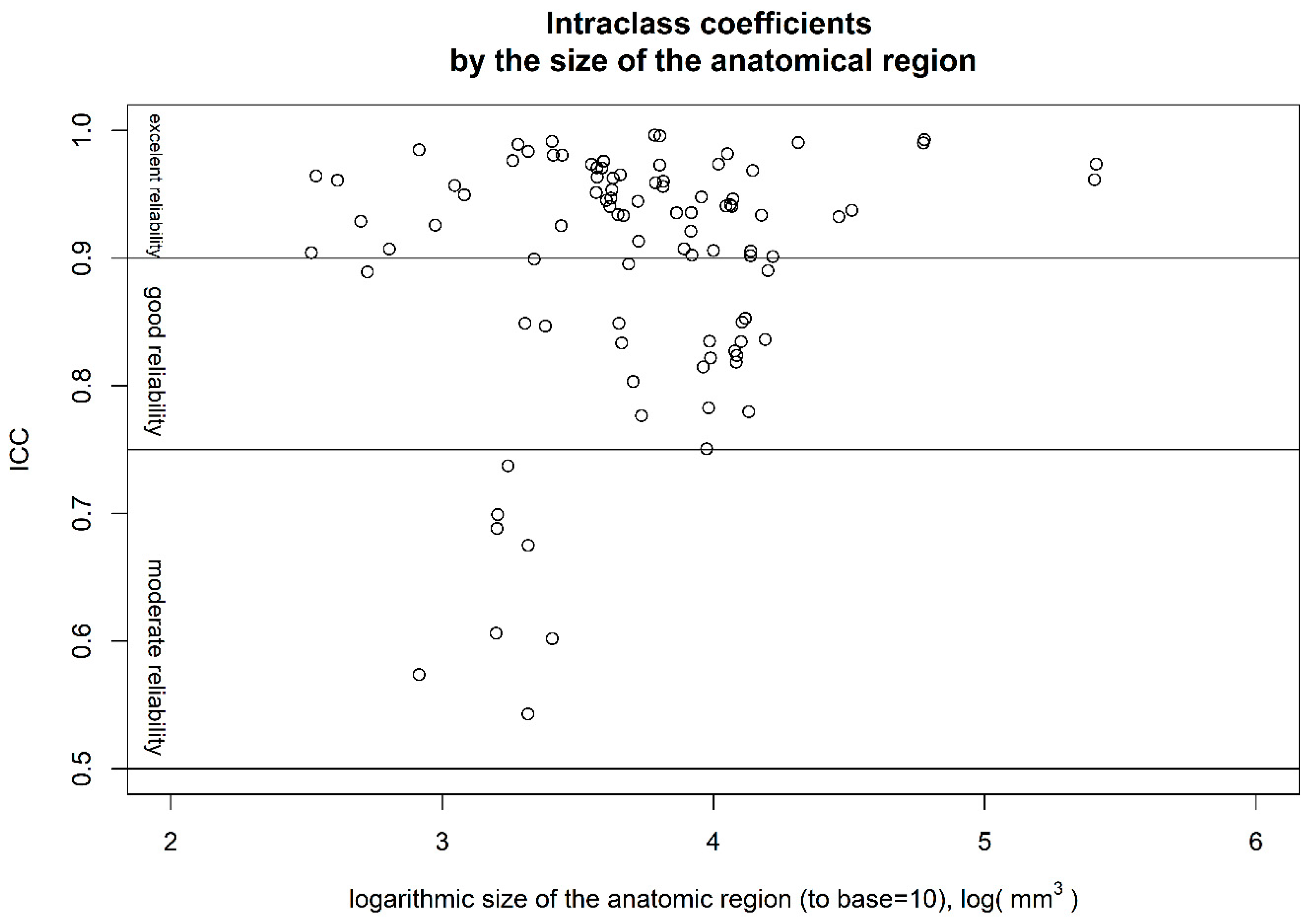
Figure 1. Intraclass correlation coefficients (ICCs) by size of anatomical region. All regions with only moderate reliability are in the lower third of the size spectrum.
Information 15 00748 g001
Table 1. Demographics.
N = 42Mean ± Standard Deviation
Sex11 Male
Age15.02 ± 1.13
BMI25.13 ± 6.94
IQ102.22 ± 11.06
Depression Score (BDI-II 1)28.88 ± 12.23
1 Beck Depression Inventory II (Raw Score range 0–63; minimal (0–13), mild (14–19), moderate (20–28), and severe (29–63) classification).
Table 2. Result of the test–retest reliability analysis by anatomical region.
Paired Regions
Anatomical Region *LeftRight
ICCMedian Absolute Error, %ICCMedian Absolute Error, %
Cerebral White Matter0.973 (0.951; 0.985)2.14 (1.43; 2.91)0.961 (0.929; 0.979)1.76 (1.36; 2.73)
Lateral Ventricle0.995 (0.992; 0.997)3.32 (2.21; 4.58)0.996 (0.993; 0.997)3.12 (1.69; 3.97)
Inf Lat Ventricle0.904 (0.828; 0.947)7.4 (4.38; 9.92)0.964 (0.934; 0.98)5.56 (4.52; 9.11)
Cerebellum White Matter0.933 (0.879; 0.963)1.92 (1; 2.97)0.968 (0.942; 0.982)1.5 (0.86; 2.24)
Cerebellum Cortex0.99 (0.981; 0.994)0.71 (0.52; 0.98)0.992 (0.986; 0.996)0.79 (0.5; 1.17)
Thalamus0.947 (0.905; 0.971)1.53 (0.69; 1.82)0.902 (0.825; 0.946)1.67 (0.96; 2.57)
Caudate0.97 (0.945; 0.984)1.04 (0.65; 1.75)0.975 (0.955; 0.986)1.03 (0.66; 1.55)
Putamen0.944 (0.899; 0.969)1.54 (1.1; 2.14)0.776 (0.62; 0.873)1.34 (0.86; 1.73)
Pallidum0.674 (0.469; 0.811)2.72 (1.81; 5.1)0.542 (0.289; 0.725)2.24 (1.41; 3.14)
Hippocampus0.933 (0.88; 0.963)1.28 (0.81; 2.12)0.965 (0.936; 0.981)1.19 (0.68; 1.63)
Amygdala0.688 (0.488; 0.819)3.42 (1.57; 5.41)0.737 (0.56; 0.849)3.16 (2.11; 6.23)
Accumbens area0.889 (0.803; 0.938)3.11 (1.93; 5.17)0.907 (0.833; 0.948)3.8 (2.58; 4.85)
Ventral diencephalon0.962 (0.931; 0.979)1.63 (0.95; 2.4)0.94 (0.891; 0.967)2.15 (1.56; 2.52)
choroid plexus0.96 (0.928; 0.978)7.34 (6.51; 11.98)0.928 (0.871; 0.961)8.5 (5.23; 10.41)
ctx caudalanteriorcing.0.973 (0.951; 0.985)2.03 (1.54; 3.76)0.98 (0.964; 0.989)2.39 (1.96; 3.61)
ctx caudalmiddlefrontal0.907 (0.834; 0.949)3.04 (2.04; 5.81)0.935 (0.883; 0.964)3.29 (2.33; 4.23)
ctx cuneus0.976 (0.956; 0.987)2.39 (1.67; 3.54)0.951 (0.911; 0.973)2.38 (1.33; 3.35)
ctx entorhinal0.698 (0.504; 0.826)6.51 (4.28; 9.27)0.606 (0.373; 0.767)8.39 (4.56; 10.93)
ctx fusiform0.921 (0.857; 0.956)2.59 (1.18; 3.63)0.935 (0.883; 0.964)1.9 (1.3; 2.54)
ctx inferiorparietal0.849 (0.737; 0.916)2.94 (2.09; 5.19)0.89 (0.805; 0.939)3.72 (2.54; 5.4)
ctx inferiortemporal0.823 (0.695; 0.901)5.05 (2.67; 6.57)0.834 (0.712; 0.907)3.57 (2.45; 4.38)
ctx isthmuscingulate0.98 (0.964; 0.989)1.35 (0.96; 2.07)0.991 (0.984; 0.995)1.9 (1.42; 2.66)
ctx lateraloccipital0.94 (0.892; 0.967)3.17 (2.28; 3.87)0.946 (0.902; 0.97)2.44 (1.28; 3.42)
ctx lateralorbitofrontal0.75 (0.58; 0.857)2.53 (1.53; 3.89)0.782 (0.63; 0.877)2.58 (1.53; 5.87)
ctx lingual0.958 (0.925; 0.977)2.17 (1.35; 3.06)0.972 (0.95; 0.985)1.46 (1.13; 2.39)
ctx medialorbitofrontal0.913 (0.844; 0.952)1.99 (1.21; 3.69)0.803 (0.662; 0.889)1.69 (1.14; 3.13)
ctx middletemporal0.852 (0.742; 0.918)5.57 (3.42; 8.18)0.779 (0.625; 0.875)5.19 (2.57; 6.74)
ctx parahippocampal0.899 (0.82; 0.944)3.83 (2.53; 5.45)0.848 (0.735; 0.915)2.58 (1.91; 4.45)
ctx paracentral0.953 (0.915; 0.974)2.65 (1.75; 3.86)0.946 (0.903; 0.971)2.31 (1.94; 3.44)
ctx parsopercularis0.933 (0.879; 0.963)3.68 (2.3; 6.03)0.833 (0.71; 0.906)4.2 (3.03; 7.11)
ctx parsorbitalis0.846 (0.732; 0.914)3.78 (2.08; 4.77)0.601 (0.367; 0.764)3.62 (2.57; 5.54)
ctx parstriangularis0.895 (0.813; 0.942)3.72 (2.13; 6.35)0.848 (0.735; 0.915)4.81 (2.89; 6.73)
ctx pericalcarine0.976 (0.956; 0.987)3.4 (2.34; 4.62)0.983 (0.969; 0.991)2.28 (1.38; 3.2)
ctx postcentral0.905 (0.831; 0.948)4.06 (2.51; 6.33)0.834 (0.713; 0.907)3.42 (2.33; 4.98)
ctx posteriorcingulate0.963 (0.933; 0.98)1.98 (1.35; 2.63)0.97 (0.946; 0.984)1.5 (0.86; 1.96)
ctx precentral0.818 (0.686; 0.898)3.53 (2.92; 6.5)0.827 (0.7; 0.903)4.77 (2.79; 6.6)
ctx precuneus0.973 (0.951; 0.985)1.56 (1.09; 2.72)0.981 (0.966; 0.99)1.32 (0.85; 2.05)
ctx rostralanteriorcing.0.945 (0.900; 0.970)2.76 (2.1; 3.54)0.925 (0.865; 0.959)3.03 (1.51; 4.4)
ctx rostralmiddlefrontal0.901 (0.824; 0.945)3.23 (2.45; 5.17)0.905 (0.83; 0.948)2.95 (1.99; 4.14)
ctx superiorfrontal0.932 (0.877; 0.963)1.59 (0.94; 2.39)0.937 (0.886; 0.965)1.96 (1.23; 3.35)
ctx superiorparietal0.94 (0.891; 0.967)2.88 (1.45; 3.98)0.941 (0.894; 0.968)2.53 (1.25; 3.35)
ctx superiortemporal0.901 (0.823; 0.945)3.57 (2.94; 4.87)0.836 (0.715; 0.908)3.99 (2.41; 5.43)
ctx supramarginal0.821 (0.691; 0.9)6.53 (5.17; 8.28)0.814 (0.68; 0.895)5.7 (3.54; 6.39)
ctx transversetemporal0.949 (0.908; 0.972)4.53 (3; 6.61)0.925 (0.866; 0.959)3.19 (2.27; 4.67)
ctx insula0.956 (0.919; 0.976)1.42 (0.79; 2.37)0.96 (0.927; 0.978)1.22 (0.8; 1.78)
Unpaired Regions
ICCMedian Absolut Error, %
3rd Ventricle0.984 (0.971; 0.991)2.29 (1.23; 3.6)
4th Ventricle0.989 (0.979; 0.994)2.18 (1.91; 2.74)
Brain Stem0.99 (0.982; 0.994)0.86 (0.65; 1.2)
CSF0.956 (0.921; 0.976)2.94 (2.21; 3.99)
white matter hypointensities0.573 (0.329; 0.745)12.8 (6.81; 18.49)
* Labels according to the Desikan-Killiany-Tourville atlas [28]: ctx—cortex, ICC—intraclass coefficient, inf—inferior, lat—lateral, cing.—cingulate, CSF—cerebral spinal fluid.
Disclaimer/Publisher’s Note: The statements, opinions and data contained in all publications are solely those of the individual author(s) and contributor(s) and not of MDPI and/or the editor(s). MDPI and/or the editor(s) disclaim responsibility for any injury to people or property resulting from any ideas, methods, instructions or products referred to in the content.