
Tortuosity Index Calculations in Retinal Images: Some Criticalities Arising from Commonly Used Approaches


Abstract
1. Introduction
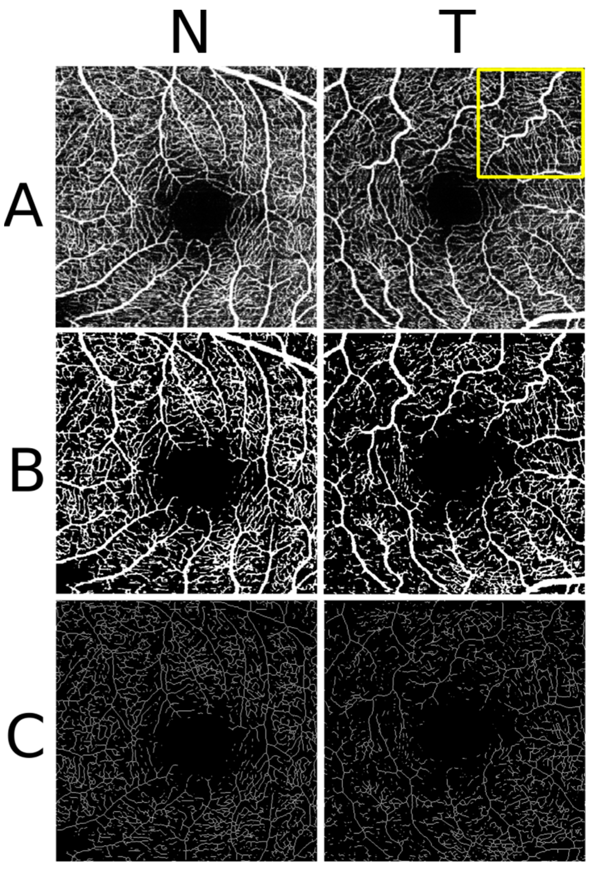
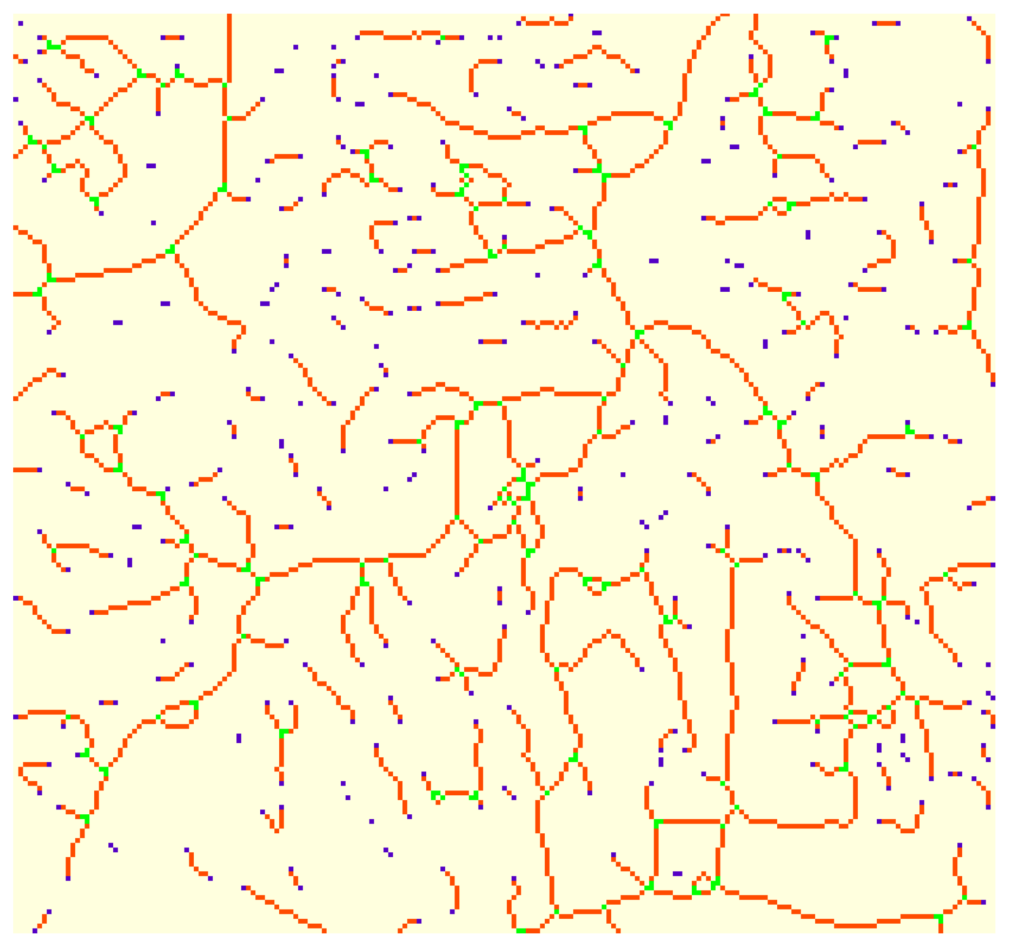
2. Materials and Methods
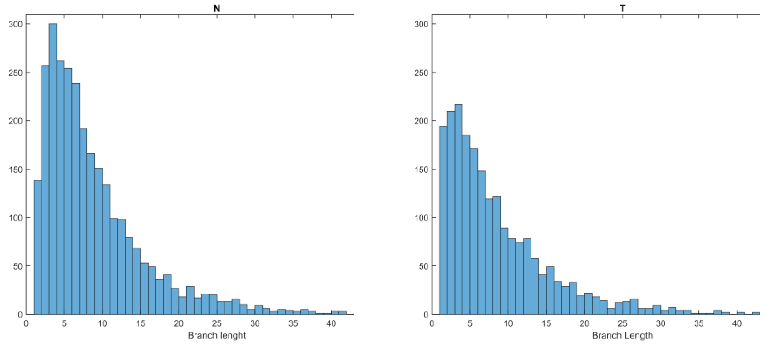
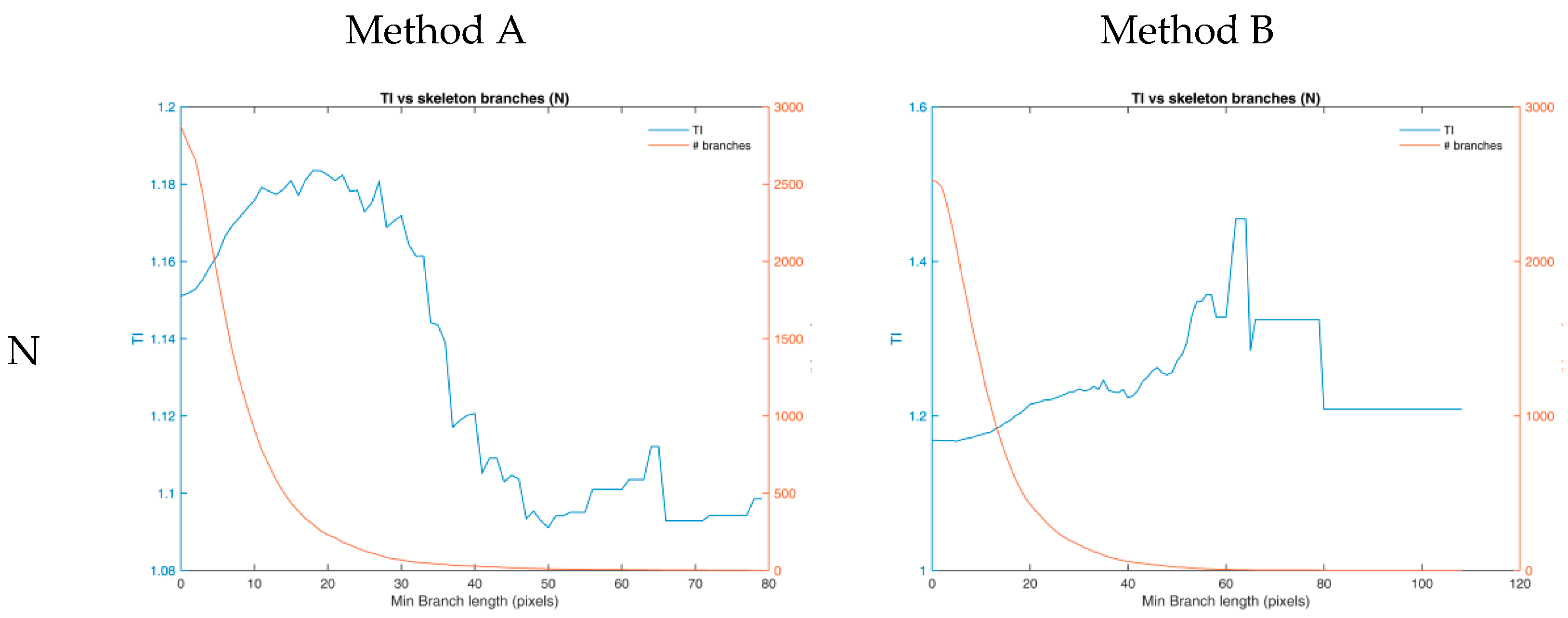
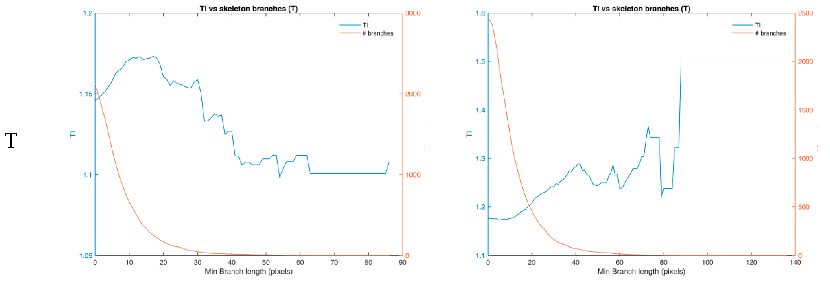
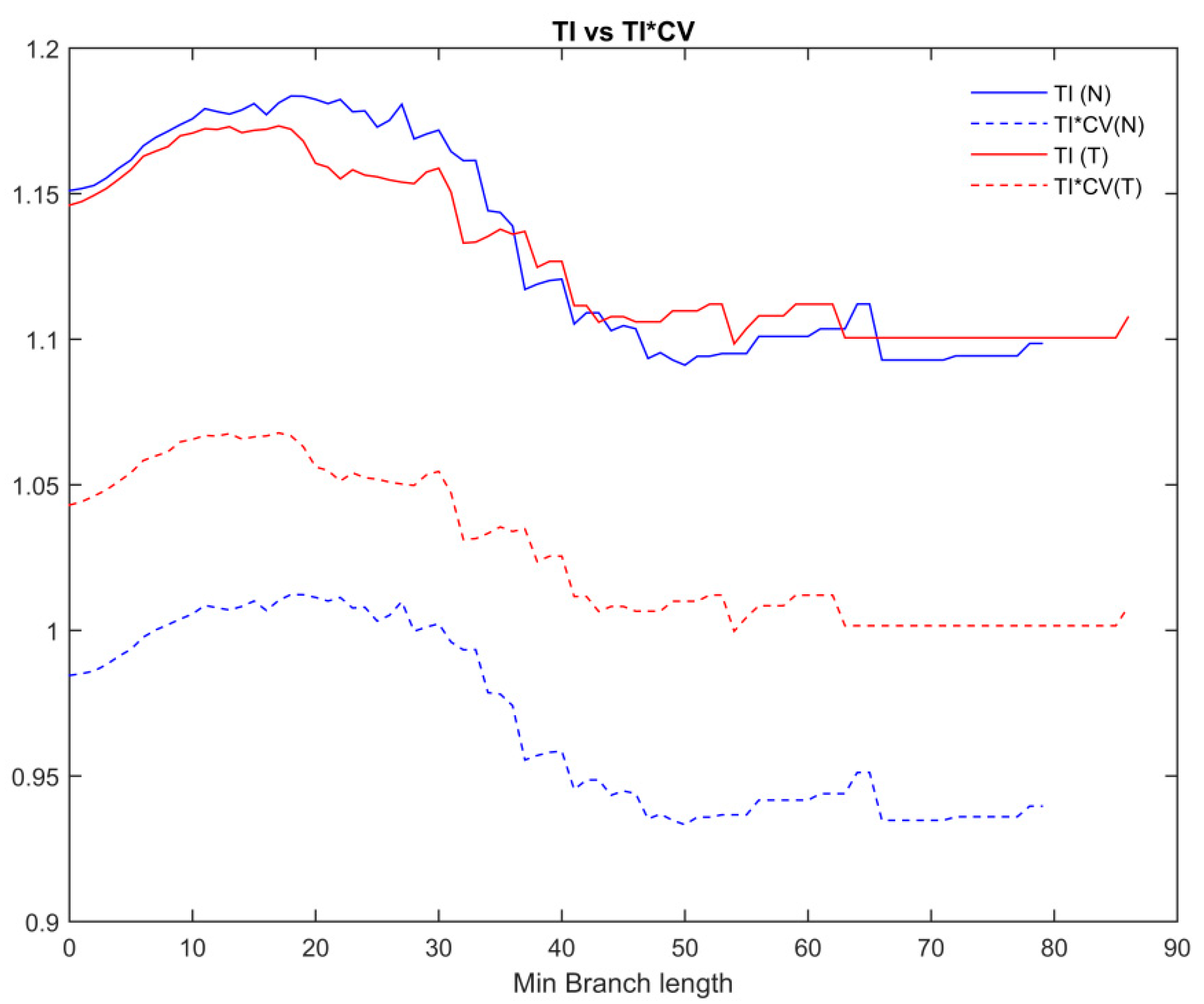
3. Results
4. Discussion
5. Conclusions
Supplementary Materials
Author Contributions
Funding
Data Availability Statement
Acknowledgments
Conflicts of Interest
References
- Kashani, A.H.; Chen, C.-L.; Gahm, J.K.; Zheng, F.; Richter, G.M.; Rosenfeld, P.J.; Shi, Y.; Wang, R.K. Optical Coherence Tomography Angiography: A Comprehensive Review of Current Methods and Clinical Applications. Prog. Retin. Eye Res. 2017, 60, 66–100. [Google Scholar] [CrossRef] [PubMed]
- Cruz-Herranz, A.; Balk, L.J.; Oberwahrenbrock, T.; Saidha, S.; Martinez-Lapiscina, E.H.; Lagreze, W.A.; Schuman, J.S.; Villoslada, P.; Calabresi, P.; Balcer, L.; et al. The APOSTEL recommendations for reporting quantitative optical coherence tomography studies. Neurology 2016, 86, 2303–2309. [Google Scholar] [CrossRef] [PubMed]
- Mendonça, L.S.M.; Perrott-Reynolds, R.; Schwartz, R.; Madi, H.A.; Cronbach, N.; Gendelman, I.; Muldrew, A.; Bannon, F.; Balaskas, K.; Gemmy Cheung, C.M.; et al. Deliberations of an International Panel of Experts on OCT Angiography Nomenclature of Neovascular Age-Related Macular Degeneration. Ophthalmology 2020, 128, 1109–1112. [Google Scholar] [CrossRef] [PubMed]
- Spaide, R.F.; Jaffe, G.J.; Sarraf, D.; Freund, K.B.; Sadda, S.R.; Staurenghi, G.; Waheed, N.K.; Chakravarthy, U.; Rosenfeld, P.J.; Holz, F.G.; et al. Consensus Nomenclature for Reporting Neovascular Age-Related Macular Degeneration Data: Consensus on Neovascular Age-Related Macular Degeneration Nomenclature Study Group. Ophthalmology 2020, 127, 616–636. [Google Scholar] [CrossRef]
- Staurenghi, G.; Sadda, S.; Chakravarthy, U.; Spaide, R.F. International Nomenclature for Optical Coherence Tomography (IN• OCT) Panel. Proposed lexicon for anatomic landmarks in normal posterior segment spectral-domain optical coherence tomography: The IN• OCT consensus. Ophthalmology 2014, 121, 1572–1578. [Google Scholar] [CrossRef]
- Balaskas, K.; Tiew, S.; Czanner, G.; Tan, A.L.; Ashworth, J.; Biswas, S.; Aslam, T. The Novel Evidenced Assessment of Tortuosity System: Interobserver Reliability and Agreement with Clinical Assessment. Acta Ophthalmol. (Copenh.) 2016, 94, e421-6. [Google Scholar] [CrossRef] [PubMed]
- Müller, P.L.; Liefers, B.; Treis, T.; Rodrigues, F.G.; Olvera-Barrios, A.; Paul, B.; Dhingra, N.; Lotery, A.; Bailey, C.; Taylor, P.; et al. Reliability of Retinal Pathology Quantification in Age-Related Macular Degeneration: Implications for Clinical Trials and Machine Learning Applications. Transl. Vis. Sci. Technol. 2021, 10, 4. [Google Scholar] [CrossRef] [PubMed]
- Tan, C.S.; Chan, J.C.; Cheong, K.X.; Ngo, W.K.; Sadda, S.R. Comparison of Retinal Thicknesses Measured Using Swept-Source and Spectral-Domain Optical Coherence Tomography Devices. Ophthalmic Surg. Lasers Imaging Retina 2015, 46, 172–179. [Google Scholar] [CrossRef]
- Ciurică, S.; Lopez-Sublet, M.; Loeys, B.L.; Radhouani, I.; Natarajan, N.; Vikkula, M.; Maas, A.H.E.M.; Adlam, D.; Persu, A. Arterial Tortuosity. Hypertension 2019, 73, 951–960. [Google Scholar] [CrossRef]
- Kemp, M. Leonardo’s Philosophical Anatomies. Lancet 2019, 393, 1404–1408. [Google Scholar] [CrossRef]
- Wells, F.C.; Crowe, T. Leonardo Da Vinci as a Paradigm for Modern Clinical Research. J. Thorac. Cardiovasc. Surg. 2004, 127, 929–944. [Google Scholar] [CrossRef] [PubMed]
- Del Corso, L.; Moruzzo, D.; Conte, B.; Agelli, M.; Romanelli, A.M.; Pastine, F.; Protti, M.; Pentimone, F.; Baggiani, G. Tortuosity, Kinking, and Coiling of the Carotid Artery: Expression of Atherosclerosis or Aging? Angiology 1998, 49, 361–371. [Google Scholar] [CrossRef]
- Hiroki, M.; Miyashita, K.; Oda, M. Tortuosity of the White Matter Medullary Arterioles Is Related to the Severity of Hypertension. Cerebrovasc. Dis. 2002, 13, 242–250. [Google Scholar] [CrossRef]
- Kahe, F.; Sharfaei, S.; Pitliya, A.; Jafarizade, M.; Seifirad, S.; Habibi, S.; Chi, G. Coronary Artery Tortuosity: A Narrative Review. Coron. Artery Dis. 2020, 31, 187–192. [Google Scholar] [CrossRef]
- Owen, C.G.; Newsom, R.S.B.; Rudnicka, A.R.; Barman, S.A.; Woodward, E.G.; Ellis, T.J. Diabetes and the Tortuosity of Vessels of the Bulbar Conjunctiva. Ophthalmology 2008, 115, e27–e32. [Google Scholar] [CrossRef]
- Pancera, P.; Ribul, M.; Presciuttini, B.; Lechi, A. Prevalence of Carotid Artery Kinking in 590 Consecutive Subjects Evaluated by Echocolordoppler. Is There a Correlation with Arterial Hypertension? J. Intern. Med. 2000, 248, 7–12. [Google Scholar] [CrossRef] [PubMed]
- Chua, J.; Sim, R.; Tan, B.; Wong, D.; Yao, X.; Liu, X.; Ting, D.S.W.; Schmidl, D.; Ang, M.; Garhöfer, G.; et al. Optical Coherence Tomography Angiography in Diabetes and Diabetic Retinopathy. J. Clin. Med. 2020, 9, 1723. [Google Scholar] [CrossRef]
- Sasongko, M.B.; Wong, T.Y.; Donaghue, K.C.; Cheung, N.; Jenkins, A.J.; Benitez-Aguirre, P.; Wang, J.J. Retinal Arteriolar Tortuosity Is Associated with Retinopathy and Early Kidney Dysfunction in Type 1 Diabetes. Am. J. Ophthalmol. 2012, 153, 176–183.e1. [Google Scholar] [CrossRef] [PubMed]
- Sasongko, M.B.; Wong, T.Y.; Nguyen, T.T.; Cheung, C.Y.; Shaw, J.E.; Kawasaki, R.; Lamoureux, E.L.; Wang, J.J. Retinal Vessel Tortuosity and Its Relation to Traditional and Novel Vascular Risk Markers in Persons with Diabetes. Curr. Eye Res. 2016, 41, 551–557. [Google Scholar] [CrossRef] [PubMed]
- Lajmi, H.; Hmaied, W.; Othmen, A.; Chelly, Z.; El Fekih, L. Optical Coherence Tomography Angiography Microvascular Changes in Diabetics without Diabetic Retinopathy. Saudi J. Ophthalmol. 2020, 34, 156. [Google Scholar] [CrossRef]
- Pierro, L.; Arrigo, A.; De Crescenzo, M.; Aragona, E.; Chiesa, R.; Castellano, R.; Catenaccio, B.; Bandello, F. Quantitative Optical Coherence Tomography Angiography Detects Retinal Perfusion Changes in Carotid Artery Stenosis. Front. Neurosci. 2021, 15, 640666. [Google Scholar] [CrossRef]
- O’Neill, R.A.; Maxwell, A.P.; Paterson, E.N.; Kee, F.; Young, I.; Hogg, R.E.; Cruise, S.; Murphy, S.; McGuinness, B.; McKay, G.J. Retinal Microvascular Parameters Are Not Significantly Associated with Mild Cognitive Impairment in the Northern Ireland Cohort for the Longitudinal Study of Ageing. BMC Neurol. 2021, 21, 112. [Google Scholar] [CrossRef] [PubMed]
- Cheung, C.Y.-L.; Ong, Y.T.; Ikram, M.K.; Ong, S.Y.; Li, X.; Hilal, S.; Catindig, J.-A.S.; Venketasubramanian, N.; Yap, P.; Seow, D.; et al. Microvascular Network Alterations in the Retina of Patients with Alzheimer’s Disease. Alzheimers Dement. 2014, 10, 135–142. [Google Scholar] [CrossRef]
- Liew, G.; Mitchell, P.; Wong, T.Y.; Lindley, R.I.; Cheung, N.; Kaushik, S.; Wang, J.J. Retinal Microvascular Signs and Cognitive Impairment. J. Am. Geriatr. Soc. 2009, 57, 1892–1896. [Google Scholar] [CrossRef]
- Allon, R.; Aronov, M.; Belkin, M.; Maor, E.; Shechter, M.; Fabian, I.D. Retinal Microvascular Signs as Screening and Prognostic Factors for Cardiac Disease: A Systematic Review of Current Evidence. Am. J. Med. 2021, 134, 36–47.e7. [Google Scholar] [CrossRef]
- Cheung, C.Y.-L.; Zheng, Y.; Hsu, W.; Lee, M.L.; Lau, Q.P.; Mitchell, P.; Wang, J.J.; Klein, R.; Wong, T.Y. Retinal Vascular Tortuosity, Blood Pressure, and Cardiovascular Risk Factors. Ophthalmology 2011, 118, 812–818. [Google Scholar] [CrossRef]
- Rosenblatt, T.R.; Ji, M.H.; Vail, D.; Ludwig, C.A.; Al-Moujahed, A.; Pasricha, M.V.; Callaway, N.F.; Kumm, J.; Moshfeghi, D.M. Key Factors in a Rigorous Longitudinal Image-Based Assessment of Retinopathy of Prematurity. Sci. Rep. 2021, 11, 5369. [Google Scholar] [CrossRef] [PubMed]
- Abbinante, G.; Plaitano, C.; Gallo, F.G.; Magli, A. A Case of Retinal Vascular Involvement in a 6-Year-Old Patient with COVID-19. Eur. J. Ophthalmol. 2021. [Google Scholar] [CrossRef] [PubMed]
- Sim, R.; Cheung, G.; Ting, D.; Wong, E.; Wong, T.Y.; Yeo, I.; Wong, C.W. Retinal Microvascular Signs in COVID-19. Br. J. Ophthalmol. 2021. [Google Scholar] [CrossRef] [PubMed]
- Abdalla, M.; Hunter, A.; Al-Diri, B. Quantifying Retinal Blood Vessels’ Tortuosity—Review. In Proceedings of the IEEE 2015 Science and Information Conference (SAI), London, UK, 28–30 July 2015; pp. 687–693. [Google Scholar]
- Kalitzeos, A.A.; Lip, G.Y.H.; Heitmar, R. Retinal Vessel Tortuosity Measures and Their Applications. Exp. Eye Res. 2013, 106, 40–46. [Google Scholar] [CrossRef] [PubMed]
- Kipli, K.; Hoque, M.E.; Lim, L.T.; Mahmood, M.H.; Sahari, S.K.; Sapawi, R.; Rajaee, N.; Joseph, A. A Review on the Extraction of Quantitative Retinal Microvascular Image Feature. Comput. Math. Methods Med. 2018, 2018, 4019538. [Google Scholar] [CrossRef]
- Fawzi, A.A. Consensus on Optical Coherence Tomographic Angiography Nomenclature: Do We Need to Develop and Learn a New Language? JAMA Ophthalmol. 2017, 135, 377–378. [Google Scholar] [CrossRef] [PubMed]
- Lee, H.; Lee, M.; Chung, H.; Kim, H.C. Quantification of Retinal Vessel Tortuosity in Diabetic Retinopathy Using Optical Coherence Tomography Angiography. Retina 2018, 38, 976–985. [Google Scholar] [CrossRef]
- Ma, Y.; Hao, H.; Xie, J.; Fu, H.; Zhang, J.; Yang, J.; Wang, Z.; Liu, J.; Zheng, Y.; Zhao, Y. ROSE: A Retinal OCT-Angiography Vessel Segmentation Dataset and New Model. IEEE Trans. Med. Imaging 2021, 40, 928–939. [Google Scholar] [CrossRef]
- Arganda-Carreras, I.; Kaynig, V.; Rueden, C.; Eliceiri, K.W.; Schindelin, J.; Cardona, A.; Sebastian Seung, H. Trainable Weka Segmentation: A Machine Learning Tool for Microscopy Pixel Classification. Bioinformatics 2017, 33, 2424–2426. [Google Scholar] [CrossRef] [PubMed]
- Goselink, R.J.M.; Schreur, V.; van Kernebeek, C.R.; Padberg, G.W.; van der Maarel, S.M.; van Engelen, B.G.M.; Erasmus, C.E.; Theelen, T. Ophthalmological Findings in Facioscapulohumeral Dystrophy. Brain Commun. 2019, 1, fcz023. [Google Scholar] [CrossRef] [PubMed]
- Minnella, A.M.; Barbano, L.; Verrecchia, E.; Martelli, F.; Pagliei, V.; Gambini, G.; Placidi, G.; Falsini, B.; Caporossi, A.; Manna, R. Macular Impairment in Fabry Disease: A Morpho-Functional Assessment by Swept-Source OCT Angiography and Focal Electroretinography. Invest. Ophthalmol. Vis. Sci. 2019, 60, 2667–2675. [Google Scholar] [CrossRef]
- Savastano, M.C.; Gambini, G.; Cozzupoli, G.M.; Crincoli, E.; Savastano, A.; De Vico, U.; Culiersi, C.; Falsini, B.; Martelli, F.; Minnella, A.M.; et al. Retinal Capillary Involvement in Early Post-COVID-19 Patients: A Healthy Controlled Study. Graefes Arch. Clin. Exp. Ophthalmol. 2021, 259, 2157–2169. [Google Scholar] [CrossRef]
- Ridler, T.W.; Calvard, S. Picture Thresholding Using an Iterative Selection Method. IEEE Trans. Syst. Man Cybern. 1978, 8, 630–632. [Google Scholar] [CrossRef]
- Otsu, N. A Threshold Selection Method from Gray-Level Histograms. IEEE Trans. Syst. Man Cybern. 1979, 9, 62–66. [Google Scholar] [CrossRef]
- Chan, T.F.; Vese, L.A. Active Contours without Edges. IEEE Trans. Image Process. Publ. IEEE Signal Process. Soc. 2001, 10, 266–277. [Google Scholar] [CrossRef] [PubMed]
- Arganda-Carreras, I.; Fernández-González, R.; Muñoz-Barrutia, A.; Ortiz-De-Solorzano, C. 3D Reconstruction of Histological Sections: Application to Mammary Gland Tissue. Microsc. Res. Tech. 2010, 73, 1019–1029. [Google Scholar] [CrossRef] [PubMed]
- Lu, Y.; Wang, J.C.; Zeng, R.; Katz, R.; Vavvas, D.G.; Miller, J.W.; Miller, J.B. Quantitative Comparison of Microvascular Metrics on Three Optical Coherence Tomography Angiography Devices in Chorioretinal Disease. Clin. Ophthalmol. 2019, 13, 2063–2069. [Google Scholar] [CrossRef] [PubMed]
- Matlab. Version 7.10.0 (R2010a); The MathWorks Inc.: Natick, MA, USA, 2010. [Google Scholar]
- Sage, D.; Prodanov, D.; Tinevez, J.-Y.; Schindelin, J. MIJ: Making Interoperability Between ImageJ and Matlab Possible. In Proceedings of the ImageJ User & Developer Conference (IUDC’12); Mondorf-les-Bains, Grand Duchy of Luxembourg, 24–26 October 2012. [Google Scholar]
- Zhou, K.; Song, S.; Legocki, A.; Cheng, Y.; Ding, L.; Rezaei, K.A.; Wang, R.K.; Cabrera, M.T. Quantitative Handheld Swept-Source Optical Coherence Tomography Angiography in Awake Preterm and Full-Term Infants. Transl. Vis. Sci. Technol. 2020, 9, 19. [Google Scholar] [CrossRef] [PubMed]
- Poplawsky, A.J.; Fukuda, M.; Kang, B.-M.; Kim, J.H.; Suh, M.; Kim, S.-G. Dominance of Layer-Specific Microvessel Dilation in Contrast-Enhanced High-Resolution FMRI: Comparison between Hemodynamic Spread and Vascular Architecture with CLARITY. Neuroimage 2019, 197, 657–667. [Google Scholar] [CrossRef] [PubMed]


| Method A | Method B | Method C | Method D | |||||
|---|---|---|---|---|---|---|---|---|
| N | T | N | T | N | T | N | T | |
| TI | 1.151 | 1.146 * | 1.169 | 1.177 | 1.174 | 1.179 | 1.187 | 1.167 * |
| TI_avg | 1.139 | 1.129 * | 1.146 | 1.146 | 1.133 | 1.135 | 1.168 | 1.162 * |
| TI*CV | 0.98 | 1.04 | 0.89 | 0.97 | 1.19 | 1.21 | 0.94 | 1.02 |
| # branches | 2874 | 2117 | 2526 | 2441 | 4235 | 3953 | 3269 | 1641 |
| Mean BL | 9.15 ± 7.83 | 8.81 ± 8.02 | 12.97 ± 9.89 | 13.28 ± 10.96 | 6.58 ± 6.69 | 6.56 ± 6.76 | 7.89 ± 6.24 | 8.49 ± 7.41 |
| Max BL | 79.36 | 86.46 | 108.05 | 135.74 | 81.04 | 76.33 | 80.18 | 59.36 |
| Min BL | 1 | 1 | 1 | 1 | 1 | 1 | 1 | 1 |
| Mean ED | 7.95 ± 6.79 | 7.69 ± 7.01 | 11.1 ± 8.34 | 11.28 ± 9.18 | 5.61 ± 5.7 | 5.56 ± 5.69 | 6.65 ± 5.37 | 7.27 ± 6.49 |
| Max ED | 72.24 | 78.10 | 89.40 | 89.94 | 65.37 | 69.53 | 72.24 | 54.20 |
| Min ED | 0 | 0 | 0 | 0 | 0 | 0 | 0 | 0 |
| # ED = 0 | 14 | 14 | 75 | 111 | 60 | 70 | 61 | 16 |
| BL < 10 | 68.16% | 68.73% | 47.23% | 48.22% | 80.85% | 80.75% | 75.13% | 72.64% |
| BL < 21 | 92.59% | 93.06% | 84.52% | 83.08% | 95.96% | 95.70% | 95.87% | 93.78% |
| BL = 1 | 3.97% | 6.57% | 0.48% | 1.11% | 14.88% | 16.27% | 2.84% | 4.57% |
Publisher’s Note: MDPI stays neutral with regard to jurisdictional claims in published maps and institutional affiliations. |
© 2021 by the authors. Licensee MDPI, Basel, Switzerland. This article is an open access article distributed under the terms and conditions of the Creative Commons Attribution (CC BY) license (https://creativecommons.org/licenses/by/4.0/).
Share and Cite
Martelli, F.; Giacomozzi, C. Tortuosity Index Calculations in Retinal Images: Some Criticalities Arising from Commonly Used Approaches. Information 2021, 12, 466. https://doi.org/10.3390/info12110466
Martelli F, Giacomozzi C. Tortuosity Index Calculations in Retinal Images: Some Criticalities Arising from Commonly Used Approaches. Information. 2021; 12(11):466. https://doi.org/10.3390/info12110466
Chicago/Turabian StyleMartelli, Francesco, and Claudia Giacomozzi. 2021. "Tortuosity Index Calculations in Retinal Images: Some Criticalities Arising from Commonly Used Approaches" Information 12, no. 11: 466. https://doi.org/10.3390/info12110466
APA StyleMartelli, F., & Giacomozzi, C. (2021). Tortuosity Index Calculations in Retinal Images: Some Criticalities Arising from Commonly Used Approaches. Information, 12(11), 466. https://doi.org/10.3390/info12110466