Communication

Paper-Based Flexible Electrode Using Chemically-Modified Graphene and Functionalized Multiwalled Carbon Nanotube Composites for Electrophysiological Signal Sensing

1
Department of Medicine, University of Connecticut School of Medicine, Farmington, CT 06030, USA
2
Center for Bioelectrics, Old Dominion University Research Foundation, Norfolk, VA 23508, USA
3
Department of Biomedical Engineering, University of Connecticut, Storrs, CT 06269, USA
*
Author to whom correspondence should be addressed.
Information 2019, 10(10), 325; https://doi.org/10.3390/info10100325
Submission received: 5 September 2019 / Revised: 30 September 2019 / Accepted: 17 October 2019 / Published: 22 October 2019
(This article belongs to the Special Issue Ubiquitous Sensing for Smart Health Monitoring)

Abstract

:
Flexible paper-based physiological sensor electrodes were developed using chemically-modified graphene (CG) and carboxylic-functionalized multiwalled carbon nanotube composites (f@MWCNTs). A solvothermal process with additional treatment was conducted to synthesize CG and f@MWCNTs to make CG-f@MWCNT composites. The composite was sonicated in an appropriate solvent to make a uniform suspension, and then it was drop cast on a nylon membrane in a vacuum filter. A number of batches (0%~35% f@MWCNTs) were prepared to investigate the performance of the physical characteristics. The 25% f@MWCNT-loaded composite showed the best adhesion on the paper substrate. The surface topography and chemical bonding of the proposed CG-f@MWCNT electrodes were characterized by scanning electron microscopy (SEM) and Raman spectroscopy, respectively. The average sheet resistance of the 25% CG-f@MWCNT electrode was determined to be 75 Ω/□, and it showed a skin contact impedance of 45.12 kΩ at 100 Hz. Electrocardiogram (ECG) signals were recorded from the chest and fingertips of healthy adults using the proposed electrodes. The CG-f@MWCNT electrodes demonstrated comfortability and a high sensitivity for electrocardiogram signal detection.

1. Introduction

The continuous monitoring of personal health is one of the most demanding areas of interest in wearable devices. For personal health monitoring devices, flexible and wearable electronics are of excessive interest, and flexible, biocompatible and comfortable electrodes have been recognized as critical components in such devices [1,2]. Conventional Ag/AgCl biopotential electrodes are widely used in clinical diagnostics and home-based health monitoring systems. Electrolytic conductive gel and skin preparation are required for conventional electrodes to reduce skin–electrode contact impedance, but this gel often dries out over time, and this creates impedance variations and a dramatic reduction in signal quality. Furthermore, it often causes skin irritation and allergic reactions, consequently bringing excessive discomfort. Sweat is another source of signal degradation for wet electrodes [3,4]. Dry electrodes with rigid metal connectors are being developed to address these issues; nevertheless, discomfort and pain are still the major issues of those electrodes due to their hardness [5,6].
Thin, flexible dry electrodes with soft connectors are considered promising alternatives to conventional metal electrodes. Recently, paper-, textile-, or conductive polymer-based Electrocardiogram (ECG) flexible electrodes are being developed in order to increase the contact area between skin and electrodes by making conformal contact, resulting in a reduction of contact impedance [6,7,8,9,10]. Paper and textile electrodes can be used for different applications and have a larger contact area compared to rigid electrodes, but they do not allow for completely conformal contact with skin [11,12,13]. On the contrary, flexible conductive polymers are made from carbon, metal and polymers, which can make stronger contacts compared to rigid electrodes. It has been stated that adhesion between metallic layers and polymer substrates is poor, and the conductivity of conductive polymers is not as good as that of metal conductors. Moreover, the breathability of polymers and the comfort of metallic layers should be improved for long-term monitoring. To improve the degree of adhesion, low-complexity-preparation technology is valuable in these flexible electrodes [2,14,15].
Graphene has been pursued for flexible electronics due to its outstanding electrical and mechanical properties including high conductivity, flexibility, and easy functionalization. A number of works have reported flexible thin film electrodes using graphene-based materials for physiological monitoring. Yapici et al. reported on a graphene material-cladded ordinary textile that enabled the acquisition of high quality ECG signals [16]. Celik et al. reported on graphene-embedded Ag electrodes which showed better ECG signals, higher signal-to-noise ratio (SNR) and lower contact impedance using chemical vapor deposition (CVD) techniques [17]. Reduced graphene oxide-cladded paper electrodes were fabricated by the Park group, and these electrodes enabled the reception of fingertip ECG signals [6]. Lou et al. developed polyethylene terephthalate (PET) substrate-coated CVD graphene electrodes for prolonged ECG signal monitoring [18]. Most of these works have used metallic connectors, which are not suitable for long term ECG signal monitoring. Furthermore, an improved degree of adhesion around dry electrodes would be more valuable for higher qualities of ECG signals. In general, the aggregation of additive materials with their percentages and aspect ratios, the uniformity in the matrix, processing methods, presence of functional groups on the surface, inter-sheet junction, and sheets morphology are all useful for the evaluation of the performance of composite-based electrodes.
Pristine graphene is produced from the mechanical cleavage of pyrolytic graphite and CVD techniques, whereas chemically-treated graphene is developed chemically, hydrothermally/solvothermally, and thermally exfoliated from graphite oxide (GO) [19,20,21]. However, using CVD-grown graphene is challenging because of its high manufacturing cost and difficulty of reproduction and mass production on the patterned substrate.
With the intention of overcoming these limitations, different methods have been performed to make similar 2D graphene-based materials from graphite oxide (GO) called chemically-modified graphene (CG) using various techniques involving chemical, thermal, electrochemical, and hydrothermal/solvothermal reduction. In these techniques, the oxide functional groups in CG cannot be remarkably removed after the reduction of GO. Furthermore, placing CG in an aqueous solution results in the aggregation of sheets due to its hydrophobic nature, and large folds in the CG surface arise after drying, whereas smaller waves tend to be an inherent feature of the structure of isolated layers [5,22]. As a result, the conductivity of the CG is lower than that predicted from theory.
Meanwhile, several carbon nanotube (CNT)-based dry electrodes have been reported in previous publications. Ruffini et al. presented multiwalled CNT (MWCNT)-based arrays on a substrate to penetrate the patient’s outer skin cell layers and reduce the measurement noise of ECG signals [23]. Lee et al. reported a metallic material layer, CNTs, and polydimethylsiloxane (PDMS) composite-based dry ECG electrode [24]. Kang et al. proposed a polymeric binding CNT paste on a metallic substrate for an ECG dry electrode to resist the peeling off of CNTs [25]. Most of these reported works used polymeric binder materials with MWCNTs to reduce the peel off from the substrate. Additionally, if CNT wires remain heterogeneous, electrical conductivity can be severely poor and hinder the use of CNTs for skin-like electronics. It is worth noting that the potential applications of MWCNTs in ECG dry electrodes is limited because they are easy to entangle and agglomerate due to their large aspect ratio, and they can easily peel off from substrates due to their bundles type structure.
The integration of CG and MWCNTs into a composite material is therefore a quite promising strategy to enhance the dispersion of CG and CNTs, to inherit the advantages of both CG and MWCNTs, and to obtain an efficient and effective electronic and thermal conductive network [26,27]. CG/MWCNT composites have been developed to effectively overcome the limitations of the use of individual materials [28,29,30,31]. MWCNTs can greatly reduce internal electrical resistance and improve overall electrical conductivity, since MWCNTs act as spacers between CG sheets and make interconnected conductive network paths [32]. As non-functionalized MWCNTs are prone to agglomerate due to their large aspect ratio, the MWCNTs and CG composite in solution is not uniform and is not stable for a long time [31,33]. To overcome this issue, functionalized MWCNTs are used to make composites in order to get uniformity and longtime usability. When functionalized MWCNTs are combined with CG, CG acts as a CNT “carrier,” and CNTs anchor the CG nano-sheets, thus achieving a uniform co-dispersion for both CNTs and CG in a solvent [32].
In this work, chemically-modified graphene and carboxylic groups-functionalized short MWCNT hybrid composites were developed by solvothermal techniques. The developed composites were drop cast on nylon filter paper, and this process was aided by the vacuum filter technique at a certain pressure. We characterized the electrodes for both material and electrical properties and demonstrated that the electrodes are suitable for human physiological signal monitoring. To our best knowledge, this is the first report to show the CG/f@MWCNTs hybrid electrodes for physiological signal sensing. Section 2 in this paper describes the fabrication process of the proposed electrodes and the characterization methods for material and electrical properties. Section 3 shows the results including actual ECG recordings. Section 4 and Section 5 follow for discussion and conclusion, respectively.

2. Materials and Methods

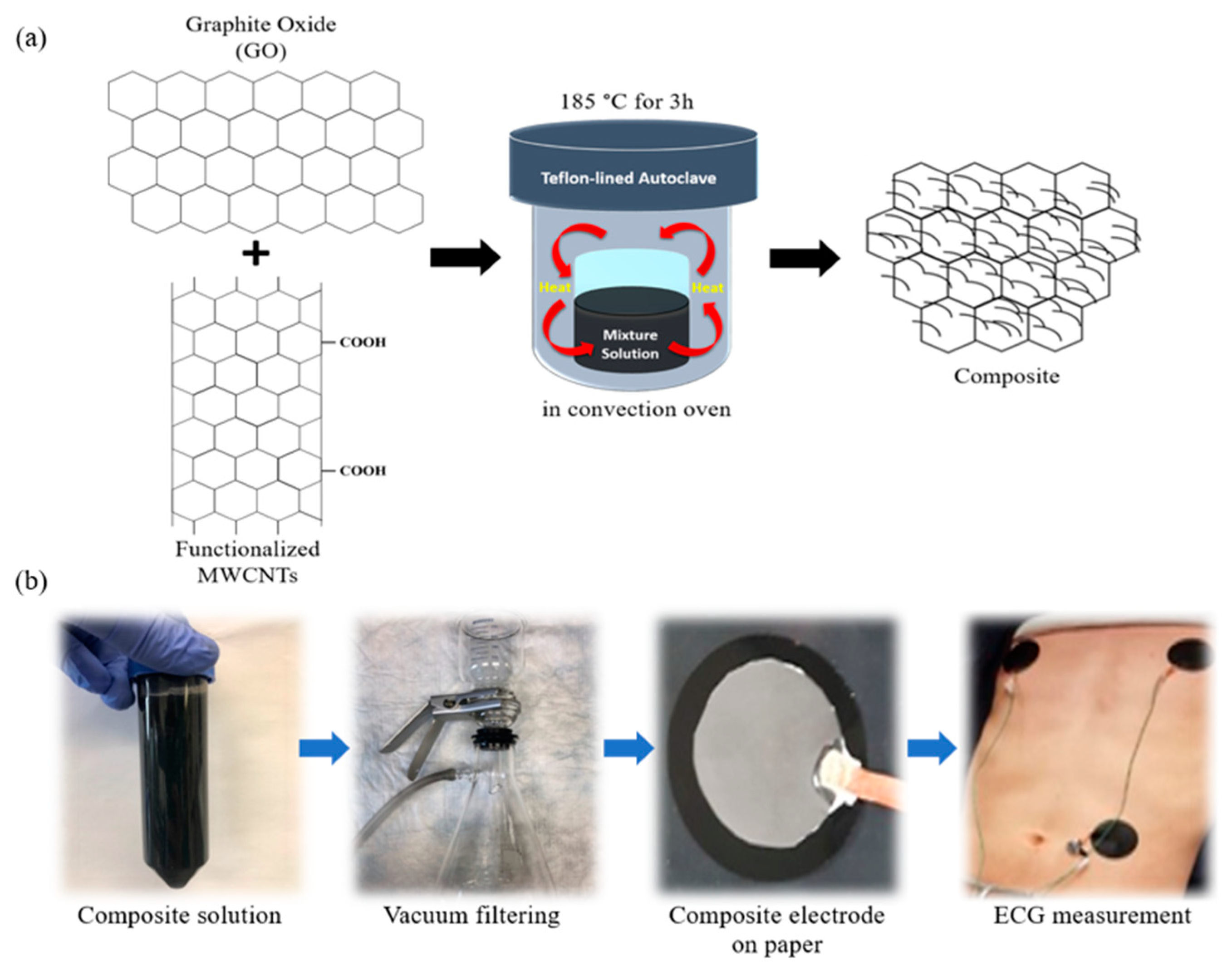
2.1. Electrode Fabrications

Both 59.5 mg of graphite oxide (ACS Material, LLC, Medford, MA, USA) and 10.5 mg of carboxylic-functionalized short MWCNTs (>95%, US Research Nanomaterials, Inc., Houston, TX) were mixed into 35 mL of Milli-Q (MQ) water, followed by ultrasonication for about half an hour (h). Then, 0.1 M glucose was added in the prepared mixture and held for 1 h. After that, the mixture was sealed in a teflon-lined autoclave and maintained at 185 °C for 3 h in a convection oven. When the temperature of the autoclave decreased to room temperature, the composite hydrogel was dispersed again into a 1 M acetic acid aqueous solution and left for 5 h. Finally, the mixture was washed with enough MQ water until reaching a pH of 7, and then it was then dried overnight in an oven at 95 °C under a vacuum condition. The dried platelet is denoted as CG/f@MWCNTs-15%. Similarly, the CG/f@MWCNTs-25% and CG-f@MWCNT-35% composites were prepared using different weight percentages of f@MWCNTs. CG was also prepared in a similar way but without using f@MWCNTs.
Exactly 1.25 mg of the CG/f@MWCNT composite platelet was dispersed into 1 mL of a dimethylformamide (DMF) and MQ water (1:1) solution by using an ultrasonicator for 3 h. Then, the mixture was stirred for a few minutes before the casting of the as-prepared composite suspension. The prepared composites (2 μL/mm2) were drop cast on 0.2 μm nylon filter paper, and the solution was filtered out using the vacuum technique. After filtration, the nylon membrane filter paper was ambiently dried. The electrode was connected to the ECG cable using a copper wire for ECG signal acquisition. An illustration of the ECG electrode and signal acquisition system is shown in Figure 1.

2.2. Physical and Electrical Characterization of Composites and Fabricated Electrodes

Each electrode sample (CG/f@MWCNTs-15%, -25%, and -35%) was characterized using SEM. For SEM analysis, the samples (5 × 4 mm in size) were bonded with conductive carbon tape on an aluminum stub using an acceleration voltage of 15 kV. The Raman spectra of the CG and CG/f@MWCNT composites were measured using a Raman spectrometer (RENISHAW SYSTEM 2000, West Dundee, IL, USA) with the wavelength of 514 nm. To measure the sheet resistance of the fabricated electrodes, a source meter with a four-point probe (Keithley 2401, Tektronix, Cleveland, OH, USA) was used. The experimental set up of the surface resistivity is shown in Figure 2a.

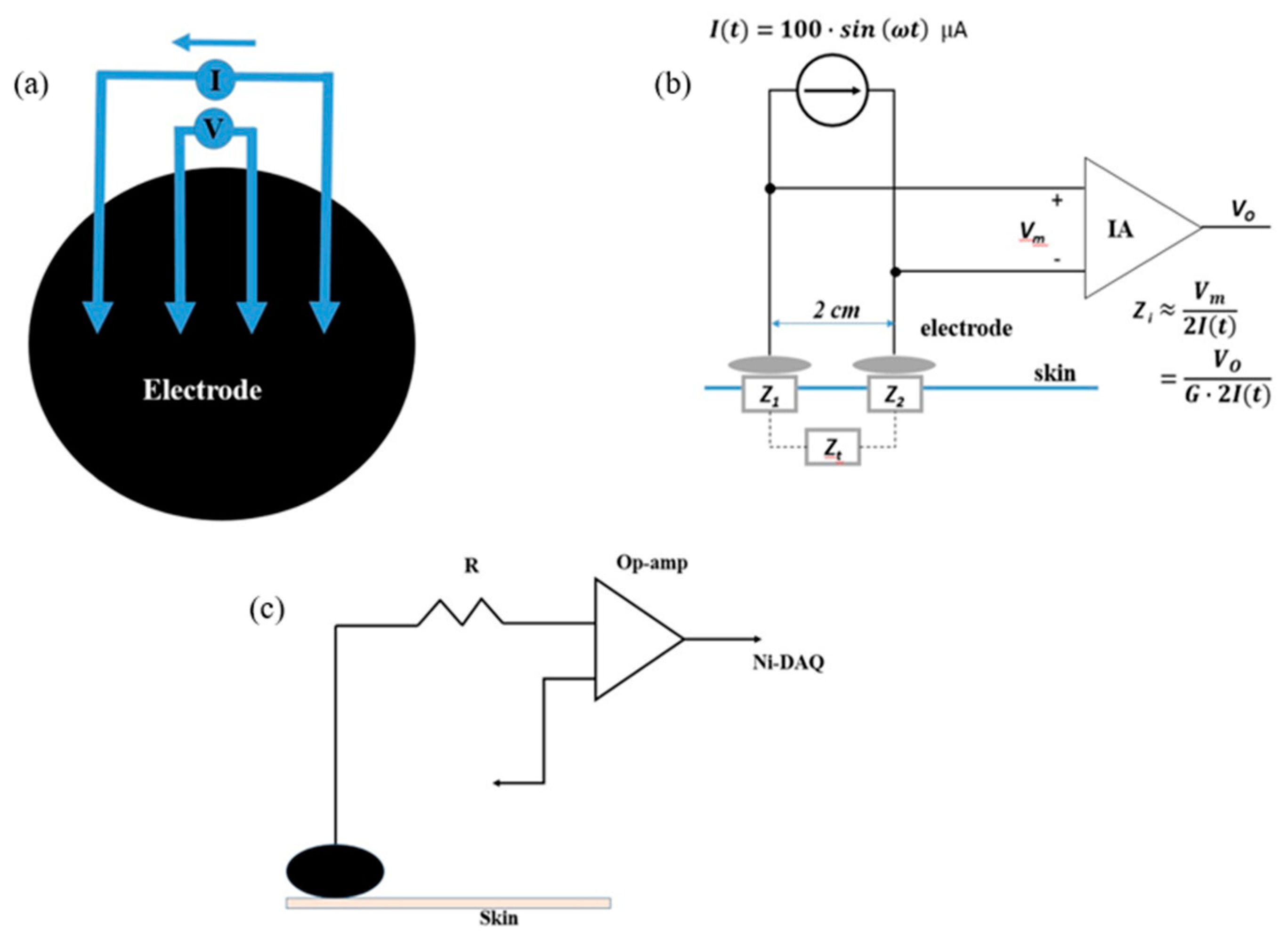
2.3. Skin–Electrode Impedance Measurement System and DC Offset

The skin–electrode contact impedance was measured without using any conductive gel and with no skin preparation. Two electrodes were placed on human participants’ forearms 2 cm apart from each other. The electrodes were placed on the left forearm and connected to the measurement setup illustrated in Figure 2b. LabVIEW was used to generate alternating current (AC) signals through a data acquisition (DAQ) device (National instruments (NI) USB-6211). The amplitude of the AC signals was set to 100 µA, and the frequencies were swept from 10, 200, 400, 600, and 800 to 1000 Hz. The skin–surface interface impedance for each electrode was labeled Z1 and Z2, and the tissue impedance between the two measurement sites was labeled Zt. We assumed that values of Z1 and Z2 were reasonably close each other and Zt was significantly less than Z1,2. Therefore, Z1,2 could be found by measuring the voltage drop between the electrodes (Vm) using an instrumentation amplifier (IA) (AD622, Analog Devices, Inc., Norwood, MA, USA). We set the gain (G) of the IA to 1.
I ( t ) = 100   s i n ( ω t ) = V m Z t o t a l
V m = I ( t ) · ( Z 1 + Z 2 + Z t )
Assuming:   Z 1 Z 2 = Z i and Z i Z t ,
Z i = V m 2   I ( t ) = V O 2   I ( t )
The DC offset of each electrode type could be determined by setting up a simple circuit to measure half-cell potential in reference to the ground. The measured DC offsets were recorded and analyzed using a DAQ device and LabVIEW, as shown in Figure 2c.

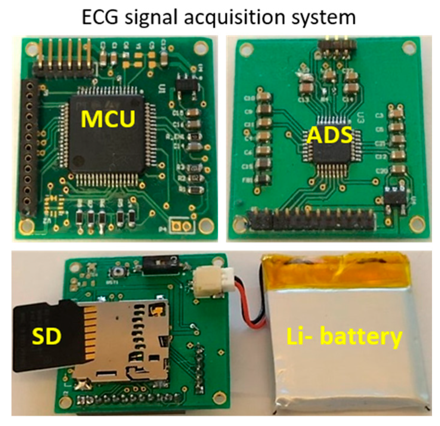
2.4. ECG Signal Measurement and Signal Processing

The performance of the fabricated electrodes was observed by ECG signal monitoring without using gel and with no skin preparation. For ECG signal acquisition, we developed a custom-designed printed circuit board (PCB) that integrated a 32-bit micro-controller (STM32L433, ST Microelectronics, Geneva, Switzerland) and a fully integrated electrocardiogram analog front-end (ADS 1191, Texas Instruments, Inc., Richardson, TX, USA), as shown in Figure 3. The electrodes received electrical signals through the electrical activity of the heart, and the electrical signals were then amplified by the analog front-end (AFE) chipset with a gain of 12. The sampling rate of the AFE was set to 128 sample/second. The ECG signals were stored on a local secure digital (SD) media card and transferred to a personal computer for further analysis and diagnosis. The measured signal was bandpass filtered with 0.5 and 45 Hz of upper and lower cutoff frequencies, respectively, to remove DC offset and high frequency noises. The signal processing was done using MATLAB 2018b.
This study recruited two healthy adults (1 male and 1 female) for skin–electrode impedance and ECG measurements; their informed consent was obtained before they participated in the study. The study was conducted in accordance with the Declaration of Helsinki, and the protocol was approved by the Institutional Review Board at the University of Connecticut (H19-010).
The ECG signal measurements were performed at room temperature with three fabricated, flexible, dry ECG electrodes, and an Ag/AgCl wet electrode (as a reference) was placed on the body of a healthy subject at three positions: the left side of the chest, the right side of the chest, and the left side of the abdomen without cleaning the hair and with no skin preparation. Another ECG signal was received from the fingertips of the subject using three fabricated dry electrodes.

3. Results

3.1. Physical Characterization of Developed Composites and Electrodes

The surface morphology of the composite decorated electrodes was characterized via SEM. The SEM images of the CG and CG-functionalized MWCNT composite-coated electrodes are shown in Figure 4. The CG and its composites were formed after three hours of ultrasonication and coated on the nylon filter paper. A number of layers of the CG sheets were anchored on the filter paper, and the as-usual wrinkle structure of the CG sheet that was generated during the chemical reduction is shown in Figure 4a. Fifteen percent of the f@MWCNTs were mixed with graphite oxide to form the CG/f@MWCNTs-15% composite sheet on the electrode, as shown in Figure 4b. An increase of the f@MWCNTs into the composites resulted in an increase in the concentration of the MWCNTs, which were more dominant in the composites shown in Figure 4c,d. It was also clear that no folded sheets of CG were available in 35% of the composites. The sheet resistance of the fabricated bioelectrodes was determined to be 180 Ω/□ for CG, 120 Ω/□ for the 15%-based composite, 82 Ω/□ for the 25%-based composites, and 75 Ω/□ for the 35%-based composites. The results indicate that after the 25% loading of f@MWCNTs into the composite, conductive networks between CG sheets and CNT wires were well established.
Additionally, in order to investigate the structure of CG and the CG/f@MWCNT composite materials that were used in the experiments, we performed Raman spectroscopy analyses at an excitation wavelength of 514 nm, as shown in Figure 5. Two intense peaks were exhibited at 1360 and 1595 cm−1 which corresponded to the D and G bands of CG, respectively. In case of the CG/f@MWCNTs, however, the D and G bands were shifted to the lower wavenumbers of 1347 and 1577 cm−1, respectively, which indicates that substantial interaction between CG and the nanotubes after adding f@MWCNTs [34,35]. In addition, by increasing f@MWCNTs contents from 0% to 35%, the relative intensity of D/G (ID/IG) gradually decreased from 1.03 to 0.88 due to the f@MWCNTs having less disordered structural defects compared to CG, indicating that a defect of the lattice may have been slightly suppressed. It could be observed that the G’ band was newly occurring in CG/f@MWCNT composites, supporting the fact that the f@MWCNT was well combined with the CG sheet.
The skin impedance versus frequency plots of the CG-based electrodes and the commercial Ag/AgCl electrodes within the 10–1000 Hz frequency range are displayed in Figure 6. It can be seen that the values of the impedance decreased a little with increasing frequency when the CG and certain amount of the f@MWCNT loading composite electrodes were used, whereas the impedance values decreased strongly with increasing frequency whenusing commercial electrodes and higher loading MWCNT composite-based electrodes, as shown in Figure 6a–e.
The impedance values showed a negligible change with the increasing frequency for CG and 15% f@MWCNT-incorporated composite electrodes, as shown in Figure 6a,b. The results imply that a smaller amount of f@MWCNTs in composite led to an inappropriate interaction between f@MWCNTs and the CG sheets, which led to a low specific capacitance. On the contrary, large amounts of MWCNTs in the composite started to increase in an unpredicted agglomeration of CNTs, which led to the inadequate interaction of f@MWCNTs with the CG sheets to improve efficient specific capacitance, as shown in Figure 6c,d [29]. It is clear from the experiment that high volume f@MWCNT-embedded composites showed frequency-dependent phenomena due to a high specific capacitance. Therefore, it was important to determine appropriate interaction of f@MWCNTs with CG to improve electrical and chemical properties. It is seen in the graphs that the 25% f@MWCNT-incorporated composite showed a low frequency dependency and a relatively low skin impedance. This result could have happened due to an appropriate interaction between f@MWCNTs and CG, which came from the uniform distribution of MWCNTs in the CG matrix, as seen in Figure 4. The contact impedance of the wet Ag/AgCl was determined to be 45 KΩ at 10 Hz and 15.05 KΩ at 1 kHz. This result is the same as that of previous reports [17,18].
The DC offset voltage of the Ag/AgCl wet electrodes was determined to be 11.738 mV, and the DC offset voltages of the fabricated electrodes were determined to be 10.264 mV for the CG-based electrodes, 11.12 mV for the CG/f@MWCNTs-15%-based electrodes, 11.824 mV for the CG/f@MWCNTs-25%-based electrodes, and 12.956 mV for the CG/MWCNTs-35%-based electrodes, as shown in Table 1. The DC offset values of all electrodes were fairly small and within range of the standard Ag/AgCl electrodes. There appeared to be a small increase in the DC offset with increasing percentages of carbon nanotubes. The 25% f@MWCNT-based, composite-embedded electrode showed a DC offset voltage similar to the conventional commercial ECG electrode.

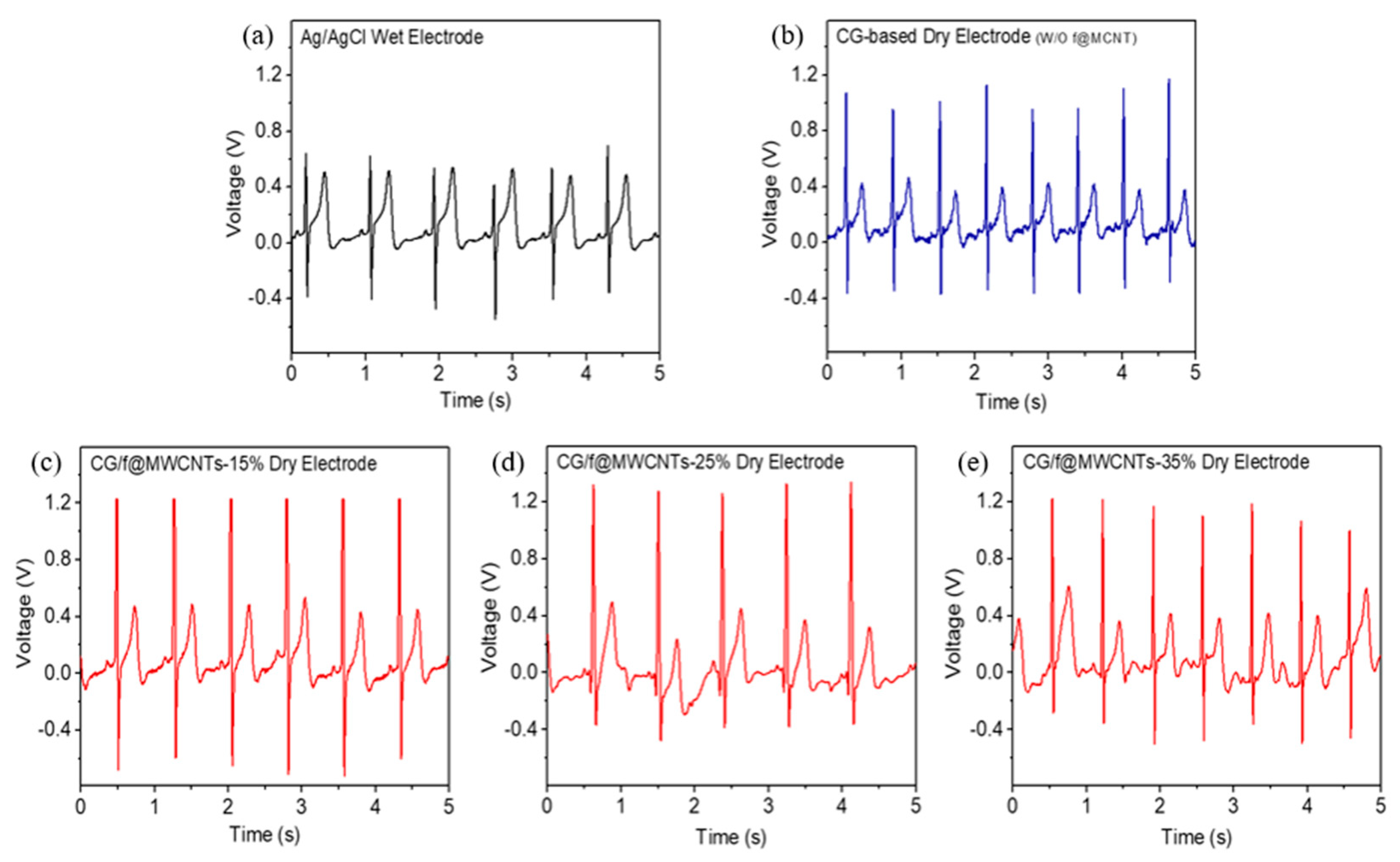
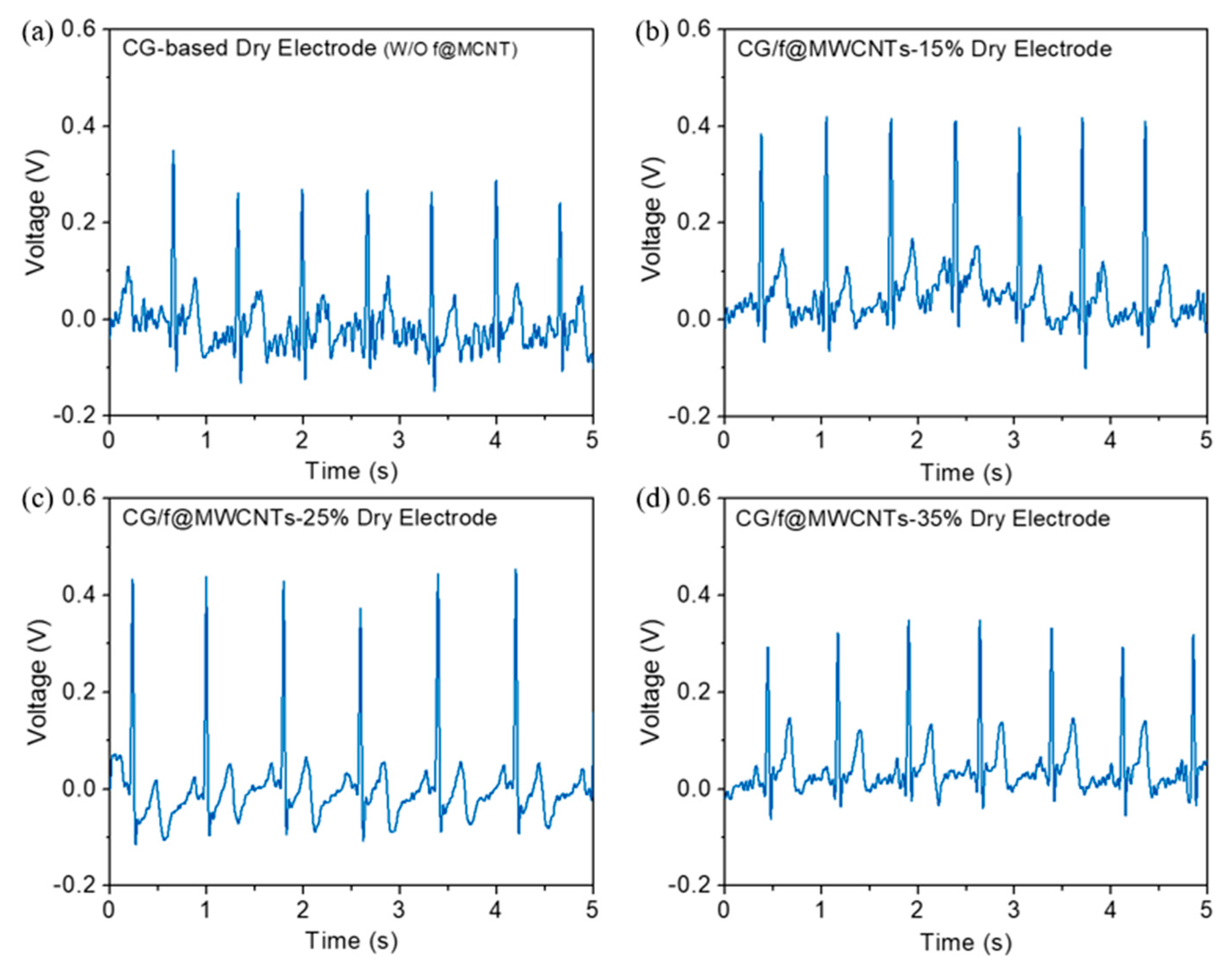
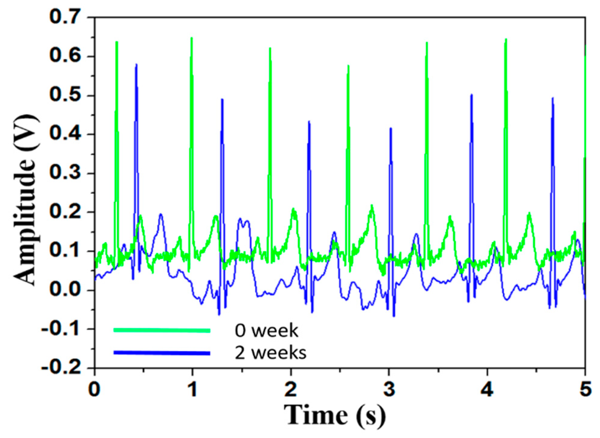
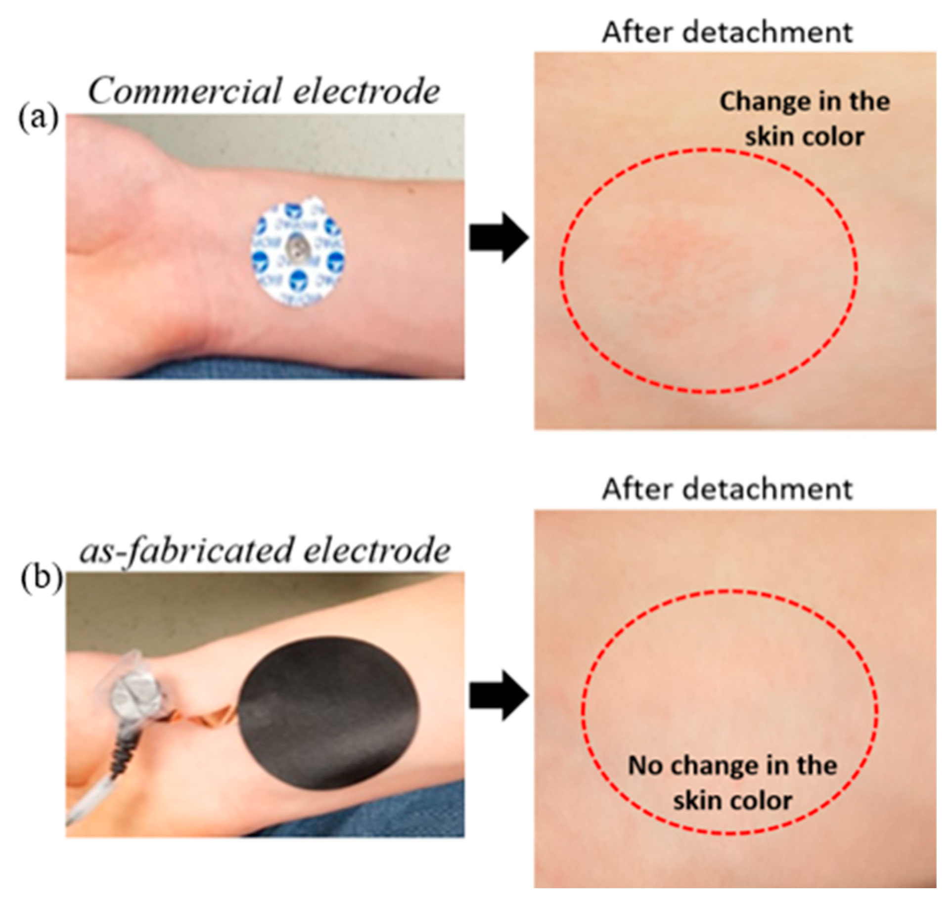
3.2. ECG Signal Measurement Using Different Electrodes

For the evaluation of the fabricated biopotential electrodes, ECG signals were received from the chest and fingertips. Typical chest ECG signals are shown in the Figure 7. It was shown that the wave form of ECG signals received from the CG-coated dry electrodes and CG and f@MWCNT composite-coated dry electrodes were similar to the wave form of ECG signals obtained from the commercial Ag/AgCl wet electrodes. P, Q, R, S and T waves can be clearly recognized in the ECG signals; these were received from fabricated flexible electrodes.
The time interval of P–Q, QRS, and Q–T waveforms were almost same as those indicated in the previous reports [36]. In addition, the ECG signals obtained from the chest using the fabricated dry ECG electrodes showed little or no variation in amplitude and had much less noise. On the contrary, the signals received from the chest using conventional electrodes showed little variation in amplitude but had no noise. No variation in the amplitude of the ECG signals obtained from the dry ECG electrodes proved that the fabricated paper-based electrodes had improved mechanical contact with the skin due to their flexible and conformal physical form factors [2,37]. It was also observed that the peak-to-peak potentials of the fabricated electrodes were 1.6, 1.5, 1.95, and 1.8 V for CG, CG/f@MWCNTs-15%, CG/f@MWCNTs-25%, and CG/f@MWCNTs-35%, respectively. We found that the peak–peak potential obtained from ECG signals using CG/f@MWCNTs-25% electrodes was higher than that of the other electrodes.
To investigate the performance of the fabricated electrodes, an ECG signal was also measured from the subject’s fingertips. The typical ECG signals were recorded from fingertips using different fabricated electrodes, as shown in Figure 8. The P, Q, R, S, and T waveforms were clearly visible in all ECG signals. Similar signals were found in previous research [11,38]. It is also clear that the CG/f@MWCNTs-25% composite-based flexible electrode showed a better quality of signal in terms of the amplitude of the signals and the visibility of the PQRST waveforms. Figure 9 shows the filtered ECG signal using the CG/f@MWCNTs-25% composite-based dry electrodes observed after two weeks. The P, Q, R, S, and T waveforms were clearly distinguished from the ECG signals. This result revealed that the fabricated dry electrode can be used long-term ECG signal monitoring. Figure 10 shows that no skin color change was observed after several hours on the subject forearm. This result indicates that flexible paper electrodes are easy to use with no skin preparation.

4. Discussion

In this study, chemically-modified graphene and functionalized MWCNT composites were developed for potential use of ECG signal acquisition. The f@MWCNTs were clearly observed to have occupied in the CG matrix network, as shown in Figure 4b,c. The MWCNTs provided good conduction pathways because MWCNTs act as spacers between the CG sheets and because they establish interconnected conductive network paths. Thus, as the loading of the f@MWCNTs increased, the volume resistivity of the composites decreased. Furthermore, CG worked as a good matrix with a large surface area to support contact between the MWCNTs and the CG sheets, resulting in a further reduction of the junction resistance [33]. At around a 30%~35% loading of f@MWCNTs in CG/f@MWCNT composites, the adhesion of the composites on the papers started to deteriorate, resulting in the peel-off effect. Therefore, the 25% loading of f@MWCNT-based electrodes was selected for a further characterization of the ECG signals.
The fabricated flexible electrode measured the ECG signals from two different positions (chest and fingertips). The P, Q, R, S, and T waveforms were clearly observed in the recorded ECG signals and were comparable to those of the commercial ECG electrode. We also found that the 25% f@MWCNT-based electrode demonstrated a good signal quality in terms of the peak-to-peak amplitude of the signal, the stability of the signal spectrum, and the visibility of signal waveforms as compared to that of other f@MWCNT-loaded electrodes. The results revealed that the 25% loading of f@MWCNTs in the CG matrix is the optimum loading for the uniform distribution of the hybrid electrode platform. Large bundles or dense agglomerates of CNTs can cause a poor performance of the composite electrode [39,40,41]. The sensitivity of the fabricated electrode was noticeable as compared to that of conventional electrodes. We observed the ECG signal for two weeks. After two weeks, it did not show a noticeable signal amplitude change (Figure 9). The wave peaks were clearly visible on the signals. In addition, we also observed skin color after removing both the commercial and fabricated electrodes from forearm. The skin color changed when the commercial electrode was attached to the subject’s forearm, but the skin remained same when the fabricated electrode was used (Figure 10).
The limitations of this study are that this study only tested two subjects (a male and a female) for ECG measurement; more participants are needed to ensure the validity of the presented data. The composites were adsorbed on nylon filter papers to fabricate flexible electrodes using a vacuum suction filter. An automatic fabrication process is possible because the developed composites easily adsorbed on the paper within 2–3 min. We did not apply the automatic fabrication.
Fabricating CG/f@MWCNTs electrodes on a garment will enable the long-term monitoring of bio-signals. However, it could be a challenge to fabricate the proposed electrodes on a fabric substrate since fabric easily absorbs CG/f@MWCNTs, and, consequently, it is difficult to control the uniformity of the electrodes.
Concerning toxicity, carbon is commonplace and has a stable structure; previous reports have demonstrated the biological compatibility of carbon group elements, including CNTs and graphene materials [42,43], but this study did not perform a biocompatibility test for human skin uses. Since the proposed electrodes are pressure and temperature sensitive, the fabricated electrodes can be used other biosignals monitoring (ECG, electromyography (EMG) and activity sensors) for robotics and prosthesis applications.

5. Conclusions

Chemically converted graphene and functionalized multiwalled carbon nanotube composites were successfully developed in a proper solvent, and these composites were deposited on flexible filter paper using a vacuum-assisted technique for biopotential electrodes. Skin impedance was measured using the fabricated electrodes without any skin preparation, and the impedance data proved that the fabricated electrodes could potentially be used to measure ECG signals. The ECG signals were measured from the chest and the fingertips of the subjects without the use of any conductive gel or skin preparation. The electrodes were attached to the chest using a sticker. The ECG signals received from the chest while using the fabricated electrode were similar to the conventional commercial electrodes. We also measured the ECG signals from the fingertips without using gel. The P, Q, R, S and T waveform were clearly visible in the signals. The CG/f@MWCNTs-25% demonstrated a better performance in terms of skin impedance and the amplitude of the signals, resulting in a uniformity of the spacer materials. The comfortability of the fabricated electrodes was much better than that of conventional electrodes. In addition, by using controlled deposition on paper electrodes, vacuum filtering enabled a good reproducibility and ease of use for ECG detection. The fabricated ECG electrode showed the capability for the longtime monitoring of ECG signals. The performance of the flexible dry electrode was excellent in comparison to the conventional wet electrodes. Thus, we believe that the fabricated flexible electrode is a good candidate for use in other physiological signal detection applications.

Author Contributions

Conceptualization, M.F.H. and I.K.; methodology, M.F.H., J.S.H., and I.K.; validation, M.F.H. and J.N.; materials characterization, M.F.H. and J.S.H.; writing—original draft preparation, M.F.H.; writing—review and editing, J.S.H. and I.K.; visualization, M.F.H. and J.S.H.; supervision, I.K.

Funding

This research received no external funding.

Acknowledgments

The study using a Raman spectroscopy presented in this work was conducted in the Institution of Material Science at the University of Connecticut.

Conflicts of Interest

The authors declare no conflict of interest.

References

  1. Kim, J.H.; Hwang, J.-Y.; Hwang, H.R.; Kim, H.S.; Lee, J.H.; Seo, J.W.; Ueon, S.S.; Lee, S.H. Simple and cost-effective method of highly conductive and elastic carbon nanotube polydimethylsiloxane composite for wearable electronics. Sci. Rep. 2018, 8, 1375. [Google Scholar] [CrossRef] [PubMed]
  2. Chlaihawi, A.A.; Narakathu, B.B.; Emamian, S.; Bazuin, B.J.; Atashbar, M.Z. Development of printed and flexible dry ECG electrodes. Sen. Bio-Sens. Res. 2018, 20, 9–15. [Google Scholar] [CrossRef]
  3. Xu, S.; Dai, M.; Xu, C.; Chen, C.; Tang, M.; Shi, X.; Dong, X. Performance evaluation of five types of Ag/AgCl bio-electrodes for cerebral electrical impedance tomography. Ann. Biomed. Eng. 2011, 39, 2059–2067. [Google Scholar] [CrossRef] [PubMed]
  4. Baek, J.Y.; An, L.H.; Choi, J.M. Flexible polymeric dry electrodes for the long-term monitoring of ECG. Sens. Act. A Phys. 2008, 143, 423–429. [Google Scholar] [CrossRef]
  5. Hossain, M.F.; Barman, S.C.; Park, J.Y. Seed-mediated growth of platinum nanoparticles anchored on chemically modified graphene and cationic polyelectrolyte composites for electrochemical multi-sensing applications. Sens. Act. B Chem. 2019, 282, 780–789. [Google Scholar] [CrossRef]
  6. Das, P.S.; Hossain, M.F.; Park, J.Y. Chemically reduced graphene oxide-based dry electrodes as touch sensor for electrocardiograph measurement. Microelectron. Eng. 2017, 180, 45–51. [Google Scholar] [CrossRef]
  7. Peng, H.; Liu, J.; Dong, Y.; Yang, B.; Chen, X.; Yang, C. Parylene-based flexible dry electrode for bioptential recording. Sens. Actuator B Chem. 2016, 231, 1–11. [Google Scholar] [CrossRef]
  8. Lee, J.W.; Yun, K.S. ECG Monitoring garment using conductive carbon paste for reduced motion artifacts. Polymers 2017, 9, 439. [Google Scholar]
  9. Liu, B.; Luo, Z.; Zhang, W.; Tu, Q.; Jin, X. Carbon nanotube-based self-adhesive polymer electrodes for wireless long-term recording of electrocardiogram signals. J. Biomater. Sci. Polym. Ed. 2016, 27, 1899–1908. [Google Scholar] [CrossRef]
  10. Peng, H.; Liu, J.; Tian, H.; Xu, B.; Dong, Y.; Yang, B.; Chen, X.; Yang, C. Flexible dry electrode based on carbon nanotube/polymer hybrid micropillars for biopotential recording. Sens. Actuator A Phys. 2015, 235, 48–56. [Google Scholar] [CrossRef]
  11. Hossain, M.F.; Das, P.S.; Park, J.Y. Development of high performance electrochemical and physical biosensors based on chemically modified graphene nanostructured electrodes. J. Electrochem. Soc. 2017, 164, B391–B396. [Google Scholar] [CrossRef]
  12. Bihar, E.; Roberts, T.; Ismailova, E.; Saadaoui, M.; Isik, M.; Sanchez-Sanchez, A.; Mecerreyes, D.; Herve, T.; De Graaf, J.B.; Malliaras, G.G. Fully printed electrodes on stretchable textiles for long-term electrophysiology. Adv. Mater. Techonol. 2017, 2, 1600251. [Google Scholar] [CrossRef]
  13. Takamatsu, S.; Lonjaret, T.; Crisp, D.; Badier, J.M.; Malliaras, G.G.; Ismailova, E. Direct patterning of organi conductors on knitted textiles for long-term electrocardiography. Sci. Rep. 2015, 5, 15003. [Google Scholar] [CrossRef] [PubMed]
  14. Kim, T.; Park, J.; Sohn, J.; Cho, D.; Jeon, S. Bioinspired, highly stretchable, and conductive dry adhesives based on 1D–2D hybrid carbon nanocomposites for all-in-one ECG electrodes. ACS Nano 2016, 10, 4770–4778. [Google Scholar] [CrossRef]
  15. Lee, S.M.; Byeon, H.J.; Lee, J.H.; Baek, D.H.; Lee, K.H.; Hong, J.S.; Lee, S. Self-adhesive epidermal carbon nanotube electronics for tether-free long-term continuous recording of biosignals. Sci. Rep. 2014, 4, 6074. [Google Scholar] [CrossRef]
  16. Yapici, M.K.; Alkhidir, T.; Samad, Y.A.; Liao, K. Graphene-Clad textile electrodes for electrocardiogram monitoring. Sens. Actuator B Chem. 2015, 221, 1469–1474. [Google Scholar] [CrossRef]
  17. Celik, N.; Manivannan, N.; Strudwick, A.; Balachandran, W. Graphene-Enabled Electrodes for Electrocardiogram Monitoring. Nanomaterials 2016, 2016, 156. [Google Scholar] [CrossRef]
  18. Lou, C.; Li, R.; Li, Z.; Liang, T.; Wei, Z.; Run, M.; Yan, X.; Liu, X. Flexible graphene electrodes for prolonged dynamic ECG monitoring. Sensors 2016, 16, 1833. [Google Scholar] [CrossRef]
  19. Novoselov, K.S.; Geim, A.K.; Morozov, S.V.; Jiang, D.; Zhang, Y.; Dubonos, S.V.; Grigorieva, I.V.; Firsov, A.A. Electric field in atomically thin carbon films. Science 2004, 306, 666–669. [Google Scholar] [CrossRef]
  20. Sprinkle, M.; Ruan, M.; Hu, Y.; Hankinson, J.; Rubio-Roy, M.; Zhang, B.; Wu, X.; Berger, C.; Heer, W.A. Scalable templated growth of graphene nanoribbons on SiC. Nat. Nanotechnol. 2010, 5, 727–731. [Google Scholar] [CrossRef]
  21. Hossain, M.F.; Park, J.Y. Novel enzymatic glucose biosensor based on distributed electrodes covered with a solvothermal synthesized graphene material and platinum nanoparticles. RSC Adv. 2016, 6, 74453–74461. [Google Scholar] [CrossRef]
  22. Mahony, C.O.; Pini, F.; Blake, A.; Webster, C.; Brien, J.O.; McCarthy, K.G. Microneedle-based electrodes with integrated through-silicon via for biopotential recording. Sens. Actuator A Phys. 2012, 186, 130–136. [Google Scholar]
  23. Ruffini, G.; Dunne, S.; Fuentemilla, L.; Grau, C.; Farres, E.; Marco-Pallares, J.; Watts, P.C.P.; Silva, S.R.P. First human trials of a dry electrophysiology sensor using a carbon nanotube array interface. Sens. Actuator A Phys. 2008, 144, 275–279. [Google Scholar] [CrossRef] [Green Version]
  24. Jung, H.C.; Moon, J.H.; Baek, D.H.; Lee, J.H.; Choi, Y.Y.; Hong, J.S.; Lee, S.H. CNT/PDMS composite flexible dry electrodes for long-term ECG monitoring. IEEE Trans. Biomed. Eng. 2012, 59, 1472–1479. [Google Scholar] [CrossRef]
  25. Kang, B.C.; Ha, T.J. Wearable carbon nanotube based dry-electrodes for electrophysiological sensors. Jpn. J. Appl. Phys. 2018, 57, 05GD02. [Google Scholar] [CrossRef]
  26. Mani, V.; Chen, S.M.; Lou, B.S. Three dimensional graphene oxide-carbon nanotubes and graphene-carbon nanotubes hybrids. Int. J. Electrochem. Sci. 2013, 8, 11641–11660. [Google Scholar]
  27. Zhao, M.Q.; Zhang, Q.; Huang, J.Q.; Tian, G.L.; Chen, T.C.; Qian, W.Z.; Wei, F. Towards high purity graphene/single-walled carbon nanotube hybrids with improved electrochemical capacitive performance. Carbon 2013, 54, 403. [Google Scholar] [CrossRef]
  28. Khan, A.; Khan, A.A.P.; Asiri, A.M.; Abu-Zied, A.M. Green synthesis of thermally stable Ag-rGO-CNT nano composite with high sensing activity. Compos. Part B Eng. 2016, 86, 27–35. [Google Scholar] [CrossRef]
  29. Yang, W.; Chen, Y.; Wang, J.; Peng, T.; Xu, J.; Yang, B.; Tang, K. Reduced graphene oxide/carbon nanotube composites as electrochemical energy storage electrode applications. Nanoscale Res. Lett. 2018, 13, 181. [Google Scholar] [CrossRef]
  30. Ponnamma, D.; Sadasivuni, K.K.; Strankowski, M.; Guob, Q.; Thomas, S. Synergistic effect of multi walled carbon nanotubes and reduced graphene oxides in natural rubber for sensing application. Soft Matter 2013, 9, 10343–10353. [Google Scholar] [CrossRef]
  31. Huang, J.R.; Her, S.C.; Yang, X.X.; Zhi, M.N. Synthesis and characterization of multi-walled carbon nanotube/graphene nanoplatelet hybrid film for flexible strain sensors. Nanomaterials 2018, 8, 786. [Google Scholar] [CrossRef] [PubMed]
  32. Fan, W.; Zhang, L.; Liu, T. Strategies for the hybridization of cnts with graphene. in: Graphene-carbon nanotube hybrids for energy and environmental applications. Springer Br. Mol. Sci 2017, 21–51. [Google Scholar]
  33. Cheng, Q.; Tang, J.; Ma, J.; Zhang, H.; Shinya, N.; Qin, L.C. Graphene and carbon nanotube composite electrodes for supercapacitors with ultra-high energy density. Phys. Chem. Chem. Phys. 2011, 13, 17615–17624. [Google Scholar] [CrossRef] [PubMed]
  34. Basu, A.K.; Sah, A.N.; Pradhan, A.; Bhattacharya, S. Poly-L-Lysine functionalized MWCNT-rGO nanosheets based 3-d hybrid structure for femtomolar level cholesterol detection using cantilever based sensing platform. Sci. Rep. 2019, 9, 3686. [Google Scholar] [CrossRef]
  35. Zeng, X.; Yu, S.; Ye, L.; Li, M.; Pan, Z.; Sun, R.; Xu, J. Encapsulating carbon nanotubes with SiO2: A strategy for applying them in polymer nanocomposites with high mechanical strength and electrical insulation. J. Mater. Chem. C 2015, 3, 187. [Google Scholar] [CrossRef]
  36. Ramli, A.B.; Ahmad, P.A. Correlation analysis for abnormal ECG signal features extraction In Proceeding of the 4th National Conference on Telecommunication Technology, Shah Alam, Malaysia, 14–15 January 2003.
  37. Achilli, A.; Bonfiglio, B. Design and characterization of screen-printed textile electrodes for ECG monitoring. IEEE Sens. J. 2018, 18, 4097–4107. [Google Scholar] [CrossRef]
  38. Mathias, D.N.; Park, J.; Kim, E.; Joung, Y.H. Development of a novel noncontact ecg electrode by mems fabrication process. Trans. Electr. Electron. Mater. 2016, 17, 150–154. [Google Scholar] [CrossRef]
  39. Faiella, G.; Musto, P.; Di Florio, G.; Buosciolo, A.; D’Orazio, L.; Antonucci, V.; Giordano, M. Monitoring the dispersion process of SWNTs in aqueous solutions by UV-Vis and Raman spectroscopies. J. Nanosci. Nanotechnol. 2009, 9, 6026–6033. [Google Scholar] [CrossRef]
  40. Zhao, H.W.; Hu, L.C.; Chang, C.R. Theoretical studies of “stabilizing” behavior about carbon nanotubes under the electrostatic force. J. Nanosci. Nanotechnol. 2013, 13, 3133–3135. [Google Scholar] [CrossRef]
  41. Hwang, J.Y.; Kim, H.S.; Kim, J.H.; Shin, U.S.; Lee, S.H. Carbon nanotube nanocomposites with highly enhanced strength and conductivity for flexible electric circuits. Langmuir 2015, 31, 7844–7851. [Google Scholar] [CrossRef]
  42. Zan, X.; Zheng, F.; Jin, W.; Fei, X.; Huo, F.; Duan, H. Freestanding graphene paper decorated with 2D-assembly of Au@Pt nanoparticles as flexible biosensors to monitor live cell secretion of nitric oxide. Biosens. Bioelectron. 2013, 49, 71–78. [Google Scholar] [CrossRef] [PubMed]
  43. Ilbasmis-Tamer, S.; Ciftci, H.; Turk, M.; Degim, T.; Tamer, U. Multiwalled Carbon Nanotube-Chitosan Scaffold: Cytotoxic, Apoptoti c, and Necrotic Effects on Chondrocyte Cell Lines. Curr. Pharm. Biotechnol. 2017, 18, 327–335. [Google Scholar] [CrossRef] [PubMed]
Figure 1. (a) Schematics of composite synthesis. (b) Illustration of electrode fabrication and measurement of ECG signal.
Information 10 00325 g001
Figure 2. The experimental set up for (a) the surface resistivity, (b) skin impedance, and (c) direct current (DC) offset potential.
Information 10 00325 g002
Figure 3. A custom-designed printed circuit board (PCB) including a 32-bit micro-controller unit (MCU), a fully integrated ECG analog front-end (ADS), a local secure digital media (SD) card, and a portable Li-battery.
Information 10 00325 g003
Figure 4. SEM images of (a) chemically-modified graphene (CG), (b) CG/carboxylic-functionalized multiwalled carbon nanotubes (f@MWCNTs)-15% composite, (c) CG/f@MWCNTs-25% composite, and (d) CG/f@MWCNTs-35% composite.
Information 10 00325 g004
Figure 5. Raman spectra of CG and CG/f@MWCNT composites with different f@MWCNTs contents: 15%, 25%, and 35%.
Information 10 00325 g005
Figure 6. Skin impedance responses of flexible dry ECG electrodes and commercial electrode at frequency range from 10 Hz to 1 KHz. Skin–electrode contact impedance of (a) CG, (b) CG/f@MWCNTs-15%, (c) CG/f@MWCNTs-25%, (d) CG/f@MWCNTs-35%, and (e) Ag/AgCl.
Information 10 00325 g006
Figure 7. The chest ECG signals recorded by the different types of electrodes. (a) Conventional Ag/AgCl, (b) CG, (c) CG/f@MWCNTs-15%, (d) CG/f@MWCNTs-25%, and (e) CG/f@MWCNTs-35%.
Information 10 00325 g007
Figure 8. The ECG signal from the fingertips received by different flexible dry electrodes after finite impulse response (FIR) filtering for (a) CG, (b) CG/f@MWCNTs-15%, (c) CG/f@MWCNTs-25%, and (d) CG/f@MWCNTs-35%.
Information 10 00325 g008
Figure 9. The stability test of the fabricated CG/f@MWCNTs-25% electrodes after two weeks in fingertips.
Information 10 00325 g009
Figure 10. The compatibility test of the electrode in the skin for (a) the commercial electrode and (b) the as-fabricated electrode.
Information 10 00325 g010
Table 1. DC offset voltage of commercial and fabricated electrodes.
Sample No.Sample NameDC Offset Voltage
1Ag/AgCl11.738 mV
2CG10.264 mV
3CG/f@MWCNTs-15%11.120 mV
4CG/f@MWCNTs-25%11.824 mV
5CG/f@MWCNTs-35%12.956 mV

